GFC Therapy: A Revolutionary Approach to Hair Restoration
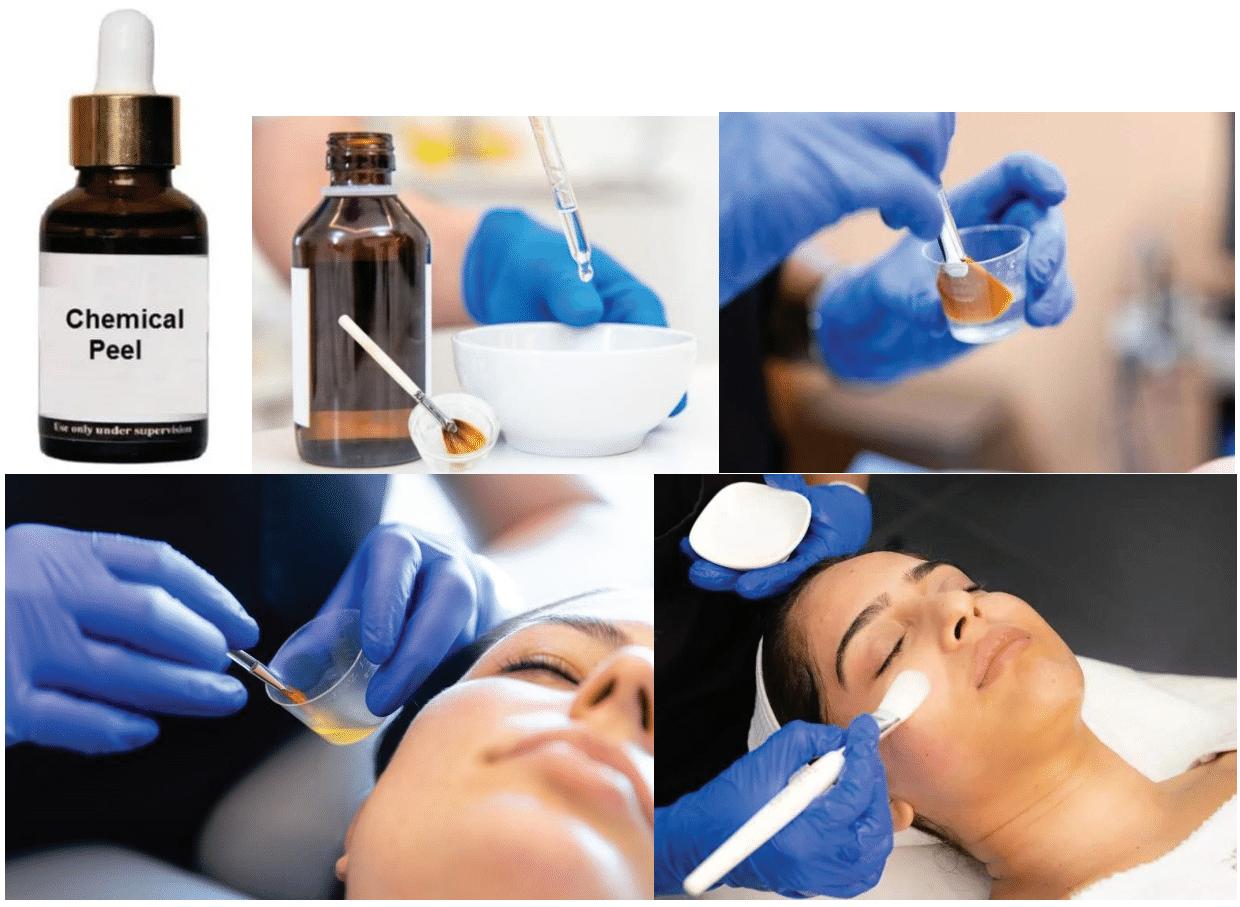
Chemical Peels: A Personalized Approach to Radiant Skin
The Future of Skin: Why Dermatologists Must Lead the Longevity Revolution An Observational Study on Safety and Effectiveness of HighIntensity Focused Ultrasound (HIFU) on Facial Skin Lift and Rejuvenation in the Indian Sub-Population
EXECUTIVE EDITOR & PUBLISHER
Dom Daniel CORPORATE OFFICE
22, Shreeji Bhavan, 275-279, Samuel Street, Masjid Bunder (W), Mumbai-4000 03, INDIA.
EMAIL: theaestheticiansjournalindia@gmail.com
Website: theaestheticiansjournal.com
Printed, Published, Edited and Owned by Dom Daniel Printed at Swastik Printer, Gala No.9 & 10, Vishal Industrial Estate, Bhandup (West), Mumbai- 400078. Published at 22 Shreeji Bhavan, 275/279, Samuel Street, Masjid Bunder (West), Mumbai - 400003. India.
“The Aestheticians Journal” takes no responsibility for unsolicited photographs or material ALL PHOTOGRAPHS, UNLESS OTHERWISE INDICATED, ARE USED FOR ILLUSTRATIVE PURPOSE ONLY.
Views expressed in this Journal are those of the contributors and not of the publisher. Reproduction in whole or in parts of texts or photography is prohibited. Manuscripts, Photographs and art are selected at the discretion of the publisher free of charge (advertising excluded). Whether published or not, no material will be returned and remains the property of the publishing house, which may make use of it as seen fit. This may include the withdrawal of publication rights to other publishing houses.
All rights reserved. Reproducing in any manner without prior written permission prohibited.
Published for the period of June -2025
Skin: The Silent Guardian of Health and Aesthetics
Skin is more than just a covering—it's our body’s largest and most expressive organ, a dynamic interface between our inner health and the external world. It shields us silently, yet speaks volumes—through texture, tone, and vitality— about our overall well-being. Aesthetically, it forms the foundation of confidence and self-expression, making its care a central concern in both clinical and cosmetic dermatology.
In this issue, we delve into cutting-edge and personalized approaches to skin and hair health, spotlighting innovations that are transforming the landscape of aesthetic dermatology. Our feature article, "The Future of Skin: Why Dermatologists Must Lead the Longevity Revolution," underscores the pivotal role of dermatologists in the evolving science of longevity. In addition, "Chemical Peels" explores how personalized peeling protocols can be customized to individual skin types, enhancing both safety and treatment efficacy.
We also present advancements in the management of hyperpigmentation, with a special focus on Q-Switched Nd:YAG Laser Therapy. This article delves into how laser-based precision techniques are becoming a game-changer in treating one of dermatology’s most persistent challenges.
In the realm of hair restoration, we highlight GFC Therapy (Growth Factor Concentrate) as a promising regenerative and non-surgical option for hair loss. With increasing interest in minimally invasive treatments, this approach is gaining momentum for its effectiveness and patient satisfaction.
Additionally, we share findings from an Observational Study on High-Intensity Focused Ultrasound (HIFU), evaluating its safety and efficacy in facial skin lifting and rejuvenation within the Indian sub-population. The insights offer a localized perspective on this globally recognized modality.
HOPE YOU HAVE A GREAT READ
Thanks & Cheers
- Dom Daniel Executive Editor & Publisher
Editorial Board
Dr. Nishita Ranka
DDVL (Dermatology)
Medical Director
Dr. Nishita’s Clinic for Skin, Hair & Aesthetics Hyderabad
The Future of Skin: Why Dermatologists Must Lead the Longevity Revolution
Dr. Nishita Ranka
DDVL (Dermatology)
Medical Director, Dr. Nishita’s Clinic for Skin, Hair & Aesthetics
Hyderabad
Opening Hook
Imagine a future where we don’t merely delay the visible signs of aging, but
" actively reverse the biological clock at the cellular level. Where skincare isn't just cosmetic, but a tool to extend both lifespan and healthspan. In this future — which is rapidly becoming reality — dermatologists are no longer confined to the realm of aesthetics. We are pioneers at the intersection of regenerative medicine, biotechnology, and preventive health."
As the science of longevity advances from experimental research to real-world applications, one truth is becoming increasingly clear: the skin is a vital biomarker and modulator of systemic aging. It’s not just a mirror of internal health but a powerful interface through which aging processes can be studied, influenced, and potentially reversed. Dermatology is stepping into a transformative era — from enhancing surface beauty to reshaping cellular youth. With our expertise in skin biology, barrier
function, inflammation, and wound healing, Dermatologists uniquely positioned to lead the clinical frontier of anti-aging and longevity interventions.
Dermatology Meets
Longevity: A Global Landscape Review
Dermatology is emerging as a vital pillar in the rapidly evolving field of longevity medicine. Globally, the perception of skin health is undergoing a paradigm shift — from being a purely aesthetic focus to becoming a measurable interface of systemic aging and biological resilience. The skin, as our largest and most visible organ, reflects cellular aging, immune function, oxidative stress, and metabolic status. This positions dermatologists at the forefront of interventions aimed not just at surface rejuvenation, but at influencing the aging process itself.
Key Scientific Mechanisms
• Cellular Senescence
Modulation: Senolytics such as quercetin and dasatinib
selectively clear senescent cells; senomorphics suppress their harmful SASP output.
• Mitochondrial ..................
Therapeutics: Urolithin A and CoQ10 support mitophagy and energy restoration.
• Sirtuin Activation: Agents such as resveratrol and NAD+ boosters enhance SIRT1/6 activity to protect against oxidative damage.
• Epigenetic .......................
Reprogramming: Skin methylation clocks (Horvath, Hannum, PhenoAge) now enable personalized biological age assessments.
• Exosome and Peptide
Regeneration: MSC-derived exosomes and synthetic peptides offer potent signals for collagen synthesis, angiogenesis, and dermal repair.
Global Integration Snapshots
United States: The U.S. leads in clinical translation of longevityfocused dermatology. Academic powerhouses such as the Buck Institute for Research on Aging, Harvard's Sinclair Lab, and Stanford's Center on Longevity are driving innovations. Clinics like Serotonin Centers, Next Health, and Upgrade Labs are already offering protocols that integrate:
• NAD+ IV infusions
• Peptide-based skin rejuvenation
• Methylation-based skin age testing
• Redox modulation and antioxidant IV drips
New York, Los Angeles, and Miami are hubs where dermatologists collaborate with biohackers, longevity physicians, and technologists.
Europe: European markets such as Switzerland, the UK, and Germany are seeing dermatology-led longevity integrated with evidence-based trials. Zurich is home to biotech startups working on topical senolytics, while London clinics are pioneering personalized skin aging assessments using DNA repair enzyme analytics. Prestige skincare brands like Augustinus Bader are investing in cellular repair actives and patenting growth factor-based topicals.
South Korea & Japan: Asia is redefining the future of regenerative dermatology. South Korea and Japan are global leaders in exosome research, stem cell-derived actives, and RF+biologics combo therapies. Companies like ExoCoBio, MEDIPOST, and BENEV are enabling the clinical adoption of mesenchymal stem cell (MSC)derived exosomes in injectables, hydrogels, and laser-adjunct serums. Korea’s regulatory agility allows early clinical use, and these innovations are often export-ready within months.
Middle East: Dubai and Riyadh have become regional hubs for luxury longevity care, integrating dermatology, genomics, and wellness. Clinics like Dubai Longevity Center offer full panels of genetic skin aging markers, telomere length testing, and precision peptide therapy, along with premium skin restoration protocols. The focus is on holistic anti-aging—where dermatologists are part of a multidisciplinary longevity team.
Australia & Singapore: These countries are regulatory innovators. The TGA (Australia) and HSA (Singapore) are drafting frameworks for cosmeceuticals
with cellular or epigenetic claims. Clinics in Sydney, Melbourne, and Singapore are offering circadian-based skin regimens, light-enhanced NAD+ therapy, and biological age testing bundled with aesthetic services. Dermatologists here are early adopters of tech-driven aging biomarkers like Skinchronomics and DermAI.
India’s Emerging Role
India, with over 15,000 boardcertified dermatologists and a vast, youthful consumer base, is witnessing a surge in demand for aesthetic procedures that combine preventive, regenerative, and wellnessdriven appeal. Major urban centers like Mumbai, Delhi, Bengaluru, and Hyderabad are now hubs for advanced skin clinics blending evidencebased dermatology with holistic wellness. The country’s dermatology sector stands at a pivotal inflection point — technologically equipped, demographically primed, and increasingly aligned with the global shift toward longevity and proactive skin health.
Strengths
• Increasing popularity of PRP, microneedling with growth factors, and RF with exosomes.
• Rise of corporate dermatology chains and wellness-linked skincare centers offering customized anti-aging solutions.
• Growing interest among younger urban demographics in biological age testing, skin biohacking, and gut-skin axis optimization.
However, significant challenges remain:
• CDSCO lacks regulatory
The
clarity on exosome or stemderived injectables, creating uncertainty for dermatologists and industry players interested in biologic-based regenerative therapies. This regulatory gap hinders clinical adoption and ethical standardization.
• There is a shortage of longterm clinical trials on Indian Fitzpatrick IV–VI skin, limiting evidence-based validation of advanced longevity therapies. Most global data remains skewed toward lighter skin phototypes.
• Biomarker integration in clinical dermatology remains minimal, with limited use of tools like DNA methylation clocks, senescence-associated secretory phenotype (SASP) profiling, or mitochondrial function panels. This restricts personalized skin aging assessment and treatment tracking.
The Science of Skin Aging: A Deep Dive
The shift from cosmetic dermatology to longevity-driven skin health is underpinned by a more advanced understanding of the molecular and cellular mechanisms of aging. Skin aging is no longer viewed simply in terms of collagen loss or photo-damage — it is now recognized as a complex, biologically regulated process involving systemic and local cellular aging pathways.
Key Drivers of Dermatologic Aging
Modern dermatologic science incorporates a systems biology approach, identifying multiple interlinked hallmarks of skin aging:
• NAD+ Decline: Loss of NAD+ impairs sirtuin activation and mitochondrial efficiency, accelerating cellular aging. Topical NMN and NR are under clinical study, while NAD+ IVs show promise in enhancing epidermal turnover and antioxidant defense. Sublingual supplementation is also gaining traction for systemic support, though more dermatologyspecific data is needed. (Sinclair, 2022).
• Senescence Burden:
Senescent cells secrete SASP factors like IL-6, IL-8, and MMPs, which degrade collagen and promote chronic inflammation. Senolytic agents like fisetin and quercetin have shown promise in lab models, though human dermatologic applications remain investigational.
• Mitochondrial Dysfunction: Declining mitochondrial function compromises ATP-dependent skin processes such as hydration and wound healing. Urolithin A, shown to enhance mitophagy (Nature Aging, 2021), is under exploration as a mitochondrial skin booster.
• Epigenetic Drift:
Age-related methylation changes alter gene expression in skin, contributing to functional decline.
Skin-specific methylation clocks (e.g., from TruDiagnostic and Clock Foundation) now allow individualized biological age tracking.
Disrupted circadian rhythms impair DNA repair and compromise skin barrier
recovery. Chronobiology-based skincare formulations, timed to align with the skin's circadian activity peaks, are currently in development.
Clinical Translation: Where Dermatologists Step In Diagnostics:
Advanced tools like epigenetic and transcriptomic skin clocks, TEWL (transepidermal water loss), inflammation markers, and real-time mitochondrial assays now enable biologically informed skin age assessment. These diagnostics are reshaping how dermatologists evaluate and track skin healthspan.
Research Frontiers:
Split-face randomized controlled trials (RCTs) using transcriptomics are revealing pathway-specific changes in response to treatment. CoQ10 and L-carnitine are being studied in post-laser recovery, while Indian longitudinal cohorts may help tailor aging interventions to Fitzpatrick IV–VI skin.
Policy and Regulation:
Regulatory clarity is urgently needed, especially for classifying exosome-based injectables under CDSCO. Dermatology must collaborate with endocrinology and genomics via cross-specialty boards to align science, ethics, and practice.
Education:
There’s a growing call to integrate the science of skin aging into dermatology residency training. CME modules focused on diagnostics, longevity biomarkers, and regenerative therapeutics can equip dermatologists for this evolving landscape.
Emerging Technologies and Cross-Specialty Synergies
Tech Innovations:
AI is now capable of analyzing facial data for aging signatures, enabling predictive dermatologic assessments. Emerging research tools such as skin-on-chip platforms and wearable diagnostics may enable future biologic testing, though their integration into clinical dermatology remains several steps away from routine application.
Synergies:
• Endocrine-Dermatology: Hormonal dysregulation and insulin resistance are key drivers of skin aging, requiring integrated care.
• Psychodermatology: ....... Stress-induced inflammaging protocols are gaining relevance in managing chronic skin conditions and promoting resilience.
Patient Personas: LongevityCentered Dermatology in India
1. Tech-Savvy Millennials (25–35):
This group is enthusiastic about biohacking, exploring skin methylation clocks, NAD+ supplementation, and data-driven skincare. They actively seek evidence-based, preventative solutions that align with longevity science.
2. Urban Women (35–50):
Focused on visible results and long-term skin resilience, they prefer highly personalized regimens backed by clinical
validation. Longevity assurance, hormonal balance, and regenerative options drive their choices.
3. HNI Wellness Seekers (40+):
High-net-worth individuals are early adopters of IV drips, peptide injectables, and exosome therapies. They seek bundled diagnostic and therapeutic programs for systemic vitality and skin youthfulness.
Global Roadmap: Dermatology-Longevity by Region
The global dermatologylongevity landscape is evolving uniquely across regions. In the USA, the emphasis lies in integrating data-driven biomarkers into clinics, though clinical evidence gaps persist— creating space for aging diagnostic platforms. Europe leads in topical innovation and structured trials, but regulatory delays slow progress, despite the potential for EU-wide standardized clinical protocols. South Korea is pioneering RF-biologic and exosome combinations, yet still requires robust efficacy data, presenting an opportunity for hybrid beauty-wellness clinical models. The Middle East focuses on luxury genomics and dermatologic protocols, but high costs limit accessibility— offering potential for regional trial satellites. Meanwhile, India stands at a strategic inflection point, with rapid expansion of Tier 1 and Tier 2 dermatology centers; although product approvals lag, the country is primed to become a clinical trial and innovation hub for longevityfocused skincare.
Vision 2030: What Lies Ahead
• Skin age diagnostics become routine.
• Epigenetic performance panels in every clinic.
• Dermatology - frontline longevity specialty.
• Indian data leads global South innovation.
The Opportunity for Dermatologists
Dermatologists are uniquely positioned to spearhead the integration of longevity science into real-world clinical care. With our understanding of skin biology, access to patients across life stages, and growing aesthetic infrastructure, we are at the frontlines of a shift from surface-level treatments to deep, cellular rejuvenation.
Key Roles for Dermatologists in This Paradigm Shift
Diagnostic Leadership
Dermatologists can pioneer the use of next-gen diagnostic tools such as:
• Skin-specific epigenetic clocks to measure biological skin age.
• Mitochondrial functional assays to detect early dysfunction.
• Proteomic and transcriptomic panels for inflammation and senescence markers.
These can allow for personalized treatment protocols and longitudinal tracking of skin healthspan.
➢ Clinical Trials and Research Leadership
Dermatologists should lead welldesigned, India-centric clinical trials focused on:
The
The Future of Skin: Why Dermatologists Must Lead the Longevity Revolution
• Longitudinal reversal of skin aging, using objective markers like dermal collagen density, transcriptomic aging signatures, and transepidermal water loss (TEWL).
• Post-procedure skin recovery studies with mitochondrial support agents.
• Topical and systemic senotherapeutics in Fitzpatrick IV–VI populations, which are underrepresented in global data.
➢ Patient Education and Reframing Aesthetics
As the public becomes more longevity-aware, dermatologists must reframe their messaging:
• Shift the focus from “looking younger” to cellular resilience and functional skin youth.
• Educate patients on how skin health reflects systemic vitality— and how targeted interventions may influence both.
This empowers patients to see dermatologic care as part of proactive, integrative health management rather than cosmetic indulgence.
Real-World Momentum in India
Examples from India show the early integration of this new science:
• Dermatology clinics in Mumbai and Bengaluru are collaborating with longevity tech startups to offer skin biological age profiling using noninvasive sampling and molecular markers.
• Aesthetic pharma companies are investing in peptide stacks, PDRN-based topicals, and mitochondrial enhancers, aiming to shift from short-term effects to regenerative claims.
• Medical colleges in South India are initiating dermatology-led clinical trials on post-laser skin recovery supported by mitochondrial co-factors like CoQ10 and L-carnitine.
These developments signal a growing scientific and commercial movement to reposition dermatology from surface aesthetics to an active player in systemic longevity and regenerative medicine.
Conclusion: Dermatology at the Helm of Healthy Aging
The dermatology-longevity axis represents one of the most transformative frontiers in modern medicine. As patients increasingly seek sustainable rejuvenation over superficial fixes, dermatologists must evolve — from aesthetic service providers to custodians of skin healthspan and biological age. We are no longer just treating the epidermis; we are interpreting the deeper signals of aging, resilience, and systemic vitality.
By grounding our practice in cellular biology, leveraging emerging biomarkers, and implementing validated interventions, dermatologists are uniquely positioned to redefine how aging is understood and managed. This new paradigm demands a shift toward biologically meaningful rejuvenation, enhanced patient literacy, and evidence-based personalization.
Let us step confidently into this leadership role — guided by science, integrity, and innovation — to ensure that dermatology is not just skindeep, but future-defining.
“The future of skin isn’t cosmetic. It’s cellular, systemic, and science-led.” – Dr. Nishita Ranka
Emerging Anti-Aging Therapies: Senolytics and Senomorphics
Senotherapeutics, encompassing senolytics and senomorphics, represent a novel class of agents targeting cellular senescence, a key driver of intrinsic aging that traditional dermatologic approaches (focused mainly on UV protection) do not address. Senolytics, such as repurposed chemotherapeutics dasatinib and navitoclax, selectively induce apoptosis in senescent cells by disrupting their anti-apoptotic pathways (e.g., p53, p21), potentially improving wound healing and reducing age-related tissue dysfunction. In contrast, senomorphics modulate senescent cell behavior without inducing cell death, primarily by suppressing the senescence-associated secretory phenotype (SASP). Agents like rapamycin, everolimus, apigenin, and metformin inhibit pathways such as mTOR, NF-κB, and IL-1α, thereby reducing inflammation and markers of senescence like SA-β-gal. Botanicals like apigenin and kaempferol, found in foods such as parsley, chamomile, kale, and beans, show promise for senomorphic effects via antioxidant and anti-inflammatory pathways. Together, these therapies aim to enhance immune clearance of senescent cells, improve skin health, and potentially delay or reverse aspects of biological aging.
Integrating Topical Skincare with Anti-Aging Treatments Enhances Skin Barrier Function Synergistically
A recent controlled clinical study involving healthy women demonstrated that combining topical reparative and anti-aging cosmetics with dermatologic procedures can significantly enhance skin rejuvenation outcomes. All participants underwent a single session of radiofrequencybased skin tightening, designed to stimulate collagen production. The test group applied topicals containing Lactobacillus Ferment Lysate, a postbiotic known for strengthening the skin barrier and promoting repair, while the control group used a basic, non-active moisturizer. Comprehensive skin assessments revealed that although there were initial, temporary increases in redness and transepidermal water loss following the procedure, these effects quickly resolved. Over the course of the study, the test group experienced greater and more consistent improvements in hydration, elasticity, skin density, fine lines, texture, pore size, and lifting—particularly in the cheeks and chin—compared to the control group. These findings highlight the synergistic potential of combining targeted topical formulations with inoffice treatments to amplify and prolong both structural and cosmetic skin improvements.
GFC Therapy: A Revolutionary Approach to Hair Restoration
Alopecia, commonly known as hair loss, is a condition that can lead to significant psychological distress and negatively impact the quality of life of affected individuals. The clinical presentations of alopecia are heterogeneous, ranging from overt manifestations, such as well-defined bald patches, to more insidious forms, characterized by diffuse thinning of the hair across the scalp. Alopecia is broadly classified into two primary categories: scarring alopecia (also referred to as cicatricial alopecia), which results in irreversible damage and destruction of hair follicles due to inflammatory processes, and non-scarring alopecia, with androgenetic alopecia being the most prevalent subtype within this classification.1 Alopecia is often regarded as a physiological consequence of aging, although its epidemiology is influenced by a variety of factors including genetic predisposition, hormonal status, and environmental influences. Androgenetic alopecia, in particular, exhibits a significant prevalence, affecting approximately 50% of males
and about 15% of females, with a marked increase noted in postmenopausal women due to the interplay of hormonal fluctuations. In the Indian demographic, studies have reported a prevalence rate of androgenetic alopecia as high as 58% among males aged 30 to 50 years, underscoring its importance as a public health issue and the necessity for effective management strategies.1 The onset of androgenetic alopecia in males typically occurs in their early twenties, often manifesting as a receding frontal hairline and bitemporal thinning, which may progress to more advanced forms of baldness, such as Norwood class VII. In females, the onset is usually later, occurring in the forties or fifties, and is characterized by a different pattern, where the anterior hairline remains intact, while there is pronounced thinning in the central scalp, commonly referred to as diffuse thinning or female pattern hair loss (FPHL). Complete baldness is a rare outcome in females, with hair loss often presenting in a more diffuse pattern.1, 2
The pathophysiology of male androgenetic alopecia is largely influenced by genetic variations, particularly those linked to the androgen receptor (AR) gene on the X chromosome. The sensitivity of hair follicles to dihydrotestosterone (DHT), a testosterone metabolite, is crucial in the development of this condition, leading to the miniaturization of hair follicles. This process transforms terminal hairs into vellus-like hairs, resulting in characteristic patterns of hair loss in affected males. In females, the pathogenesis is less clear, but it
Diagnosis
A comprehensive assessment of hair loss begins with proper patient positioning and a thorough examination of the scalp, including analysis of hair loss patterns to identify specific types of alopecia and assessment of overall hair density to quantify thinning. A detailed examination of the frontal hairline is essential to differentiate androgenetic alopecia from other forms of hair loss. Close inspection also highlights clinical signs such as
involves genetic predisposition and hormonal changes, particularly the decline in estrogen levels during menopause, which affects the androgen-to-estrogen ratio and enhances androgen effects on hair follicles. Accurate diagnosis, patient education, and effective management strategies are essential for improving treatment adherence and clinical outcomes. Building long-term relationships with patients and their families is also critical for providing sustained support in managing alopecia.1,2
Case report
A 45-year-old male with androgenetic alopecia received Growth Factor Concentrate (GFC) therapy following comprehensive baseline assessments. After the first session, the patient experienced a significant increase in hair growth, with further enhancements after a second session. The patient reported high satisfaction with the outcomes, noting improvements in hair density and overall aesthetic appearance. These findings suggest that GFC therapy is an effective treatment for managing androgenetic alopecia in this patient.
erythema, hyper- or hypopigmentation, scaling, crusting, and the presence of papules or pustules, which may indicate underlying dermatological conditions. Functional tests like the hair pull test, hair card test, and hair tug test provide valuable insights into hair shedding, follicular stability, and overall hair health, helping to diagnose different types of alopecia and guide treatment strategies.3 Trichoscopy is a non-invasive diagnostic tool that uses dermatoscopy to examine follicular anatomy, hair shaft morphology, and scalp pathologies, enabling the identification of features like follicular miniaturization, perifollicular inflammation, and characteristic signs of alopecia. For definitive diagnoses, scalp biopsies may be performed, particularly in suspected cases of cicatricial alopecia, severe dermatitis, or neoplastic lesions. Biopsies are taken from areas of active disease, including both lesional and non-lesional skin for comparison. Together, trichoscopy and scalp biopsy provide critical insights for accurately diagnosing and managing
Baseline
After the first session of GFC After the second session of GFC
Figure 1: Growth Factor Concentrate (GFC) for androgenetic alopecia
hair and scalp disorders.4,5
Blood tests are crucial for identifying underlying conditions contributing to hair loss, such as thyroid dysfunction, iron deficiency anemia, and hormonal imbalances. Key assessments include CBC, thyroid function tests, iron studies, and hormonal profiles, particularly in androgenetic alopecia. Additionally, the phototrichogram, a non-invasive diagnostic tool, evaluates hair density and the hair growth cycle, distinguishing anagen (growth phase) from telogen (resting phase) hairs, with a reduced anagen proportion commonly observed in androgenetic alopecia.6,7
By accurately identifying the underlying etiology of hair loss, clinicians can formulate individualized management strategies that address the specific needs of each patient. Prompt intervention is crucial, as it can markedly enhance clinical outcomes and improve the patient's overall quality of life.
Treatment
The goal of hair loss treatment is to halt or reverse alopecia while promoting hair regrowth, improving hair density, and enhancing hair quality, which can boost psychological well-being. Treatments like minoxidil, finasteride, PRP therapy, and exosome therapy address different aspects of hair growth through stimulation, hormonal regulation, and cellular activation. Techniques such as LLLT, microneedling, and supplementation with biotin and vitamins provide additional support for hair health. Hair transplantation offers permanent, natural-looking results, making
it a valuable solution for many patients.8
Hair replenishment is achieved through the innovative approach of growth factor concentrate (GFC) therapy. GFC is meticulously formulated and concentrated from an individual’s own blood, yielding significant results in the treatment of hair loss. Platelets, a component of blood, inherently contain numerous growth factors that promote tissue repair and regeneration. A specialized method has been developed to extract and concentrate these growth factors from the patient's blood, resulting in a potent GFC formulation created using purpose-built kits.9
To prepare for the treatment, 20 to 25 mL of peripheral blood is collected from the individual and evenly distributed into four GFC tubes (vacuettes). The blood is mixed by gently inverting the tubes 6 to 10 times and then allowed to stand for approximately 30 minutes. Subsequently, the prepared GFC is injected using a syringe. The GFC treatment employs a platelet-activating solution contained within the tube, which effectively activates the platelets and facilitates the release of various plasmaderived growth factors, including vascular endothelial growth factor (VEGF), epidermal growth factor (EGF), insulin-like growth factor (IGF), and other relevant growth factors. The tubes are then centrifuged at 3400 rpm for 10 minutes, allowing for the separation of pure growth factors from red blood cells, white blood cells, and other cellular components present in the blood. This method enhances
the therapeutic potential of the GFC by concentrating the active growth factors necessary for effective hair restoration.9
The mechanism of action of Growth Factor Concentrate (GFC) therapy for hair loss involves several biological processes that leverage the properties of growth factors derived from the patient's own blood. Initially, GFC is obtained through the collection of peripheral blood, where platelets release numerous growth factors upon activation, including vascular endothelial growth factor (VEGF), epidermal growth factor (EGF), insulin-like growth factor (IGF), plateletderived growth factor (PDGF), and transforming growth factorbeta (TGF-β). These factors promote angiogenesis, thereby enhancing blood supply to hair follicles and improving nutrient and oxygen delivery essential for healthy hair growth. Additionally, EGF and IGF stimulate the proliferation and differentiation of keratinocytes and dermal papilla cells, vital for maintaining the anagen (growth) phase of the hair cycle. The anti-inflammatory effects of TGF-β mitigate inflammation associated with hair loss conditions, while other growth factors inhibit apoptosis in hair follicle cells, prolonging their viability. GFC therapy also stimulates hair follicle stem cells, aiding in the reactivation of dormant follicles. Clinically, GFC therapy has shown promise in treating hair loss, often involving multiple sessions spaced weeks apart to allow for cumulative benefits. Furthermore, it can be effectively combined with other treatments, such as topical minoxidil or oral finasteride, to
enhance overall results, making GFC a valuable approach in hair restoration by promoting follicle health and stimulating hair growth.9
Discussion
Alopecia, or hair loss, significantly affects psychological well-being, self-esteem, and quality of life across all demographics. It is influenced by genetic factors, hormonal changes, autoimmune disorders, and environmental factors. The most common form, androgenetic alopecia, is linked to DHT, while hormonal fluctuations, conditions like alopecia areata, and stress also contribute. The psychological impact can include low selfesteem, social withdrawal, and anxiety. As awareness grows, there is a need for effective treatments that address both the physical and emotional aspects of alopecia, highlighting the importance of ongoing research to improve quality of life for those affected.1,2
Growth Factor Concentrate (GFC) therapy is an advanced method for treating hair loss
References
1. Singh N, Reddy M, Jajapuram G.Growth factor concentrate therapy for management of hair loss: a prospective, real-world study.Int J Res Dermatol2023; 9:27-3.
2. Al Aboud AM, Syed HA, Zito PM. Alopecia. 2024 Feb 26. In: StatPearls [Internet]. Treasure Island (FL): StatPearls Publishing; 2024 Jan–. PMID: 30844205.
3. Singh S, Muthuvel K. Practical Approach to Hair Loss Diagnosis. Indian J Plast Surg. 2021; 54(4):399-403. Published 2021 Dec 27. Doi: 10.1055/s0041-1739240.
4. Khutsishvili N, Rudnicka L, Ovcharenko Y, Starace M, Buchukuri I, Pataraia S, Lortkipanidze N. Trichoscopy - a valuable tool for identifying conditions mimicking
by harnessing the body’s natural healing mechanisms. This treatment uses the patient’s own blood, which is processed to concentrate bioactive growth factors that promote hair follicle function, enhance blood circulation, and improve follicle health. Unlike traditional treatments like minoxidil and finasteride, which focus on blood flow and hormone regulation, GFC therapy works through multiple pathways—stimulating cellular growth, promoting angiogenesis, and reducing inflammation—to address the root causes of hair loss. As a safe, autologous treatment, GFC therapy minimizes the risk of allergic reactions and complications, offering a promising alternative for many patients.10 While GFC therapy offers significant advantages in treating hair loss by enhancing hair follicle activity and scalp vascularity, its effectiveness can vary among individuals due to factors like the underlying cause of hair loss and the biological response of the patient. Multiple sessions are often required to achieve optimal results, which can be logistically challenging and costly. Continued clinical research is necessary to fully understand the long-term efficacy and safety of GFC therapy in managing alopecia, making it a promising option in the field of hair restoration.9, 10
Conclusion
Growth Factor Concentrate (GFC) therapy represents a significant advancement in the management of hair loss and alopecia, harnessing the body’s intrinsic regenerative processes to stimulate hair follicle activity and improve scalp health. The therapy has demonstrated efficacy in enhancing hair density and quality, yielding substantial benefits for patients' psychological well-being and overall self-perception. Future research should focus on elucidating the long-term safety and efficacy profile of GFC therapy, optimizing treatment protocols, and investigating potential synergistic effects when combined with existing therapeutic modalities. Such research endeavours are essential for enhancing the therapeutic options available for alopecia and ultimately improving patient outcomes.
androgenetic alopecia. Int J Dermatol. 2024 Jan; 63(1):23-31. doi: 10.1111/ijd.16895. Epub 2023 Nov 11. PMID: 37950461.
5. Sperling LC. The role of the scalp biopsy in the evaluation of alopecia. J Am Acad Dermatol. 2023 Aug; 89(2S):S16-S19. doi: 10.1016/j.jaad.2023.05.047. PMID: 37591560.
6. Jackson AJ, Price VH. How to diagnose hair loss. Dermatol Clin. 2013 Jan; 31(1):218. doi: 10.1016/j.det.2012.08.007. Epub 2012 Sep 29. PMID: 23159173.
7. Hillmann K, Blume-Peytavi U. Diagnosis of hair disorders. Semin Cutan Med Surg. 2009 Mar; 28(1):33-8. doi: 10.1016/j. sder.2008.12.005. PMID: 19341940.
8. Nestor MS, Ablon G, Gade A, Han H, Fischer DL. Treatment options for
androgenetic alopecia: Efficacy, side effects, compliance, financial considerations, and ethics. J Cosmet Dermatol. 2021 Dec; 20(12):37593781. doi: 10.1111/jocd.14537. Epub 2021 Nov 6. PMID: 34741573; PMCID: PMC9298335.
9. Bhargava A, Singh VK, Tiwari R, Arya A, Chokshi K. Revitalizing Hair Growth: A New Regimen Utilizing Growth Factor Concentrate for Hair Loss Treatment. Cureus. 2024; 16(6):e63354. Published 2024 Jun 28. doi:10.7759/cureus.63354
10. Bhagat N, Punga R, Gupta A, Singh AK, Acikgoz MM. Use of growth factor concentrate using derma roller in treatment of androgenetic alopecia: A literature review. J Dent Spec 2024; 12(2):92-103.
The Expanding Role of GLP-1 RECEPTOR AGONISTS in Modern Medicine
Glucagon-like peptide-1 receptor agonists (GLP-1RAs) have emerged as a cornerstone in metabolic enhancement and insulin modulation, offering transformative potential across both clinical and aesthetic domains. Semaglutide, a leading agent in this class, is utilized in different doses to achieve targeted outcomes-lower doses are used for insulin and metabolic regulation, while a higher-dose formulation (2.4 mg weekly) is FDA-approved for weight management in individuals with excess adiposity or those pursuing body composition refinement. This dose-specific application highlights its growing versatility in both endocrine and aesthetic care. GLP-1, a hormone secreted by L-cells in the lower gut in response to food, acts on the pancreas, brain, and GI tract to regulate metabolism and energy balance. Its natural secretion can be boosted by fermentable
fibers (from oats, legumes, fruits), unsaturated fats (like olive oil, fatty fish), proteins (from whey, soy, fish), low-glycemic carbs, and probiotics like Lactobacillus reuteri. A diet rich in fiber, healthy fats, lean proteins, and probiotics can enhance GLP-1 activity and support GLP-1 receptor agonist therapy for better metabolic outcomes.
In aesthetic and regenerative medicine, higher-dose semaglutide is increasingly used in non-invasive body contouring to reduce fat, trim waistlines, and refine physique. Its flexibility for targeting either insulin balance or fat loss makes it a valuable tool in personalized, integrative care.
Frequently Asked Question
1. What is GLP-1 and how is it naturally produced in the body?
GLP-1 (glucagon-like peptide-1) is a gut-derived incretin hormone secreted by intestinal L-cells in response to food intake, especially carbohydrates and fats. It acts as a nutrient-sensing signal that helps coordinate insulin regulation and energy balance. By linking the digestive tract to pancreatic and central pathways, GLP-1 plays a key role in metabolic homeostasis—not only
improving glycemic control but also influencing appetite and body composition.
2. How does GLP-1 activate its receptor?
GLP-1 activates its receptor, GLP-1R—a class B G-protein-coupled receptor—by binding to its extracellular domain, inducing a conformational change that triggers intracellular Gs protein activation. This leads to increased cyclic AMP (cAMP) production and downstream activation of protein kinase A (PKA) and exchange protein directly activated by cAMP (EPAC). These signalling pathways enhance insulin secretion,
inhibit glucagon release, and promote ß-cell survival, forming the molecular basis of GLP-1 therapeutic effects on glucose metabolism and energy balance.
3. How do GLP-1 receptor agonists influence body composition and metabolic regulation?
GLP-1 receptor agonists modulate body composition and metabolic function primarily through appetite suppression and delayed gastric emptying, resulting in reduced energy intake and sustained weight loss. They promote fat mobilization, enhance thermogenic activity through browning of white adipose tissue, and attenuate chronic inflammation in adipose depots. These agents facilitate a favourable shift in fat distribution—preferentially reducing visceral adiposity—and support improvements in lipid metabolism and overall metabolic efficiency.
4. How does controlling glycemic levels affect adiposity?
Controlling blood glucose levels is crucial for managing adiposity. GLP-1 helps maintain blood glucose homeostasis by stimulating insulin secretion in a glucose-dependent manner. It also inhibits glucagon release, which reduces hepatic glucose production, particularly during fasting or between meals. These regulatory effects of GLP-1 contribute to a balanced energy state, appetite suppression, and reduced caloric intake. Additionally, GLP-1 enhances lipolysis while inhibiting lipogenesis. Collectively, these actions facilitate the reduction of visceral adiposity and improve overall body composition, highlighting the importance of glycemic control in adiposity management.
5. What is semaglutide?
Semaglutide is a medication that mimics the natural hormone GLP-1, helping the body regulate blood sugar and metabolism. It supports the pancreas by increasing insulin release when blood sugar levels are high and reducing the amount of glucagon—a hormone that raises blood sugar. Semaglutide also acts on areas of the brain that control hunger, helping to reduce appetite, and slows down how quickly the stomach empties after eating. Together, these effects lead to better blood sugar control and gradual loss of excess body fat.
6. How does semaglutide promote adipose tissue remodelling and fat reduction?
Semaglutide, a GLP-1 receptor agonist, supports adipose tissue remodeling by promoting the transformation of white adipose tissue (WAT)— which mainly stores energy—into a more metabolically active form resembling brown adipose tissue (BAT). This “browning” process involves increased mitochondrial activity and expression of uncoupling protein 1 (UCP1), which enhances thermogenesis. By activating the GLP1/AMPK/SIRT1 signaling pathway, semaglutide boosts fatty acid oxidation and encourages the formation of new mitochondria within adipose cells. These actions lead to greater energy expenditure, a reduction in visceral and excess adiposity, and improved metabolic function, contributing to effective fat loss and better body composition.
7. How does semaglutide improve insulin sensitivity and reduce excess adiposity in insulin resistance?
Semaglutide enhances insulin sensitivity through several mechanisms, primarily by stimulating insulin secretion in response to meals while inhibiting glucagon release, thereby reducing hepatic glucose output. It also promotes weight loss by increasing feelings of fullness and reducing appetite, leading to a decrease in excess adiposity. This reduction in adipose tissue helps alleviate obesity-associated insulin resistance, as excess fat, particularly visceral adiposity, exacerbates inflammation and disrupts insulin signalling pathways. By targeting both insulin sensitivity and excess adiposity, semaglutide offers a dual mechanism for improving glucose homeostasis in patients with obesity and insulin resistance, contributing to better overall glycemic control and metabolic health.
8. What is the approved semaglutide dosing regimen for adiposity reduction?
The approved dosage of semaglutide for body contouring is 2.4 mg subcutaneously once weekly. Treatment begins with a starting dose of 0.25 mg weekly, gradually increasing every 4 weeks to 0.5 mg, 1 mg, and 1.7 mg, until reaching the maintenance dose of 2.4 mg. If a patient has difficulty tolerating a particular dose, a 4-week
delay in dose escalation may be considered. If the maintenance dose of 2.4 mg is not well tolerated, it can be temporarily reduced to 1.7 mg, with the goal of resuming the full dose. Persistent intolerance should prompt discontinuation of the treatment. This structured approach supports metabolic enhancement and body contouring while prioritizing patient safety and adherence to treatment protocols.
9. What is the role of semaglutide in influencing gut-brain signalling, and how does this impact appetite regulation and metabolic health?
Semaglutide plays a key role in regulating gutbrain communication, where it enhances the release of GLP-1 from the intestines. This stimulates receptors in the brain that modulate hunger signals, leading to reduced food intake. By influencing the hypothalamic circuits responsible for appetite control, semaglutide helps reduce cravings and promotes a sense of fullness. This, in turn, leads to a reduction in calorie consumption and weight loss. Through its effects on both appetite regulation and energy balance, semaglutide supports improvements in metabolic health and overall glycemic control in individuals with obesity and resistance.
10. How does GLP-1 affect skeletal muscle metabolism and support lean body mass during fat reduction?
Lean body mass is maintained during physique refinement through mechanisms that support muscle preservation and minimize muscle breakdown. GLP-1 receptor agonists like semaglutide enhance nutrient utilization and improve insulin sensitivity in skeletal muscle, promoting muscle integrity and functional strength. These effects encourage the body to preferentially mobilize fat stores for energy rather than degrading muscle tissue. Furthermore, the suppression of catabolic pathways helps preserve lean mass during adipose reduction, ensuring that muscle structure is retained while enhancing body contour and composition.
An Observational Study on Safety and Effectiveness of High-Intensity Focused Ultrasound (HIFU) on Facial Skin Lift and Rejuvenation in the Indian Sub-Population
Dr. Nithya Raghunath
MD (Dermatology)
Consultant Dermatologist
Contura Clinic, Bengaluru
Dr. Syeda Sarwath Saniya
DVD (Dermatology)
Consultant Dermatologist
Contura Clinic, Bengaluru
Dr. Sreekar Harinatha
MS (General Surgery), MCh (Plastic and Reconstructive Surgery), DNB (Plastic and Reconstructive Surgery)
Consultant Plastic and Cosmetic Surgeon
Contura Clinic, Bengaluru
Abstract
Introduction: Facial skin rejuvenation and skin lifting procedures pose challenges, especially for individuals with Fitzpatrick skin types III-V, which are common in the Indian population. Acquired hyperpigmentation is prevalent after skin lifting procedures, leading to the exploration of High-Intensity Focused Ultrasound (HIFU) as a non-
invasive treatment option. HIFU targets deeper skin layers to improve skin elasticity and appearance.
Aim: To evaluate the safety and effectiveness of HIFU for facial skin lifting and rejuvenation in the Indian sub-population, assessing treatment outcomes, monitoring side-effects and analysing demographic
influences on efficacy.
Materials and Methods:
The study was designed as a prospective observational investigation involving 15 patients aged 30 and above, primarily comprising individuals with Fitzpatrick skin types Ill to V, characterized by varying degrees of skin pigmentation and sensitivity. Data collection encompassed detailed patient demographics, including age, gender and medical history; alongside treatment specifics such as type, duration and frequency of interventions.
Clinical assessments were performed to evaluate each patient's Fitzpatrick skin type and Glogau scale ratings. The primary outcomes measured in this study focused on improvement in skin laxity and monitoring the occurrence of side effects to ensure patient safety and treatment tolerability. Through this comprehensive approach, the study aimed to gather valuable insights into the efficacy and safety of the treatments for individuals with different skin types.
Results: The study included 15 patients, predominantly female (86.66%), to evaluate treatment efficacy and side effects. Improvement in skin laxity and tone was noted: for cheek lift, 7% had no change, 33% improved, and 60% showed significant enhancement; for jawline definition, 13% reported no change, 47% improved, and 40% experienced substantial enhancement. In skin tone, 60% had no change, 33% improved, and 7% reported significant improvement. For skin laxity, 27% noted no change, 33%
improved, and 40% showed significant enhancement. Mild side effects occurred in 33% of patients, while 67% reported no adverse reactions. Overall, treatments were well-tolerated and effective for improving skin quality in patients with Fitzpatrick skin type Ill-V, emphasizing the need for ongoing monitoring for side effects.
Conclusion: HIFU is a safe and effective non-invasive treatment for facial rejuvenation in the Indian sub-population, showing promising results and minimal adverse effects.
Limitations: Small sample size, short follow-up and subjective assessments limit generalizability.
Keywords: HIFU, facial rejuvenation, Indian subpopulation, safety, effectiveness, non-invasive treatment.
Introduction
Skin laxity presents as mid-facial sagging, prominence of nasolabial folds, due to the weaker facial skeletal support, gravitational descent of soft tissues and malar fat pad ptosis.1 Demand for facial skin lift and rejuvenation procedures is on the rise, although various options such as fractional lasers, microdermabrasion, chemical peeling and radiofrequency treatments are available, none of them are the ideal treatment option for lax skin and these procedures can also be associated with acquired hyperpigmentation which is especially common in Fitzpatrick skin types III -V (which is predominant in the Indian sub-
population). Asian skin is also different from Caucasian skin as it is thicker and has a more fibrous dermis, higher epidermal melanin content, as well as more dispersed melanosome distribution within the epidermis.
High-Intensity Focused
Ultrasound (HIFU) is an emerging non-ablative treatment modality for ageing and lax skin. Ultrasound waves induce vibration in the composite molecules of a given tissue, and the friction between the molecules generates heat. Deep energy delivery to the level of the SMAS (superficial musculoaponeurotic system) in a fractionated pattern - which leads to denaturation of collagen, new collagen synthesis and skin tightening.2 The action of HIFU is independent of skin colour and chromophores. Only minimal energy absorption and heating of the tissue occurs in the epidermis reducing complications.
HIFU was approved by the Food and Drug Administration in 2009 for use in brow lifting. Currently, it is being used for facial rejuvenation, lifting, tightening, and body contouring, which are considered 'off-label' use.3
HIFU being a non-invasive treatment can therefore be used for facial lift in Indian skin types with minimal adverse effects.
Material and Methods
This study employs a prospective observational design, 15 patients aged above 30 years with face wrinkles and skin laxity having Fitzpatrick skin types III-V, seeking non-invasive facial rejuvenation. Individuals
were excluded if they had recent bacterial or viral infections over the treatment area, open wounds over treatment area, undergone a surgical facelift, implants over treated area, bleeding diathesis, or pregnant and lactating women.
The study predominantly involved women (13/15), the patients’ ages ranged from 34 to 67 years. Glogau scale ratings reflect a range of photoaging severity, with most patients classified as levels 2 and 3. Treatment primarily targeted the face and neck with tailored approaches based on individual needs.
In terms of treatment details, patients received a range of sessions categorised as full treatments and touch-ups. Patients were treated with Du:Sonic Bi-High Intensity Focused Ultrasound (SNJ Co., Ltd; South Korea). Topical anaesthetic (Prilocaine 2.5% w/w and lidocaine 2.5% w/w) was applied over the face 30 minutes before the procedure. Ultrasound gel was applied over the treatment area after cleansing. The energy per ultrasound pulse ranged from 0.5 to 1J. A 3-4.5 mm probe was used for the cheek and jawline area. After the treatment, the ultrasound gel was wiped off and moisturiser was applied. The patients were advised to apply mild topical steroid creams for 2-3 days post-procedure. Follow-up was done at 1 month and 3 months intervals.
Specifically, 6 patients received 1 full session, 6 patients underwent 2 full sessions, and 3 patients opted for 3 full sessions along with 1 touch-up, illustrating
a preference for either 1 or 2 full sessions. Clinical assessments included Fitzpatrick skin type categorization, Glogau scale rating for photoaging, and efficacy ratings based on improvements in skin laxity, jawline definition, cheek lift, and skin tone, rated from 1 (no change) to 3 (much improved). The primary outcome focused on improvement in facial rejuvenation as assessed by clinical observations and patient-reported outcomes at specified intervals posttreatment, while the secondary outcome addresses the incidence and severity of side effects.
Frontal and 45-degree still digital photographs of the face and neck were obtained before treatment, after treatment, at 30 days and at 90 days.
Statistical analysis involves descriptive statistics to summarize patient demographics and treatment outcomes, comparative analysis to evaluate the relationship between demographic factors and treatment efficacy, and assessment of side effects using chi-square tests or other appropriate methods. Ethical considerations include obtaining informed consent from all participants before enrollment, along with rigorous evaluation and approval of the study protocol by an appropriate institutional ethics committee.
Results
The results of the study involving 15 patients, comprised of 86.66% females and 13.33% males, indicated a diverse range of outcomes concerning the efficacy of treatments and
the occurrence of side effects. Participants exhibited varying degrees of improvement in skin laxity and overall skin tone, as evaluated by Glogau scale ratings, with results ranging from "no change" to "much improved." Specifically, in assessing cheek lift efficacy, 7% (1 patient) demonstrated no change, 33% (5 patients) exhibited some improvement, and 60% (9 patients) showed significant enhancement. Regarding jawline definition, 13% (2 patients) reported no change, 47% (7 patients) demonstrated some improvement, and 40% (6 patients) indicated substantial enhancement. For skin tone, 60% (9 patients) experienced no change, while 33% (5 patients) exhibited improvement, and 7% (1 patient) reported much improvement. Concerning skin laxity, 27% (4 patients) noted no change, 33% (5 patients) showed improvement, and 40% (6 patients) experienced significant enhancement. Treatments targeting facial and neck areas yielded positive outcomes, with many patients reporting considerable improvements in skin laxity and jawline definition. Importantly, 33% (5 patients) experienced mild side effects, including migraines, tingling sensations, parotid enlargement, tooth pain, and vertigo, while the remaining 67% (10 patients) reported no adverse effects.
Overall, these findings suggest that the treatments were generally well-tolerated and effective for enhancing skin quality in patients with Fitzpatrick skin types Ill to V; however, vigilant monitoring for potential side effects remains imperative.
Table 1: Patient details and degrees of improvement in skin laxity and overall skin tone including followup assessments at 3 and 6 month intervals
TU-Touch up, 1: no change; 2: improved; 3: much improved, L – Laxity, J- Jawline, C- Cheek Lift and T- Tone
Patient 1
Discussion
Unlike surgical methods, noninvasive tissue lifting by heating mechanisms such as HIFU is believed to occur after the application of sufficient thermal injury (65°C-70°C) to collagen fibers, which enables them to reorganize and contract later, usually by 3 months posttreatment, with results lasting up to 1 year. Temperatures higher than 80°C result in coagulation and fibrosis of collagen fibers.
An Observational Study on Safety and Effectiveness of High-Intensity Focused Ultrasound (HIFU) on
The presented clinical study has demonstrated that High-Intensity Focused Ultrasound (HIFU) can significantly improve facial skin lift and rejuvenation, with many patients reporting noticeable enhancements in sagging, texture, and tone. In a cohort study involving patients aged 34-67 years, treatment regimens that combined full sessions with touch-ups yielded positive results, particularly in enhancing cheek lift, jawline definition, and overall skin appearance. The safety profile of HIFU is generally favourable, although some patients may experience mild side effects such as temporary redness, swelling, and tingling sensations. However, these effects are typically transient. The non-invasive nature of HIFU presents a significant advantage over traditional surgical interventions, making it an appealing option for individuals in carefully selected individuals.
References
1. Chan NP, Shek SY, Yu CS, Ho SG, Yeung CK, Chan HH. Safety study of transcutaneous focused ultrasound for non-invasive skin tightening in Asians. Lasers Surg Med. 2011 Jul;43(5):36675. doi: 10.1002/lsm.21070. PMID:21674541.
Conclusion
The study highlights the effectiveness of High-Intensity Focused Ultrasound (HIFU) in facial skin lift and rejuvenation, particularly within the Indian sub-population. Among the 15 patients evaluated, significant improvements were observed in skin laxity, jawline definition, and overall facial contour, indicating HIFU's ability to enhance skin elasticity and firmness. Many patients reported noticeable changes in their cheek lift and skin tone, with efficacy scores reflecting a range of improvements, from modest to substantial. This non-invasive procedure not only stimulates collagen production but also caters to the unique skin characteristics and aesthetic concerns prevalent in this demographic, such as the effects of environmental stressors and ageing. The individualized treatment plans, which often included a combination of full
sessions and touch-ups, proved essential in achieving optimal outcomes while minimizing side effects. Overall, HIFU stands out as a promising option for facial rejuvenation, offering tailored solutions that resonate with the needs and preferences of the Indian sub-population.
Limitations: The small sample size may limit generalizability and the short follow up may inadequately assess long term effects. Additionally, the reliance on subjective assessments may introduce variability.
2. Suh DH, Shin MK, Lee SJ, Rho JH, Lee MH, Kim NI, Song KY. Intense focused ultrasound tightening in Asian skin: clinical and pathologic results. Dermatol Surg. 2011 Nov;37(11):1595-602. doi: 10.1111/j.15244725.2011.02094.x. Epub 2011 Aug 1. PMID: 21806707.
3. White WM, Makin IR, Barthe PG, Slayton MH, Gliklich RE. Selective creation of thermal injury zones in the superficial musculoaponeurotic system using intense ultrasound therapy: a new target for noninvasive facial rejuvenation. Arch Facial Plast Surg. 2007;9:22–29. doi: 10.1001/archfaci.9.1.22.
Research Demonstrates Thiamidol Effectiveness and Safety in Treating Hyperpigmentation.
A recent presentation highlighted thiamidol, a novel tyrosinase inhibitor, as a promising treatment for pigmentary disorders such as melasma, solar lentigines, and post-inflammatory hyperpigmentation. Identified as the most potent reversible human tyrosinase inhibitor through screening a large number of molecules, thiamidol demonstrated significant efficacy in reducing hyperpigmentation, outperforming traditional treatments like hydroquinone. Clinical trials with patients applying thiamidol-based serum and SPF day cream twice daily showed notable improvements, with a reduction in hyperpigmentation observed early and continuing progress throughout the study. The Melasma Area and Severity Index (MASI) scores showed significant reductions, and digital imaging confirmed visible lightening of pigmented areas. The treatment was well-tolerated, with minor transient skin irritation as the only reported adverse effect. While some regression of hyperpigmentation occurred post-treatment, levels remained significantly lower than baseline, suggesting sustained benefit. Thiamidol high efficacy, safety, and consistency across various skin types and hyperpigmentation severities indicate its potential for over-the-counter use. However, further research is needed to assess its long-term effects, optimal concentrations, and potential for combination therapies.
OCT and D-OCT Imaging Identify Unique Dermal and Vascular Characteristics in Different Skin Photoaging Types.
A recent study demonstrated the effectiveness of optical coherence tomography (OCT) and dynamic OCT (D-OCT) in noninvasively identifying distinct dermal and vascular characteristics associated with different types of skin photoaging, specifically atrophic and hypertrophic forms. Conducted in Italy, the study involved middle-aged Caucasian women who were clinically categorized into photoaging phenotypes. Imaging of the cheek using OCT and D-OCT, alongside reflectance confocal microscopy (RCM) and clinical photography, revealed that individuals with atrophic photoaging exhibited increased vascular density and moderate collagen fragmentation. In contrast, those with hypertrophic photoaging showed more pronounced collagen disorganization, fibrotic thickening, and specific RCM features such as curled and clustered collagen fibers. D-OCT findings correlated with erythema and structural skin changes, supporting the use of these imaging tools to reflect underlying biological mechanisms of aging. The study highlights the potential of OCT and D-OCT as noninvasive diagnostic aids and monitoring tools in personalized dermatologic care. Although limited by sample size and study design, the results encourage further prospective research to validate these findings and establish standardized clinical thresholds for imaging-based assessment of photoaging.
Advancements in the Management of Hyperpigmentation: A Focus on Q SWITCHED Nd: YAG Laser Therapy
Dr. N Padmaja Nirmala
MD, DVL
Consultant
Dermatologist
Dr. Padmaja's Skin & Hair Clinic
Hyderabad
Introduction
Skin pigmentation is determined bymelanin production in melanocytes, its transfer to keratinocytes, and melanosome degradation. Tyrosinase converts tyrosine to eumelanin (UV-protective) or pheomelanin (photo-unstable). Eumelanin absorbs UV radiation, reducing DNA damage and skin cancer risk, while pheomelanin increases UV-induced damage. Variations in melanin types can lead to pigmentation disorders. Hyperpigmentation is caused by excess melanin in localized skin areas and is often linked to dysregulation in melanogenesis, involving tyrosinase and tyrosine-related proteins (TRPs) regulated by the microphthalmiaassociated transcription factor (MITF). Aging leads to reduced melanin synthesis and melanocyte senescence, contributing to conditions like age spots, melasma, and postinflammatory hyperpigmentation, often triggered by UV exposure or hormonal changes.1,2
The causes of hyperpigmentation are
multifactorial and can be attributed to a combination of genetic, environmental, and lifestyle factors. Prolonged exposure to UV radiation is a major contributor, as UV rays trigger melanocytes to produce more melanin as a protective mechanism. This overproduction can lead to sun spots or age spots, especially in areas frequently exposed to sunlight. Hormonal changes, particularly during pregnancy or menopause or as a result of oral contraceptive use, can trigger hyperpigmentation. Inflammation, injury, or conditions such as acne can cause postinflammatory hyperpigmentation as the skin darkens during the healing process.2
Additionally, genetic factors and aging can contribute to the development of hyperpigmentation, with older individuals often experiencing more pigmentation changes due to cumulative sun exposure over time. The pathophysiology of hyperpigmentation involves an overactive production and deposition of melanin in the
Advancements in the Management of Hyperpigmentation: A Focus on Q SWITCHED Nd:
skin. Melanin synthesis occurs within melanocytes through a complex process that involves the enzyme tyrosinase, which converts the amino acid tyrosine into melanin. In hyperpigmentation, this process becomes dysregulated, leading to either an overproduction of melanin or an accumulation of melanin in certain areas. In conditions like melasma, pigmentation is often triggered by hormonal fluctuations, while UV radiation or inflammation can contribute to the overproduction of melanin in response to damage or irritation.3
The impact of hyperpigmentation extends beyond physical appearance. Individuals affected by visible pigmentation changes often experience emotional and psychological effects, such as lowered self-esteem, anxiety, and depression. The stigma associated with visible pigmentation abnormalities, especially in societies where a uniform skin tone is considered the standard of beauty, can lead to social isolation and discrimination. Furthermore, conditions like melasma and post-inflammatory hyperpigmentation can cause individuals to feel self-conscious about their appearance, impacting their social interactions and professional lives.1,2
Case 1
A 27 year old female presented with post-inflammatory hyperpigmentation (PIH) and post inflammatory erythema secondary to acne. She underwent four sessions of Q-Switched Nd:YAG laser (1064 nm) spaced two weeks
apart. Adjunct therapy included topical depigmenting agents and sunscreen. Significant improvement in pigmentation and skin texture was observed after treatment, with no adverse effects. Maintenance with sun protection and periodic laser sessions was advised. The Q-switched Nd:YAG laser proved safe and effective for treating PIH in this case. Even post inflammatory erythema reduced and patient was very happy with the results.
Case 2
A 42 year female patient presented with seborrheic melanosis, predominantly on the forehead. She was treated using a Q-Switched Nd:YAG laser (1064 nm) over four sessions spaced two weeks apart. Adjunct therapy included topical depigmenting agents and sunscreen. Post-treatment, there was a significant reduction in pigmentation with an even skin tone and no adverse effects. The Q-Switched Nd:YAG laser demonstrated efficacy and safety in managing seborrheic melanosis.
Figure 1: Post-inflammatory hyperpigmentation (PIH) treated with Q-Switched Nd:YAG laser
Figure 2: Facial hyperpigmentation treated using a Q-Switched Nd:YAG laser
Case 3
A 32 year male presented with diffuse facial melanosis primarily on the forehead. He underwent treatment with a Q-Switched Nd:YAG laser over six sessions spaced two weeks apart. Adjunctive care included topical depigmenting agents and sunscreen. Post-treatment evaluation revealed a marked improvement in pigmentation with a more even skin tone and no adverse effects. The Q-Switched Nd:YAG laser proved to be a safe and effective treatment for facial melanosis.
Before treatment
After treatment
Figure 3: Hyperpigmentation on the forehead treated using a Q-Switched Nd:YAG laser
Case 4
A 30-year-old female presented with concerns about hyperpigmentation on her forehead. Clinical examination revealed brownish discoloration post para-phenylenediamine (PPD) containing hair color usage. The patient was treated with a series of Q-switched Nd:YAG laser sessions. Patient was also instructed to use para-phenylenediamine (PPD) free hair dyes. We also prescribed few depigmenting creams and sunscreen. Following the treatment, significant improvement in pigmentation was observed. The patient reported high satisfaction with the results.
Before treatment
After treatment
4: Hyperpigmentation with diffuse brownish discoloration treated with Q-switched Nd:YAG laser
Diagnosis
The aim of diagnosis is to accurately identify the underlying condition, determine its etiology, and differentiate it from other potential disorders, thereby guiding the selection of an appropriate treatment strategy and informing prognosis.
Diagnostic tools for pigmentation include —
1) Wood's Lamp, which uses UV light to differentiate superficial and deep pigmentation.
2) Dermatoscopy, for magnified examination of pigmentation patterns and vascular features.
3) Skin biopsy assesses melanocytes and cellular features.
4) Digital imaging documents and monitors hyperpigmentation over time.
6) Blood tests evaluate hormonal or systemic causes like thyroid dysfunction, diabetic and pre diabetic condition of patient.
7) Reflectance confocal microscopy offers in vivo imaging of skin layers.
8) Genetic testing diagnoses rare inherited disorders like albinism.
9) Cross-polarized UV photography highlights suninduced pigmentation not visible in normal light.4,5,6,7.8,9,10
In this case, we did woods lamp evaluation, skin biopsy. Also took pre and post pics.
Treatment
The aim of treatment for hyperpigmentation is to inhibit excessive melanogenesis,
Figure
reduce pigmentation, restore uniform skin tone, and prevent recurrence or further dyschromia by addressing underlying etiological factors.
Topical treatments for hyperpigmentation focus on reducing melanin production and improving skin texture. Some of the topical applications we used are11 –
- Hydroquinone inhibits tyrosinase, the key enzyme in melanin synthesis.
- Retinoids boost cell turnover and reduce the thickness of skin.
- Azelaic acid and vitamin C reduce melanin and provide antioxidant protection.
- Kojic acid, arbutin and niacinamide lighten pigmentation, with niacinamide also blocking melanin transfer.
- AHAs exfoliate to fade dark spots and enhance skin renewal.
Oral treatments like tranexamic acid inhibit melanogenesis, while antioxidants (e.g., vitamin C, polypodium leucotomos, beta-carotene, glutathione) reduce oxidative stress and UV-induced pigmentation.
Q switched Nd:YAG laser (1064 nm) effectively treats deep hyperpigmentation using selective photothermolysis. It targets and breaks down dermal melanin, which is cleared naturally, while sparing surrounding tissue. This laser also stimulates collagen production, enhancing skin texture and tone, making it ideal for conditions like melasma and PIH.12
Procedure
1. Pre-Procedure: A thorough assessment of the patient's skin type, medical history,
and pigmentation disorder is performed. protective eye shield is provided for patient.
2. Laser Application: The settings are adjusted based on the patient's skin type (Fitzpatrick scale) and pigmentation condition. Treatment will typically take 15–30 minutes depending on the treatment area.12
3. Post-Procedure: Posttreatment effects may include erythema and mild edema which subside within hours. Patients are advised to use broadspectrum sunscreen and may experience mild exfoliation as pigment is broken down.12
Q switched Nd YAG laser is effective for treating PIH, solar lentigines, and other deep pigmentation disorders, with a reduced risk of side effects. Multiple sessions, spaced 2-3 weeks apart, are usually required for optimal results, with periodic maintenance recommended to maintain results. The Q switched Nd:YAG laser offers long-term benefits such as significant pigmentation reduction, improved skin tone, and enhanced skin texture, with minimal complications, especially in darker skin tones.
Discussion
Hyperpigmentation, a common skin concern in Indians arises from intrinsic factors like genetics, hormones and inflammation, and extrinsic factors such as UV radiation, trauma, acne, or medications. Melasma appears as brown or grey-brown macules on sunexposed areas, often linked to hormonal changes like pregnancy. PIH occurs after skin injury or inflammation, while solar lentigines (age spots) result from
chronic UV exposure. Ephelides (freckles) are small brown spots common in lighter skin tones, typically developing in childhood.5 Hyperpigmentation, though benign, can significantly impact emotional well-being, especially on visible areas like the face. This has driven interest in alternative treatments, including lasers, to enhance the efficacy and safety of management.2
Q switched Nd:YAG laser has emerged as a highly effective treatment modality for a range of hyperpigmentation disorders. The laser operates at a wavelength of 1064 nm, which is highly absorbed by melanin in the skin, allowing it to penetrate deep into the dermis while minimizing damage to the surrounding epidermal tissue. The selective absorption of the laser energy by melanin results in the fragmentation of melanin granules within the melanocytes and melanosomes, leading to their subsequent elimination by macrophages through phagocytosis. This mechanism is particularly effective in conditions such as melasma, PIH, and solar lentigines, where excess melanin deposition occurs within both the epidermis and dermis.12
The Nd:YAG laser effectively targets melanin in both superficial and deep skin layers, making it suitable for all skin types, including darker tones (Fitzpatrick IV-VI). It's a non-invasive, rapid treatment offering quick results after a few sessions, unlike topical therapies that take months. Nd:YAG laser reduces the risk of hyperpigmentation recurrence, particularly in PIH, by
addressing both epidermal and dermal pigmentation. It is safe, well-tolerated, with minimal downtime and side effects, making it an ideal choice for diverse patient populations.12
Conclusion
Hyperpigmentation significantly impacts patients' quality of life, and while topical treatments are standard, the Nd:YAG laser offers an effective alternative or adjunct. It targets both superficial and deep pigmentation, making it suitable for various skin types and disorders like melasma, PIH, and solar lentigines. With minimal adverse effects and continued advancements in laser technology, Nd:YAG laser therapy is a significant step forward in the dermatologic treatment of hyperpigmentation.
2. Ko D, Wang RF, Ozog D, Lim HW, Mohammad TF. Disorders of hyperpigmentation. Part II. Review of management and treatment options for hyperpigmentation. J Am Acad Dermatol. 2023 Feb; 88(2): 291-320. doi: 10.1016/ j.jaad.2021.12.065. Epub 2022 Feb 12. Erratum in: J Am Acad Dermatol. 2023 Jul; 89(1):195. doi: 10.1016/ j.jaad.2023.04.004. PMID: 35158001.
3. Moolla S, Miller-Monthrope Y. Dermatology: how to manage facial hyperpigmentation in skin of colour. Drugs Context. 2022 May 31; 11:202111-2. doi: 10.7573/ dic.2021-11-2. PMID: 35720052; PMCID: PMC9165630.
4. Gilchrest BA, Fitzpatrick TB, Anderson RR, Parrish JA. Localization of malanin pigmentation in the skin with Wood's lamp. Br J Dermatol. 1977 Mar; 96(3):2458. doi: 10.1111/ j.1365-2133.1977. tb06132.x. PMID: 857837.
5. Vinay K, Bishnoi A, Parsad D, Saikia UN, Sendhil Kumaran M. Dermatoscopic evaluation and histopathological correlation of acquired dermal macular hyperpigmentation. Int J Dermatol. 2017 Dec; 56(12): 1395-1399. doi: 10.1111/ ijd.13782. Epub 2017 Oct 3. PMID: 28971471.
6. Gordon G, Sparano BM, Iatropoulos MJ. Hyperpigmentation of the skin associated with minocycline therapy. Arch Dermatol. 1985 May; 121(5):618-23. PMID: 3158285.
7. Kojima K, Shido K, Tamiya G, Yamasaki K, Kinoshita K, Aiba S. Facial UV photo imaging for skin pigmentation assessment using conditional generative adversarial networks. Sci Rep. 2021 Jan 13; 11(1):1213. doi: 10.1038/ s41598-020-79995-4. PMID: 33441756; PMCID: PMC7806902.
8. Ghuse VM, Someshwar S. Patch Testing as a Corroborative and Diagnostic Tool in Patients Suspected of Contact Allergen Induced Facial Melanosis. Indian J Dermatol. 2021 Jul-Aug; 66(4):337-342. doi: 10.4103/ ijd.IJD_559_19. PMID: 34759389; PMCID: PMC8530067.
10. Zhong C, Liang G, and Li P, et al. Inflammatory response: The target for treating hyperpigmentation during the repair of a burn wound. Front Immunol. 2023; 14:1009137. Published 2023 Feb 1. doi:10.3389/ fimmu.2023.1009137.
11. Nautiyal A, Wairkar S. Management of hyperpigmentation: Current treatments and emerging therapies. Pigment Cell Melanoma Res. 2021 Nov; 34(6): 1000-1014. doi: 10.1111/ pcmr.12986. Epub 2021 Jun 3. PMID: 33998768.
12. Piccolo D, Fusco I, Crisman G, Zingoni T, Conforti C. Efficacy and Safety of Q-Switched 1064/532 nm Nd:YAG Lasers on Benign Hypermelanosis in Dark-Skinned Individuals-A Preliminary Study. J Clin Med. 2024; 13(6): 1615. Published 2024 Mar 12. Doi: 10.3390/ jcm13061615.
Innovative Topical Treatment Enhances Facial Skin Condition in Individuals on GLP-1 Receptor Agonists
A novel topical serum has been developed to address facial skin quality deterioration associated with the use of glucagon-like peptide-1 receptor agonists (GLP-1 RAs) and sodium-glucose cotransporter-2 (SGLT-2) inhibitors, medications increasingly prescribed for type 2 diabetes, obesity, and cardiovascular conditions. The serum, referred to as GLPSGLT, was evaluated in a split-face, double-blind, placebo-controlled pilot study involving adult female participants with signs of accelerated skin aging, including loss of elasticity, volume depletion, fine lines, enlarged pores, and photodamage. Participants applied GLPSGLT to one side of the face and a placebo to the other, twice daily, alongside a standardized skincare regimen. Assessments using the Global Ranking Scale and blinded photographic analysis revealed significant improvements in hydration, pigmentation, pore size, texture, and vascular features on the treated side, with changes becoming clinically evident midway through the treatment period and continuing thereafter. The formulation, which includes a combination of peptides, sodium hyaluronate, botanical agents, and a novel bioavailable retinoic acid derivative, is designed to enhance collagen and elastin production, increase cell turnover, and promote keratinocyte proliferation. No adverse effects were reported, and patient feedback indicated enhanced skin radiance and firmness. While epidermal thickening is suspected, further histological studies are planned to validate these findings in larger cohorts. This serum represents the first clinically tested product specifically targeting skin changes linked to GLP-1 RA use.
Herbal-Based Supplement Shows Improvement in Acne.
Acne vulgaris has traditionally been viewed as a localized condition, but recent research highlights the significant role of systemic factors like stress, hormonal fluctuations, diet, and immune dysregulation in its development. To address these underlying contributors, a multi-targeting nutraceutical was developed. In a randomized, doubleblind, placebo-controlled trial involving women with mild to moderate acne, the supplement, used alongside a standard skincare regimen, led to notable improvements in skin clarity, texture, and a consistent reduction in lesions, suggesting long-term benefits. The supplement was well tolerated with minimal adverse effects. These findings underscore the potential of targeting systemic factors in acne treatment, complementing traditional approaches. Botanicals, vitamins, minerals, and probiotics have been explored for their ability to address these systemic contributors. Compounds like holy basil for stress modulation, berberine for metabolic support, zinc and vitamin D for their anti-inflammatory effects, and probiotics for promoting a balanced microbiome have shown early promise in clinical and preclinical studies as adjunctive treatments.
Chemical Peels: A Personalized Approach to Radiant Skin
Dr. Vijaya Erande
MBBS, DVD
Aesthetic Laser Consultant
Amey Skin and Laser Center
Ahmednagar, Maharashtra
Introduction
Chemoexfoliation, or chemical peeling, is a targeted dermatological procedure that utilizes specific caustic agents to induce controlled, predictable, and uniform ablation of the skin at a desired depth, promoting skin rejuvenation while minimizing complications such as scarring and pigmentation issues. The procedure entails the application of a chemical agent that promotes exfoliation of the compromised epidermal layers, facilitating the renewal of healthier underlying skin. Chemical peels are designed to improve skin appearance and texture, addressing concerns such as acne, hyperpigmentation, sun damage, and fine lines by encouraging exfoliation and the growth of smoother, more youthful skin. The process targets the epidermis and sometimes extends into the dermis, removing damaged skin cells and activating the skin's natural healing mechanisms. This leads to the production of new skin, enhancing surface texture and stimulating collagen synthesis, resulting in firmer, rejuvenated skin over time.1
Chemical peels utilize caustic agents that induce controlled keratocoagulation and protein denaturation in the epidermis and dermis, triggering a localized inflammatory response. This response activates the healing cascade, stimulating collagen and elastin formation, reorganization of dermal connective tissue, and keratinocyte regeneration, leading to epidermal thickening and increased dermal volume. The process improves superficial and mediumdepth dyspigmentation and enhances skin appearance by reducing rhytides, acne scars, inflammatory acne lesions, and dyspigmentation. Chemical peels are categorized based on the depth of tissue injury: Superficial chemical peels target the epidermis and epidermal-dermal junction, inducing controlled necrosis of the stratum corneum and upper epidermis through keratocoagulation. This disrupts corneocyte adhesion, leading to the shedding of the stratum corneum, thinning of the epidermis, enhanced turnover, and improved melanin distribution. The mild dermal
stimulation also promotes collagen production, helping to reduce fine wrinkles and minor photoaging. Medium-depth peels extend into the papillary and upper reticular dermis, inducing protein denaturation and cellular necrosis within both the epidermis and dermal layers. This leads to oedema, homogenization of the dermis, and increased collagen production. The inflammatory response stimulates fibroblasts, resulting in improved skin texture, reduced wrinkles, acne scars, and photo-damage. Deep chemical peels reach the mid-reticular dermis, causing complete epidermolysis and mid-dermal injury. This stimulates a strong inflammatory response, leading to significant collagen production and remodelling.1, 2
Additionally, the duration of exposure is inversely related to the concentration of the chemical agent. Higher concentrations of the agent achieve the target depth of keratocoagulation in a shorter exposure time. Downtime, healing rate, and the risk of adverse effects are directly related to the peel’s depth and inversely related to the desired cosmetic outcome. Deeper peels generally involve longer recovery periods and have a higher risk of scarring and dyspigmentation, but they often yield more pronounced improvements in skin tone and texture. When used appropriately and administered with correct technique, chemical peels demonstrate considerable clinical efficacy in improving facial skin tone and texture, remaining a vital tool in the dermatologist’s aesthetic practice.2
Specific components and their functions
Superficial chemical peels utilize caustic agents, including alpha-hydroxy acids (AHAs), beta-hydroxy acid (salicylic acid), and alpha-keto pyruvic acid, to induce controlled keratocoagulation in the epidermis. These peels are indicated for the treatment of hyperpigmentation, mild photoaging, and fine wrinkles, offering the advantage of minimal recovery time.1, 2
Glycolic acid, a commonly used AHA, facilitates superficial exfoliation by breaking down corneocytes and enhancing epidermal turnover. Lactic acid, another AHA, is preferred for its milder exfoliation with reduced irritation compared to glycolic acid, while mandelic acid, a
Chemical Peels:
gentler alternative, is beneficial for patients with sensitive skin or darker Fitzpatrick skin types.1, 2
Salicylic acid, a beta-hydroxy acid, promotes keratolysis and is particularly effective in the treatment of acne due to its ability to penetrate the sebaceous follicles, clear blockages, and reduce inflammation. Lipohydroxy acid, a derivative of salicylic acid, intensifies the keratolytic effects, making it advantageous in managing oily skin and acne vulgaris.1, 2
Jessner’s solution, a combination of salicylic acid, resorcinol, lactic acid, and ethanol, is frequently employed to enhance the penetration of more aggressive peeling agents, such as trichloroacetic acid (TCA). Resorcinol in Jessner’s solution provides both keratolytic and bactericidal properties, making it effective for treating acne, scars, and melasma. The resultant frosting observed during treatment indicates deeper penetration, leading to enhanced exfoliation.1, 2
Pyruvic acid, with its potent exfoliative properties, stimulates collagen synthesis and promotes skin tightening, contributing to the improvement of skin texture and tone. Resorcinol’s dual keratolytic and bactericidal action further supports its use in the treatment of acne, scars, and melasma, with its ability to induce frosting being useful in deeper peel applications.1, 2
Medium-depth chemical peels includes Trichloroacetic acid (TCA) is a hydrophilic agent that induces controlled protein denaturation within the epidermis, facilitating exfoliation and promoting the removal of
damaged skin layers. This process results in the regeneration of healthier skin. TCA is frequently combined with adjunctive agents, such as Jessner’s solution or glycolic acid, to augment its therapeutic effects. Jessner’s solution, comprising salicylic acid, resorcinol, and lactic acid, enhances the penetration and efficacy of TCA, while glycolic acid, an alpha-hydroxy acid, further facilitates exfoliation, thereby optimizing the depth and uniformity of the peel. Frosting, a whitening of the skin, indicates peel depth, with medium-depth peels causing Level 2 frosting. High-concentration TCA is also used for targeted treatments like acne scars and xanthelasma.1,2
Deep peeling involves Phenol-based deep peeling has been a standard treatment for moderate to severe photoaging, including deep rhytides and significant hyperpigmentation. Phenol is a powerful keratolytic agent that induces deep exfoliation by penetrating the dermis, thereby promoting the removal of damaged skin layers. The Baker-Gordon phenol formula, which includes croton oil and hexachlorophene, serves to enhance the penetration of phenol, allowing for more effective and consistent exfoliation. This combination enables the treatment to reach the midreticular dermis, resulting in significant dermal remodeling and improved skin texture. Croton oil acts as a penetration enhancer, while hexachlorophene provides additional antimicrobial properties, ensuring both effective and safe treatment outcomes.1, 2
Indications
Chemical peels are indicated for aesthetic conditions such as dilated pores, photoaging, fine rhytides, superficial scars, and epidermal proliferations like actinic keratoses, seborrheic keratoses, and sebaceous hyperplasia. They also treat inflammatory dermatoses, including acne vulgaris, comedonal acne, and pseudofolliculitis barbae, by reducing inflammation and unclogging pores. Chemical peels are effective in managing pigmentary disorders, including ephelides, lentigines, melasma, and post-inflammatory pigmentation, by enhancing skin turnover and promoting even melanin distribution for improved skin tone and reduced discoloration.3 Photo-aging
Procedure
Chemical peeling is a dermatological procedure to improve skin texture, tone, and appearance by applying a peeling agent to exfoliate the skin. Patients are positioned supine with the head elevated, and sensitive areas are protected with petroleum jelly and eyewear. The peeling agent is applied in layers, beginning on the forehead and moving to the cheeks, chin, nose, and periocular areas. Superficial peels (e.g., glycolic or salicylic acid) treat mild concerns; medium-depth peels (e.g., TCA) address wrinkles, sun damage, and scars; and deep peels (e.g., phenol) target severe wrinkles and scars.3
After the chemical application, the peel is neutralized, and post-treatment care includes moisturizing, soothing ointments, and cooling compresses. Superficial peels require 4-6 sessions spaced 2-4 weeks apart, while medium-depth peels may be done yearly. Deep peels are performed less frequently due to longer recovery periods.4
Post-procedure, patients may experience redness, swelling, and peeling, with recovery varying by peel depth. Maintenance includes sun protection, gentle skincare, and antiviral therapy if needed. Re-epithelialization begins within 3-4 days, with collagen remodeling continuing over months. Treatment plans are customized based on the patient's skin type and goals.5
Acne Vulgaris
Melasma
Acne Scars
Chemical Peels: A Personalized Approach to Radiant Skin
Contraindications
Chemical peels, particularly medium and deep peels, have several contraindications and precautions. General contraindications include active infection, Fitzpatrick skin types III to VI, history of allergic reactions to peeling agents, open lacerations or wounds at the treatment site, active isotretinoin therapy, and body dysmorphic disorder. Additionally, phenol-based peels should be avoided in patients with renal or hepatic disease. Specific to medium and deep peels, contraindications include recent isotretinoin use (within the past 6 months), pregnancy or breastfeeding, psoriasis, connective tissue disorders, atopic dermatitis, poor wound healing, recent facial surgery, uncontrolled diabetes, immunosuppression, malnutrition, protein deficiency, chronic glucocorticoid therapy, smoking, prior radiation therapy, and a known predisposition to keloid formation.3, 4
Discussion
Chemical peels are effective in managing acne vulgaris, acne scars, photodamage, and melasma. For active acne, peels reduce comedones, enhance exfoliation, and address inflammation. In acne scarring, they stimulate collagen remodeling and improve texture. Peels help treat photodamage by reducing fine lines, wrinkles, and pigmentation while promoting collagen production. For melasma, they remove excess pigmentation but require caution in darker skin types to avoid post-inflammatory hyperpigmentation. Combining peels with therapies like microneedling, lasers, or topical agents can improve results. A personalized treatment plan is essential for safety and optimal outcomes.3
Chemical peels are personalized aesthetic treatments tailored to the patient's concerns, goals, skin type, and tolerance for recovery. They address photo-aging, acne, pigmentation, and pre-malignant epidermal neoplasms. The choice of peel and depth depends on the severity of the condition, guided by tools like the Glogau Scale for photo-aging and the Fitzpatrick Skin Type Classification, which assesses skin sensitivity and predicts risks like hyperpigmentation or scarring in darker skin types (IV-VI). Superficial peels work for
minor issues like solar lentigines, while medium-depth or deep peels address advanced concerns like dermatoheliosis. Diagnostic tools include patch testing to check for allergies and Wood's lamp examination to assess pigment depth, aiding in treatment planning. Proper evaluation ensures safe and effective outcomes.4,5
Chemical peels are often combined with cosmetic procedures like microdermabrasion, microneedling, BTX-A injections, or laser resurfacing to enhance skin resurfacing and rejuvenation. These combinations target concerns like photoaging, acne scars, pigmentation, and actinic keratosis. Superficial peels pair well with hyaluronic acid fillers, while medium/deep peels avoid non-hyaluronic fillers due to biofilm risks. Phenol peels require anesthesiology and liver/renal tests due to systemic absorption risks.3,6 Pre-procedural evaluation by a qualified provider ensures patient suitability, while a coordinated healthcare team optimizes safety and outcomes. Post-treatment care, including proper skincare, sun avoidance, and education, is critical to minimize risks and maximize results.
Conclusion
Chemical peels are highly effective for treating acne, acne scars, photodamage, and melasma, improving skin appearance and function. They reduce comedones, stimulate collagen synthesis, lighten pigmentation, and enhance texture. Success depends on selecting the right peel, tailoring
the frequency, and incorporating adjunctive therapies. Clinicians must evaluate the patient’s skin type, condition severity, and complication risks for safe and effective outcomes. When performed correctly, chemical peels provide a non-invasive option for skin rejuvenation and correcting pigmentation disorders.
References
1. Soleymani T, Lanoue J, Rahman Z. A Practical Approach to Chemical Peels: A Review of Fundamentals and Step-by-step Algorithmic Protocol for Treatment. J Clin Aesthet Dermatol. 2018; 11(8):21-28.
2. O'Connor AA, Lowe PM, Shumack S, Lim AC. Chemical peels: A review of current practice. Australas J Dermatol. 2018 Aug; 59(3):171-181. doi: 10.1111/ ajd.12715. Epub 2017 Oct 24. PMID: 29064096.
3. Rendon MI, Berson DS, Cohen JL, Roberts WE, Starker I, Wang B. Evidence
and considerations in the application of chemical peels in skin disorders and aesthetic resurfacing. J Clin Aesthet Dermatol. 2010; 3(7):32-43.
4. Sharad J. Glycolic acid peel therapy - a current review. Clin Cosmet Investig Dermatol. 2013; 6:281-288. Published 2013 Nov 11. doi:10.2147/CCID.S34029.
5. Samargandy S, Raggio BS. Chemical Peels for Skin Resurfacing. 2023 Oct 29. In: StatPearls [Internet]. Treasure Island (FL): StatPearls Publishing; 2024 Jan–. PMID: 31613532.
6. Pathak A, Mohan R, Rohrich RJ. Chemical Peels: Role of Chemical Peels in Facial Rejuvenation Today. Plast Reconstr Surg. 2020 Jan; 145(1):58e-66e. doi: 10.1097/PRS.0000000000006346. PMID: 31881607.
MUMBAI 2025
MUMBAI 2025
MUMBAI 2025
1 Day Conference, Hands on Workshop and Exhibition
1 Day Conference, Hands on Workshop and Exhibition
CON, a 1 Day Conference, Hands on Workshop and Exhibition focused on practical learning experiences in Aesthetic Dermatology.
AESTHETICCON, a 1 Day Conference, Hands on Workshop and Exhibition focused on practical learning experiences in Aesthetic Dermatology.
Brought to you by “The Aestheticians Journal” serving you since 2010 with 12 years in print and digital publications and over a 100 educational workshops and conferences.
Brought to you by “The Aestheticians Journal” serving you since 2010 with 12 years in print and digital publications and over a 100 educational workshops and conferences.
AESTHETICCON Mumbai 2025 is just the event for you with practical insights shared Conference, tips while training in the Hands on workshop and interaction with product manufacturers.
AESTHETICCON Mumbai 2025 is just the event for you with practical insights shared Conference, tips while training in the Hands on workshop and interaction with product manufacturers.
Spend the day catching up and meeting with your fellow Dermatologists colleagues.
Spend the day catching up and meeting with your fellow Dermatologists colleagues.