Optimizing Aesthetic Outcomes in Melasma Treatment
Intravenous Glutathione in Dermatology: : Safety, Potential, Controversies and Future Perspectives
Hair Loss and Regeneration: Exploring the Potential of PRP and Microneedling
CO2 Laser Treatment for Acne Scars: A Comprehensive Solution for Skin Resurfacing
COVER GIRL LOOK with Dermatologist Advice
EXECUTIVE EDITOR & PUBLISHER
Dom Daniel CORPORATE OFFICE
22, Shreeji Bhavan, 275-279, Samuel Street, Masjid Bunder (W), Mumbai-4000 03, INDIA.
EMAIL: theaestheticiansjournalindia@gmail.com
Website: theaestheticiansjournal.com
Printed, Published, Edited and Owned by Dom Daniel Printed at Swastik Printer, Gala No.9 & 10, Vishal Industrial Estate, Bhandup (West), Mumbai- 400078. Published at 22 Shreeji Bhavan, 275/279, Samuel Street, Masjid Bunder (West), Mumbai - 400003. India.
“The Aestheticians Journal” takes no responsibility for unsolicited photographs or material ALL PHOTOGRAPHS, UNLESS OTHERWISE INDICATED, ARE USED FOR ILLUSTRATIVE PURPOSE ONLY.
Views expressed in this Journal are those of the contributors and not of the publisher. Reproduction in whole or in parts of texts or photography is prohibited. Manuscripts, Photographs and art are selected at the discretion of the publisher free of charge (advertising excluded). Whether published or not, no material will be returned and remains the property of the publishing house, which may make use of it as seen fit. This may include the withdrawal of publication rights to other publishing houses.
All rights reserved. Reproducing in any manner without prior written permission prohibited.
Published for the period of April -2025
Cover Credit Model: Payal Rangar
Advancements in Skin Rejuvenation: Redefining the Future of Dermatology
Skin rejuvenation has witnessed a paradigm shift, moving from merely surface-level enhancements to deeply restorative and regenerative solutions. This evolution is driven by advancements in dermatological science, innovative technologies, and an increased emphasis on personalized treatment approaches.
Modern techniques, such as energy-based devices (lasers, radiofrequency), biostimulating injectables, and sophisticated chemical peels, are redefining how we perceive and achieve youthful skin. Emerging therapies, including platelet-rich plasma (PRP), exosome-based treatments, and collagen biostimulators, are particularly notable for their ability to harness the body's natural regenerative potential, ensuring subtle yet long-lasting results.
The rise of non-invasive and minimally invasive procedures has further expanded accessibility, allowing patients to choose treatments tailored to their comfort levels and aesthetic goals. Additionally, skin rejuvenation is no longer confined to cosmetic applications—it plays a crucial role in therapeutic dermatology, offering effective solutions for scars, pigmentation disorders, and photodamage.
As technology continues to evolve, the future of skin rejuvenation will likely bring even more precise, customized, and biologically compatible treatments, further solidifying its place at the intersection of aesthetics and medical dermatology.
In this issue, we delve into some of the most advanced and impactful developments in aesthetic dermatology, focusing on innovations that are redefining treatment paradigms and patient outcomes. This issue brings forward evidence-based insights into some of the most promising advancements in the field. From non-surgical hair regrowth solutions and CO2 laser resurfacing to optimizing melasma treatment and exploring the potential of PRP and microneedling, cutting-edge treatments like intravenous glutathione these articles reflect the synergy of science and innovation. By addressing both emerging trends and clinical challenges, we aim to equip dermatologists with the knowledge to enhance patient care and achieve optimal aesthetic outcomes.
Skin color plays a significant role in shaping societal perceptions of beauty and self-confidence, particularly for women. Beyond its biological function, skin tone carries sociopsychological implications, often influencing self-esteem and personal identity. While individuals with lighter skin may seek tanning options, those with darker skin tones often explore methods to achieve a brighter, more even complexion. This growing demand has fueled the widespread use of skin-lightening products, particularly in Asia, where such treatments are highly sought after. Among the many available depigmenting agents, glutathione has emerged as a key ingredient in the cosmetic industry due to its antioxidant, detoxifying, and skin-brightening properties. A naturally occurring tripeptide composed of glutamate, cysteine, and glycine, reduced glutathione (GSH) plays a crucial role in maintaining cellular health. In addition to its systemic benefits—such as liver protection, improvement in diabetic complications, antiviral effects, and potential antitumor
activity—glutathione is known for its antimelanogenic properties, which contribute to skin lightening. It inhibits tyrosinase, a key enzyme in melanin synthesis, and shifts the balance of melanin production from eumelanin (dark brown pigment) to pheomelanin (yellow-red pigment), leading to a lighter skin tone. Glutathione is available in topical, oral, and injectable forms, each offering benefits for skin brightening. Topical formulations enhance radiance with consistent use, while oral supplements, especially sublingual forms, provide gradual and sustained results (1-2 g/day). Injectable glutathione is promoted for faster effects, though careful medical supervision is advised. For individuals seeking faster outcomes, intravenous (IV) glutathione is often promoted, with doses ranging from 6001200 mg once or twice weekly. However, while IV administration may offer rapid visible results, careful consideration is required regarding dosage standardization and administration safety. Ensuring proper medical supervision can
help minimize potential risks, including reactions related to dosage excess or injection additives. As research on systemic glutathione continues to evolve, selecting the most suitable and well-regulated formulation remains key to achieving safe and effective skinbrightening outcomes.1,2,3
Glutathione’s Popularity and Mechanism of Action
Glutathione plays a significant role in skin brightening through its ability to regulate melanin synthesis. It exists in two forms: reduced glutathione (GSH) and oxidized glutathione (GSSG), with GSH being the active form responsible for its depigmenting effects. GSH influences melanin production in multiple ways. First, it inhibits tyrosinase, the key enzyme involved in melanin synthesis, thereby reducing the formation of eumelanin, which is responsible for darker skin tones. Second, it promotes a shift in melanin production from eumelanin (brown-black pigment) to pheomelanin (yellow-red pigment), leading to a lighter complexion. Additionally, as a potent antioxidant, glutathione combats oxidative stress, which can contribute to hyperpigmentation. This antioxidative action helps protect skin cells from damage and supports an even skin tone over time. These combined effects make glutathione a sought-after ingredient in skin-brightening treatments, with its efficacy widely promoted in managing hyperpigmentation conditions like melasma.2,3
Intravenous Glutathione: Efficacy and Safety
Intravenous (IV) glutathione has gained attention for its potential
skin-lightening effects, but concerns regarding safety and efficacy remain. While some studies suggest temporary improvements in skin tone, evidence supporting longterm benefits is limited. Zubair et al. reported that 37.5% of participants receiving 1200 mg IV glutathione twice weekly for six weeks observed skin-lightening effects compared to 18.7% in the placebo group. However, these effects diminished within six months. Additionally, 32% of participants experienced adverse events, including liver dysfunction and one case of anaphylaxis, highlighting significant safety concerns. Compared to oral and topical formulations, which are generally well-tolerated, IV glutathione carries a higher risk of serious complications, including hepatic injury, allergic reactions, and systemic toxicity. Reports of liver failure in a third of patients and severe hypersensitivity reactions underscore the need for caution in its use. Furthermore, systematic reviews indicate no superior skin-lightening benefits of IV administration over oral or topical routes, raising concerns about its risk-benefit profile. Given the absence of standardized dosing protocols and potential for misuse, IV glutathione should be restricted to controlled clinical environments with strict regulatory oversight. Further well-designed studies are necessary to establish clear guidelines on safety, dosing, and long-term efficacy before recommending its broader use in dermatology.4
Limitations of Intravenous Glutathione3
Intravenous administration of
glutathione comes with several significant drawbacks:
• High Cost: The expensive infusion vials make it an inaccessible option for many.
• Lack of Scientific Support: There is insufficient evidence confirming its efficacy as a skinlightening agent.
• Unclear Dosage Guidelines: The optimal dose and duration of IV therapy remain undefined.
• Regulatory Concerns: The US FDA has not approved IV glutathione, and the Philippines FDA has issued warnings regarding its use due to potential safety risks.
•Adverse Effects: IV glutathione carries serious health risks, including liver toxicity, renal impairment, thyroid dysfunction, life-threatening dermatologic reactions, and fatal complications such as air embolism and sepsis.
Discussion
Glutathione has gained significant popularity as a skinbrightening agent, particularly in countries like the Philippines and India, where its demand has surged due to increased awareness through social media and online platforms. Many individuals seeking a fairer complexion or solutions for refractory facial melanosis are well-informed about its potential benefits, leading to widespread adoption. This demand has also been fueled by pharmaceutical companies promoting intravenous (IV) glutathione as a faster and more effective option, despite limited clinical evidence and safety concerns. As a naturally occurring antioxidant, glutathione plays a vital role in detoxification and melanin
regulation. It is present in various dietary sources such as leafy vegetables, fruits, walnuts, tomatoes, and whey protein. Glutathione is available in topical, oral, and injectable forms, each offering different degrees of efficacy. Topical applications, when used consistently, can enhance skin tone and texture, though results may vary among individuals. Oral formulations, including tablets and solutions, provide a gradual yet effective approach, with the sublingual route offering superior absorption. The recommended daily dose ranges from 20–40 mg/kg (approximately 1–2 g in divided doses), with visible results typically observed within three months. While oral and topical glutathione are generally regarded as safe, IV administration is associated with greater risks. Despite its widespread promotion for faster results, IV glutathione lacks standardized dosing guidelines and long-term safety data. Commonly used in doses of 600–1200 mg once or twice weekly, its administration has been linked to severe adverse effects, including StevensJohnson syndrome, toxic epidermal necrolysis, liver and kidney dysfunction, thyroid
abnormalities, air embolism, and sepsis due to improper sterile techniques. Recognizing these risks, regulatory agencies such as the US FDA and the Philippines FDA have issued warnings against IV glutathione, while its oral counterpart remains classified as generally recognized as safe (GRAS) under Section 201(s) of the US FDA’s Federal Food, Drug, and Cosmetic Act. Beyond glutathione, various alternative depigmentation therapies have emerged, including L-cysteine peptide, tranexamic acid, and plant-based extracts. Additionally, over-the-counter skincare agents like Vitamin C, niacinamide, arbutin, glycolic acid, kojic acid, and hydroquinone are widely used for pigmentation management. While these agents provide alternative or complementary options, their efficacy also depends on individual skin types and response. Despite its increasing global popularity, systemic glutathione for skin lightening remains an area of ongoing research. While oral and topical formulations are widely accepted, the long-term safety and standardization of IV glutathione require further clinical investigation. As its use
References
1. Sitohang IBS, Ninditya S. Systemic Glutathione as a Skin-Whitening Agent in Adult. Dermatol Res Pract. 2020;2020:8547960. Published 2020 Apr 24. doi:10.1155/2020/8547960
2. Mohan, Shobhit & Mohan, Lalit & Sangal, Renu & Singh, Neelu. (2020). Glutathione for skin lightening for dermatologists and cosmetologists. International Journal of Research in Dermatology. 6. 284. 10.18203/issn.2455-4529. IntJResDermatol20200615.
3. Mahmood, Methaq. (2022). The Effectiveness of Glutathione on Skin Lightening: A Review.
4. Alzahrani T F, Alotaibi S M, Alzahrani A A, et al. (January 27, 2025) Exploring the Safety and Efficacy of Glutathione Supplementation for Skin Lightening: A Narrative Review. Cureus 17(1): e78045. doi:10.7759/cureus.78045
continues to expand, welldesigned trials are essential to determine its optimal dosing, efficacy, and long-term dermatological impact, ensuring that its application in cosmetic dermatology is both effective and safe.2,3,5
Conclusion
Glutathione therapy has gained recognition for its antioxidant and skin-brightening properties, offering potential benefits in depigmentation and overall skin health. This analysis provides valuable insights into the dosage, duration, and clinical applications of glutathione, helping dermatologists make informed treatment decisions. While intravenous glutathione is popular for its rapid effects, ensuring its safe and appropriate use remains essential. Oral and topical formulations present accessible and well-tolerated alternatives, though their effectiveness may vary among individuals. Further large-scale, long-term research is necessary to establish standardized protocols and optimize treatment outcomes. Until then, a cautious, patient-centered approach is recommended to maximize benefits while ensuring safety in dermatological practice.
5. Sonthalia S, Daulatabad D, Sarkar R. Glutathione as a skin whitening agent: Facts, myths, evidence and controversies. Indian J Dermatol Venereol Leprol. 2016;82(3):262-272. doi:10.4103/03786323.179088
Which is Trending: Lip Filler or Lip Flip? A Look at the Latest in Lip Augmentation
A recent Google Trends study comparing public interest in "lip filler" versus "lip flip" over the past decade reveals a greater and more consistent increase in interest for dermal lip fillers, emphasizing their growing popularity and acceptance in aesthetic medicine. The rising search queries for both procedures indicate a societal shift towards minimally invasive lip augmentation, likely influenced by cultural norms and advancements in cosmetic medicine. While the "lip flip" showed a slower increase in interest, it appeals to a more niche group seeking subtle enhancement. This trend highlights the rising demand for lip fillers, which are seen as more versatile for enhancing lip volume and shape. The study suggests the need for future research to refine lip augmentation techniques, focusing on patient cosmetic desires while ensuring safety and satisfaction.
Efficacy of Fractional CO2 Laser in Treating Postburn Hypertrophic Scars in Skin of Colour
A cohort study assessed the effectiveness of fractional CO2 laser therapy for postburn hypertrophic scars in patients with Fitzpatrick skin types III and IV. Participants received multiple sessions of fractional CO2 laser treatment, with scar evaluations using the Vancouver Scar Scale (VSS) and Patient and Observer Scar Assessment Scale (POSAS). Results showed significant improvements in scar vascularity, pigmentation, and pliability, with the most noticeable pigmentation improvements in skin type III. Treatment was most effective for scars treated within 1-5 years post-injury. The overall POSAS score decreased significantly, indicating substantial improvement in both observer and patient assessments. While fractional CO2 laser was effective, pigmentation outcomes were more variable in darker skin types. Limitations included the small sample size and limited representation of Fitzpatrick skin type V.
Non-surgical Treatment for Hair Regrowth
Dr. Ayesha Faizan
MBBS, DVD
Director, Founder and Consultant Dermatologist
Indian Skin, Nagpur
Introduction
Androgenetic
Alopecia is a genetically predetermined
condition characterized by an exaggerated response to androgens, affecting approximately 50% of both males and females. This disorder is defined by a progressive loss of terminal hair on the scalp that can commence any time after puberty, with distinct patterns of hair loss observed in males and females. In males, hair loss is most pronounced in the vertex and frontal temporal regions, while females typically experience diffuse thinning at the crown and top of the head, with the frontal hairline often remaining intact. This diffuse loss is frequently associated with a widening of the central part.
The term Female Pattern Hair Loss (FPHL) is now preferred for androgenetic alopecia in women, reflecting the uncertain relationship between androgens and this condition. FPHL is the most common hair loss disorder in women, often starting in adolescence and leading to progressive hair loss with characteristic patterns. It is classified as nonscarring diffuse
alopecia due to the miniaturization of hair follicles, primarily affecting the central, frontal, and parietal scalp regions. The main clinical manifestations include diffuse thinning of the upper biparietal and vertex regions while preserving the anterior hairline, thinning with frontal accentuation that creates a triangular pattern, and significant recession of the frontal-temporal hairline, which, though more common in men, can occasionally occur in women.1
The pathophysiology of androgenetic alopecia in both males and females is predominantly influenced by genetic predisposition and hormonal factors, particularly androgens. In males, dihydrotestosterone (DHT), a potent androgen derived from testosterone, plays a pivotal role in the miniaturization of hair follicles, resulting in the shortening of the anagen phase of the hair growth cycle. This leads to progressively thinner and shorter hair until the follicles may ultimately cease producing terminal hair. In females, the relationship between androgens and hair loss is more intricate;
while DHT is implicated, overall androgen levels tend to be lower. Female pattern hair loss is characterized by diffuse thinning across the crown and frontal regions while preserving the frontal hairline. Genetic factors, including polymorphisms in the androgen receptor gene, can heighten the sensitivity of hair follicles to androgens. Additionally, alterations in the local scalp environment, such as inflammation and modified dermal papilla function, further contribute to the pathophysiology of AGA in both sexes.2
The psychological impact of androgenetic alopecia can be substantial, significantly affecting individuals' self-esteem, body image, and overall quality of life. Patients often associate hair loss with aging and diminished attractiveness, which can result in heightened anxiety, depression, and social withdrawal. Many individuals report decreased self-confidence and increased emotional distress, further exacerbated by societal pressures and cultural standards that prioritize a full head of hair. These psychological effects may lead to maladaptive coping mechanisms, such as excessive grooming behaviours or avoidance of social interactions. Consequently, it is imperative for healthcare providers to recognize the psychological ramifications of AGA and to offer appropriate support and resources to assist affected individuals in managing their experiences and improving their emotional well-being.3
Case Report
A 29-year-old female patient with a history of diabetes presented with androgenetic alopecia, characterized by female pattern
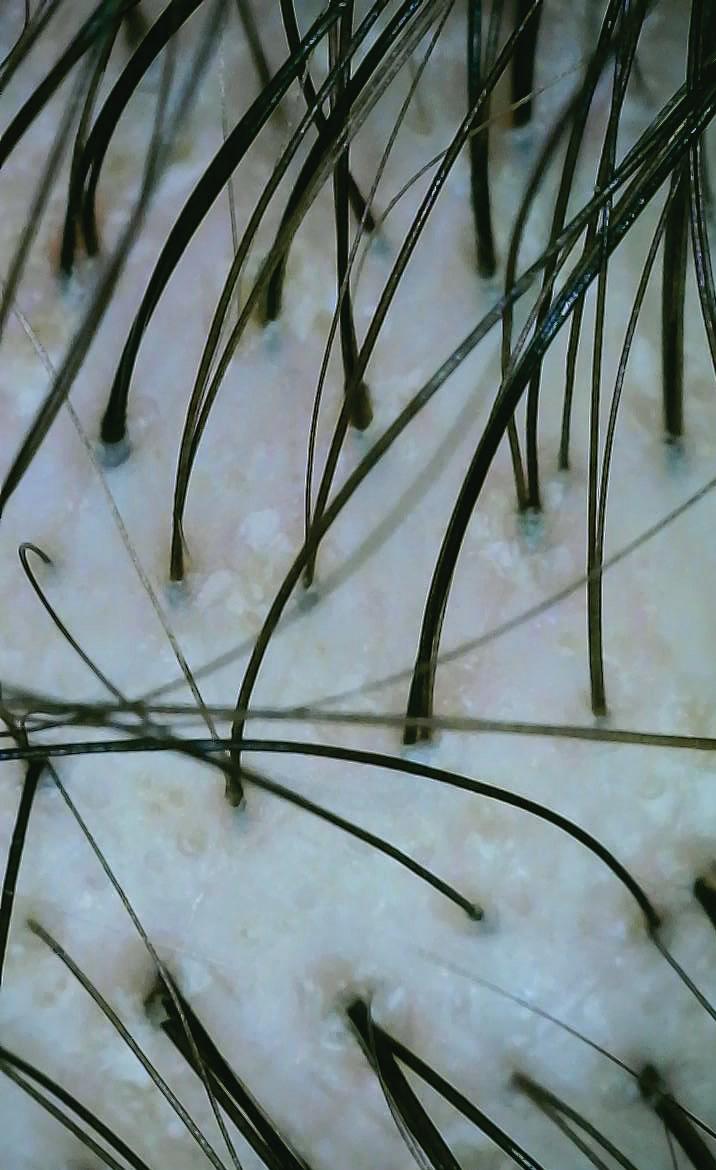
hair loss. The therapeutic regimen consisted of a topical combination of minoxidil and finasteride, cyclical nutraceutical therapy, and supplementation with Vitamin D and B12, all while adhering to a healthy lifestyle and undergoing no further diagnostic investigations. The treatment initiated with mesotherapy utilizing copper peptide and fibroblast growth factor, which involved micro-injection of the peptide into the scalp at three-week intervals to stimulate cellular regeneration and promote hair growth. Trichoscopic imaging was performed on the central area of the scalp exhibiting thinning before and after the treatment protocol. Following four sessions, trichoscopic findings demonstrated a significant increase in the number of hairs per follicle and an overall enhancement in hair count, accompanied by a reduction in focal atrichia, characterized by fewer areas of scalp devoid of hair and a decrease in hair thickness. These observations indicate a favorable response to the combined treatment approach, underscoring the efficacy of integrating advanced therapies, such as copper peptide and fibroblast growth factor delivered through mesotherapy, with conventional methods in the management of female androgenetic alopecia.
Before treatment
Trichoscopy before treatment
After Treatment
Trichoscopy after treatment Figure 1:
Figure 1: Significant result in hair growth after four sessions of copper peptide and fibroblast growth factor delivered through mesotherapy.
Diagnosis
Diagnosing androgenetic alopecia involves a combination of clinical evaluation, patient history, and sometimes additional diagnostic tools to confirm the condition and rule out other types of hair loss. A thorough medical history is obtained to assess the onset and progression of hair loss, family history of AGA, and any associated symptoms, such as itching or redness. Additionally, a physical examination of the scalp is conducted to evaluate hair density, the pattern of hair loss, and any signs of scarring or inflammation. Together, these elements provide a comprehensive assessment for accurate diagnosis and management of AGA.4
In diagnosing hair loss, several key tests help distinguish androgenetic alopecia from other conditions. The hair pull test is a simple, non-invasive method where clinicians gently pull a small group of hairs; if more than 2-3 hairs come out, it suggests a high shedding rate, often associated with AGA. The hair pluck test involves extracting individual hairs to examine miniaturized follicles and assess the anagen-to-telogen ratio, with a higher telogen count indicating shedding. A trichogram further evaluates hair cycle phases by plucking a small sample and microscopically analyzing anagen, telogen, and catagen hairs, where an elevated telogen rate suggests AGA. Trichoscopy is a noninvasive imaging technique that
enhances diagnostic precision by identifying features such as exclamation mark hairs and yellow dots, which help differentiate alopecia areata from AGA, guiding targeted treatment approaches.4,5
Scalp biopsy is a diagnostic procedure for assessing suspected androgenetic alopecia. It involves excising a small segment of the scalp from an area of hair loss for histopathological analysis. The biopsy can reveal features of AGA, such as follicular miniaturization and a reduced anagen-to-telogen ratio, while also excluding other forms of alopecia, including scarring and inflammatory types.6 Blood tests are essential in the assessment of androgenetic alopecia as they identify systemic conditions that may contribute to hair loss. Key evaluations include measuring serum testosterone, dihydrotestosterone......... (DHT), and dehydroepiandrosterone sulfate (DHEAS) levels, along with thyroid function tests and iron studies. These tests effectively exclude secondary causes of alopecia and guide appropriate treatment strategies.1
The Norwood classification system is a standardized tool for diagnosing androgenetic alopecia in males, comprising seven stages. It ranges from Type I, indicating minimal frontal hairline recession, to Type VII, characterized by advanced balding in the crown and frontal areas. This system aids clinicians in assessing hair loss severity and pattern, facilitating communication about treatment options and informing individualized management strategies.7
Treatment
Treatment modalities for androgenic alopecia encompass minoxidil, finasteride, and dutasteride, as well as emerging therapeutic options. Minoxidil functions as a potassium channel blocker, promoting vasodilation and enhancing dermal blood flow to hair follicles. Finasteride, a 5-alpha reductase type 2 inhibitor, demonstrates efficacy primarily in the vertex region of the scalp. Dutasteride exhibits greater potency than finasteride and may be employed in patients who do not respond adequately to finasteride therapy.8 Topical antiandrogens are utilized for hormonal modulation, while latanoprost has been investigated for its potential efficacy in promoting hair growth. Topical antibiotics and antifungals may be indicated to manage secondary infections or dermatological conditions that exacerbate hair loss.8 Growth factors are also under evaluation for their role in stimulating hair regrowth. Lowlevel laser therapy represents a nonsurgical intervention aimed at enhancing hair density. Surgical interventions, including hair transplantation, offer a more permanent solution for individuals with sufficient donor hair density.8
The combination of mesotherapy with copper peptide and fibroblast growth factor represents a powerful therapeutic strategy for hair loss treatment and other regenerative applications, effectively leveraging their unique properties to enhance healing, promote tissue regeneration, and improve overall outcomes. Mesotherapy is a minimally invasive cosmetic procedure
that involves injecting a mixture of vitamins, minerals, amino acids, and beneficial substances directly into the mesoderm (the middle layer of the skin) of the scalp, aiming to rejuvenate hair follicles, particularly in conditions like androgenetic alopecia. This technique delivers essential nutrients—such as biotin, zinc, and amino acids.9
This synergistic approach demonstrates efficacy in promoting hair restoration by reducing hair loss and enhancing hair density through the stimulation of hair follicle activity and the facilitation of scalp healing. The combination of mesotherapy and peptide growth factors has been shown to accelerate hair regrowth, improving outcomes in patients with androgenetic alopecia and other forms of hair loss. Notable peptides, such as Copper peptides, are recognized for their ability to enhance hair follicle health and stimulate tissue regeneration, while fibroblast growth factors (FGFs) are known for their role in promoting cellular growth and metabolic activity within hair follicles.10 Overall, the integration of mesotherapy with copper peptide and fibroblast growth factor provides a robust and customizable therapeutic option for individuals seeking effective non-surgical interventions for hair restoration. It is imperative for practitioners to conduct thorough consultations to develop individualized treatment plans, as patient responses may vary. This innovative combination not only enhances therapeutic outcomes in hair restoration but also presents opportunities for continued advancements in the field of dermatology and regenerative medicine focused
on hair health.9, 10
Discussion
Alopecia, particularly androgenic alopecia, commonly known as female pattern baldness, poses a significant burden on individuals and society due to its widespread prevalence and profound psychological impact. Characterized by thinning hair and receding hairlines, male and female pattern hair loss is increasingly common across all age groups, with contributing factors including pollution, lifestyle choices, stress, and poor dietary habits. For those experiencing extreme hair loss, treatments like Peptide Growth Factors are effective, as they stimulate fibroblasts and rejuvenate the hairline.1
The combination of copper peptide and fibroblast growth factor has emerged as a promising treatment in the field of regenerative medicine, particularly for hair restoration. Peptides enriched with growth factors are essential for stimulating healing, regeneration, and hair growth. When the copper peptide and fibroblast growth factor formulation is injected into specific areas, such as the scalp, it stimulates various biological processes that contribute to hair restoration. The fibroblast growth factor in the Copper Peptide formulation promotes the proliferation of dermal papilla cells, which are critical for hair follicle development and maintenance, leading to the activation of hair follicles and promoting hair regrowth. Enhanced blood circulation facilitates the delivery of essential nutrients and oxygen to hair follicles, which is crucial for healthy hair growth.9,10
The activation of natural fibroblasts by these growth factors stimulates an increase in collagen synthesis, thereby improving the structural integrity of the scalp and hair follicles and fostering a more conducive environment for hair growth. Certain growth factors also possess anti-inflammatory properties, helping to reduce inflammation in the scalp. By minimizing inflammation, peptide growth factor therapy creates a favourable environment for hair follicles to thrive. Furthermore, this therapy has been shown to extend the anagen (growth) phase of the hair cycle, leading to longer-lasting hair growth.9,10
This therapy is widely used for treating various forms of hair loss, including androgenetic alopecia (male and female pattern baldness), alopecia areata, and hair thinning due to stress or other factors. The addition of copper peptides to peptide growth factor formulations significantly enhances their effectiveness for hair restoration. Copper peptides activate dormant hair follicles, promote cell proliferation, and extend the anagen phase, resulting in increased hair density. They also boost collagen and elastin production, maintain scalp integrity, and reduce inflammation, creating an optimal environment for growth. Their antioxidant properties protect hair follicles from oxidative stress, while promoting blood vessel formation improves nutrient delivery. Together with growth factors, copper peptides maximize treatment outcomes, leading to a comprehensive approach to combating hair loss.4, 5
Peptide growth factor therapy, which includes the combination of copper peptide and fibroblast growth factor, is a non-surgical procedure that does not require blood withdrawal; instead, it involves injections directly into the treatment area. This approach results in minimal downtime and quick recovery for patients. By harnessing the body’s natural healing mechanisms, this therapy promotes regeneration without the use of synthetic drugs or chemicals, aligning with the growing trend towards natural and holistic health approaches. For individuals experiencing hair loss, the combination of copper peptide and fibroblast growth factor can lead to significant improvements in hair density and quality, ultimately boosting self-esteem and confidence.
References
1. Ho CH, Sood T, Zito PM. Androgenetic Alopecia. 2024 Jan 7. In: StatPearls [Internet]. Treasure Island (FL): StatPearls Publishing; 2024 Jan–. PMID: 28613674.
2. Ramos PM, Miot HA. Female Pattern Hair Loss: a clinical and pathophysiological review. A Bras Dermatol. 2015 Jul-Aug; 90(4):529-43. doi: 10.1590/ abd18064841.20153370. PMID: 26375223; PMCID: PMC4560543.
3. Dhami L. Psychology of Hair Loss Patients and Importance of Counseling. Indian J Plast Surg. 2021 Dec 31; 54(4): 411-415. doi: 10.1055/ s-0041-1741037. PMID: 34984078; PMCID: PMC8719979.
4. Vidal CI. Overview of Alopecia: A Dermato pathologist's Perspective. Mo Med. 2015 Jul-Aug; 112(4): 308-12. PMID: 26455063; PMCID: PMC6170065.
The psychological impact of regaining hair can be profound, enhancing social interactions and overall well-being.10
As awareness of peptide growth factor therapy increases, patients become more informed about their options for managing hair loss. This empowerment enables individuals to take proactive steps toward their health and appearance. Many patients report sustained results from peptide growth factor therapy, with ongoing maintenance treatments providing longlasting improvements. This contrasts with other hair loss treatments that may require continuous use to maintain effectiveness.10
Conclusion
The combination of copper peptide and fibroblast growth
factor formulation represents a significant advancement in the management of hair loss disorders, particularly androgenetic alopecia. By harnessing the body’s inherent healing processes through the application of peptide formulations enriched with growth factors, this therapy offers a promising intervention for individuals seeking effective and lasting solutions to hair thinning and baldness. As the demand for hair restoration therapies continues to rise, the formulation containing copper peptide with fibroblast growth factor emerges as a credible and innovative modality that addresses not only the physiological aspects of hair loss but also the psychological and social ramifications associated with this condition.
5. Al-Dhubaibi MS, Alsenaid A, Alhetheli G, Abd Elneam AI. Trichoscopy pattern in alopecia areata: A systematic review and meta-analysis. Skin Res Technol. 2023 Jun;29(6):e13378. doi: 10.1111/ srt.13378. PMID: 37357664; PMCID: PMC10236002.
6. Pinedo - Moraleda F, Tristán-Martín B, Dradi GG. Alopecias: Practical Tips for the Management of Biopsies and Main Diagnostic Clues for General Pathologists and Dermato pathologists. J Clin Med. 2023 Jul 29; 12(15):5004. doi: 10.3390/ jcm12155004. PMID: 37568407; PMCID: PMC10419566.
7. Wirya CT, Wu W, Wu K. Classification of Male-pattern Hair Loss. Int J Trichology. 2017 Jul-Sep; 9(3):95-100. doi: 10.4103/ ijt.ijt_46_17. PMID: 28932058; PMCID: PMC5596658.
8. Devjani S, Ezemma O, Kelley KJ, Stratton E, Senna M. Androgenetic Alopecia: Therapy Update. Drugs. 2023 Jun; 83(8):701-715. doi: 10.1007/ s40265-023-01880-x. Epub 2023 May 11. PMID: 37166619; PMCID: PMC10173235.
9. Tang Z, Hu Y, Wang J, Fan Z, Qu Q, Miao Y. Current application of mesotherapy in pattern hair loss: A systematic review. J Cosmet Dermatol. 2022 Oct;21(10) :4184-4193. doi: 10.1111/ jocd.14900. Epub 2022 Mar 14. PMID: 35253335.
10. Pyo HK, Yoo HG, Won CH, Lee SH, Kang YJ, Eun HC, Cho KH, Kim KH. The effect of tripeptide-copper complex on human hair growth in vitro. Arch Pharm Res. 2007 Jul;30(7):834-9. doi: 10.1007/ BF02978833. PMID: 17703734.
The inclusivity observed in melasma clinical trials establishes a model for enhancing diversity in research for other Dermatologic conditions.
A study on melasma clinical trials highlighted significant progress in patient representation, particularly for women and individuals with skin of color. Historically, dermatologic research has been criticized for underrepresenting non-white populations. However, in the examined trials, there was notable inclusivity, with a substantial proportion of Hispanic, Asian, and Black or African American participants, as well as individuals with Fitzpatrick skin types III to IV. This trend reflects a shift toward more diverse representation in dermatology research, particularly for conditions like melasma, which predominantly affects women and individuals with darker skin tones. This approach sets an important precedent for other dermatologic conditions, emphasizing the need for broader diversity in clinical trials.
Polyherbal Lip Hydrant Effectively Heals Chapped and Rough Lips, Outperforming Petroleum Gel
A recent study investigated the benefits of a novel herbal lip hydrant formulation made from six plant extracts: peppermint oil, perilla extract, emblica fruit extract, guava leaf oil, galanga rhizome extract, and green tea leaf extract. The formulation was applied to participants with chapped lips, and the results showed significant improvements in lip hydration, reduction in roughness, and enhanced appearance. The herbal extracts demonstrated antiinflammatory, antioxidant, and emollient properties that contributed to the healing process. Notably, the herbal lip hydrant was shown to elevate Nuclear Factor Erythroid 2-Related Factor 2 (Nrf2) protein levels, which helps protect the skin barrier, and strengthened cell adherence and renewal. These findings highlight the potential benefits of using natural herbal ingredients for lip care, offering a safe and effective alternative to synthetic treatments.
NICOTINAMIDE ADENINE DINUCLEOTIDE (NAD+): A UNIVERSAL COENZYME.
Nicotinamide adenine dinucleotide (NAD+) is a universal and indispensable coenzyme present in all living cells, playing a fundamental role in numerous biological processes essential for sustaining life. As a central player in cellular metabolism, NAD+ is involved in energy production, genome maintenance, and intracellular communication, ensuring the proper functioning of various physiological systems. This crucial molecule influences key biological mechanisms, including oxidative stress response, immune system regulation, mitochondrial function, and DNA repair, all of which are vital for cellular homeostasis.Beyond its metabolic functions, NAD+ plays a pivotal role in preserving cellular integrity by activating key enzymes such as sirtuins and poly (ADP-ribose) polymerases (PARPs). These enzymes are essential for regulating inflammation, longevity, and overall cellular resilience, making NAD+ a key factor in maintaining tissue health and function. However, NAD+ levels naturally decline with age, leading to a reduction in cellular efficiency an increased susceptibility to age-related diseases, including neurodegenerative disorders, metabolic syndromes, and skin aging.With growing interest in the role of NAD+ in aging
and disease prevention, various strategies are being explored to replenish its levels and restore cellular vitality. Therapeutic approaches such as oral supplementation, intravenous (IV) therapy, and lifestyle modifications, including diet and exercise, have emerged as potential interventions to enhance NAD+ levels. These strategies aim to counteract the effects of aging, improve cellular function, and promote longevity, particularly in tissues that are highly dependent on NAD+, such as the skin, muscles, and brain. As ongoing discoveries continue to uncover the broad impact of NAD+ on human health, its therapeutic potential remains an exciting frontier in the fields of longevity science and regenerative medicine.
FAQ on Nicotinamide Adenine Dinucleotide
1.What is Nicotinamide Adenine Dinucleotide (NAD+) and why is it important?
Nicotinamide Adenine Dinucleotide (NAD+) is a vital coenzyme found in all living cells. It plays a crucial role in energy metabolism by participating in redox reactions, where it alternates between its oxidized form (NAD+) and reduced form (NADH). NAD+ is essential for ATP production, mitochondrial function, DNA repair, and cellular signalling pathways. It also activates enzymes like sirtuins and poly (ADP-ribose) polymerases (PARPs), which influence aging, inflammation, and overall cellular health
2. How does NAD+ contribute to energy production in cells?
NAD+ is a key player in cellular metabolism. It serves as a cofactor in glycolysis, the tricarboxylic acid (TCA) cycle (Krebs cycle), and oxidative phosphorylation. During glycolysis and the TCA cycle, NAD+ accepts electrons to become NADH, which then donates these electrons to the electron transport chain in mitochondria. This process generates ATP, the cell’s primary energy source, making NAD+ indispensable for sustaining life and cellular function.
3. Why Do NAD+ Levels Decrease with Age, and What Are the Key Biological Factors Involved?
Nicotinamide Adenine Dinucleotide (NAD+) levels
peak in the early 20s but progressively decline with age, decreasing by 40–50% by the time individuals reach 50. This decline results from both increased consumption and reduced production. Key enzymes, such as Poly (ADP-ribose) Polymerases (PARPs) and Cluster of Differentiation 38 (CD38), play a significant role in depleting NAD+. PARPs become more active in repairing DNA damage, while CD38 activity rises due to age-related inflammation. At the same time, the body's ability to synthesize NAD+ diminishes as essential precursors—Nicotinamide (NAM), Nicotinic Acid (NA), Nicotinamide Mononucleotide (NMN), Nicotinamide Riboside (NR), and tryptophan—become less available. Additionally, the enzymes responsible for NAD+ production slow down. This imbalance in NAD+ levels leads to reduced cellular energy, weakened cell function, and an increased risk of age-related diseases, highlighting its crucial role in maintaining overall health and longevity.
4. What is the role of NAD+ in regulating sirtuins?
Sirtuins (SIRT1–SIRT7) are a family of NAD+-dependent enzymes that regulate key biological processes, including metabolism, inflammation, stress resistance, and longevity. Sirtuins rely on NAD+ to function effectively, meaning that declining NAD+ levels impair their ability to promote healthy aging. Activation of sirtuins by NAD+ has been shown to enhance mitochondrial function, improve insulin sensitivity, and reduce inflammation, contributing to overall well-being.
5. How does NAD+ impact skin health and aging?
NAD+ plays a crucial role in maintaining skin health by supporting cellular energy production, DNA repair, and antioxidant defense mechanisms. It helps to protect skin cells from oxidative damage caused by UV exposure and environmental pollutants. Additionally, NAD+-activated sirtuins promote collagen synthesis and reduce inflammation, which can improve skin elasticity and reduce signs of aging, such as wrinkles and fine lines.
6. How does the decline of NAD+ contribute to skin aging?
NAD+ is crucial for maintaining cellular energy, supporting DNA repair, and defending against oxidative stress in skin cells. As NAD+ levels decrease with age, skin fibroblasts become less effective in synthesizing collagen and elastin, resulting in the formation of wrinkles, reduced skin firmness, and slower wound healing. Moreover, NAD+ depletion negatively impacts Sirtuins (especially SIRT1 and SIRT6), which are involved in cellular longevity and UV damage protection. This disruption contributes to the acceleration of photoaging, pigmentation irregularities, and a weakened skin barrier, leaving the skin more vulnerable to environmental stressor.
7. How does NAD+ depletion affect skin barrier function and wound healing?
NAD+ is essential for lipid metabolism, keratinocyte proliferation, and moisture retention, all of which are crucial for maintaining a healthy skin barrier. When NAD+ levels decline, the epidermis weakens, leading to increased trans epidermal water loss (TEWL), dryness, and heightened sensitivity to irritants and allergens. Additionally, NAD+ supports fibroblast activity, collagen remodeling, and angiogenesis, all of which are vital for effective wound healing. Decreased NAD+ availability results in delayed tissue repair, increased scarring, and a compromised immune response, which can impair overall healing.
8. What other specific health conditions can result from NAD+ deficiency?
NAD+ deficiency has been associated with various health conditions, including metabolic disorders such as obesity and type 2 diabetes due to reduced insulin sensitivity, cardiovascular diseases caused by endothelial dysfunction and increased oxidative stress, and neurodegenerative disorders like Alzheimer's and Parkinson’s disease due to impaired neuronal function. It can also lead to chronic fatigue and muscle weakness from mitochondrial dysfunction, immune system dysfunction that weakens immune responses and increases infection susceptibility, cognitive decline due to impaired brain cell energy production and repair mechanisms, and liver damage as NAD+ is crucial for detoxification and maintaining healthy liver function.
9. Dietary Sources of NAD+ Precursors?
Nicotinamide adenine dinucleotide (NAD+) is not directly available from dietary sources but is synthesized from precursors through metabolic pathways. Key precursors include tryptophan, converted via the de novo pathway, found in poultry, red meat, fish, eggs, dairy, soy products, nuts, seeds, and legumes. Nicotinic acid (niacin, vitamin B3), utilized in the Preiss-Handler pathway, is abundant in organ meats, poultry, fish, legumes, whole grains, nuts, and fortified cereals. Nicotinamide (niacinamide), processed via the salvage pathway, is present in meat, poultry, fish, nuts, seeds, whole grains, mushrooms, and leafy greens. These dietary sources support NAD+ biosynthesis, essential for cellular metabolism, DNA repair, and overall physiological function.
10. What are the approaches to restoring NAD+ levels that also enhance skin health and appearance?
To restore NAD+ levels and improve skin health, different approaches can be used. Topical treatments like nicotinamide (Vitamin B3), NMN, and NR can boost NAD+ production and improve hydration. Oral supplements such as nicotinamide riboside (NR) and nicotinamide mononucleotide (NMN) help replenish NAD+ throughout the body, improving skin elasticity and radiance. Sirtuin-activating compounds like resveratrol and CoQ10 help repair skin cells and protect against oxidative damage. Antioxidants such as vitamin C and glutathione also help neutralize free radicals, preserving skin health. Lifestyle changes, including exercise and a healthy diet, support NAD+ recycling. NAD+ IV therapy can also directly increase NAD+ levels, promoting better skin health.
Optimizing Aesthetic Outcomes in Melasma Treatment
Dr. Neetu Sidana
MD (Dermatology)
Consultant Dermatologist
Apex
Hospitals, Jaipur
Introduction
Melasma is an acquired ......... hyperpigmentation disorder predominantly affecting photoexposed areas of the skin, particularly the face, and is most commonly observed in women of reproductive age. Clinically, it presents as symmetric, brown to grayishbrown macules or patches, primarily localized to the cheeks, forehead, upper lip, and chin, although it can also affect extrafacial areas. The condition is strongly associated with sun exposure, hormonal fluctuations (such as during pregnancy or the use of oral contraceptives), and medications that increase photosensitivity. While generally asymptomatic, melasma can cause significant psychological distress due to its visible nature, leading to low self-esteem, anxiety, and social embarrassment. Its clinical course is marked by fluctuations in pigmentation, with exacerbations often triggered by sun exposure or hormonal changes. In many cases, melasma becomes chronic, necessitating ongoing management.1 The pathophysiology of
melasma, primarily associated with photoaging, remains incompletely understood but is driven by multiple etiological factors that enhance melanogenesis. Ultraviolet (UV) radiation plays a central role by penetrating the epidermis, inducing the production of reactive oxygen species, and stimulating melanocyte activity.2 Furthermore, shorter wavelengths of visible light, such as blue and violet light, have been implicated in persistent hyperpigmentation, especially in individuals with darker skin phototypes. A familial predisposition is notable, as a significant proportion of affected individuals have a positive family history. Hormonal factors, particularly oestrogen and progesterone, are key contributors to melasma, with a heightened prevalence observed during pregnancy, oral contraceptive use, menopausal hormone therapy, and topical oestrogen treatments, with oestrogen being the primary mediator. Phototoxic reactions associated with certain medications, including anti-epileptic, antimalarial, antipsychotic, and cytotoxic
drugs, can also precipitate melasma. Additionally, heat exposure, particularly from occupational environments or cooking, has been identified as a potential exacerbating factor.1,2 Melasma has also been linked to endocrinological conditions, such as thyroid dysfunction, where elevated levels of thyroidstimulating hormone and specific autoantibodies have been noted in affected patients. At the molecular level, melasma pathogenesis involves several signalling pathways, including melanocyte-stimulating hormone/cyclic adenosine monophosphate, KIT, and Wnt pathways, which upregulate tyrosinase and microphthalmiaassociated transcription factor, thereby stimulating melanogenesis. Clinically, melasma typically presents as bilateral, asymptomatic macules of varying shades of light to dark brown with irregular borders, most commonly localized to the malar cheeks, forehead, upper lip, and/or mandible. The clinical presentation is usually distinctive, allowing for a straightforward diagnosis based on visual examination. Melasma can be classified into several patterns, including the centrofacial pattern, involving the cheeks, nose, forehead, and upper lip (excluding the philtrum); the malar pattern, affecting the nose and malar cheeks; and the mandibular pattern, located on the mandible and chin. An extrafacial pattern, primarily affecting sun-exposed areas such as the upper extremities, is also recognized, along with an erythematous or inflamed variant known as erythrosis pigmentosa faciei.1, 2
Melasma can have a profound impact on an individual's quality of life, primarily due to its visible and chronic nature, which often results in significant psychological distress. The aesthetic concerns associated with the condition, particularly the presence of facial pigmentation, can lead to reduced selfesteem, social withdrawal, and heightened anxiety, with many patients experiencing diminished psychosocial functioning. The recalcitrant and relapsing course of melasma, in conjunction with its limited responsiveness to conventional treatments, can further contribute to patient frustration and emotional distress. Additionally, the ongoing necessity for strict sun protection and long-term management can impose a significant psychosocial and practical burden, compromising overall well-being and life satisfaction.2
Before treatment
Case Report
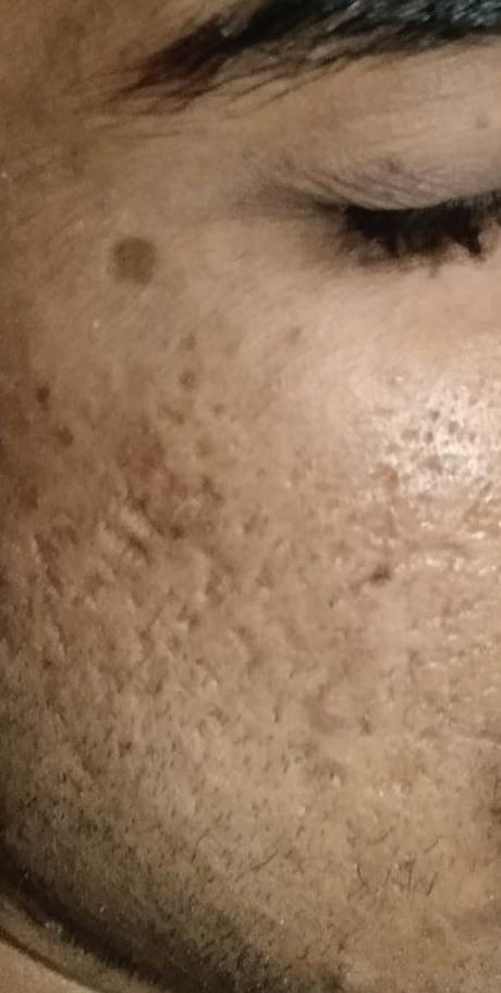
A 30-year-old female presented with facial hyperpigmentation, predominantly affecting the malar region and upper lip, consistent with melasma. She had no significant medical history, but reported gradual darkening of the skin, worsened by sun exposure. Her treatment regimen included oral tranexamic acid 500 mg once daily to inhibit melanocyte activity, hydroquinone lotion applied nightly for its tyrosinase inhibition and depigmenting effects, and coconut oil for moisturizing. The patient was instructed to avoid soap, face washes, and direct sunlight, emphasizing strict sun protection. She was also advised against using home remedies that could potentially aggravate the condition. At follow-up, the patient showed improvement in pigmentation, demonstrating the efficacy of this treatment combination.
After treatment
Figure 1: Hyperpigmentation on malar region of the face
Diagnosis
The aim of diagnosing melasma is to confirm the condition based on clinical evaluation, considering the patient's history, typical pigmentation patterns, and triggering factors. It is essential to differentiate melasma from other pigmentary disorders to ensure accurate diagnosis and guide appropriate treatment and preventive
measures.
Dermoscopy and Wood’s lamp examination are essential tools for evaluating melasma by assessing melanin depth and distribution. Dermoscopy reveals a reticular brown pattern in epidermal melasma, diffuse bluish-grey pigmentation in dermal melasma, and combined features in mixed melasma, aiding treatment planning. Wood’s lamp uses ultraviolet light to distinguish types, with epidermal melasma appearing darker, dermal melasma showing minimal contrast, and mixed melasma displaying variable intensities, guiding diagnosis and therapy monitoring.3 Reflectance confocal microscopy (RCM) is a non-invasive imaging technique that provides highresolution, real-time visualization of skin layers, aiding in melasma assessment. It allows detailed imaging of the epidermis and dermis, distinguishing epidermal melasma with increased basal layer pigmentation and dermal melasma with melanophages. RCM enhances diagnostic accuracy by offering a clear view of melanin distribution and differentiating melasma from other hyperpigmentation disorders.3 Histopathology in melasma reveals increased melanin in the basal epidermal layer for epidermal melasma, while dermal melasma shows melanin within dermal melanophages, with mixed melasma involving both layers. Though not routinely needed, it aids in assessing pigmentation depth.4Advanced imaging techniques like highfrequency ultrasound and optical coherence tomography (OCT) provide non-invasive evaluation of skin layers and melanin
distribution, distinguishing between epidermal, dermal, and mixed melasma. These methods are primarily research-oriented and not commonly used in clinical practice.5
Treatment
Hydroquinone functions by blocking tyrosinase, an enzyme essential for melanin synthesis in melanocytes. By blocking the conversion of tyrosine to melanin, hydroquinone reduces hyperpigmentation in the skin. This action decreases the synthesis of melanin, thereby lightening the skin and helping to treat conditions like melasma. Additionally, hydroquinone may interfere with the melanocyte’s ability to transfer melanin to surrounding keratinocytes, further contributing to its depigmenting effect. Tranexamic acid works in the treatment of melasma by inhibiting the binding of plasminogen to keratinocytes, which decreases the activation of melanocytes and reduces melanin production. It also modulates the vascular response by preventing the release of inflammatory mediators and reducing the formation of new blood vessels, which can contribute to hyperpigmentation.6
Kligman’s formula, commonly used for treating melasma, combines hydroquinone, tretinoin, and dexamethasone. Hydroquinone reduces melanin production by inhibiting tyrosinase activity in melanocytes.6 Tretinoin, a retinoid, accelerates epidermal turnover, promoting the shedding of pigmented skin cells and enhancing hydroquinone penetration. Dexamethasone, a corticosteroid, provides anti-
inflammatory effects, reducing irritation and inflammation that may result from other active ingredients. This combination is especially effective for moderate to severe cases of melasma. Azelaic acid, a naturally occurring acid, inhibits tyrosinase activity, which reduces melanin production and lightens hyperpigmented areas, making it effective for epidermal melasma. Kojic acid also inhibits tyrosinase, preventing the formation of melanin and thereby reducing pigmentation.6 Ascorbic acid (Vitamin C) acts as an antioxidant, neutralizing reactive oxygen species, stabilizing melanin production, and improving overall skin tone. Cysteamine, used in melasma treatment, inhibits tyrosinase activity and alters melanosome function, leading to a reduction in melanin synthesis. Metimazole, a potent peroxidase inhibitor, blocks melanin production by hindering the enzymatic steps involved in melanin synthesis. Iron oxide sunscreens are beneficial for blocking visible light, which is known to exacerbate pigmentation, and are an essential part of melasma management.6 Tretinoin also increases keratinocyte turnover, helping to shed pigmented skin cells and enhancing the effectiveness of other depigmenting agents. Hydrocortisone, another corticosteroid, reduces inflammation and inhibits melanogenesis, decreasing hyperpigmentation associated with melasma.6 Systemic treatments for melasma include oral tranexamic acid, which decreases melanocyte activity by inhibiting vascular growth factors and reducing pigmentation.
Oral glutathione, an antioxidant, reduces tyrosinase activity and shifts melanin production toward lighter pheomelanin. Carotenoids such as lutein and zeaxanthin also act as antioxidants, helping to reduce oxidative stress and potentially preventing melasma progression. These treatments, when used in combination with sun protection and other therapies, offer comprehensive management of melasma.6
Chemical peels, microneedling, and laser treatments are effective therapeutic modalities for melasma management. Chemical peels, utilizing agents such as glycolic acid, trichloroacetic acid, or salicylic acid, promote skin turnover and reduce pigmentation. Microneedling stimulates collagen production and enhances the absorption of topical depigmenting agents.6
Laser treatments, including ablative lasers (CO2, Er:YAG) and non-ablative fractional lasers (fractional CO2, fractional erbium), target melanin in the skin. Ablative lasers remove outer skin layers, promoting regeneration, while non-ablative lasers stimulate collagen remodelling and improve skin tone without damaging the epidermis. Non-ablative lasers are preferred for darker skin types due to a lower risk of postinflammatory hyperpigmentation (PIH).7
Discussion
Melasma can affect individuals of all racial backgrounds, though it is more prevalent in individuals with darker skin types, particularly those with light brown skin. The condition occurs with a higher frequency in women, with a female-tomale ratio of approximately 9:1.
Melasma is rare before puberty and is most commonly seen during the reproductive age. The condition is present in 15% to 50% of pregnant individuals. Its prevalence varies between 1.5% and 33%, depending on the specific population being studied.1
Differential diagnoses for melasma include conditions like actinic lichen planus (violaceous papules linked to sun exposure), acanthosis nigricans (dark, thickened patches associated with insulin resistance), discoid lupus erythematosus (erythematous plaques with pigmentation changes), and drug-induced photosensitivity (UV-induced pigmentation from medications like tetracyclines). Other considerations are exogenous ochronosis (bluishblack discoloration from prolonged hydroquinone use), frictional melanosis (hyperpigmentation from repetitive trauma), mastocytosis (brownish macules from increased mast cells), nevi of Ito and Ota (blue-gray patches), pigmented contact dermatitis (irritant-induced pigmentation), poikiloderma of Civatte (photoinduced pigmentation with telangiectasias), and postinflammatory hyperpigmentation (darkening after inflammation). Sun exposure, a leading trigger for melasma, underscores the importance of strict sun protection. UVB, UVA, and visible light drive melanogenesis, necessitating high-SPF (≥50) sunscreens with strong UVA protection, crucial due to UVA’s potent melanogenic effects.2,8
Occupational heat exposure may contribute to melasma chronicity, and broad-spectrum
tinted sunscreens are effective in reducing severity. Makeup with sun protection can help camouflage lesions but should be layered over standard sunscreens for optimal protection. Photosensitizing agents, like contraceptives and estrogen therapy, should be avoided as they may exacerbate melasma. While melasma is benign with no malignancy risk, dermal pigmentation resolves more slowly than epidermal, but sustained inhibition of melanogenesis aids resolution. Recurrence is common without strict sun avoidance. Overuse of potent corticosteroids should be avoided to prevent worsening pigmentation or exogenous ochronosis, and depigmenting agents should be applied only to affected areas. Though various treatments exist, none is definitively superior, and lasers may worsen the condition; sun protection alone often leads to gradual improvement.1,3
Conclusion
Melasma is a chronic dermatological condition that significantly affects an individual’s aesthetic appearance, often leading to psychological distress, particularly in women and individuals with darker skin. It poses a challenge for both patients, who must adhere to strict sun protection and treatment regimens, and physicians, who face difficulties with treatment resistance and recurrence. While various treatments exist, no single approach has proven universally effective, and some may worsen the condition. Further research is needed to develop more targeted therapies and preventive strategies to reduce recurrence and improve outcomes for
patients and healthcare providers.
References
1. Basit H, Godse KV, Al Aboud AM. Melasma. 2023 Aug 8. In: StatPearls [Internet]. Treasure Island (FL): StatPearls Publishing; 2024 Jan–. PMID: 29083744.
2. Doolan BJ, Gupta M. Melasma. Aust J Gen Pract. 2021 Dec; 50(12):880-885. doi: 10.31128/AJGP-05-21-6002. PMID: 34845463.
3. Ogbechie-Godec OA, Elbuluk N. Melasma: an Up-to-Date Comprehensive Review. Dermatol Ther (Heidelb). 2017; 7(3):305-318. Doi: 10.1007/s13555-0170194-1.
4. Ritter CG, Fiss DV, Borges da Costa JA, de Carvalho RR, Bauermann G, Cestari TF. Extra-facial melasma: clinical, histopathological, and immunohistochemical case-control
5. Wang Z, Chen Y, Pan S, et al. Quantitative classification of melasma with photoacoustic microscopy: a pilot study. J Biomed Opt. 2024; 29(Suppl 1):S11504. doi:10.1117/1.JBO.29.S1.S11504.
6. Jiryis B, Toledano O, Avitan-Hersh E, Khamaysi Z. Management of Melasma: Laser and Other Therapies-Review Study. J Clin Med. 2024 Mar 3; 13(5):1468. doi: 10.3390/jcm13051468. PMID: 38592701; PMCID: PMC10932414.
SR, Nisticò SP, Zerbinati N, Rongioletti F, Pellacani G. Melasma and reflectance confocal microscopy: from baseline to treatment monitoring. Int J Dermatol. 2024 Aug; 63(8):1007-1012. doi: 10.1111/ijd.17117. Epub 2024 Mar 6. PMID: 38448367.
8. Cassiano DP, Espósito ACC, da Silva CN, et al. Update on Melasma-Part II: Treatment. Dermatol Ther (Heidelb). 2022; 12(9):1989-2012. doi:10.1007/ s13555-022-00780-4
The assessment of skin characteristics such as Fitzpatrick skin type, hyperpigmentation, redness, and wrinkle severity is crucial for determining the appropriate laser therapy. With the growing demand for cosmetic procedures, accurate evaluations have become more important. Artificial intelligence (AI) and machine learning have been explored to improve dermatologic assessments. To fill the gap, researchers created a
novel dataset, Skin Analysis, which includes labeled images of various skin characteristics, annotated by a Dermatologist. The final AI model demonstrated high accuracy and strong generalization capabilities, suggesting that AI-driven assessments could enhance the precision of laser therapy planning. Integrating such models into clinical practices could improve safety and treatment effectiveness. This advancement in AI-driven Dermatology has the potential to reduce complications associated with improper treatments and support more personalized, effective cosmetic procedures. Future research should focus on expanding the dataset and refining AI-based treatment planning to further enhance patient safety and treatment outcomes.
Lower-Dose Doxycycline for Lymphocytic Scarring Alopecia: A Viable Alternative
Recent findings suggest that low-dose doxycycline may effectively manage symptoms of lymphocytic scarring alopecia while reducing side effects. Lymphocytic scarring alopecia, a rare immune-mediated condition, leads to permanent hair loss due to immune system attacks on hair follicles. The study aimed to compare the efficacy and safety of low and high-dose doxycycline. While highdose doxycycline has traditionally been used, it is associated with side effects like gastrointestinal issues and rashes, leading to discontinuation.
The study reviewed patient records from a major healthcare institution. Results showed no significant difference in clinical outcomes, with both regimens improving hair loss severity, scalp inflammation, and hair quality. However, the low-dose treatment was associated with fewer adverse events and a lower rate of treatment discontinuation due to gastrointestinal problems. This is the first study to compare doxycycline doses for treating lymphocytic scarring alopecia, showing that lower doses offer similar benefits with fewer side effects. Reducing doses may also help mitigate the risk of antibiotic resistance.
Payal
Rangar - A Dedicated Nutritionist, passionate about transforming health through personalized, home-based, highly nutritious diet plans tailored to unique bio-individuality, sharing her skincare and haircare routine under the guidance of her Dermatologist.
In this exclusive interview, Payal Rangar discusses her approach to achieving optimal skin and hair health through a combination of balanced nutrition, hydration, and personalized routines. She emphasizes the importance of antioxidants, healthy fats, and protein while maintaining a holistic skincare regimen. Payal highlights the connection between emotional well-being, stress management, and skin health. By incorporating her Dermatologist guidance, including regular followups, she ensures her skin and hair receive the best care. Staying updated on the latest advancements in Nutrition and Dermatology, Payal effectively addresses concerns such as acne, dryness, and aging through her diet, along with expert guidance from her Dermatologist.
1. As a Nutritionist, what are the key dietary habits you follow to maintain healthy skin and hair?
As a Nutritionist, I follow a balanced nutrition plan with plenty of antioxidants, healthy fats, and protein to maintain healthy skin and hair. I ensure a daily intake of omega-3-rich foods such as flaxseeds and walnuts, vitamin C sources like citrus fruits and bell peppers, and biotin-rich foods including nuts, seeds, and eggs. By maintaining this nutrient-rich diet, I effectively support optimal skin and hair health.
2. How do you balance nutrition with external skincare routines to achieve optimal skin health?
I balance nutrition with external skincare routines by following a holistic approach, as I believe good skin starts from within. While I adhere to a minimalist skincare routine consisting of cleansing, moisturizing, and sunscreen, I prioritize anti-inflammatory and gut-friendly foods to prevent breakouts and maintain skin elasticity. I never follow shortcuts and always believe in balanced, wholesome meals.
3. What role does hydration play in your personal skincare and haircare routine, and how do you ensure you get enough water daily?
Hydration plays a crucial role in both my skincare and haircare routine. I ensure I drink 2-3 litres of water daily, incorporating herbal teas, infused water, and hydrating fruits such as watermelon and cucumber to maintain optimal moisture levels. Additionally, I am mindful of my caffeine consumption, ensuring I do not overindulge, and I consciously moderate my intake of wine, reserving it for occasional enjoyment to support overall hydration and well-being.
4. Do you take any supplements specifically for your skin and hair, and how do you determine what best for you?
I prefer a food-first approach to nutrition, ensuring I get the necessary nutrients through my diet. However, I take collagen, omega-3, and vitamin D supplements when needed to support my skin and hair health. I determine what best for me by first assessing my individual needs through blood tests and identifying specific deficiencies, rather than blindly following trends or popular recommendations.
5. What daily habits, apart from diet, do you practice to support your skin and hair health?
Apart from diet, I practice several daily habits to support my skin and hair health. I prioritize quality sleep, stress management through meditation and yoga, and regular exercise, which includes walks, strength training, and cardio. I also incorporate regular scalp massages with oil and limit heat styling, such as ironing and blow-drying, to help maintain hair health. Additionally, I make an effort to minimize hair colouring to preserve its overall condition.
6. How do you address specific skin concerns like acne, dryness, or aging through your diet?
To address specific skin concerns like acne, dryness, and aging through my diet, I make targeted adjustments. For acne, I avoid excess sugar and dairy while including anti-inflammatory foods such as turmeric, green tea, and probiotics. To combat dryness, I increase healthy fats, such as avocados, nuts, and seeds, and ensure proper hydration. For aging, I focus on collagen-boosting foods like bone broth, berries, and leafy greens to support skin elasticity and reduce the visible signs of aging.
7. What are some of the most important nutrients that you believe are often overlooked for skin and hair health?
Some of the most important nutrients often overlooked for skin and hair health include zinc, silica, and vitamin E. Zinc, which is essential for acne control and hair growth, can be found in pumpkin seeds and lentils. Silica, crucial for hair strength, is present in cucumbers and oats. Vitamin E, which supports skin elasticity, is abundant in almonds and sunflower seeds. These nutrients play a significant role in maintaining the health and appearance of skin and hair.
8. How do you manage stress through your diet, and what impact does it have on your skin and hair?
I manage stress through my diet by including magnesiumrich foods such as dark chocolate, nuts, and leafy greens, as well as adaptogens like ashwagandha to help regulate stress hormones. Since stress directly affects hair fall, acne, and dull skin, managing cortisol levels is crucial to maintaining healthy skin and hair. By focusing on these dietary adjustments, I can better manage stress and support the overall health of my skin and hair.
9. Have you ever experienced a situation where a Dermatologist advice significantly improved your skin or hair, and how did you incorporate it with your nutritional approach?
Yes, I have experienced a situation where my Dermatologist advice significantly improved my skin. My Dermatologist
consistently provides excellent guidance, recommending supplements and topically applied creams for better results. A treatment for my pigmentation was recommended, which worked out perfectly. I incorporated my Dermatologist advice into my nutritional approach by continuing to focus on a balanced diet that supports skin health, ensuring that both my skincare routine and nutrition worked synergistically for optimal results.
10. How do you stay updated on both Nutritional science and Dermatology to provide the best care for your skin and hair?
To stay updated on both Nutritional science and Dermatology, I always follow Dermatologist and Nutrition advice to stay informed about the latest findings in skin and hair nutrition. This approach helps me continuously enhance my knowledge and ensures I provide the best care for my skin and hair.
The Significance of Immunohistochemical Stains in Mohs Micrographic Surgery
Immunohistochemical (IHC) stains are increasingly used in Mohs micrographic surgery (MMS) to improve diagnostic accuracy, especially in cases involving poorly differentiated tumors or melanoma, where conventional hematoxylin and eosin (H&E) staining may be insufficient. IHC involves the use of antibodies to detect specific antigens within cells, allowing for enhanced visualization of neoplastic cells. Commonly used IHC stains in MMS include MART-1 and SOX10 for melanoma, Ber-EP4 for basal cell carcinoma (BCC), and PanCK for squamous cell carcinoma (SCC). These stains are particularly helpful in identifying tumor cells that may be challenging to distinguish using H&E alone, ensuring accurate tumor excision and clearer margin assessment. Despite the advantages, IHC staining has some limitations, including increased procedural costs, extended processing times, and the potential for nonspecific staining or background artifact. Additionally, it requires specialized training and histotechnological expertise. However, when used selectively in complex cases, IHC can significantly improve the precision of MMS, leading to better long-term outcomes for patients by reducing recurrence rates and minimizing unnecessary tissue removal.
At-Home Skincare
Devices Face Growing Scrutiny over Efficacy and Safety
As the popularity of at-home skincare devices continues to rise, Dermatologists are urging caution regarding their safety and effectiveness. Tools like LED masks, microcurrent devices, and at-home laser systems have become staples for many seeking to replicate professional treatments. However, experts warn that improper use of these devices can lead to adverse effects such as burns or skin irritation. While convenient, patients are advised to consult with medical professionals before using advanced at-home treatments, particularly for those with sensitive or complex skin conditions.
Pilot Study Demonstrates Safety and Efficacy of Intradermal Botulinum Toxin A for Melasma Treatment.
A pilot study demonstrated that intradermal Botulinum Toxin A (BoNT-A) is both safe and effective for the treatment of melasma, without compromising cellular integrity or patient safety. A randomized, split-face, placebocontrolled trial was conducted on female patients with Fitzpatrick skin types III and IV, all diagnosed with melasma. BoNT-A injections were administered to one side of the face, while the contralateral side received a normal saline placebo. Outcomes included the melasma area and severity index (MASI), physician global assessment (PGA), and patient satisfaction. The results indicated a significant reduction in MASI scores on the BoNT-A-treated side compared to the control, with notable improvements in melasma severity. Patient satisfaction scores were consistently higher for the treated side. The only reported adverse event was mild erythema, observed in a subset of patients. In vitro data corroborated these findings, showing that BoNT-A reduces melanin content and inhibits tyrosinase activity in UVA-irradiated B16F0 cells, without inducing cytotoxicity. Given the promising outcomes, further studies are warranted, particularly those exploring higher concentrations of BoNT-A, extended follow-up periods, and the potential benefits of combination therapies to optimize treatment for melasma.
Innovative Monopolar Radiofrequency Device Proven to Be Safe and Effective for Treating Facial Wrinkles
A novel monopolar radiofrequency device has demonstrated promising results in reducing fine wrinkles around the eyes and cheeks. This device works by emitting radiofrequency energy through a single active electrode, which passes current through the skin to generate heat in the tissues, promoting collagen remodeling and skin tightening. With its larger tip, it offers deeper penetration into the dermis, targeting both superficial and deeper layers of the skin for effective wrinkle and skin laxity treatment. The device provides a minimally invasive, non-surgical solution with natural-looking results, offering smoother skin, reduced wrinkle depth, and improved skin texture without the need for dramatic changes. It is a safe treatment with low risk of adverse effects, typically resulting in only mild, temporary effects such as erythema or swelling, while no severe side effects like scarring were reported. The procedure is convenient and efficient, delivering noticeable results in a single session with minimal discomfort. Additionally, the treatment stimulates collagen production, providing long-term skin benefits. This makes the device an attractive option for individuals seeking subtle, non-invasive aesthetic enhancements, with further studies needed to explore its full potential in larger, more diverse patient populations.
Hair Loss and Regeneration: Exploring the Potential of PRP and Microneedling
Dr. Arif Iqbal
MD (Dermatology, Venereology and Leprosy)
Dermatologist
Delhi Introduction
Alopecia refers to hair loss, which can be temporary or
permanent and affects all ages and genders. It is classified into nonscarring and scarring types. Nonscarring alopecia, where hair follicles remain intact and regrowth is possible, includes conditions like androgenetic alopecia, alopecia areata, telogen effluvium, anagen effluvium, traction alopecia, trichotillomania, and alopecia syphilitica. Scarring alopecia (cicatricial alopecia) involves irreversible follicular destruction, leading to permanent hair loss. Primary scarring alopecias include frontal fibrosing alopecia, lichen planopilaris, and central centrifugal cicatricial alopecia, while secondary scarring alopecias result from conditions like scleroderma, neoplasms, radiation, trauma, or infections.1
Androgenic alopecia (AGA), also referred to as male-pattern baldness (MPB) in men and female-pattern hair loss (FPHL) in women, is the most prevalent form of hair loss, driven by both genetic and hormonal factors. It
is characterized by progressive hair thinning, primarily resulting from the action of androgens, particularly dihydrotestosterone (DHT), on genetically predisposed hair follicles. The clinical presentation of AGA differs between sexes, with distinct patterns observed in men and women. In men, MPB typically begins with recession of the hairline at the temples and thinning at the vertex (crown), which may progress to more extensive baldness, often culminating in a "U-shaped" pattern, leaving a band of hair around the sides and back of the scalp. This pattern is closely associated with the influence of DHT on androgen-sensitive hair follicles. In women, FPHL typically manifests as diffuse thinning, especially at the crown, without significant recession of the frontal hairline. The thinning is often subtle and gradual, typically resulting in a widening of the part.2
The pathophysiology of AGA involves the process of follicular miniaturization, in which terminal hairs (thick, long) are replaced by vellus hairs (fine, short).
This occurs as a result of DHT binding to androgen receptors on the hair follicles, leading to a shortened anagen (growth) phase, a prolonged catagen (regression) phase, and an extended telogen (rest) phase. Over time, this results in the progressive shrinking of the hair follicles, producing finer, shorter hairs, which eventually become dormant, leading to permanent hair loss if not addressed. Genetic predisposition plays a crucial role in the development of AGA, with the androgen receptor (AR) gene and additional genetic loci influencing the sensitivity of hair follicles to DHT. In men, the genetic susceptibility is typically inherited from both parents, while in women, the inheritance pattern is more complex, involving multiple genetic factors that contribute to the condition. Alopecia areata is an immunemediated condition causing patchy hair loss, potentially progressing to alopecia totalis or universalis, with remission and recurrence. Telogen effluvium, triggered by stress, illness, hormonal changes, medications, or metabolic issues, leads to diffuse shedding about three months after the trigger.2,3
Accurate diagnosis through detailed clinical history, physical examination, and specific diagnostic testing is essential for determining the underlying etiology and guiding appropriate management, as treatment strategies vary significantly between nonscarring and scarring forms, with scarring alopecia often leading to permanent follicular destruction.
Diagnosis
The diagnosis of alopecia begins with a detailed patient
history, including onset, pattern, progression, and any associated symptoms. Family history, recent illnesses, medication use, stress, and hormonal changes are also important. Physical examination focuses on hair loss distribution and scalp condition. Several diagnostic tools are employed to evaluate hair loss, each offering valuable insights into its underlying pathology. The Hair Pull Test helps assess excessive shedding by gently tugging on a small section of hair, with a positive result indicating conditions like telogen effluvium. Trichoscopy, a noninvasive technique, is essential in androgenetic alopecia, revealing features such as follicular miniaturization, frontal hairline recession, and hair shaft thinning, facilitating early detection of follicular changes. When the diagnosis remains uncertain, a Scalp Biopsy becomes crucial, particularly for distinguishing scarring from non-scarring alopecia. Histopathological analysis evaluates follicular inflammation, perifollicular fibrosis, and structural alterations, ensuring diagnostic precision. The Trichogram aids in assessing the hair growth cycle by examining plucked hairs under a microscope to determine their distribution across the anagen, catagen, and telogen phases, helping differentiate telogen effluvium from other alopecias. The Card Test quantifies hair shedding by collecting telogen hairs using an adhesive card post-combing, assisting in distinguishing normal shedding from pathological hair loss. Each of these diagnostic modalities plays a pivotal role in understanding alopecia, enabling precise diagnosis and targeted
therapeutic strategies for optimal patient management.3,4,5,6
Treatment
We treated several patients with Platelet-Rich Plasma (PRP) therapy and PRP combined with microneedling using a dermaroller for androgenetic alopecia (AGA), observing promising outcomes in hair regrowth and follicular regeneration. Platelet-Rich Plasma (PRP) is a promising treatment for androgenetic alopecia (AGA) that promotes hair regrowth using growth factors derived from autologous blood. PRP works by harnessing the regenerative potential of platelets, specifically the alpha granules, which are rich in key growth factors such as plateletderived growth factor (PDGF), vascular endothelial growth factor (VEGF), transforming growth factor-beta (TGF-ß), and insulin-like growth factor (IGF-1). These factors play a crucial role in stimulating stem cell differentiation, follicular cell proliferation, and angiogenesis, supporting hair follicle regeneration. PRP is particularly effective due to the release of PDGF, which reduces follicular apoptosis and enhances hair follicle survival. Additionally, IGF-1 in PRP counteracts the inhibitory effects of dihydrotestosterone (DHT), a key factor responsible for hair follicle miniaturization in AGA. Since DHT reduces local IGF-1 production in hair follicles, the introduction of IGF-1 through PRP helps maintain follicular health and stimulates regrowth. The PRP treatment procedure involves collecting a small blood sample from the patient, which is then processed in a centrifuge to obtain a concentrated PRP
Hair Loss and Regeneration: Exploring
solution. This solution is injected into the scalp, targeting areas affected by thinning or hair loss. The growth factors within PRP activate dormant or weakened hair follicles, promoting regrowth. For optimal results in AGA treatment, three to five sessions are typically recommended, spaced at onemonth intervals. PRP is a safe and well-tolerated procedure with minimal downtime, making it a widely accepted therapeutic option for hair restoration.7,8,9,10
In addition to PRP therapy, microneedling with a dermaroller enhances treatment efficacy by creating multiple microchannels in the scalp, increasing transdermal drug penetration and stimulating the dermis. While studies evaluating the combination of PRP and microneedling in hair restoration are limited, this approach has shown promising results. Microneedling is performed using a dermaroller, typically with 192 titanium alloy–coated needles arranged in 24 rows, available in sizes ranging from 0.5 mm to 3 mm, with 1.5 mm being the preferred size for AGA patients. The procedure involves rolling the device sequentially and multidirectionally over the scalp with firm pressure until pinpoint bleeding occurs, signifying effective penetration. The combination of PRP and microneedling has demonstrated superior outcomes in AGA management by enhancing hair follicle regeneration and patient satisfaction. This synergistic approach represents an effective and innovative treatment option for individuals seeking improved hair restoration outcomes.7,8,9,10
Several treatments are also available for androgenetic alopecia (AGA), targeting different mechanisms to promote hair
regrowth. Topical minoxidil enhances follicular proliferation and prolongs the anagen phase, while topical finasteride locally inhibits 5-alpha-reductase, reducing DHT levels. Oral dutasteride and spironolactone further block androgenic effects, with spironolactone being particularly useful for female pattern hair loss. Botulinum toxin (Botox) improves scalp circulation, while stem cell therapy focuses on follicular regeneration. Low-Level Laser Therapy (LLLT) stimulates cellular metabolism and blood flow, enhancing minoxidil's effects when combined. Microneedling boosts collagen production and increases topical treatment absorption. For permanent restoration, hair transplantation via FUE or FUT remains the gold standard. These treatments, alone or in combination, help improve hair density and slow progression in AGA.7
Result
Patient 1 (Significant hair regrowth after using PRP with Dermaroller)
Before treatment After 3rd sitting After 5th sitting
Patient 2 (Significant hair regrowth after using PRP)
Before treatment
After Treatment
Hair Loss and Regeneration: Exploring the Potential of PRP and Microneedling
Patient 3 (Significant hair regrowth after using PRP)
Before treatment
After 4th sitting
Patient 4 (Significant hair regrowth after using PRP)
Before treatment
Discussion
After 3 months of treatment
Alopecia refers to hair loss with various subtypes, often requiring precise diagnosis. Key types include androgenetic alopecia, alopecia areata, telogen effluvium, female pattern alopecia (FFA), lichen planopilaris (LPP), and central centrifugal cicatricial alopecia, each with distinct causes and progression.1 Male-pattern hair loss (MPHL), assess its severity, several classification systems have been developed, with prominent models including the Hamilton, Norwood-Hamilton, Adapted Norwood-Hamilton, and Basic and Specific (BASP) classifications. Classification systems for male pattern hair loss (MPHL) help standardize diagnosis and staging. The Hamilton Classification divides MPHL into "not bald" (Types I–III) and "bald" (Types IV–VIII). The Norwood-Hamilton Classification refines this into seven types with five subdivisions, addressing frontotemporal recession and vertex involvement, while the Adapted Norwood-Hamilton simplifies the model with A (anterior) and V (vertex) designations. The BASP Classification categorizes hair loss by anterior hairline shape (L, M, C, U) and thinning location (F/V1–V3) for a detailed yet practical approach.11 Overlap between F and V types can create diagnostic ambiguity, but each system has unique strengths and limitations, providing a practical framework for diagnosing and tracking MPHL progression. Differentiating male pattern hair loss (MPHL) from other alopecias like telogen effluvium, diffuse alopecia areata, FFA and LPP is crucial, as conditions like chronic cutaneous lupus erythematosus, central centrifugal cicatricial alopecia, and folliculitis decalvans require distinct management approaches.1,2 Hair loss can impact mental well-being, particularly in women, children, and adolescents. The SALT score assesses severity by dividing the scalp into quadrants and classifying alopecia into six stages (S0–S5) for treatment guidance. Nutritional support, including biotin, vitamin D, iron, zinc, and omega-3 fatty acids, promotes follicular health. Preventive measures like reducing
heat styling, avoiding tight hairstyles, managing stress, and maintaining a balanced diet help minimize hair shedding.1,7
Platelet-Rich Plasma (PRP) therapy is a promising non-invasive treatment for alopecia, promoting follicular regeneration, scalp microcirculation, and hair growth. It counteracts DHT-induced follicular miniaturization, enhancing outcomes when combined with minoxidil or low-level laser therapy (LLLT).8 Early diagnosis and intervention optimize outcomes and reduce psychosocial impact. Nurses educate, pharmacists ensure medication safety, and a collaborative team improves care and quality of life.
Conclusion
Platelet-Rich Plasma (PRP) therapy is a promising treatment for alopecia, particularly androgenetic alopecia. It utilizes growth factors and cytokines from autologous platelets to stimulate hair follicle activity and enhance hair regrowth. Clinical studies show its effectiveness in increasing hair density with minimal side effects. However, standardized protocols and long-term safety data are needed. Future research should focus on optimizing PRP techniques and identifying the most responsive patient populations to improve treatment outcomes.
Hair Loss and Regeneration: Exploring
References
1. Al Aboud AM, Syed HA, Zito PM. Alopecia. In: StatPearls. Treasure Island (FL): StatPearls Publishing; February 26, 2024.
2. Ho CH, Sood T, Zito PM. Androgenetic Alopecia. In: StatPearls. Treasure Island (FL): StatPearls Publishing; January 7, 2024.
3. Kaiser M, Abdin R, Gaumond SI, Issa NT, Jimenez JJ. Treatment of Androgenetic Alopecia: Current Guidance and Unmet Needs. Clin Cosmet Investig Dermatol. 2023; 16:1387-1406. Published 2023 May 31. doi:10.2147/ CCID.S385861.
4. Singh S, Muthuvel K. Practical Approach to Hair Loss Diagnosis. Indian J Plast Surg. 2021; 54(4):399-403. Published 2021 Dec 27. Doi: 10.1055/ s-0041-1739240
5. Jain N, Doshi B, Khopkar U. Trichoscopy in alopecias: diagnosis simplified. Int J Trichology. 2013; 5(4):170-178. doi:10.4103/0974-7753.130385
6. Sperling LC. The role of the scalp biopsy in the evaluation of alopecia. J Am Acad Dermatol. 2023; 89(2S):S16-S19. doi:10.1016/ j.jaad.2023.05.047.
7. Serrano-Falcón C, Fernández-Pugnaire MA, Serrano-Ortega S. Hair and scalp evaluation: the trichogram. Actas Dermosifiliogr. 2013; 104(10):867-876. doi:10.1016/ j.ad.2013.03.004.
8. Sharma A, Chouhan K, Bhatia S, Dashore S. Platelet-Rich Plasma in Androgenetic Alopecia. Indian Dermatol Online J. 2021; 12(Suppl 1):S31-S40. Published 2021 Nov 25. doi:10.4103/ idoj.idoj_328_21
9. Shah KB, Shah AN, Solanki RB, Raval RC. A Comparative Study of Microneedling with Platelet-rich Plasma Plus Topical Minoxidil (5%) and Topical Minoxidil (5%) Alone in Androgenetic Alopecia. Int J Trichology. 2017 Jan-Mar;9(1):14-18. doi: 10.4103/ ijt.ijt_75_16. PMID: 28761259; PMCID: PMC5514790.
10.Jha, Abhijeet Kumar et al. Androgenetic alopecia and microneedling: Every needling is not microneedling. Journal of the American Academy of Dermatology, Volume 81, Issue 2, e43 - e44
11. Wirya CT, Wu W, Wu K. Classification of Male-pattern Hair Loss. Int J Trichology. 2017; 9(3):95-100. doi:10.4103/ ijt. ijt_46_17
The Natural Beauty Shift: Subtle Enhancements in Aesthetic
Procedures
A new trend is reshaping the world of aesthetic treatments, where the focus is shifting toward subtle, natural-looking results that don’t immediately reveal the use of surgery or injectables. This emerging movement, known as the “undetectable era,” signifies a change in patient preferences, with many now opting for subtle improvements that enhance their natural beauty without dramatic alterations. These procedures aim for minimal enhancement,
using less filler carefully placed to refine features rather than change them. The objective is simple: deliver results that are barely noticeable to others but make a significant difference for the individual. This trend reflects a broader cultural shift toward authenticity, where individuals strive to look like the best version of themselves rather than someone entirely different. With a growing acceptance of aesthetic enhancements, the focus has moved away from drastic changes to subtle adjustments that boost confidence while maintaining a natural, refined appearance.
The likelihood of adverse alopecia areata outcomes is increased in patients with comorbid asthma or atopic dermatitis.
A recent study explored the impact of comorbid atopic and nonatopic chronic inflammatory disorders on the prognosis of alopecia areata. The presence of comorbidities such as asthma, atopic dermatitis, Hashimoto’s thyroiditis, and allergic rhinitis was associated with higher odds of early-onset, severe, and prolonged alopecia areata. Asthma was identified as a particularly strong risk factor. The study also found that each additional atopic comorbidity increased the likelihood of these adverse outcomes. Patients with multiple atopic comorbidities experienced an earlier onset of alopecia areata, with those having asthma, atopic dermatitis, or rhinitis experiencing the earliest onset. These findings suggest that patients with alopecia areata and comorbid chronic inflammatory diseases may benefit from more frequent monitoring and earlier intervention. The study also highlights the role of Th2 inflammation in modulating disease severity and suggests potential for Th2-targeted therapies.
CO2 Laser Treatment for Acne Scars: A Comprehensive Solution for Skin Resurfacing
Dr. Sarita Tippannawar MBBS, DDV Dermatologist
Renaissance Derma Clinic, Pune
Introduction
AAcne scars are common sequelae of acne vulgaris that can significantly impact a patient's quality of life. The presence of multiple forms of atrophic acne scars often coexists, requiring a customized treatment approach that is informed by the specific scar phenotype. Acne scars primarily arise from dermal damage incurred during the inflammatory phase of active acne lesions. The scarring process reflects the body’s healing response, characterized by alterations in collagen production and remodelling. These scars can be broadly classified based on the net change in collagen— either leading to atrophic (loss of collagen) or hypertrophic (excessive collagen) scarring. Atrophic scars are the most prevalent type of acne scarring, affecting 80% to 90% of individuals with acne scars, resulting from insufficient collagen synthesis during the healing process and leading to skin depressions.1 These scars can be categorized into
three distinct types: ice-pick scars, which account for 60% to 70% of atrophic scars, are characterized by their deep, narrow, and sharply defined appearance, resembling small puncture marks typically caused by severe inflammatory acne; boxcar scars, comprising 20% to 30% of atrophic scars, are wider with defined edges, resembling small pits and usually found on the cheeks and temples due to collagen loss; and rolling scars, which represent 15% to 25% of atrophic scars, exhibit a wave-like appearance with smooth edges caused by fibrous tethering of the skin to underlying structures, affecting normal movement and impacting skin texture. In contrast, hypertrophic scars and keloids, which result from excessive collagen production during healing, are less common in the context of acne scarring, occurring approximately three times less frequently than atrophic scars.2 The severity and type of acne scarring are closely linked to the inflammatory response during active acne. Severe lesions, particularly deep nodules and
cysts, significantly increase the risk of dermal damage and scarring. Delayed treatment can prolong inflammation and worsen collagen destruction. Factors like genetic predisposition, skin type, and healing capacity also play a critical role in scar formation. Timely intervention is essential for prevention and correction of acne scars and their psychosocial effects. A systematic approach with individualized combination therapies provides the best potential for improving scar morphology.1,2
Case Report
A male patient presented with atrophic acne scars predominantly on the cheeks, characterized by dermal tissue loss and uneven skin texture, which impacted both cosmetic appearance and psychological well-being. The patient underwent fractional CO2 laser therapy, a proven treatment for acne scar management. This procedure involved controlled ablation using a fractional laser to precisely target scarred tissue while preserving surrounding healthy skin, and collagen stimulation to promote remodelling and improve skin texture and elasticity. Pretreatment images showed visible atrophic scars and irregular skin texture. Post-treatment outcomes demonstrated significant improvement, with reduced scar depth, smoother skin texture, and a more uniform skin tone. Fractional CO2 laser therapy proved effective by enhancing dermal remodelling and stimulating collagen synthesis, leading to noticeable scar reduction and skin rejuvenation.
laser therapy
Diagnosis
The evaluation and diagnosis of acne scars are essential for effective treatment planning and involve a detailed clinical examination by a dermatologist. Visual inspection identifies scar types, distribution, and severity, while palpation helps distinguish atrophic scars (with tissue loss) from hypertrophic scars (raised due to excess collagen). Advanced tools like 3D photography document scar morphology, aiding in monitoring outcomes and analyzing underlying structures. Dermatoscopy provides magnified views of pigmentation and vascularity, while ultrasound imaging assesses scar depth and dermal changes. Scoring systems such as the Manchester and Vancouver Scar Scales standardize severity assessment, enhancing treatment precision. Additional tools like Optical Coherence Tomography (OCT) and Laser Doppler imaging offer non-invasive insights into scar structure and vascular changes, further guiding individualized therapeutic strategies.3,4,5,6,7,8
Treatment
The primary goal of acne scar treatment is to improve skin appearance, texture, and patient confidence. Management includes reducing scar erythema and using therapies like retinoids for cell turnover, AHAs/BHAs for exfoliation, and chemical peels for collagen stimulation. Hydroquinone treats hyperpigmentation, while peptides strengthen the skin. Techniques like subcision, microneedling, and dermabrasion address atrophic scars, with PRP, fillers, and RF therapy boosting collagen and restoring volume. Combination treatments, such as PRP with microneedling, offer enhanced results.9
The CO2 (carbon dioxide) laser is a well-established and effective therapeutic modality for the treatment of acne scars, particularly atrophic scars characterized by dermal tissue loss. This laser technology employs a highly focused beam of light to precisely
Before treatment
After treatment
Figure 1: Atrophic acne scars predominantly on the cheeks treated with CO2
ablate layers of skin, facilitating skin rejuvenation and promoting collagen remodeling. CO2 lasers function by emitting a specific wavelength of light that is preferentially absorbed by water within the skin, resulting in the thermal vaporization of the targeted tissue. The laser energy penetrates the epidermis and extends into the dermis, inducing controlled microinjuries that activate the body’s intrinsic healing mechanisms. This process encompasses several key components: epithelialization, where the ablation of damaged skin allows for the regeneration of new epidermal cells, facilitating the repair of the skin surface; collagen remodeling, in which the thermal effects of the laser stimulate fibroblasts within the dermis to synthesize new collagen, thereby enhancing the structural integrity and elasticity of the skin; and inflammation reduction, as CO2 laser treatment can attenuate inflammation associated with acne, leading to an overall improvement in skin texture and appearance. This combination of effects positions CO2 laser therapy as a potent intervention for the management of atrophic acne scars, supporting both aesthetic and functional outcomes in affected individuals.10 CO2 lasers (10600 nm) are the gold standard for treating acne scars, offering benefits like reduced tissue damage and faster recovery compared to surgery. However, thermal trauma to surrounding tissues is a concern. Candidates should avoid oral retinoids for a year, be free from active herpes
infections for six months, and have no history of keloids or hypertrophic scars. Patients with higher skin phenotypes are more prone to post-inflammatory hyperpigmentation than those with lighter skin types.11
Discussion
Acne vulgaris is a chronic skin condition affecting over 80% of adolescents and many adults, caused by abnormal keratinization, increased sebum, bacterial growth, and hormonal imbalances. Severe cases can result in scarring, which forms through the wound healing process in three phases: inflammatory, proliferative, and remodeling. The inflammatory phase (6–72 hours) involves erythema and leukocyte activity for debris clearance. The proliferative phase (2–6 weeks) sees fibroblast-driven granulation tissue formation and collagen synthesis, initially type III collagen, later replaced by type I. The remodeling phase (months to a year) involves granulation tissue replacement, wound contraction, and scar maturation. Acne scars often lead to psychological distress, affecting self-esteem and causing social anxiety or depression, underscoring the importance of effective management for both cosmetic and emotional well-being.1,2 The CO2 laser is highly effective for treating acne scars, offering precise ablation of damaged skin layers while preserving healthy tissue. Its thermal effects stimulate collagen production, improving skin texture, elasticity, and pigmentation issues over
time. Ideal for atrophic scars and skin rejuvenation, CO2 laser treatment delivers lasting results, with most patients experiencing manageable downtime of about a week. Contraindications include active acne, infections, keloid history, autoimmune disorders, pregnancy, and uncontrolled conditions like diabetes. Fractional CO2 lasers enhance healing and reduce downtime by targeting skin in a grid pattern, creating controlled micro-injuries to stimulate collagen synthesis and improve skin texture and tone.9,10,11
Conclusion
CO2 laser therapy has proven to be an effective intervention for the treatment of acne scars, offering significant improvements in skin texture, tone, and overall appearance for affected patients. Its ability to precisely target and ablate damaged skin while promoting collagen regeneration makes it an invaluable tool for dermatologists in their quest to enhance patient outcomes. The efficacy of CO2 lasers contributes to the physical improvement of scars and has profound psychological benefits, as patients experience increased self-esteem and quality of life following treatment. Ongoing research into the optimization of CO2 laser parameters, combination therapies, and the exploration of new technologies will continue to enhance the understanding and application of this modality, further solidifying its role in comprehensive acne scar management and expanding its benefits to a broader patient demographic.
References
1. Chilicka K, Rusztowicz M, Szyguła R, Nowicka D. Methods for the Improvement of Acne Scars Used in Dermatology and Cosmetology: A Review. J Clin Med. 2022; 11(10):2744. Published 2022 May 12. Doi: 10.3390/jcm11102744.
2. Fabbrocini G, Annunziata MC, D'Arco V, De Vita V, Lodi G, Mauriello MC, Pastore F, Monfrecola G. Acne scars: pathogenesis, classification and treatment. Dermatol Res Pract. 2010; 2010:893080. doi: 10.1155/2010/893080. Epub 2010 Oct 14. PMID: 20981308; PMCID: PMC2958495.
3. Kim J. Topographic computer analysis for acne scar treatment on face accompanying biopsy study after dermal injection of hydrotoxin mixture. J Cosmet Dermatol. 2021 Jan; 20(1):75-83. doi: 10.1111/jocd.13462. Epub 2020 Jun 21. PMID: 32359014; PMCID: PMC7818118.
4. Arsiwala SZ, Arsiwala N. Role of Dermoscopy in Laser Therapy. Indian Dermatol Online J. 2023; 14(5):585-593. Published 2023 Aug 29. doi:10.4103/ idoj.idoj_325_22
5. Lacarrubba F, Verzì AE, Tedeschi A, Catalfo P, Nasca MR, Micali G. Clinical and ultrasonographic correlation of acne scars. Dermatol Surg. 2013 Nov; 39(11):1683-8. doi: 10.1111/dsu.12321. Epub 2013 Oct 4. PMID: 24118266.
6. Dreno B, Khammari A, Orain N, Noray C, Mérial-Kieny C, Méry S, Nocera T. ECCA grading scale: an original validated acne scar grading scale for clinical practice in dermatology. Dermatology. 2007; 214(1):46-51. doi: 10.1159/000096912. PMID: 17191047.
7. Bray R, Forrester K, Leonard C, McArthur R, Tulip J, Lindsay R. Laser Doppler imaging of burn scars: a comparison of wavelength and scanning methods. Burns. 2003 May; 29(3):199-206. doi: 10.1016/s03054179(02)00307-8. PMID: 12706611.
8. Baran U, Li Y, Choi WJ, Kalkan G, Wang RK. High resolution imaging of acne lesion development and scarring in human facial skin using OCT-based microangiography. Lasers Surg Med. 2015 Mar; 47(3):231-8. doi: 10.1002/lsm.22339. Epub 2015 Mar 5. PMID: 25740313; PMCID: PMC4376563.
9. Tahiliani S, Mysore V, Ganjoo A, et al. Practical Aspects of Acne Scar Management: ASAP 2024. Cureus. 2024; 16(3):e55897. Published 2024 Mar 10. doi:10.7759/cureus.55897.
10. Petrov A, Pljakovska V. Fractional Carbon Dioxide Laser in Treatment of Acne Scars. Open Access Maced J Med Sci. 2016; 4(1):38-42. doi:10.3889/oamjms.2016.004
11. Qian H, Lu Z, Ding H, Yan S, Xiang L, Gold MH. Treatment of acne scarring with fractional CO2 laser. J Cosmet Laser Ther. 2012 Aug; 14(4):162-5. doi: 10.3109/14764172.2012.699679. PMID: 22671310.