Early Small Airway Obstruction Due to Tuberculosis of the Airway: A Case Report
EXECUTIVE EDITOR & PUBLISHER
Dom Daniel CORPORATE OFFICE
22, Shreeji Bhavan, 275-279, Samuel Street, Masjid Bunder (W), Mumbai-4000 03, INDIA.
EMAIL: info@residerm.com
TEL: + 91 22 2345 1404
Printed, Published, Edited and Owned by Dom Daniel Printed at Swastik Printer, Gala No.9 & 10, Vishal Industrial Estate, Bhandup (West), Mumbai- 400078. Published at 22 Shreeji Bhavan, 275/279, Samuel Street, Masjid Bunder (West), Mumbai - 400003. India.
“Residerm ” takes no responsibility for unsolicited photographs or material
ALL PHOTOGRAPHS, UNLESS OTHERWISE INDICATED, ARE USED FOR ILLUSTRATIVE PURPOSE ONLY.
Views expressed in this Journal are those of the contributors and not of the publisher. Reproduction in whole or in parts of texts or photography is prohibited. Manuscripts, Photographs and art are selected at the discretion of the publisher free of charge (advertising excluded). Whether published or not, no material will be returned and remains the property of the publishing house, which may make use of it as seen fit. This may include the withdrawal of publication rights to other publishing houses.
All rights reserved. Reproducing in any manner without prior written permission prohibited.
Published for the period of November -2025
YOUR WINDOW INTO CLINICAL DERMATOLOGY
Residerm continues to serve as an insightful platform dedicated to the detailed exploration of clinical cases in dermatology. It aims to guide and inspire the young generation of dermatologists by offering practical insights, diagnostic perspectives, and real-world clinical experiences that shape confident clinical decisionmaking.
In the November issue, we present two intriguing and educational case reports that underscore the diversity and depth of dermatological practice. The first, “One Hand Two Feet Tinea Syndrome: A Case Report,” revisits a classic dermatophyte infection pattern, emphasizing clinical awareness and comprehensive management strategies to prevent recurrence. The second, “Early Small Airway Obstruction Due to Tuberculosis of the Airway: A Case Report,” highlights the diagnostic challenges posed by rare pulmonary manifestations and the importance of early recognition in preventing complications.
Each article in this issue reinforces the value of clinical acumen, timely diagnosis, and evidence-based treatment in addressing both common and uncommon dermatological presentations.
Residerm remains committed to nurturing continuous learning through the sharing of authentic cases — because in dermatology, every case tells a story and every story enriches the clinician’s journey.
We look forward to your contributions for the next edition.
Hope you have a great read!
Thanks & Cheers
- Dom Daniel Executive Editor & Publisher
One Hand Two Feet Tinea Syndrome: A Case Report
Dr. Newshree Rout
3rd Year Resident (2024)
Department of Dermatology and Sexually Transmitted Diseases
Jawaharlal Institute of Postgraduate Medical Education and Research (JIPMER), Puducherry, India
Dr. Logamoorthy Ramamoorthy
MBBS, MD, DNB
Assistant Professor
Department of Dermatology
Sri Manakula Vinayagar Medical College and Hospital, Puducherry
Early Small Airway Obstruction Due to Tuberculosis of the Airway: A Case Report
Dr. Digambar Dashatwar
M.D. (Dermatology)
Consultant Dermatologist
Chandrapur, Maharashtra
One Hand Two Feet Tinea Syndrome: A Case Report
One Hand Two Feet Tinea Syndrome: A Case Report
Dr. Newshree Rout
3rd Year Resident (2024)
Department of Dermatology and Sexually Transmitted Diseases
Jawaharlal Institute of Postgraduate Medical Education and Research (JIPMER)
Puducherry, India
Dr. Logamoorthy Ramamoorthy
MBBS, MD, DNB
Assistant Professor
Department of Dermatology
Sri Manakula Vinayagar Medical College and Hospital
Puducherry
Introduction
"One hand two feet" syndrome, also known as "tinea manuum et pedis," is a dermatological condition characterized by the simultaneous presence of tinea (fungal infection) on one hand and both feet. It is a relatively rare condition but can occur in individuals who are susceptible to fungal infections or have certain risk factors. Tinea infections are commonly caused by dermatophytes, which are a group of fungi
that can invade and multiply in the skin, hair and nails. Tinea pedis, also known as athlete's foot, affects the feet, while tinea manuum affects the hands. The simultaneous occurrence of both infections on one hand and both feet can be seen in this syndrome.1,2,3,4,5,6
The symptoms of tinea manuum et pedis may include:1,2,3,4,5,6
• Itching: The affected areas may be itchy, causing discomfort and a strong
urge to scratch.
• Redness: The skin on the hands and feet may appear reddened or inflamed.
• Scaling: The skin may show scaling or flaking, which can sometimes be mistaken for dry skin.
• Peeling or cracking skin: In severe cases, the affected skin may peel or crack, leading to pain and discomfort.
• Blister formation: In some instances, fluid-filled blisters may develop on the skin, especially between the toes or fingers.
The exact cause of the "one hand two feet" syndrome is not well understood, but it is thought to be related to a combination of factors, including the individual's susceptibility to fungal infections, environmental factors and possible predisposing conditions such as immunosuppression or diabetes.1,2,3,4,5,6
In the case of tinea manuum and tinea pedis, the involvement of the unilateral hand and bilateral feet, respectively, is commonly referred to as the "two feetone hand" syndrome. This characteristic distribution pattern can aid physicians in
One Hand Two Feet Tinea Syndrome: A Case Report
making a correct diagnosis. The presence of tinea manuum in combination with tinea pedis or vice versa, provides a clue for the diagnosis of ringworm, as more than 80% of patients with tinea manuum also have tinea pedis.7
Superficial fungal skin infection that involves bilateral plantar tinea pedis (fungal infection of the feet) with coexistent unilateral tinea manuum (fungal infection of the hand). It is also possible for the toenails and fingernails to be affected.8
The most common causative agents of this syndrome are anthropophilic fungal species, particularly Trichophyton rubrum, Trichophyton interdigitale and Epidermophyton floccosum. These fungi are often isolated more frequently in males than in females. Additionally, non-dermatophytic filamentous fungi such as Hendersonula toruloidea and Scytalidium hyalinum have been identified as the confirmed etiologic agents of palm, sole and nail infections. The two feet-one hand syndrome is highly associated
with onychomycosis, with approximately 6% of patients with onychomycosis developing this syndrome. Typically, tinea pedis (foot infection) occurs at an earlier age than tinea manuum (hand infection). The infection is transmitted from one foot to the hand through activities such as excoriating the soles of the feet and picking toenails and then it can be transferred from the hand to the other foot. In some cases, tinea manuum may develop in both hands, contrary to the name "one hand."6
Understanding the etiology, transmission and associated conditions of the two feet-one hand syndrome is important for accurate diagnosis and appropriate management of this fungal skin infection. Prompt and accurate diagnosis of ringworm is essential for initiating appropriate treatment, which usually involves the use of antifungal medications, either topically or orally, depending on the severity and location of the infection.7
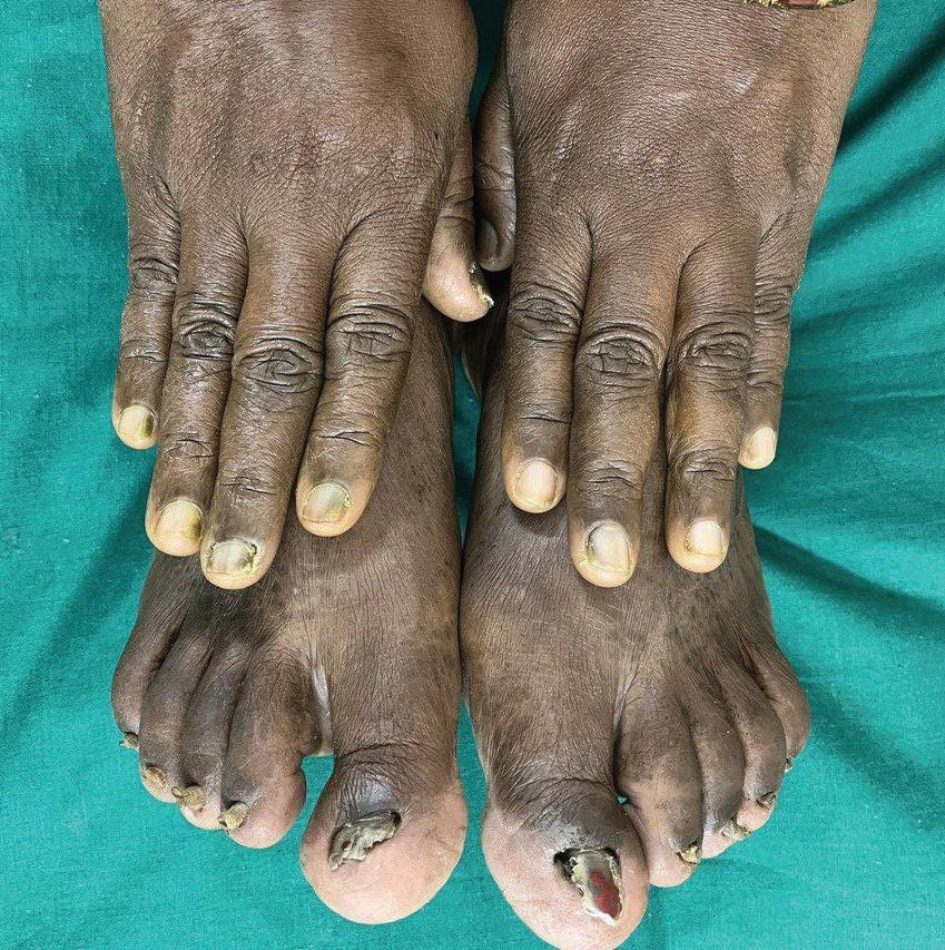
A 52-year-old female presented with discoloration of the right hand and bilateral toenails for three months. The patient also gives a history of itchy, scaly lesions over the bilateral foot for two months. On examination, the patient had dystrophy of all the right fingernails and bilateral toenails with sparing left-hand fingernails (Figure 1). The patient also had tinea pedis over the bilateral toe web spaces. Based on the typical distribution, the patient was diagnosed with a case of one hand and two feet syndrome. The patient was started on topical antifungals and itraconazole pulse therapy.
Diagnosis
"One Hand Two Feet Tinea Syndrome" is not a recognized medical condition or syndrome. Tinea refers to a group of fungal skin infections commonly known as ringworm. These infections can occur on various parts of the body,
including the hands and feet, as well as other areas like the scalp, groin and body. The diagnosis of ringworm can sometimes be challenging due to its ability to mimic other skin conditions, leading to difficulties in distinguishing it from other rashes or infections. Ringworm infections can cause symptoms such as itching, redness, scaling and sometimes the formation of raised circular or ringshaped lesions on the skin. The infection can occur on different parts of the body, including the scalp (tinea capitis), body (tinea corporis), groin area (tinea cruris or jock itch), feet (tinea pedis or athlete's foot) and hands (tinea manuum). It is important for healthcare professionals to consider the possibility of ringworm and perform appropriate diagnostic tests, such as skin scrapings or fungal cultures, to confirm the diagnosis. Indeed, the recognition of the two feet-one hand syndrome, characterized by bilateral tinea pedis with coexistent unilateral tinea manuum, is important for physicians in making a diagnosis of ringworm infection. The presence of this typical rash
Figure 1: One hand two feet tinea syndrome involving nails of the right hand and bilateral feet
distribution raises suspicion of a fungal infection. The syndrome may also exhibit a relapsing course and inappropriate treatment, such as the use of topical steroids under an incorrect diagnosis, can worsen the condition. Hence, proper diagnosis through methods such as KOH testing is crucial. Physicians should be familiar with the typical distribution of the two feetone hand syndrome, as this recognition can aid in making a diagnosis even in settings with limited medical resources. It is important not to overlook the examination of the feet, as it is sometimes neglected due to time constraints or oversight. This case emphasizes the significance of thorough physical examination as a routine practice. Absolutely, a crusted rash distributed in two feet and one hand should raise suspicion of ringworm infection. To confirm the diagnosis, physicians should perform KOH testing, which involves microscopic examination of skin scrapings or samples using potassium hydroxide (KOH) solution. This testing can help visualize the presence of fungal elements
One Hand Two Feet Tinea Syndrome: A Case Report
such as hyphae, confirming the diagnosis of ringworm. Considering the numerous mimics of ringworm, it is crucial to make a correct and immediate diagnosis. Prompt identification of the infection can help initiate appropriate treatment and prevent the condition from worsening or spreading.5,7
Treatment
Treatment for tinea manuum et pedis usually involves antifungal medications, which may be applied topically as creams or ointments or taken orally in more severe cases. The specific treatment approach may vary depending on the severity and extent of the infection. It is important to follow the recommended treatment regimen and take preventive measures to avoid recurrence and spread of the infection.7
When nail involvement, particularly toenail involvement, is present, it is often challenging to treat with topical therapy alone. In such cases, oral antifungal agents may be considered as they have been found to be more effective in treating nail infections. Oral antifungal agents like itraconazole, terbinafine and
fluconazole have shown better efficacy in treating fungal nail infections. These medications are able to penetrate the nail bed and reach the site of infection. However, it is important to note that relapses are common in this syndrome and long-term management and preventive measures may be necessary to control the condition effectively. The availability of new and more effective antifungal drugs brings hope for better management of this troublesome and sometimes difficult-totreat disorder. Continued research and advances in antifungal therapy may further improve outcomes and provide better control of the two feet-one hand syndrome. The specific treatment approach may vary depending on the severity of the infection and individual factors. Here are some common treatment options:8
1. Topical antifungal..... medications: In mild to moderate cases, topical antifungal creams, lotions or sprays are usually prescribed. These medications contain antifungal agents such as
One Hand Two Feet Tinea Syndrome: A Case
clotrimazole, terbinafine, miconazole or econazole. They are applied directly to the affected areas, including the feet, hands and nails, as directed by a healthcare professional. It's important to follow the instructions for application and continue treatment for the prescribed duration, even if symptoms improve.9,10,11
2. Oral antifungal..... medications: In cases of severe or persistent infections or when there is involvement of the nails (onychomycosis), oral antifungal medications may be prescribed. Medications such as terbinafine, itraconazole or fluconazole are commonly used. Oral antifungal therapy is typically taken for a longer duration and requires close monitoring for potential side effects.11,12,13
3. Combination therapy:
In some cases, a combination of topical and oral antifungal medications may be recommended to effectively treat the infection. This approach can help target the fungal infection from both external and systemic perspectives.11,13,14
4. Adjunctive treatments:
In addition to antifungal therapy, supportive
measures can be taken to improve the overall condition and prevent recurrence. These may include keeping the affected areas clean and dry, wearing clean socks and shoes made of breathable materials, practicing good foot and hand hygiene, avoiding sharing personal items and maintaining a healthy immune system.11,13,15
It's important to consult a healthcare professional, such as a dermatologist, for an accurate diagnosis and appropriate treatment plan tailored to your specific condition. They can assess the severity of the infection and provide recommendations for the most suitable treatment options.
Discussion
Tinea pedis, commonly known as athlete's foot, is a prevalent skin infection, particularly among soldiers and individuals involved in recreational or professional sports activities. The clinical and mycological point prevalence of tinea pedis among soldiers were found to be 60.1% and 27.3%, respectively. Among athletes, the prevalence of tinea pedis was reported
to be 26%. The increased prevalence in men may be attributed to the more frequent use of occlusive footwear, which promotes the development of the infection. Tinea pedis can present in different forms. The interdigital type, often referred to as "athlete's foot," typically manifests as scaling, maceration, fissuring or erythema in the web spaces between the toes, with the fourth and fifth toes commonly affected. Moccasin-type tinea pedis presents as generalized scaling and hyperkeratosis of the plantar surface of the foot and is often associated with nail involvement. The inflammatory or vesiculobullous type is characterized by painful, pruritic vesicular eruptions on the arch or side of the feet. The fourth ulcerative type, seen in immunocompromised ....... and diabetic patients, is characterized by rapidly spreading vesiculopustular lesions and ulcers, primarily in the web spaces.3
The "two feet-one hand syndrome" is a relatively rare condition and there have been limited large case series investigating this specific syndrome. In one
study, Trichophyton rubrum was the most commonly isolated pathogen (93.3%), followed by Trichophyton mentagrophytes (4.0%) and Epidermophyton floccosum (2.7%). The clinical presentation of the syndrome usually involves a chronic bilateral, papulosquamous form, characterized by minimal inflammation and patchy or diffuse moccasin-like scaling on the soles. However, variations in the presentation can also occur. Tinea pedis typically develops before tinea manus (hand infection) in the "two feet-one hand syndrome." Studies have shown that approximately 80.5% of patients develop tinea pedis several years before the onset of tinea manus, with an average time interval of about 8.8 ± 1.3 years. It is common for tinea manus to develop on the hand that has been involved in scratching the pruritic feet or picking the toenails, especially when onychomycosis (fungal infection of the nails) is present.3
Risk factors for tinea pedis include wearing socks, footwear and leather shoes, as they create a warm, moist and
One Hand Two Feet Tinea Syndrome: A Case Report
enclosed environment that promotes the growth of dermatophytes. In contrast, hands are exposed to a relatively dry and open environment and are washed more frequently, which helps remove pathogens from the surface. Early diagnosis, prompt treatment of tinea pedis, education on prophylaxis measures, regular foot care and seeking medical assistance when needed can help minimize this problem in individuals predisposed to the infection.3
Topical antifungal agents are typically chosen for the treatment of the two feet-one hand syndrome. However, it is important to be aware that these agents can sometimes cause contact dermatitis, which can be difficult to diagnose based solely on visual inspection. Differential diagnoses to consider include psoriasis, chronic eczema and hand-foot syndrome. Topical antifungal agents are typically the first-line treatment for ringworm infections. They can be applied directly to the affected areas, such as creams, lotions or powders. In more severe or extensive cases, oral
antifungal medications may be prescribed. Treatment duration can vary depending on the severity and location of the infection.
It is important to note that patients may not always report skin changes or seek medical attention due to embarrassment or a lack of awareness about the condition. Therefore, it is essential for physicians to proactively inquire about any skin symptoms and conduct a thorough physical examination, including an inspection of the feet. Examination of the feet is often overlooked, but it is necessary to identify any signs of infection or skin abnormalities.7
In the case of older patients, a thorough physical examination becomes even more critical during the initial encounter. This is because age-related changes and comorbidities may increase the risk of infections and other dermatological conditions. A comprehensive examination can help identify and address any skin issues promptly.
By being vigilant, performing appropriate diagnostic tests and
One Hand Two Feet Tinea Syndrome: A Case Report
conducting thorough physical examinations, physicians can make accurate diagnoses and provide timely treatment for ringworm infections, ultimately improving patient outcomes.7
Conclusion
One hand, two feet syndrome is a superficial fungal skin infection involving both feet and one hand. Distal and lateral subungual onychomycosis (DLSO) is the commonest clinical variant affecting both finger and toenails caused by Trichophyton rubrum followed by Trichophyton mentagrophytes , ............ Trichophyton tonsurans and Epidermophyton floccosum. ′One hand two feet′ tinea syndrome is a distinct clinical pattern in DLSO caused by Trichophyton rubrum in which the fungus spreads from the plantar and palmar surface of feet and hands, similar to our case.
Typically, tinea manuum develops in the hand that has been involved in excoriating the feet or picking toenails. Patients with occupations that involve a high intensity of hand use are more likely to develop the syndrome at
an earlier age. They tend to seek medical attention once tinea manuum has developed, especially if there is a family history of tinea infection. Contact between the hands and feet can lead to the transmission of dermatophytes from the infected feet to the scratching hand. When an individual has tinea pedis (athlete's foot) or onychomycosis (fungal infection of the nails), scratching or touching the affected areas can transfer the fungal spores to the hands. The act of scratching or picking the toenails can cause the spores to adhere to the hands, facilitating their spread to other parts of the body, including the other foot or even the nails of the opposite hand. This contact transmission is one of the mechanisms through which dermatophytes can be transmitted and lead to the development of tinea manuum (hand infection) in the context of the two feetone hand syndrome.
References
1. Mizumoto J. Two FeetOne Hand Syndrome. Cureus. 2021;13 (12):e20758. Published 2021 Dec 27. doi:10.7759/ cureus.20758
2. Chamorro MJ, House SA. Tinea Manuum. [Updated 2023 Aug 7]. In: StatPearls [Internet]. Treasure Island (FL): StatPearls Publishing; 2024 Jan-. Available from: https://www.ncbi.nlm.nih. gov/books/NBK559048/
3. Bjekić M. Two Feet-One Hand Syndrome: A Case Report. Acta facultatis medicae Naissensis 2015;32(3):215-219. DOI: 10.1515/afmnai-2015-0022
4. Al Hasan, M., Fitzgerald, S.M., Saoudian, M. et al. Dermatology for the practicing allergist: Tinea pedis and its complications. Clin Mol Allergy 2, 5 (2004). https:// doi.org/10.1186/1476-7961-2-5
5. Singal A, Khanna D. Onychomycosis: Diagnosis and management. Indian J Dermatol Venereol Leprol 2011;77:659672. DOI:10.4103/03786323.86475
6. Ugalde-Trejo, N.X., Delgado Moreno, K.P., Alfaro-Sánchez, A. et al. Two Feet-One Hand Syndrome: Tinea Pedis and Tinea Manuum. Curr Fungal Infect Rep 16, 117–125 (2022). https:// doi.org/10.1007/s12281-02200447-9
8. Seeburger J, Scher RK. Long-term remission of two
feet-one hand syndrome. Cutis. 1998 Mar;61(3):149-51. PMID: 9538957.
9. Crawford F, Hollis S. Topical treatments for fungal infections of the skin and nails of the foot. Cochrane Database Syst Rev. 2007;2007(3):CD001434. Published 2007 Jul 18. doi:10.1002/14651858. CD001434.pub2
10. ELY JW, ROSENFELD S, STONE MS. Diagnosis and Management of Tinea Infections. American Family Physician. Am Fam Physician. 2014;90(10):702-711. Antifungal agents for common paediatric infections. Paediatr Child Health. 2007;12(10):875-883. doi:10.1093/pch/12.10.875
11. Banerjee M, Ghosh AK, Basak S, Das KD, Gangopadhyay DN. Comparative evaluation of effectivity and safety of topical amorolfine and clotrimazole in the treatment of tinea corporis. Indian J Dermatol. 2011;56(6):657-662. doi:10.4103/0019-5154.91823
12. Kota SSN, Bandhakavi S, Anjali C, Athlete’s foot disease: A comparative study on marketed products, Journal of Drug Delivery and Therapeutics. 2022; 12(3):1-4. DOI https://doi. org/10.22270/jddt.v12i3.5455
13. Antifungal agents for common paediatric infections. Paediatr
14. J.-L. Kienzler, C. Queille Roussel, C. Mugglestone, J. P. Ortonne & C. Larnier (2007) Stratum corneum pharmaco¬kinetics of the antifungal drug, terbinafine, in a novel topical formulation, for single-dose application in dermatophytoses, Current Medical Research and Opinion, 23:6, 1293-1302, DOI: 10.1185/030079907X199664
15. Gupta AK, Simkovich AJ, Hall DC. The March Against Onychomycosis: A Systematic Review of the Sanitization Methods for Shoes, Socks, and Textiles. J Am Podiatr Med Assoc. 2022;112(4):21-223. doi:10.7547/21-223
Early Small Airway Obstruction Due to Tuberculosis of the Airway: A Case Report
Early Small Airway Obstruction Due to Tuberculosis of the Airway: A Case Report
Dr. Digambar Dashatwar M.D. (Dermatology)
Consultant Dermatologist
Chandrapur,
Maharashtra
Introduction
Tuberculosis (TB) remains a critical global health challenge, contributing significantly to morbidity and mortality. Despite advancements in treatment, TB's long-term sequelae can lead to serious respiratory complications. One such complication is early airway obstruction, often referred to as tuberculosis-associated obstructive pulmonary disease (TOPD). This condition is a significant concern due to its adverse effects on pulmonary function and overall quality of life.1 Early airway obstruction related to TB is characterized by a persistent form of airflow limitation that can continue even after the resolution
of active TB. Patients with TOPD often experience symptoms akin to those of chronic obstructive pulmonary disease (COPD), including persistent cough, breathlessness, wheezing, and increased sputum production. Unlike primary TB, which presents with acute symptoms such as fever and weight loss, TOPD primarily involves chronic respiratory symptoms that may develop gradually. The airflow obstruction associated with TOPD is progressive and, if not addressed promptly, can lead to significant functional impairment.1
The pathogenesis of early airway obstruction due to TB involves several interconnected
mechanisms. TB primarily affects the lung parenchyma, leading to granulomatous inflammation and fibrosis. This inflammatory response can cause scarring and damage to the bronchial structures, resulting in airway narrowing and obstruction. The fibrotic changes, along with a loss of elastic recoil in the lungs, contribute to impaired airflow and decreased pulmonary function. Additionally, TBrelated airway damage can sustain chronic inflammation, exacerbating airway obstruction and increasing susceptibility to secondary infections and persistent respiratory symptoms. The impact of TB on the small airways— non-cartilaginous airways with an internal diameter of less than 2 mm—is particularly significant, as these structures are heavily affected by TB lesions, leading to airflow obstruction. The persistence of abnormalities such as gas trapping, even after the initial infection has resolved, highlights the ongoing impact of TB on respiratory health. Furthermore, the interaction between TB and non-tuberculous mycobacterial infections complicates the clinical scenario.2
Early Small Airway Obstruction Due to Tuberculosis of the Airway: A Case Report
The prevalence of early small airway obstructive pulmonary disorders is notable. While the connection between smoking and chronic airflow limitation is wellestablished, the role of past TB as a risk factor for chronic airflow obstruction is gaining recognition. This underscores the importance of considering a patient’s TB history in the assessment and management of chronic respiratory conditions. The convergence of TB and early small airway obstructive disorders exacerbates the public health challenge, particularly in regions with limited healthcare resources. Addressing this dual burden requires a comprehensive approach that integrates TB history into the evaluation and management of patients with chronic respiratory conditions. Advances in understanding the pathophysiology of early airway obstruction due to TB are essential for improving patient outcomes and developing effective treatment strategies.2
Case Report
A 19-year-old woman presented with persistent, irritating, and disabling nocturnal cough lasting for six weeks. She had a fouryear history of dust allergy
and was known to have lactose intolerance. Initial treatment by her general practitioner yielded minimal relief. Due to the persistence of symptoms, she was referred to an internist and subsequently to a respiratory medicine specialist. Over six months, she was treated with antibiotics, systemic corticosteroids, and inhaled bronchodilators and corticosteroids, but her symptoms showed little improvement. Spirometry revealed early small airway obstruction, with FEF 25–75% predicted and PEFR <70%. A chest radiograph showed resolving pneumonitis. Her medical history was also notable for seborrheic dermatitis of the scalp and menstrual irregularities associated with ovarian cysts. Her father had type 2 diabetes mellitus. After few months, she developed new symptoms, including severe oral ulcers, redness in both eyes, and recurrent epistaxis, predominantly from the left nostril. She also reported feeling unwell, mildly feverish, and had gained approximately five kilograms over three weeks. On physical examination, she had multiple superficial ulcers over the floor of the mouth, gums, palate, and lips, with diffuse lip swelling.
Early Small Airway Obstruction Due to Tuberculosis of the Airway: A Case Report
Conjunctival examination revealed severe bilateral bulbar and palpebral redness, and the nasal septum had dry crusts and blood clots. No cervical lymphadenopathy was noted, and systemic examination was unremarkable.
A clinical diagnosis of oral herpes simplex infection with suspected partial Kawasaki syndrome was made. Investigations showed a raised ESR of 29 mm at the end of the first hour and a positive Mantoux test with 20 mm of erythema and induration. Chest radiography continued to show resolving pneumonitis.
Diagnosis
Airway obstruction due to tuberculosis (TB) requires a multimodal diagnostic approach, combining imaging, pulmonary function testing, endoscopic evaluation, microbiological studies, and blood investigations.
A chest X-ray is a fundamental tool, revealing characteristic TB-related abnormalities such as cavitary lesions,
lung consolidation, and enlarged lymph nodes that may compress airways. It also detects bronchial wall thickening from granulomatous inflammation, fibrotic changes from previous infections, pleural effusion compressing lung parenchyma, and findings like miliary nodules or atelectasis, all of which suggest airway compromise.2 For more detailed assessment, a CT scan provides highresolution images that help identify cavitary lesions, bronchial narrowing or thickening, fibrotic changes, pleural effusion, and atelectasis. CT is particularly useful in visualizing small airway disease and evaluating the extent of lung involvement, which is crucial for accurate diagnosis and treatment planning.3 Pulmonary Function Tests (PFTs) are essential to quantify functional impairment. They detect reduced airflow (e.g., decreased FEV1 and FEV1/ FVC ratio), altered lung volumes (increased residual volume and decreased total lung capacity), and diminished diffusion capacity (DLCO), indicating impaired gas exchange. PFTs can also assess bronchial hyperreactivity, suggesting
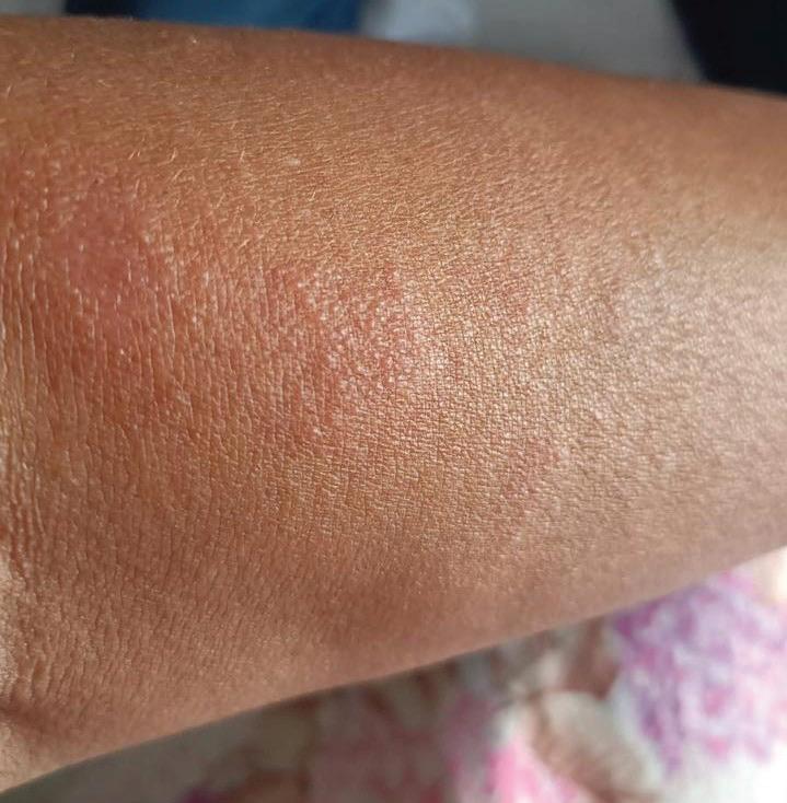
Figure 1: Blisters on the left side of the upper arm
airway sensitivity related to TB-induced inflammation.4 Bronchoscopy allows direct visualization of the airways and can reveal endobronchial TB, granulomas, mucosal thickening, or cavitary lesions. It facilitates targeted sputum sampling and biopsy for microbiological culture and histopathological examination. Sputum smear microscopy is a frontline diagnostic method, detecting acidfast bacilli (AFB) to confirm Mycobacterium tuberculosis. AFB quantification reflects disease burden and airway involvement.5 Molecular tests, such as nucleic acid amplification, provide rapid and specific detection of M. tuberculosis DNA or RNA. These tests identify drug resistance mutations and allow bacterial load assessment, aiding early diagnosis and individualized therapy.6 Histopathology confirms TB through identification of granulomas, caseous necrosis, chronic inflammation, and AFB in tissue. It also evaluates structural damage contributing to airway obstruction.7 Blood tests support diagnosis and monitor systemic involvement. Interferongamma release assays
Early Small Airway Obstruction Due to Tuberculosis of the Airway: A Case Report
(IGRAs) detect latent or active TB, while raised ESR and CRP suggest ongoing inflammation. CBC may show leukocytosis or eosinophilia, and liver function tests (ALT, AST) help assess drug-related toxicity. TB serology, though less common, may provide additional evidence in complex cases.8 Together, these tools offer a comprehensive framework for diagnosing and managing TB-related airway obstruction.
Treatment
In treating early airway obstruction due to tuberculosis (TB), the focus is on targeting the TB infection to reduce inflammation and lung damage. This is achieved using a combination of medications that work together to eliminate the bacteria and improve lung function, which helps relieve airway obstruction and prevent further lung damage.
Isoniazid inhibits mycolic acid synthesis, essential for the TB bacterial cell wall, leading to bacterial death and reduced airway inflammation and obstruction. Rifampicin blocks bacterial RNA synthesis by inhibiting DNA-dependent RNA polymerase, helping
eliminate the infection and improve airflow. Pyrazinamide, converted to pyrazinoic acid inside the bacteria, disrupts membrane transport and energy production, especially in acidic environments like macrophages, enhancing treatment efficacy and reducing lung inflammation. Ethambutol targets arabinosyl transferase, inhibiting cell wall synthesis, preventing resistance, and supporting other antitubercular drugs— ultimately lowering bacterial load and early airway obstruction.
Fluoroquinolones and Aminoglycosides are vital in treating TB-related airway obstruction, particularly in drug-resistant cases.9
Fluoroquinolones inhibit bacterial DNA gyrase and topoisomerase IV, enzymes essential for DNA replication and repair, effectively blocking bacterial growth. They are key second-line agents for treating multidrug-resistant (MDR) and extensively drug-resistant (XDR) TB, helping reduce airway inflammation and obstruction caused by resistant strains. Aminoglycosides bind to bacterial ribosomes, inhibiting protein synthesis and causing bacterial death.
Early Small Airway Obstruction Due to Tuberculosis
Used in MDR-TB and XDRTB, they are often combined with other second-line drugs to enhance efficacy. Both drug classes help lower bacterial load and inflammation, improving respiratory function and relieving airway obstruction.9
Bronchodilators, such as short-acting beta-agonists (e.g., albuterol), provide rapid relief by relaxing bronchial muscles and opening the airways. Longacting beta-agonists offer sustained relief in chronic obstruction. Corticosteroids help reduce airway inflammation—systemic ... steroids like prednisone are used in severe cases, while inhaled corticosteroids, often combined with bronchodilators, manage chronic symptoms. Endobronchial interventions using bronchoscopy can remove obstructions like granulomas or mucus and assess airway narrowing. In more severe cases, balloon dilation or stenting may be performed to restore airflow. Together, these approaches help relieve TB-related airway obstruction and improve breathing.9
Valacyclovir, a prodrug of acyclovir, treats herpes simplex stomatitis by inhibiting viral DNA polymerase, blocking HSV
replication. This reduces viral spread and eases symptoms such as painful lesions and inflammation.10 Its higher bioavailability allows for less frequent dosing, making it effective and convenient—especially when initiated within 48 hours of symptom onset.
Discussion
Patients with tuberculosis (TB) often experience restricted airflow, presenting with symptoms such as chest pain, cough, and shortness of breath. This restriction is typically identified by a decrease in forced vital capacity (FVC) and/or an elevated FEV1/FVC ratio. In one study, airflow restriction was observed in 57% of patients at baseline and in 24% at the end of TB treatment, though the study lacked long-term followup. Airflow obstruction in TB is associated with dyspnea, reduced exercise capacity, and chronic bronchitis. The severity is commonly assessed using forced expiratory volume in 1 second (FEV1), expressed as an absolute value and a percentage of predicted normal. A decline of 100 mL in FEV1 is considered clinically significant. Research has shown a mean annual FEV1
decline of 38.2±8 mL in cured TB patients, similar to the 33±2 mL/year seen in COPD patients without TB. A comparative study also reported similar FEV1 declines in TB patients versus age-matched controls. Moderate-tosevere airflow obstruction (FEV1 <60% predicted) has been observed in about half of TB patients at baseline, with only a modest 14.8% improvement after treatment. Small airways (<2 mm in diameter) are often affected by TB lesions, contributing to obstruction. Im et al. documented that 95% of individuals with pulmonary TB showed centrilobular nodules, “treein-bud” patterns, and poorly defined nodules, which typically resolved within five months of chemotherapy. In TB-endemic regions, TB is a major cause of obliterative bronchiolitis—92% of cases are post-infectious, and 78% are attributed to TB. Radiologically, bronchiolitis appears as gas trapping on CT, visualized as areas of reduced attenuation on expiratory imaging.6
Tuberculosis of the airways should be considered in any otherwise healthy patient presenting with a chronic, dry, hacking cough that does not respond to
standard treatment for upper respiratory tract infections. Additionally, co-infection with herpes simplex virus, which can affect the oral cavity, nasal septum, and conjunctiva, may be present. In the case described, the patient was managed with systemic and inhaled corticosteroids over a six-month period without significant improvement in symptoms. This therapeutic approach may have inadvertently predisposed the patient to herpes simplex virus infection of the mouth and upper airway tuberculosis due to suppressed local and systemic immunity. It is crucial to consider this potential risk when managing patients who do not respond adequately to conventional treatments.
Herpes simplex virus (HSV) infection can complicate early airway obstruction through several mechanisms. HSV, particularly in the form of herpes simplex stomatitis, may cause mucosal lesions in the oral cavity and upper airway that become inflamed and contribute to obstruction. It can also lead to secondary infections and worsen pre-existing respiratory conditions. In patients receiving
Early Small Airway Obstruction Due to Tuberculosis of the Airway: A Case Report
immunosuppressive therapies (e.g., systemic corticosteroids) for airway obstruction or other respiratory illnesses, susceptibility to HSV increases due to compromised immune responses. This interaction between HSV and airway inflammation can complicate both diagnosis and treatment. Early airway obstruction is a complex issue with multiple causes and significant implications for respiratory health. Recognizing co-infections like HSV, identifying at-risk populations, and understanding its epidemiology are essential for effective management.10
Conclusion
Small airway obstruction due to tuberculosis (TB) poses ongoing challenges, including persistent pulmonary function impairment and chronic respiratory symptoms, even after active TB resolves. It often overlaps with conditions like COPD, complicating diagnosis and treatment. This leads to reduced quality of life and increased healthcare burden, particularly in resource-limited settings. Effective management requires a focused approach that addresses the long-
term impact of TB on small airways. Future research should aim to improve diagnostics, develop targeted therapies, and create strategies to prevent or minimize TB-related airway damage—ultimately enhancing patient outcomes and reducing the long-term burden.
References
1. Singh D. Small Airway Disease in Patients with Chronic Obstructive Pulmonary Disease. Tuberc Respir Dis (Seoul). 2017; 80(4):317-324. doi:10.4046/ trd.2017.0080.
2. Amaral AF, Coton S, Kato B, et al. Tuberculosis associates with both airflow obstruction and low lung function: BOLD results. Eur Respir J. 2015; 46(4):1104-1112. doi:10.1183/13993003.023252014.
3. Bhalla AS, Goyal A, Guleria R, Gupta AK. Chest tuberculosis: Radiological review and imaging recommendations. Indian J Radiol Imaging. 2015; 25(3):213-225. doi:10.4103/0971-3026.161431
4. Ranu H, Wilde M, Madden B. Pulmonary function tests. Ulster Med J. 2011; 80(2):84-90.
5. Desikan P. Sputum smear microscopy in tuberculosis: is it still relevant? Indian J Med Res. 2013; 137(3):442-444.
6. Nurwidya F, Handayani D, Burhan E, Yunus F. Molecular Diagnosis of Tuberculosis.
Early
Small Airway Obstruction Due to Tuberculosis of the Airway:
Chonnam Med J. 2018; 54(1):19. doi:10.4068/cmj.2018.54.1.1
7. Kashyap S, Solanki A. Challenges in endobronchial tuberculosis: from diagnosis to management. Pulm Med. 2014; 2014:594806. doi:10.1155/2014/594806.
8. Bhalla AS, Goyal A, Guleria R, Gupta AK. Chest tuberculosis: Radiological review and imaging recommendations. Indian J Radiol Imaging. 2015; 25(3):213-225. doi:10.4103/0971-3026.161431.
9. Jindal SK, Jindal A, Agarwal R. Upper Respiratory Tract Tuberculosis. Microbiol Spectr. 2016; 4(6):10.1128/ microbiolspec.TNMI70009-2016. doi:10.1128/ microbiolspec.TNMI7-00092016.
10. Piperi E, Papadopoulou E, Georgaki M, et al. Management of oral herpes simplex virus infections: The problem of resistance. A narrative review. Oral Dis. 2024; 30(3):877-894. doi:10.1111/odi.14635.