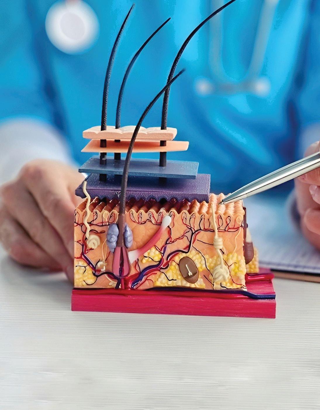
Pigmentosa with Squamous Cell Carcinoma of the Back: A Rare Case
Xeroderma
EXECUTIVE EDITOR & PUBLISHER
Dom Daniel CORPORATE OFFICE
22, Shreeji Bhavan, 275-279, Samuel Street, Masjid Bunder (W), Mumbai-4000 03, INDIA.
EMAIL: info@residerm.com
TEL: + 91 22 2345 1404
Printed, Published, Edited and Owned by Dom Daniel Printed at Swastik Printer, Gala No.9 & 10, Vishal Industrial Estate, Bhandup (West), Mumbai- 400078. Published at 22 Shreeji Bhavan, 275/279, Samuel Street, Masjid Bunder (West), Mumbai - 400003. India.
“Residerm ” takes no responsibility for unsolicited photographs or material
ALL PHOTOGRAPHS, UNLESS OTHERWISE INDICATED, ARE USED FOR ILLUSTRATIVE PURPOSE ONLY.
Views expressed in this Journal are those of the contributors and not of the publisher. Reproduction in whole or in parts of texts or photography is prohibited. Manuscripts, Photographs and art are selected at the discretion of the publisher free of charge (advertising excluded). Whether published or not, no material will be returned and remains the property of the publishing house, which may make use of it as seen fit. This may include the withdrawal of publication rights to other publishing houses.
All rights reserved. Reproducing in any manner without prior written permission prohibited.
Published for the period of August -2025
"UNLOCKING CLINICAL INSIGHTS: RARE CASES FOR THE INQUISITIVE RESIDENTS"
We are pleased to present the latest issue of ResiDerm, a journal dedicated to nurturing academic curiosity and clinical acumen among dermatology residents. This edition highlights the importance of rare case presentations in sharpening diagnostic skills and broadening clinical perspective.
In this issue, we delve into two intriguing case reports:
�� Xeroderma Pigmentosum with Squamous Cell Carcinoma of the Back: A Rare Case Report –underscoring the oncogenic potential in genodermatoses and the need for vigilant monitoring.
�� Kaposi’s Varicelliform Eruption: A Case Report of Dermatological Complications and Treatment Approach – offering insights into this uncommon yet critical viral complication and its therapeutic management. These cases not only reflect the diverse spectrum of dermatological disorders but also reinforce the importance of early recognition and a multidisciplinary approach in patient care. We encourage residents to continue contributing unique and educational cases to foster a collaborative learning environment.
We hope these contributions inspire learning, discussion, and further exploration in your dermatology journey.
Hope you have a great read!
Thanks & Cheers
- Dom Daniel Executive Editor & Publisher
Kaposi's
Varicelliform Eruption: A Case Report of Dermatological Complications and Treatment Approach
Dr. Sachin Choudhary
3rd Year Resident (2024)
Department of Dermatology, Venereology and Leprosy ESI-PGIMSR, New Delhi
Dr. Damanpreet Kaur
2nd Year Resident
Department of Dermatology, Venereology and Leprosy ESI-PGIMSR, New Delhi
Dr. Kanchan Dhaka
Senior Resident (2024)
Department of Dermatology, Venereology and Leprosy ESI-PGIMSR, New Delhi
Dr. Paschal Dsouza
MD, Dermatology
Professor & Head of Department
Department of Dermatology, Venereology and Leprosy ESI-PGIMSR, New Delhi
Xeroderma Pigmentosa with Squamous Cell Carcinoma of the Back: A Rare Case Report
Dr. Rupa Raote
MBBS, DDV
Dermatologist and Hair Transplant Surgeon
The Wellness Co, Mumbai
Kaposi's Varicelliform Eruption: A Case Report of Dermatological Complications and Treatment Approach
Kaposi's Varicelliform Eruption: A Case Report of Dermatological
Complications and Treatment Approach
Dr. Sachin Choudhary
3rd Year Resident (2024)
Department of Dermatology, Venereology and Leprosy
ESI-PGIMSR, New Delhi
Dr. Damanpreet Kaur
2nd Year Resident
Department of Dermatology, Venereology and Leprosy
ESI-PGIMSR, New Delhi
Dr. Kanchan Dhaka
Senior Resident (2024)
Department of Dermatology, Venereology and Leprosy
ESI-PGIMSR, New Delhi
Dr. Paschal Dsouza
MD, Dermatology
Professor & Head of Department
Department of Dermatology, Venereology and Leprosy
ESI-PGIMSR, New Delhi
Introduction
Eczema herpeticum, also known as Kaposi's
varicelliform eruption (KVC), is a severe and disseminated cutaneous infection primarily
caused by herpes simplex virus (HSV). It typically occurs in patients with preexisting skin diseases, which is majority of the cases atopic dermatitis being the most common underlying condition. However, eczema herpeticum can also arise in a range of other dermatological conditions, including darier disease, pemphigus foliaceous, psoriasis, seborrheic dermatitis, contact dermatitis, and cutaneous T-cell lymphoma. This condition often presents in individuals during their second or third decade of life.1
Kaposi's varicelliform eruption is characterized by vesico-pustules, some umbilicated and other eroded, and extended in clusters. It may also show haemorrhagic crusts with an erythematous base. As the condition progresses, the vesiculopustules evolve into painful hemorrhagic lesions that develop crusts and form punched-out erosions. The lesions are painful and they are often associated with fever, malaise, and regional lymphadenopathy. These erosions can merge to create extensive denuded areas of skin, which are highly susceptible to secondary bacterial
infections. The primary etiological agent in Kaposi's varicelliform eruption is herpes simplex virus type 1 (HSV-1), although herpes simplex virus type 2 (HSV2), Coxsackie A16, vaccinia virus, varicella-zoster virus, and smallpox virus have also been implicated in some cases. The most commonly affected sites are head, neck, and trunk and it is more common in children due to its association with atopic dermatitis, although cases in healthy adults have also been reported. Although the pathogenesis of KVE is not completely elucidated, it is believed to be due to humoral and cellular immunity dysfunction. The Th2 cytokine environment typically associated with atopic dermatitis plays a crucial role in the disease's pathogenesis. Recent studies have also suggested a genetic predisposition, indicating that genetic factors may influence susceptibility to KVE. Despite its rarity, KVE can be potentially life-threatening due to the widespread nature of the vesicular eruptions and the associated risk of complications, including severe secondary bacterial infections. The precise incidence of KVE is difficult to ascertain due to
its infrequent occurrence and the lack of extensive case series. The disease appears to affect both genders equally and does not show significant ethnic predilection. Prompt recognition and treatment are essential to manage the condition effectively and prevent serious outcomes.2
Case report
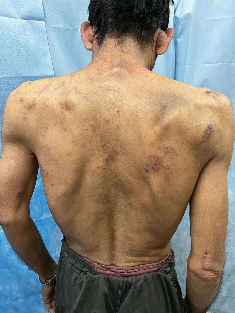
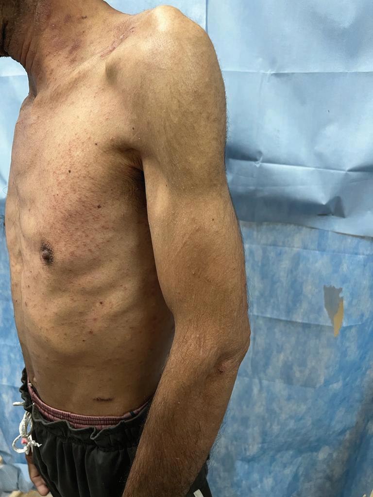
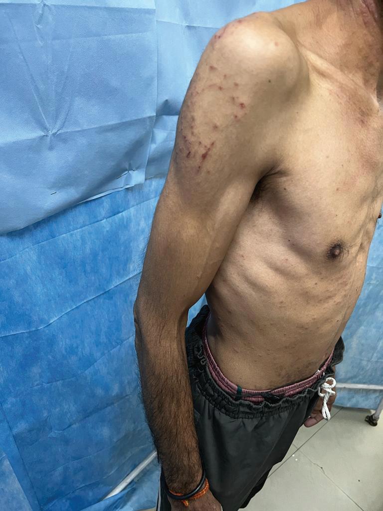
We present a case of a 45-year-old male with longstanding history of atopic dermatitis, who presented with a 5-day history of eruption of painful vesicles that had started on his face and gradually spread to involve the rest of his body and were accompanied by systemic symptoms such as fever, malaise, headache. There was past history of atopy, recurrent respiratory tract infections and flairs of dermatitis, which were treated as required. On examination his face, chest, arms, abdomen, and back were packed with confluent erythematous vesicopapules which evolved into crusted, haemorrhagic, and punched out skin erosions and some areas were weeping a yellow serous fluid. The diagnosis of Kaposi varicelliform eruption is made on the basis of a clinical examination and Tzanck smear was
Kaposi's Varicelliform Eruption: A Case Report of Dermatological Complications and Treatment Approach
performed which showed multinucleated giant cells. The patient was promptly treated with oral Acyclovir, 800 mg five times a day, to combat the viral infection. In addition to antiviral therapy, antipyretics were administered to manage fever, and topical antibiotics were used to prevent or treat secondary bacterial infection. Remarkably, the patient showed significant clinical improvement, with a complete resolution of the skin lesions by the fourth day of treatment. This rapid response underscores the importance of early recognition and intervention in managing Kaposi varicelliform eruption, especially in individuals with underlying atopic conditions.
the abdomen, both the side of chest and neck region
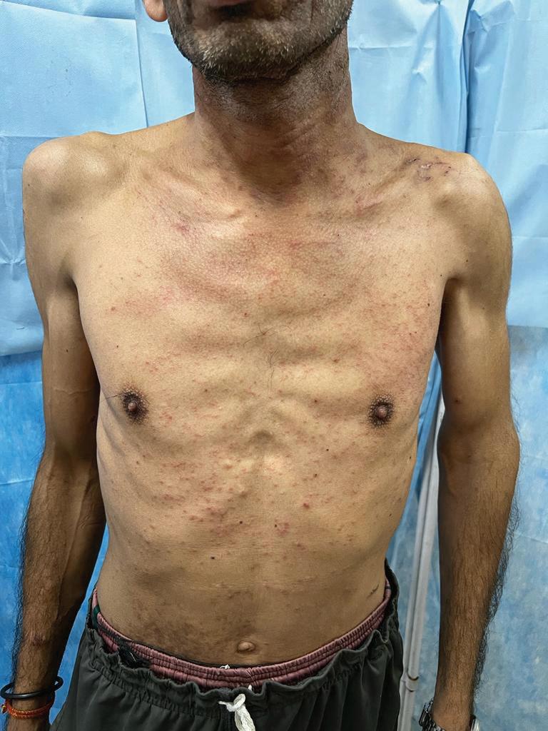
Figure 1: Crusted erythematous vesicopapules on the back
Figure 2: Crusted erythematous vesicopapules on the right side of chest
Figure 4: Crusted erythematous vesicopapules on the right side of upper arms
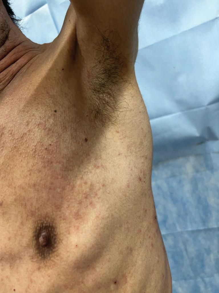
Figure 3: Vesico-papules on left under arms
Figure 5: Crusted erythematous vesicopapules on
Kaposi's Varicelliform Eruption: A Case Report of Dermatological Complications and Treatment Approach
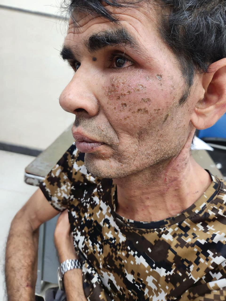
Figure 6: Erythematous vesico-papules that evolved into crusted, hemorrhagic, and punched-out skin erosions, with areas weeping yellow serous fluid on the left side of the face and neck region
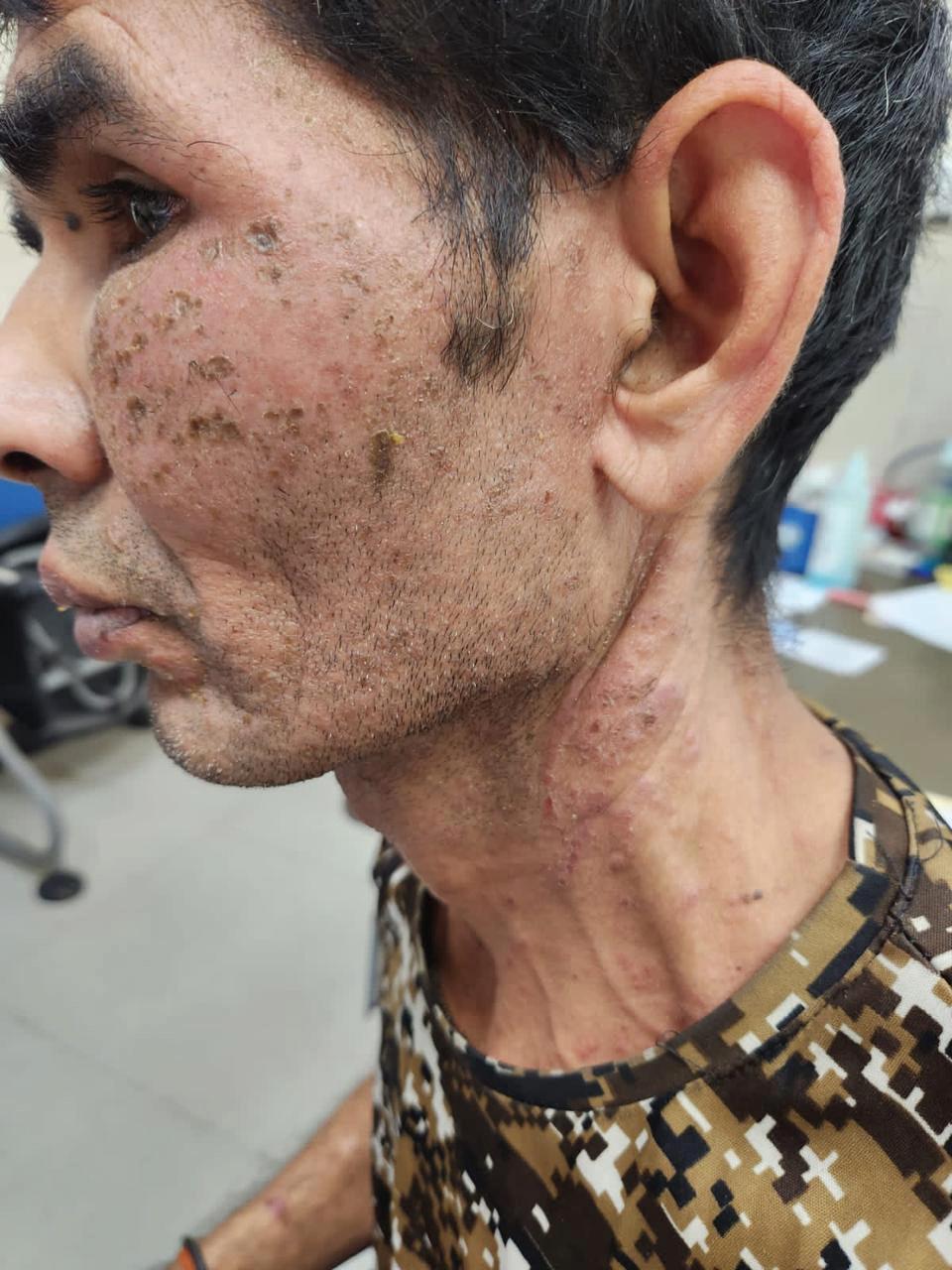
Figure 8 (A & B): After the treatment with Acyclovir 800 mg, antipyretics, and topical antibiotics. Improvement was noted, with skin lesions clearing completely by the fourth day
Diagnosis
Diagnosing Kaposi varicelliform eruption involves a combination of clinical assessment and laboratory testing. A
Figure 7: Erythematous vesico-papules
Fig. 8 (A)
Fig. 8 (B)
Kaposi's Varicelliform Eruption: A Case Report of Dermatological Complications and Treatment Approach
thorough patient history, particularly the presence of pre-existing conditions like atopic dermatitis, and a detailed physical examination are crucial. Kaposi's varicelliform eruption is characterized by painful, edematous clusters of umbilicated vesiculopustules that progress to hemorrhagic, crusted erosions. Laboratory confirmation can be achieved through viral culture, which detects herpes simplex virus (HSV) by identifying cytopathic effects such as cell rounding and syncytia formation. Alternatively, polymerase chain reaction (PCR) is a highly sensitive and specific method that detects HSV DNA, providing rapid and accurate differentiation between HSV types and other pathogens.3,4
The Direct Fluorescent Antibody (DFA) assay detects herpes simplex virus (HSV) antigens in lesion samples using fluorescently labeled antibodies, revealing bright structures under a fluorescence microscope. Though rapid and specific, DFA is less sensitive than PCR or viral culture. Serologic testing measures HSV-specific IgM and IgG antibodies in the blood,
identifying recent or past infections, but it is less effective for acute diagnosis of Kaposi varicelliform eruption as it does not directly detect the virus in lesions. Skin biopsy can reveal histopathological features of HSV, such as multinucleated giant cells and Cowdry type A bodies, providing supportive evidence for diagnosis.4,5,6
Immunohistochemistry (IHC) detects herpes simplex virus (HSV) antigens in tissue samples using colored or fluorescent staining, offering direct visual evidence of HSV infection to support the diagnosis of Kaposi varicelliform eruption. Although useful, IHC is typically supplemented with more sensitive techniques like PCR. A Tzanck smear, which involves examining stained skin lesion samples for signs of HSV infection such as multinucleated giant cells and ballooning degeneration, provides quick results but lacks the specificity and sensitivity of PCR, which directly identifies viral DNA.7,8
Treatment
Antiviral therapy is crucial in the treatment of Kaposi varicelliform eruption, as it targets the herpes simplex virus (HSV) responsible for the infection. The
primary goal is to reduce viral replication, alleviate symptoms, and accelerate healing. The treatment of Kaposi varicelliform eruption primarily involves antiviral medications to manage herpes simplex virus infection. Acyclovir is the main antiviral drug, inhibiting HSV DNA polymerase to reduce viral replication; it is typically administered orally but may be given intravenously for severe cases. Valacyclovir, a prodrug of acyclovir with improved oral bioavailability, offers effective antiviral activity with a more convenient dosing schedule. Famciclovir, converted into penciclovir, is another option that inhibits HSV DNA polymerase and serves as an alternative to acyclovir or valacyclovir. These drugs are crucial for reducing symptom severity and preventing complications in Kaposi's varicelliform eruption.9
In the treatment of Kaposi varicelliform eruption, antipyretics are used to manage fever and reduce systemic discomfort. Acetaminophen (paracetamol) is commonly used to alleviate mild to moderate pain, while ibuprofen, a nonsteroidal anti-inflammatory drug
Kaposi's Varicelliform Eruption: A Case Report of Dermatological Complications and Treatment Approach
(NSAID), helps reduce pain and inflammation.10
Topical antibiotics are used to prevent or manage secondary bacterial infections that can complicate the skin lesions. Given that the eruption can lead to open erosions and weeping areas, applying topical antibiotics helps reduce the risk of bacterial colonization and infection in these vulnerable areas. In addition to pharmacological treatments, the management of Kaposi varicelliform eruption involves several supportive measures.1
Proper skin care is crucial, necessitating gentle cleansing with mild, nonirritating agents to prevent secondary infections and facilitate healing. The application of emollients or moisturizers helps to alleviate skin irritation and prevent excessive dryness. Cooling measures, such as the use of cool compresses, can offer symptomatic relief from itching and discomfort. Avoidance of potential irritants, including harsh soaps and abrasive scrubs, is important to prevent exacerbation of skin irritation. Adequate rest and hydration are essential to support overall recovery
and immune function. Additionally, rigorous hygiene practices, including regular handwashing, are vital to minimize the risk of infection spread and secondary bacterial complications.10
Discussion
Austrian dermatologist
Moriz Kaposi gave the first description of Kaposi's varicelliform eruption or eczema herpeticum. Some authors distinguish between these two conditions: eczema herpeticum refers to a disseminated herpes simplex virus infection that complicates an eczematous skin disease, while Kaposi's varicelliform eruption describes a similar vesicular eruption occurring in individuals with pre-existing skin infections with the HSV type 1or 2.8
Kaposi varicelliform eruption can complicate or coexist with various dermatological conditions due to their impact on the skin barrier and immune response, especially atopic dermatitis.3 In psoriasis, the chronic inflammation may heighten susceptibility to herpes simplex infections, while pityriasis rubra pilaris and Darier’s disease, with their skin abnormalities, can lead to more severe manifestations of the virus.
Grover’s disease and Hailey–Hailey disease, with their disrupted skin integrity, also increase vulnerability to herpes simplex, potentially resulting in Kaposi varicelliform eruption. Additionally, both irritant and allergic contact dermatitis can compromise the skin barrier, making it more prone to secondary infections like herpes simplex. Managing Kaposi varicelliform eruption in these contexts necessitates careful coordination of care, considering how these underlying conditions can influence the eruption's presentation and progression.4 The average disease duration is 16 days and most of the lesions heal without scarring within 2–6 weeks. Differential diagnosis for Kaposi varicelliform eruption includes chickenpox, impetigo, and contact dermatitis. Chickenpox, caused by the varicellazoster virus, presents with a generalized rash that evolves through macules, papules, vesicles, and crusts, often in a centripetal pattern affecting the trunk and face more than the limbs, and is usually accompanied by systemic symptoms such as malaise. Impetigo, a bacterial infection typically
caused by Staphylococcus aureus or Streptococcus species, begins with red sores or blisters that rupture and form honeycolored crusts, commonly localized around the nose and mouth, and generally lacks systemic symptoms. Contact dermatitis results from exposure to an irritant or allergen, presenting with localized erythema, itching, and vesicular eruptions confined to the area of contact, with symptoms often resolving once the offending substance is removed. Accurate diagnosis of KVE requires a thorough clinical examination, detailed patient history, and, if needed, laboratory tests to differentiate it from these similar conditions.1 Tzank smear can be performed which can demonstrate multinucleated giant cells on the Wright-Giemsa stain. Direct fluorescent antibody staining, polymerase chain reaction (PCR), and viral culture techniques are utilized to identify the causative viral agent. Such investigations were not performed in the present case and the diagnosis of Kaposi varicelliform eruption was made on the basis of clinical presentation plus findings of wright-giemsa
satin which showed multinucleated giant cells.5, 8
Kaposi varicelliform eruption is a serious condition with the potential for fatal outcomes. The recurrent form, typically occurring in adults, tends to be milder and more localized, generally without viremia. Secondary bacterial infection of cutaneous lesions can exacerbate morbidity and mortality, with common pathogens including Staphylococcus aureus, group A betahemolytic streptococcus, Peptostreptococcus, and Pseudomonas aeruginosa. Ocular involvement is a risk when the herpes simplex virus-associated Kaposi varicelliform eruption affects the facial area, potentially leading to uveitis, conjunctivitis, keratitis, and blepharitis. Herpetic keratitis is the most serious ophthalmic complication and may result in vision loss due to corneal scarring. Treatment should be initiated without delay due to the potentially lifethreatening nature of the condition. Most patients will experience resolution of skin lesions over several days. Prophylactic measures are recommended to prevent secondary bacterial infections.
Clinicians must be vigilant for adverse drug reactions and monitor treatment progress closely. Ongoing monitoring until lesion resolution is necessary, and an interprofessional approach to management is optimal.1 The most common complication is bacterial infection that can lead to sepsis and viremia, with involvement of other organs. Therefore, prophylactic antibiotic therapy is recommended.
Conclusion
Kaposi's varicelliform eruption is a rare and severe condition often seen in patients with underlying immunosuppression or pre-existing skin diseases, marked by widespread, vesicular lesions that resemble varicella. Doctors face significant challenges in diagnosing and managing Kaposi varicelliform eruption due to its resemblance to other vesicular eruptions and the complexity of treatment in immunocompromised patients. Patients with KVE may experience substantial physical discomfort, secondary bacterial infections, and psychological distress due to the appearance and severity of the rash. Future research should
Kaposi's Varicelliform Eruption: A Case Report of Dermatological Complications and Treatment Approach
focus on understanding the pathophysiology of Kaposi varicelliform eruption, improving diagnostic techniques, and developing targeted therapies to manage the condition effectively while minimizing complications.
References
1. Karray M, Kwan E, Souissi A. Kaposi Varicelliform Eruption. In: StatPearls. Treasure Island (FL): StatPearls Publishing; September 12, 2022.
2. Vora RV, Pilani AP, Jivani NB, Kota RK. Kaposi varicelliform eruption. Indian Dermatol Online J. 2015; 6(5):364-366. doi:10.4103/2229-5178.16448.
3. Külcü Çakmak S, Alli N, Yilmaz E, Artüz F. A Case of Kaposi's Varicelliform Eruption in a Patient with Psoriasis Receiving Cyclosporine Therapy. Ann Dermatol. 2015; 27(3):345-356. doi:10.5021/ad.2015.27.3.345.
4. Hong S, Kim EH, Cho SB, Rha SY. Kaposi's VaricelliformLike Eruption in a Patient Treated with Everolimus for Metastatic Renal Cell Carcinoma: Report of a Rare Case. Case Rep Oncol. 2014; 7(2):337-342. Published 2014 May 27. Doi: 10.1159/000362925.
5. Saleh D, Yarrarapu SNS, Sharma S. Herpes Simplex Type 1. In: StatPearls. Treasure Island (FL): StatPearls Publishing; August 28, 2023.
6. El-Masry H, Essam S, Gaber H, et al. Kaposi varicelliform eruption: an unusual presentation caused by varicella zoster virus in a healthy adult patient - a case report. BMC Infect Dis. 2024; 24(1):244. Published 2024 Feb 22. Doi: 10.1186/s12879-02409115-4.
8. Nath AK, Sori T, Thappa DM. A case series of kaposi's varicelliform eruption in dermatology in-patients in a tertiary care centre. Indian J Dermatol. 2011; 56(1):110-115. doi:10.4103/0019-5154.77572.
9. Martín-Galache M, EscalonaGil AM, Posado-Domínguez L, et al. Kaposi's Varicelliform Eruption: A Potentially LifeThreatening Complication of Atopic Dermatitis. Eur J Case Rep Intern Med. 2024; 11(5):004392. Published 2024 Mar 28. Doi: 10.12890/2024_004392.
10. Shenoy MM, Suchitra U. Kaposi's varicelliform eruption. Indian J Dermatol Venereol Leprol. 2007;73(1):65. doi:10.4103/0378-6323.30664.
Xeroderma Pigmentosa with Squamous Cell Carcinoma of the Back: A Rare Case Report
Dr. Rupa Raote
MBBS, DDV
Dermatologist and Hair Transplant Surgeon
The Wellness Co, Mumbai
Abstract
Introduction: Xeroderma pigmentosum (XP) is a rare autosomal-recessive disorder that appears in childhood. Squamous cell carcinoma is not uncommon in patients with xeroderma pigmentosum and mostly involves the sun exposed parts that is face, head, neck, and scalp. It is a rare group of disorder with reported incidence in US and Europe being 1:250,000 and Japan 1:40,000 while its incidence is not significant in Indian in context to other parts of the world.1
Case Report: Here, we present a case of a thirtysix years-old female with xeroderma pigmentosum with a huge squamous cell carcinoma on the right side
of the back. In addition, we illustrate a female with xeroderma pigmentosum who grows up in a sunny environment (Konkan) where the possibility of early onset of squamous cell carcinoma is extremely high in any suspected skin lesion.
A 36-year-old female presented with progressively increasing hypopigmentary skin lesions from the age of 3-5 years of age predominantly over the sun-exposed areas. On examination, the skin was dark, dry, atrophic, freckled, scaly, and rough. Discrete, multifocal patchy hypopigmentation (macules and patches) with some hyperpigmented hyperkeratotic papules and plaques were seen predominantly over the sun-
Xeroderma Pigmentosa with Squamous Cell Carcinoma of the Back: A Rare Case Report
exposed areas with nail involvement where there was complete dystrophy of nails. Based on these clinical findings, the patient was diagnosed as xeroderma pigmentosum and confirmed with a skin biopsy. The patient's 27-year-old brother was also diagnosed with xeroderma pigmentosum based on clinical examination. There was history of consanguineous marriage of parents.
Defective nucleotide excision repair of ultravioletinduced DNA damage is most commonly observed in clinical assessments. Clinical presentation comprises of dermatological, ocular, and neurological manifestations. Up to 60% of persons with xeroderma pigmentosum eventually develop skin cancer. Basal cell carcinoma is mostly associated with xeroderma pigmentosum in the majority of the reported cases in Indian literature, and very few patients have squamous cell carcinoma thereby signifying the rarity of our case. Cutaneous neoplasms are typically managed through curettage or surgical excision. However, in the case reported above, excision was done for this extensive
ulceroproliferative lesion.
However, unfortunately, patient lost follow-up.
Conclusion: In patients with xeroderma pigmentosum, squamous cell carcinoma of the back may manifest at an earlier stage and exhibit an unusually aggressive clinical course. In sun-exposed regions, comprehensive education for both patients and their families regarding ultraviolet light protection and early detection of suspicious lesions is crucial for potentially life-saving outcomes.
Xeroderma pigmentosum (XP) is a rare autosomal recessive disorder that typically manifests in childhood. Patients with xeroderma pigmentosum are at an increased risk for developing squamous cell carcinoma (SCC), particularly in sunexposed areas such as the face, head, neck, and scalp. The incidence of xeroderma pigmentosum is approximately 1 in 250,000 in the US and Europe, and 1 in 40,000 in Japan, while it is comparatively less prevalent in India context
relative to other regions.1 Xeroderma pigmentosum resulting from defective nucleotide excision repair of UV-induced DNA damage, leading to a broad spectrum of clinical manifestations that vary by subtype. Cutaneous symptoms include early-onset freckling in sun-exposed areas, severe sunburns from minimal UV exposure, and an increased risk of skin cancers, such as basal cell carcinoma (BCC) and squamous cell carcinoma (SCC), with a median onset age of about 9 years. BCCs typically present as pearly papules with rolled borders and central ulceration, while SCCs manifest as scaly, hyperkeratotic macules or papules. Keratoacanthomas, a variant of SCC, grow rapidly with a central hyperkeratotic plug, and malignant melanoma, if present, appears as an irregular, changing brownto-black macule or patch. Additional skin changes may include actinic damage features like telangiectasias, actinic keratoses, skin atrophy, xerosis, and wrinkling. Ocular manifestations often include photophobia, dry eyes, and hyperpigmentation of the eyelids or conjunctiva, with complications such as
telangiectasias, ectropion, corneal vascularization or opacification, and both benign and malignant ocular lesions. Oral manifestations may involve squamous cell carcinoma of the anterior tongue. Neurological symptoms, particularly in xeroderma pigmentosum subtypes XPA and XPD, affect about 20% of patients and include developmental deficiencies, sensorineural hearing loss, microcephaly, hyporeflexia, spasticity, ataxia, and premature death, resulting from neuronal loss, cortical atrophy, and ventricular dilation.2
Xeroderma pigmentosum (XP) is a rare genetic disorder caused by mutations in genes involved in nucleotide excision repair (NER), leading to the accumulation of UVinduced DNA damage. There are eight XP subtypes (XP-A to XP-G and XPV), each with distinct genetic and clinical features. In the U.S., XPC mutations are most common and increase the risk of skin and mucous membrane cancers without neurological symptoms. XP patients are particularly prone to aggressive squamous cell carcinoma (SCC) on sun-exposed areas.1,3,4 We report a case of a large SCC on the back in an XP patient, emphasizing
the importance of sun protection.
Case report
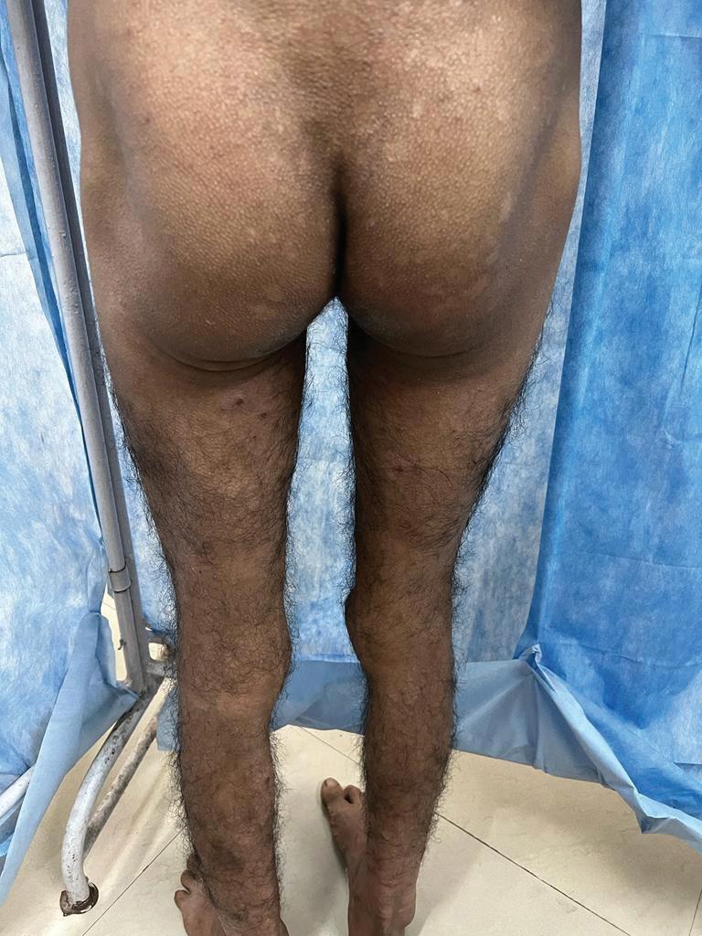
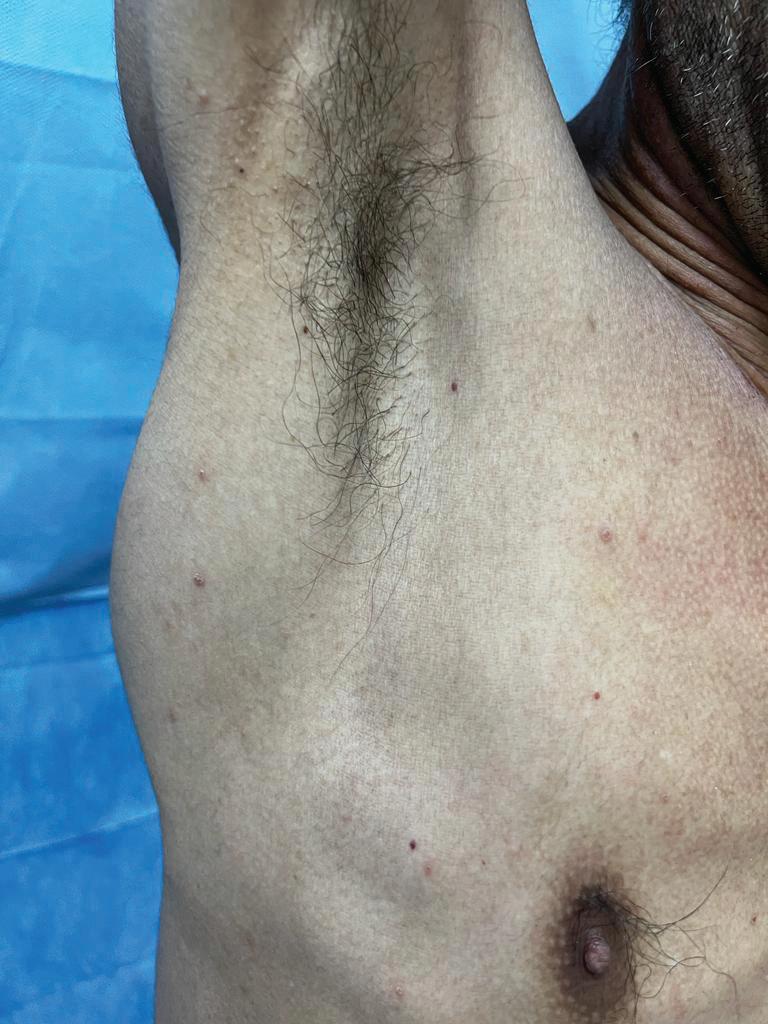
A 36 year-old female, known case of XP (Biopsy proven), presented to our OPD with an ulceroproleferative mass in the back. The ulcer was round in shape, measured about 8 - 10 cm in diameter, with punched out edges and a pedunculated base. There was no discharge or bleeding, at presentation. There were diffuse hyperpigmented and hypopigmented lesions over the face, trunk, and extensor surface of the upper extremities. The skin was unnaturally dry and rough all over the previously mentioned areas. Hyperpigmented hyperkeratotic changes were observed over some lesions. There was no congestion of conjunctivae in either eye. Neurological examination was normal. She has a family history of similar case. There is a positive history of consanguinity. Biopsy showed moderately to poorly differentiated squamous cell carcinoma with proliferative growth over back. Tumor necrosis was seen and moderate mononuclear infiltrate was noted in intra tumoral and peritumoral tissue. The patient presented with a late diagnosis of SCC that has been progressing for 2 years. Hyperpigmentation (freckles) on the face and extremities. Previously, she sought medical advice that has poor diagnostic facilities near her town. Excision of the lesion with 1 cm safety margin circumferentially was done. Reconstruction was performed using local flaps and skin graft. A written informed consent was obtained from the patient for excision, reconstructive procedure and publication of this case report and any supplementary image.
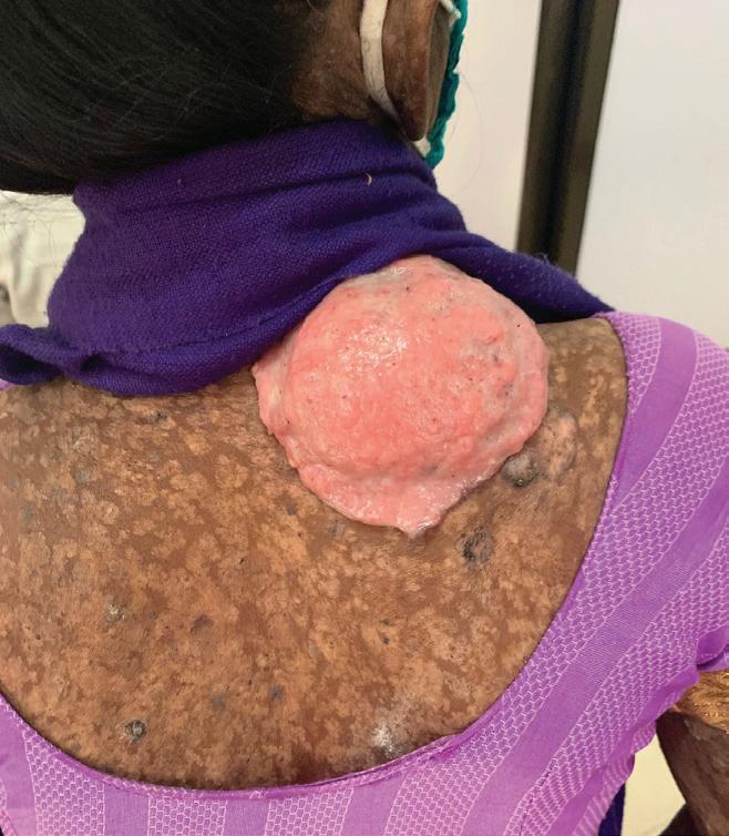
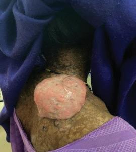
Figure 1: Ulceroproliferative mass in the back
Xeroderma Pigmentosa with Squamous Cell Carcinoma of the Back: A Rare Case Report
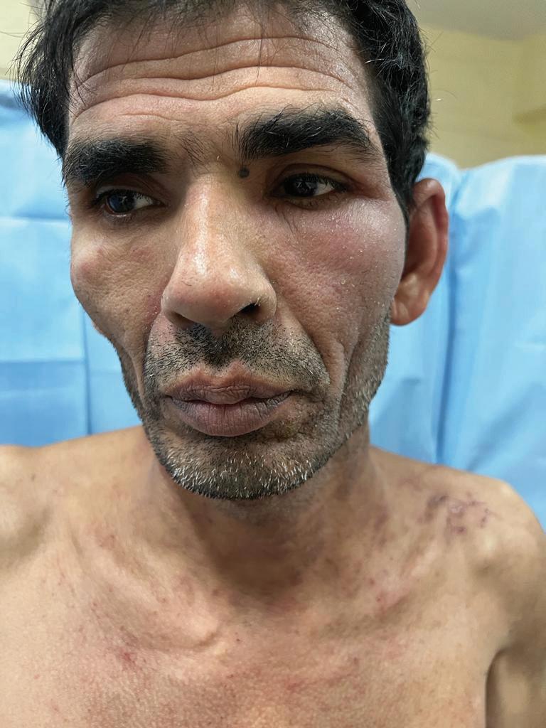
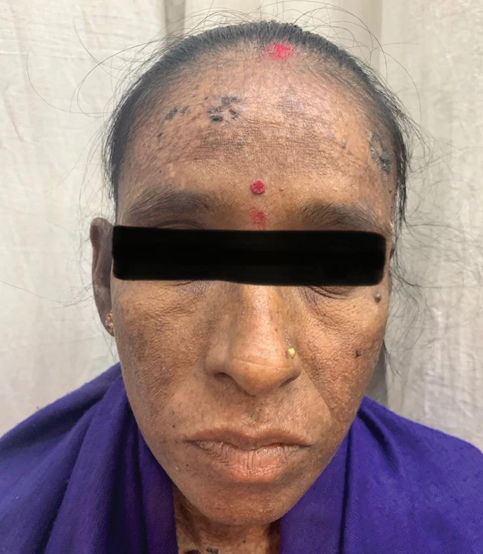
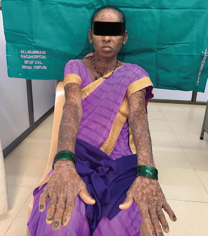
2: Hyperpigmented and hypopigmented lesion on the face
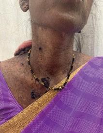
Figure 3: Hyperpigmentation and hypopigmentation on neck
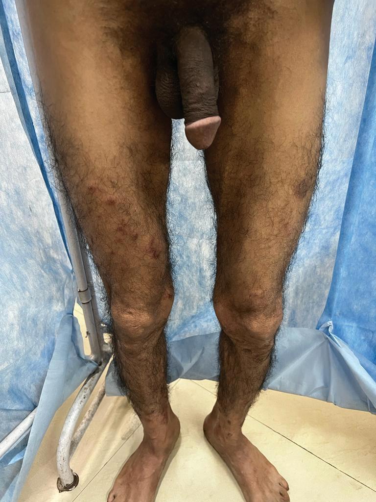
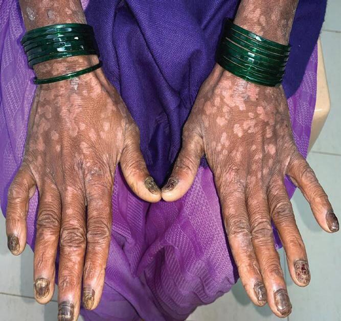
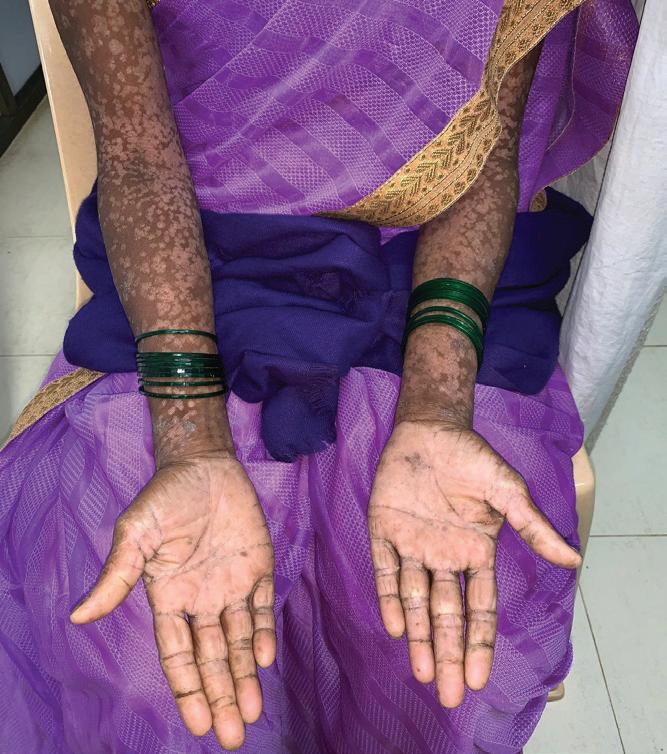
4 (A, B & C):............. Hypopigmentation is observed on the arms and the dorsal surfaces of both hands
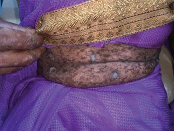
Figure 6: Hypopigmentation of the skin on abdomen
Diagnosis
In the diagnosis of xeroderma pigmentosum, dermatology plays a crucial role by assessing key clinical features associated with the condition. Dermatological evaluation focuses on identifying early-onset freckling in sunexposed areas, which is a hallmark of xeroderma pigmentosum. The presence of hyperpigmented or hypopigmented lesions, along with signs of premature skin aging such as atrophy, xerosis, and wrinkling, are also critical indicators. Dermatologists examine the skin for the development of nonmelanoma skin cancers, including basal cell carcinoma (BCC) and squamous cell carcinoma (SCC), characterized by pearly papules, scaly
Figure
Fig. 4 (A)
Figure
Fig. 4 (B)
Fig. 4 (C)
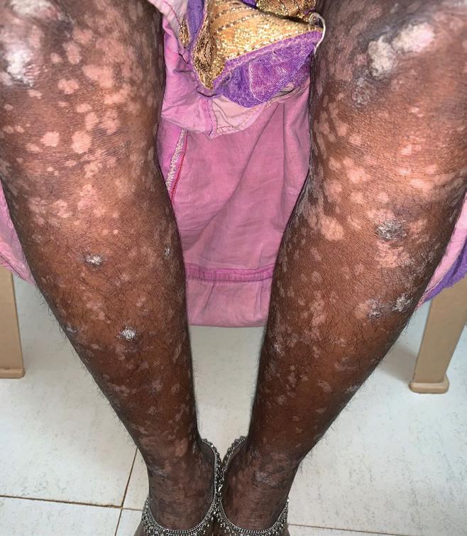
Figure 5: Dark spots with hypomelanosis on the legs
Xeroderma Pigmentosa with Squamous Cell Carcinoma
erythematous macules, and rapidly growing keratoacanthomas. ......... This comprehensive assessment helps in diagnosing xeroderma pigmentosum, monitoring disease progression, and guiding appropriate management strategies. Ocular examination is critical in diagnosing xeroderma pigmentosum (XP) by identifying UV-induced eye damage, such as conjunctivitis, pterygium, and signs of ocular malignancies like basal cell carcinoma. ENT evaluations help detect UV-related complications, including squamous cell carcinoma in the tongue and oral cavity, and tumors in the ear canal. Neurological assessments are essential to identify sensorineural hearing loss, reflex abnormalities, and neurodegenerative symptoms in XP patients. Molecular diagnostics involve functional assays, genetic analysis, and protein expression studies to assess DNA repair efficiency and identify mutations. Genetic counselling supports understanding XP's autosomal recessive inheritance, aiding in family planning and early diagnosis. This comprehensive approach ensures accurate diagnosis
and tailored management of XP, improving patient outcomes.5
Treatment
Managing xeroderma pigmentosum (XP) requires a comprehensive approach to minimize sun exposure, address skin cancers, and manage other complications. Key strategies include avoiding outdoor activities during peak sunlight, using high-SPF broadspectrum sunscreens, and wearing protective clothing, such as wide-brimmed hats and UV-blocking sunglasses, to reduce UV damage and skin cancer risk. Chemoprevention with medications like oral isotretinoin can decrease the number and size of skin tumors by influencing keratinocyte behavior, though regular monitoring is needed to manage potential side effects.
Topical imiquimod, an immune-modulating agent, is applied directly to the skin to stimulate the local immune response, effectively treating superficial basal cell carcinomas and actinic keratoses, although it may cause temporary inflammation and irritation.
Topical fluorouracil, a chemotherapeutic agent,
targets actinic keratoses and superficial basal cell carcinomas by inhibiting DNA synthesis in rapidly dividing cells, helping manage precancerous lesions and prevent their progression to invasive cancers, though it may lead to redness, peeling, and discomfort in the treated areas. Managing poikiloderma and actinic keratoses involves several effective therapies. Chemical Peeling exfoliates damaged skin layers, improving texture and reducing pigmentation. Dermabrasion uses a rotating device to smooth skin and remove surface irregularities. Laser resurfacing with CO2 or erbium-YAG lasers targets damaged layers and stimulates collagen production, with CO2 lasers for deeper resurfacing and erbiumYAG lasers for precise treatment. Cryotherapy freezes and destroys actinic keratoses, causing them to slough off. Fractional/ Pulsed Laser Therapy targets specific skin areas to improve appearance, and Photodynamic Therapy (PDT) uses a photosensitizing agent activated by light to destroy abnormal cells, treating actinic keratoses and
Xeroderma Pigmentosa with Squamous Cell Carcinoma of the Back: A Rare Case Report
superficial cancers.6
In xeroderma pigmentosum (XP), surgical excision is essential for removing large, resistant, or cancerous skin lesions, with skin grafts and local flaps used to cover defects and restore damaged areas. Skin grafts transplant skin from a donor site, while local flaps use nearby skin, offering better color and texture matching. For ocular complications, treatments include methylcellulose eyedrops for dryness, UV-protective contact lenses, and surgical resection or cryotherapy for tumors. Managing skin cancers involves using oral vismodegib for basal cell carcinoma and pembrolizumab for squamous cell carcinoma. Additionally, topical T4 endonuclease-V, oral nicotinamide, and gene therapy address UVinduced DNA damage, while genetic and psychological counseling support overall patient care and quality of life.6
Discussion
Xeroderma pigmentosum (XP) is a rare autosomalrecessive disorder, first described by Hebra and Kaposi in 1874, with a prevalence of about 1 in 1,000,000 in the United
States. XP typically presents in early childhood with multiple pigmented lesions, severe photosensitivity, and dry skin. Patients face a dramatically increased risk of non-melanoma skin cancers, with a 10,000fold higher incidence by age 20 compared to the general population. Factors that exacerbate skin conditions and increase cancer risk include sunny weather, outdoor living, fair skin, smoking, limited diagnostic access, delayed diagnosis, and inadequate sun protection.1,7,8,9,10,11 Patients with xeroderma pigmentosum (XP) who smoke face an increased risk of lung cancer, as cigarette smoke causes mutagenic damage similar to UV exposure. Skin cancers in XP can also develop in less sun-exposed areas, such as the tongue, with higher rates of anterior tongue malignancies noted in African and African-American patients. Diagnosing XP involves differentiating it from similar conditions like Cockayne syndrome and Trichothiodystrophy, which have distinct neurological and physical traits. Diagnosis can be straightforward in severe cases but more challenging in milder ones where
pigmentation changes might only appear in adolescence. Differential diagnosis also involves excluding other conditions with pigmentation patterns, such as RothmundThompson Syndrome and Peutz-Jeghers Syndrome, aided by thorough family history and DNA repair tests.12,13 In xeroderma pigmentosum (XP) patients, skin cancer typically begins around 8 years of age, much earlier than the average onset age of 60 in unaffected individuals. Common malignancies include basal cell carcinoma and squamous cell carcinoma (SCC), particularly affecting the face, head, and neck, with SCC often showing aggressive behaviour. Preventive measures, such as strict sun avoidance, wearing protective clothing, using broad-spectrum sunscreens, and UVblocking eyewear, are essential. Early diagnosis, regular follow-up for the detection and removal of pre-cancerous lesions, and genetic counselling for atrisk families are crucial. In sunny regions, like Konkan, rigorous sun protection and timely medical attention are vital to managing XP effectively and reducing the risk of rapid cancer
progression.8,14,15
Conclusion
Xeroderma pigmentosum presents significant challenges due to its high risk of UV-induced skin cancers and complex diagnostic needs, requiring strict sun protection, regular screenings, and chemoprevention. The rarity and genetic diversity of XP complicate diagnosis, but advances in genetic and molecular research offer hope for better diagnostics and targeted therapies. Clinicians should be vigilant for early signs of aggressive squamous cell carcinoma (SCC) in XP patients and educate parents on strict UV protection, especially in sunny regions. Early detection of lesions on the scalp, face, and neck can be life-saving, emphasizing the need for a multidisciplinary approach to improve outcomes and quality of life for those with Xeroderma pigmentosum.
Footnotes:
Source of Support: Nil.
Conflict of Interest: None declared.
References
1. Pagon R, Bird T, Dolan C, Stephens K, Adam M. Xeroderma Pigmentosum – Gene Reviews™ 1993 [Google Scholar]
2. Lucero R, Horowitz D. Xeroderma Pigmentosum. In: StatPearls. Treasure Island (FL): StatPearls Publishing; July 4, 2023.
3. Tamura D, DiGiovanna JJ, Khan SG, Kraemer KH. Living with xeroderma pigmentosum: comprehensive photoprotection for highly photosensitive patients. Photodermatol Photoimmunol Photomed. 2014; 30(2-3):146152. doi:10.1111/phpp.12108.
4. Lang PG, Braun MA, Kwatra R. Aggressive squamous carcinomas of the scalp. Dermatol Surg. 2006; 32:1163–70. [PubMed] [Google Scholar]
5. Lehmann J, Schubert S, Emmert S. Xeroderma pigmentosum: diagnostic procedures, interdisciplinary patient care, and novel therapeutic approaches. J Dtsch Dermatol Ges. 2014; 12(10):867-872. doi:10.1111/ ddg.12419.
6. Leung AK, Barankin B, Lam JM, Leong KF, Hon KL. Xeroderma pigmentosum: an updated review. Drugs Context. 2022; 11:2022-2-5. Published 2022 Apr 25. doi:10.7573/dic.2022-25.
7. Awan BA, Alzanbagi H, Samargandi OA, Ammar H. Scalp squamous cell carcinoma in xeroderma pigmentosum. N Am J Med Sci. 2014; 6(2):105-106. doi:10.4103/1947-2714.127754.
carcinoma of face in a child with xeroderma pigmentosa: A case report. Austral-Asian J Cancer. 2009; 8:133–135. [Google Scholar]
9. Kleijer WJ, Laugel V, Berneburg M, et al. Incidence of DNA repair deficiency disorders in western Europe: Xeroderma pigmentosum, Cockayne syndrome and trichothiodystrophy. DNA Repair (Amst). 2008;7(5):744750. doi:10.1016/j. dnarep.2008.01.014.
10. Halpern J, Hopping B, Brostoff JM. Photosensitivity, corneal scarring and developmental delay: Xeroderma Pigmentosum in a tropical country. Cases J. 2008;1(1):254. Published 2008 Oct 20. Doi: 10.1186/1757-1626-1-254.
11. Bradford PT, Goldstein AM, Tamura D, et al. Cancer and neurologic degeneration in xeroderma pigmentosum: long term follow-up characterises the role of DNA repair. J Med Genet. 2011; 48(3):168-176. doi:10.1136/jmg.2010.083022.
12. Black JO. Xeroderma Pigmentosum. Head Neck Pathol. 2016; 10(2):139-144. Doi: 10.1007/s12105-016-0707-8.
13. Cleaver JE. Diagnosis of Xeroderma Pigmentosum and Related DNA Repair-Deficient Cutaneous Diseases. Curr Med Lit Dermatol. 2008; 13(2):41-48.
14. Akdeniz, Necmettin &
Bilgili, Serap Gunes & Omer, Calka & KARADAĞ, AYŞE. Xeroderma pigmentosum in eastern Turkey: A review of 15 cases. Turkish Journal of Medical Sciences. (2012); 42. 719-723. 10.3906/sag-1012-2.
15. Lehmann AR, McGibbon D, Stefanini M. Xeroderma pigmentosum. Orphanet J Rare Dis. 2011;6:70. Published 2011 Nov 1. doi:10.1186/1750-11726-70
Xeroderma Pigmentosa with Squamous Cell Carcinoma of the Back: A Rare Case Report