
Vo lume 45, Issue 1 • 2023
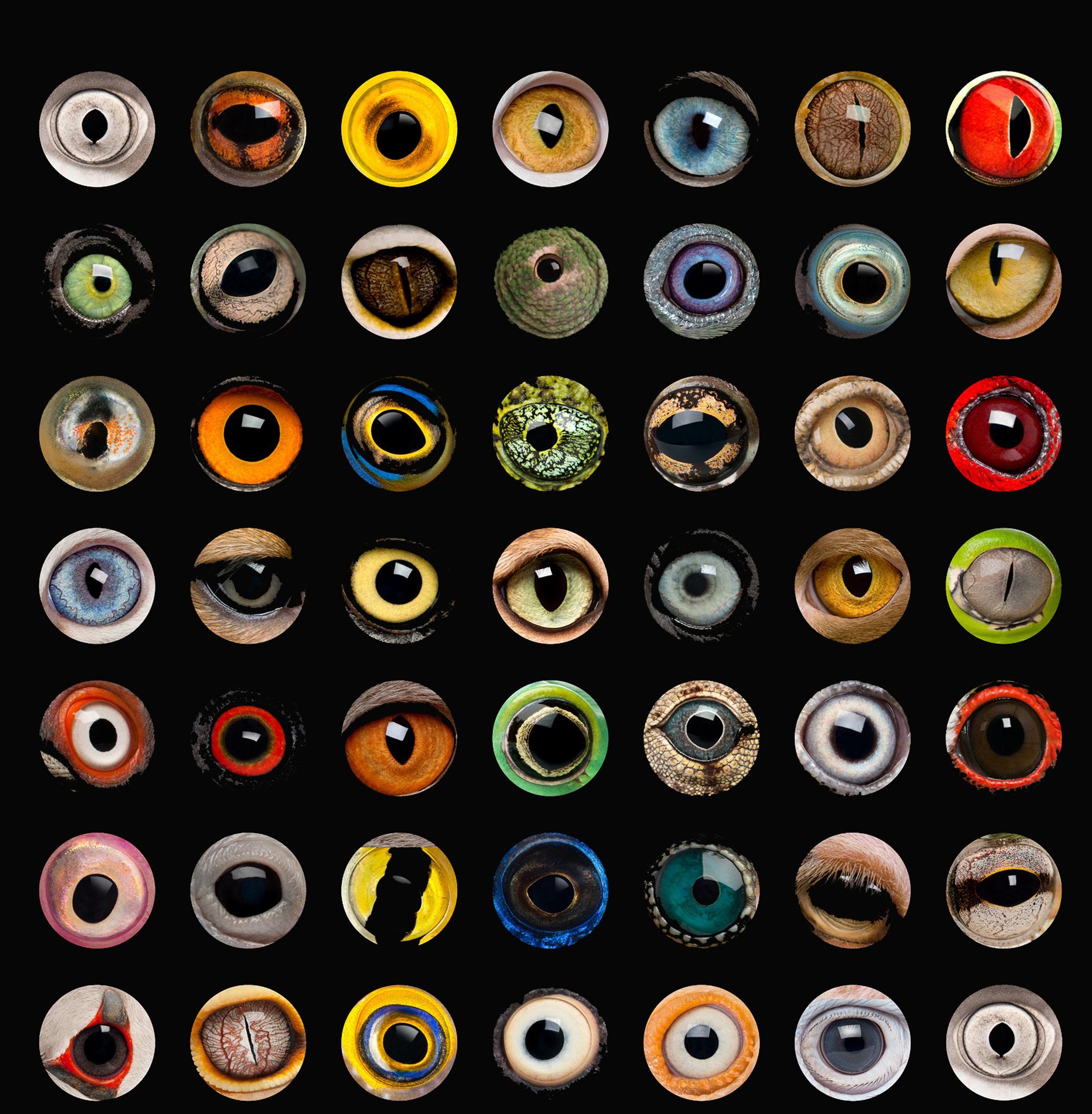
Ophthalmology Pupils of Different Land Animals. Adapted image courtesy of Wanderlust2003, Wikimedia Commons, CC-BY-SA-4.0
A Look Into the World of Animal

Volume 45, Issue 1, 2023
Editorial: Dear OPS Members Monica Motta 5
Original Article: Vision and Field Trial Performance: The Effect of Optical Defocus on Your Field Trial Retriever
Monica Motta, BS RVT; Steve Hollingsworth, DVM; Allyson Groth, DVM; John Doval BPA, Senior Artist; Philip H Kass DVM, PhD; Ron Ofri DVM, PhD; Christopher J Murphy DVM, PhD
Article Abstract: Snake Spectacle Vessel Permeability to Sodium Fluorescein Roy W Bellhorn; Ann R Strom; Monica J Motta; John Doval; Michelle G Hawkins; Joanne Paul-Murphy


7
Case Report: In An Emergency – Veterinary Cases from the ER Federica Maggio, DVM, Diplomate ACVO 8
Original Article: Amazing Animal Eyes Kathleen Warren
Case Report: An Afternoon with Dr. Brooklynn Lafoon Brooklynn LaFoon, DVM, DACVO
2022 OPS Scientific Exhibit
Original Article: Dynamic Tilt Contact Angle Goniometry of Animal Globes Kevin Aguirre Gamarra; Erin A Hisey; Brian C Leonard
Case Report: Unique and Dazzling Ocular Clinical Case of a Great Horned Owl
Monica J Motta, BS, RVT, LATG; Andrea Minella, DVM; Piyaporn Eiamcharoen, DVM; Brian Murphy, DVM; Christopher Reilly, DVM; Christopher Murphy, DVM, PhD ; Danielle K Tarbert, DVM, DACZM
Diving into Whales’ Ability to Tolerate Low Oxygen
Case Report: Purtscher Retinopathy in Nonhuman Primate


6
10
. . . . . . . . . . . . . . . . . . . . . . . . . . . . 12
.
16
33
. . . . . . . . . . . . . . . . . . . . . . . . . . . 34
Giselle
. . . . . . . . . . . . . . . . . . . . . . . . . . . . . . . 35
Wang; Angela Spivey
Tzu-Ni Sin;
Yiu . . . . . . . . . . . . . . . . . . . . . . . . . . . . . . . . . . . . 36
Ralph
Clevenger 38
Glenn
Photo Essay
A.
Jody Troyer, CRA Ralph Eagle Jr., MD
James Gilman, CRA, FOPS
2022 Scientific Exhibit Winners: page 16
Jaime Tesmer, CRA, OCT-C
Editor in Chief
Kathleen Warren Duke University Eye Center Department of Ophthalmology 2351 Erwin Road Durham, NC 27710 kathleen.warren@duke.edu
Art Director
Jennifer Manning 5682 Dunnigan Road Lockport, NY 14094 jenmanningJOP@gmail.com


Editorial Review Board
Case Report / Technical Tactics Editor


Michael P. Kelly, FOPS Duke University Eye Center 2351 Erwin Road, Suite 209 Durham, NC 27710 michaelpkellydukeeye@gmail.com
Elizabeth Affel, MT, MS, OCT-C, FOPS Jefferson University Hospital Philadelphia, PA president@opsweb.org

Lisa Dennehy, BS Mass Eye and Ear Boston, MA lisa_dennehy@meei.Harvard.edu
Alan Frohlichstein, BFA, BS, CRA - ret., FOPS Retinal Angiography Services Morton Grove, IL alanfroh@gmail.com


Christiaan Lopez-Miro Duke University Eye Center Durham, NC christiaan.lopezmiro@duke.edu
Paula Morris, BS, CRA, FOPS John A. Moran Eye Center Salt Lake City, UT paula.morris@hsc.utah.edu
Assistant Editor
Monica Motta, SRA III, Supervisor I, BS, RVT, LATG UC Davis School of Veterinary Medicine Comparative Ophthalmology Vision Science Lab Davis, CA 95616 Ocular Services on Demand Madison, WI mjmotta@ucdavis.edu


Advertising Editor
Barbara McCalley 1621 East Jody Circle, Republic, MO 65738 Phone: 417.725.0181 Fax: 417.724.8450 ops@opsweb.org

Medical Advisor
Akbar Shakoor, MD John A. Moran Eye Center University of Utah School of Medicine Salt Lake City, UT Akbar.Shakoor@hsc.utah.edu

Nicholas Patterson, CPT Duke University Eye Center Durham, NC nicholas.patterson@duke.edu

Kasi Sandhanam Singapore National Eye Centre Singapore kasi.sandhanam@snec.com.sg
Robert G. Shutt, CRA, OCT-C Connecticut Eye Consultants Danbury, CT shutteye@optimum.net
Jennifer Struck Duke University Eye Center Durham, NC jennifer.struck@duke.edu
Paola Torres, COT, CPT, CRA, OCT-C Duke University Eye Center Durham, NC paola.belleau-torres@duke.edu
Founded in 1977 by the Ophthalmic Photographers’ Society, Inc.; Don Wong, RBP, FOPS, Founding Editor
Monica Motta, BS RVT, LATG

UC Davis School of Veterinary Medicine
Comparative Ophthalmology Vision Science Lab Davis, CA 95616
mjmotta@ucdavis.edu
Dear OPS Members
Iwould like to take a moment to introduce myself and tell you a bit about my career history and how I came to be a part of the OPS.
I began working in private veterinary clinics when I was 17 just out of high school and attending junior college to get my Registered Vet Tech certification.
I graduated CSU, Chico in 2003 with a Bachelors in Biological Sciences, and went on to work at IDEXX for two years as a histology technician. From IDEXX, I made my way to the UC Davis School of Veterinary Medicine Companion Avian and Exotics Department where I worked for almost four years and found my love for exotic veterinary medicine. A combined case with the ophthalmology department performing cataract surgery on a bald eagle led me to ophthalmology. I began working in the ophthalmology department and in the midst of the
2008 crisis, Dr. Christopher Murphy joined the department as research faculty. I quickly became intrigued, and in January 2010 I was hired as the Murphy-Russel Vision Lab Staff Research Associate. What is now COVSL (Comparative Ophthalmology Vision Science Lab) has been my home and family for the past 13 years. Over the past 13 years, I have developed an expertise in imaging a wide range of species, including primates, rabbits, dogs, horses, birds, snakes and mice. I attended my first OPS annual program in 2011, and again at various times in the years thereafter. In 2019, I joined the OPS on the Board of Directors. My goal while I am with the OPS is to share the knowledge, skill, work and beauty of ophthalmic photography in animals and animal-based research, and expand the diversity of the OPS to all aspects of ophthalmic photography.
Subscription Information
The Journal of Ophthalmic Photography (ISSN 0198-6155) is published biannually by the Ophthalmic Photographers’ Society, Inc. Membership in the Ophthalmic Photographers’ Society includes an annual subscription to the The Journal of Ophthalmic Photography. Membership requests, subscription inquiries, past issue requests and changes of address should be sent to Barbara McCalley, Executive Director, 1621 East Jody Circle, Republic, MO 65738; Phone: 417.725.0181; Email: ops@opsweb.org.
Instructions to Authors
Instructions to Authors can be found at http://www.opsweb.org/?page=journal
The Ophthalmic Photographers’ Society
The Ophthalmic Photographers’ Society (OPS) is a nonprofit international organization dedicated to the advancement of photography as applied to ophthalmology and visual science. Our membership, numbering over 1000, includes a broad spectrum of professionals in eye health care, including ophthalmologists, optometrists, photographers, nurses, ophthalmic medical personnel, and basic researchers. The Society’s educational programs, designed to supplement the professional expertise of our members, have earned the OPS recognition as being an organization dedicated to the elevation of standards in the craft. The Society’s major publication, The Journal of Ophthalmic Photography, presents tutorials and articles on photographic techniques, instrumentation, and related topics in ophthalmology. Visit us online at www.opsweb.org.
Copyright
The Ophthalmic Photographers’ Society. All rights reserved. Printed in
cal or electronic processes, stored in a retrieval system or transmitted by
5 Editorial
Editorial
the U.S.A. None of the contents may be reproduced by mechani-
any means without prior written permission from the Editor.
Original Article
Monica Motta, BS RVT, LATG
 Steve Hollingsworth, DVM
Allyson
John
Philip
Groth, DVM
Doval BPA, Senior Artist
H Kass DVM, PhD
Ron Ofri DVM,
Christopher
PhD
J Murphy DVM, PhD
UC Davis School of Veterinary Medicine
Comparative Ophthalmology
Vision Science Lab Davis, CA 95616
mjmotta@ucdavis.edu
Vision and Field Trial Performance: The Effect of Optical Defocus on Your Field Trial Retriever
It’s not just something you do, it’s something you live for. Field trialing is a way of life for trainers, owners and their Labradors, which is why it’s so important to reveal the growing knowledge of the effects of myopia in Labrador retrievers. Through diligent research it has become known that myopia (nearsightedness) is an ocular disorder found in canines, and has a relatively high prevalence in Labrador retrievers. Recent studies conducted in both the US and New Zealand support myopia to be of genetic origin.

Of all the breeds affected with myopia, only the Labrador retriever has been documented to possess an axial myopia. This means that the cause of the myopia is due to a dysregulated growth of the eye, such that the axial dimensions are excessively elongated. So, what does it mean to be nearsighted? Nearsightedness is the inability to image objects accurately at the retina that are more than approximately 20 feet from the eye. If the eye is one diopter myopic (commonly detected in Labradors) that means that objects one meter from the eye are in sharp focus but that objects placed at a distance will not be accurately focused on the retina, creating a “blurry” image. Having myopia decreases visual acuity (acuteness or clearness of vision), which directly impacts the ability to perform visually challenging tasks dependent on fine imaging of distant objects. A recent study was conducted by UC Davis School of Veterinary Medicine in 2010 (In press American J Veterinary Research, 2011) that found Labrador retrievers to have a strong tendency to worsen field trial performance with increasing optical defocus. Eight field trial Labrador retrievers were fit with 3 sets of contact lens (0.0, +1.5 and +3.0) to induce increasing states of myopia. Field trial judges, masked to the myopic changes, judged each dog on a 150-yard and 200-yard retrieval. It was no surprise to find that when the judge’s performance evaluations were combined, the results of this study strongly indicated a significant relationship between the degree of myopic change and the rank with which each Labrador placed for the two field trial
retrieves. That is, the greater degree of defocus, the lower the ranking the Labrador received by the field trial judges. So, what does this mean? Studies show that myopic defocus can significantly affect performance under field trial conditions. It has been observed that some myopic Labs do well out to 100 or 150 yards, but then fall apart, failing to mark accurately at longer distances. The trainer is then faced with difficult decisions, and must determine if this behavior is an issue that can be addressed through training, or if there is an underlying organic reason for the falloff in the dog’s performance. If the dog is moderately to severely myopic, it may not be able to adequately mark a target at long distances. Further research being conducted at the UC Davis School of Veterinary Medicine will attempt to identify gene(s) responsible for myopia in the Labrador retriever. If the gene(s) can be identified then they could be tested for to the betterment of breeding programs of performance dogs.
The Journal of Ophthalmic Photography Volume 45, Issue 1 • 2023 6
Image courtesy of Monica Motta and California Waterfowl Association
So what does it mean? The impact of myopia will depend on the expectations for visual performance. For a pet in a home environment, mild-moderate degrees of myopia, may not result in any detectable change in behavior. A myopic dog presented with visually challenging tasks, however, may not be able to perform adequately. The presence of myopia can be easily and rapidly detected by a veterinary ophthalmologist familiar with retinoscopy. More and more veterinary ophthalmologists are becoming familiar with this technique, and trainers interested in having this procedure done are inquiring with their local professional. Myopia can be detected in puppies and could be measured at the time a CERF
Article Abstract
Roy W Bellhorn
Ann R Strom
Monica J Motta
John Doval

Michelle G Hawkins
Joanne Paul-Murphy
(Canine Eye Registration Foundation) exam is performed. Although vision plays a significant role in the performance of a field trial Labrador, it of course is only one factor in the makeup of a champion. Intelligence, natural athletic ability and fitness, training, desire and motivation, and inherent sensory capabilities, including vision, must come together in any champion’s performance.
Co-Authors and Field Trial Professionals
Julie Cole; Luann Pleasant, 22 year field trial trainer, titled field champions and master hunters; Ron Foley, 8-point AKC field trial judge for retrievers, and Alice Woodyard.
UC Davis School of Veterinary Medicine
Comparative Ophthalmology
Vision Science Lab
Davis, CA 95616
Snake Spectacle Vessel Permeability to Sodium Fluorescein
Keywords
Python regius; ball python; ecdysis; fluorescein angiography; permeability; snake spectacle.
Objective
Assess vascular permeability of the snake spectacle to sodium fluorescein during resting and shedding phases of the ecdysis cycle.
Animal Studied
Ball python (Python regius).
Procedures
The snake was anesthetized, and spectral domain optic coherence tomography was performed prior to angiographic procedures. An electronically controlled digital single-lens reflex camera with a dual-head flash equipped with filters suitable for fluorescein angiography was used to make images. Sodium fluorescein (10%) solution was administered by intracardiac injection. Angiographic images were made as fluorescein traversed the vasculature of the iris and spectacle. Individually acquired photographic frames were assessed and sequenced into pseudo video image streams for further evaluation.
Conclusions
Fluorescein angiograms of the snake spectacle were readily obtained. Vascular permeability varied with the phase of ecdysis. Copious leakage of fluorescein occurred during the shedding phase. This angiographic method may provide diverse opportunities to investigate vascular aspects of snake spectacle ecdysis, dysecdysis, and the integument in general.
7
Vision
and Field Trial Performance: The Effect of Optical Defocus on Your Field Trial Retriever
Case Report
Federica Maggio, DVM, Diplomate ACVO

Tufts Veterinary Emergency Treatment and Specialties
525 South Walpole Street
Walpole, MA 02081 508.668.5454
Federica.Maggio@tufts.edu
In An Emergency: Veterinary Cases from the ER
Case 1
Severe lens subluxation in the right eye of a 5-year-old MN mix breed dog. The pupil had been accidentally pharmacologically dilated.
The cornea shows a central focal area of mild edema, cause unknown (Figure 1). Uncharacteristically for cases of primary lens luxation, the lens has not been displaced by gravity (in the anterior chamber or posterior segment), but is still partially connected to the dorsotemporal zonular ligaments.
A focal posterior synechia is also visible, furtherly anchoring the lens to the iris. The lens itself, as it is expected in chronic lens luxation cases, is starting to show the presence of cataracts, equatorial and diffuse cortical. The large aphakic crescent visible on the right of the lens shows an out-of-focus fundus, with a prominent pinkish optic nerve head and dorsal green-yellow tapetum.
The owner of the pet elected conservative miotic treatment and the dog was lost to follow up.
NIKON D70, f/14, 1/60’’, focal length 105 mm, 3.8 max aperture, auto flash.
Case 2
Uveal cyst in the vitreous of a 12-year-old MN Scottish terrier.
The finding was incidental during a routine ophthalmic examination, as the dog was admitted for assessment of ocular allergies.
Figure 2 shows the ocular fundus, with its greenyellow dorsal tapetum, robust retinal veins and arteries, and a healthy optic nerve head. The uveal cyst is visible in the dorsal vitreous, lightly pigmented, and superimposed over the retina, which is visible through it. The cyst is also casting a shadow over the retina.
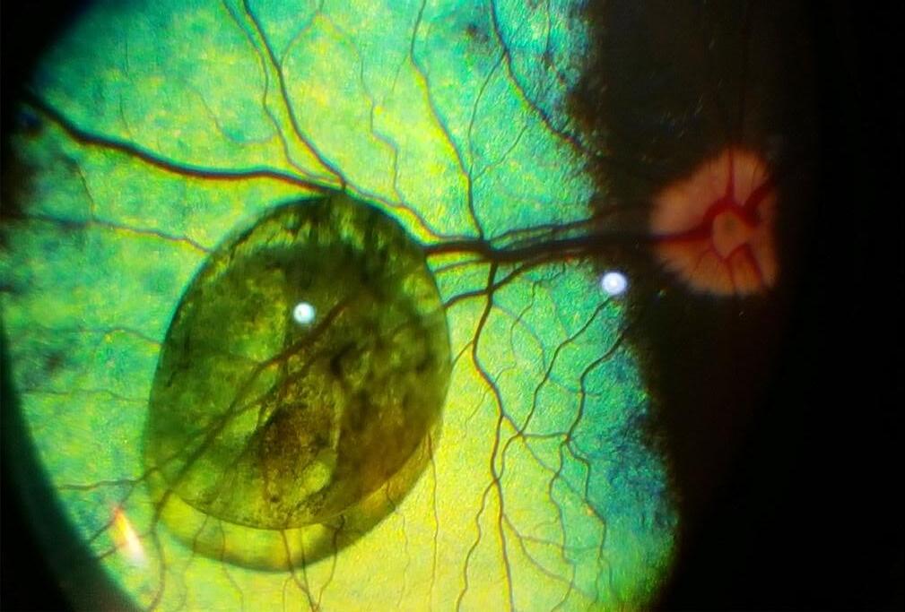
Uveal cysts are quite common in dogs, and originate from the epithelia of the ciliary body or posterior iris. They are either congenital or acquired. Once present, they may float and locate in the anterior segment or, more rarely, in the posterior segment of the eye, as in the present case. They often spontaneously implode, and fade.

In most cases, they are very well tolerated and, as in the present case, do not require any treatment. The dog had undiminished visual acuity.
The image was obtained using the camera of a Samsung smartphone (SM-G925G), with its light source, with an interposed Volk 20D fundus lens.
The Journal of Ophthalmic Photography Volume 45, Issue 1 • 2023 8
Figure 1
Figure 2
Case 3
Traumatic cataract in the right eye of a 3-year-old FS Siberian Husky mix breed dog.


The pet was adopted by the owners a few weeks prior to presentation. She had a history of injuries on both eyes, cause unknown, and was almost completely blind. The left eye showed such severe end-stage lesions that enucleation was recommended.
The right eye, which is represented in Figures 3 and 4, was still partially visual.
The eye shows minor conjunctival hyperemia. The pupil is pharmacologically dilated. The lens presents immature diffuse anterior and posterior cortical cataracts. A solid, roughly round dark foreign body is visible in a dorsal/central position within the lens (Figure 3). A radiograph of the dog’s head revealed multiple lesions from penetrated shotgun pellets. It was assumed that the small round foreign body within the right lens was indeed a birdshot and the cause of the cataract. Due to the absence of corneal scars, it was assumed that the pellet had penetrated the lens from the side and the back, perforating the sclera. The left eye had suffered much more severe injuries, and was not viable anymore. Lead is usually well tolerated within the eye, but unfortunately the trauma resulted in the presence of the cataract in the right eye. The owner elected cataract surgery and the removal of the foreign body for the right eye, and removal of the left eye. It was confirmed that the foreign body was indeed a lead pellet from a shotgun.
Figure 4 shows the same eye after extracapsular lens extraction surgery. Due to the damage present in the posterior lens capsule and anterior vitreous, caused by the trajectory of the pellet, it was elected not to insert an artificial intraocular lens (IOL). The corneal incision seen dorsally (few sutures) is larger than usual, as it was necessary for the removal of the pellet. The large capsulorexis is visible, as is the focal vitreal fibrotic strand. The dog was visual and comfortable after the procedure.
NIKON D3200, f/16, 1/200”, ISO-2200, 105mm focal length, 4 max aperture, auto flash.
About the Author
A specialist in veterinary ophthalmology, Dr. Federica Maggio was inducted into the American College of Veterinary Ophthalmology in 2005. She has authored numerous ophthalmology articles that have appeared in the Journal of the American Veterinary Medical
Association, the UK Veterinary Journal, Veterinary Ophthalmology and Veterinary Clinics of North America—Small Animal Practice. Dr. Maggio’s most important contribution has been her research on systemic hypertension and associated ocular lesions in cats. Her main current focus is glaucoma, cataract surgery and reconstructive eyelid surgery.
Born and raised in Italy, Dr. Maggio attended veterinary school in Bologna and graduated with her Doctorate of Veterinary Medicine in 1991. Following veterinary school, she completed a residency program in veterinary ophthalmology at North Carolina State University in 2004. Since then, Dr. Maggio has been teaching ophthalmology at the Cummings School of Veterinary Medicine at Tufts University and practicing ophthalmology at Tufts VETS.
9 In An Emergency: Veterinary Cases from the ER
Figure 3
Figure 4
Kathleen Warren Duke University Eye Center Department of Ophthalmology
 2351 Erwin Road Durham, NC 27710
kathleen.warren@duke.edu
Amazing Animal Eyes
Planet earth is home to close to nine million animal species, all with eyes that function in a plethora of ways. By detecting light and shadows, the simplest eyes can help avoid collisions, while the most advanced eyes, utilizing a vast range of the light spectrum, may perceive this world in ways far beyond what any human eye can see.
According to the Smithsonian Institute, the biggest eyes in the animal kingdom belong to the giant squid, whose eyes measure up to 10 inches, using them to swim the ocean’s dark, murky waters. One study even proved the giant squid could detect a sperm whale nearly 400 feet away with its large eyes.
While humans possess only three types of photoreceptors; colors red, blue and green; the mantis shrimp, a crustacean, has close to 16 kinds of photoreceptors. These tiny shrimps can perceive light in ways far beyond the capability of other animals, yet they have tiny brains. With such small brain capacity, what do mantis shrimps do with all of that visual range?
To add to this, floating around without a brain, the box jellyfish has 24 eyes, but the species cannot see much beyond light, shadows and shapes.
Soaring hundreds of feet in the air, a bald eagle can spot a fish swimming in the river, then dive down and grab its lunch with such precision. How is this possible? While each human eye has only one fovea with about 200,000 cones, a bald eagle eye possesses a massive quantity of visual receptors. With two foveae’s, each one offering a million cones, and shaped somewhat like a telephoto lens, with long-range vision, eagles are lucky enough to have some of the best sight abilities in the animal kingdom.

Owls cannot move their eyeballs and are the only bird that can see the color blue. This has resulted in the distinctive way they turn their heads almost 360 degrees.
As evolution has seen fit, horses, sheep and goats can be seen as prey animals. Their pupils are horizontal and elliptical, offering a wider range of vision to help them spot predators seeking a meal.
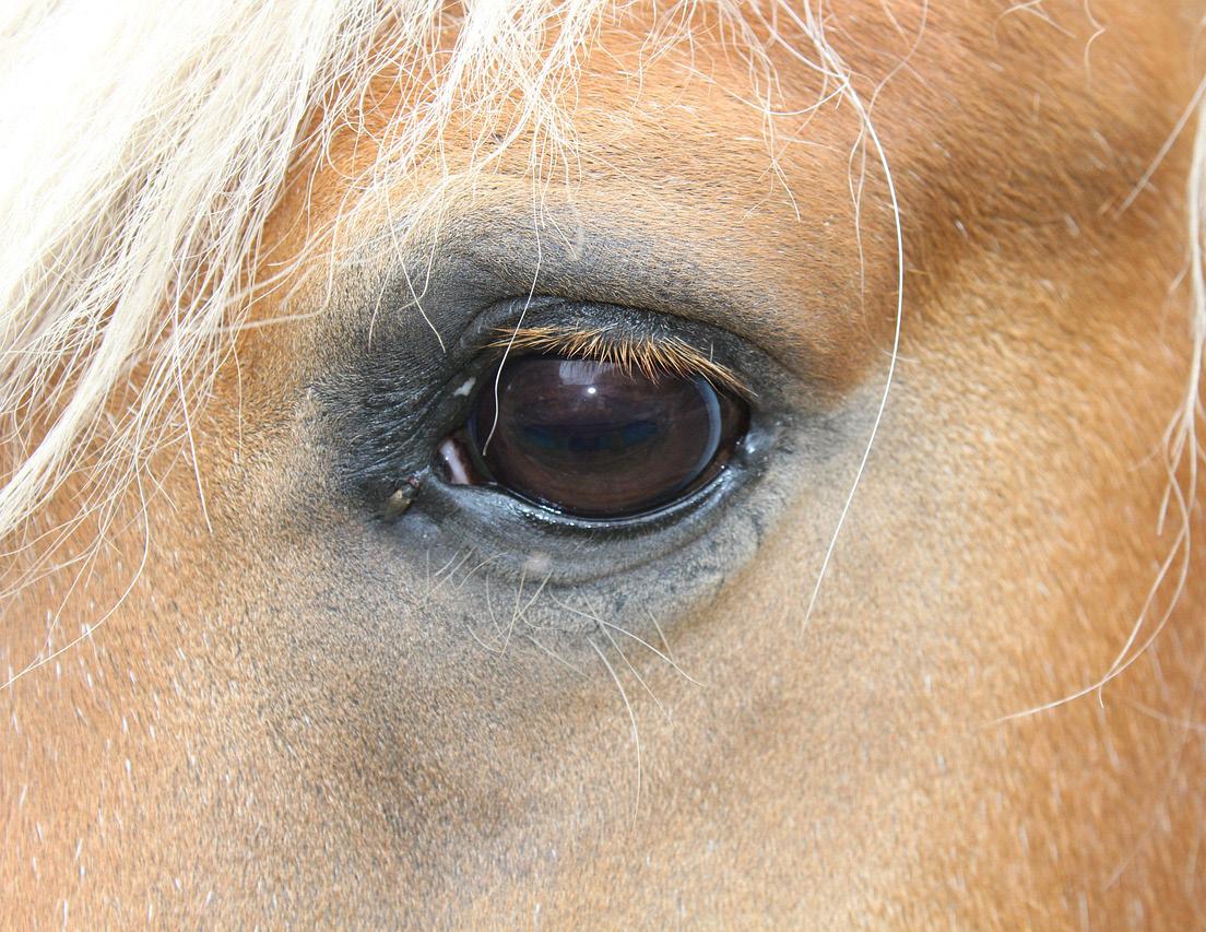
Horses have expansive peripheral vision. Positioned on the sides of its skull, a horse’s vision provides a 340-degree field of view (out of 360). That means a horse can see almost all the way around itself in whichever direction it may be facing. Riding a horse is a bit like having
The Journal of Ophthalmic Photography Volume 45, Issue 1 • 2023 10
Image courtesy of pixnio.com, CC0
Image courtesy of Roland Sehm, Pixabay.com
Original Article
eyes in the back of your head… literally. However, horses also have a blind spot — directly in front of its nose! Have you ever seen the eyes of animals at night, maybe while camping? Animals like deer, horses, lions, tigers, and bears (oh my) have retinas with a mirror-like region, known as the tapetum lucidum, which reflects light waves back into their eyes, boosting night vision. If you have ever shone a light into the direction of these animals’ eyes, those regions are activated… giving their eyes a zombie-like, ghostly glow.
With such precise and remarkable color detection, another special feature belongs to the sweet little honeybee. With a field of vision nothing like that of a human, honeybees can detect colors five times faster. At this rate of speed — the fastest in the animal kingdom — bees find flowers quickly, zeroing in on their prized nectar. Each of the honeybee’s compound eyes has between 6,900 and 8,600 lenses, or facets, which allows them to see the world like a mosaic, or kaleidoscope format.

Fun Facts About Eyes
• A worm does not have any eyes at all. Scorpions can have as many as twelve eyes.
• Guinea pigs are born with their eyes already open, while hamsters can only blink one eye at a time.
• A chameleon’s eyes are independent, meaning it can look in two different directions at once.

• An ostrich’s brain is smaller than its actual eye.
• Camels have three eyelids and extra-long lashes (10cm long) to help protect their eyes from blowing sands and desert debris.
• Similar to human corneas, shark corneas have been used in transplants.
• A scallop has close to 100 eyes around the edge of its shell to help detect predators.
• Dolphins sleep with one eye open. Polar bears have a third eyelid to help filter UV light.
• A dragonfly has over 30,000 lenses in its eyes, perfect for motion detection and not-so-perfect for predators to kill.
• A moth’s eyes are covered with an anti-reflective and water-repellant coating.
• The night vision of tigers is almost six times better than humans.

• A cat’s eye has a 285 degrees of sight, in three dimensions, making peripheral vision ideal for hunting.
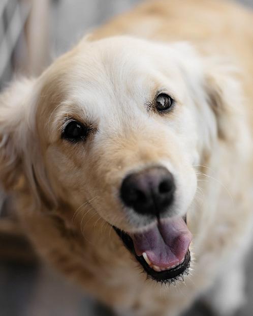
• Dogs can’t distinguish between red and green.
11 Amazing Animal Eyes
Image courtesy of christels, pixnio.com, CC0
Image courtesy of pixnio.com, CC0.
Image courtesy of collinsvisual, Pixabay.com
Image courtesy of Marko Milivojevic, pixnio.com, CC0.
Case Report
Brooklynn LaFoon, DVM, DACVO Carolina Veterinary Specialists

501 Nicholas Road
Greensboro, NC 27409
blafoon0318@gmail.com
An Afternoon with Dr. Brooklynn Lafoon
Sunny, an 8-year-old Mouse Lemur presented for blindness OU. The patient was evaluated initially in the cage for visual responses and ability to navigate the enclosure. The was an absent menace and tracking but present dazzle response and PLR OU. The mouse lemur was sedated for ophthalmic examination via slit lamp biomicroscopy and indirect ophthalmoscopy. On presentation, there was a mid-immature cataract OD and a late immature cataract OS. Anisocoria with a focal area of posterior synechia causing dyscoria OS. On fundic examination there was advanced retinal degeneration as appreciated by diffuse hyperreflectivity (due to increased tapetal reflection) and severe vascular attenuation (normal is a radial holangiotic retinal vasculature pattern). The optic nerve was moderately degenerated. Tonometry performed by TonoPen (applanation tonometer): 10 mmHg OD, 4 mmHg OS.


About the Duke Lemur Center
Founded in 1966, the Duke Lemur Center (DLC) is a noninvasive research center—the most diverse population of lemurs on Earth, outside their native Madagascar.
Currently, there are more than 200 rare lemurs and bush babies living at the Duke Lemur Center. Currently, 13 species are housed at the DLC, with 39 species of prosimian (lemurs, lorises, and bush babies) being housed over the history of the DLC. A total of 3,285 endangered prosimian primates have been born at the Duke Lemur Center over the past 50 years.
Because all of its research is non-invasive, the DLC is open to the public and educates more than 35,000 visitors annually. Its highly successful conservation breeding program seeks to preserve vanishing species such as the aye-aye, Coquerel’s sifaka, and blue-eyed black lemur, while its Madagascar Conservation Programs study and protect lemurs—the most endangered group of mammals on Earth—in their native habitat. The Division of Fossil Primates examines primate extinction and evolution over time and houses over 35,000 fossils, including
extinct giant lemurs and one of the world’s largest and most important collections of early anthropoid primates.
Over 1,000 students have been trained at the DLC over the course of our 50-year history, while more than 400 students have been trained in research, husbandry, and conservation, in the past four years alone.
In 2016, the DLC celebrated 50 years of trail-blazing lemur research and conservation. Much of that knowledge – from research data to ground-breaking insights on health, genomics, paleontology, and more – is showcased at www. lemur.duke.edu (Extracted from www.lemur.duke.edu)
The Journal of Ophthalmic Photography Volume 45, Issue 1 • 2023 12
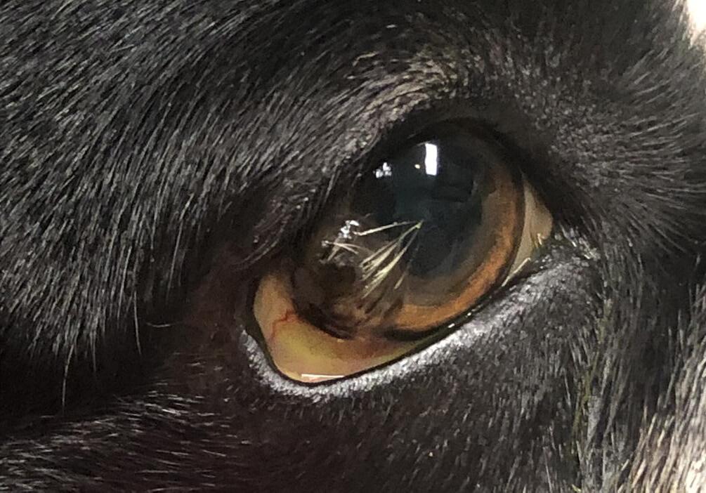
Additional Exams Performed by Dr. LaFoon Marshall, a 1.5 yo mixed breed canine, is shown with a corneal dermoid of the right eye adjacent to the limbus. Note the haired pigmented keratinized epithelium causing the mass effect approximately 8 mm in diameter adjacent to the limbus OD. Keratectomy was recommended for removal, which was performed uneventfully under general anesthesia.


13 An Afternoon
with
Dr. Brooklyn Lafoon
6-year-old Schnauzer presented for diabetic cataracts OU. Images are post bilateral phacoemulsification with artificial lens implantation.


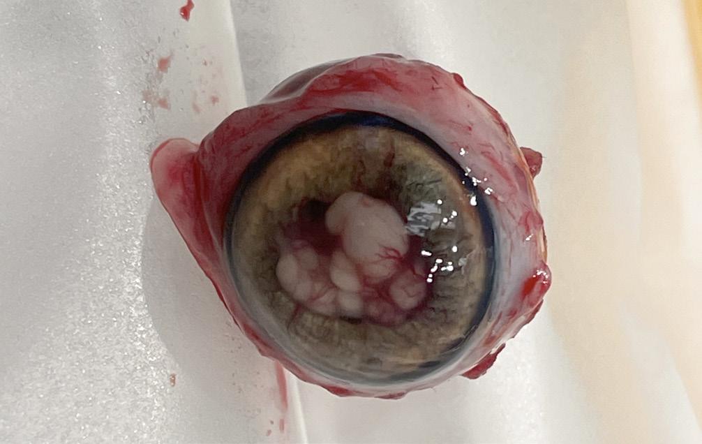
Cat presented for lens capsule rupture with mature cataract and proliferative like tissue extruding through the pupil with significant preiridal fibrovascular membrane. Enucleation showed intralenticular hyphae.


About the Author
Dr. Brooklynn LaFoon is a North Carolina native, originally from Raleigh, NC. She attended East Carolina University for undergraduate studies where she achieved a BS in Biology and a minor in Business. She graduated from Mississippi State University College of Veterinary Medicine in 2014. Following veterinary school, she completed a small animal medicine and surgical internship with BluePearl in Southfield, MI and an ophthalmology specialty internship at Animal Eye Care in Chesapeake, VA with a focus in retinal surgery. She then completed an ophthalmology internship and residency at MedVet Medical and Cancer Center for Pets in Columbus, OH. During that time, she pursued additional training at Ohio State University College of Veterinary Medicine, Bluegrass Veterinary Vision Equine Ophthalmology services and worked closely with the Columbus Zoo and Aquarium as well as The Wilds. After completion of her residency training and board certification, Dr. LaFoon and her family returned to North Carolina and joined Carolina Veterinary Specialists in Greensboro, NC.
The Journal of Ophthalmic Photography Volume 45, Issue 1 • 2023 14
Quality Without Question
SPECTRALIS® multimodal imaging platform, the answer for unparalleled image quality, is uniquely designed to empower you to see more and do more for patients.
■ Pinpoint pathology through precise visualization and segmentation of the retinal layers


■ Reveal real change from visit-to-visit automatically with AutoRescan
■ Take patient comfort to the next level with TruTrack Active Eye Tracking
■ Preserve your investment as a customizable and upgradeable imaging platform
And so much more… Experience the SPECTRALIS difference.
www.HE-Lounge.com • 800-931-2230
303174-001 US.AE23 © Heidelberg Engineering GmbH
2022 OPS Scientific Exhibit
First Place Winners: Print Division
The purpose of the annual OPS Scientific Exhibit is to encourage members to share their outstanding images with the ophthalmic community. The entire exhibit, featuring 42 categories in the print and stereo divisions, is displayed as part of the Scientific Exhibit at the Annual Meeting of the American Academy of Ophthalmology. For further information contact:
(For information only; please do not send entries to the addresses below.)
Print Division
John Grybas, CRA 313.916.4456
johngrybas67@gmail.com
Stereo Division

Tony Medina, CRA 248.408.7821 ops.scientificexhibit@gmail.com
Video Division
Tim Steffens, CRA 734.936.2283
tjsteffe@umich.edu
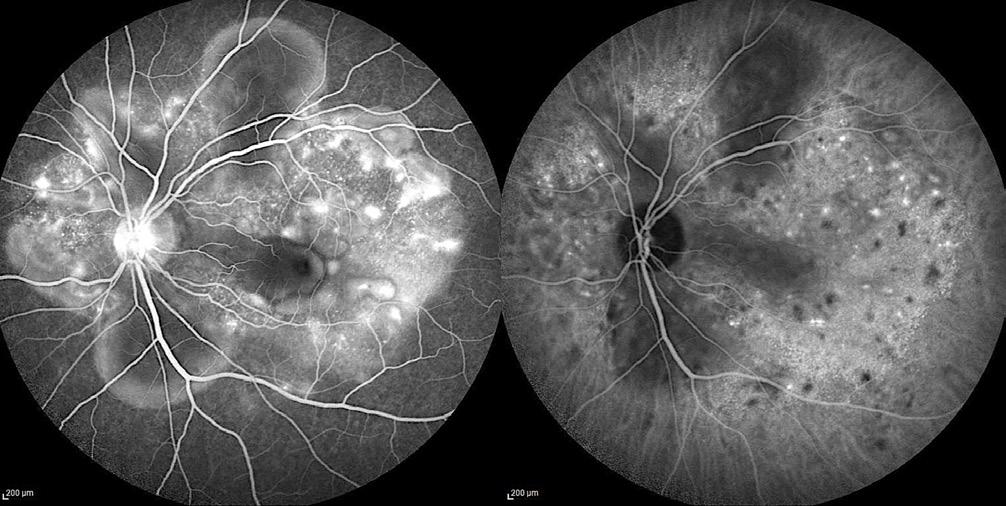
Indocyanine Green Angiography

Vogt-Koyanagi-Harada Disease

Nipan Yodmanee
Mettapracharak Watraikhing Hospital
Nakhon Prathom, Thailand
BEST OF PRINT DIVISION
Slit Lamp Photography
Post Yag Laser Capsulotomy
Kasi Sandhanam
Singapore National Eye Centre, Singapore
Cross Categories
AZOOR
Dena Harris, CRA
University of Michigan Kellogg Eye Center
Ann Arbor, Michigan
The Journal of Ophthalmic Photography Volume 45, Issue 1 • 2023 16
2022 OPS Scientific Exhibit
First Place Winners: Print Division
Slit Lamp Photography
Corneal Ulcer


Jenny Kellogg COMT, CRA, OCT-C, ROUB
Flaum Eye Institute, University of Rochester
Rochester, New York
Fundus Photography Normal 30°- 40° Brachytherapy
Jaime Tesmer, CRA, OCT-C Mayo Clinic
Rochester, Minnesota
Fundus Photography Wide Angle 45°+ Pseudohypopyon Vitelliform Lesion
Sarah Skiles, CRA University of Iowa Hospitals and Clinics Iowa City, Iowa
Fundus Photography Wide Angle 45°+ Leukemic Retinopathy


Jaime Tesmer, CRA, OCT-C Mayo Clinic
Rochester, Minnesota
2022 OPS Scientific Exhibit 17
2022 OPS Scientific Exhibit
First Place Winners: Print Division
Fluorescein Angiography
PDR with VH
Bradley Stern, CRA, OCT-C
Henry Ford Health System
Detroit, Michigan
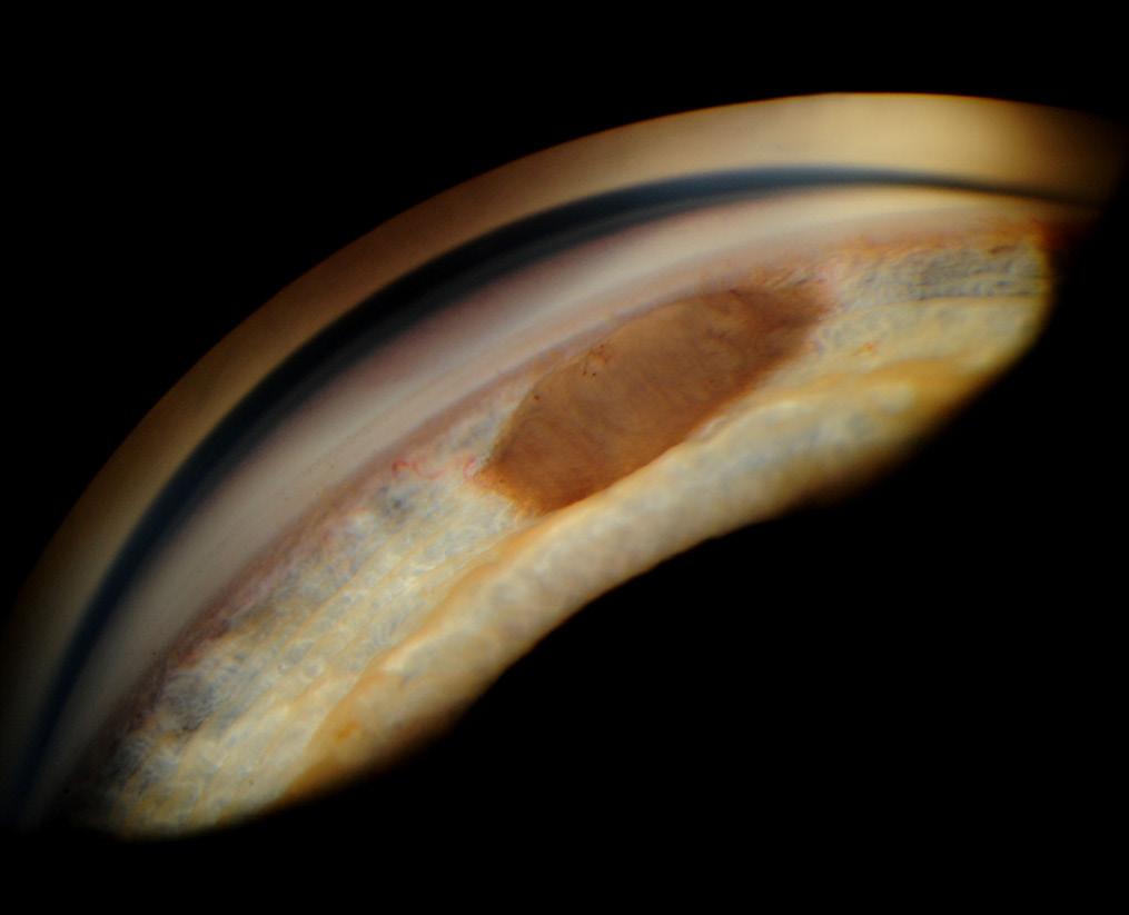
Gonio Photography
Iris Tumor
James Gilman, CRA, FOPS
Moran Eye Center
Salt Lake City, Utah
Fluorescein Angiography

PDR (AV Phase)
Bradley Stern, CRA, OCT-C
Henry Ford Health System
Detroit, Michigan
Monochromatic Photography
Congenital Retinal Macrovessel
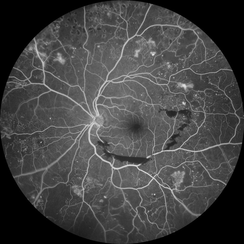
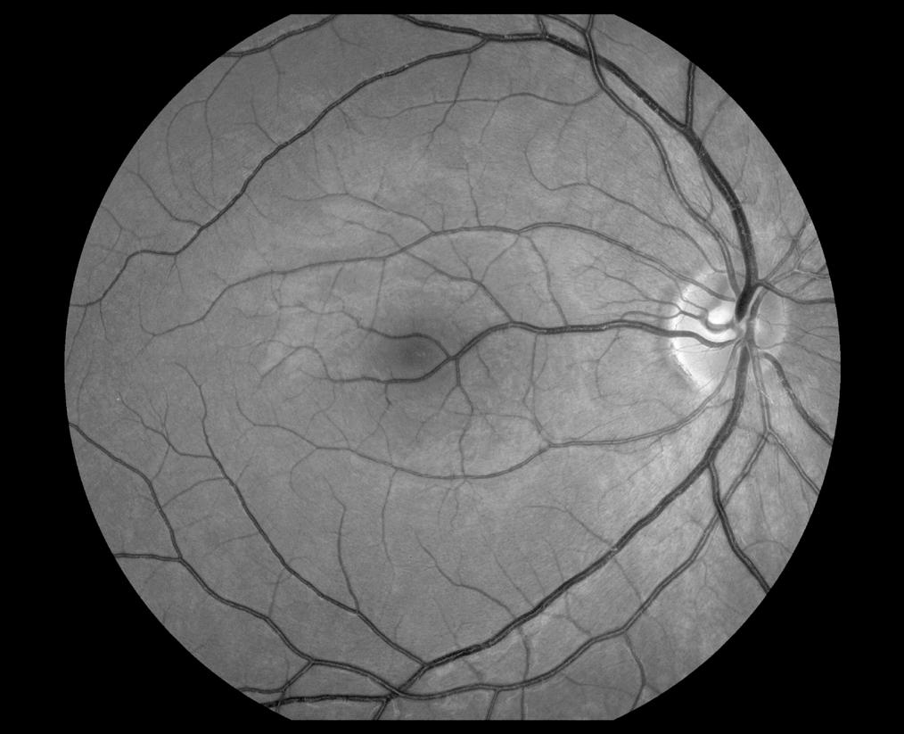

Robert Mays, CRA, OCT-C
National Eye Institute
Bethesda, Maryland
The Journal of Ophthalmic Photography Volume 45, Issue 1 • 2023 18
2022 OPS Scientific Exhibit
First Place Winners: Print Division
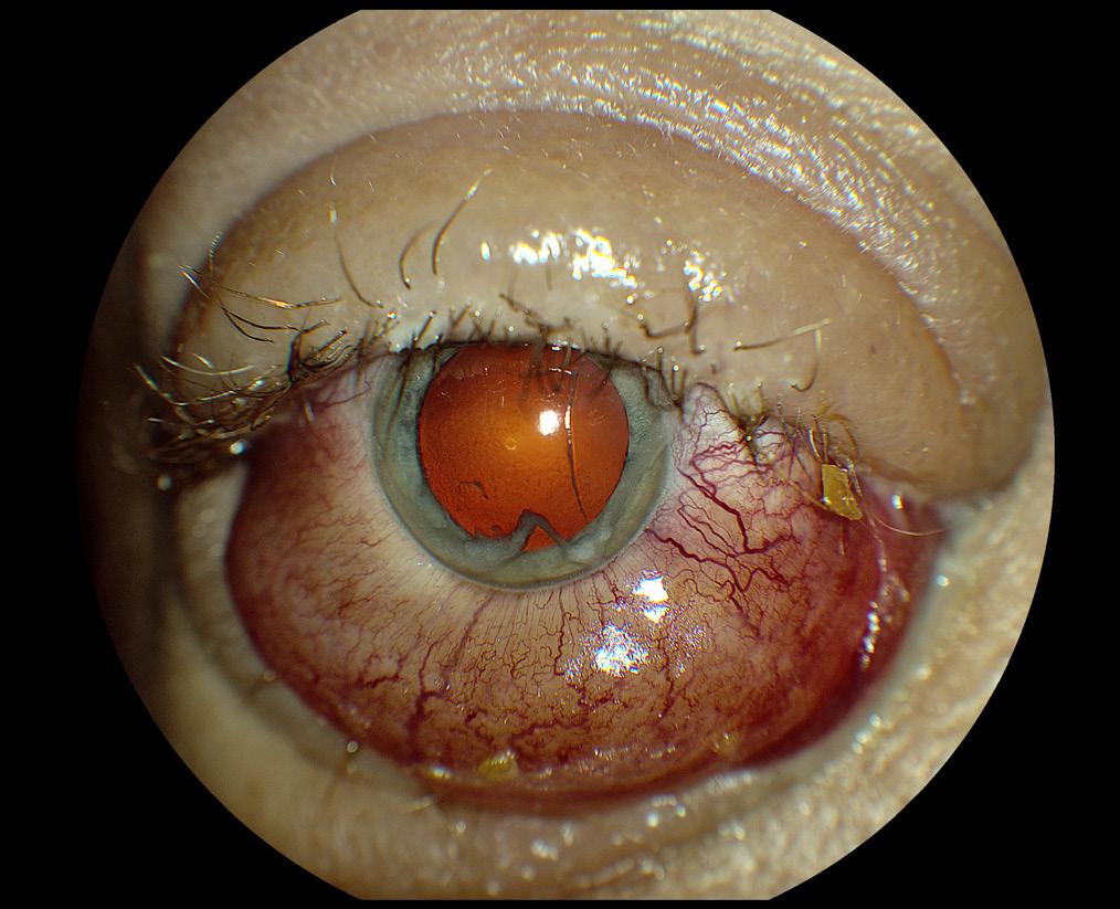
External Photography
Complete Ophthalmoplegiea
Sarah Skiles, CRA
University of Iowa Hospitals and Clinics
Iowa City, Iowa
The Eye as Art Proliferative Diabetic Retinopathy


Jody Troyer, CRA
University of Iowa Hospitals and Clinics
Iowa City, Iowa
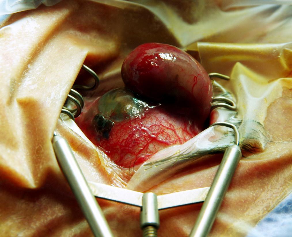
Gross Specimen Photography
Uveal Melanoma with Extra Ocular Extension

Belinda Rodriquez
Murray Ocular Oncology and Retina
Miami, Florida
Composite Image
Combined Hamartoma
Jason P. Hall, CRA, OCT-C Mayo Clinic
Rochester, Minnesota
2022 OPS Scientific Exhibit 19
2022 OPS Scientific Exhibit
First Place Winners: Print Division
Optical Coherence Tomography
Endothelial Transplant after DMEK
Barbara Klemenc
University Eye Hospital Ljubljana
Ljubljana, Slovenia
Optical Coherence Tomography
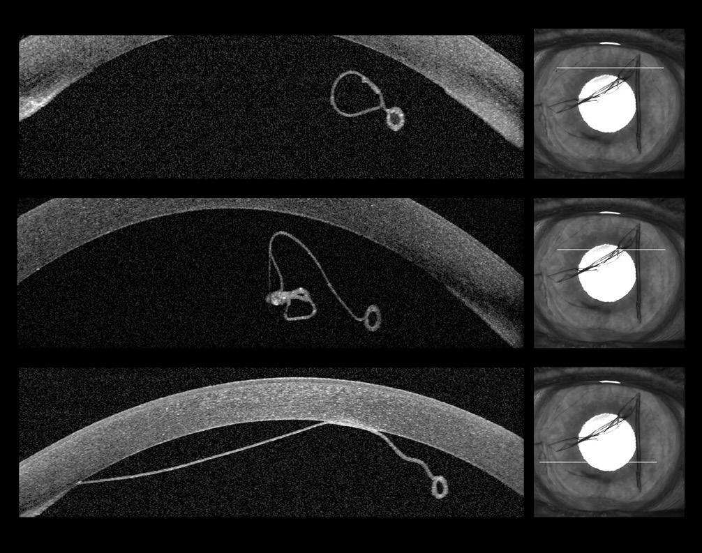
MEK Retinopathy

Barbara Klemenc
University Eye Hospital Ljubljana
Ljubljana, Slovenia
OCT Angiography
Untitled
Jody Troyer, CRA
University of Iowa Hospitals and Clinics
Iowa City, Iowa
OCT Angiography

Myopic Choroidal Neovascularization
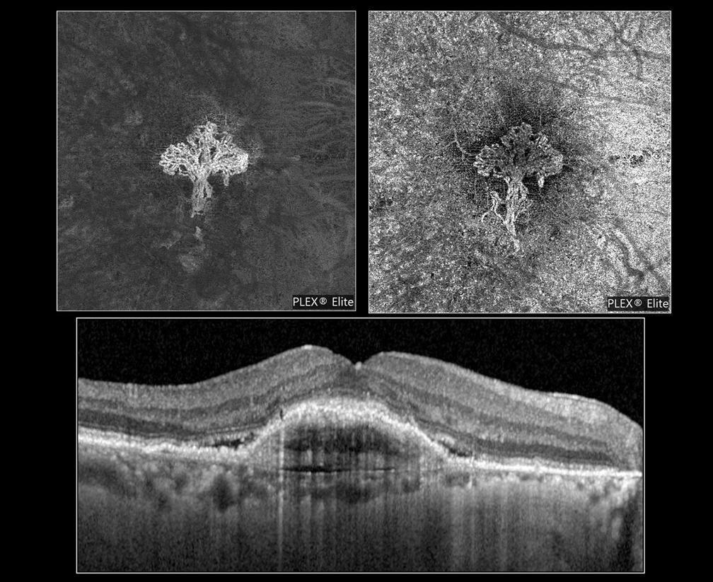

Kasi Sandhanam
Singapore National Eye Centre
Singapore
The Journal of Ophthalmic Photography Volume 45, Issue 1 • 2023 20
2022 OPS Scientific Exhibit
First Place Winners: Print Division
Fundus Autofluorescence

Submacular Hemorrhage

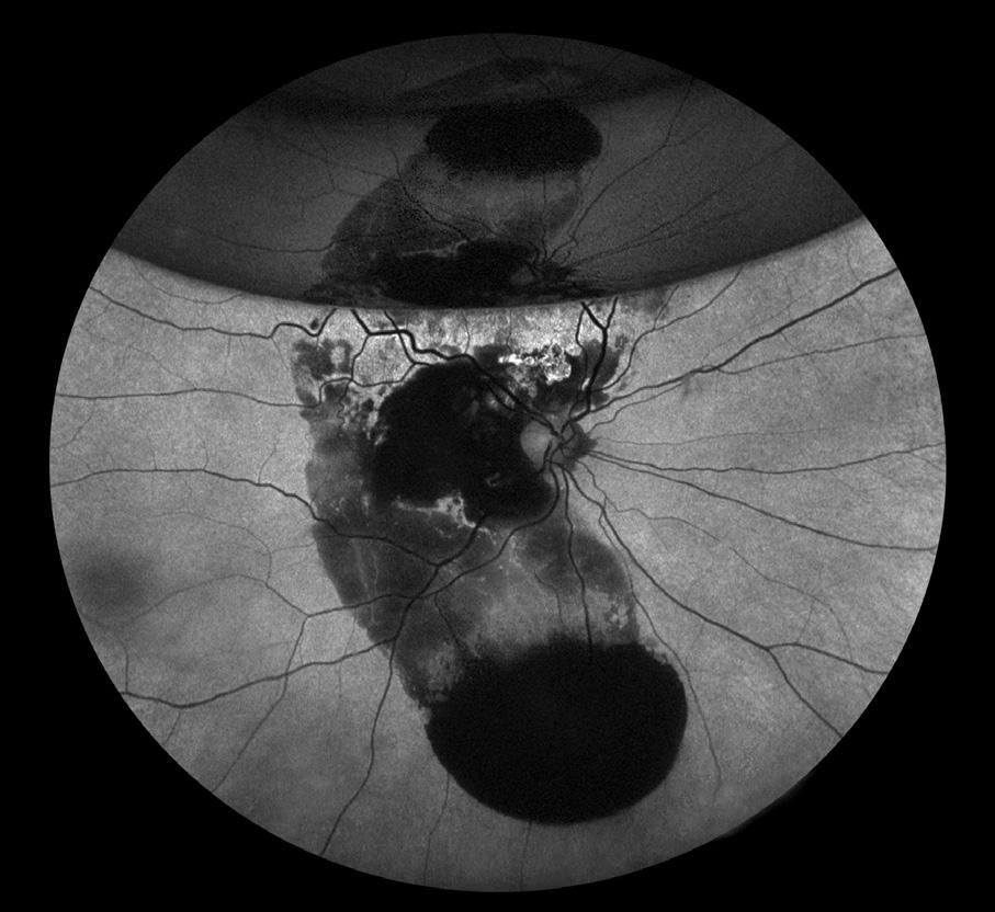
Dena Harris, CRA
University of Michigan Kellogg Eye Center
Ann Arbor, Michigan
Ultra-Widefield Imaging
Hollenhorst Plaque
Veronica T. Jones OCT-C, CPT
Duke Eye Center
Durham, North Carolina
Photo/Electron Micrography
Histology of Outer Retina, RPE & Choroid
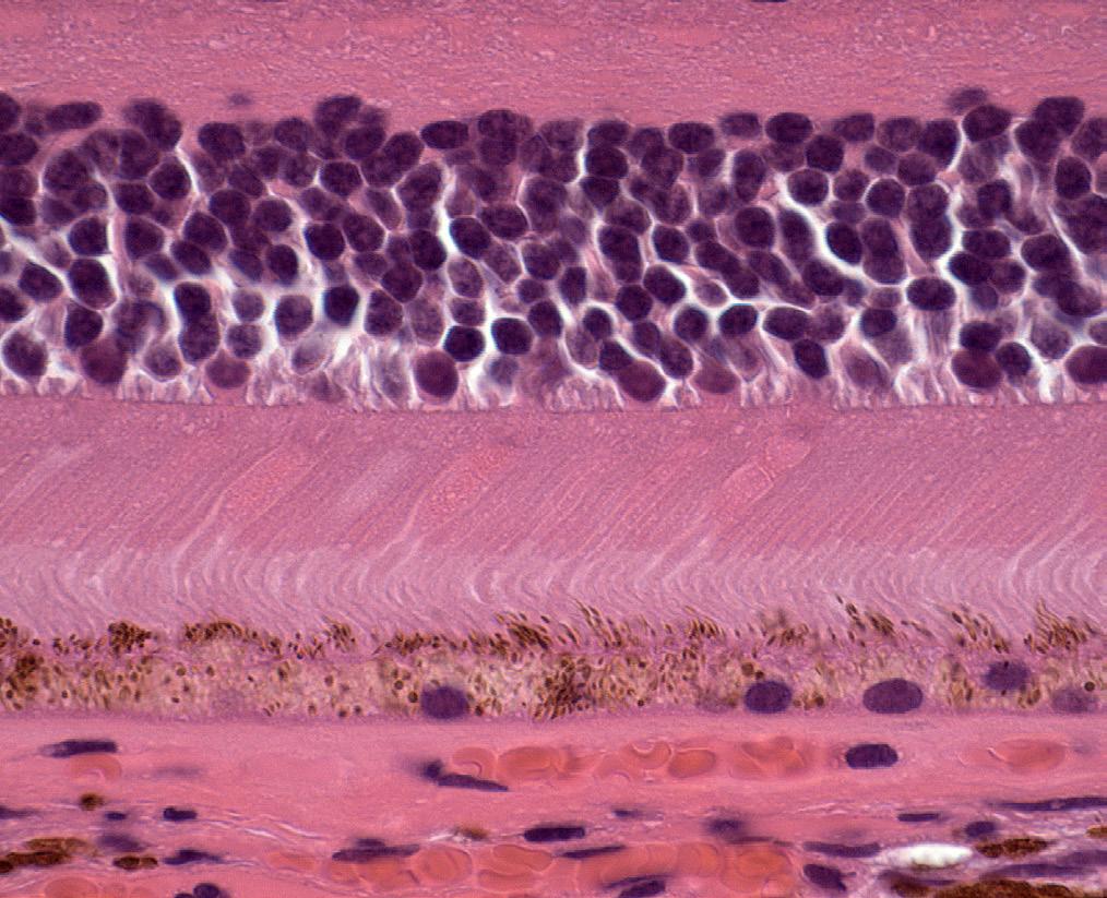

Ralph Eagle Jr., MD
Wills Eye Hospital
Philadelphia, Pennsylvania
Clinical Setting
Untitled
Ralph Eagle Jr., MD
Wills Eye Hospital
Philadelphia, Pennsylvania
2022 OPS Scientific Exhibit 21
2022 OPS Scientific Exhibit
Second Place Winners: Print Division
Fluorescein Angiography
CRAO
Bradley Stern
Henry Ford Health System
Detroit, Michigan
Fluorescein Angiography

CRVO
Timothy Costello CRA, COA Kellogg Eye Center
Ann Arbor, Michigan
Indocyanine Green Angiography
Prominent Choroidal Vessel

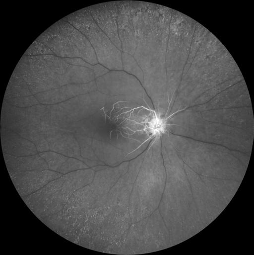
Jaime Tesmer CRA, OCT-C Mayo Clinic
Rochester, Minnesota
Fundus Photography Normal 30°- 40°

Cat Scratch with Mac Star
Joseph Morales, CRA, OCT-C
UT Southwestern
Dallas, Texas
Slit Lamp Photography
Corneal Ulcer
Melissa Field, COA
Flaum Eye Institute
Rochester, New York
Fundus Photography Wide Angle 45°+ Choroidal Neovascular Membrane

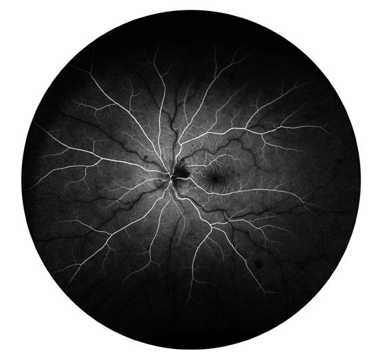
Jody Troyer, CRA
University of Iowa Hospitals and Clinics Iowa City, Iowa
External Photography
Corneal Ulcer


Ben Serar, CRA, M.A. Serar Photography
Prescott, Arizona
Slit Lamp Photography
Lens Pigment Deposits

Dena Harris, CRA
Univ of Michigan Kellogg Eye Center
Ann Arbor, Michigan
Slit Lamp Photography

Dislocated Lens
Megan Walsh, CRA, OCT-C Eyecare Medical Group
Portland, Maine
The Journal of Ophthalmic Photography Volume 45, Issue 1 • 2023 22
2022 OPS Scientific Exhibit
Second Place Winners: Print Division
Photo/Electron Micrography Growth on Conjunctiva

Joseph Morales, CRA, OCT-C UT Southwestern Dallas, Texas
Photo/Electron Micrography
Retinoblastoma
Ralph Eagle Jr., MD Wills Eye Hospital
Philadelphia, Pennsylvania
Gross Specimen Photography
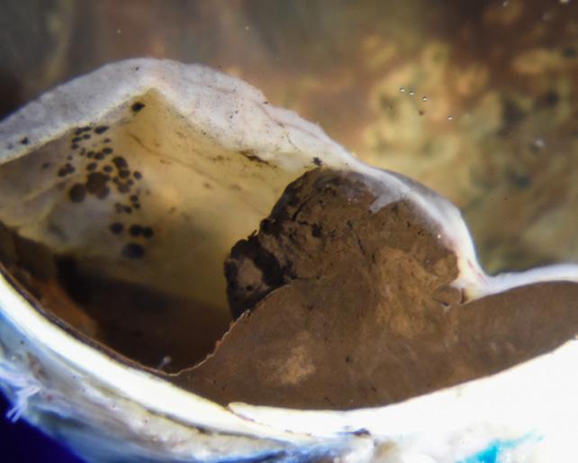
Uveal Melanoma w/RD and Subretinal Seeds
Ralph Eagle Jr., MD Wills Eye Hospital, Philadelphia, PA
Composite Image
Preretinal Hemorrhage in PDR
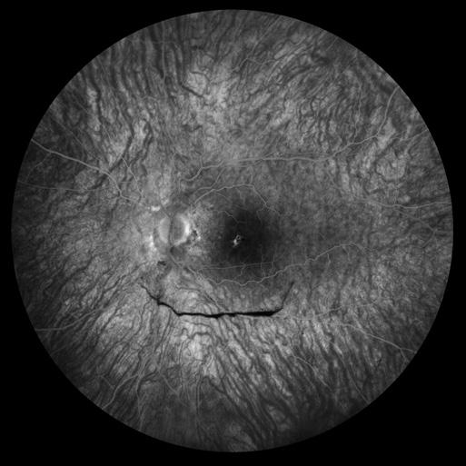
Jason P. Hall, CRA, OCT-C Mayo Clinic
Rochester, Minnesota
The Eye as Art Galax (eye)

Meghan Menzel, CRA
University of Iowa Hospitals and Clinics Iowa City, Iowa
The Eye as Art
Lisa Frank’s IOL
Nicole Radunzel, CRA
University of Iowa Hospitals and Clinics
Iowa City, Iowa
Fundus Autofluorescence
MIDD Related Maculopathy

Barbara Klemenc
University Eye Hospital Ljubljana
Ljubljana, Slovenia
Fundus Autofluorescence


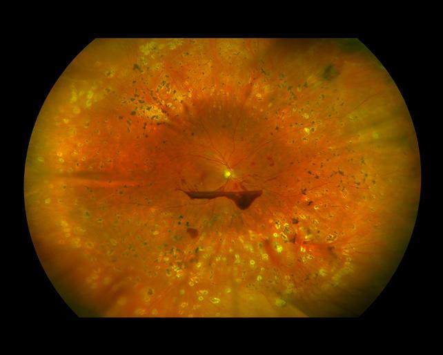
Choroideremia
Dena Harris, CRA
Univ of Michigan Kellogg Eye Center
Ann Arbor, Michigan
Ultra-Widefield Imaging
Vitreous Hemorrhage

Dena Harris, CRA
Univ of Michigan Kellogg Eye Center
Ann Arbor, Michigan
2022 OPS Scientific Exhibit 23
2022 OPS Scientific Exhibit

Second Place Winners: Print Division
Gonio Photography
Iris Lesion
Melissa Field, COA
Flaum Eye Institute
Rochester, New York
Cross Categories
Wyburn Mason Syndrome
Megan Menzel, CRA
Univ of Iowa Hospitals and Clinics
Iowa City, Iowa
Cross Categories
Retinal Detachment w Multiple Breaks

Sarah Skiles, CRA
Univ of Iowa Hospitals and Clinics
Iowa City, Iowa
Gonio Photography
Iris Tumor
James Gilman, CRA, FOPS Moran Eye Center Salt Lake City, Utah
Optical Coherence Tomography


Ectasia of Cornea
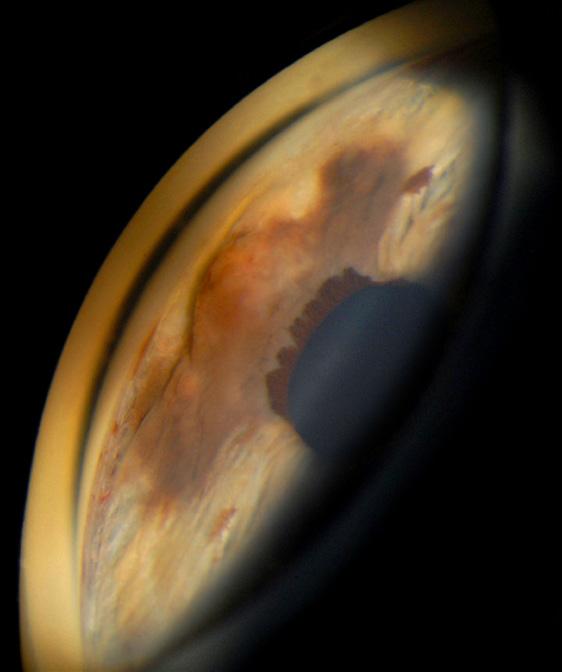


Tim Steffens, CRA, OCT-C, FOPS
University of Michigan Kellogg Eye Center Ann Arbor, Michigan
The Journal of Ophthalmic Photography Volume 45, Issue 1 • 2023 24
Third Place/Honorable Mention: Print Division
Fluorescein Angiography
3rd: Choroidal Malignant Melanoma, Dena Harris, CRA
3rd: PDR, Jason P. Hall, CRA, OCT-C
3rd: PDR, Jason P. Hall, CRA, OCT-C
HM: PDR, Jaime Tesmer, CRA, OCT-C
HM: Untitled, Jody Troyer, CRA
HM: Von Hippel Lindau Syndrome, Nicole Radunzel, CRA
HM: DME w/ PDR, Timothy Costello, CRA, COA
Indocyanine Green Angiography
HM: Unusual Atrophic Changes Around Optic Nerve After Meningitis, Barbara Klemenc
Fundus Photography High Magnification 20°
HM: Cotton Wool Spots on Nerve, Meghan Menzel, CRA
HM: Tortuous Veins on the Optic Nerve, Joseph Morales, CRA, COA
Fundus Photography Normal 30°-40°
3rd: Laser Burn, Meghan Menzel, CRA
3rd: Congenital Anomaly of the Optic Nerve, Sarah Skiles, CRA
HM: Juxtapapillary Astrocytic Hamartoma, Sarah Skiles, CRA
HM: Asteroid Hyalosis, Ben Serar, MA, CRA
HM: Best Disease, Megan Menzel, CRA
Fundus Photography Wide Angle 45°+
3rd: PDR with Severe CME, Jody Troyer, CRA
3rd: Adult Onset Vitelliform Dystrophy, Sarah Skiles, CRA
Slit Lamp Photography
3rd: Corneal Bubbles, Dena Harris, CRA
3rd: Lattice Dystrophy, Debra Cantrell, OCT-C, COA
HM: Dermis Fat Graft, Shannon Howard, CDOS, ROUB, COA
HM: Corneal Sutures, Katie Lachut-Yevich
External Photography
HM: Conjunctival Papilloma, Nicole Radunzel, CRA
Gonio Photography
HM: S/P Iris Melanoma Removal with Sutured Iris, Jason P. Hall, CRA, OCT-C
Monochromatic Photography
HM: Retinal Capillary Hemangioblastoma, Robert Mays, CRA, OCT-C
Gross Specimen Photography
3rd: Lens Molding, Ralph Eagle Jr., MD
HM: Enucleated Specimen, Scleral Shell, Ciliary Body, Belinda Rodriguez
Surgical Photography
HM: Iris Melanoma Plaque Brachytherapy, Shannon Howard, CDOS, ROUB, COA
Composite
3rd: AMPEE, Jody Troyer, CRA
The Eye as Art

HM: Vitreous Hemorrhage Tree, Jaime Tesmer, CRA, OCT-C
HM: Coloboma, Jody Troyer, CRA
HM: Macular Schisis, Kasi Sandhanam
Cross Categories
HM: Choroidal Rupture, Nicole Radunzel. CRA
Optical Coherence Tomography
3rd: Optic Disc Pit, Kasi Sandhanam
HM: Pellucid Marginal Degeneration of Cornea, Jody Troyer, CRA
OCT – Angiography
3rd: Diabetic Retinopathy with Neovascularization, Kasi Sandhanam
HM: Choroideremia, Jody Troyer, CRA
Fundus Autofluorescence
HM: Multifocal Choroiditis with many Scars, Sarah Skiles, CRA
HM: Serpiginous Choroiditis, Sarah Skiles, CRA
Ultra-Widefield Imaging
3rd: Scleral Buckle, Alexandra Copple
HM: Retinitis Pigmentosa, Becky Weeks, CRA
HM: PDR w Chorioretinal Scar, Bradley A. Stern, CRA, OCT-C
HM: Chronic Temporal Schisis Detachment, Sarah Skiles, CRA
Instrumentation
HM: Unusual Contact Lens, Sarah Skiles, CRA
HM: Pediatric Intacs, Removed, Katie Lachut-Yevich
2022 OPS Scientific Exhibit 25
2022 OPS Scientific Exhibit
2022 OPS Scientific Exhibit
First Place Winners: Stereo Division
Gonio Photography
Iridectomy s/p Iris Melanoma

Tim Steffens, CRA, OCT-C, FOPS
Kellogg Eye Center, Ann Arbor, Michigan
Slit Lamp Photography
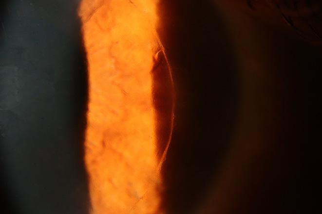


Scleritis Due to Granulomatosis


Sarah Skiles, CRA
University of Iowa
Hospitals and Clinics
Iowa City, Iowa
External Photography
Neoplasm of Uncertain Behavior
Dena Harris, CRA
University of Michigan
Kellogg Eye Center
Ann Arbor, Michigan
Tim Steffens, CRA, OCT-C, FOPS
University of Michigan
Kellogg Eye Center
Ann Arbor, Michigan
The Journal of Ophthalmic Photography Volume 45, Issue 1 • 2023 26
CSABA MARTONYI BEST OF SHOW AWARD and BEST OF STEREO DIVISION
The Eye as Art DSEK
2022 OPS Scientific Exhibit
First Place Winners: Stereo Division
Fluorescein Angiography
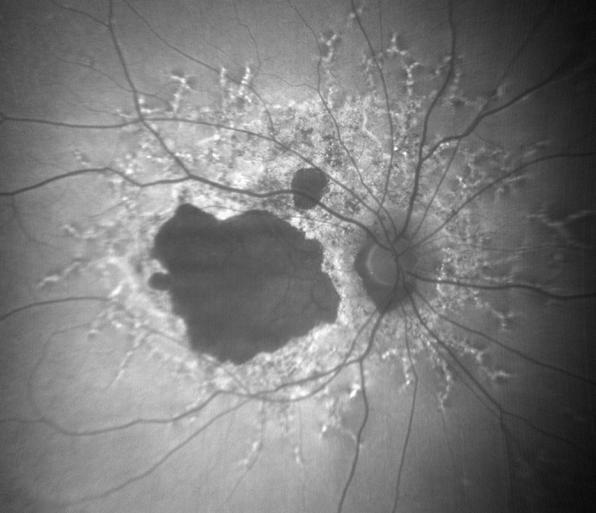

Subretinal Fluid with Macular Oedema
Kasi Sandhanam
Singapore National Eye Centre
Singapore
Autofluorescence Photography
Stargardts
Tony Medina, CRA, FOPS
University of Michigan
Kellogg Eye Center
Grand Blanc, Michigan
Indocyanine Green Angiography
Choroidal Neovasularization


Kasi Sandhanam
Singapore National Eye Centre
Singapore
Monochromatic Photography
Terson Syndrome with Preretinal Hemorrhage
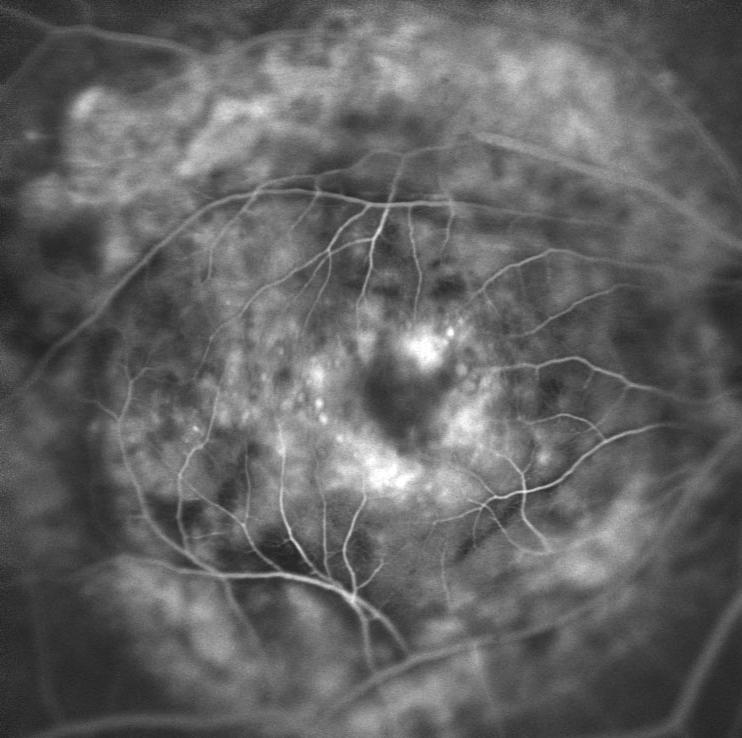


Kasi Sandhanam
Singapore National Eye Centre
Singapore
2022 OPS Scientific Exhibit 27
2022 OPS Scientific Exhibit
First Place Winners: Stereo Division
Fundus Photography High Magnification 20° Optic Disc Coloboma
Pabla Gili, MD Hospital Universitario
Fundación Alcorcón Madrid, Spain
Fundus Photography Normal 30°- 40° Optic Disc Drusen
Nicole Radunzel, CRA
University of Iowa Hospitals and Clinics
Iowa City, Iowa
Ultra-Widefield Imaging
Sub Retinal Fluid

Tony Medina, CRA, FOPS
University of Michigan
Kellogg Eye Center
Grand Blanc, Michigan
Fundus Photography Wide Angle 45°+ Choroidal Melanoma with a Collar-Button Configuration
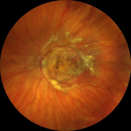


Nicole Radunzel, CRA University of Iowa Hospitals and Clinics Iowa City, Iowa

The Journal of Ophthalmic Photography Volume 45, Issue 1 • 2023 28
2022 OPS Scientific Exhibit
First Place Winners: Stereo Division
Instrumentation Photography
Unusual Contact Lens


Sarah Skiles, CRA
University of Iowa
Hospitals and Clinics
Iowa City, Iowa
Composite Optic Nerve Lesion

Dena Harris, CRA
Jaime Tesmer, CRA, OCT-C Mayo Clinic
Rochester, Minnesota
Clinical Setting Photography
Autologous Blood Injection


Tony Medina, CRA, FOPS
University of Michigan
Kellogg Eye Center
Grand Blanc, Michigan
Cross Categories
Choroidal Melanoma


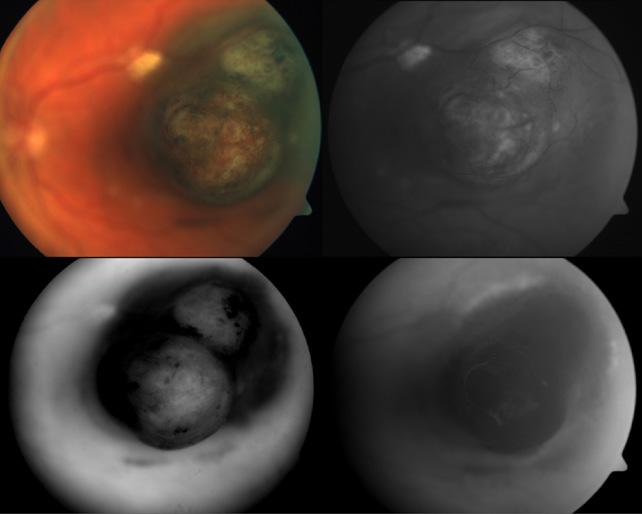
Pabla Gili, MD
Hospital Universitario
Fundación Alcorcón
Madrid, Spain
2022 OPS Scientific Exhibit 29
2022 OPS Scientific Exhibit
Second Place Winners: Stereo Division
Fluorescein Angiography
Choroidal Melanoma
Pablo Gili, MD
Hospital Universitario Fundación Alcorcón, Madrid, Spain
Fluorescein Angiography


Malignant Melanoma
Tim Steffens, CRA, OCT-C, FOPS
University of Michigan Kellogg Eye Center, Ann Arbor, Michigan
Indocyanine Green Angiography
Choroidal Melanoma
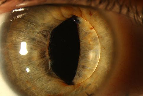


Pablo Gili, MD
Hospital Universitario Fundación Alcorcón, Madrid, Spain
Fundus Photography High Magnification 20°

Metal in Eye


Tony Medina, CRA, FOPS
Univ of Michigan Kellogg Eye Center, Grand Blanc, Michigan
Fundus Photography Normal 30°-40°
Untitled
Tim Steffens, CRA, OCT-C, FOPS
University of Michigan Kellogg Eye Center, Ann Arbor, Michigan
Fundus Photography Wide Angle 45°+ ERM
Angela Chappell
Flinders Medical Centre, South Australia
Slit Lamp Photography
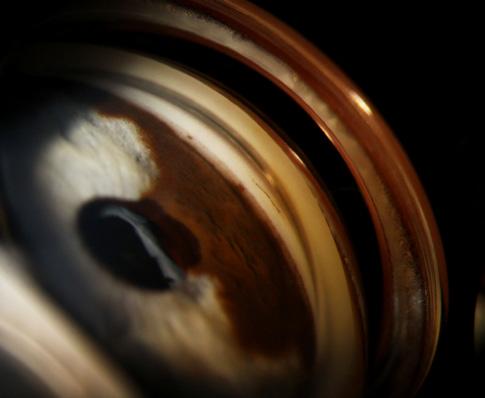

Lens Dislocation
Dena Harris, CRA
University of Michigan Kellogg Eye Center, Ann Arbor, Michigan
Gonio Photography
Iris Lesion
Jody Troyer, CRA
University of Iowa Hospitals and Clinics, Iowa City, Iowa
The Journal of Ophthalmic Photography Volume 45, Issue 1 • 2023 30
2022 OPS Scientific Exhibit
Second Place Winners: Stereo Division
External Photography
Scleroderma
Dena Harris, CRA
University of Michigan Kellogg Eye Center, Ann Arbor, Michigan
Composite
Optic Nerve Disorder


Jason P. Hall, CRA, OCT-C Mayo Clinic, Rochester, Minnesota
Monochromatic Photography
Posterior Vitreous Detachment


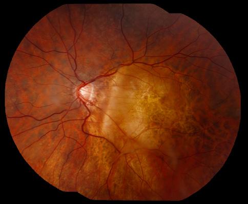


Kasi Sandhanam
Singapore National Eye Center, Singapore
Monochromatic Photography
Papilledema


Pablo Gili, MD
Hospital Universitario Fundación Alcorcón, Madrid, Spain
Ultra-Widefield Imaging
Choroidals
Tony Medina, CRA, FOPS
Univ of Michigan Kellogg Eye Center, Grand Blanc, Michigan
2022 OPS Scientific Exhibit 31
2022 OPS Scientific Exhibit
Third Place/Honorable Mention: Stereo Division
Fluorescein Angiography
HM: Hamartoma, Jason P. Hall, CRA, OCT-C
Indocyanine Green Angiography
3rd: Polypoidal Choroidal Vasculopathy, Kasi Sandhanam

HM: Polypoidal Choroidal Vasculopathy , Pablo Gili, MD
Fundus Photography High Magnification 20°
3rd: Optic Nerve Neovascularization, Jaime Tesmer, CRA, OCT-C
HM: Disc Edema from Mass Effect, Jason P. Hall, CRA, OCT-C
Fundus Photography Normal 30°-40°
3rd: Giant Retinal Tears & Inverted Retina, Pablo Gili, MD
3rd: Myopic Peripapillary Staphyloma, Pablo Gili, MD
HM: Vitreous Mass, Ben Serar, MA, CRA
Fundus Photography Wide Angle 45°+
3rd: Mushroom-Shaped Juxtapapillary Choroidal Melanoma, Nicole Radunzel, CRA
HM: Serous Detachment around Optic Nerve, Jaime Tesmer, CRA, OCT-C
Gonio Photography
3rd: Iris Lesion, Jody Troyer, CRA
Coming Next Issue
Under the Surface: A Clear Look into the Cornea
Slit Lamp Photography
3rd: Corneal Ulcer, Tim Steffens, CRA, OCT-C, FOPS
HM: Acute Anterior Uveitis with Synechia, Tim Steffens, CRA, OCT-C, FOPS

HM: Anterior Capsular Phimosis with Dislocated IOL, Dena Harris, CRA
External Photography
3rd: Conjunctival Lesion, Dena Harris, CRA
HM: s/p Wide Excision of Melanoma, Tim Steffens, CRA, OCT-C, FOPS
Monochromatic Photography
HM: Optic Disc Coloboma, Pablo Gili, MD
Composite
3rd: Extensive Neovasculariztion, Jaime Tesmer, CRA, OCT-C
HM: ERM with Star Fold in Temporal Macula/Mid Periphery, Tony Medina, CRA, FOPS
Ultra-Widefield Imaging
3rd: Familial Exudative Vitreopathy, Tim Steffens, CRA, OCT-C, FOPS
HM: Tractional Fibrosis, Tony Medina, CRA, FOPS
The Journal of Ophthalmic Photography Volume 45, Issue 1 • 2023 32
Corneal Epithelial Ingrowth, image courtesy of Kathleen Warren, 3rd place, 2022 Haag-Streit International photo competition.
Kevin Aguirre Gamarra
 Erin A Hisey
Brian C Leonard
Department of Surgical and Radiological Sciences
School of Veterinary Medicine
University of California, Davis Davis, CA 95616
Corresponding Author: BC Leonard
275 Medical Sciences Drive
Davis, CA 95616
bcleonard@ucdavis.edu
530.752.3676
Dynamic Tilt Contact Angle Goniometry of Animal Globes
These images are representative of dynamic tilt contact angle goniometry of animal globes. Figures 1a and 1b show a globe submerged in quartz environmental chamber filled with PBS, one from a rabbit (a) and the other one from a mouse (b). Figure 1b depicts the mouse globe in the tilting stage. Figures 1c-f were taken after a 10 uL droplet of perfluorodecalin was deposited on two knockout mouse globes. Globes were enucleated within three hours of euthanasia and mounted on glass slides with the axial cornea facing upward, perpendicular to the slide. The goniometer stage was titled by 0.5° per second until the droplet was displaced. Pictures chosen for measurement were taken right before detachment (Figures 1d and 1f). These images were captured on a Ramé-Hart 290 goniometer (Ramé-Hart Instruments, Succasunna, NJ). The aim of these experiments was to assess the adherence capability of the ocular surface in acyl-CoA: wax alcohol acyltransferase 2 (Awat2) knockout mice. This gene encodes
an enzyme which is critical for the synthesis of long-chain wax esters of the tear film by the meibomian glands, thus a deficiency in Awat2 leads to tear film abnormalities. Normally, the tear film lipid layer provides lubrication and protection to the cornea. However, if this layer is altered, ocular surface irritation and inflammation can develop. Measurements of droplet attachment duration, tilt of the stage, and difference in advancing (Figure 1d, marked in red), receding (Figure 1d, marked in yellow), and hysteresis (difference between the previous two) angles were taken for this study. When comparing the two mice (Figures 1c and 1d vs Figures 1e and 1f), the most notable difference is the presence of a darker lesion on the surface of Figures 1e and 1f (blue arrow) and a cloudier corneal appearance, which indicate corneal degeneration due to an abnormal tear film. This abnormal tear film causes a loss in the adherence properties of the ocular surface, leading to clinical signs of evaporative dry eye disease (EDED).

33 Dynamic Tilt Contact Angle Goniometry of Animal Globes
Article Figure 1 a b c d e f
Original
Case Report
Monica Motta, BS RVT, LATG

Andrea Minella, DVM
Piyaporn Eiamcharoen, DVM
Brian Murphy, DVM
Christopher Reilly, DVM
Christopher Murphy, DVM, PhD
Danielle K Tarbert, DVM, DACZM
UC Davis School of Veterinary Medicine
Comparative Ophthalmology
Vision Science Lab
Davis, CA 95616
mjmotta@ucdavis.edu
Unique and Dazzling Ocular Clinical Case of a Great Horned Owl


Examination Findings
Ajuvenile Great Horned Owl found down was brought to the UC Davis Veterinary Medical Teaching Hospital by a good Samaritan. On initial assessment, the patient presented as bright and alert. On ophthalmic examination, the corneas and lens were clear, with trace pigmented cells found in the anterior chamber. Both pupils were miotic with a yellow-brown iris color observed with intermittent anisocoria. A direct PLR was present in both eyes. On posterior examination, the vitreous was normal with multifocal hyper-pigmented foci with sharp borders observed in the fundus. No significant findings were observed in the pecten visible. Optical
coherence tomography using the Heidelberg Spectralis OCT revealed bilateral multifocal retinal detachments. The en-face image confirmed an intact, normal pecten.

Histopathology Findings
Histopathological findings revealed bilateral multifocal retinal separation and retinal epithelial hyperplasia, in addition to photoreceptor attenuation, consistent with retinal detachment. Lymphocytes infiltrated the choroidal and pecten vessels. The black pigmentation of the iris was attributed to a densely melanotic posterior pigmented iris epithelium.
The Journal of Ophthalmic Photography Volume 45, Issue 1 • 2023 34
Figure 1: Normal great horned owl Iris and pupil.
Figure 2: Affected great horned owl Iris and pupil.
Figure 3: Heidelberg OCT image (OCT-IR, Cube) of abnormal and detached retinal layers and normal pecten.
Diving into Whales’ Ability to Tolerate Low Oxygen
By Giselle Wang and Angela Spivey
Imagine you take a long deep breath after breakfast, then hold it. You go about your day, walk to classes or work, all without taking another breath. Finally, just before lunch, having exhausted all the oxygen in your body, you exhale and then inhale more precious air.
For the Cuvier’s beaked whale, an elusive deepdiving marine mammal, this is just routine. In 2020, a group of researchers from Duke University recorded the longest dive ever for this species — 3 hours and 42 minutes — and published their findings in the Journal of Experimental Biology.
Now Duke cancer biologist Jason Somarelli, PhD, will collaborate with this group of Duke marine scientists to explore the cellular adaptations these whales’ possess that gives them such an amazing ability to tolerate low-oxygen (hypoxic) environments as they dive and forage for food. “These whales are worldrecord divers; they dive down the equivalent of seven Empire State Buildings,” said Somarelli, assistant professor in medicine.
Somarelli’s lab has used his molecular biology expertise to create new cell culture models of whales, using small tissue samples that the marine lab researchers collect as part of their field work.
With funding received in December 2022 from the United States Office of Naval Research, he will use those cell culture models to study the cellular and molecular mechanisms involved when whales hold their breath, and how stress from ship noise may affect this ability at a cellular level.

Somarelli hopes the study will reveal ways to protect whales. It may also provide clues to combating human health challenges that involve low-oxygen states, he said.
Previously published studies have shown that man-made noise levels in the ocean have doubled every decade since the 1950s, mainly from ship traffic, seismic surveys, and industrial activities, he said. In the aftermath of the 9/11 attacks, ship traf-
fic decreased dramatically in the Atlantic Ocean, making for a much quieter environment underwater. This presented an ideal natural experiment for marine biologists, who observed that anthropogenic noise can induce stress in whales.
Somarelli will work closely with Andy Read, PhD, director of the Duke Marine Lab; Andreas Fahlman, PhD, and Tom Schultz, PhD, of Duke’s Nicholas School of the Environment; and Nicola Quick, PhD, of the University of Plymouth. “We might be able to figure out how whales tolerate these low oxygen environments and how stress interplays with that,” Somarelli said. “We think that our findings can inform which species we focus on for conservation.”
As a surfer and husband to a Duke-educated marine biologist, Meagan Dunphy-Daly, PhD, Somarelli has always felt a connection to the ocean. He and Dunphy-Daly started the Scholars in Marine Medicine Program at Duke to offer undergraduate students research experience at the intersection of medicine and marine science.
The experienced team of undergraduates and researchers at Somarelli’s lab will expose the whale cell cultures to various conditions and observe how the cells change in response to their environment.
“We can modulate the stress signals and levels of oxygen we expose these cells to in a rigorous way through controlled lab experiments,” Somarelli said.
Low-oxygen states are relevant to many human health challenges, including heart attack, stroke, COVID-19, prolonged surgical procedures, and cancer, Somarelli said. In the long term, studying how whales tolerate low-oxygen environments could help lead to discovery of a druggable target that could be manipulated to help humans better tolerate hypoxia.
Giselle Wang is recent Duke University graduate who interned in the lab of Jason
Somarelli, PhD. Angela Spivey is a senior science writer in the School of Medicine’s Office of Strategic Communications.
35 Diving into Whales’ Ability to Tolerate Low Oxygen
Image courtesy of pixnio.com, CC0
Case Report
Tzu-Ni Sin Glenn Yiu

Department of Ophthalmology and Vision Science
University of California, Davis Davis, CA 95616
Corresponding Author: Glenn
Department of Ophthalmology and Vision Science
University of California, Davis 4860 Y St., Suite 2400
Sacramento, CA, 95817 916.734.6602
gyiu@ucdavis.edu
Purtscher Retinopathy in Nonhuman Primate
Purtscher retinopathy is often associated with blunt trauma, but may also occur in other systemic conditions like acute pancreatitis, hemolytic uremic syndrome, and renal failure where acute complementmediated leukoembolization likely contributes to the typ-
 Yiu, MD, PhD
ical appearance of cotton-wool spots, retinal hemorrhage, optic disc edema, and inner retinal whitening. Affected patients also experience reduced vision acuity with possibilities of eyesight recovery, depending on the disease progression in other affected organs.1 However, the
The Journal of Ophthalmic Photography Volume 45, Issue 1 • 2023 36
a c d e b
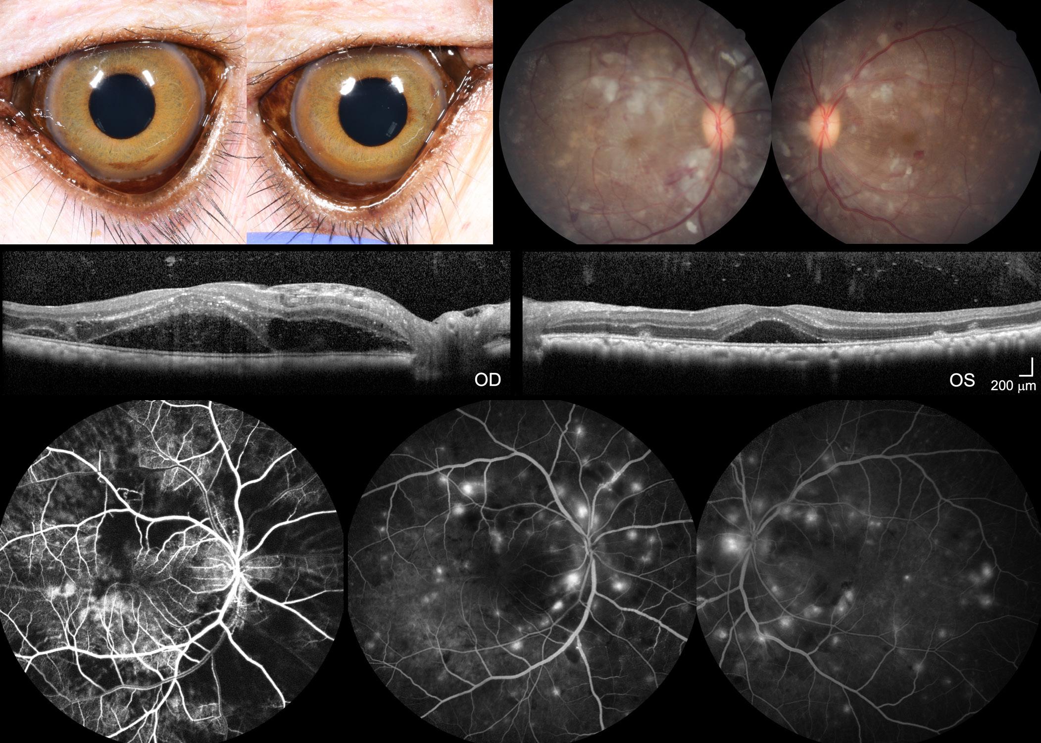
Figure 1: Multimodal imaging of NHP Purtscher-like retinopathy. (a) External photographs, (b) color fundus photographs (FP), (c) spectral-domain optical coherence tomography (OCT), (d) early-phase fluorescein angiography (FA), and (e) late-phase FA of the left and right eyes of the affected macaque during the clinical eye examination at 16 years old. Abbreviations: OD, right eye; OS, left eyes. Scale bars, 200µm.
pathogenesis of Purtscher retinopathy remains unclear. Here, we report a unique case of Purtscher retinopathy in a nonhuman primate (NHP) with multiple body system complications found at necropsy.

A 16-year-old female rhesus macaque (Macaca mulatta) was diagnosed with severe Purtscher-like retinopathy during a routine eye examination. Intraocular pressures were 7 and 8 mmHg for the right and left eye, respectively, but with no other anterior segment abnormalities (Figure 1a). Fundus examination and photography revealed diffuse cotton-wool spots bilaterally extending across both macula and periphery, which corresponded to the nerve fiber layer (NFL) thickening on optical coherence tomography (OCT) suggesting NFL infarction (Figures 1b and 1c). OCT also showed severe retinal edema with intraretinal and subretinal fluid which was more prominent in the right eye (Figure 1c). Intraretinal hyperreflective foci were also apparent on OCT, suggesting intracellular infiltrates that are likely inflammatory in nature. Early-phase fluorescein angiography (FA) of the right eye showed choroidal filling defects suggestive of
patchy choroidal ischemia while late-phase FA exhibited multiple foci of leakage, suggestive of increased retinal vessel permeability in both eyes (Figures 1d and 1e). The affected macaque died one month after the eye examination. At necropsy, the cause of the sudden death was attributed to left ventricular hypertrophy. Damage to the kidneys and the lungs were found with glomerulosclerosis and interstitial fibrosis in the kidneys along with tubular casts, blood, and high levels of protein found in the urine. Acute pulmonary edema was also noted. Furthermore, the tricavitary fibrinous effusion and focal acute myocardial necrosis suggested that this animal was septic although the source of infection was unknown. We hypothesize that the sepsis and renal failure were the cause of the Purtscher-like retinopathy in nonhuman primates.
References
37
Purtscher Retinopathy in Nonhuman Primate
1. Campo, S.M.A., Gasparri, V., Catarinelli, G., and Sepe, M. (2000). Acute pancreatitis with Purtscher’s retinopathy: case report and review of the literature. Digest Liver Dis 32, 729-732.
Photo Essay
Ralph A. Clevenger

www.ralphclevenger.com
Instagram: @ralphwildshot
Ralph Clevenger grew up on the coast of North Africa and began diving in the waters of the Mediterranean Sea at an early age with his father. He went on to study zoology and worked as a diver/biologist for the Scripps Institution of Oceanography in La Jolla, California. He holds a BS degree from San Diego State University and a BA degree from Brooks Institute of Photography. Based in Santa Barbara, California, Ralph is now retired from his commercial photography business but still travels extensively sharing images via his website and Instagram account, @ralphwildshot. Much of his photography is dedicated to supporting environmental and social issues he’s passionate about. He is the author of the book “Photographing Nature”, published by Peachpit Press.
Ralph was a senior faculty member at Brooks Institute
of Photography for over 33 years, teaching courses in Natural History Photography, Location Lighting, Video Production, and Undersea Photography.

He has traveled throughout the world on assignments including Australia, Antarctica, Brazil, Chile, Canada, Costa Rica, Ireland, Mexico, New Zealand, Norway and Africa. His print publication credits include Audubon, Backpacker, Islands, Oceans, Outside, National Geographic, Nature’s Best, National Geographic Traveler, Orion Nature Quarterly, National Geographic Books, Smithsonian Books, Sierra Club Books, and many other national and international publications.
His photography is exclusively represented by Tandem Stills & Motion, https://tandemstillsmotion.com/licensing/.
The Journal of Ophthalmic Photography Volume 42, Issue 2 • 2020 38
Harbor Seal: Channel Islands National Marine Sanctuary, California


39
Photo Essay
Polar Bear: Churchill, Canada
Redback Jumping Spider: California
Green Parrot Snake: Costa Rica
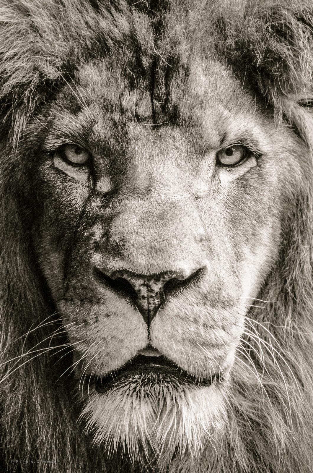
African Lion: Captive


 2351 Erwin Road Durham, NC 27710
kathleen.warren@duke.edu


 Erin A Hisey
Brian C Leonard


 Yiu, MD, PhD


