

s PIE magazine embarks on a new year, we are excited to bring you the latest developments and breakthroughs in the ever-evolving field of medical and surgical retina.
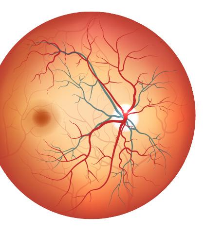
Our commitment to keeping you informed and engaged drives us to explore the frontiers of retinal research. In this issue, we are thrilled to present a comprehensive overview of the latest updates in medical and surgical retina— from cutting-edge treatments to groundbreaking discoveries.
Our network of experts in the field helped us curate a collection of articles that aims to deepen your understanding of these critical areas.
For instance, innovations in vitreoretinal imaging continue to shape the landscape of ophthalmology, offering new insights and avenues for diagnosis and treatment. Also, we are excited to explore mobile and home monitoring, a burgeoning field poised to transform how we manage retinal conditions. Discover how advancements in technology are empowering patients to play an active role in their healthcare, while simultaneously providing healthcare professionals real-time data to tailor more personalized treatment strategies.
Furthermore, the diagnosis and imaging of retinal conditions lie at the heart of our pursuit of excellence in eye care. With numerous developments in this area in the past year alone, we hope to bring you the latest in diagnostic tools and imaging modalities— shedding light on how these advancements enhance our ability to detect and monitor retinal diseases with unprecedented precision.
As we step into 2024 and beyond, PIE magazine is dedicated to providing you with the knowledge essential for navigating the complexities of diagnosis and treatment in the rapidly evolving field of retina care.
As always, we are grateful for your continued support and trust. It is our privilege to serve as your source of information and insight into the dynamic world of medical and surgical retina, and we hope you find inspiration in the discoveries and innovations shared within.

Best regards,
Gloria D. Gamat Chief Editor, Media MICE PIE, CAKE and COOKIE magazines

Matt Young
Hannah Nguyen
Gloria D. Gamat

A New Approach to Old Challenges in Modern nAMD and DME Treatment
Despite over two decades of breakthroughs with anti-VEGF agents, unmet needs abound in the treatment of exudative retinal diseases. Is sustained disease control on the horizon?

Riddles of IRD Experts at APVRS 2023 share insights into the mysteries of inherited retinal diseases
New Long-Term Faricimab Data Boosts Drug’s Profile for nAMD, DME and RVO

Brandon Winkeler
Robert Anderson
Sven Mehlitz
Media
Innovations in Sight
Cutting-edge solutions and revolutionary advancements poised to reshape retinal medicine in 2024
Revolutionizing Retinal Care
Latest developments in MacTel, polypoidal choroidal vasculopathy treatments, and advancements in vitrectomy tools
Ocular Imaging
Discovering groundbreaking studies that stole the spotlight at APVRS 2023
Visionary Healer
With an unwavering passion for diabetic retinopathy research and patient care, Dr. R. Rajalakshmi aims to redefine the standards of care in ophthalmology
Seeing Beyond Strife
Orbis and Sightsavers bring eye care to the victims of war Conference Highlights
Sight-saving Innovations
From ROP advancements to ocular oncology strategies, experts highlighted cutting-edge ophthalmic breakthroughs at APVRS 2023
Visionary Ocular Oncology
Could we soon see a cure for ocular cancer?


Dr. Alay S. Banker
Banker’s Retina Clinic and Laser Centre Ahmedabad, India
alay.banker@gmail.com
Dr. Barbara Parolini Eyecare Clinic Milan, Italy
parolinibarbara@gmail.com


Prof. Gemmy Cheung
Singapore National Eye Centre (SNEC) Singapore
gemmy.cheung.c.m@singhealth.com.sg

Prof. Mark Gillies
University of Sydney Sydney, Australia
mark.gillies@sydney.edu.au

Dr. Hudson Nakamura
Bank of Goias Eye Foundation Goiânia, Brazil
hudson.nakamura@gmail.com

Dr. Saad Waheeb
King Faisal Specialist Hospital & Research Centre, Riyadh, Saudi Arabia
saadwaheeb@hotmail.com




Despite over two decades of breakthroughs with anti-VEGF agents, unmet needs abound in the treatment of exudative retinal diseases. Is sustained disease control on the horizon?
by Matt HermanOphthalmic science has come a long way in the treatment of diseases like neovascular AMD (nAMD) and diabetic macular edema (DME). Anti-VEGF therapies have set new benchmarks in sight preservation for patients around the world, but treatment burden remains a major obstacle. Experts at a Bayer-sponsored symposium at the 23rd EURETINA Congress (EURETINA 2023) discussed the state of unmet needs in exudative retinal diseases and how results from the recent PHOTON and PULSAR trial results suggest that FDAand EMA-approved aflibercept 8mg may be able to address these challenges.

The success of the anti-VEGF revolution in the treatment of fluid-based retinal diseases has sparked heated debates amongst retinal specialists around the world about how much further patient outcomes can be pushed. Leaps in visual acuity gain have come at the cost of high treatment burdens, and ophthalmic meetings and conferences have devoted vast swathes of their programs to debating unmet needs and their potential solutions.
Bayer’s symposium in Amsterdam was no different. But with new approaches to managing these diseases and results from the PHOTON and PULSAR trials for aflibercept 8mg, the presentations and discussion from a panel of worldrenowned retinal research clinicians took on a new dimension.
Against this backdrop, symposium chair Prof. Carel Hoyng (Netherlands), Prof. Richard Gale (UK), Prof. Paolo Lanzetta (Italy) and Dr. Peter Kaiser (USA) convened to discuss gaps in exudative retinal disease treatment, approaches to sustained disease control and the impact of the new aflibercept 8mg data on this potential turning point in nAMD and DME treatment.
Prof. Richard Gale of the York and Scarborough Teaching Hospitals NHS Foundation Trust in the United Kingdom set the table by defining current unmet needs in nAMD and DME – and looking at what it would take to meet them.
For Prof. Gale, the necessity for effective management of these diseases is glaring, and he began by citing a battery of troubling statistics. In the industrialized world, nAMD represents 10% of all AMD cases, but


is responsible for around 90% of all cases of AMD-related blindness.
Despite this, challenges remain in delivering optimal outcomes with existing treatments for these ailments and action must be taken. “Solutions are required to address these in order to give better outcomes for patients and caregivers,” he commented. “These are principally put into several themes: Unmet needs around high treatment burden, poor longterm vision maintenance, suboptimal disease control, non-adherence and noncompliance. The cost of therapy and visits is burdensome, and the number of patients is increasing.”
With the scene set for candidate drugs in exudative retinal disease by Prof. Gale, Prof. Lanzetta defined the waypoints on the road to sustained disease control. Binding affinity, ocular clearance, potency and disease pathway targets are all necessary ingredients for success in the nAMD and DME arena. But Prof. Lanzetta put the focus firmly on molar dose.
“Solutions are required to address these in order to give better outcomes for patients and caregivers.”
– Prof. Richard Gale
According to Prof. Gale, the solution to overcoming these demands is sustained disease control, a concept with three pillars at its base: Lasting vision gains, rapid and resilient fluid control and extended treatment intervals.
“An agent that provides sustained disease control could address these key unmet needs,” he said. Any agent that offers sustained disease control must also have a similar safety profile to the cast of well-established antiVEGF agents in use for over a decade. And though Prof. Gale believes a high bar has already been set with aflibercept 2mg, he turned over the podium to Prof. Paolo Lanzetta for a rundown of the design features of what such an agent would look like.
He believes that data from the PULSAR aflibercept 8mg trial supports the rationale behind raising molar dose to achieve key sustained disease control milestones. PULSAR examined higher molar dose aflibercept 8mg. In the study, patients were randomly assigned to extended treatment intervals of either 12 or 16 weeks following 3-monthly loading injections. With aflibercept 2mg, patients received fixed 2q8 injections after 3-monthly loading doses. Prof. Lanzetta began with lasting vision gains, where aflibercept 8mg showed non-inferiority to its 2mg counterpart. “The high dose of aflibercept 8mg was non-inferior to 2mg at week 48 and week 96 – and this was achieved with less injections,” he noted.
Prof. Lanzetta also sees rapid and resilient fluid control, with superior drying compared to aflibercept 2mg in a pre-specified secondary endpoint in the PULSAR study. At week 16, 52% of aflibercept 8-week interval (q8) 2mg patients were free of intra- and
subretinal fluid in the central subfield, versus 62% and 65% for aflibercept 8mg at q12 and q16, respectively. This showed clear one-sided superiority, with a strong statistical significance of p=.0002. And at week 48, 8mg maintained its advantage– 59% for 2mg q8 versus 71% and 67% for q12 and q16.
“We also want to know how fast the fluid is resolved once we start treating the patient,” he continued. PULSAR’s answer to this query? Fast. For aflibercept 8mg, the median time to a fluid free central subfield was 4 weeks, compared to 8 weeks for 2 mg.
And in perhaps the most telling data supporting a higher molar dose’s impact on sustained disease control, Prof. Lanzetta turned to PULSAR’s extended treatment interval data. At weeks 48 and 96, 8q16 patients received 5.2 and 8.2 injections, respectively compared to 6.9 and 12.8 for 2q8 aflibercept 2mg patients. This represents a more than 33% reduction in injections at week 96 –meaning 33% less visits to the clinic and a significant upgrade in quality of life for nAMD sufferers.
This effect also proved to be lasting. In the group of patients randomly assigned to the 8mg q16 arm, a groundbreaking 8 out of 10 patients achieved a last-assigned treatment interval of 4 months or greater. And for the first time, last assigned treatment intervals of 5 and 6 months were achieved by 53% and 31% of patients, respectively – representing new milestones in the management of patients with nAMD.
The bottom line for Prof. Lanzetta is that the data all adds up to one
thing. “We believe that aflibercept 8mg has the potential for greater and longer lasting control in patients with nAMD through sustained disease control.”
With the case for a higher molar dose in nAMD laid out by Prof. Lanzetta, Dr. Peter Kaiser moved the discussion to DME and the aflibercept 8mg PHOTON results.
The DME PHOTON results largely echoed PULSAR’s, with additional data supporting aflibercept 8mg’s role in sustained disease control in DME. In PHOTON, aflibercept 8mg patients were randomly assigned to treatment intervals of 12 or 16 weeks after 3 initial monthly loading doses. These patients were compared to aflibercept 2mg patients who received 5 initial monthly loading doses, after which they received fixed 2q8 injections. Dr. Kaiser began with vision gains, where aflibercept 8mg was found to be non-inferior to aflibercept 2mg.
“This is really important because as we start to look at the dosing regimens and the treatment intervals, the visual acuity outcomes were the same, but if you look at that q16 arm – with six less injections,” he commented on the data.
Fluid reduction data told the same story. “We had a very rapid reduction in fluid, as we saw in all of our previous studies with aflibercept, and this was maintained all the way out to week 96,” he continued.
And just like with PULSAR, the impact of higher molar dose on treatment intervals was marked. PHOTON data showed 6 fewer injections for the q16 group compared to the q8 2mg group.
For treatment intervals and sustained disease control, the extension possible with aflibercept 8mg had the most profound impact on Dr. Kaiser. He noted that almost 90% of patients in the q16 group were maintained at 4-month intervals at week 96. But a closer look at the data revealed something more impressive.
and their loved ones from the chains that these diseases can put on their daily lives.
“We believe that aflibercept 8mg has the potential for greater and longer lasting control in patients with nAMD through sustained disease control.”
- Prof. Paolo Lanzetta
“A third of [q16] patients were at 6-month intervals to get those visual acuity results,” he said, pointing to the patient subgroup extended to q24. “And to me, that’s a very powerful number and one that I know my diabetic patients will enjoy.”
As Profs. Gale and Lanzetta and Dr. Kaiser indicated, sustained disease control stands at the vanguard of new approaches to managing sightrobbing retinal disease like nAMD and DME.
By getting fluid under control rapidly and extending treatment intervals with lasting visual acuity gains, patients can achieve lower treatment burdens and unshackle themselves

All patients stand to benefit from this reduction in treatment burden. It’s clear that patients already receiving frequent injections will see their shorter intervals pushed out even further. But aflibercept 8mg also has the potential to redefine what it is to be on a ‘long’ interval. Patients currently on what we consider ‘long’ intervals can also be pushed out even further– for some up to 5 months, a first for any intravitreal treatment for these diseases.*
The recent data from aflibercept 8mg’s PHOTON and PULSAR trials shows that one route to sustained disease control runs through an increased molar dose. And with a safety profile that mirrors that of aflibercept 2mg, one way forward in changing the lives of millions of nAMD and DME sufferers worldwide has materialized.
“We want to have the same vision, the same safety – but with less injections,” summarized Dr. Kaiser at the end of the symposium. “And I hope you saw in the presentations today that both in the PHOTON and PULSAR studies, we can get sustained disease control with 8mg of aflibercept.”
The 23rd Annual EURETINA Congress took place on October 5 to 8, 2023, in RAI Amsterdam Convention Centre, Amsterdam, The Netherlands. Reporting for this story took place during the event.

Due to the diverse and uncommon nature of inherited retinal diseases, diagnosing them can be challenging. Patients with IRD don’t often present with the same traits, and cases can sometimes stump ophthalmologists.

Asymposium on inherited retinal diseases (IRD), which took place on the final day of the Asia-Pacific Vitreo-retina Society (APVRS 2023) in Hong Kong in December 2023, brought together experts in the field. They discussed groundbreaking research and advancements, focusing on the understanding and treatment of these conditions.
The talks covered a diverse range of topics, including distinguishing between IRD and autoimmune retinopathy (AIR). The session also explored the evaluation of tinlarebant’s (Belite Bio; California, USA) effects on Stargardt disease based on promising results from a Phase 1b/2 study. Additionally, the discussions also included insights into essential components of IRD care.
The clinical features of AIR can appear similar to those of IRD, making differentiation between the two challenging. Misdiagnosing an IRD as an AIR can lead to unnecessary treatment with immunosuppressive agents.
In differentiating IRD from AIR, Dr. Tharikarn Sujirakul, associate professor of ophthalmology from Mahidol University, Thailand, suggested considering AIR in patients with acute/subacute progressive, painless visual loss caused by photoreceptor dysfunction.
“As a rule, it is important to conduct proper cancer surveillance in every patient suspected of autoimmune retinopathy to rule out paraneoplastic autoimmune retinopathy,” she noted.
AIR is a spectrum of retinal degenerative disorders that includes the paraneoplastic AIR (PAIR) and non-paraneoplastic (nPAIR) subtypes. Both subtypes are triggered by molecular mimicry, wherein similarities in sequence exist between foreign antigens and self-antigens, and self-antigens potentially cause an immune response.
In PAIR, molecular mimicry occurs between tumor antigens and retinal proteins; whereas in nPAIR, the
mimicry is between retinal proteins and presumed infectious (bacterial or viral) antigens. Both subtypes of AIR lead to retinal degeneration and may result in visual loss.
“As a rule, it is important to conduct proper cancer surveillance in every patient suspected of autoimmune retinopathy to rule out paraneoplastic autoimmune retinopathy.”
characteristics (aged ≤18 years) demonstrated sustained lower DDAF lesion growth in tinlarebanttreated subjects over the 24-month treatment period (p<0.001).
“Though IRD and AIR share numerous clinical features, comprehensive history-taking and proper investigations can help provide the right diagnosis to avoid treatment delay in AIR and unnecessary treatment in IRD patients,” she added.
– Dr. Tharikarn Sujirakul
A Phase 2 study evaluating the effects of tinlarebant in adolescent subjects with Stargardt disease has shown promising results.
According to Dr. Ruifang Sui, a professor at the Department of Ophthalmology in Peking Union Medical College Hospital, China, a daily dose of 5 mg tinlarebant reduces RBP4 levels by 80% to 90% and is found to be safe and well-tolerated after 24 months of treatment.
Tinlarebant is an orally administered tablet designed to slow disease progression in patients with Stargardt disease (STGD1) and geographic atrophy (GA) in advanced dry age-related macular degeneration (dry AMD).
In the Phase 2 study of tinlarebant, a total of 12 adolescent subjects with STGD1, aged 12 to 18 years, completed 24 months of treatment.
Retinal imaging showed that, after 24 months of tinlarebant treatment, five of 12 subjects remained free of atrophic lesions, referred to as definitely decreased autofluorescence (DDAF).
A comparison of the 24-month DDAF lesion growth between tinlarebanttreated subjects and ProgStar participants with similar baseline
A two-year, multi-center, randomized, double-masked, placebocontrolled Phase 3 DRAGON study is currently underway to evaluate the safety and efficacy of tinlarebant in the treatment of Stargardt disease in adolescents. Recruitment for this study has been completed with 104 subjects, and interim data for the first year are expected in mid- to late 2024.
Tinlarebant is a novel oral therapy that is intended to reduce the accumulation of vitamin A-based toxins, known as bisretinoids, that cause retinal disease in STGD1 and also contribute to disease progression in GA or advanced dry AMD. Bisretinoids are byproducts of the visual cycle, which is dependent on the supply of vitamin A (retinol) to the eye.
Tinlarebant works by reducing and maintaining levels of serum retinol-binding protein 4 (RBP4), which is the sole carrier protein for retinol transport from the liver to the eye. By modulating the amount of retinol entering the eye, tinlarebant reduces the formation of bisretinoids.
Dr. Chan Hwei Wuen, senior consultant and ophthalmic surgeon at the Department of Ophthalmology, National University Hospital (NUH), Singapore, leads a dedicated IRD service at the hospital for both local and international patients. During the session, she shared her experience of starting the first IRD service in Singapore.
In assembling the team, she noted that it was ideal to have a retinal specialist, clinical geneticist, genetic counselor, electrophysiologist, trained technicians, and research coordinators.
The service should aim to raise public awareness by organizing communitybased public talks, holding patient support groups, and contributing to media outlets through articles and media interviews.

Dr. Chan stressed the importance of genotyping, highlighting its role in providing unequivocal diagnosis, informing prognosis and inheritance patterns, and facilitating accurate counseling. Genotyping also enables the selection of appropriate therapies, including gene therapy, cell replacement therapy, and bionic implants.
“The other important thing that I will urge you to do is to maintain a robust database,” she advised. “You need to be meticulous and systematic in collecting data. Collect everything from patient demographics to phenotype and genotype details.”
Stargardt disease (STGD1) is the most common inherited retinal dystrophy in both adults and children. The disease, characterized by the blurring or loss of central vision, is caused by mutations in a retinaspecific gene (ABCA4). This results in the progressive accumulation of bisretinoids, causing retinal cell death and progressive loss of central vision. Currently, there are no FDAapproved treatments for STGD1.
The Asia-Pacific Vitreo-Retina Society Congress (APVRS 2023) was held from December 8 to 10, 2023, at the Hong Kong Convention and Exhibition Centre, Hong Kong. Reporting for this story took place during the event.


Aswathe of promising new data for Roche’s (Basel, Switzerland) Vabysmo (faricimab-svoa) at the virtual 21st Annual Angiogenesis, Exudation and Degeneration 2024 Conference (Angiogenesis 2024) on February 4 has unveiled promising outcomes in some of the world’s most debilitating retinal diseases.
The data drop comes from three key studies evaluating the drug’s efficacy in neovascular AMD (nAMD), diabetic macular edema (DME) and retinal vein occlusion (RVO).
VOYAGER is a newer noninterventional, prospective, multicenter study that aims to bolster Vabysmo’s clinical trial chops with real-world nAMD and DME data. The preliminary 3-month data from VOYAGER, presented by Prof. Robyn Guymer from the Centre for Eye Research Australia, demonstrated promising visual acuity (VA) gains and rapid reduction of central subfield thickness (CST).
BALATON and COMINO are Phase III studies looking at branching RVO (BRVO: BALATON) and hemiretinal and central RVO (HRVO and CRVO:
COMINO), respectively. The data on display at Angiogenesis 2024 was new, long-term 72-week data presented by investigator Dr. Ramin Tadayoni (France) and demonstrated excellent maintenance of VA gains, drying and extended treatment intervals.
Vabysmo, or faricimab, as it is also commonly known, had a coming-out party of sorts with study leader Prof. Guymer’s preliminary VOYAGER trial data announcement. This investigation will examine how the

drug’s stellar clinical trial results compare to the harsh light of realworld outcomes in nAMD and DME in a minimum of 5,000 patients across 28 countries.
Initial data from the US, Canada and Japan for 220 eyes of 174 patients with nAMD and 107 eyes of 69 patients with DME indicate improved visual acuity (VA) at month 3. The nAMD patients who switched at study enrollment saw a mean VA improvement of about 6 ETDRS letters from baseline (58.9 to 65), with DME patient ETDRS letter gains of about 2.5 (69.7 to 72.2).
CST also decreased rapidly through month 3 for those who switched to faricimab at enrollment. Faricimabnaive nAMD patients racked up a mean CST loss of about 51 µm, while DME patients saw a mean thinning of 41.7 µm.
Also, investigator tolerance of fluid varied by disease. Prof. Guymer also reported that among the researchers involved, 83.3% and 61.1% favored treat-and-extend regimens for nAMD and DME, respectively.
The VOYAGER study primarily assesses changes in VA over a year. Secondary and exploratory analyses cover anatomical features, treatment impact, outcomes influenced by treatment regimens, drivers of treatment decisions and imaging algorithms. Currently, 5,863 images are available, and future data analysis will include information from more countries.
In addition to the VOYAGER trial, the 72-week data from the Phase 3 BALATON and COMINO studies aimed to shed light on the longterm durability and effectiveness of faricimab.
The BALATON study was conducted in 553 people with BRVO while the COMINO study was conducted in 729 people with CRVO or HRVO.
The studies demonstrate a substantial extension of treatment intervals, with nearly 60% in BALATON and up to 48% in COMINO extending intervals to three or four months.
Participants maintained the vision gains and robust retinal drying achieved in the first 24 weeks of the studies for more than one year, a crucial measure as retinal swelling, measured by CST, relates to visual defects.
For BRVO, vision gains at 72 weeks reached 18.1 letters with a 310.9 µm reduction in CST. CRVO results showed vision gains of 16.9 letters and a 465.9 µm CST reduction at 72 weeks.
Dr. Levi Garraway, Genentech’s (South San Francisco, USA) chief medical officer and head of global product development, noted that these results mark the first instance of maintaining vision and anatomical improvements for over
a year in global Phase 3 studies for both BRVO and CRVO, reinforcing Vabysmo’s efficacy as a longterm and effective treatment for retinal conditions. Genentech codeveloped the drug with Roche as an independent subsidiary of the Swiss pharmaceutical giant.
“These long-term results build on the strong clinical and real-world data reinforcing Vabysmo as an effective treatment option for people affected by retinal conditions that can cause vision loss,” Dr. Garraway said.
Vabysmo’s novel feature is its dualpathway inhibition of angiopoietin-2 (Ang-2) and vascular endothelial growth factor A (VEGF-A), and the latest data gives faricimab promising evidence for this unique approach, its potential in treating exudative retinal diseases.
FDA-approved on January 28, 2022, it was the first bispecific antibody to be approved for treating eye disorders. Faricimab’s expansion into the RVO treatment arena underscores its versatility and potential to address a broader spectrum of retinal diseases.
With the promising preliminary results from the VOYAGER trial and the comprehensive insights from the BALATON and COMINO studies, faricimab has positioned itself as a formidable player in a highly competitive medical retina landscape.
1. Genentech Press Release. New LongTerm Data for Genentech’s Vabysmo Show Sustained Retinal Drying and Vision Improvements in Retinal Vein Occlusion (RVO) Available at: https://www.gene.com/media/ press-releases/15017/2024-01-31/new-longterm-data-for-genentechs-vabysm Accessed on 09 February 2024.
A version of this article was first published on piemagazine.org

From novel drugs and gene therapies to the integration of artificial intelligence and advanced surgical tools, 2024 promises groundbreaking advancements in retinal care. Experts shared the latest trends that ophthalmologists are buzzing about, providing a glimpse into the transformative landscape of retinal medicine for the year ahead.
One of the most dynamic and fast-paced fields in medicine, retina is constantly evolving and improving, with new cutting-edge solutions emerging seemingly every minute. These solutions often prove to be gamechangers for surgeons and patients, leading to better diagnoses, shorter surgery times, smaller incisions, improved surgical safety and better outcomes.
Exploring the latest advancements, such as drugs, artificial intelligence (AI), 3D visualization and gene therapy, we take a look at the innovations and trends that ophthalmologists are discussing and eagerly anticipating in the retina field for the coming year.

The competition is ramping up in the battle against retinal diseases, thanks to the emergence of a couple of promising anti-vascular endothelial growth factor (anti-VEGF) agents. These additions bolster retina’s armamentarium in the fight against conditions such as neovascular age-related macular degeneration (nAMD) and diabetic macular edema (DME).
“We have probably seen a ‘ceiling effect’ with regard to the efficacy of the anti-VEGF treatments available now,” shared Dr. Lee Mun Wai, vitreoretinal surgeon and medical director of Lee Eye Centre. “Newer molecules coming out are focused on reducing the burden of treatment by extending the intervals between treatments.”
Faricimab is one such treatment, gaining in popularity for its unique mechanism of action, which involves blocking both angiopoietin-2 and VEGF-A. It has the potential for extended durability with 16-week dosing.
Dr. Mae-Lynn Catherine Bastion, a vitreoretinal surgeon at the National University of Malaysia (UKM), shared that the YOSEMITE and RHINE phase 3 randomized clinical trials concluded that robust vision gains and anatomical
improvements with faricimab were achieved through adjustable, with dosing up to every 16 weeks. This demonstrates the potential for faricimab to offer extended durability in the treatment of patients with DME.
“This is the newest agent on the Malaysian market,” she said. “It provides an option for patients looking to increase the interval between treatments, and it’s also suitable for those in whom other agents have proven ineffective.”
Dr. Manish Nagpal, a vitreoretinal consultant at Retina Foundation, India, said: “New anti-VEGFs are emerging, and I would be pleased to see one with a long-acting or sustained release profile. Faricimab has just been launched in India, and we have started using it.” Though it is still early, he said he expects it to exhibit superior efficacy compared to other currently utilized anti-VEGF agents.
Dr. Alay Banker, a vitreoretinal surgeon at Banker’s Retina Clinic and Laser Centre, India, couldn’t agree more. “Faricimab [in my opinion] is a superior molecule with a better outcome,” he said.
“This is a definitive positive for our patients,” added Dr. Lee. He said he looks forward to hearing about experiences from retinal specialists in the Asia-Pacific region, as disease prevalence, phenotypes and response to treatment can vary significantly in different ethnic groups, as well as patient behavior and compliance to treatment.
In addition to faricimab, there is an anticipated increase in the use of higher-dose aflibercept compared to aflibercept 2 mg. “I look forward to having more options available, such as the aflibercept 8 mg,” said Dr Lee.
“Aflibercept 8mg is another agent to watch because the increase in its concentration is hoped to translate into a longer treatment interval for more patients, compared to the 2mg form,” added Dr. Bastion.
Another new tool that surgeons are anticipating is the port delivery system (PDS), which offers continuous passive diffusion of drugs into the vitreous cavity. By
continuously delivering medication over an extended period, the PDS aims to maintain the clinical benefits of monthly intravitreal anti-VEGF therapy while decreasing the injection burden.
“PDS is another exciting development that we hope will reduce the number of injections patients get without compromising efficacy. This includes depot antiVEGF repositories with slow release,” shared Dr. Bastion.
The PDS was originally designed for ranibizumab in the treatment of nAMD, with the potential for future use with other molecules.
Developments in gene therapy are being closely monitored for potential new treatments targeting inherited retinal diseases (IRDs), such as retinitis pigmentosa (RP) and Stargardt disease, which are caused by broken or mutated genes.
While still in its infancy, many are optimistic that gene therapy has the potential to significantly benefit patients suffering from IRDs and provide positive outcomes. Gene therapy technology is directed to the appropriate part of the eye through a delivery vehicle, typically a safe and modified virus called a viral vector.
“Gene therapy is evolving, and I would be happy to see some concrete developments, especially in dystrophies such as Stargardt disease and retinitis pigmentosa,” said Dr. Nagpal.
The trial, known as STELLAR, is scheduled for enrollment in the first half of 2024. Unlike genetic therapies that deliver an entire healthy gene to replace the mutated one or edit DNA, ACDN-01 rewrites RNA, the genetic message derived from DNA that cells read to make proteins.
Belite Bio, a San Diego, US-based biopharmaceutical company, is also enrolling patients with Stargardt disease in DRAGON, its Phase 3 clinical trial for tinlarebant—an emerging oral medication designed to slow disease progression and vision loss. The medication aims to inhibit a protein known as retinolbinding protein 4 to reduce the uptake of vitamin A to the retina, thereby decreasing the production and accumulation of toxic vitamin Z byproducts, a telltale sign of Stargardt disease.
Nanoscope Therapeutics (Dallas, USA), a biotech company, has dosed at least six participants in its Phase 2 STARLIGHT clinical trial of its optogenetic therapy for individuals with advanced Stargardt disease. Some patients have reported vision improvements following this emerging treatment, administered via intravitreal injection. The therapy uses a human-engineered virus to deliver copies of the multicharacteristic opsin (MCO) gene to bipolar cells—cells that do not normally sense light but often survive after photoreceptors are lost to advanced retinal disease.
“Gene therapy is evolving, and I would be happy to see some concrete developments, especially in dystrophies such as Stargardt disease and retinitis pigmentosa.”
Below are several ongoing developments in research on Stargardt disease and RP.
– Dr. Manish Nagpal
Ascidian Therapeutics (Boston, USA) is set to launch a Phase 1/2 clinical trial for ACDN-01, its RNA exon editing therapy designed for people with Stargardt disease, an inherited form of macular degeneration caused by mutations in the ABCA4 gene.
Alkeus (Cambridge, USA), another biotech company, is conducting a multi-center Phase 2 clinical trial for ALK-001, a drug designed to target the toxic buildup in the retina believed to cause degeneration and vision loss. This emerging therapy involves a modified form of vitamin A, which, when metabolized in the retina, results in less waste production.
Spark Therapeutics’ (Philadelphia, USA) vision-restoring RPE65 gene therapy, LUXTURNA (voretigene
neparvovec), has received marketing approval from the US FDA, making it the first gene therapy to gain regulatory approval in the US for the eye or any inherited condition.
LUXTURNA demonstrated its efficacy in restoring vision during a clinical trial for patients with Leber congenital amaurosis caused by mutations in the RPE65 gene. The treatment is also designed to work for patients with RP caused by RPE65 mutations.
GenSight, Bionic Sight, and Nanoscope have launched separate clinical trials for their optogenetic therapies for RP and potentially other retinal diseases.
“Gene therapy and stem cells are always in the conversation when it comes to retinal diseases, and I look forward to seeing continued progress in this field,” said Dr. Lee. “I believe it will be the ultimate solution if a safe and effective method for delivering gene or stem cell therapy to the retina [can be established].”
Dr. Bastion noted, “I think the anticipated developments would be in therapies for inherited retinal diseases such as retinitis pigmentosa, in which programmed cell death occurs, and for which no other therapy is available.
“Hence, we have heard of gene therapy in which a viral vector reprograms the defective cells, thus halting the progression of the disease. These interventions need to be introduced prior to irreversible damage.”
However, Dr. Bastion acknowledged that challenges persist in safely introducing the viral vector to the subretinal space. “Hence, there is growing interest in suprachoroidal and subretinal access for these therapies,” she added.
While some may fear the onslaught of AI in this day and age, with concerns mounting about the potential for this disruptive technology to take away jobs, ophthalmologists not only expect further advancement of artificial intelligence (AI) this year but also anticipate its positive impact in the ophthalmic industry.
AI has the potential to further penetrate several aspects of healthcare, including imaging diagnostics, surgical platforms, telemedicine and administrative tasks.
Deep learning algorithms can be used to screen for conditions such as diabetic retinopathy, retinopathy of prematurity and other diseases. They can also predict future disease progression from optical coherence tomography (OCT) scans.
According to Dr. Banker, future innovations could look into AIbased systems that facilitate improved screening of common retinal diseases in remote areas. These systems may also predict outcomes and determine the need for treatment in patients.
“In the coming years, I think operating and imaging systems will become more affordable, with a lot of AI incorporated into their platforms. This advancement will aid surgeons and diagnostics, facilitating better decision-making and improving patient outcomes,” Dr. Banker predicted.
“Artificial intelligence is already being used to screen and diagnose retinal diseases, and I look forward to the development of more mature algorithms and seamless integration into our clinical practices, which will ultimately benefit our patients,” chimed in Dr. Lee.
While medical retina has enjoyed groundbreaking advancements, the field of surgical retina has also seen its fair share of innovations—from faster cutters to improved viewing systems.
“Vitrectomy systems have progressed to improve surgical efficiency and safety,” shared Dr. Lee. “Newer designs of vitrectomy cutters with a beveled tip allow for better tissue manipulation, while smaller gauge cutters of 27G and high-speed cutting of up to 20,000 cuts per minute contribute to improved safety for our patients,” he added.
Higher cutting rates reduce pulsatile fluid flow, an important source of vitreoretinal traction.
The HYPERVIT Dual Blade
Vitrectomy Probe by Alcon (Geneva, Switzerland) features a continuous duty cycle with a speed of 20,000 cuts per minute. The beveled-tip design is available in 23, 25+ and 27+ gauge sizes, boosting cutting efficiency while minimizing fluidic turbulence.
Recent advances in endoillumination technology focus not only on improving safety and brightness but also on enhancing the durability and versatility of tools.
“In retinal surgery, visualization is everything, and the improved illumination systems integrated with our machines have improved visualization significantly,” said Dr. Lee.
Digital imaging in vitreoretinal surgery is also becoming more accessible to the masses, and this has the potential to revolutionize the way surgeons operate. Instead of depending solely on microscopic viewing, this technology offers a promising alternative, he added.
Dr. Nagpal concurred. “Surgically, new high-speed 20,000 cutters have been recently introduced in India. They are excellent in increasing efficacy and safety for the patient,” he said.
New generation vitrectomy machines are in development, and Dr. Nagpal expects to see some emerging in the next few years that would improve the technology used as well as enhance safety.
“Imaging systems, of course, keep evolving, and we might see new ones or more advanced versions of the same in the coming years,” he noted.
Dr. Bastion is also anticipating new developments in state-of-the-art imaging and viewing systems. A ‘heads-up’ approach in surgery has many advantages, including an increased depth of field, ergonomic benefits, reduced fatigue and teaching benefits.
One heads-up system, the Alcon NGENUITY, can be used with any
microscope. It features a 55-inch viewing LED screen with four colors per pixel, providing customized images, hues, colors and resolution. The system is continuously upgraded.

“Viewing methods have advanced further, with 3D viewing preferred for surgery, especially macula surgery. Three-dimensional viewing systems make the surgery more comfortable for surgeons and a delight for the trainees, as they can all see the surgery in 3D, not just the assistant,” shared Dr. Bastion.
She added that in this area of macular surgery, intraoperative OCT also remains helpful. “However, the cost remains a challenge for both these technologies in Malaysia. Therefore, 3D viewers need to be truly affordable before they can be widely utilized,” she noted.
When it comes to retinal imaging, Dr. Bastion looks forward to wide-field imaging non-mydriatic cameras, wide-field OCTs and widerfield OCTA— all with more easily interpretable and motion-adjusted capacities.
There you have it. As technology continues to progress, we can anticipate further advances in retinal treatment that will enhance outcomes for patients. Indeed, 2024 is shaping up to be a very exciting year.

Contributors
Dr. Lee Mun Wai is a graduate of the University of Manchester and started his ophthalmic training in the UK and completed his advanced training in Singapore. He underwent a vitreoretinal fellowship at Lions Eye Institute, Australia. He is currently the medical director at LEC Eye Centre, Malaysia and is also the chair of the Malaysian Society of Cataract and Refractive Surgery.
munwai_lee@lec.com.my
Dr. Mae-Lynn Catherine Bastion graduated from the University of Sydney, Australia, with MBBS (First Class Honours) in 1999. In 2004, she received the Doctor of Ophthalmology degree from the Universiti Kebangsaan Malaysia (UKM). And in 2007, she completed her clinical fellowship in vitreoretinal surgery at The Eye Institute, Singapore. Following that until today, she’s been serving as head of vitreoretinal services at UKM. In 2009 she became the head of the Department of Ophthalmology, for which she served two terms. She was appointed UKM professor of ophthalmology (vitreo-retina) in 2014 and received the Academy of Medicine (AMM) Fellowship in 2016. She teaches undergraduate and postgraduate ophthalmology while maintaining private practice at UKM Specialist Centre. She currently serves on the committees of the Malaysian Universities Conjoint Committee of Ophthalmology, College of Ophthalmologists of the AMM, and Malaysian Society of Ophthalmology. This is finely balanced with a busy family life of three kids, two dogs and a vegetable garden.
2010 and the Best of Show at the annual AAO meeting held at New Orleans in October 2013. At the annual All India Congress (AIOS) he won the best paper in retina four times, including the S. Natarajan award in 2008 and the C.S. Reshmi award for the best video presentation in 2012. He was also the recipient of the IIRSI special gold medal in 2012, as well as the Young Achievers award from BOA in 2012. He has numerous peer reviewed articles to his credit, along with numerous chapters in various international books. He is a regular reviewer with ophthalmology journals such as Retina, American Journal of Ophthalmology (AJO), EYE, European Journal of Ophthalmology and IJO.
drmanishnagpal@yahoo.com

mae-lynn@ppukm.ukm.edu.my

Dr. Manish Nagpal is a vitreoretinal consultant at Retina Foundation, Ahmedabad, since 1999. After completing his ophthalmology training at the M&J institute of Ophthalmology, Ahmedabad in 1996, he further went on to accomplish the FRCS (Ed) at U.K. in 1997. He is a recipient of the prestigious Distinguished Service Award of APAO in 2007. He has a keen interest in making surgical videos and has received the Rhett Buckler award for best video at the annual meeting of ASRS (6 times) 2009 in New York; 2010 in Vancouver; 2011 in Boston; followed by Las Vegas in 2012, Toronto in 2013 and Seattle in 2020. He received the best video award at the film festival held at the 25th APAO meet held at Beijing, China in
Dr. Alay S. Banker is the director of Banker’s Retina Clinic and Laser Centre in Ahmedabad, India and his practice has served the city since 2007. He started off his career as a clinical instructor and fellow at the Department of Diseases of Retina and Vitreous, Uvea and Inflammation of Eye at University of California, San Diego, USA. He was the first Indian to receive the “International Scholar Award” from AAO in 2010 and also the youngest Indian to receive the Achievement Award by the AAO in 2006. His contributions toward his medical peers and community services have also earned him the Senior Achievement Award from AAO (2013) and the Dr. Piyush Patel Award for Service to Society and Mankind from Ahmedabad Medical Association (2013). He is the senior founding editor of the Retina Image Bank (ASRS 2012), has presented at more than 250 international and national conferences with over 40 papers published in peer-reviewed medical journals, and has published five book chapters in international book publications.
alay.banker@gmail.com
The most recent findings from crucial trials in macular telangiectasia, polypoidal choroidal vasculopathy treatment, vitrectomy technology, and diabetic macular edema management were unveiled during the Late-Breaking Keynote Symposium at the 16th Asia-Pacific Vitreo-retina Society Congress (APVRS 2023).
Dr. Emily Chew (USA) presented compelling results from two phase 3 controlled clinical trials in macular telangiectasia (MacTel) type 2, highlighting the efficacy of the NT-501 treatment in preserving photoreceptors. Dr. Tien-Yin Wong (Singapore) revealed sustained visual acuity gains with aflibercept in polypoidal choroidal vasculopathy (PCV).
Additionally, Dr. Harvey Uy (Philippines) showcased the enhanced precision of the 20,000 cuts per minute (cpm) vitrectomy probe, offering promising avenues for improved surgical outcomes.
Last but not least, Dr. Stela Vujosevic (Italy) shared results from the
ALTIMETER Phase 2b trial, showing improvement in vision outcomes in diabetic macular edema (DME) patients with faricimab.
Dr. Chew highlighted significant progress in macular telangiectasia type 2 (MacTel) treatment through two Phase 3 controlled clinical trials using the ciliary neurotrophic factor (CNTF) implant.
According to her, the progressive loss of the ellipsoid zone (EZ) has become an important feature of MacTel. Understanding that, under
pathological conditions, Müller glial cells can shield photoreceptors from cell death by generating neurotrophic factors, including CNTF, Dr. Chew and her colleagues embarked on a groundbreaking study involving firstin-class encapsulated cell therapy, revankinagene taroretcel (NT-501). NT-501 was surgically implanted into the vitreous cavity through a capsule anchored to the sclera to produce sustained levels of CNTF.
The identically designed NTMT-03-A and NTMT-03-B trials employed a randomization ratio of 1:1. Patients were allocated to NT-501 (n=58 and 59, respectively) or a sham procedure in the study eye (n=57 and 54).
The results are compelling, showing a 56.4% and 29.2% reduction in loss of EZ over two years with NT-501 treatment in NTMT-03-A and NTMT03-B, respectively.
“Treatment with NT-501, compared to the sham, preserved more photoreceptors through 24 months in both Phase 3 clinical studies, meeting the primary endpoint. Retinal sensitivity was preserved in participants receiving NT-501 versus the sham in NTMT-03-A. Additionally, reading speed was better preserved in participants receiving NT-501 compared to the sham in both studies,” she explained.
“Treatment-related serious ocular adverse events were uncommon and predominantly expected and typical of surgery. There were no serious treatment-emergent adverse events (TEAEs) of endophthalmitis or ischemic optic neuropathy,” she added.

“Our results demonstrate that intraocular delivery of CNTF using NT-501 encapsulated cell therapy is safe and effective for the treatment of MacTel,” Dr. Chew concluded.
Currently, the standard of care for the vast majority of patients with polypoidal choroidal
vasculopathy (PCV) is anti-vascular endothelial growth factor (anti-VEGF) therapy, either as monotherapy or in combination with photodynamic therapy.
Dr. Wong presented results from a 96-week subgroup analysis of the phase 3 PULSAR trial, highlighting the sustained efficacy of aflibercept 8 mg monotherapy in treating PCV. In this trial, patients were randomly assigned in a 1:1:1 ratio to receive intravitreal aflibercept 8 mg every 12 or 16 weeks (8q12, 8q16) or 2 mg every 8 weeks (2q8), each after three initial monthly injections.
boasts a continuously open port, is powered by a dual-pneumatic engine and features a bevel tip design.
“The 20,000 cpm dualblade vitrectomy cutter probe is capable of all retinal indications. You can achieve a high vitreous flow rate, which allows for efficient removal of core vitreous.”
The study demonstrated consistent visual acuity gains, with the aflibercept 8q12, 8q16, and 2q8 subgroups maintaining gains of +8.4, +8.2, and +9.6 letters, respectively, from baseline to week 96. Notably, both doses of aflibercept effectively reduced the total polypoidal lesion area, emphasizing their therapeutic impact.
– Dr. Harvey UyTo assess the clinical outcomes and surgical performance of the 20,000 cpm probe, Dr. Uy and his colleagues conducted a study on 55 adult eyes undergoing vitrectomy with the 20,000 cpm 25-gauge probe (Hypervit; Alcon, Geneva, Switzerland). The results were compared to 50 eyes operated on with a 10,000 cpm probe of a similar design. Results indicate that the 20,000 cpm probe significantly enhances surgical precision and safety by reducing tractional forces and pulsatile motion.
“The 20,000 cpm dual-blade vitrectomy cutter probe is capable of all retinal indications. You can achieve a high vitreous flow rate, which allows for efficient removal of core vitreous,” shared Dr. Uy.
from the ALTIMETER phase 2b trial, showing significant improvement in vision and anatomic outcomes in patients with diabetic macular edema (DME) through the inhibition of VEGF-A and Ang-2 with faricimab.
During this 24-week Phase 2 exploratory study, conducted in an open-label, single-arm fashion, 99 eyes received 6 mg of faricimab every four weeks. Vision gains and reductions in central subfield thickness (CST) were notable, persisting until week 24, resulting in a mean best-corrected visual acuity (BCVA) gain of +9.2 letters and a CST reduction of -200.2µm by week 24.
Meanwhile, the median intraretinal fluid (IRF) volume decreased by -343.3nL at week 24, and the macular leakage area significantly diminished from 28.6mm² (baseline) to 2.8mm² at week 20.
“In the ALTIMETER study, faricimab demonstrated meaningful improvements in vision and anatomy, consistent with outcomes observed in the YOSEMITE and RHINE trials in patients with DME,” shared Dr. Vujosevic.
Moreover, the data indicate an extended dosing interval for aflibercept 8 mg, with 72% of patients qualifying for ≥20 weeks, suggesting prolonged durability compared to aflibercept 2 mg.
Importantly, the safety profile of aflibercept 8 mg remains comparable to that of aflibercept 2 mg in both the PCV subgroup and the overall study population, affirming its reliability as a treatment option.
Meanwhile, Dr. Uy highlighted the evolving landscape of vitrectomy probe technology, emphasizing advancements geared toward smaller, faster probes with reduced distances between the port opening and the tip.
The focus of his talk was on the latest technological iteration—a dual-blade, bevel tip cutter probe operating at 20,000 cpm. This cutting-edge tool
“The tool also contributes to reduced peak traction and pulsatile flow, maintaining stable globe intraocular pressure (IOP). Tissues are more stable during shave vitrectomy, allowing precise dissection and low adverse events. The bevel tip enhances tissue dissection and reduces ancillary instrument exchanges, enhancing surgical efficiency,” he added.
Dr. Uy recommended starting with the regular 10,000 cpm setting and adjusting it down, asserting that the 20,000 cpm vitrectomy probe is definitely a welcomed addition to the vitreoretinal toolkit—offering enhanced capabilities for improved surgical outcomes.
VEGF-A serves as a pivotal factor in neovascularization and vascular permeability, while angiopoietin-2 (Ang-2) plays a crucial role in regulating vascular stability.
Dr. Vujosevic presented findings
High macular leakage correlated with elevated Ang-2-related proteins and immune regulators at baseline, which showed a reduction following faricimab treatment.
“The observed safety profile was consistent with the known safety parameters of faricimab. Further exploratory analyses are planned to delve into imaging and aqueous humor biomarkers of vascular stability associated with dual Ang2/VEGF-A inhibition,” she added, concluding her presentation.
The Asia-Pacific Vitreo-Retina Society Congress (APVRS 2023) was held from December 8 to 10, 2023, at the Hong Kong Convention and Exhibition Centre, Hong Kong. Reporting for this story took place during the event.
On the third day of the Asia-Pacific Vitreo-retina Society (APVRS 2023) Congress, a Rapid Fire session explored the latest studies on ocular imaging, intraocular inflammation and uveitis and scleritis.
The symposium featured several groundbreaking studies promising to usher in a new era of retinal therapies.
Dr. Weiting Liao’s (China) exploration of Vogt-KoyanagiHarada (VKH) disease shed light on age-specific patterns, offering a comprehensive understanding of the disease evolution in pediatric, adult and elderly patients.
An investigation from Dr. Ting Yu’s (China) into the role of galactomannan (GM) in Aspergillus endophthalmitis (AE) highlighted the diagnostic potential of GM testing in intraocular fluid. Dr. Yue Zhang (China) challenged traditional categorizations of central serous chorioretinopathy (CSC) subtypes, emphasizing the need to distinguish acute and chronic forms based on choroidal characteristics.
And, finally, Dr. Gabriel Yang (Hong Kong, China) highlighted the potential of using deep learning on OCT images to predict the effectiveness of anti-VEGF therapy in eyes with center-involved diabetic macular edema (CI-DME).
VKH disease represents the predominant cause of panuveitis in China. According to Dr. Liao, the clinical features of VKH disease, as reported in the current literature, predominantly focus on adult patients. As such, she and her team performed a study to compare the clinical characteristics, complications, and visual prognosis across different age groups (pediatric, adult, and elderly) among 2,571 Chinese VKH patients.
The findings revealed that within the first two weeks after the onset of uveitis, patients in all age groups (pediatric, adult, and elderly) exhibited choroiditis (100%), retinal detachment (75%, 84.2%, and 85.7%), and optic disc hyperemia. Notably, no anterior segment inflammation was observed in these VKH patients during this early stage. Between two weeks and two months after disease onset, the majority of VKH patients displayed mild to moderate nongranulomatous anterior uveitis.
Subsequently, after two months, granulomatous anterior uveitis became a prevalent manifestation across all age groups, accompanied by the significant presence of DalenFuchs nodules.
“In conclusion, pediatric, adult, and elderly VKH patients demonstrated a similar disease evolutionary process. Children with VKH disease exhibited a lower frequency of manifestations in the neurological and auditory systems,” Dr. Liang shared.
“Improved visual acuity (VA) was observed across all three age groups. The association between disease onset age and a poor visual outcome revealed a reversed u-shape, with 32 years identified as the highest risk age for the development of BCVA lower than 6/18.”
In summary, this study offers a novel insight into the complete spectrum of clinical features associated with VKH disease.
Fungal endophthalmitis poses a critical ophthalmic emergency, demanding swift and accurate diagnosis for therapeutic success. Dr. Yu shed light on the diagnostic efficacy of GM levels in intraocular fluid, a heat-stable antigen constituting the cell wall of the Aspergillus fungi.
In a unique investigation of GM testing specifically for AE in intraocular fluid, this study aimed to provide evidence for early disease diagnosis. The retrospective analysis involved three patient groups–an AE group with positive intraocular fluid culture (n=17), a non-AE intraocular infection (NAII) group (n=20), and a negative control group (n=19).

The results unveiled significantly elevated GM levels in the AE group (5.77±1.73) compared to the NAII (0.19±0.11), and negative control (0.29±0.27) groups. The test demonstrated notable sensitivity and specificity, leading to Dr. Yu’s conclusion that GM testing in intraocular fluid serves as a rapid and reliable diagnostic modality for AE.
Next, Dr. Zhang challenged the traditional categorization of CSC into acute and chronic forms based on the duration of subretinal fluid (SRF) resolution. Highlighting the absence of a consensus despite various novel classification systems emerging, Dr. Zhang emphasized the importance of distinguishing between acute and chronic CSC due to potential differences in pathophysiology and management approaches.
To address this, Dr. Zhang and her colleagues set out to investigate choroidal characteristics in four recently defined subtypes of 83 CSC eyes (acute, non-resolving, recurrent and chronic CSC) through a crosssectional observational study using swept-source optical coherence tomography angiography (SS-OCTA).
They found significant differences in best corrected visual acuity (BCVA) among the four subtypes and healthy control groups. The visual acuity (VA) of the complex CSC subgroup was significantly worse than the other four groups.
Meanwhile, complex CSC had significantly lower central choroidal thickness, choroidal vascular volume (CVV), choroidal vascularity index (CVI), and choriocapillaris vessel density (CCVD).
seems more reasonable to define chronic CSC as morphological change rather than the duration of disease, with distinctive choroid features. Traditional acute CSC tends to be ‘simple’, with less retinal pigment epithelial disruption, and chronic CSC tends to be ‘complex’ with more [retinal pigment epithelial disruption].
Also, threedimensional CVI and CCVD have demonstrated efficacy in evaluating choroidal vasculature, serving as a promising tool for disease assessment,” Dr. Zhang explained.
“This may be used as a guiding tool for therapeutic selection, thereby reducing the impact of invasive and burdensome treatment for patients in whom anti-VEGF therapy is likely to be ineffective.”
– Dr. Gabriel Yang
In order to identify patients who will benefit more from anti-VEGF therapy, Dr. Yang and his team developed, validated and externally tested an integral deep learning framework based on pretreatment OCT images and clinical variables (i.e., VA and central subfield thickness [CST]) to predict the therapeutic response in patients with center-involved DME (CI-DME) after anti-VEGF injections.
Last but not least, Dr. Gabriel Yang addressed the challenge of variable responses to anti-VEGF treatment in diabetic macular edema (DME), where 40% to 50% of patients do not experience improved visual acuity.

“From the results of our study, it
Data received from three hospitals in Hong Kong were used for training and validating the algorithm. Their proposed algorithm attained the best performance in the primary validation and the external testing sets by using the first definition: ≥ 1-line gain on Snellen VA chart or >10% reduction in CST after 3 antiVEGF injections, followed by the second definition: ≥ 1-line gain on Snellen VA chart after 3 anti-VEGF injections, and the third definition: >10% reduction in CST after 3 antiVEGF injections.
“We demonstrated the potential use of deep learning on OCT images to predict ‘good-response’ vs. ‘noresponse’ of anti-VEGF therapy in eyes with CI-DME,” Dr. Yang shared.
“This may be used as a guiding tool for therapeutic selection, thereby reducing the impact of invasive and burdensome treatment for patients in whom anti-VEGF therapy is likely to be ineffective.”
The Asia-Pacific Vitreo-retina Society Congress (APVRS 2023) was held from December 8 to 10, 2023, at the Hong Kong Convention and Exhibition Centre, Hong Kong. Reporting for this story took place during the event.

With an unwavering passion for diabetic retinopathy research and patient care, Dr. R. Rajalakshmi aims to redefine the standards of care in ophthalmologyby April Ingram
From a personal tragedy that sparked her passion for medicine to becoming a prominent figure in diabetic retinopathy research, Dr. R. Rajalakshmi’s story unfolds with insights into mentorship, challenges faced by women in ophthalmology, and the profound impact of patient confidence.
Dr. R. Rajalakshmi is a senior consultant ophthalmologist and the head of Medical Retina at Dr. Mohan’s Diabetes Specialities Centre in Chennai, India. Additionally, she serves as a clinical researcher at Madras Diabetes Research Foundation.
Her journey into medicine began at an early age, driven by a personal tragedy. “I lost my father when I was three years old,” she shared. “He was a research scientist from the Indian Institute of Technology, and he died when he was just 32 years old due to a sudden heart attack.
“I grew up listening to my mother and grandmother telling me how timely medical help could have possibly saved my father’s life. So, the thought of becoming a physician stayed deeply rooted in me—and I am glad I became one.”
fascinated me a lot. Thus, I chose ophthalmology as my specialization,” she continued.
Support and mentorship from women is truly a game changer. Seeing themselves in roles that may still be predominantly held by males can help women access opportunities and cultivate their confidence.
Dr. Rajalakshmi found this support in Dr. Rema Mohan. “After completing my post-graduation in ophthalmology from Madras Medical College, I joined Dr. Rema Mohan, the chief of ophthalmology at Dr. Mohan’s Diabetes Specialities Centre and Madras Diabetes Research Foundation, as a junior ophthalmologist,” she shared.

Dr. Rajalakshmi believes ophthalmology is one of the most fascinating specialties of all. “While pursuing my Bachelor of Medicine and Bachelor of Surgery
(MBBS) and especially during my residency, the ocular manifestations of various systemic diseases and the microsurgery of the eye
Dr. Mohan, a woman pioneer in diabetic retinopathy research in India, served as Dr. Rajalakshmi’s mentor. “Working with her inspired my interest in retina/ diabetic retinopathy research. As the daughter of a research scientist who died very early, I was inspired to pursue my Ph.D. in retina under Dr. Mohan’s guidance,” Dr. Rajalakshmi shared.
“Unfortunately, we lost her to cancer about 10 years ago, and I am now continuing the retina research work that she started at the institution where I currently work.”
Despite the increasing equalization of opportunities between men and women in ophthalmology, does Dr. Rajalakshmi still experience any unique challenges as a woman compared to her male counterparts? Has she observed changes in this aspect over the course of her career?
“Being a woman professional has its challenges, as we, women, need to know how to strike a work-life balance,” she replied. “In every field of ophthalmology, women have to work harder to prove ourselves. And this requires a lot of selfconfidence, consistent hard work, and support from family. My mother, my husband, and my sweet daughter have always been supportive and encouraging—even when I have faced challenges in my career.”
Support and collaboration are important pillars of career success, recognizing that we can achieve more together. “Specializing in the unique field of ophthalmology, medical retina, with a special focus on diabetic retinopathy, has created opportunities for collaborative research in this field and has helped me grow in my career,” enthused Dr. Rajalakshmi.
Dr. Rajalakshmi offered words of wisdom for women who may be early in their ophthalmology career or contemplating the specialty. She shared some valuable advice drawn from her personal journey.
“My passion for retinal research helped me grow in my career. Being passionate about what you do and maintaining consistency in your efforts are key,” she said.
She also highlighted the importance of perfecting balance. “Personal and professional life are equally important for women, and balancing both is an art. Ophthalmology is one of the best fields, especially for women, where it is possible to balance career growth and personal life—being productive in both without burning out,” she continued.
Finally, building your team and surrounding yourself with others who share the same passion and goals is crucial. “Understanding that managing patients with diabetes and retina problems, as well as eye disorders at the hospital, is a team effort and not an individual task,” she noted. “Teamwork works!”
Dr. Rajalakshmi also shared her passion for researching the nuances of the diabetic eye. “Yes, diabetic retinopathy research is my passion. The use of smartphones for retinal imaging (cost-effective retinal imaging), artificial intelligence (AI), and telemedicine/teleophthalmology are my key interest areas of research,” she shared. “In recent years, I have been doing a lot concerning the use of AI in the diagnosis and management of diabetic retinopathy.”
Indeed, this is such an important and timely focus to manage the growing number of people with diabetes, and Dr. Rajalakshmi’s work in the years to come is highly anticipated.
As the world cautiously opened up after the pandemic, providing opportunities to attend conferences and share insights, Dr. Rajalakshmi also expressed her preferences for conferences.
According to her, it’s all about learning and translating these learnings into local practices for maximum effectiveness, and, of course, seizing opportunities for collaboration.
“In international conferences, I look forward to learning something new from the Western world and exploring how the same innovation can be applied cost-effectively in a low- to middle-income country like India,” she noted.
“When attending big conferences, I look for opportunities to collaborate and network with other renowned international ophthalmology researchers for collaborative multicentric ophthalmology research projects.”
The power of patient confidence in ocular care Words are powerful, particularly when they come from our patients. Dr. Rajalakshmi shared some of the most meaningful expressions patients have conveyed to her.
“The confidence that my patients have in me is most gratifying as a treating ophthalmologist. Many of them express that they find a healing touch in my warm smile, and they mention that they find it very reassuring given that diabetic retinopathy management is not a one-time procedure,” she enthused.
These are the patients who refer and bring many more patients through word of mouth for treatment, owing to their faith in ocular management and holistic approach.
Dr. Rajalakshmi shared a specific example: “A lady who had already visited four or five eye hospitals across India came to me for diabetic retinopathy management. She said,
‘My friend told me, go and see Dr. Rajalakshmi. You will be perfectly fine. When I came, I was unsure.”
“Now, after being treated by you, I know that she was 100% right!’ These words are more valuable to me than anything else in this world. Even now, this patient travels more than 1000 km and has been coming to me for regular follow-ups, as advised for more than 15 years,” she reflected.
It is evident that Dr. Rajalakshmi is not only admired by her colleagues but also adored by her patients—and for good reason.
“Some patients treat us like demigods after they get better. Next to the loss of life, the loss of sight is a famous old proverb. The gratification of symptomatic relief and visual improvement in every patient after successful surgery is beyond what can be explained in words,” she said.
“The satisfaction of providing treatment and cure in the medical profession cannot be obtained in any other field,” she opined. A true and beautiful sentiment, indeed
Of course, Dr. Rajalakshmi also shared sentiments from the other end of the spectrum. “There are patients who read miscellaneous information from the internet, become confused with too much information regarding diagnosis or treatment options, and consider themselves as ‘all-knowing, Google doctors’. They can be annoying at times with their remarks when they make a self-diagnosis and come to us.”
When she’s not working, Dr. Rajalakshmi loves to read.“I enjoy both fiction and non-fiction, with Atomic Habits by James Clear being one of my current favorites.” The book is a practical guide to developing small sustainable habits that evolve into immense personal growth.
Finally, if she hadn’t become an ophthalmologist, what career path does Dr. Rajalakshmi envision for herself? “I can’t think of anything else right now as ophthalmology and research are my passion.”


In a world where providing eye care remains a significant challenge, particularly in war-torn regions, organizations like Sightsavers and Orbis shine as beacons of hope. Sightsavers, operating in Africa and South Asia, employs an adaptive management process to deliver care in dangerous areas, while Orbis addresses the vital need for eye care among refugees.
Saving a patient’s threatened vision is already a challenge under the best of circumstances. But when the stark realities of global politics get involved, this task becomes even more daunting. Providing eye care to the underserved remains one of the greatest obstacles faced by the medical profession to this day. And few places are so critically underserved as those affected by war, including those to which refugees have been forced to flee.
Healthcare providers working in conflict zones were recognized during the Orbis Symposium: Global Ophthalmology in War Zones at the recently concluded European Society of Cataract and Refractive Surgeons Congress (ESCRS 2023). Amid the numerous symposia at this year’s Congress in Vienna that focused on the technical aspects of ophthalmology, the Orbis Symposium took a different turn. This session shifted its focus to the political and economic challenges faced by
doctors struggling to bring even basic services to those who are most in need.
Due to the tremendous challenges associated with delivering these services to far-flung and often dangerous locations, these eye care professionals often work as part of non-government organizations (NGOs) and charitable entities. Without these structures, the task of securing financial, logistical and legal support for their incredible work would likely be impossible.
Representatives from two of these important organizations introduced their missions and discussed the numerous obstacles they have to overcome to preserve patients’ vision.
Ms. Sumrana Yasmin introduced the audience to Sightsavers, an international NGO that works in over 30 developing countries in Africa and South Asia to treat and prevent avoidable blindness.
“Our primary focus is on creating a world where no one is blind from avoidable causes, and where people with disabilities can participate equally in society,” said Ms. Yasmin, the deputy technical director for Eye Health and Unaddressed Refractive Errors (URE) of Sightsavers. “Within that sphere, we focus on protecting sight and fighting disease, as well
as disability programs where social inclusion of people with disabilities is ensured.”
To address this tall task, Sightsavers employs an adaptive management process that focuses on conducting a landscape assessment of the area where they intend to deliver care. The first step is to identify marginalized communities and analyze their needs in terms of health services and required products. In addition, the organization must evaluate the risks implicit in operating in dangerous areas as well as security options that are available.
Part of this risk analysis involves carefully prioritizing safety, including mitigation plans, as well as evacuation strategies for healthcare staff. These precautions aren’t without valid reason. “In the last eight months,” Ms. Yasmin explained, “I can actually remember three instances where we had to apply these plans to take out our people from these situations. In these cases, the most important factor was having the right resources, including both financial support and competent, secure individuals to deliver these services.”
Amid these concerns for the physical and mental well-being of caregivers, quality of care becomes a real concern as well. Delivering primary care, which these eye care workers have come onsite to provide, as well as managing follow-up care, becomes increasingly challenging due to the inherent danger of working in conflict zones. On top of this, the ongoing threat of resource theft is a further
concern, and the cost of delivering eye care in these zones is at least 20% to 25% higher than it is in a usual clinical setting.
However, on the flip side, Ms. Yasmin noted that the opportunities to help people make all these challenges worthwhile. “We have a very strong focus on leaving no one behind,” she said. “Working in these locations helps us reach the most vulnerable population.”
Through their frontline work in these war zones and the strengthening of partnerships with local caregivers, Sightsavers is a true game-changer in the lives of millions of patients whose vision may otherwise have been lost amid the strife of local conflict.
While patients within a war zone are likely in the most peril, the challenges for victims of conflict do not instantly get easier once they have fled from the threat of harm.
As a leading NGO dedicated to saving sight worldwide, Orbis International, the host of the symposium, is at the forefront of providing ophthalmic care to refugees.
Dr. Robert Walters, the chairman of Orbis in the Middle East, spoke on the threats faced by refugees who are fleeing conflict.
“Among those displaced people,if you are a refugee, you instantly become poor,” Dr. Walters began “And you

experience everything that comes with poverty. These people are flung together, often within walls.”
Dr. Walters introduced the work that Orbis has done in cooperation with the United Nations High Commission for Refugees (UNHCR), providing eye care services among the Rohingya refugees who have fled Myanmar to neighboring Bangladesh.
The Rohingya, like many refugees, faced significant discrimination in the country from which they had fled. Notably, they faced medical discrimination, with most having received little to no primary care before escaping into the dire situation of a refugee camp.
It is in such situations that care is so vital. And though the challenges presented by providing care in a refugee camp are not as violent as in an active conflict zone, they nonetheless present obstacles. Notably, the UNHCR needed to establish roads, water and septic services and provide basic structure before care could be delivered.
The need for care among these neglected refugee populations is enormous. Dr. Walters reported that one in seven Rohingya refugees has sight-threatening cataracts. Not only is this condition devastating, but it is also one of the most treatable. By establishing clinics that service the refugee community, Orbis is able to preserve the vision of individuals whose lives are already faced with overwhelming challenges.
Amid the dangers presented by global conflict, organizations like Sightsavers and Orbis bring hope and sight to patients who are in the greatest peril. Their work is truly visionary and deserving of our highest respect and admiration.
The 41st European Society of Cataract and Refractive Surgeons Congress (ESCRS 2023) took place on September 8 to 12, 2023, in Vienna, Austria. Reporting for this story took place during the event.

From ROP advancements to ocular oncology strategies, experts highlighted cutting-edge ophthalmic breakthroughs at
The fourth Rapid Fire Session at APVRS 2023 called in the research artillery on subjects ranging from rare cancers to sight-saving innovations and techniques in neonatal and trauma eye care.
Rapid fire sessions have become a staple of ophthalmic congresses with their whirlwind approach to sharing the latest research in eye care.
The 16th Asia-Pacific Vitreo-retina Society (APVRS 2023) Congress in Hong Kong, held from December 8-10, 2023, was no different.
On the last day of the Congress, Rapid Fire Session #4 saw experts from across the region present the latest sight-saving research in ocular oncology, trauma, emergencies and infections, pediatrics, and more.
Retinopathy of prematurity (ROP) was a well-represented topic at APVRS as a whole, and Rapid Fire Session #4 continued the trend.
New advancements in multimodal imaging and analysis are benefitting the tiniest patients in ophthalmology. Dr. Akash Belenje’s (India) research showed how modern swept-source optical coherence tomography (SSOCT) biomarkers can help predict responses to anti-VEGF treatment in infants with aggressive ROP (AROP).

“The red flag is the ischemic variant of AROP with thin choroids—less than 150 microns—with hyperreflectivity of the inner layers. These patients had a higher chance of anti-VEGF crunch, reactivation, avascular retina, and a higher chance of retreatment with laser.”– Dr. Akash Belenje
One of the key findings of his study involved classifying patients with AROP based on the presence of ischemia. “We identified two important non-ischemic variants of AROP with hyper-reflective cystic spaces or good retinal layers with thickened choroid that always did well [with anti-VEGF treatment],” Dr. Belenje said.
“The red flag is the ischemic variant of AROP with thin choroids—less than 150 microns—with hyper-
reflectivity of the inner layers. These patients had a higher chance of anti-VEGF crunch, reactivation, avascular retina, and a higher chance of retreatment with laser,” he concluded.
Dr. Sushma Jayanna (India) discussed another imaging modality in ROP— ultra-widefield color fundus (UWCF) imaging. Her work, co-authored with Dr. Subhadra Jalali and Dr. Tapas Padi, sought to validate the usefulness of these fundus photographs for everything—from assessing the stage and severity of the disease to recommending potential treatment interventions.
Using Optos (Dunfermline, UK) UWCF technology, Dr. Jayanna’s group retrospectively compared images taken with the Optos camera and those with binocular indirect ophthalmoscopy (BIO).
They found that only 11% of the UWCF images were discordant with BIO images, with notable missed findings in zone 2 ROP.

“Ultra-wide field cameras are quick and non-contact. They can be a great tool in busy OPD [outpatient department] setups for documentation, training and medical-legal purposes,” Dr. Jayanna concluded.
And speaking of zone 2, Dr. Yong Cheng (China) rounded out the ROP slate with a comparison of conbercept (Chengdu Kanghong Biotech, Sichuan, China) and photocoagulation laser therapy in zone 2 ROP patients.
“In two groups with zone 2 ROP, intravitreal conbercept and laser therapy were both well tolerated,” said Dr. Cheng. “But compared with laser therapy, intravitreal conbercept may have a potential advantage due to the easy and quick nature of the procedure.”
Ocular oncology advancements also received a large chunk of podium time. Dr. Neiwete Lomi (India) gave two consecutive presentations, sharing insights into his use of ruthenium-106 plaque brachytherapy in two types of ocular cancers—oneeyed retinoblastoma recalcitrant to chemotherapy and thick choroidal melanomas.
Dr. Lomi said that ruthenium-106 delivers lower doses of radiation on
the non-tumoral structures of the eye compared to the more commonly used iodine-125 plaques, making it an attractive therapeutic option.
In the first study, ruthenium-106 was combined with transpupillary thermotherapy (TTT) to treat thick (≥5mm, ≤8mm) choroidal melanomas, a treatment approach previously deemed unwise due to the high dose of radiation required to the outer sclera.
The study demonstrates promise for this combined therapy, with the TTT moderating the mean scleral dose to 518 Gy and the mean dose at the tumor apex to 78.5 Gy. Tumor control and globe salvage rates reached 100% at one year, with a survival rate of 0.8571 at one year (CI=95%).
Dr. Lomi’s next study was on retinoblastoma, where he thinks a paradigm shift from mutilating enucleation to globe and vision salvage is occurring. With this paradigm shift comes more chemotherapy, and thus more chemotherapy-recalcitrant cases.
Against this backdrop, he used ruthenium-106 plaque brachytherapy to treat such cases. Out of his initial group of 10 eyes in 10 children, he found tumor control and eye salvage rates of 80% at 12 and 18 months.
“Ruthenium-106 plaque brachytherapy has been found to be an effective and safe focal therapy in [these chemotherapy-recalcitrant cases],” he said, emphasizing the need for a randomized controlled trial to strengthen confidence in his results.
Concluding the session’s cancer section, Dr. Arnold Chee (Hong Kong) presented his work on using tyrosine kinase inhibitors (TKIs) to treat choroidal metastasis from EGFR mutated non-small-cell lung cancer.
Dr. Chee found TKI monotherapy to be effective in controlling this kind of metastasis, which, if left unchecked, could drastically reduce vision and quality of life in patients.
Dr. Chee highlighted that TKI monotherapy was non-inferior to chemoradiotherapy and TKI with adjuvant radiotherapy, with one important advantage. “TKI
monotherapy is promising in controlling tumor progression and improving visual acuity while having comparable efficacy to the patient,” he said.
The subsequent presentations spanned research on a slate of ocular emergencies and traumas. Dr. Vivek Dave (India) discussed his pilot trial on intravitreal bevacizumab as a prophylactic in posterior segment open globe injuries.
Dr. Dave’s research showed great potential for bevacizumab in these types of injuries, demonstrating notable reductions in post-trauma vitreous hemorrhaging and retinal detachment, as well as positive benefits to final visual outcomes.
In a separate session, Dr. Manabjyoti Barman (India) delivered a spirited overview of the rapid identification and management of parasitic eye disease. He shared valuable retrospective lessons learned from case sheets of ocular parasitic diseases of the last few years.
While Dr. Barman’s treatment of the topic was expansive, he emphasized the importance of live examination of parasites over the serology. Nevertheless, he did note that serology still had a role to play in assisting diagnosis.
Regardless of the method, Dr. Barman emphasized one key aspect of management: “Timely management can save both eye and vision.”
The Asia-Pacific Vitreo-retina Society Congress (APVRS 2023) was held from December 8 to 10, 2023, at the Hong Kong Convention and Exhibition Centre, Hong Kong. Reporting for this story took place during the event.

Dr. Dan S. Gombos believes so. Dr. Gombos, a prominent figure in ocular oncology, delivered his keynote lecture at the Ocular Oncology Symposium held during the 127th American Academy of Ophthalmology (AAO) Annual Meeting. He highlighted groundbreaking advancements, including the application of immunotherapy and the transformative potential of liquid biopsy in diagnosing and treating ocular cancers.

With a shift towards personalized treatments and innovative approaches like neo-adjuvant therapy, Dr. Gombos envisions a future where every ocular cancer can be cured, offering hope for countless patients and fundamentally changing the landscape of cancer care.
“Many named lectures are about a long journey of the past,” began Dr. Gombos. “The critical trials, the critical studies–this named lecture is a vision: A vision of the future.”
A professor and section chief for over 20 years, as well as a collaborator on over 125 clinical trials, Dr. Gombos is widely recognized as a leader in the field of oncology, specifically ocular oncology. This year, Dr. Gombos was honored with the Zimmerman Medal in recognition of his visionary work, which led to his keynote lecture.
The Zimmerman Medal, presented annually by the AAO, pays tribute to an ophthalmologist whose work has made outstanding contributions to ophthalmic pathology. By recognizing
Dr. Gombos, the AAO not only acknowledges his pioneering efforts but also sheds light on an area of medicine too often perceived as incurable—cancer.
“The principles that we see curing patients in medical oncology today are slowly being applied to ocular oncology.”
– Dr. Dan S. Gombos
Speaking at the climax of the Ocular Oncology Symposium at AAO 2023 held in San Francisco, California, USA, Dr. Gombos presented his Zimmerman Lecture, entitled Liquid Biopsy and Personalized Medicine: The Keys that Will Cure Ocular Cancers.
“The principles that we see curing patients in medical oncology today are slowly being applied to ocular oncology,” he said. “Even the most challenging situations will lead to a cure. After decades in the field, I’m convinced that every single ocular cancer will be cured in the future.”
The landscape of cancer treatment was extremely limited several years ago, especially when dealing with cancers that affect the eye. While radiation and chemotherapy treatments showed some success in reducing and even eliminating cancerous growths, they always came with the tradeoff of being extremely hazardous to a patient’s ocular tissue.
But from the era when we risked compromised vision in order to save a patient’s life, Dr. Gombos said the tide has turned. He attributes the catalyst for this revolution in cancer treatment to the work of his colleague, Dr. Jim Allison.
It was Dr. Allison who first successfully applied immune therapy to cancer treatment, winning the 2018 Nobel Prize in Physiology or Medicine for doing so. According to Dr. Gombos, “this is one of the most significant paradigm shifts in cancer medicine today.”
By identifying targeted pathways of cancer formation in patients, oncologists have seen a precipitous decline in deaths over the past five years, particularly among patient groups where fatalities had previously been on the rise.
Dr. Gombos described a seminal event that made him realize a significant shift in treatment had transpired. A middle-aged, nonsmoking patient had suddenly presented with multifocal pulmonary adenocarcinoma, which was present not only in her lungs but also in her spine and her eye. Ordinarily, he said, he would have consulted the medical oncologist, and they would have had a lengthy discussion about the course of treatment to pursue immediately.
However, this time, the oncologist made an unusual request: She asked him to delay therapy for this critical patient for two to three weeks. Immediately, he was confused by the request and asked why.
“She said, ‘I want to know the molecular profiling of this tumor’,” Dr. Gombos recounted. “If this patient has a PD L1 tumor, I can treat that, and I can save this patient’s life. We’d never had a conversation like that, where having the right molecular pathway meant there’s now a cure. Without the right molecular pathway, I would have had to subject this patient to traditional chemotherapy, and the outcome wouldn’t have been the same,” he continued.
Dr. Gombos added that this incident was the beginning of his new work in precision medicine—treating cancers through a personalized approach tailored to each tumor’s organic pathway, allowing for much more effective treatment for each patient’s unique cancer.
According to Dr. Gombos, another crucial element that has made a world of difference in ocular oncology is liquid biopsy. “For those of you who aren’t oncologists, let me just simplify this very clearly: In the blood, primarily, although there are many other liquid media, there are circulating cancer cells
and circulating DNA,” he explained. “And we know from other cancers that when you have higher rates of circulating tumor cells, they often correlate with higher rates of metastatic disease.”
Through analysis of circulating tumor cells (CTCs), oncologists have already made enormous strides in the diagnosis of various cancers. This method has been especially useful in diagnosing non-small cell lung cancer, as well as other cancers that are especially difficult to detect, such as gastrointestinal, colorectal, and prostate cancer. Liquid biopsy provides not only an early detection method but also a very clear indication of the cancer’s origin.
Taken in conjunction with the aforementioned targeted therapy, this approach provides a radically new treatment method. Instead of attempting to solve the mystery of the origin of a difficult-to-biopsy ocular tumor, liquid biopsy allows ocular oncologists to determine the tumor’s organic pathway and treat the cancer directly.
To illustrate this point, Dr. Gombos shared the case of a patient who, he said, really brought this reality home for him. This patient presented with melanoma on her skin, her liver, and her eye. Instead of asking whether it was a primary uvular cancer, an uvular melanoma, or a cutaneous melanoma that had spread, they simply drew blood.
In that blood sample, they found cell-free DNA, and in analyzing that DNA, they identified the unmistakable signature of cutaneous melanoma. With specific targeted therapy, they directly treated her skin cancer. The results, as witnessed by Dr. Gombos, were startling.
Dr. Gombos proceeded to describe the numerous unique personalized, targeted treatments available in the market today and at various stages of development. All of these share the same clinical framework: Drugs that reorient the immune system, in contrast to surgery or chemotherapy.
“So we’re undergoing another paradigm shift,” he emphasized. “Instead of treating the patient as a surgeon upfront, we’re administering neoadjuvant treatment upfront—and that has the potential to improve survival.”
As the pioneering work in identifying and treating cancers through their unique DNA signatures progresses, Dr. Gombos envisions a future where more ocular cancers are cured. This translates to preserving vision for countless patients and saving many lives.
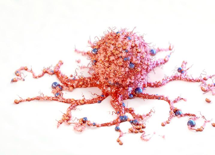
“This tumor didn’t shrink, we’ve all seen tumors shrink—this tumor evaporated,” he said.
The 127th American Academy of Ophthalmology (AAO) Meeting was held on November 3 to 6, 2023, in San Francisco, California, USA. Reporting for this story took place during the event.
