
Deep Learning in Anterior Segment OCT
Optimizing Biometric Analysis and Scleral Spur Detection
Abstract
Swept-source anterior segment optical coherence tomography (AS-OCT) is increasingly becoming a central tool in ophthalmology, providing high-resolution, crosssectional images of the anterior chamber. Swept-source provides the depth and clarity that other types of OCT and Scheimpflug imaging do not. However, the extraction of quantitative biometric data and the identification of the scleral spur remain time-consuming and susceptible to high variability. This limitation hinders the widespread adoption of quantitative AS-OCT analysis in anterior chamber angle and metrics assessment in clinical practice.
Deep learning (DL) algorithms offer a revolutionary solution to this challenge. Recent studies have demonstrated that DL models can achieve expertlevel performance in both quantitative biometry and scleral spur detection tasks. This paper delves into the application of DL for SS-OCT image analysis, specifically focusing on its ability to optimize these processes.
The advantages of DL compared to traditional methods are multifaceted. First, DL algorithms can achieve
superior accuracy in measurements and scleral spur detection due to their ability to learn complex relationships within large datasets. Second, DL offers a significant gain in efficiency by automating previously manual tasks, allowing for faster analysis and improved workflow. Finally, DL methodologies provide an objective approach, potentially reducing the subjectivity inherent in human interpretation.
This paper explores the potential of DL-powered ASOCT analysis to transform clinical practice. Using the ANTERION® Multidisciplinary Imaging Platform (Heidelberg Engineering GmbH, Heidelberg, Germany) as an example, we showcase the performance of DL algorithms in real-world clinical settings. By demonstrating the feasibility and benefits of DL integration, we pave the way for a future where automated, objective and efficient AS-OCT analysis empowers clinicians to provide enhanced patient care.
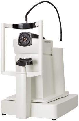
ANTERION® Multidisciplinary Imaging Platform

Keywords
Angle-closure, Anterior chamber measurements, Sweptsource OCT (SS-OCT), Anterior segment OCT (AS-OCT), Deep learning, ANTERION OCT
Abbreviations
AC (anterior chamber), ACA (anterior chamber angle), ACD (anterior chamber depth), AOD (angle opening distance), AS-OCT (anterior segment OCT), CNN (convolutional neural network), DL (deep learning), ICC (intraclass correlation coefficients), ICL (implantable collamer lens), PAC (primary angle-closure), PACD (primary angleclosure disease), PACG (primary angle-closure glaucoma), PACS (primary angle-closure suspect), RC (repeatability coefficient), ROI (region of interest), SC (Schlemm’s canal), SS (scleral spur), TISA (trabecular iris space area), TM (trabecular meshwork)
Introduction
Anterior segment optical coherence tomography (ASOCT) has become indispensable in clinical practice and research in ophthalmology. It provides high-resolution, cross-sectional images of the anterior segment, enabling detailed assessment of structures like the cornea, iris and angle. There is clinical utility of AS-OCT across a wide range of anterior segment pathologies, such as conjunctival neoplasia, pterygium, scleritis, keratoconus, corneal dystrophies and infectious/non-infectious keratitis.1 However, unlocking the full potential of AS-OCT relies on increasing the accuracy of image analysis.
The role of deep learning algorithms in optimizing the precision of biometric measurements associated with scleral spur identification
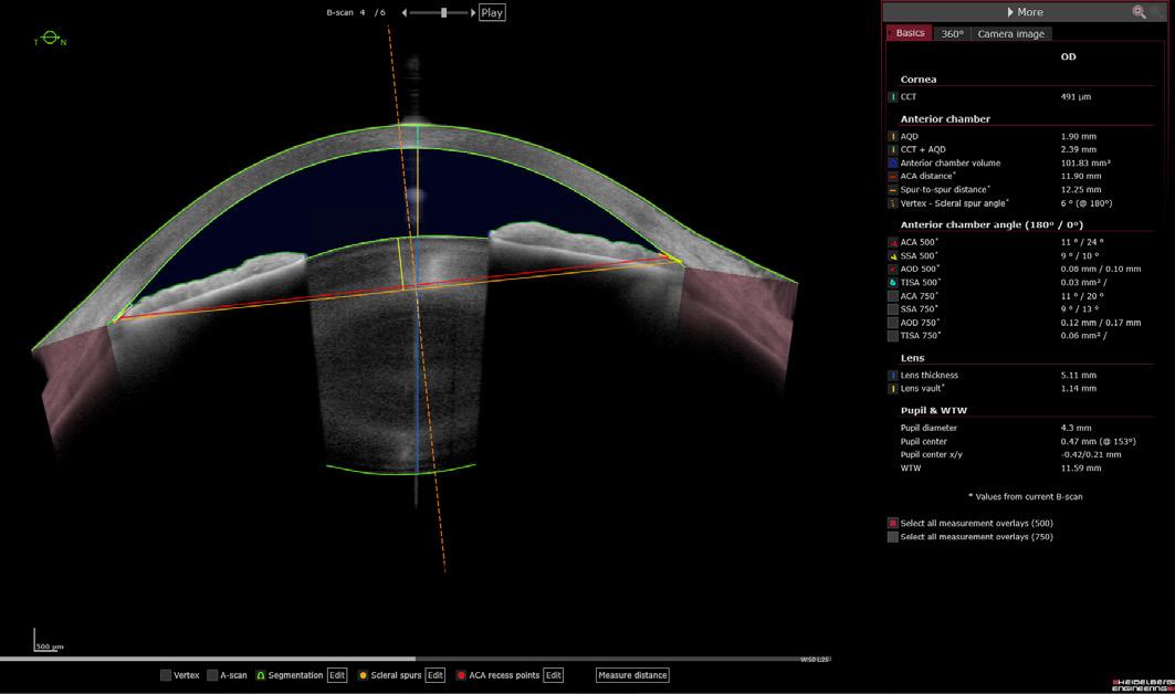
The scleral spur (SS) is a rigid projection of the sclera containing elastic, collagenous fibers and myofibroblasts. It is situated within the ciliary muscle fibers at the base of the trabecular meshwork (TM) and is thought to play a crucial role in maintaining the structure of both the TM and Schlemm’s canal (SC), critical components of aqueous humor outflow pathways.2,3The scleral spur is a crucial landmark in AS-OCT imaging, influencing various biometric parameters.4 Recent advances in automated scleral spur detection incorporating deep learning (DL) algorithms, particularly convolutional neural networks (CNNs)5 into quantitative OCT, represent a powerful new approach to the in-depth evaluation of the anterior segment. DL algorithm-enhanced quantitative OCT offers several advantages over traditional methods.6
Firstly, the use of quantitative OCT with scleral spur detection offers objectivity and repeatability compared to subjective and variable gonioscopy for assessing the anterior chamber angle (ACA)1,7, reducing human error and improving the consistency of clinical data. Secondly, scleral spur-based parameters like angle opening distance (AOD) and trabecular iris space area (TISA) have shown promise in reflecting anatomical variations and potentially predicting disease progression in primary angle-closure glaucoma (PACG).8 Additionally, these measurements can improve IOL power calculations by providing more precise anterior chamber depth (ACD) measurements, leading to better surgical outcomes.9
In a recent publication in the British Journal of Ophthalmology, Bolo and collaborators investigated whether DL-predicted spur locations, when used for biometric measurements on the ANTERION Multidisciplinary Imaging Platform (Heidelberg Engineering GmbH, Heidelberg, Germany), could achieve consistency comparable to manual marking by multiple human experts.10
Validating a convoluted neural network for automated scleral spur detection in AS-OCT
Dr. Bolo and colleagues developed and internally tested DL algorithms for automated scleral spur detection in ASOCT images.10 A set of nearly 4800 AS-OCT images from over 360 patients was divided into training and testing datasets to train the algorithm. An expert ophthalmologist marked the scleral spur locations in the training images, which were then used to train a convolutional neural network (CNN) to predict these locations within a defined region of interest (ROI) based on anatomical landmarks.
Next, the researchers employed data augmentation techniques to improve the robustness of the CNN. Finally, they selected two operating points for further analysis based on the trade-off between minimizing false positive detections and ensuring a high rate of true positive detections.
The validation dataset utilized in the study was acquired from patients undergoing routine eye examinations at two clinics. A total of 40 patients were recruited from March 2021 to August 2021, with exclusion criteria including corneal opacities and prior ocular trauma. AS-OCT images were captured using a standardized protocol by trained technicians. Three graders with varying experience levels marked scleral spur locations in all images, and automated algorithms also predicted these locations.
Next, eight scleral spur-based biometric parameters were measured automatically by the ANTERION software. A subset of images with narrow angles was also identified based on a predefined AOD500 threshold.
Superior scleral spur detection and biometric measurements in AS-OCT images by DL algorithms
The study (Boloet al.) investigated the agreement between human graders and deep learning (DL) algorithms (TPR95 and FPR4) for identifying scleral spurs and measuring anterior chamber angles (ACAs) using high-resolution OCT images from ANTERION.10 A total of 1308 images from 117 participants were analyzed.
The results showed that the reference grader marked fewer scleral spurs compared to other graders and the TPR95 algorithm. Post-hoc analysis revealed that most of these discrepancies involved images with eyelid artifacts or other obscuring features. In addition, the median differences in scleral spur location between the reference grader and other graders/algorithms ranged from 52.6 μm to 79.4 μm. Notably, the ANTERION® DL algorithms exhibited the smallest median differences compared to the reference grader.
The agreement between the reference grader and all graders/algorithms in measuring various biometric parameters (AOD500, TISA500, ACW, LV) was excellent
(ICC range: 0.918-0.997). This high level of agreement held true even for narrow angles (AOD500 < 150 μm), although ICCs for anterior chamber angle width measurements were generally lower in this subset. Bland-Altman plots further confirmed consistent agreement across the entire measurement range. The ANTERION DL algorithms demonstrated performance comparable to or exceeding the agreement between human experts for narrow-angle images.
These findings suggest that both human graders and DL algorithms can achieve reliable scleral spur detection and biometric measurements from AS-OCT images. The ANTERION TPR95 algorithm, in particular, exhibited high accuracy and may offer advantages over human graders, especially for narrow-angle images.
ANTERION® DL-based OCT image analysis offers benefits for clinical practice
Limited access to accurate and automated measurements of scleral spur-based biometrics has hampered clinical advancements. The study by Bolo and colleagues demonstrates that DL algorithms like those on the ANTERION system can generate biometric measurements highly correlated with those obtained by human graders, including for narrow angles. This opens doors for broader application of quantitative AS-OCT analysis in clinical settings.10
Automated, expert-level biometric measurements hold promise for improved evaluation and management of various eye conditions, including primary angle-closure disease (PACD), refractive errors, and cataracts. Biometric parameters associated with scleral spur location, such as ICL sizing potential, can inform treatment decisions for PACD and IOL selection procedures. Furthermore, the scleral spur could serve as a reference plane to better inform the propensity for IOL tilt or decentration.
The study by Bolo et al. offers several advantages over previous investigations into automated scleral spur detection.10 Firstly, the DL algorithms utilized maintained expert-level performance in a real-world clinical setting. This setting involved a diverse patient cohort, recruited across various ages and ethnicities from comprehensive and glaucoma clinics during routine eye care procedures. Importantly, the validation cohort and environment were entirely independent of those used to develop the algorithm.
These findings support the generalizability and potential for widespread implementation of DL algorithms across diverse clinical practices. This contrasts with prior studies that utilized smaller and more homogenous patient populations.11,12
Secondly, the validation dataset included images containing eyelids or other imaging artifacts. This approach allowed the research team to assess the variability in both the human grader and algorithm confidence levels for scleral spur detection and to evaluate the impact of these artifacts on performance.
Conclusions and future directions
DL has emerged as a powerful tool for optimizing quantitative biometric analysis in AS-OCT. The DL algorithm on the ANTERION system achieved expertlevel performance by automating scleral spur detection and segmentation of relevant structures, leading to more efficient, objective, and consistent clinical evaluations.
Despite significant advancements in the field, specific challenges remain. Firstly, the generalizability of DL models across different AS-OCT devices and image acquisition protocols needs further investigation. Additionally, incorporating explainability into DL models would enhance trust and understanding of their decisionmaking processes. Therefore, future research should explore utilizing DL for disease classification based on AS-OCT features and integrating these algorithms into clinical workflows for seamless adoption. As research progresses, DL can potentially revolutionize the analysis of AS-OCT data, impacting clinical practice and furthering our understanding of anterior segment diseases.
References
1. Chong YJ, Azzopardi M, Hussain G, et al. Clinical Applications of Anterior Segment Optical Coherence Tomography: An Updated Review. Diagnostics (Basel). 2024;14(2):122.
2. Tamm ER, Koch TA, Mayer B, Stefani FH, Lütjen-Drecoll E. Innervation of myofibroblast-like scleral spur cells in human monkey eyes. Invest Ophthalmol Vis Sci. 1995;36(8):1633-1644.
3. Lütjen-Drecoll E. Functional morphology of the trabecular meshwork in primate eyes. Prog Retin Eye Res. 1999;18(1):91-119.
4. Salim S. The role of anterior segment optical coherence tomography in glaucoma. J Ophthalmol. 2012;2012:476801.
5. Ronneberger O, Fischer P, Brox T. U-net: Convolutional networks for biomedical image segmentation. Lecture Notes in Computer Science (including subseries Lecture Notes in Artificial Intelligence and Lecture Notes in Bioinformatics). 2015;9351:234-241.
6. Soh ZD, Tan M, Nongpiur ME, et al. Deep Learning-based Quantification of Anterior Segment OCT Parameters. Ophthalmol Sci. 2023;4(1):100360.
7. Pujari A, Sharma N. The Emerging Role of Anterior Segment Optical Coherence Tomography in Cataract Surgery: Current Role and Future Perspectives. Clin Ophthalmol. 2021;15:389-401.
8. Yang G, Li K, Yao J, et al. Automatic measurement of anterior chamber angle parameters in AS-OCT images using deep learning. Biomed Opt Express. 2023;14(4):1378-1392.
9. Triolo G, Barboni P, Savini G, et al. The Use of Anterior-Segment Optical-Coherence Tomography for the Assessment of the Iridocorneal Angle and Its Alterations: Update and Current Evidence. J Clin Med. 2021;10(2):231.
10. Bolo K, Apolo Aroca G, Pardeshi AA, et al. Automated expert-level scleral spur detection and quantitative biometric analysis on the ANTERION anterior segment OCT system. Br J Ophthalmol. 2023:bjo-2022-322328. [Epub ahead of print]
11. Pham TH, Devalla SK, Ang A, et al. Deep learning algorithms to isolate and quantify the structures of the anterior segment in optical coherence tomography images. Br J Ophthalmol. 2021;105(9):1231-1237.
12. Xu BY, Chiang M, Pardeshi AA, Moghimi S, Varma R. Deep Neural Network for Scleral Spur Detection in Anterior Segment OCT Images: The Chinese American Eye Study. Transl Vis Sci Technol. 2020;9(2):18.