Atlas anatomie člověka


Končetiny, stěna trupu Limbs, Body Wall
Miloš Grim
Ondřej Naňka
Ivan Helekal
Miloš Grim, Ondřej Naňka, Ivan Helekal

Upozornění pro čtenáře a uživatele této knihy Všechna práva vyhrazena. Žádná část této tištěné či elektronické knihy nesmí být reprodukována a šířena v papírové, elektronické či jiné podobě bez předchozího písemného souhlasu nakladatele. Neoprávněné užití této knihy bude trestně stíháno
Notice to readers
All rights reserved. No part of this printed or electronic publication may be reproduced, stored and soled in any form without the prior written permission of the Publisher. Unauthorized use of this book will be prosecuted
I.
Končetiny, stěna trupu ● Limbs, Body Wall
Autoři ● Authors:
Prof. MUDr. Miloš Grim, DrSc.
Doc. MUDr. Ondřej Naňka, Ph.D.
Ak. mal. Ivan Helekal
Ilustrace ● Illustrations:
Ak. mal. Ivan Helekal, Mgr. Jan Kacvinský, Archiv Anatomického ústavu, zdroje RTG, CT a MRI snímků –podrobněji viz str. XVI ● Ak. mal. Ivan Helekal, Mgr. Jan Kacvinský, the Archive of Institute of Anatomy, sources of X-ray, CT and MRI images see page XVI
Ilustrace na obálce ● Cover Illustration: Ak. mal. Ivan Helekal
Recenze ● Review: Prof. MUDr. Jan Bartoníček, DrSc.
Konzultace terminologie v anglickém jazyce ● Consultations on English terminology: Prof. MUDr. David Sedmera, D.Sc.
Texty na stranách V, IX–XV, 320 přeložil Timothy W. P. Jackson. ● Texts on the pages V, IX–XV, 320 were translated by Timothy W. P. Jackson.
Vydání odborné knihy schválila Vědecká redakce nakladatelství Grada Publishing, a.s.
© Grada Publishing, a.s., 2014
Cover Design © Grada Publishing, a.s., 2014
Vydala Grada Publishing, a.s.
U Průhonu 22, Praha 7 jako svou 5600. publikaci
Sazba a zlom Ak. mal. Ivan Helekal, Jan Šístek
Počet stran 336
1. vydání, Praha 2014
Vytiskla tiskárna FINIDR s.r.o., Český Těšín
Při přípravě publikace bylo věnováno velké úsilí a pozornost správnosti informací v ní obsažených. Ani vydavatel ani autoři však nenesou zodpovědnost za chyby nebo důsledky vyplývající z použití informací obsažených v knize, nepřebírají jakoukoli odpovědnost za případné následky za použití této publikace a rovněž z toho pro ně nevyplývají žádné právní důsledky.
Every effort has been taken to confirm the accuracy of the information presented. Neither the Publisher nor the authors can be held responsible for errors or for any consequences arising from the use of the information contained herein, and make no warranty, expressed or implied, with respect to the contents of the publication.
ISBN 978-80-247-9538-6 (pdf)
ISBN 978-80-247-4012-6 (print)
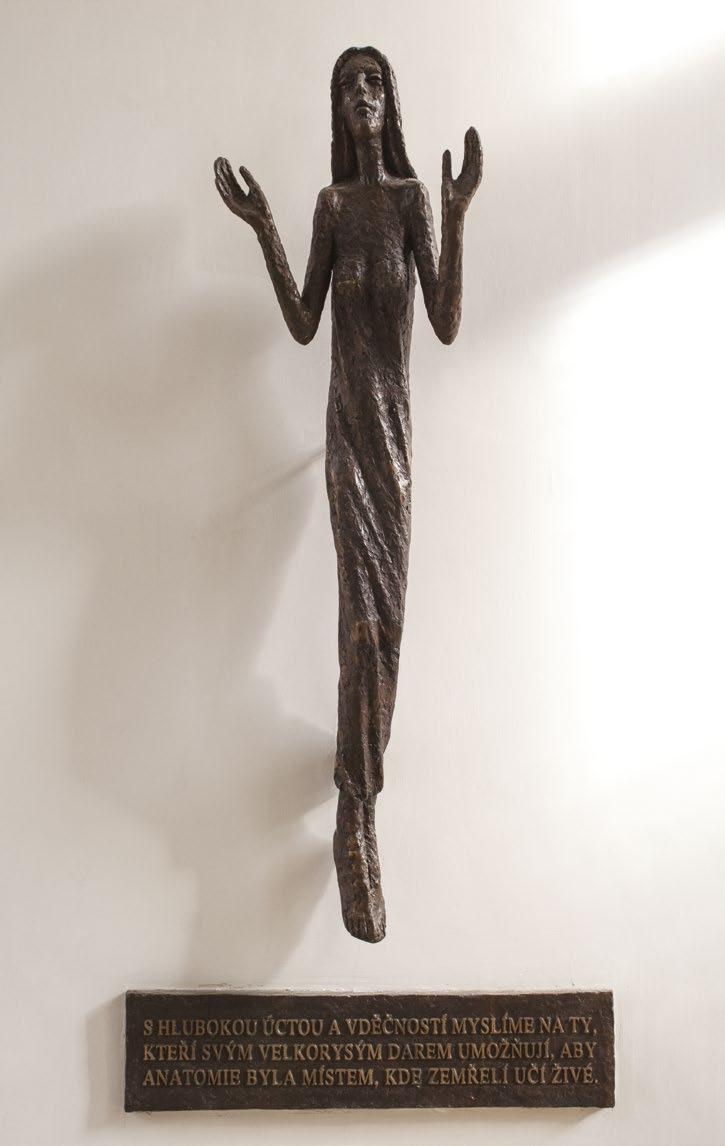
Památce zemřelých, kteří darovali své tělo pro vzdělávání posluchačů lékařství v anatomii, je věnována plastika Olbrama Zoubka nazvaná Thysia. Byla instalována v Anatomickém ústavu 1. lékařské fakulty Univerzity Karlovy v Praze v roce 1998. Její výraz a název tlumočí myšlenku obětování. Připojený nápis vyjadřuje úctu a vděčnost dárcům, kteří umožňují, aby anatomie byla místem, kde zemřelí učí živé. Ani na začátku 21. století nelze tento přístup rovnocenně nahradit. Bez něho by nebyl možný ani vznik tohoto atlasu, který je proto věnován památce našich dárců.
Olbram Zoubek dedicated his sculpture „Thysia“, installed in the Institute of Anatomy of the First Faculty of Medicine, Charles University in Prague in 1998, to the memory of those who donated their bodies for the Anatomy teaching programme. Its name and expression represent the idea of sacrifice –the ultimate offering. The inscription expresses the respect and gratitude to the donors, who made it possible for anatomy to become the place where „the dead teach the living“. Even at the beginning of the 21st century, this gift is irreplaceable; we thus dedicate this atlas to the memory of our donors.
Historie anatomických ilustrací na pražské
lékařské fakultě
Zdroje ilustrací, RTG, CT a MRI snímků
Nomina generalis anatomiae
Části lidského těla
Anatomické krajiny těla
Projekce vnitřních orgánů na povrch těla
Kostra dospělého muže
Přehled kosterního svalstva
Centrální a periferní nervový systém
Hlavní kmeny tepen
Hlavní žilní kmeny a vena portae
Lymfatický systém
Kosti a klouby
Přehled skeletu
Skelet pletence
Skelet volné horní končetiny
Spoje pletence a ramenní kloub
Loketní kloub a předloktí
Zápěstí a ruka
Svaly
Přehled svalů
Svaly ramene a paže
Svaly předloktí
Svaly ruky
Topografie, cévy a nervy
Kožní inervace a povrchové žíly
Fossa infraclavicularis, axilla
Plexus brachialis
A. subclavia et a. axillaris, regio scapularis
Paže, cévy a nervy
Předloktí a loket, cévy a nervy
Zápěstí a ruka, cévy a nervy
DOLNÍ KONČETINA
Kosti a klouby Přehled
Dedication
Contents
Preface
History of anatomical illustrations on the Prague Medical Faculty
Origin of illustrations, radiographs, CT, and MRI
General terms of anatomy
Parts of the human body
Regions of the human body
Surface projections of the thoracic and abdominal viscera
Adult male skeleton
Overview of skeletal muscles
Central and peripheral nervous system
Principal systemic arteries
Principal systemic veins and hepatic portal vein
Lymphatic system
UPPER LIMB
Bones and joints
Overview of the skeleton
Skeleton of shoulder girdle
Skeleton of free part of upper limb
Shoulder girdle joints, shoulder joints
Elbow joint and forearm
Wrist and hand
Muscles
Overview of muscles
Muscles of the shoulder and arm
Muscles of the forearm
Muscles of the hand
Topography, vessels and nerves
Skin innervation and superficial veins
Infraclavicular fossa, axillary region
Brachial plexus
Subclavian and axillary artery, scapular region
Brachial region, vessels and nerves
Forearm and elbow, vessels and nerves
Wrist and hand, vessels and nerves
LOWER LIMB
Bones and joints
Overview of the skeleton
Pánev
Kyčelní kloub
Kolenní kloub
Bérec
Hlezno a noha
Svaly
Přehled svalů
Svaly kyčle a stehna
Svaly bérce
Svaly nohy
Topografie, cévy a nervy
Povrchové žíly, kožní inervace, tepny
Hýždě a stehno, cévy a nervy
Pánev, cévy a nervy
Koleno, cévy a nervy
Bérec, cévy a nervy
Hlezno a noha, cévy a nervy
Kosti a klouby
Páteř a kostra hrudníku
Obratle, kost křížová, kostrč Spojení na páteři
Žebra, sternum a jejich spoje
Svaly Svaly hrudní a břišní stěny Bránice
Tříselný kanál
Pánevní dno
Svaly zad
Topografie, cévy a nervy
Kožní inervace stěny trupu, Headovy zony
Trigonum suboccipitale, páteřní kanál
Cévy stěny trupu
Prs, mléčná žláza
Latinsko-anglicko-český slovník obecných anatomických termínů
Pelvis Hip joint
Knee joint Leg
Ankle and foot
Muscles
Overview of muscles
Muscles of the hip and thigh
Muscles of the leg
Muscles of the foot
Topography, vessels and nerves
Superficial veins, skin innervation, arteries
Gluteal and femoral regions, vessels and nerves
Pelvis, vessels and nerves
Knee, vessels and nerves
Leg, vessels and nerves
Ankle and foot, vessels and nerves
BODY WALL
Bones and joints
Vertebral column and thorax
Vertebrae, sacrum, coccyx
Joints and ligaments of vertebral column
Ribs, breastbone and its connections
Muscles
Muscles of the thoracic and abdominal wall
Diaphragm
Inguinal canal
Pelvic floor
Muscles of the back
Topography, vessels and nerves
Cutaneous innervation, Head´s zones
Suboccipital triangle, vertebral canal
Vessels of the body wall
Breast, mammary gland
Latin-English-Czech dictionary of general terms of anatomy
Jsme rádi, že můžeme předložit čtenářům prvý díl Atlasu anatomie člověka. Je věnován anatomii končetin a stěny trupu. Prvá kapitola je úvodem do anatomie. V dalších kapitolách následuje anatomie horní končetiny, dolní končetiny a stěny trupu. Nejprve je vždy popsán skelet, pak jeho vazivové spoje a klouby, dále svaly a nakonec poloha a průběh nervů a cév v rámci kapitoly topografická anatomie. Ke každému vyobrazení je připojen vysvětlující text v češtině/latině a zároveň v angličtině, aby atlas nalezl uplatnění u všech studentů. Je připojen slovník nejčastěji používaných latinsko-řeckých anatomických termínů a jejich překlad do angličtiny a češtiny.
Každý oddíl má své logo a každá kapitola má svou signální barvu. Obrázky nejsou samostatně číslovány. Vztahuje se na ně číslo stránky, na které jsou otištěny, a tak je na ně odkazováno v rejstříku. Pokud je na jedné stránce více obrázků, jsou v abecedním pořadí označeny malými písmeny. Anatomické útvary jsou popsány v současné mezinárodní Terminologia Anatomica (1998). Pro jednotnou orientaci je vždy znázorněna končetina pravé strany. Celkem je v atlase obsaženo 476 obrázků. Jsou to anatomické obrazy a schémata (413) a snímky (63) pořízené některou ze zobrazovacích metod (RTG, CT, MRI).
Druhý díl zahrnuje anatomii hlavy, krku, hrudníku, břicha a pánve. Jeho úprava i grafické provedení je shodné s prvním dílem.
Atlas jsme připravili během let 2011–2013 v Anatomickém ústavu 1. lékařské fakulty Univerzity Karlovy v Praze. Vedla nás k tomu snaha vytvořit moderní anatomický atlas, protože dosud jediným atlasem české provenience je Janošíkův atlas z roku 1904. Dosud byla potřeba anatomického atlasu opakovaně řešena zakoupením licence k vydání zahraničního díla, ke kterému byly do češtiny překládány doprovodné a vysvětlující texty. Například my sami jsme před šesti lety připravili překlad Sobottova Atlasu anatomie člověka. Přitom jsme se začali zabývat úmyslem vytvořit vlastní atlas. Nakladatelství Grada Publishing nám nabídlo jeho
We are pleased to present our readership with the first volume of the Atlas of Human Anatomy. It contains the anatomy of limbs and body wall. The first chapter is an introduction to anatomy and the following chapters deal with the anatomy of the upper limb, lower limb and body wall. Each chapter starts with the skeletal description followed by the description of joints and of relevant muscles. The position and passage of nerves and blood vessels are to be found in the chapter dedicated to topographical anatomy. Each image is accompanied by an explanatory text in Czech, Latin and English. As such, the atlas serves as a universal tool for all students. The attached glossary of frequently used Latin/Greek terms carries their English and Czech equivalents.
The atlas is divided into parts distinguished by different logos and reference colours. The plates are not numbered individually, instead, they carry a reference to the page on which they can be found. In instances where multiple images are found on the same page, they are indexed in alphabetical order by lower case letters. Anatomical entities are described using current international terminology, Terminologia Anatomica (1998). The right limb is consistently presented for the purpose of unified orientation. The atlas contains 476 images, of which 413 are anatomical images and schemes and 63 are results of medical imaging (X-ray, CT, MRI).
The second volume of the atlas presents the anatomy of the head, neck, chest, abdomen and pelvis. Its presentation is identical to the first volume.
This atlas has been compiled between 2011 and 2013 at the Institute of Anatomy of the First Faculty of Medicine, Charles University in Prague. It has been our aim to create an up to date atlas of anatomy, as the only other atlas of Czech provenience was that of Janošík, dating from 1904. The need for an anatomy atlas has been thus far met by obtaining a licence to publish foreign titles that were accompanied by explanatory texts
realizaci. Rozhodli jsme se přijmout tuto výzvu, neboť jsme věděli, že můžeme využít rozsáhlý soubor anatomických ilustrací v archivu Anatomického ústavu a že můžeme počítat s tvorbou nových ilustrací v našem výtvarném kabinetu. Akademický malíř Ivan Helekal a Mgr. Jan Kacvinský se zde věnují vědecké ilustraci již více než 20 let a jsou v tom velmi dobří.
Archiv Anatomického ústavu obsahuje rozsáhlý soubor anatomických ilustrací. Jsou to sépie a kolorované akvarely vytvořené podle preparátů, které připravili anatomové působící na ústavu během 20. století. Autory těchto ilustrací jsou zejména Emil Illing a Stanislav Macháček, kteří je připravovali pro Topografickou anatomii Karla Weignera (1. vydání 1915–1926), Soustavnou anatomii Ladislava Borovanského (1. vydání 1955) a pro stručnou topografickou anatomii, kterou měl v úmyslu napsat Ladislav Borovanský. Další část archivu tvoří kresby, které pocházejí z pracovišť spoluautorů Borovanského učebnice, zejména z Brna a z Plzně. Jejich autoři jsou uvedeni v kapitole o historii anatomických ilustrací.
Prohlédli jsme a roztřídili archivované kresby. Ivan Helekal je ofotografoval a převedl do elektronické podoby. Pomocí grafického programu je zrestauroval a obohatil o barvu. Doplnili jsme je, popsali a připravili k barevné reprodukci. Většina z nich byla dosud publikována pouze černobíle a bez využití moderních technik knihtisku. Těší nás, že jejich nynější publikace připomene mnohaleté úsilí řady našich předchůdců. Několik těchto kreseb jsme vložili do atlasu jako frontispis na volné listy v původním stavu před restaurováním. Celkem pochází z archivu 224 ilustrací.
Významnou součást prvního dílu tvoří 165 nových obrazů zhotovených Ivanem Helekalem (iniciály IH) a 24 obrazů zhotovených Janem Kacvinským (JK). V jejich ilustracích jsou anatomické struktury zobrazovány ve výtvarné zkratce, která potlačuje detail a zdůrazňuje funkci. Ilustrace tak přechází od popisného provedení k výkladovému pojetí a dokládá potřebu anatomické kresby i v době
translated into Czech. As an example, six years ago we translated by our own means Sobotta’s Atlas of Human Anatomy and that is where the idea to compile our own atlas originated. Grada Publishing offered us their assistance in this venture, and we decided to accept the challenge, as we were confident that we could use our extensive archive of medical imaging and that new images could be provided by our creative arts department. The resident artist Ivan Helekal and Jan Kacvinský, MA, dedicated more than 20 years of their work to scientific illustration.
The Institute of Anatomy archive carries an extensive collection of anatomical illustrations, sepia prints and colour aquarelles of preserved body parts, supplied by anatomists, who worked in the institute during the 20th century. These illustrations were created predominantly by Emil Illing and Stanislav Macháček in preparation for the Topographical Anatomy by K. Weigner (1st edition 1915–1926), the Systematic Anatomy of the Man by L. Borovanský (1st edition 1955) and the Concise Topographical Anatomy, intended to be compiled by L. Borovanský. Another part of the archive carries drawings created in workplaces of co-authors of the Borovanský textbook, namely in Brno and Pilsen. The individual authors are named in the chapter on the history of anatomical illustrations.
We inspected and sorted archived illustrations. Ivan Helekal photographed and digitalized them. Graphic software helped us restore the images and add colour. The images were completed, labeled and prepared for colour publication. Most of the images were thus far published only in black and white, without the use of technological advances of contemporary book printing. We are delighted that their publication now will remind all of the historic efforts of many of our predecessors. We have included several drawings in their re-prestoration state on unbound pages as a frontispiece of our atlas. In total, 224 illustrations originate from the archive.
digitální fotografie. Často je používáno didaktické schéma, které je konfrontováno s obrazem získaným výpočetní tomografií a technikou MRI.
Poslední součástí atlasu jsou RTG, CT a MRI snímky získané z řady pražských klinických pracovišť. Jsou mezi nimi i 3D rekonstrukce pořízené z těchto snímků, dále snímky z digitální subtrakční angiografie a CT angiografie. Jsou zobrazeny struktury dospělých jedinců a také vyvíjející se skelet v růstovém období. Tato část atlasu zdůrazňuje klinický aspekt anatomie v současném pojetí tohoto oboru jako základního předmětu preklinické výuky lékařství.
Děkujeme všem, kteří se podíleli na přípravě atlasu a přispěli k jeho vydání. Za spolupráci při tvorbě ilustrací děkujeme Mgr. Janu Kacvinskému, který je autorem schémat k výkladu obrazů získaných technikou MRI.
Za odbornou spolupráci při popisování ilustrací děkujeme našim kolegům prof. Dr. med. Zdeňku Halatovi, MUDr. Zdeňce Novákové, MUDr. Pavlu Šnajdrovi, Ph.D., a Petru Krůpovi, lektoru a posluchači 1. LF UK. Za revizi anglických textů v názvech obrázků děkujeme prof. MUDr. Davidu Sedmerovi, D.Sc. Slovník laskavě přehlédla PhDr. Dana Svobodová z Ústavu dějin lékařství a cizích jazyků 1. LF UK. Děkujeme jí za cenné připomínky.
Za přenos popisek do elektronické podoby obrázků děkujeme Evě Davidové, Martině Línové a posluchačům 1. LF UK Markétě Válkové, Kláře Steffalové, Michalu Žigovi, Janu Novákovi a Filipu Jonášovi.
Za poskytnutí RTG, CT a MRI snímků děkujeme prof. MUDr. Josefu Vymazalovi, DrSc., z radiodiagnostického oddělení Nemocnice Na Homolce, MUDr. Jiřímu Benešovi z Radiodiagnostická kliniky
1. LF UK a VFN, primářce MUDr. Janě Votrubové, CSc., z radiodiagnostického oddělení Thomayerovy nemocnice a prof. MUDr. Janu Bartoníčkovi, DrSc., z Kliniky traumatologie pohybového aparátu 1. LF UK a ÚVN v Praze-Střešovicích.
165 new images created by Ivan Helekal (initialed IH) and 24 images created by Jan Kacvinský (initialed JK) represent a substantial part of the first volume of the atlas. The illustrations of anatomical structures are simplified, suppressing detail in order to illustrate their function. An illustration thus progresses from descriptive rendering to explanatory conceptualization and documents the persisting need for anatomical drawing in times of digital photography. Didactic schemes are often used and compared with computed tomography and MRI images.
X-ray, CT and MRI images from various clinical institutions in Prague form the concluding part of the atlas. Among these, we can also find 3D image reconstructions, images of digital subtraction angiographies, and CT angiographies. Images include structures of adult individuals and skeletal structures in development. This part of the atlas underlines the clinical aspect of anatomy, currently perceived as the basis of pre-clinical education in medicine.
We would like to extend our thanks to all who worked on the compilation of this atlas and contributed to its publication. We would like to thank Jan Kacvinský, MA, for his cooperation in creating the illustrations and for creating the schemes for interpretation of MRI images.
We would like to thank our colleagues for their professional cooperation in the description of illustrations, namely Prof. MUDr. Zdeněk Halata, MUDr. Zdeňka Nováková, MUDr. Pavel Šnajdr, Ph.D., and Petr Krůpa, lecturer and student of the First Faculty of Medicine. We are grateful to Prof. MUDr. David Sedmera, D.Sc., for his revision of the English texts in the titles of illustrations and to PhDr. Dana Svobodová, of the Institute for History of Medicine and Foreign Languages, for her kind revision of the dictionary and for her valuable recommendations.
Our thanks also go to Eva Davidová, Martina Línová and students of the First Faculty of Medicine
Vedoucímu zdravotnické redakce nakladatelství Grada Publishing MUDr. Miroslavu Lomíčkovi děkujeme za návrh vydat tento atlas, za řešení jeho technických parametrů a za ošetření autorských a majetkových práv vázaných na kresby pocházející z archivu. Naše poděkování za recenzi a cenné připomínky patří prof. MUDr. Janu Bartoníčkovi, DrSc.
Atlas je určen především medikům, kteří se anatomii učí také studiem a kreslením obrázků, neboť anatomický poznatek tlumočený obrazem je názornější a určitější než text. Věříme, že atlas najde uplatnění také u lékařů různých oborů, u studentů přírodních věd i pracovníků nelékařských zdravotnických oborů. Lze ho využít jako doplněk učebnic anatomie i jako samostatnou publikaci. Přáli bychom si, aby atlas splnil svůj účel při studiu anatomie i jako průvodce v otázkách klinické potřeby anatomie. Budeme rádi za informace o nalezených nepřesnostech, kterých jsme se nechtěně dopustili, a uvítáme návrhy k jeho zlepšení. Doufáme, že atlas naplní v přijatelné míře očekávání, se kterým k němu bude přistupováno.
Miloš Grim, Ondřej Naňka, Ivan Helekal Praha, červen 2014
Markéta Válková, Klára Steffalová, Michal Žiga, Jan Novák and Filip Jonáš for the transfer of descriptions into digital images.
We would like to address thanks to Prof. MUDr. Josef Vymazal, DrSc., of the Department of Radiology of the Na Homolce Hospital, MUDr. Jiří Beneš, Ph.D., of the Department of Radiology of the First Faculty of Medicine and General Teaching Hospital, MUDr. Jana Votrubová, CSc., of the Department of Radiology of the Thomayer´s Hospital, and Prof. MUDr. Jan Bartoníček, DrSc., Department of Orthopaedic Trauma First Faculty of Medicine and Military University Hospital in Prague, for providing us with X-ray, CT and MRI images.
Sincere thanks to MUDr. Miroslav Lomíček, the editor in chief of the Grada Publishing health department for his suggestion to publish this atlas, for his technical expertise and for his resolution of copyright and property rights relating to the archived images. Our thanks go to Professor Jan Bartoníček for his revision of the manuscript and invaluable recommendations.
The atlas is mainly destined for students of medicine, who are also learning anatomy through the study of images and drawing. Acquisition of anatomical knowledge through study of images is more descriptive and precise than the study of text alone. We believe that the atlas will serve physicians of various specializations, students of natural sciences and those working in non-medical health related occupations. The atlas can be used as a supplement of an anatomy textbook or as an independent publication. It is our wish that the atlas serves its purpose in the study of anatomy and in being a guide in the questions of clinical necessity of anatomy. We would welcome notification of inaccuracies that occurred without our intent and any recommendations for improvement. We hope that the atlas meets all expectations and does not disappoint.
Miloš Grim, Ondřej Naňka, Ivan HelekalPrague, June 2014
V české anatomické literatuře nemají anatomické atlasy výraznou tradici, přestože se anatomické ilustraci věnovala celá řada výtvarníků. Nejstarším a jediným samostatným atlasem je Anatomický atlas prof. Jana Janošíka. Autor ho vydal vlastním nákladem ve dvou částech v roce 1900 a 1904. Ilustrace nakreslil Josef Rejsek, technický a výtvarný spolupracovník prof. Janošíka. Atlas obsahuje 532 zčásti kolorovaných vyobrazení, která Janošík velmi oceňoval po věcné i výtvarné stránce. Dalších vydání se atlas nedočkal, ale prof. Janošík poskytl prof. Weignerovi 166 Rejskových vyobrazení pro I. až III. díl Topografické anatomie, ve které prof. Weigner spojil učebnici s atlasem.
Prvé vydání všech pěti dílů Topografické anatomie prof. Weignera vyšlo postupně v letech 1915–1926 a obsahuje celkem 680 ilustrací. Ve druhém vydání (1930–1938) to již je 916 obrázků, které většinou pocházejí z ruky Weignerova výtvarníka akad. malíře Emila Illinga. V blíže neurčené míře se na ilustracích Topografické anatomie podíleli i další ilustrátoři. V předmluvě k 1. vydání V. dílu (1924) uvedl prof. Weigner jména vysokoškolských studentů, které učil kreslit anatomické obrazy a které se nepříliš úspěšně snažil zapojit do přípravy ilustrací. V předmluvě ke 2. vydání 5. dílu (1938) uvádí prof. Borovanský jméno jednoho z nich, L. Bartla, který přispěl jako „host“ k ilustracím ve 2. vydání.
Výtvarným provedením si jsou ilustrace v Topografické anatomii navzájem velmi podobné. Lze předpokládat, že jejich provedení ovlivňoval Emil Illing, který měl výtvarné vzdělání. Individuální autorství není vyjádřeno, protože žádná z nich není signována. Řada těchto vyobrazení se v našem archivu zachovala a pro první díl atlasu jsme některá z nich restaurovali a použili. Citujeme je podle zdroje jako ilustrace z Topografické anatomie Karla Weignera.
Soustavná anatomie člověka I a II byla připravena za redakce Ladislava Borovanského (pět vydání
The atlas of anatomy is not a traditional publication in Czech anatomical literature despite the fact that numerous artists have created anatomical illustrations. The only independent atlas, and the oldest, is the Atlas of Anatomy by Professor Jan Janošík, published and financed by the author himself in two volumes, in 1900 and 1904. Josef Rejsek, Professor Janošík’s technical and artistic collaborator, created illustrations included in the atlas. The atlas contained 532 partly coloured images that Janošík greatly appreciated for their factual and artistic value. No further editions of this atlas were published, however, Professor Janošík provided Professor Weigner with 166 images created by Rejsek in order to publish them in the three volumes of the Topographical Anatomy. In this book Professor Weigner combined a textbook with an atlas.
The first edition of the Topographical Anatomy in five volumes by Professor Weigner was gradually published between 1915 and 1926 and included 680 illustrations. The second edition (1930–1938) included 916 illustrations, most of which were created by Weigner’s artist, painter Emil Illing. Other illustrators worked to a certain extent on illustrations for the Topographical Anatomy. K. Weigner taught anatomical drawing to university students and he tried to involve them in the preparation of illustrations without much success. These students were named in the preface to the first edition of the fifth volume (1924). L. Borovanský names one of them in the preface to the second edition of the fifth volume, L. Bartl, who as a “guest” contributed to the illustrations in the second edition.
As far as the artistic execution of the illustrations in the Topographical Anatomy is concerned, these illustrations are very similar. It is possible to assume that E. Illing, an artist by education, influenced their execution. The individual illustrations are not signed and therefore not assigned to particular authors. Many of the works were preserved in
v letech 1955–1976). Většina ilustrací v této učebnici byla nově vytvořena panem Stanislavem Macháčkem, vědeckým ilustrátorem prof. Borovanského. Kromě toho byly použity ilustrace z Weignerovy Topografické anatomie a také ilustrace od malířů z pracovišť spoluautorů této kolektivně připravené knihy. Byly použity obrazy od malířů (V. Gabrielová, J. Indra, V. Kacerovský, I. Luklová-Marková, Z. Placheta, R. Smetanová) z pracovišť spoluautorů této kolektivně připravené knihy. Signatura je však pouze na obrazech, které zhotovil S. Macháček, V. Kacerovský a R. Smetanová. Řadu těchto ilustrací jsme použili v prvém dílu atlasu. Pokud jsou signovány, citujeme je podle autorů, nesignované kresby citujeme podle zdroje.
V letech 1995–1996 bylo publikováno 12 dílů Systematické, topografické a klinické anatomie, které připravili pracovníci Anatomického ústavu 1. LF UK za redakce prof. Pavla Petrovického. Později byly tyto texty staženy do tří svazků, které byly vydány na Slovensku v roce 2001. Ilustrace tvoří převážně černobílé kresby od různých autorů, včetně některých autorů textu.
V roce 1995 připravil prof. Václav Seichert Malý atlas anatomie zaměřený na topografickou anatomii. Obsahuje 185 schematických černobílých perokreseb, které autor sám navrhl a nakreslil. Barevně jsou na nich zdůrazněny arterie, vény, žlázy, mízní kmeny a uzliny.
Další anatomické ilustrace Ivana Helekala a Jana Kacvinského jsou obsaženy v kolektivní publikaci Základy anatomie (redakce prof. Miloš Grim a prof. Rastislav Druga). V letech 2001–2005 vyšel 1., 3. a 5. díl a v roce 2011 a 2013 další dva svazky odpovídající 4. dílu. Helekalovy a Kacvinského ilustrace obsahuje také publikace Základy anatomie, kterou pro posluchače bakalářských oborů napsali doc. Ondřej Naňka a doc. Miloslava Elišková. Jan Kacvinský ilustroval také Aplikovanou anatomii pro fyzioterapeuty a maséry, kterou napsali doc. Miloslava Elišková a prof. Oldřich Eliška.
our archive and some of them were restored and used in the first volume of the atlas. The images are cited according to their origin, i.e. illustrations from the Topographical Anatomy by K. Weigner.
Systematic Anatomy of Man I and II were prepared for publication under the editorship of L. Borovanský (five editions in 1955–1976). Most of the illustrations in the textbook were newly created by Stanislav Macháček, professor Borovanský’s scientific illustrator. Illustrations from Weigner’s Topographical Anatomy were also used, as were illustrations created by artists working alongside co-authors of this book. Illustrations by artists, such as V. Gabrielová, J. Indra, V. Kacerovský, I. Luklová-Marková, Z. Placheta and R. Smetanová, who worked with co-authors, were added. We used many of these illustrations in the first volume of the atlas. Signed illustrations are cited by their authors, unsigned illustrations are cited by their origin.
12 volumes of the Systematic, Topographic and Clinical Anatomy were prepared for publication by Professor Pavel Petrovický of First Faculty of Medicine and his colleagues, and were published in 1995–1996. The text was subsequently condensed into 3 volumes, and published in Slovakia in 2001. Illustrations consisted predominantly of black and white drawings by various authors, and several authors of the texts created some of the drawings themselves.
Professor Václav Seichert prepared for publication his Small Atlas of Anatomy that focused on topographical anatomy in 1995. It contained 185 schematic black and white ink drawings, created and designed by the author himself. Only arteries, veins, glands, lymphatic vessels and nodes were highlighted by colour.
Ivan Helekal and Jan Kacvinský created further anatomical illustrations and these were included in a collective publication named Basic Anatomy (prepared for publication by Professor Milos Grim and Professor Rastistav Druga). The 1st, 3rd and 5th volumes were published between 2001 and
Od roku 1987 vychází učebnice soustavné anatomie Anatomie 1–3 prof. Radomíra Čiháka. V současné době vyšlo již 3., upravené a doplněné vydání jejího 2. dílu. První a druhý díl ilustroval akad. malíř Milan Med a jeho spolupracovnice Ivona Šebelková a Helena Fügnerová. Jejich kresby doplňuje 30 ilustrací od Stanislava Macháčka. Výtvarníkem třetího dílu je Ivan Helekal. V posledním vydání obsahuje Anatomie 1–3 celkem 1096 anatomických ilustrací.
Přicházíme-li nyní téměř přesně po 110 letech se samostatným anatomickým atlasem, máme tedy z čeho vycházet a na co navazovat. Připravovali jsme tento atlas s vědomím, že je to pomůcka, kterou potřebuje každý lékař.
2005, and two more volumes were published in 2011 and 2013, which represented volume 4 of the publication. Further illustrations by Helekal and Kacvinský can be also found in Basic Anatomy, written for students of bachelor programmes, by Associate Professor Ondřej Naňka and Associate Professor Miloslava Elišková. Jan Kacvinský also illustrated Applied Anatomy for Physiotherapists and Masseurs, which was written by Associate Professor Miloslava Elišková and Professor Oldřich Eliška.
A systematic anatomy textbook, Anatomy 1–3 by Professor Radomír Čihák has been repeatedly published since 1987. Its 3rd amended and supplemented edition of the 2nd volume has been published recently. Artist – painter Milan Med and his colleagues, Ivona Šebelková and Helena Fügnerová, illustrated the first and second volumes. Their drawings were supplemented by 30 illustrations by Stanislav Macháček. Ivan Helekal was the illustrator of the third volume. The last edition of Anatomy 1–3 contains a total of 1096 anatomical illustrations.
We are introducing an independent atlas of anatomy after almost precisely 110 years. We have started from a solid base of previous works. We prepared this atlas knowing that it presents a tool that every physician needs.
Nové ilustrace • New illustrations
Jan Kacvinský: 72b, 79a, 90a, 93a, 102a, b, c, 103a, 105a, 125c, 127b, 173b, 182a, 194a, b, 196a, 202a, 208a, 210a, 211a, 252b, c, 276b, 279b
Ilustrace z archivu Anatomického ústavu (AAU) restaurované Ivanem Helekalem • Illustrations from the Archive of Institute of Anatomy restored by Ivan Helekal
Autor originálu Stanislav Macháček • Originally painted by Stanislav Macháček: 18, 22b, c, 24a, b, 25a, c, 27a, b, 29a, 31a, 34a, b, c, 35a, b, 36a, 38a, b, 40b, 41b, 63c, 64, 65a, b, 68b, 77, 80, 81, 82, 83, 84, 86, 87, 88, 89, 96, 97, 101, 106, 109a, b, d, 117a, b, 118a, b, 120c, 122c, 124a, b, 125a, b, 126, 128a, b, 130a, b, 132a, b, c, d, e, 136a, 139a, 140a, b, 177, 178, 179, 183, 184, 185, 186, 190a, b, 192, 197, 203, 207, 209b, 213, 214, 219a, b, 229d, h, 232b, 234a, 238a, b, 239a, c, 240, 245a, 248a, b, c, d, 254, 258a, c, 259a, b, 261b, 266a, b, 268, 271a, 275, 279a, 283
Autor originálu Václav Kacerovský • Originally painted by Václav Kacerovský: 85, 125c, 199
Ilustrace pocházející z Topografické anatomie K. Weignera • Illustrations from Topographic anatomy of K. Weigner: 32a, b, 63a, b, 66a, b, 67a, b,
Ilustrace pocházející ze Soustavné anatomie L. Borovanského • Illustrations from Systemic anatomy of L.
270, 271b
Zdroje RTG, CT a MRI snímků • Sources of X-ray, CT and MRI images
Radiodiagnostické oddělení Nemocnice Na Homolce (prof. MUDr. Josef Vymazal, DrSc., MUDr. Alena Šnajdrová) • Department of Radiology, Na Homolce Hospital: 79b, 90b, 93b, 102d, 103b, 104a, b, 182b, 191a, b, 193a, b, 195a, b, 196b, 202b, 209a, 210b, 211b, 228, 241a, b, 242a, b, 243a, b
Radiodiagnostická klinika 1. LF UK a VFN (MUDr. Jiří Beneš) • Department of Radiology of the First Faculty of Medicine and General University Hospital: 29b, 74a, 92b, 98b, 111a, 135a, b, 169a, b, 189a, b, 198b, 201a, 224b
Radiodiagnostické oddělení Thomayerovy nemocnice v Praze-Krči (MUDr. Jana Votrubová, CSc.) • Department of Radiology of the Thomayer´s Hospital, Prague-Krč: 30a, b, c, d, e, f, 33a, b, c, 37a, b, c, d, 39a, b, 42d, e, f, 111b, c, 121a, b, 122e, 123a, b, c, d, 133a, b, c, d, e, f, 137a, b, c, 224a, 232a, 235a, b, c, 237a, b, c, 238c, d, 239b, d, 240, 245c, 286b
Klinika traumatologie pohybového aparátu 1. LF UK a ÚVN v Praze-Střešovicích (prof. MUDr. Jan Bartoníček, DrSc.) • Department of Orthopaedic Trauma First Faculty of Medicine and Military University Hospital in Prague: 21a, b, c, d, e, f, 23a, b, 143a, b
Anatomický ústav 1. LF UK v Praze (doc. MUDr. Rastislav Hromádka, Ph.D.) • Institute of Anatomy: 71b, 73a, b, 105b
Nomina generalis anatomiae
Části lidského těla
Anatomické krajiny těla
Projekce vnitřních orgánů na povrch těla
Kostra dospělého muže
Přehled kosterního svalstva
Centrální a periferní nervový systém
Hlavní kmeny tepen
Hlavní žilní kmeny a vena portae
Lymfatický systém
Parts of the human body
Regions of the human body
Surface projections of the thoracic and abdominal viscera
Adult male skeleton
Overview of skeletal muscles
Central and peripheral nervous system
Principal systemic arteries
Principal systemic veins and hepatic portal vein Lymphatic system
a.– arteria, aa. – arteriae
ant. – anterior
dx. – dexter
dist. – distalis
dors. – dorsalis
ext. – extensor, externus
ggl. – ganglion
gl. – glandula
inf. – inferior
lat. – lateralis
lig. – ligamentum, ligg. – ligamenta
m. – musculus, mm. – musculi
med. – medialis
n. – nervus, nn. – nervi post. – posterior proc. – processus prof. – profundus prox. – proximalis r. – ramus, rr. – rami sin. – sinister sup. – superior sperf. – superficialis v. – vena, vv. – venae ventr. – ventralis
anterior, anterius – anterior, situated in front of, or in the forward part of an organ – přední posterior, posterius – posterior, situated in back of, or in the back part of an organ – zadní ventrālis, e – ventral, pertaining to the belly – břišní dorsālis, e – dorsal, pertaining to the back – hřbetní, zádový superior, superius – superior, situated above – horní inferior, inferius – inferior, situated bellow – dolní crāniālis, e – cranial, pertaining to the superior end of the body – situovaný směrem k hlavě, horní caudālis, e – caudal, pertaining to the inferior end of the body – situovaný směrem k „ocasu”, dolní mediālis, e – medial, situated nearer to the median plane – situovaný blíže středové linii laterālis, e – lateral, denoting the position farther from the median plane – boční, postranní mediānus, a, um – intermediate – ležící ve střední čáře sagittālis, e – sagittal, parallel to the median plane of the body – šípový frontālis, e – coronal, pertaining to the forehead – rovnoběžný s čelem (frōns) trānsversālis, e – transversal, placed crosswise – příčný palmāris, e – palmar, pertaining to the palm of the hand – dlaňový plantāris, e – plantar, pertaining to the sole of the foot – chodidlový proximālis, e – proximal, closer to any point of reference (e. g. to the trunk) – situovaný na končetině blíže k trupu
distālis, e – distal, farther from any point of reference – situovaný na končetině dále od trupu dexter, tra, trum – right(side) – pravý (pravostranný) sinister, tra, trum – left(side) – levý (levostranný)
cranialis
lateralis
M – planum medianum
T – planum transversale
F – planum frontale
S – planum sagittale
Nomina generalis anatomiae, plana et directiones
tibialisfibularis
General terms of anatomy, planes and directions
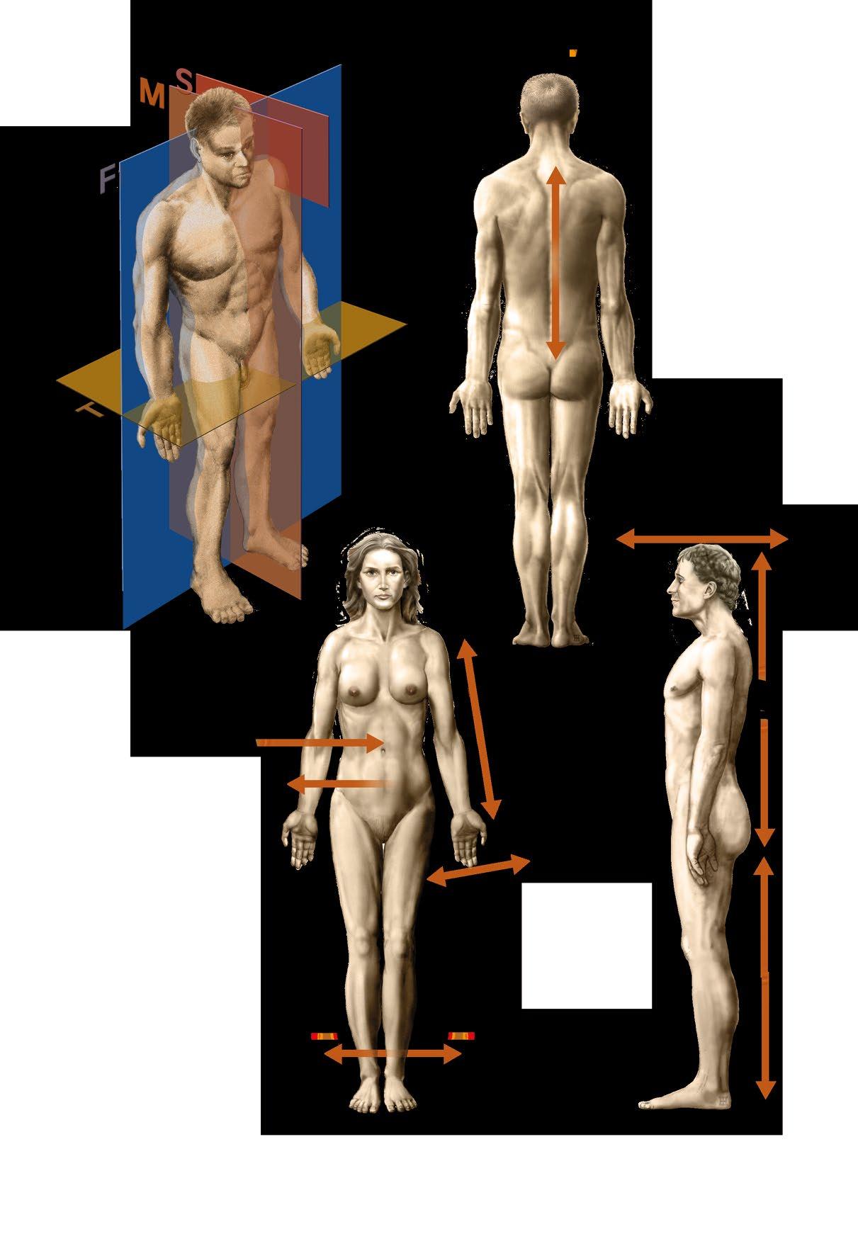
caudalis
ventralis anterior dorsalis posterior
b pohled zezadu
Parts of the human body
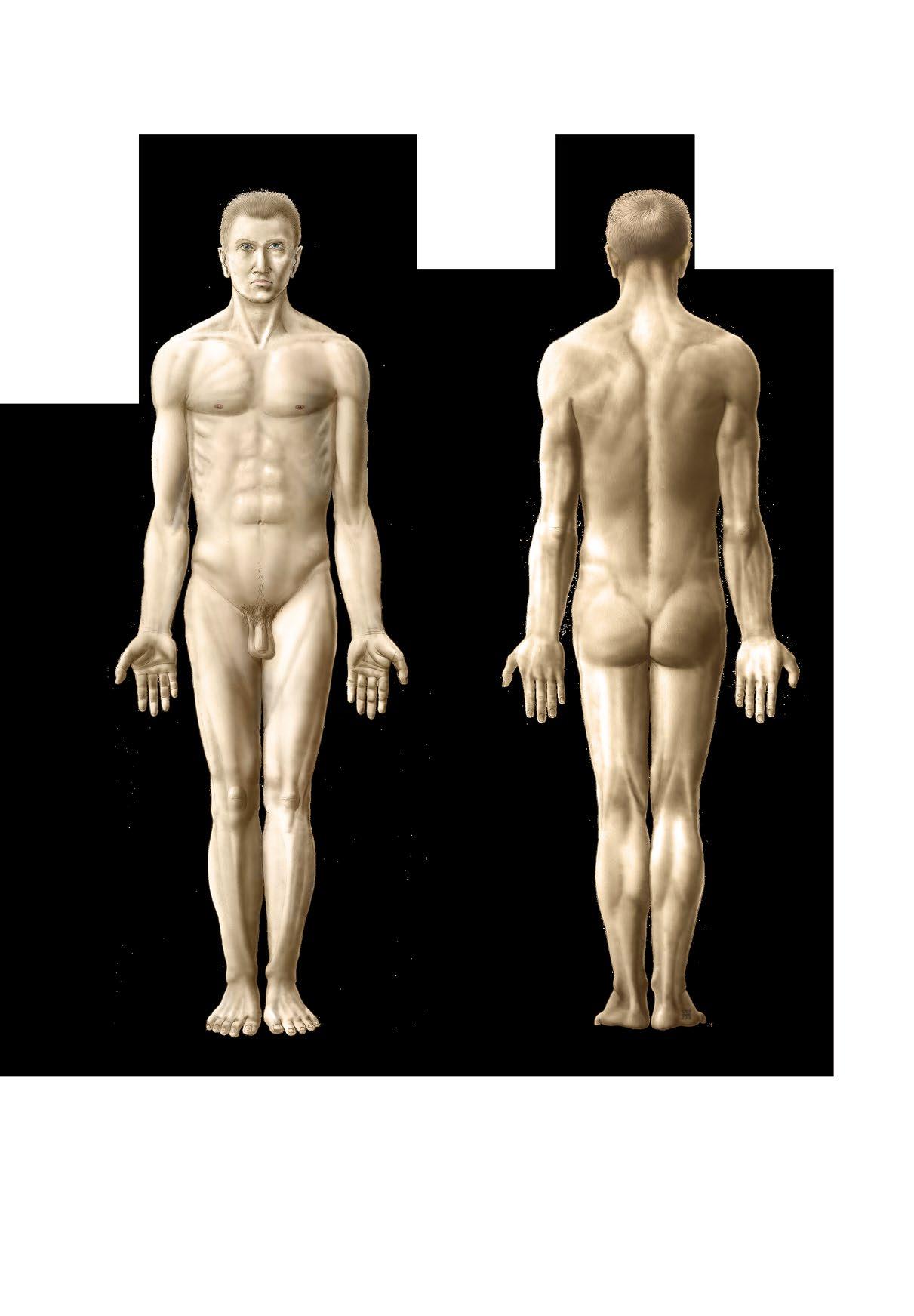
a anterior view
b posterior view
trigonum clavipectorale
regio pectoralis
regio mammaria
regio brachii anterior
regio cubitalis anterior
regio abdominalis lateralis
regio antebrachii anterior
regio carpalis anterior
regio palmaris
regiones digitorum
regio femoris anterior
regio genus anterior
regiones capitis
regiones cervicales anteriores
regiones cervicales laterales
regio deltoidea
regio presternalis
regio brachii posterior
regio inframammaria
regio epigastrica
regio parietalis
regiones capitis
regiones cervicales posteriores regio suprascapularis (var.)
regio scapularis
regio vertebralis
trigonum auscultationis
regio infrascapularis
regio cruris anterior
regio talocruralis anterior
dorsum pedis
regio umbilicalis
regio antebrachii posterior
regio hypochondriaca regio inguinalis
regio pubica
trigonum femoris
regio
regio cubitalis posterior
regio lumbalis
regio glutealis regio sacralis
regio carpalis posterior regio dorsalis manus
regio femoris posterior
regio genus posterior, fossa poplitea regio cruris posterior
regio retromalleolaris medialis
talocruralis posterior digiti pedis
b pohled zezadu
margo lateralis pedis
regio retromalleolaris lateralis
regio calcanea
Regions of the human body
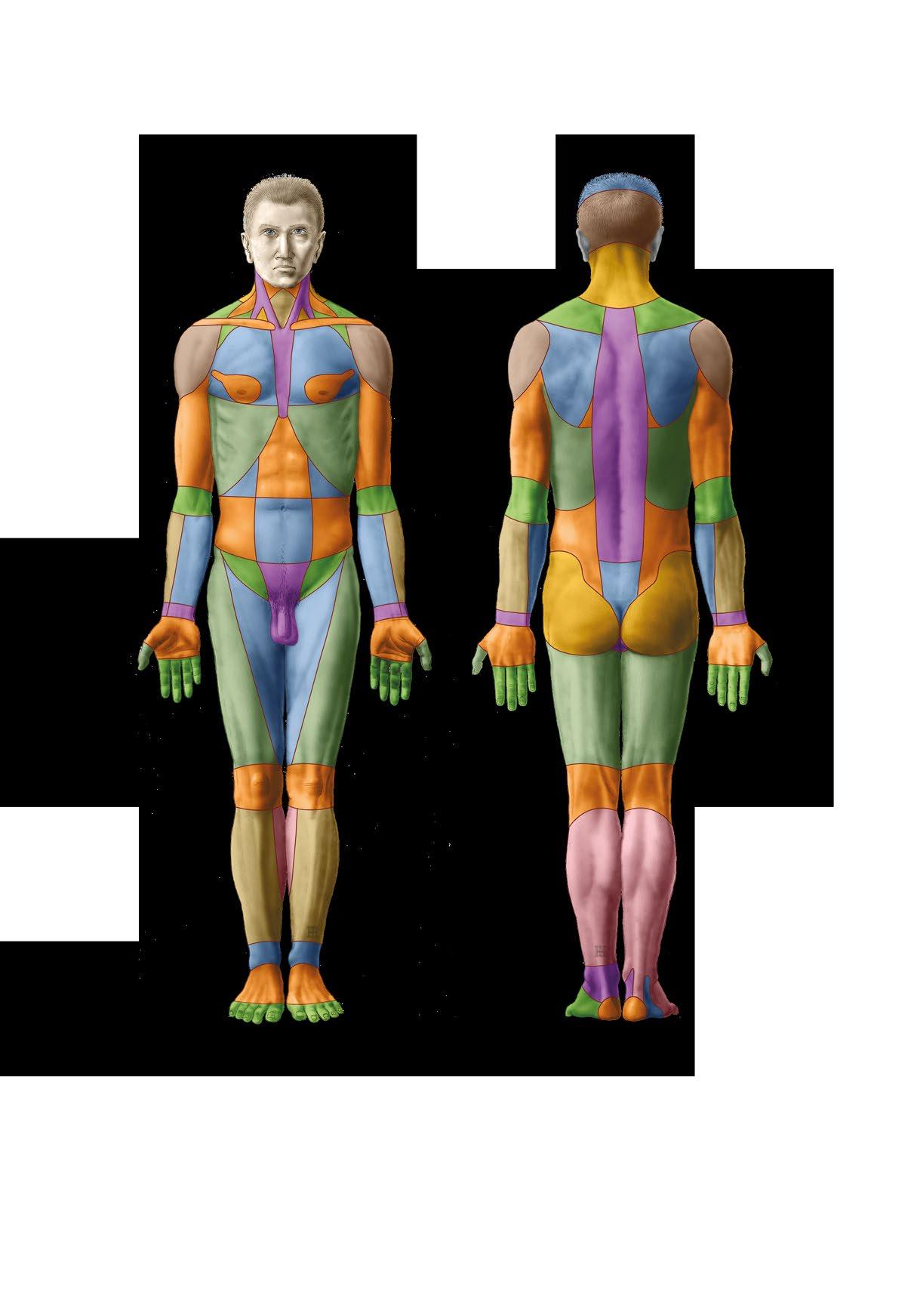
a anterior view
b posterior view
pulmo
hepar duodenum
colon ascendens
Projekce vnitřních orgánů na povrch těla, pohled zpředu
cor pleura gaster
colon descendens vesica urinaria
Surface projections of the thoracic and abdominal viscera, anterior view
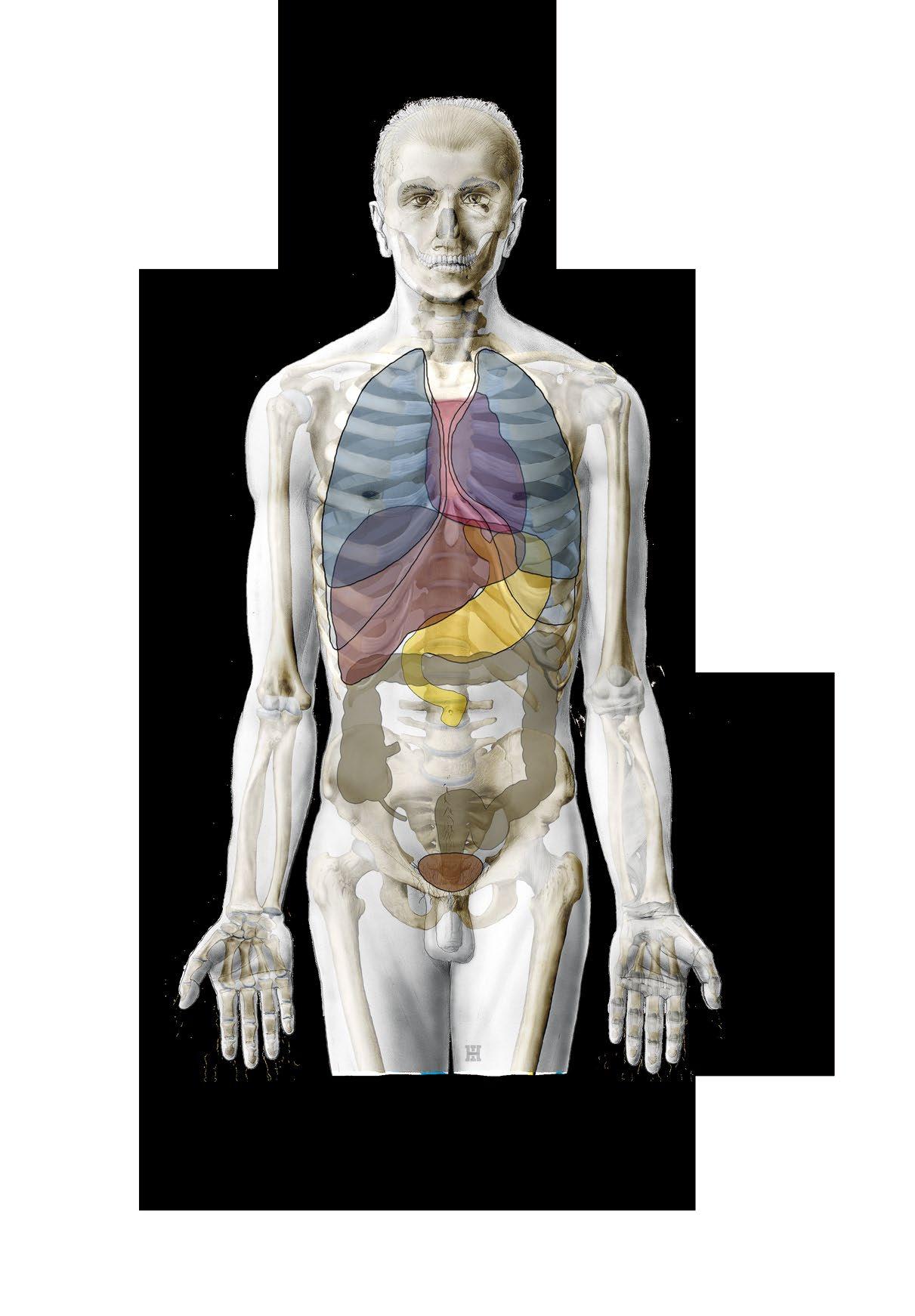
cor
hepar gaster
duodenum
colon ascendens
colon descendens
vesica urinaria
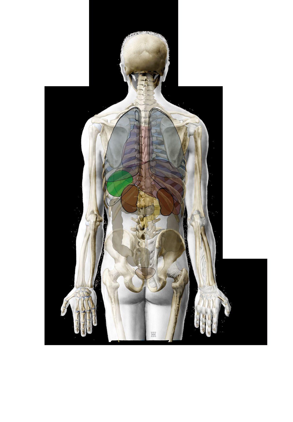
Projekce vnitřních orgánů na povrch těla, pohled zezadu
Surface projections of the thoracic and abdominal viscera, posterior view
clavicula
humerus
cranium
mandibula
columna vertebralis, pars cervicalis
articulatio humeri
sternum
costae
radius
ulna
ossa carpi
ossa metacarpi
ossa digitorum (phalanges)
femur
articulatio cubiti columna vertebralis, pars lumbalis
os coxae
membrana interossea
os sacrum
articulatio coxae
articulatio radiocarpalis
articulationes metacarpophalangeae articulationes interphalangeae manus
patella
articulatio genus
fibula
tibia
membrana interossea
ossa tarsi
ossa metatarsi
ossa digitorum (phalanges)
articulationes pedis
cranium
mandibula
columna vertebralis, pars cervicalis
clavicula
scapula
humerus
radius
ulna
ossa carpi
ossa metacarpi
ossa digitorum (phalanges)
femur
articulatio humeri
columna vertebralis, pars thoracica
costae
columna vertebralis, pars lumbalis
os coxae
os sacrum
articulatio coxae
articulatio radiocarpalis
membrana obturatoria
articulatio genus
fibula
tibia
ossa tarsi
articulatio talocruralis
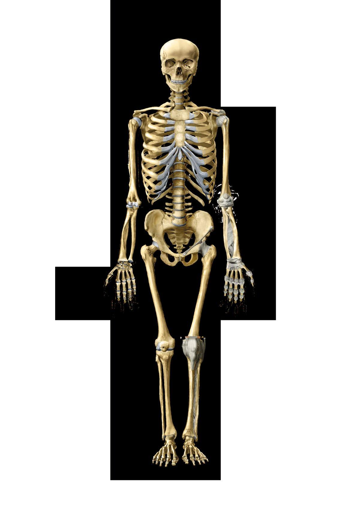
Kostra dospělého muže, pohled zezadu Adult male skeleton, posterior view

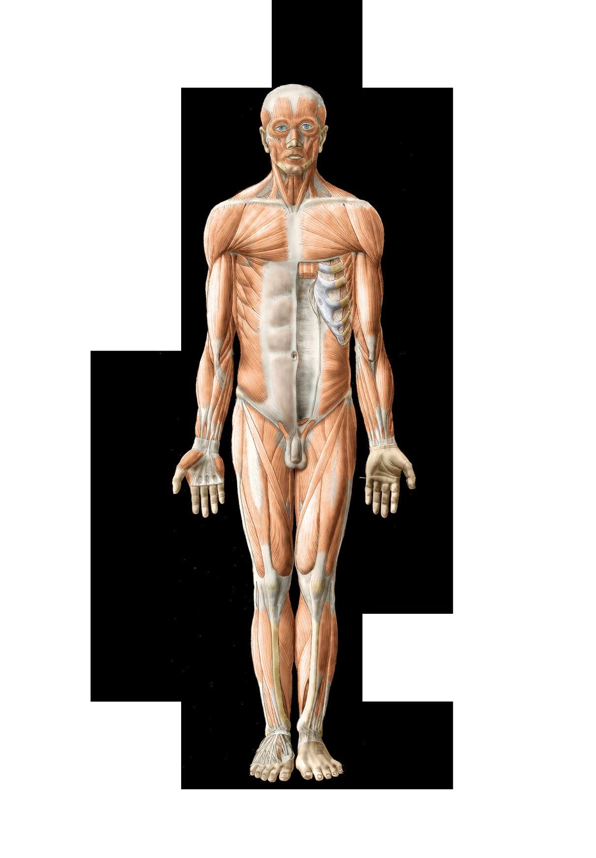
mm. capitis et mm. colli
m. deltoideus
m. pectoralis major
m. serratus anterior
mm. thoracis et mm. abdominis
vagina musculi recti abdominis, lamina anterior
m. obliquus externus abdominis
vagina musculi recti abdominis, lamina posterior
mm. manus
mm. brachii
mm. membri superioris
mm. antebrachii
mm. femoris
m. quadriceps femoris
m. tibialis anterior
mm. cruris
mm. membri inferioris
mm. pedis
Přehled kosterního svalstva, pohled zpředu Overview of skeletal muscles, anterior view