INSIDE
Updates on Biomechanics of Cervical Disc Arthroplasty
Recurrent Lumbar Disc Herniation: To Fuse or not to Fuse?
The Impact of Spinal Pathology on Sleep
Use of Image Navigation and Robotic Surgery in Training Programs Telemedicine and Spine Care in 2022: What Have We Learned?
Vertebral COLUMNS
International Society for the Advancement of Spine Surgery
TRANSITIONING TO THE Ambulatory Surgical Center
Patient Safety, Cost Efficacy, and the Patient Perspective
OUTPATIENT
FALL 2022
Editor in Chief
Kern Singh, MD
Editorial Board
Peter Derman, MD, MBA
Brandon Hirsch, MD
Sravisht Iyer, MD
Yu-Po Lee, MD
Sheeraz Qureshi, MD, MBA Managing
Vertebral Columns is published quarterly by the International Society for the Advancement of Spine Surgery.

©2022 ISASS. All rights reserved.
Opinions of authors and editors do not necessarily reflect positions taken by the Society.
This publication is available digitally at www.isass.org/news/vertebralcolumns-Fall-2022
Editor
3 8 12 16 20 22 isass.org Fall 2022 Vertebral Columns EDITORIAL Transitioning to the Ambulatory Surgical Center, Part 1: Patient Safety, Cost Efficacy, and the Patient Perspective CERVICAL SPINE Updates on Biomechanics of Cervical Disc Arthroplasty LUMBAR SPINE Recurrent Lumbar Disc Herniation: To Fuse or not to Fuse? PAIN The Impact of Spinal Pathology on Sleep ROBOTICS Use of Image Navigation and Robotic Surgery in Training Programs CLINICAL PRACTICE Telemedicine and Spine Care in 2022: What Have We Learned? Become a member today https://www.isass.org/about/membership/
Audrey Lusher Designer CavedwellerStudio.com
Transitioning to the Ambulatory Surgical Center, Part 1: Patient Safety, Cost Efficacy, and the


Patient Perspective
Chronic spine problems among patients, including patients with either neck or back complaints, compose a considerable burden on healthcare costs globally.1 As healthcare costs continue to rise in the United States, examination of measures to reduce costs for patients and surgeons alike is of utmost importance. Single-day surgery in an ambulatory surgical center (ASC) represents one such manner in which costs can be reduced. ASCs have grown by more than 60% within the past decade with more than 54 million procedures performed within the outpatient setting in the United States alone. 2 As part of a 2-part series, this first article focuses on the patient aspect of the ambulatory surgical experience with regard to safety, cost efficacy, and other patient perspectives. The rapid increase in popularity of ASCs has been attributed to a variety of factors, including lowered costs for patients along with wider use of enhanced recovery after surgery and multimodal analgesia protocols. Addi-
tionally, these protocols have resulted in increased patient satisfaction as patients are able to recover in the comfort of their own homes. 2–6 As a result, the prevalence of both lumbar and cervical surgeries performed at ASCs continues to increase in relation to in-hospital procedures.7–10 Over a 5-year period, Baird et al reported a 60.5% increase in cervical procedures performed at an ASC whereas the same procedures only increased by 8.7% within the inpatient hospital setting.10 Additionally, the variety of spine procedures performed at ASCs has increased from microdiscectomies to multilevel cervical and lumbar fusions, including newer approaches and minimally invasive and nonfusion techniques. 3
While indications for ASC interventions continue to expand, one of the foremost concerns of patients remains the same: is it safe? Over the past 2 decades, a multitude of studies have evaluated concerns of safety and efficacy of spine procedures at ASCs.
3 isass.org Fall 2022 Vertebral Columns
EDITORIAL
From the Department of Orthopaedic Surgery, Rush University Medical Center in Chicago, Illinois.
Kern Singh, MD
Timothy J. Hartman, MD
James W. Nie, BS
Keith R. MacGregor, BS
Omolabake O. Oyetayo, BS
Eileen Zheng, BS
Arash Sayari, MD
Even in 3- and 4- level procedures, anterior cervical discectomy and fusions have been demonstrated to be remarkably effective when performed in the ASC setting, with similar safety profiles and even lower complication rates when compared to inpatient care.11–18 Similar studies have echoed these findings with regard to minimally invasive lumbar fusion procedures.19–22 However, these studies consistently repeat a similar mantra: proper patient selection is mandatory. Patient selection criteria may vary slightly between surgeon groups but are overall similar. Age, body mass index, and history of cardiac or other serious conditions are necessary to take into consideration before scheduling patients to undergo surgery at an ASC. 2,9,19,23,24 Coordination of care with an experienced anesthesiologist grants further insight as to appropriateness for outpatient care. Some ASCs have a case review team of nurses and anesthesiologists to ensure each patient meets safety criteria for surgical care at an ASC. 2 Furthermore, in higher-risk procedures, such as anterior lumbar interbody fusions in which the great vessels are in close proximity to the operative space, it is necessary for ASCs to have agreements
with local hospitals as a failsafe when rare complications do arise. Taking time to educate patients on the general safety and specific risks of their procedure, as well as the preventative or treatment measures in place, allows for a safe transition of surgical cases to an ASC.
As studies generally report ASC procedures to be more cost effective than hospital outpatient procedures (eg, $2000-$3500 in cost savings in patients undergoing lumbar decompression 25), there is a clear financial incentive for the healthcare system and patients for transitioning care to ASCs when appropriate.11,26,27 The cost savings garnered at ASCs in comparison to hospital-based outpatient departments (HOPDs) is attributed to a variety of factors, but it may be most importantly due to the reduced surgical time in ASCs, generally reported to take 31.8 fewer minutes. 28 Fabricant et al reported approximately 80% of the cost savings of orthopedic surgeries performed at ASCs to be attributed to this time efficacy. 29 Furthermore, such efficiency may allow for more surgical procedures by a surgical team in a given day. Financial factors such as efficient supply utilization and decreased overhead costs allow for a leaner system without the need for rarely-used equipment. Because of their functioning size, ASCs can be nimble and make rapid adjustments to simultaneously meet the fiscal needs of the system and the surgical needs of the surgeon. 30 With utilization of ASCs leading to Medicare saving more than $4.2 billion annually—a figure expected to increase over time—patients are being incentivized toward ASC care

4 isass.org Fall 2022 Vertebral Columns EDITORIAL
with lower copays. 34 Further reducing costs by safely shifting patients to ASCs benefits both third-party and government payers. 31–35 In some examples, patients may see copays reduced by 40% when receiving care at an ASC compared to HOPDs. 34 In addition to direct savings, ASCs can allow earlier intervention for patients who may then see an earlier return to work.
Beyond the demonstrated safety and cost benefit in appropriate patients, patients may seek care at an ASC for other factors. In situations where surgeons are able to safely perform more surgeries at an ASC, patients may be scheduled sooner than if they sought care through a hospital-based inpatient or outpatient system. While this convenience poses a cost benefit with regard to return-towork times, the intangible benefit of receiving earlier care for painful and potentially disabling conditions may further drive patients toward ASCs. Additionally, conveniently located ASCs interspersed throughout a region may be a considerable factor in how patients select care options, as opposed to seeking care at a less convenient centralized
hospital-based location. When surveyed, patients cited more factors affecting selection of ASCs over HOPDs, including same-day discharge allowing peaceful recovery at home and avoiding being surrounded by patients with other illnesses, a prominent aspect affecting patient care location during the COVID-19 pandemic. 36 Additionally, during the COVID-19 pandemic, patients who had their surgical cases cancelled in the hospital setting were able to undergo surgery far sooner in the ASC setting.
Overall, spine surgery in the ASC setting poses significant benefits to patients with regard to safety, cost efficacy, and a variety of conveniences. Ultimately, proper patient selection is the most important factor in predicting successful outcomes. Educating patients about these benefits is necessary to allow patients to make the most appropriate, informed decision for their care. While the patient perspective is a key aspect of the rapid transition to ASCs for spine surgery, there are additionally a variety of surgeon factors that remain important and will be discussed in the second part of this series. n
References
1. Martin BI, Deyo RA, Mirza SK, et al. Expenditures and health status among adults with back and neck problems. JAMA . 2008;299(6):656–664.
2. Pendharkar AV, Shahin MN, Ho AL, et al. Outpatient spine surgery: defining the outcomes, value, and barriers to implementation. Neurosurg Focus . 2018;44(5):E11.
3. Gerling MC, Hale SD, White-Dzuro C, et al. Ambulatory spine surgery. J Spine Surg. 2019;5(Suppl 2):S147–S153.
4. Young S, Pollard RJ, Shapiro FE. Pushing the envelope: new patients, procedures, and personal protective equipment in the ambulatory surgical center for the COVID-19 era. Adv Anesth. 2021;39:97–112.
5. Silvers HR, Lewis PJ, Suddaby LS, Asch HL, Clabeaux DE, Blumenson LE. Day surgery for cervical microdiscectomy: is it safe and effective? J Spinal Disord. 1996;9(4):287–293.
6. Licina A, Silvers A. Perioperative multimodal analgesia for adults undergoing surgery of the spine—a systematic review and meta-analysis of three or more modalities. World Neurosurg. 2022;163:11–23.
7. Gray DT, Deyo RA, Kreuter W, et al. Population-based trends in volumes and rates of ambulatory lumbar spine surgery. Spine 2006;31(17):1957–1963; discussion 1964.
5 isass.org Fall 2022 Vertebral Columns EDITORIAL
References, continued
8. Best MJ, Buller LT, Eismont FJ. National trends in ambulatory surgery for intervertebral disc disorders and spinal stenosis: a 12-year analysis of the national surveys of ambulatory surgery. Spine . 2015;40(21):1703–1711.
9. Mundell BF, Gates MJ, Kerezoudis P, et al. Does patient selection account for the perceived cost savings in outpatient spine surgery? A meta-analysis of current evidence and analysis from an administrative database. J Neurosurg Spine . 2018;29(6):687–695.
10. Baird EO, Egorova NN, McAnany SJ, Qureshi SA, Hecht AC, Cho SK. National trends in outpatient surgical treatment of degenerative cervical spine disease. Global Spine J. 2014;4(3):143–150.
11. Adamson T, Godil SS, Mehrlich M, Mendenhall S, Asher AL, McGirt MJ. Anterior cervical discectomy and fusion in the outpatient ambulatory surgery setting compared with the inpatient hospital setting: analysis of 1000 consecutive cases. J Neurosurg Spine . 2016;24(6):878–884.
12. Lee MJ, Kalfas I, Holmer H, Skelly A. Outpatient surgery in the cervical spine: is it safe? Evid Based Spine Care J. 2014;5(2):101–111.
13. Liu JT, Briner RP, Friedman JA. Comparison of inpatient vs. outpatient anterior cervical discectomy and fusion: a retrospective case series. BMC Surg. 2009;9:3.
14. Martin CT, Pugely AJ, Gao Y, Mendoza-Lattes S. Thirty-day morbidity after single-level anterior cervical discectomy and fusion: identification of risk factors and emphasis on the safety of outpatient procedures. J Bone Joint Surg Am. 2014;96(15):1288–1294.
15. Mullins J, Pojskić M, Boop FA, Arnautović KI. Retrospective single-surgeon study of 1123 consecutive cases of anterior cervical discectomy and fusion: a comparison of clinical outcome parameters, complication rates, and costs between outpatient and inpatient surgery groups, with a literature review. J Neurosurg Spine . 2018;28(6):630–641.
16. Yerneni K, Burke JF, Chunduru P, et al. Safety of outpatient anterior cervical discectomy and fusion: a systematic review and meta-analysis. Neurosurgery. 2020;86(1):30–45.
17. McGirt MJ, Godil SS, Asher AL, Parker SL, Devin CJ. Quality analysis of anterior cervical discectomy and fusion in
the outpatient versus inpatient setting: analysis of 7288 patients from the NSQIP database. Neurosurg Focus . 2015;39(6):E9.
18. McClelland S 3rd, Oren JH, Protopsaltis TS, Passias PG. Outpatient anterior cervical discectomy and fusion: a meta-analysis. J Clin Neurosci. 2016;34:166–168.
19. Schlesinger S, Krugman K, Abbott D, Arle J. Thirty-day outcomes from standalone minimally invasive surgery—transforaminal lumbar interbody fusion patients in an ambulatory surgery center vs. hospital setting. Cureus. 2020;12(9):e10197.
20. Emami A, Faloon M, Issa K, et al. Minimally invasive transforaminal lumbar interbody fusion in the outpatient setting. Orthopedics . 2016;39(6):e1218–e1222.
21. Sivaganesan A, Hirsch B, Phillips FM, McGirt MJ. Spine surgery in the ambulatory surgery center setting: value-based advancement or safety liability? Neurosurgery. 2018;83(2):159–165.
22. Helseth Ø, Lied B, Halvorsen CM, Ekseth K, Helseth E. Outpatient cervical and lumbar spine surgery is feasible and safe: a consecutive single center series of 1449 patients. Neurosurgery 2015;76(6):728–737; discussion 737–738.
23. Walid MS, Robinson JS 3rd, Robinson ERM, Brannick BB, Ajjan M, Robinson JS Jr. Comparison of outpatient and inpatient spine surgery patients with regards to obesity, comorbidities and readmission for infection. J Clin Neurosci. 2010;17(12):1497–1498.
24. Jenkins NW, Parrish JM, Nolte MT, et al. Multimodal analgesic management for cervical spine surgery in the ambulatory setting. Int J Spine Surg. 2021;15(2):219–227.
25. Malik AT, Xie J, Retchin SM, et al. Primary single-level lumbar microdisectomy/decompression at a free-standing ambulatory surgical center vs a hospital-owned outpatient department-an analysis of 90-day outcomes and costs. Spine J. 2020;20(6):882–887.
26. Lewandrowski KU. Incidence, management, and cost of complications after transforaminal endoscopic decompression surgery for lumbar foraminal and lateral recess stenosis: a value proposition for outpatient ambulatory surgery. Int J Spine Surg. 2019;13(1):53–67.
27. Purger DA, Pendharkar AV, Ho AL, et al. Analysis of outcomes and cost of inpatient and ambulatory anterior cervical disk
replacement using a state-level database. Clin Spine Surg. 2019;32(8):E372–E379.
28. Munnich EL, Parente ST. Procedures take less time at ambulatory surgery centers, keeping costs down and ability to meet demand up. Health Aff. 2014;33(5):764–769.
29. Fabricant PD, Seeley MA, Rozell JC, et al. Cost savings from utilization of an ambulatory surgery center for orthopaedic day surgery. J Am Acad Orthop Surg. 2016;24(12):865–871.
30. Kelso L, Govil A. Cost savings at ambulatory surgical centers. Published October 14, 2020. Accessed November 19, 2022. https://avantgardehealth.com/blog/cost-savings-at-ambulatory-surgical-centers/
31. Waddill K. Humana expands bundled payment models for spinal, joint surgeries. HealthPayerIntelligence . Published July 23, 2019. Accessed November 19, 2022. https://healthpayerintelligence. com/news/humana-expands-bundled-payment-models-for-spinal-joint-surgeries
32. Waddill K. Ambulatory surgery centers could save private payers $3B. HealthPayerIntelligence . Published December 18, 2020. Accessed November 19, 2022. https://healthpayerintelligence. com/news/ambulatory-surgery-centers-could-save-private-payers-3b
33. Waddill K. How ambulatory surgery centers lower payer outpatient spending. HealthPayerIntelligence . Published September 8, 2021. Accessed November 19, 2022. https://healthpayerintelligence. com/news/how-ambulatory-surgery-centers-lower-payer-outpatient-spending
34. Ambulatory Surgery Center Association. Medicare cost savings. Accessed November 19, 2022. https://www.ascassociation.org/advancingsurgicalcare/ reducinghealthcarecosts/costsavings
35. Ambulatory Surgery Center Association. Commercial Insurance Cost Savings in Ambulatory Surgery Centers. Published June 14, 2016. Accessed November 19, 2022. https://www.ascassociation.org/advancingsurgicalcare/reducinghealthcarecosts/ privatepayerdata/healthcarebluebookstudy
36. Silva M, Silva J, Novo J, Oliveira F, Carneiro E, Mourão J. The patient perspective regarding ambulatory surgery: an observational study. Acta Med Port . 2022;35(10):743–748.
6 isass.org Fall 2022 Vertebral Columns
EDITORIAL


JUNE 1 – JUNE 3, 2023 MARRIOTT MARQUIS • SAN FRANCISCO, CA • Interactive meeting led by world-class faculty • Focused on new technologies and techniques • AMA PRA Category 1 Credit(s)™ available 23RD ANNUAL CONFERENCE ISASS23 LEARN MORE: ISASS.ORG Program Chair: Han Jo Kim, MD Program Chair: Luis Tumialan, MD
Mummaneni, MD
MD JOINTLY PROVIDED BY:
Massimo Balsano, MD
Vice Chair: Praveen
Vice Chair: Peter Passias,
President:
CERVICAL SPINE
Updates on Biomechanics of Cervical Disc Arthroplasty


Anterior cervical discectomy and fusion has historically been considered the gold standard for neural decompression and disc height restoration among patients with cervical spondylosis who have exhausted conservative measures.1 In recent years, however, the aging population and growing focus on value-based treatment spurred the advent of motion-sparing technologies. Cervical disc arthroplasty (CDA) was developed as a means of preserving physiologic biomechanics and mitigating the shortcomings of traditional fusion (namely, adjacent segment degeneration, restriction of motion, and long-term cost effectiveness). 2
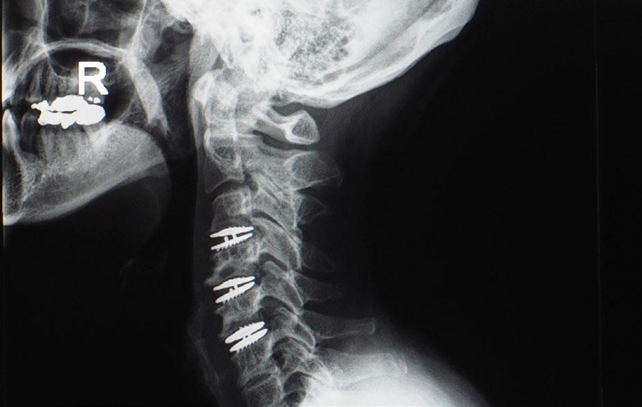
The US Food and Drug Administration (FDA) has thus far approved 9 artificial discs (Prestige ST, Prodisc-C, Bryan, Secure-C, PCM, Mobi-C, Prestige LP, M6-C, and Simplify) for single-level arthroplasty and 2 artificial discs (Mobi-C and Prestige LP) for 2-level arthroplasty. 3 CDA has subsequently garnered extensive level I and II evidence supporting its use and is now considered an important technique in the spine surgeon’s armamentarium. Despite its safety and efficacy, however, CDA requires meticulous technical proficiency and preoperative planning. Implant-specific variables such as the
degree of constraint, number of components, type of articulation, material composition, and endplate surface constructs largely influence postoperative functionality and kinematics.4,5 As such, a thorough understanding of implant design is critical to optimize device selection, spinal biomechanics, and patient outcomes.
Degree of Constraint
The motion of an object is defined by the number of independent components governing its movement within a 3-dimensional space, otherwise known as degrees of freedom (DOFs). DOFs of artificial cervical disc prostheses are conjointly determined by the number of components entailed in the construct, their mode of articulation with one another, and the intrinsic constraint produced by the bearing. Constraint is a kinematic term used to describe the limitations placed on an object’s DOFs and distinguishes implants into 3 categories: constrained, semi-constrained, and unconstrained.6 Whereas an unconstrained implant exhibits supra-physiologic motion that allows it to both translate and rotate independently along 3 orthogonal axes (6 DOFs), semi-constrained and constrained implants allow for physiologic and

8 isass.org Fall 2022 Vertebral Columns
From the Hospital for Special Surgery in New York, New York.
Sheeraz A. Qureshi, MD, MBA
Omri Maayan, BS
Kasra Araghi, BS
Olivia Tuma, BS
Robert Kamil, BS
Pratyush Shahi, MBBS, MS(Ortho)
subphysiologic motion, respectively. These differences in motion underlie important tradeoffs in implant stability and load distribution. Constrained devices exhibit a fixed axis of rotation that minimizes facet load at the expense of more challenging implant placement and decreased range of motion (ROM). 2,7 Semi-constrained discs likewise entail physical stops that reduce shear and torsional load compared to unconstrained implants without the strict ROM limitations of constrained devices.7,8 As such, these prostheses represent most artificial cervical discs available on the market today.
Number of Components and Type of Articulation
The number of DOFs provided by each artificial cervical disc also depends on the quantity of individual components and articulation modality of the bearing surfaces. Nonarticulating prostheses such as the Bryan (Medtronic, Minneapolis, MN), M6-C (Orthofix, Lewisville, TX), Rhine Cervical Disc (Stryker, Portage, MI), and CP-ESP discs (Spine Innovations, Lyon, France) replicate the physiologic kinematics of the functional spinal unit by providing 6 DOFs. Their viscoelastic core allows for disc height compression along 3 orthogonal axes and therefore mimics the shock-absorbing properties of the native disc. Despite increased resistance against excessive translational and angular motion, however, nonarticulating designs have been associated with greater risk of implant migration postoperatively. This is likely a result of increased shear stress along the vertebra-implant interface.4,6,9,10
In contrast, articulating implants are composed of 2 or 3 distinct components and can thus disperse shear stress across multiple constituents. The Discover Artificial Cervical Disc (DePuy Spine, Raynham, MA), ProDisc-C, and porous coated motion (PCM) discs are 2-piece prostheses with ball-andsocket joints. The absence of an independent bearing forces the 2 components to maintain contact throughout their motion, providing 3 DOFs through flexion-extension, lateral bending, and axial rotation.9-11 Ball-and-socket articulations in 2 component prostheses do not allow for independent translation and thus have a fixed center of rotation. As a result, positioning of the sphere within the vertebral disc space must precisely mirror the anatomic center of rotation. Failure to do so may lead to excess shear and torsional load, thereby stimulating degeneration of adjacent segments.6,11
Three-component cervical disc prostheses entail a mobile core and 2 articulating bearings that allow for independent translation between the vertebrae. This mobile center of rotation provides additional leeway regarding the restoration of physiologic kinematics, as it is less dependent on intraoperative device positioning. The Mobi-C (Zimmer Biomet, Austin, TX) and Simplify (Nuvasive, San Diego, CA) implants comprise 3 components that provide 5 DOFs through flexion-extension, lateral bending, axial rotation, and independent translation in both the sagittal and coronal planes.11 The biconvex mobile core of the Simplify prosthesis allows for 3 independent angular motions and 2 independent translational motions. In contrast, the
9 isass.org Fall 2022 Vertebral Columns
CERVICAL SPINE
CERVICAL SPINE
CDA has garnered extensive evidence supporting its use. However, a thorough understanding of implant design is critical to optimize device selection, spinal biomechanics, and patient outcomes.

ball-in-trough articulation of the Mobi-C is characterized by a ball-and-socket bearing superiorly and a planar bearing inferiorly. This allows for 3 independent angular motions through the superior bearing and independent translation along 2 orthogonal axes via the inferior bearing.12
Material Composition and Osteointegration
Biomaterials for CDA prostheses have traditionally included titanium, stainless steel, polyurethanes, polyethylene, and cobalt-chromium alloy.13 Cobalt-chromium and titanium are stronger than stainless steel and are simultaneously less likely to corrode. However, titanium’s susceptibility to abrasion is associated with production of polyethylene debris, which can result in periprosthetic inflammation and increase
the risk of postoperative complications such as hardware failure and implant migration.14 The superior wear characteristics of cobalt-chromium alloy have contributed to its increased popularity and incorporation in several FDA-approved devices, including the ProDisc-C, Secure-C, Mobi-C, and PCM cervical discs.15,16 Furthermore, implants containing poly-ether-ether-ketone (PEEK), ceramic, silicon nitride, and polycrystalline diamond have been developed in recent years to counteract the suboptimal corrosion resistance and significant interference produced on magnetic resonance imaging by traditional metal components. For instance, a majority of CDA implants used today include ultra-high molecular weight polyethylene on titanium or cobalt chromium endplates. The initial stainless-steel components of the Prestige ST disc have likewise been replaced with titanium-ceramic alloy, and the Simplify disc has a biconvex ceramic core enclosed between PEEK endplates.13,17
Osteointegration additionally relies on intraoperative fixation and endplate surface biocompatibility as predeterminants of short- and long-term prosthetic stability, respectively. Modalities of fixation can be differentiated by their degree of disruption to cancellous and cortical bone. Rails (Prestige LP) and keels (Secure-C, ProDisc-C, M6C) require extensive endplate preparation but offer immediate press-fit stability. The consequent release of osteoinductive factors has been associated with elevated rates of heterotopic ossification and fusion among these devices. On the other hand, spikes (activC) and teeth (Mobi-C, PCM, CerviCore)
10 isass.org Fall 2022 Vertebral Columns
do not result in significant bony disruption but require more time to settle, increasing the risk of implant movement in the immediate postoperative period. 4,18 Long-term osteointegration is also contingent upon the endplate surface coating. For example, the endplate surfaces of the Mobi-C, M6-C, ProDisc-C, Prestige LP, Secure-C, Bryan, and PCM Cervical Disc contain an overlay of porous titanium-alloy spray that enhances biocompatibility. Other materials used for this purpose include hydroxyapatite, calcium phosphate, porous cobalt-chromium, and titanium wire mesh. 9,19
References
1. Zhang Y, Lv N, He F, et al. Comparison of cervical disc arthroplasty and anterior cervical discectomy and fusion for the treatment of cervical disc degenerative diseases on the basis of more than 60 months of follow-up: a systematic review and meta-analysis. BMC Neurol. 2020;20(1):143.
2. Leven D, Meaike J, Radcliff K, Qureshi S. Cervical disc replacement surgery: indications, technique, and technical pearls. Curr Rev Musculoskelet Med. 2017;10(2):160-169.
3. Shin JJ, Kim KR, Son DW, et al. Cervical disc arthroplasty: What we know in 2020 and a literature review. J Orthop Surg (Hong Kong). 2021;29(1_ suppl):23094990211006934.
4. Staudt MD, Das K, Duggal N. Does design matter? Cervical disc replacements under review. Neurosurg Rev. 2018;41(2):399-407.
5. Choi H, Purushothaman Y, Baisden J, Yoganandan N. Unique biomechanical signatures of Bryan, Prodisc C, and Prestige LP cervical disc replacements: a finite element modelling study. Eur Spine J. 2020;29(11):2631-2639.
6. Patwardhan AG, Havey RM. Biomechanics of cervical disc arthroplasty devices. Neurosurg Clin N Am. 2021;32(4):493-504.
7. Villavicencio AT, Burneikiene S, Pash -
Conclusion
CDA has emerged as a viable motion-sparing alternative to anterior cervical discectomy and fusion in the treatment of cervical spondylosis. The DOFs, constraint, axis of rotation, and biocompatibility conferred by these devices ultimately determine whether they adequately preserve motion and mitigate the risk of adjacent segment degeneration. As cervical disc prostheses continue to be refined, it will become increasingly crucial to understand the minutiae of implant design to optimize patient kinematics and clinical outcomes. n
man R, Johnson JP. Spinal artificial disc replacement: cervical arthroplasty: part I: history, design, and types of artificial discs. Contempor Neurosurg. 2007;29(12):1-5. doi:10.1097/01. CNE.0000271890.82993.b9
8. Cunningham BW, Gordon JD, Dmitriev AE, Hu N, McAfee PC. Biomechanical evaluation of total disc replacement arthroplasty: an in vitro human cadaveric model. Spine . 2003;28(20):S110–S117.
9. Wellington IJ, Kia C, Coskun E, et al. Cervical and lumbar disc arthroplasty: a review of current implant design and outcomes. Bioengineering (Basel). 2022;9(5):227.
10. Choi D, Petrik V, Fox S, Parkinson J, Timothy J, Gullan R. Motion preservation and clinical outcome of porous coated motion cervical disk arthroplasty. Neurosurgery. 2012;71(1):30-37.
11. Patwardhan AG, Havey RM. Biomechanics of cervical disc arthroplasty—a review of concepts and current technology. Int J Spine Surg. 2020;14(suppl 2):S14-S28.
12. Davis RJ, Kim KD, Hisey MS, et al. Cervical total disc replacement with the Mobi-C cervical artificial disc compared with anterior discectomy and fusion for treatment of 2-level symptomatic degenerative disc disease: a prospective, randomized,
controlled multicenter clinical trial. J Neurosurg Spine . 2013;19(5):532-545
13. Saini M, Singh Y, Arora P, Arora V, Jain K. Implant biomaterials: a comprehensive review. World J Clin Cases . 2015;3(1):52-57.
14. Head WC, Bauk DJ, Emerson RH Jr. Titanium as the material of choice for cementless femoral components in total hip arthroplasty. Clin Orthop Relat Res . 1995;(311):85-90.
15. Pham MH, Mehta VA, Tuchman A, Hsieh PC. Material science in cervical total disc replacement. Biomed Res Int . 2015;2015:719123.
16. Bydon M, Michalopoulos GD, Alvi MA, Goyal A, Abode-Iyamah K. Cervical total disc replacement: Food and Drug Administration-approved devices. Neurosurg Clin N Am. 2021;32(4):425-435.
17. Warburton A, Girdler SJ, Mikhail CM, Ahn A, Cho SK. Biomaterials in spinal implants: a review. Neurospine . 2020;17(1):101-110.
18. Mehren C, Suchomel P, Grochulla F, et al. Heterotopic ossification in total cervical artificial disc replacement. Spine (Phila Pa 1976). 2006;31(24):2802-2806.
19. Phillips FM, Garfin SR. Cervical disc replacement. Spine (Phila Pa 1976) 2005;30(17 Suppl):S27-S33.
11 isass.org Fall 2022 Vertebral Columns
CERVICAL SPINE
LUMBAR SPINE
Recurrent Lumbar Disc Herniation
To Fuse or not to Fuse?


Lumbar disc herniation (LDH) is one of the leading causes of functional disability worldwide. The prevalence of LDH is reported around 2% to 3%, predominantly affecting men (57% men vs 43% women) with a mean age of 41 years. 1 Surgical intervention forms the mainstay of treatment for patients not responding to conservative measures.
Recurrent lumbar disc herniation (rLDH) is defined as the recurrence of herniation at the same intervertebral level after a definite pain-free period of 6 months following surgery. 2 The rate of rLDH following surgery may range from 0.5% to 25%. 3,4 This often results in a massive healthcare cost burden with debilitating clinical outcomes when compared with primary LDH management. 5
Risk Factors for Recurrent Lumbar Disc Herniation
The risk factors for rLDH can be grouped into modifiable patient-related factors and nonmodifiable surgical and biomechanical factors. First, patient-related factors such as male gender, smoking, diabetes mellitus, and obesity are found to be predictive
for rLDH after spine surgery. 5-8
Second, surgical factors such as limited disc removal and endplate changes following microdiscectomy can alter the disc height, which can increase the incidence of rLDH. 9,10 Cinotti et al reported that the presence of an annular incision makes the disc more susceptible to herniation due to the increased stress and mechanical load on the compromised nucleus pulposus fragment.11-14 Additional evidence was provided by Thome et al, wherein the use of an annular closure device significantly reduced the risk of recurrence from 31.6% to 18.8%.15
Third, biomechanical factors such as increased sagittal motion, heavy axial loading, and facet joint degeneration lead to spinal instability. These factors in turn predispose approximately one-fourth of patients to develop reherniations after surgical interventions. 6,16
Treatment Options for Management of Recurrent Lumbar Disc Herniation


A subanalysis of the SPORT trial by Leven et al found that over 8 years following a surgically treated disc

12 isass.org Fall 2022 Vertebral Columns
From the Hospital for Special Surgery in New York, New York.
Sravisht Iyer, MD
Apurv Gabrani, MBBS, MS (Ortho)
Nishtha Singh, MBBS
Anthony Pajak, BS
Max Korsun, BS
Pratyush Shahi, MBBS, MS(Ortho)
herniation, 15% of patients had a reoperation, with 62% of those patients having a recurrent disc herniation as the indication for surgery. 3 Patients who underwent reoperation reported significantly worse outcomes. Discectomy alone and fusion revision procedures have both demonstrated successful short-term patient-reported outcomes and leg pain relief when treating rLDH.14,17

Discectomy
Discectomy alone is a less invasive procedure, inherently carrying a set of benefits. Previous studies have shown that discectomies alone present with the advantage of lower operative times, faster recovery, less intraoperative blood loss, lower total
cost of the procedure, and a lower reliance on blood transfusions in the treatment of rLDH.14 In terms of clinical complications, they mitigate the risks of loss of motion segment, adjacent segment disease, and pseudarthrosis, all of which are associated with fusion procedures. 17 These factors pose discectomy procedures as an intervention with a lower risk of postoperative complications in comparison with fusion procedures. However, nonfusion procedures in the treatment of rLDH have been repeatedly shown to pose a greater risk of subsequent herniation and the incidence of mechanical instability over the long term.14,15,18 For example, Tanavalee et al 19 reported a revision rate of 9.09% in discectomy procedures as opposed to 2.0%
13 isass.org Fall 2022 Vertebral Columns
LUMBAR SPINE
LUMBAR SPINE
in spinal fusion. Despite the lower prevalence of intraoperative and postoperative complications, discectomy alone may be an inferior treatment when solely focusing on mitigating the risk of re-rLDH.
Discectomy and Fusion

Discectomy and fusion procedures are generally performed for the treatment of painful instability of the lumbar spine, which usually manifests as chronic low back pain with or without radicular symptoms. One major advantage of performing these procedures is eliminating the risk of adjacent level instability and minimizing recurrence. 20 However, the risk of loss of a motion segment or pseudoarthrosis or adjacent level disease can be seen. 21 A recent analysis conducted by Fu et al 22 compared discectomy alone (n = 23) with posterior lumbar interbody fusion (n = 18) for treating rLDH and found good to
excellent clinical outcomes in both groups with no statistically significant differences. They did, however, report an increased operative time, higher blood loss, and longer hospital stay for the fusion group. Similar findings were also reported by Guan et al, who also reported lower hospital charges for the discectomy only group. 23 Although dural tear was reported to be one of the most common intraoperative complications in both groups of treatments, no study reported any significant difference in the incidence of these tears.
Ahsan et al 14 compared the clinical outcomes and complications in fusion vs discectomy alone for rLDH in 135 patients and found no differences in postoperative outcomes except for significantly higher low back pain and radicular pain scores in the discectomy alone group. Also, 11 of the 110 patients of the discectomy alone group went on to have a fusion procedure due to re-recurrence of LDH or instability. Hence, it may be argued that discectomy alone may be preferred as the initial surgical treatment for rLDH, which can be escalated to a fusion procedure in selected cases.14
Conclusion
Despite the existing evidence supporting both discectomy alone and discectomy with fusion procedures as satisfactory treatment modalities for rLDH, patient-specific factors add an extra layer of complexity to reaching successful management. The less invasive nature of a discectomy-alone procedure may appeal to patients, whereas fusion procedures may be favored for patients
14 isass.org Fall 2022 Vertebral Columns
The decision-making process should involve a conversation with the patient about the risks and benefits of both modalities to reach the optimal individualized treatment plan.
history, comorbidities, demographic factors, and the nature of their diagnosis. The decision-making process should involve a conversation with the patient about the risk and benefits of both modalities to reach the optimal individualized treatment plan. n
References
1. Cummins J, Lurie JD, Tosteson TD, et al. Descriptive epidemiology and prior healthcare utilization of patients in the Spine Patient Outcomes Research Trial’s (SPORT) three observational cohorts disc herniation, spinal stenosis, and degenerative spondylolisthesis. Spine . 2006;31(7):806-814.
2. Ye YP, Hu JW, Zhang YG, Xu H. Impact of lumbar interbody fusion surgery on postoperative outcomes in patients with recurrent lumbar disc herniation: analysis of the US national inpatient sample. J Clin Neurosci. 2019;70:20-26.
3. Leven D, Passias PG, Errico, TJ, et al. Risk factors for reoperation in patients treated surgically for intervertebral disc herniation: a subanalysis of eight-year SPORT data. J Bone Joint Surg Am. 2015;97(16):1316-1325.
4. Ambrossi GLG, McGirt MJ, Sciubba DM, et al. Recurrent lumbar disc herniation after single level lumbar discectomy: incidence and health care cost analysis. Neurosurgery. 2008;65(3):574-578.
5. Shimia M, Ghazani AB, Sadat BE, Habibi B, Habibzadeh. Risk factors of recurrent lumbar disc herniation. Asian J Neurosurg. 2013;8(2):93-96.
6. Huang W, Han Z, Liu JL, Yu L, Yu X. Risk factors for recurrent lumbar disc herniation: a systematic review and meta-analysis. Medicine . 2016;95(2):1-10.
7. Kim CH, Chung CK, Park CS, Choi B, Kim MJ, Park BJ. Reoperation rate after surgery for lumbar herniated intervertebral disc disease: nationwide cohort study. Spine . 2013;38(7):581-590.
8. Siccoli A, Staartjes VE, Klukowska AM, Muizelaar JP, Schröder ML. Overweight and smoking promote recurrent
lumbar disc herniation after discectomy. Eur Spine J. 2022;31:604-613.
9. McGirt M, Eustacchio S, Varga P, et al. A prospective cohort study of close interval computed tomography and magnetic resonance imaging after primary lumbar discectomy: factors associated with recurrent disc herniation and disc height loss. Spine . 2009;34(19):2044-2051.
10. Shepard N, Cho W. Recurrent lumbar disc herniation: a review. Global Spine J. 2019;9(2):202-209.
11. Yaman ME, Kazanci A, Yaman ND, Bas F, Ayberk G. Factors that influence recurrent lumbar disc herniation. Hong Kong Med J. 2017;23(3):258-263.
12. Cinotti G, Roysam GS, Eisenstein M, Postacchini F. Ipsilateral lumbar disc herniation: a prospective, controlled study. J Bone Joint Surg Br. 1998;80-B(5):825-832.
13. Samartzis D, Karppinen J, Cheung JP, Lotz J. Disk degeneration and low back pain: are they fat-related conditions? Global Spine J. 2013;3(3):133-144.
14. Ahsan K, Khan SI, Zaman N, Ahmed N, Montemurro N, Chaurasia B. Fusion versus nonfusion treatment for recurrent lumbar disc herniation. J Craniovertebr Junction Spine . 2021;12(1):44-53.
15. Thomé C, Kuršumovic A, Klassen PD, et al. Effectiveness of an annular closure device to prevent recurrent lumbar disc herniation: a secondary analysis with 5 years of follow-up. JAMA Netw Open. 2021;4(12):e2136809.
16. Wang F, Chen K, Lin Q, et al. Earlier or heavier spinal loading is more likely to lead to recurrent lumbar disc herniation after percutaneous endoscopic lumbar disectomy. J Orthop
Surg Res . 2022;17(1):356-362.
17. Dave BR, Degulmadi D, Krishnan A, Mayi S. Risk factors and surgical treatment for recurrent lumbar disc prolapse: a review of the literature. Asian Spine J. 2020;14(1):113-121.
18. Arif S, Brady Z, Enchev Y, Peev N. Is fusion the most suitable treatment option for recurrent lumbar disc herniation? A systematic review. Neurol Res . 2020;42(12):1034-1042.
19. Tanavalee C, Limthongkul W, Yingsakmongkol W, Luksanapruksa P, Singhatanadgige W. A comparison between repeat discectomy versus fusion for the treatment of recurrent lumbar disc herniation: systematic review and meta-analysis. J Clin Neurosci. 2019;66:202-208.
20. El Shazly AA, El Wardany MA, Morsi AM. Recurrent lumbar disc herniation: a prospective comparative study of three surgical management procedures. Asian J Neurosurg. 2013;8(3):139-146.
21. Galal A, Elsayed AM, Ahmed OE. Recurrent lumbar disc herniation: is fusion necessary? Internet J Neurosurg 2019;15(1). doi:10.5580/IJNS.54187
22. Fu TS, Lai PL, Tsai TT, Niu CC, Chen LH, Chen WJ. Long term results of disc excision for recurrent lumbar disc herniation with or without posterolateral fusion. Spine (Phila Pa 1976). 2005;30(24):2830-2834.
23. Guan J, Ravindra VM, Schmidt MH, Dailey AT, Hood RS, Bisson EF. Comparing clinical outcomes of repeat discectomy versus fusion for recurrent disc herniation utilizing the N2QOD. J Neurosurg Spine . 2017;26(1):39-44.
15 isass.org Fall 2022 Vertebral Columns
presenting with underlying instability, chronic lower back pain, and a history of recurring herniations. However, there is still a paucity of level 1 evidence comparing both modalities. Patients should also be carefully assessed based on their medical LUMBAR SPINE
The Impact of Spinal Pathology on Sleep


Sleep is an essential process that plays an important role in our physical and mental wellbeing.1–6 There is growing evidence of a bidirectional relationship between pain and disturbed sleep—pain disturbs sleep, and decreased sleep enhances pain.7–13
Sleep disturbance has been reported by patients with multiple spine conditions, including lumbar spinal stenosis (LSS), cervical myelopathy, adult spinal deformity (ASD), ankylosing spondylitis (AS), scoliosis, neck pain, and low back pain.12,14–18 In the context of spinal conditions, the bidirectional relationship between sleep and pain manifests in multiple ways. Specifically, studies have demonstrated that concurrent spinal and sleep pathologies may be associated with significant differences in pain, health-related quality of life (HRQoL), and possibly outcomes following spine surgery.19–22
Lumbar Stenosis and Low Back Pain
Sleep disturbances are common in patients with low back pain (58.7%) and LSS (66%).15 In patients with sleep disturbance and lumbar spinal stenosis, higher rates of pain, disability, and depression have been reported.15
In a study of 230 patients with LSS, radiographic measures were analyzed in relation to sleep disturbance.15 Patients with sleep disorders demonstrated a significantly decreased dural cross-sectional area with more severe central and foraminal LSS. In these patients, radiographs revealed both significantly increased pelvic tilt and sagittal vertical axis as well as decreased lumbar range of motion. The authors hypothesized that while supine positioning during sleep may alleviate mechanical back pain by minimizing axial load on the spine, the loss of lordosis (relative extension) may decrease the size of the foramen and central spinal canal, thereby increasing the rate of nocturnal pain and sleep disturbance.
In a separate prospective study by Kim et al, the authors compared the differences in sleep quality in LSS patients treated with surgical versus conservative modalities. 29 Following cohort matching, the minimal clinical improvement difference in sleep improvement at 6 months was reported in 85.4% of surgical patients and 50% of patients undergoing conservative care. The subgroup analysis of patients in the conservative treatment group demonstrated that patients with continued sleep disturbance had higher rates of depression and more severe foraminal stenosis than the patients whose sleep improved. Furthermore, patients in the conservative treatment group had significantly


16 isass.org Fall 2022 Vertebral Columns
PAIN
From the Texas Back Institute in Plano, Texas.
Junyoung Ahn, MD
Peter B. Derman, MD, MBA
Mary P. Rogers-LaVanne, PhD
Alexander M. Satin, MD
higher Oswestry Disability Index scores at 6 months than those in the surgical group. This finding complements a separate study of 508 patients treated with spine surgery for a myriad of conditions—patients who did not experience improvement in sleep also reported significantly less improvement in pain, physical function, and satisfaction with their social interactions. 20
While pain is often referenced as an interrupter of sleep, research also highlights the effects of sleep quality on pain. In a study evaluating patients with low back pain, greater pain intensity was recorded following a night of sleep disturbance.9 This result is supported by a prospective study in which significantly increased pain scores were reported following a night of poor sleep in patients with chronic pain. 23 One explanation of such findings may be related to modulation of pain thresholds associated with sleep quality. 24
Cervical Myelopathy
Seventy-one percent of patients with cervical myelopathy exhibit sleep disturbance. 26 In a cross-sectional study of 203 patients with cervical myelopathy, Kim et al reported several characteristics identified as independent risk factors for sleep disturbance: depression, lower modified Japanese Orthopedic Association score, chronic shoulder pain, smaller cross-sectional area of the spinal cord at the most stenotic level, and decreased cervical range of motion. In contrast to their previous study on sleep and LSS discussed above, the authors demonstrated no association between nocturnal pain, arm/neck pain, or disability and sleep disturbance. The authors hypoth-
esized that the etiology of sleep disturbance in cervical myelopathy may be similar to the etiologies of sleep disorders in spinal cord injury patients such as paresthesias, abnormal breathing during sleep, disruption of endogenous melatonin production, and sleep-related movement disorders.
In a subsequent prospective study, the change in sleep quality following surgical versus nonsurgical treatment of cervical myelopathy was compared.14 While sleep quality initially improved regardless of treatment, the improvement in sleep quality score was not maintained in the conservative treatment group. Significantly lower neck disability index scores and better sleep quality scores were observed at 6-month follow-up in patients who underwent operative intervention compared to the conservative treatment group. The reported improvement in sleep in patients undergoing surgery for cervical myelopathy was similar to the result from the previous study, which demonstrated significantly greater improvement in sleep following surgical treatment of LSS.29 These studies serve as case studies for how surgical invention may both decrease pain and improve sleep.14,29
Adult Spinal Deformity
The data on the association between sleep disturbance and ASD are limited. However, in a report by Kim et al, 44 patients with ASD were compared to 45 patients with LSS via propensity matching.16 In this study, ASD and LSS patients demonstrated sleep disturbance rates of 75.0% and 72.7%, respectively. The authors then compared results within each cohort according to whether the patients had
17 isass.org Fall 2022 Vertebral Columns
PAIN
PAIN
sleep disturbance. The ASD patients with poor sleep reported significantly greater levels of back pain while the LSS patients with sleep disturbance reported greater rates of disability and depression. Although similar rates of sleep disturbance were reported between the cohorts, the authors hypothesized that ASD patients may have sleep disturbance related to the back pain while sleep disturbance in LSS patients may be related to disability and neurogenic symptoms.
Ankylosing Spondylitis
The prevalence of sleep disturbance in patients with AS is 50% to 82% depending on the sample. 30,31,32 In a survey-based study, Hultgren et al compared the results of a sleep questionnaire between patients with AS and the general population. 30 The authors demonstrated significantly increased rates of sleep disturbance in the AS patients with greater issues initiating and maintaining sleep secondary to pain. They hypothesized that nocturnal pain was the main contributing factor to the sleep disturbance in AS.
Subsequently, Batmaz et al compared sleep in 80 patients with AS to a propensity-matched control group of 52 patients. 31 Nearly 50% of patients with AS reported significant sleep disturbance. Following multivariate analysis, sleep disturbance was correlated with increasing severity of AS symptoms as well as decreased range of motion of the spine.
In a retrospective study, Hu et al reported on the change in sleep quality in 62 patients with AS following surgical intervention. 32 Preoperatively, 82% of patients reported sleep disturbance. Postoperatively, 46.8% of
patients reported significant improvement in their sleep quality. There was a significant improvement in supine dysfunction from 89% preoperatively to 15% following corrective surgery. Furthermore, the improvement in sleep demonstrated significant positive correlation with correction of the sagittal alignment and pelvic parameters. In contrast, the pain levels did not differ significantly between pre- and post-surgery. As such, the authors attributed the postoperative improvement in sleep quality to the improvement in the resting sagittal alignment, which allows patients to maintain a restful posture in the supine position.
In summary, the etiology of sleep disturbance in the context of AS may be multifactorial. However, a stiff spine that is not amenable to a comfortable resting posture appears to contribute significantly to sleep disturbance.
Conclusion
Sleep is an integral part of maintaining health in multiple domains. Greater understanding of the bidirectional relationship between pain and sleep has increased our appreciation of sleep disturbances observed in patients with common spine pathologies. In addition, recent data suggest that continued sleep disturbance may decrease quality of life outcomes following spine surgery. Surgical treatment of spinal conditions can often improve pain, physical and mental function, as well as sleep quality. Accurate diagnosis and characterization of sleep disturbance via future research holds the potential to improve pain and HRQoL outcomes following spine surgery. n
18 isass.org Fall 2022 Vertebral Columns
References
1. Cirelli C, Tononi G. Is sleep essential? PLoS Biol. 2008;6(8):e216.
2. Dattilo M, Antunes HKM, Medeiros A, et al. Sleep and muscle recovery: endocrinological and molecular basis for a new and promising hypothesis. Med Hypotheses . 2011;77(2):220-222.
3. Jaussent I, Bouyer J, Ancelin ML, et al. Insomnia and daytime sleepiness are risk factors for depressive symptoms in the elderly. Sleep. 2011;34(8):1103-1110.
4. Luyster FS, Strollo PJ Jr, Zee PC, Walsh JK; Boards of Directors of the American Academy of Sleep Medicine and the Sleep Research Society. Sleep: a health imperative. Sleep. 2012;35(6):727-734.
5. Schubert CR, Cruickshanks KJ, Dalton DS, Klein BEK, Klein R, Nondahl DM. Prevalence of sleep problems and quality of life in an older population. Sleep. 2002;25(8):889-893.
6. Reimer MA, Flemons WW. Quality of life in sleep disorders. Sleep Med Rev. 2003;7(4):335-349.
7. Roehrs T, Hyde M, Blaisdell B, Greenwald M, Roth T. Sleep loss and REM sleep loss are hyperalgesic. Sleep. 2006;29(2):145-151.
8. Krause AJ, Prather AA, Wager TD, Lindquist MA, Walker MP. The pain of sleep loss: a brain characterization in humans. J Neurosci. 2019;39(12):2291-2300.
9. Alsaadi SM, McAuley JH, Hush JM, et al. The bidirectional relationship between pain intensity and sleep disturbance/ quality in patients with low back pain. Clin J Pain. 2014;30(9):755-765.
10. Lautenbacher S, Kundermann B, Krieg JC. Sleep deprivation and pain perception. Sleep Med Rev. 2006;10(5):357-369.
11. Lavigne GJ, Nashed A, Manzini C, Carra MC. Does sleep differ among patients with common musculoskeletal pain disorders? Curr Rheumatol Rep. 2011;13(6):535-542.
12. Kovacs FM, Seco J, Royuela A, et al. Patients with neck pain are less likely to improve if they experience poor sleep quality: a prospective study in routine practice. Clin J Pain. 2015;31(8):713-721.
13. Frohnhofen H. Pain and sleep : A bidirectional relationship. Z Gerontol Geriatr. 2018;51(8):871-874.
14. Kim J, Kim G, Kim SW, et al. Changes in sleep disturbance in patients with cervical myelopathy: comparison between surgical treatment and conservative treatment. Spine J. 2021;21(4):586-597.
15. Kim J, Park J, Kim SW, et al. Prevalence of sleep disturbance in patients with lumbar spinal stenosis and analysis of the risk factors. Spine J. 2020;20(8):1239-1247.
16. Kim HJ, Hong SJ, Park JH, Ki H. Sleep disturbance and its clinical implication in patients with adult spinal deformity: comparison with lumbar spinal stenosis. Pain Res Manag. 2020;2020:6294151.
17. Agmon M, Armon G. Increased insomnia symptoms predict the onset of back pain among employed adults. PLoS One . 2014;9(8):e103591.
18. Yang H, Haldeman S. Behavior-related factors associated with low back pain in the US adult population. Spine . 2018;43(1):28-34.
19. Jansson KA, Németh G, Granath F, Jönsson B, Blomqvist P. Health-related quality of life (EQ-5D) before and one year after surgery for lumbar spinal stenosis. J Bone Joint Surg Br. 2009;91(2):210-216.
20. Marrache M, Harris AB, Puvanesarajah V, et al. Persistent sleep disturbance after spine surgery is associated with failure to achieve meaningful improvements in pain and health-related quality of life. Spine J. 2021;21(8):1325-1331.
21. Battié MC, Jones CA, Schopflocher DP, Hu RW. Health-related quality of life and comorbidities associated with lumbar spinal stenosis. Spine J. 2012;12(3):189-195.
22. Stundner O, Chiu YL, Sun X, et al. Sleep apnoea adversely affects the outcome in patients who undergo posterior lumbar fusion: a population-based study. Bone Joint J. 2014;96-B(2):242-248.
23. O’Brien EM, Waxenberg LB, Atchison JW, et al. Intraindividual variability in daily sleep and pain ratings among chronic pain patients: bidirectional association and the role of negative mood. Clin J Pain. 2011;27(5):425-433.
24. Onen SH, Alloui A, Gross A, Eschallier A, Dubray C. The effects of total sleep deprivation, selective sleep interruption and sleep recovery on pain tolerance thresholds in healthy subjects. J Sleep Res . 2001;10(1):35-42.
25. Salvi FJ, Jones JC, Weigert BJ. The assessment of cervical myelopathy. Spine J. 2006;6(6 suppl):182S-189S.
26. Kim J, Oh JK, Kim SW, Yee JS, Kim TH. Risk factors for sleep disturbance in patients with cervical myelopathy and its clinical significance: a cross-sectional study. Spine J. 2021;21(1):96-104.
27. Lin WS, Huang TF, Chuang TY, Lin CL, Kao CH. Association between cervical spondylosis and migraine: a nationwide retrospective cohort study. Int J Environ Res Public Health. 2018;15(4):587.
28. Stoffman MR, Roberts MS, King JT Jr. Cervical spondylotic myelopathy, depression, and anxiety: a cohort analysis of 89 patients. Neurosurgery 2005;57(2):307-313; discussion 307-313.
29. Kim J, Lee SH, Kim TH. Improvement of sleep quality after treatment in patients with lumbar spinal stenosis: a prospective comparative study between conservative versus surgical treatment. Sci Rep. 2020;10(1):14135.
30. Hultgren S, Broman JE, Gudbjörnsson B, Hetta J, Lindqvist U. Sleep disturbances in outpatients with ankylosing spondylitisa questionnaire study with gender implications. Scand J Rheumatol. 2000;29(6):365-369.
31. Batmaz İ, Sarıyıldız MA, Dilek B, Bez Y, Karakoç M, Çevik R. Sleep quality and associated factors in ankylosing spondylitis: relationship with disease parameters, psychological status and quality of life. Rheumatol Int . 2013;33(4):1039-1045.
32. Hu F, Song K, Hu W, et al. Improvement of sleep quality in patients with ankylosing spondylitis kyphosis after corrective surgery. Spine . 2020;45(23):E1596-E1603.
19 isass.org Fall 2022 Vertebral Columns
PAIN
Use of Image Navigation and Robotic Surgery in Training Programs

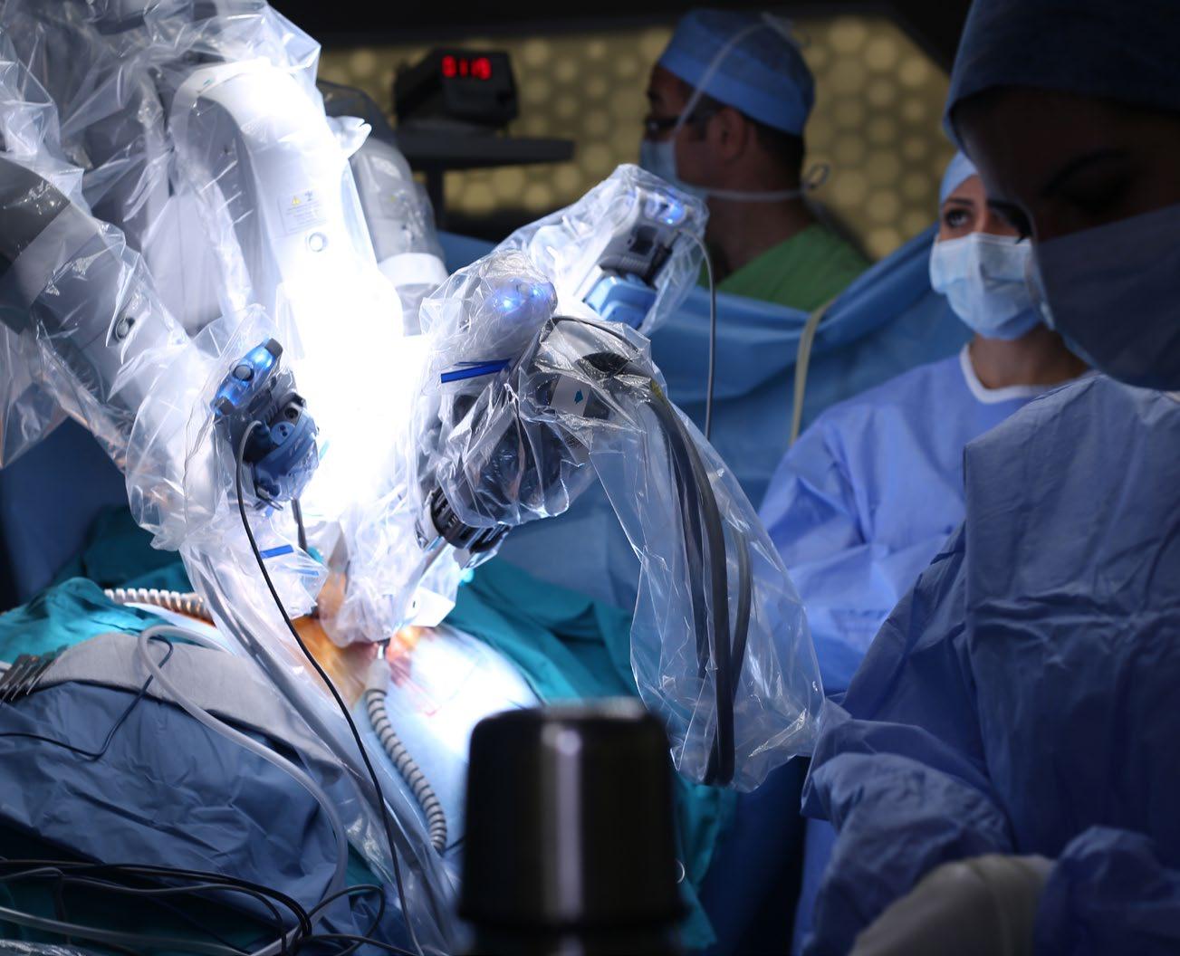
The use of image navigation and robotic-assisted surgery is becoming more common in spine surgery as these tools can improve efficiency, shorten surgical time, and decrease morbidity.1,2
For example, Waschke et al evaluated pedicle screw placement accuracy comparing computed tomography (CT) navigation versus fluoroscopy-assisted placement of pedicle screws.1 The authors noted that the placement accuracy was 96.4% for CT-navigated screws and 93.9% for pedicle screws placed under fluoroscopy. There was a much greater discrepancy in accuracy in the thoracic spine, with a placement accuracy of 95.5% in the CT-navigation group compared to 79.0% in the fluoroscopy group.
Additionally, the use of image navigation and robotic surgery can potentially improve accuracy in the placement of pedicle screws during scoliosis correction surgeries throughout the cervicothoracic spine. The placement of screws can be more complex in these cases because the anatomy can be distorted or because the anatomy prevents the surgeons from obtaining good fluoroscopic images. Studies have also shown that the use of image navigation and robotic surgery can shorten operative time and can decrease morbidity from errant placement of the pedicle screws.1,2
Image navigation and robotic surgery also have the potential benefit of decreasing radiation exposure to the operative staff. In a study by Rampersaud et al, the authors noted that the use of fluoroscopy in spine surgery has the potential to expose the surgeons up to 10 to 12 times more radiation than other orthopaedic surgeons who do not do spine surgery. 3 Because there is increasing use of image navigation and robots in spine surgery, it is reasonable that residents and fellows in training gain exposure to these tools.
However, image navigation and robotic surgery is not foolproof. Chenin et al evaluated the accuracy of lumbar pedicle screw placement with the use of a robot.4 Of the 110 screws placed, 101 (91.8%) were completely within the pedicle, 5 (4.5%) had a pedicle wall breach <2 mm, 2 (1.8%) had a pedicle wall breach of 2–4 mm, and 2 had pedicle wall breach >4 mm (grade D). One screw was replaced during surgery. So, although this study does show good accuracy of pedicle screw placement with a robot, robotic screw placement is not 100% accurate.
In cases in which pedicle screw placement is not accurate, a surgeon needs to determine whether there is a breach and, if so, whether it is clinically significant. Prior experience with traditional open or percutaneous pedicle screw placement is very important as the surgeon will need to draw
20 isass.org Fall 2022 Vertebral Columns
From UCI Health in Orange County, California.
ROBOTICS
Yu-Po Lee, MD
upon his or her experience with traditional pedicle screw placement for repositioning. If a surgeon has little or no experience with traditional pedicle screw placement, this will make repositioning the screw or screws very challenging. Additionally, there can be instances in which a surgeon does not have access to such equipment, such as at an outpatient surgery center, or instances in which equipment malfunctions. In these instances, experience with traditional pedicle screw placement is important.
Thus, while it is important to expose residents and fellows to the latest technology, it is equally important to provide young trainees a firm background in traditional techniques to give them a broad base of knowledge and experience to draw upon when cases do not go as planned. This is especially problematic with reduced duty hours due to the work hour guidelines from the Accreditation Council for Graduate Medical Education. In a study by Jagannathan et al, the authors surveyed 122 chief residents. 5 Most chief residents and program directors believed the 80-hour workweek compromised resident training (96%) and decreased resident surgical ex-
References
1. Waschke A, Walter J, Duenisch P, Reichart R, Kalff R, Ewald C. CT-navigation versus fluoroscopy-guided placement of pedicle screws at the thoracolumbar spine: single center experience of 4,500 screws. Eur Spine J. 2013;22:654–660.
2. Gelalis ID, Paschos NK, Pakos EE, et al. Accuracy of pedicle screw placement: a systematic review of prospective in vivo studies comparing free hand, fluoros -
perience (98%). In a surgical specialty that is technically demanding, experience and repetition are very important and may be limited by these duty hour restrictions. The use of image navigation and robotic surgery may exacerbate this problem even further if young trainees are not exposed to traditional techniques. Further study is required to determine how best to integrate this new technology into surgeon training. n
copy guidance and navigation techniques. Eur Spine J. 2012;21:247–255.
3. Rampersaud YR, Foley KT, Shen AC, Williams S, Solomito M. Radiation exposure to the spine surgeon during fluoroscopically assisted pedicle screw insertion. Spine . 2000;25:2637–2645.

4. Chenin L, Capel C, Fichten A, Peltier J, Lefranc M. Evaluation of screw placement accuracy in circumferential lumbar
arthrodesis using robotic assistance and intraoperative flat-panel computed tomography. World Neurosurg. 2017;105:86–94.
5. Jagannathan J, Vates GE, Pouratian N, et al. Impact of the Accreditation Council for Graduate Medical Education work-hour regulations on neurosurgical resident education and productivity. J Neurosurg. 2009;110(5):820-827.
21 isass.org Fall 2022 Vertebral Columns
ROBOTICS
Telemedicine and Spine Care in 2022
What Have We Learned?
Brandon P. Hirsch, MD

The COVID-19 pandemic had a dramatic impact on healthcare delivery worldwide. Hospital systems were pushed to their breaking point by overwhelming demand for care and increasingly scarce resources. Widespread social distancing restrictions also left many physicians unable to offer the most fundamental building block of healthcare: a visit to the doctor’s office. To overcome this obstacle, many physicians and patients began to utilize virtual visits, otherwise known as “telemedicine” or “telehealth.” Seemingly overnight, demand for virtual office visits skyrocketed. Prior to March 2020, Medicare beneficiaries utilized approximately 13,000 telemedicine encounters per week; by April 2020, this increased to 1.5 million telemedicine encounters per week.1 Like their peers in other surgical specialties, spine surgeons embraced this trend. 2 A recent AO Spine survey of 485 spine surgeons spanning 75 countries revealed that 60% of surgeons performed at least 1 telemedicine visit during 2020. More than 35% of survey respondents utilized virtual visits for more than half of their patient appointments. 3 As obstacles to in-person care fade now, more than 2 years after the first reports of COVID-19, demand for virtual care remains. As telemedicine
becomes more widespread, it is important for spine surgeons to adapt their practices accordingly.
Physical Examination
For all physicians, the physical examination is the area of an office visit most impacted by a transition to telemedicine. Spine surgeons who are hesitant to adopt telemedicine often cite the inability to physically examine patients in the virtual setting as an obstacle. 3 The diagnosis of spine-related complaints is challenging, and many aspects of the physical examination are helpful when assessing a patient’s source of symptoms. Patterns of gait and physical examinations of the shoulder, hip, wrist, and elbow are often needed to differentiate spine-related pain from other musculoskeletal sources. While many patients with spinal disorders have a normal sensory and motor examination, it is important for clinicians to be able to detect significant motor or sensory abnormalities during a patient’s diagnostic work-up.
Iyer et al published one of the earliest articles on adaptations to the physical examination of the spine patient in the telehealth setting. 4 The authors suggested that the patient stand approximately 10 steps from the camera to assess gait, postural balance, muscle tone, and any skin abnormalities.
22 isass.org Fall 2022 Vertebral Columns
From the Core Institute in Mesa, Arizona.
CLINICAL PRACTICE
Many commonly performed “special tests” (Spurling’s test, straight leg raise, etc) are described in a modified manner. Iyer et al suggested that the virtual examination not be viewed as a substitute for in-person examination but rather a supplementary piece of the diagnostic evaluation to be used alongside the patient’s history and imaging findings. Satin and Lieberman described additional modifications to common physical examination maneuvers, including the use of household objects for motor testing. 5 These authors also recommended the ability to perform “screen-sharing” to allow patients to see their relevant imaging findings. Two recent studies compared the results of virtual examinations with patients’ in-person examinations and found good intrarater reliability and high levels of patient satisfaction. 6,7
Surgical Plan
Development of an accurate, appropriate surgical plan is critical to obtaining a successful postoperative result and is one of the common objectives of a patient visit, whether virtual or in person. Two recent studies have evaluated the validity of surgical plans for patients seen both virtually and subsequently in person. Lightsey et al evaluated 33 patients who underwent spine surgery after having both a telemedicine examination and an in-person examination. 8 The authors found that in all but 2 patients, the surgical plans remained the same. In the cases where the surgical plan was altered, both patients were recommended an additional level of fusion following an in-person visit.
In another recent study, 65 patients who had both a telemedicine and an in-person visit prior to spine surgery were evaluated. 9 In this cohort, 61 of 65 (94%) patients had no change to their diagnosis at the in-person visit and 52 patients (80%) had no change to their surgical plan. Of the 13 patients who had a change to their surgical plan after an in-person visit, 10 were related to new imaging findings, 2 were related to new findings on physical examination, and 1 was changed due to patients’ preferences regarding the surgical approach. Both studies’ results suggest that a telemedicine evaluation is capable of generating highly accurate surgical plans for spine patients.

23 isass.org Fall 2022 Vertebral Columns
CLINICAL PRACTICE
CLINICAL PRACTICE
Patient Satisfaction
Several published analyses suggest that patients have relatively high levels of satisfaction with virtual spine care. Hobson et al evaluated patient satisfaction in 128 virtual visits and found that 80% of patients would choose to utilize telehealth visits in the future.10 In addition, 58% of patients felt their virtual visit was as good as or preferable to an in-person visit. Interestingly, patient age did not affect overall level of satisfaction with virtual care. In a similar study across 2 large centers, Satin et al surveyed 772 patients regarding their perception of telemedicine care during the pandemic.11 In this series, 88% of patients were satisfied or very satisfied with their telemedicine visit and 45% of patients reported a preference for telemedicine compared to an in person visit. In this series, patients age 59 years or younger had a statistically significant preference for a telemedicine visit, where -
as patients older than 70 years preferred in-person evaluation. This is unsurprising given that virtual care relies heavily on technology with which some seniors may not be familiar.
Conclusion
Greater adoption of telemedicine in spine surgery will be a lasting effect of the COVID-19 pandemic. The current early literature suggests a high level of patient satisfaction and relatively high diagnostic accuracy. Techniques for in-person evaluation of patients must be modified to suit a virtual encounter. As healthcare technology continues to evolve and improve, spine surgeons are likely to see a growing demand for virtual services. Further potential areas of study should include evaluation of differences in long-term patient outcomes following surgery between patients evaluated virtually versus face-to-face. n
References
1. Verma S. Early impact of CMS expansion of Medicare telehealth during COVID-19. Health Affairs Forefront July 15, 2020. https://doi.org/10.1377/ FOREFRONT.20200715.454789
2. Swiatek PR, Weiner JA, Johnson DJ, et al. COVID-19 and the rise of virtual medicine in spine surgery: a worldwide study. Eur Spine J. 2021;30:2133–2142.
3. Riew GJ, Lovecchio F, Samartzis D, et al. Telemedicine in spine surgery: global perspectives and practices. Global Spine J. 2021;21925682211022310.
4. Iyer S, Shafi K, Lovecchio F, et al. The spine telehealth physical examination: strategies for success. HSS J. 2021;17:14–7.
5. Satin AM, Lieberman IH. The virtual spine examination: telemedicine in the era of COVID-19 and beyond. Global Spine J. 2021;11:966–974.
6. Jansen T, Gathen M, Touet A, et al. Spine examination during COVID-19 pandemic via video consultation. Z Orthop Unfall. 2021;159:193–201.
7. Goyal DKC, Divi SN, Schroeder GD, et al. Development of a telemedicine neurological examination for spine surgery: a pilot trial. Clin Spine Surg. 2020;33:355–369.
8. Lightsey HM, Crawford AM, Xiong GX, et al. Surgical plans generated from telemedicine visits are rarely changed after in-person evaluation in spine patients. Spine J. 2021;21:359–365.
9. Bovonratwet P, Song J, Kim YE, et al. Telemedicine visits can generate highly accurate diagnoses and surgical plans for spine patients. Spine (Phila Pa 1976). 2022;47:1194–202.
10. Hobson S, Aleem IS, Bice MJ, et al. A multicenter evaluation of the feasibility, patient/provider satisfaction, and value of virtual spine consultation during the COVID-19 pandemic. World Neurosurg. 2021;154:e781–e789.
11. Satin AM, Shenoy K, Sheha ED, et al. Spine patient satisfaction with telemedicine during the COVID-19 pandemic: a cross-sectional study. Global Spine J. 2022;12:812–819.
24 isass.org Fall 2022 Vertebral Columns


