
International Research Journal of Engineering and Technology (IRJET) e-ISSN: 2395-0056
Volume: 12 Issue: 03 | Mar 2025 www.irjet.net p-ISSN: 239-0072
International Research Journal of Engineering and Technology (IRJET) e-ISSN: 2395-0056
Volume: 12 Issue: 03 | Mar 2025 www.irjet.net p-ISSN: 239-0072
Akshat Jaiswal*1 , Pratik Sao*2 , Suman Yadav*3 , Vikas Bharti Pandey*4 , Anuyoksha Singh*5
*1,2,3,4 B.Tech Student, Department of Computer Science and Engineering, LCIT Bilaspur (C.G.) *5 Assistant Professor, Department of Computer Science and Engineering, LCIT Bilaspur (C.G.) ***
Abstract - Pancreatic cancer is one of the most lethal cancers due to its late diagnosis and aggressive nature. Early detection significantly improves survival rates, but conventional diagnostic methods are often time-consuming and expensive. This project leverages deep learning techniques to develop an AI-based system capable of detecting pancreatic cancer from CT images. The model classifies images into normal, benign, or malignant cases and also predicts the stage of cancer. Additionally, the system estimates the future risk of developing pancreatic cancer based on patient details. A userfriendly interface is developed using Streamlit, allowing easy interaction for medical professionals. This AI-driven approach aims to provide rapid, cost-effective, and accurate pancreatic cancer detection
Key Words: Deep Learning, Pancreatic Cancer Detection, Medical Image Analysis, Convolutional Neural Networks (CNN), Computer-Aided Diagnosis (CAD), CT Scan Classification, Tumor Identification, Artificial Intelligence (AI)inHealthcare
1.INTRODUCTION
Pancreaticcancerisoneofthemostaggressiveanddeadly formsofcancer,contributingsignificantlytoglobalcancerrelated mortality. The primary challenge associated with pancreatic cancer is its late-stage detection, as symptoms oftenremainunnoticeduntilthediseasehasadvanced.This late diagnosis drastically reduces treatment options and survivalrates,makingearlydetectioncrucialforimproving patient outcomes. Current diagnostic techniques, such as biopsy,MRI,andCTscans,relyheavilyonexpertradiologists forinterpretation.However,thesetraditionalmethodsmay not always provide early-stage detection, especially in resource-limited settings where access to specialized medicalprofessionalsisrestricted.
Inrecentyears,artificialintelligence(AI)anddeeplearning haverevolutionizedmedicalimagingbyenablingautomated, accurate, and efficient disease diagnosis. Deep learning models, particularly Convolutional Neural Networks (CNNs), have demonstrated exceptional performance in medical image analysis, making them highly suitable for detecting and classifying cancerous tissues. By leveraging CNN-basedarchitectures,thisprojectaimstodevelopan AIpowered system for early pancreatic cancer detection usingCTscanimages.Thesystemnotonlyclassifiescases into Normal, Benign, and Malignant categories but also
assesses the severity of the cancer and predicts the patient's future risk basedonvarioushealthparameters.
Beyond classification, this project introduces a predictive component that evaluates an individual’s likelihood of developingpancreaticcancerinthefuture.Byanalyzingkey healthindicatorssuchasage,smokinghistory,familyhistory ofcancer,andBodyMassIndex(BMI),a machinelearningbasedriskassessmentmodel isincorporated.Thisfeature empowers individuals with proactive health insights, allowing for early medical intervention and lifestyle modifications.
To ensure accessibility and ease of use, the system is designedwitha multi-step userinterface developedusing Streamlit, a lightweight web framework for interactive applications. The interface follows a structured workflow, including secure login authentication, patient detail entry, image upload, real-time processing, result visualization, andprecautionary guidance.Thisstep-bystepprocessensuresanefficient,user-friendlyexperience forbothmedicalprofessionalsandgeneralusers.
Theintegrationofdeeplearning,machinelearning,andan intuitive interface makes this project a powerful tool in assisting radiologists, reducing diagnostic workload, and improving early detection rates.Byautomatingthe analysis of CT scans and providing predictive risk assessment,thissystemhasthepotentialtoenhancecancer diagnostics,improvepatientoutcomes,andcontributetothe ongoingadvancementsinAI-drivenhealthcaresolutions.
Pancreatic cancer is typically diagnosed using traditional medicalimagingtechniquesandlaboratorytests.Oneofthe most common approaches is CT scan analysis, where radiologistsexaminecross-sectionalimagesofthepancreas todetectabnormalities.Whileeffective,thismethodishighly dependentontheexpertiseofradiologists,makingitboth time-consumingandsusceptibletohumanerror,especially incaseswhereearly-stagetumorsaresubtleanddifficultto identify.
In addition to CT scans, MRI (Magnetic Resonance Imaging) and ultrasound are also widely used for pancreaticcancerdetection.MRIprovidesdetailedimagesof
International Research Journal of Engineering and Technology (IRJET) e-ISSN: 2395-0056
Volume: 12 Issue: 03 | Mar 2025 www.irjet.net p-ISSN: 239-0072
soft tissues and is particularly useful in detecting small tumorsthatmaynotbevisibleonaCTscan.However,the highcostandlimitedavailabilityofMRImachinesmakethis optionlessaccessible,especiallyinregionswithinadequate healthcare infrastructure. Ultrasound, while non-invasive andrelativelyaffordable,oftenlackstheprecisionrequired for definitive pancreatic cancer diagnosis, particularly in obesepatientsorthosewithexcessiveintestinalgas.
Anotherwell-establishedmethodis biopsy,whichinvolves extracting a small tissue sample from the pancreas for laboratoryanalysis.Whilethistechniqueprovidesahighly accuratediagnosis,itisaninvasiveprocedurethatrequires significant time for sample collection, processing, and evaluation.Delaysinobtainingbiopsyresultscanslowdown treatmentdecisions,whichisacriticalconcernforarapidly progressingdiseaselikepancreaticcancer.
These traditional methods, although effective, come with significantchallenges,includinghighcosts,longprocessing times,andrelianceonhumanexpertise.Thishascreateda growing demand for AI-driven diagnostic tools that can automate the detection process, reduce human error, and enhanceearly-stagedetectionrates.
The emergence of deep learning has significantly transformedthefieldofmedicalimaging,particularlyinthe detection and classification of diseases. Convolutional Neural Networks(CNNs) havebecomethebackboneofAIdrivenimage analysisdue totheirabilityto automatically learn and extract complex patterns from medical scans. Unlike traditional image processing techniques, which requiremanualfeatureextraction,CNNscandetectintricate variations in medical images that may not be easily noticeabletothehumaneye.
Several studies have demonstrated that deep learning modelscanachievediagnosticaccuracy comparable to, or even surpassing, that of human radiologists.Bytraining modelsonlargedatasetsofmedicalimages,AIcanrecognize cancerous patterns with high precision, leading to faster and more reliable diagnoses. Moreover, the use of transfer learning where pre-trained models such as ResNet, VGG16, and Inception arefine-tunedformedical applications has proven effective in improving performance, particularly when working with limited datasets.
To enhance generalization and prevent overfitting, techniques like data augmentation are widely applied, includingrotation,flipping,contrastadjustments,andnoise addition to create more diverse training samples. Furthermore, ensemble learning which combines multiple deep learning models has shown promise in increasingdiagnosticaccuracybyleveragingthestrengthsof differentarchitectures.
With these advancements, deep learning is now being integrated into radiology systems, providing computeraided diagnosis (CAD) tools thatassistdoctorsinmaking moreinformeddecisions.TheabilityofAItoanalyzelarge volumesofmedicaldataquicklyandaccuratelymakesita valuableassetinmodernhealthcare,especiallyinoncology.
Deeplearninghasbeenincreasinglyexploredfor pancreatic cancer detection, with researchers leveraging CNN architecturesforaccurateclassificationofpancreatictissues. Several studies have focused on training AI models to differentiate between normal, benign, and malignant pancreatic tissues using CT scans. Popular CNN architectures,suchas VGG16,ResNet,andInception,have been employed to improve classification accuracy. These modelshavedemonstratedpromisingresultsinidentifying pancreatic tumors, reducing dependency on manual radiologicalassessments.
Beyondimage-basedclassification,someresearchershave developed hybrid AImodels thatintegratemedicalimaging with clinical parameters such as patient age, smoking history,geneticpredisposition,andotherhealthfactors.By combining both imaging and patient-specific data, these models enhance diagnostic precision and provide a more comprehensive risk assessment.
AI-basedpancreaticcancerdetectionsystemsarealsobeing integrated into radiology workstations to assist medical professionals.Thesesystemsactas decision-supporttools, flagging suspicious regions in CT scans and offering secondaryopinionstoradiologists.Byautomatingtheinitial screening process, AI not only reduces the workload of radiologists butalsohelpsin early-stagedetection,which iscrucialforimprovingsurvivalrates.
Despitetheseadvancements,challengesremaininAI-driven pancreatic cancer detection. The availability of large, annotated medical datasets remainsalimitingfactor,as acquiring high-quality labeled data is both expensive and time-consuming.Additionally,AImodelsmustberigorously validatedacrossdiversepopulationstoensure robustness and reliability inclinicalsettings.
Nevertheless, the potential of AI in pancreatic cancer detection is undeniable. With continued improvements in deeplearningarchitectures,datasetavailability,andmodel interpretability,AI-powereddiagnostictoolsareexpectedto playasignificantrolein enhancing early detection rates, improving patient outcomes, and reducing the global burden of pancreatic cancer.
Pancreatic cancer remains one of the most challenging diseases to diagnose in its early stages, contributing to its
International Research Journal of Engineering and Technology (IRJET) e-ISSN: 2395-0056
Volume: 12 Issue: 03 | Mar 2025 www.irjet.net p-ISSN: 239-0072
high mortality rate. One of the primary difficulties is late detection,assymptomsoftenappearonlywhenthedisease hasalreadyprogressedtoanadvancedstage,leavinglimited treatmentoptions.Traditionaldiagnosticmethodsheavily relyon radiologists formanualanalysisofCTscans,which can be both time-consuming and prone to human error. Additionally, cost and accessibility pose significant barriers, as advanced imaging techniques and biopsy procedures are expensive and may not be available in all healthcarefacilities,especiallyinresource-limitedsettings.
Toaddressthesechallenges,thisprojectintroducesan AIbaseddeeplearningmodel fortheautomateddetectionof pancreatic cancer from CT scan images. The system is designed to classify images into normal, benign, or malignant caseswithhighaccuracy,reducingdependency on radiologists and expediting the diagnostic process. It furtherassessesthe severity ofdetected cancer,providing valuable insights for treatment planning. Beyond classification, the system incorporates a future risk prediction component,utilizingpatientdatasuchasage, smokinghistory,andfamilymedicalbackgroundtoestimate thelikelihoodofdevelopingpancreaticcancer.Additionally, itoffers precautionaryguidelines tohigh-riskindividuals, empowering them with preventive measures and early intervention strategies. By integrating deep learning with medicalimagingandpredictiveanalytics,thisprojectaimsto enhance early detection rates, improve accessibility to diagnostictools,andultimatelycontributetobetterpatient outcomes.
Theproposedsystemisdesignedtoleveragedeeplearning andmachinelearningtechniquesfortheearlydetectionof pancreaticcancer,futureriskassessment,andprecautionary guidance through an intuitive user interface. This section outlines the steps involved in dataset preparation, model development, risk prediction, interface design, and evaluation.
ThedatasetconsistsofCTscanimages,whicharecollected fromreliablemedicalimagingsourcesandcategorizedinto three distinct classes: Normal, Benign, and Malignant Proper data organization ensures structured learning, allowing the model to differentiate between healthy and canceroustissueseffectively.
Beforetraining,datapreprocessingisperformedtoenhance the quality and consistency of input images. The preprocessingstepsinclude:
Image Resizing: Standardizingimagedimensions toensureuniformityacrossthedataset.
Normalization: Scalingpixelvaluesbetween0and 1toimprovemodelperformanceandconvergence.
Data Augmentation: Applying transformations suchasrotation,flipping,contrastadjustment,and noise addition to artificially increase the dataset sizeandimprovegeneralization.
Thesepreprocessingtechniquesenhancethemodel’sability to recognize patterns in varying conditions, improving robustnessandreducingoverfitting.
Thecoreofthissystemisa Convolutional NeuralNetwork (CNN),adeeplearningmodelwidelyusedforimageanalysis duetoitsabilitytoextractmeaningfulfeaturesfrommedical images. The CNN is responsible for learning spatial hierarchies of features from CT scans, distinguishing betweennormalandcanceroustissues.
Toimproveaccuracyandoptimizeperformance, Transfer Learning is applied using pre-trained models such as ResNet,VGG16,andInceptionV3.Thesemodels,trainedon large-scale image datasets, provide powerful feature extraction capabilities, reducing the need for extensive trainingonalimitedmedicaldataset.
Thetrainingprocessfollowsthesekeysteps:
Feature Extraction: The CNN layers extract lowlevel and high-level features from CT images, identifyingpatternsindicativeofcancer.
Classification: A fully connected layer classifies images into one of the three categories: Normal, Benign,orMalignant.
Optimization: The model is trained using the cross-entropy loss function,whichmeasuresthe difference between predicted and actual labels, while the Adam optimizer is used to adjust learningratesdynamicallyforfasterconvergence.
The final model is fine-tuned through hyperparameter adjustments,includinglearningrateoptimization,dropout regularization, and batch normalization, ensuring better performanceonunseendata.
InadditiontoclassifyingCTimages,thesystempredictsan individual's future risk of developing pancreatic cancer based on patient-specific factors. A separate machine learning model is integrated for this purpose, using demographicandlifestyleattributessuchas:
Age
Smoking history
Family history of cancer
Body Mass Index (BMI)
International Research Journal of Engineering and Technology (IRJET) e-ISSN: 2395-0056
Volume: 12 Issue: 03 | Mar 2025 www.irjet.net p-ISSN: 239-0072
Thesefeaturesserveasinputstoaclassificationmodel,such as LogisticRegression, DecisionTree,orRandomForest, whichcalculatestheprobabilityoffuturepancreaticcancer occurrence. The output provides a risk assessment score, enabling proactive monitoring and early medical intervention.
Tomakethesystemaccessibleandeasytouse,a Streamlitbasedwebapplication isdeveloped,offeringastep-by-step workflowforusers.Theinterfaceis designedwitha clean medicalthemeandintuitivenavigation,allowingseamless interactionforbothmedicalprofessionalsandgeneralusers.
Thekeyfunctionalitiesoftheuserinterfaceinclude:
Login Page: Implements user authentication to ensuresecureaccess.
Patient Details Form: Collects essential health data,includingname,age,medicalhistory,andrisk factors.
Image Upload: Allows users to upload CT scan imagesforanalysis.
Processing Page: Displays real-time progress of image analysis, showing loading indicators and modelprocessingsteps.
Result Page: Presents classification results (Normal, Benign, or Malignant) along with the predicted future risk score.
Precautionary Guidance: Ifapatientisclassified ashigh-riskordiagnosedwithcancer,personalized medicalrecommendations,lifestylemodifications, andfollow-uptestingsuggestionsareprovided.
Thisstructuredflowensuresasmoothuserexperience,from data input to result interpretation and medical recommendations.
To ensure the reliability and accuracy of the proposed system,themodelundergoesrigorousevaluationusingwellestablishedperformancemetrics.Theprimarymetricsused forassessmentinclude:
Accuracy: Measures the overall correctness of predictions.
Precision: Evaluateshowmanypositivelyclassified casesaretrulycancerous,reducingfalsepositives.
Recall (Sensitivity): Measureshoweffectivelythe model identifies cancerous cases, reducing false negatives.
F1-score: Provides a balanced evaluation of precision and recall, useful for dealing with imbalanceddatasets.
The trained model is tested on a separate test dataset, ensuring its ability to generalize to new, unseen cases. Additionally,Grad-CAM(Gradient-weightedClassActivation Mapping)visualizationisutilizedtohighlightcriticalregions in CT images that influenced the model’s decision. This explainability feature enhances trust in AI predictions by providing visual insights into how the model detects cancerousregions.
TheAI-basedpancreaticcancerdetectionsystemwastested onadatasetofCTimages.Thefollowingobservationswere recorded:
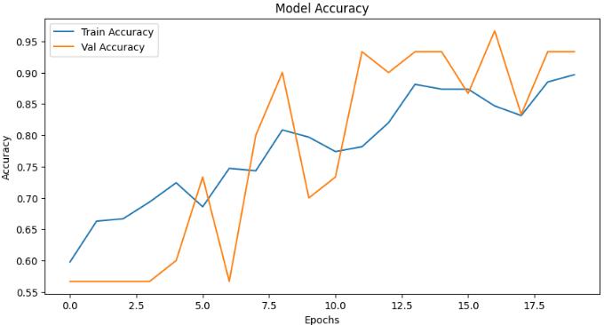
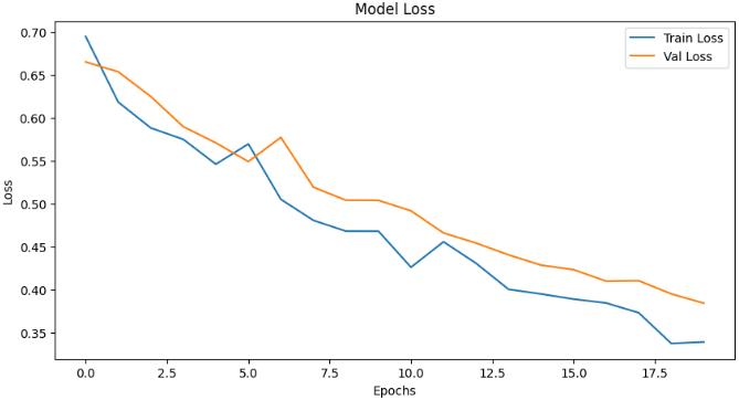
ModelAccuracy:ThetrainedCNNmodelachieved 93% accuracy onthetestdataset.
Model Loss:Themodel'slossvalueconvergedtoa low value, indicating effective learning and generalization.
Precision and Recall: High precision ensured fewer false positives, while high recall minimized falsenegatives.
International Research Journal of Engineering and Technology (IRJET) e-ISSN: 2395-0056
Volume: 12 Issue: 03 | Mar 2025 www.irjet.net p-ISSN: 239-0072
Themodelaccuratelypredictedfuturecancerrisk basedonpatientdetails,providingvaluableinsights intopotentialhealthrisks.
The Streamlit interface provided an easy-to-use experience, allowing smooth interaction with the system.
The precautionaryguidancesystem helpedusers understand preventive measures if cancer was detected.
The results indicate that AI-based detection is a fast, reliable, and affordable alternative to traditional methods.
ThisprojectpresentsanAI-baseddeeplearningapproach for the early detection of pancreatic cancer using CT scan images.Thesystemeffectivelyclassifiesimagesintonormal, benign, or malignant categories, helping medical professionalsidentifyanddiagnosecaseswithhighaccuracy. Beyondclassification,italsodeterminestheseverityofthe detected cancer and predicts the likelihood of future risk based on patient-specific details. By integrating deep learningtechniqueswithauser-friendlyinterface,thistool bridges the gap between medical expertise and AI-driven assistance, ensuring faster, more reliable, and accessible diagnoses.
Oneofthemostsignificantadvantagesofthissystemisits abilitytoautomatecancerdetection,reducinghumanerror andminimizingdependency onradiologists.Inareaswith limitedaccesstospecializedmedicalprofessionals,thisAIdriven approach can serve as a valuable screening tool, allowing for early intervention and improving patient outcomes.Additionally,themodel’spredictivecapabilities enable individuals to assess their risk of developing pancreatic cancer in the future, empowering them with
crucial health insights. This proactive approach to cancer monitoring is further strengthened by the system’s precautionaryguidance,whichprovideshigh-riskpatients withlifestylerecommendations,dietaryadvice,andmedical guidelines to help mitigate their risk and take preventive measures.
Withearlydetectionbeingakeyfactorinimprovingcancer treatment success rates, this AI-powered system has the potentialtorevolutionizecancerdiagnosticsbyenhancing prognosis and increasing survival rates. The ability to analyze large volumes of medical images with precision makesitapowerfultoolinthefightagainstcancer,offering hopeforimprovedhealthcareoutcomes.
While this project primarily focuses on pancreatic cancer, futureadvancementscouldincludeexpandingthemodelto detectmultipletypesofcancers,suchaslung,kidney,and brain cancer, using similar AI techniques. Enhancing predictionaccuracywithlargerandmorediversedatasets, integratingreal-timeAIassistanceintohospital networks, anddevelopingacloud-basedplatformforremoteaccessare also promising directions. Additionally, incorporating explainable AI techniques can help medical professionals betterunderstandandtrustthesystem’sdecisions,fostering greateracceptanceofAIinclinicalpractice.
As artificial intelligence continues to evolve, its role in medical diagnostics will only become more significant. By refining and expanding this technology, AI-driven cancer detection tools can make healthcare more efficient, accessible,andaccurate,ultimatelysavinglivesandshaping thefutureofmodernmedicine.
1. Ian Goodfellow, Yoshua Bengio, and Aaron Courville,DeepLearning,MITPress,2016.
2. Rajalingappaa Shanmugamani, Deep Learning for Computer Vision: Expert Techniques to Train AdvancedNeuralNetworksUsingTensorFlowand Keras,PacktPublishing,2018.
3. S. Suzuki, Medical Imaging: Principles and Applications,CRCPress,2012.
4. Dey, N., Ashour, A. S., & Borra, S. (Eds.), Machine Learning in Bio-Signal Analysis and Diagnostic Imaging,AcademicPress,2018.
5. Murphy, K. P., Machine Learning: A Probabilistic Perspective,MITPress,2012.
6. Suri, J. S., & Liu, K. (Eds.), Medical Imaging and MachineLearning,CRCPress,2020.
7. Bishop, C. M., Pattern Recognition and Machine Learning,Springer,2006.
8. Aggarwal, C. C., Neural Networks and Deep Learning:ATextbook,Springer,2018.