
International Research Journal of Engineering and Technology (IRJET) e-ISSN: 2395-0056
Volume: 12 Issue: 04 | Apr 2025 www.irjet.net p-ISSN: 2395-0072
International Research Journal of Engineering and Technology (IRJET) e-ISSN: 2395-0056
Volume: 12 Issue: 04 | Apr 2025 www.irjet.net p-ISSN: 2395-0072
Ayush Patel1 , Prof. (Dr.) Tejas Shah2
1Ayush Patel, Research Scholar, Instrumentation & Control Dept., L.D College of Engineering, Ahmedabad, Gujarat, India
2Prof. (Dr.) Tejas Shah, Associate Professor, Instrumentation & Control Dept., L.D College of Engineering, Ahmedabad, Gujarat, India ***
Abstract - This work introduces a super-resolution method for improving low-qualitychest X-rayimages witha fine-tuned Real-ESRGAN model. High-resolution chest X-rays from a public database were artificially degraded to produce lowresolution images, which were restored using the trained super-resolution model. Thequalityoftherestoredimages was assessed by comparison with ground truth using PSNR, SSIM, and MSE metrics. Also, a pre-trained CNN-based pneumonia classifier was evaluated with both low-resolution and superresolved images. Outcomes show that the Real-ESRGAN model effectively enhances image quality without sacrificing diagnostic characteristics.
Key Words: Real-ESRGAN, Medical Image Enhancement, Chest X-Ray, Convolutional Neural Networks (CNN), Medical Imaging
1.INTRODUCTION
ChestX-rayimagingisarguablythemostapplieddiagnostic tool used to detect diverse lung conditions, thanks to its speed, affordability, and access. Nevertheless, in most realistic situations i.e., in mobile health setups, remote settings, or telemedicine the resultant images can exhibit low resolution, which can result in impeded accurate interpretation as well as computerized analysis. It is paramounttoimprovesuchlow-resolutionimagequalityto help ensure diagnostic credibility in such limiting environments.
Super-resolution (SR) methods, especially deep learningbasedSRmethods,havebeenhighlypromisinginrecovering visual details from degraded medical images. Here, we investigate the application of Real-ESRGAN, a strong realworldimagerestorationgenerativeadversarialnetwork,to improvelow-resolutionchestX-rays.
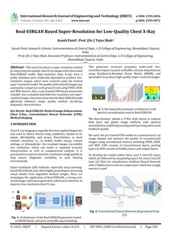
This generator structure processes multi-scale lowresolutioninputsviapixelunshuffle,extractsdeepfeatures using Residual-in-Residual Dense Blocks (RRDB), and upsamplestoproducehigh-qualitysuper-resolvedimages.
Fig -2:U-Net-baseddiscriminatorarchitecturewith spectralnormalizationusedinReal-ESRGAN.
The discriminator adopts a U-Net style layout to capture both local and global image artifacts, with spectral normalizationstabilizingtrainingandimprovingadversarial feedbackquality.
Weused thepretrainedCNNmodelonapneumoniaX-ray image dataset and measure the quality of reconstructed images using conventional metrics including PSNR, SSIM, and MSE. CNN consists of Convolutional layers, pooling layers&ANNconsistsofhiddenlayersandoutputlayers.
To develop the model author have used 5 Conv2D layers whicharefollowedbymaxpoolinglayersforeveryConv2D layer [1] Then for classification Artificial Neural Network with2hiddenlayersandoneoutputlayerwhichhasasingle neuronisused.
Fig -3:ConvolutionalNeuralNetwork(Reproducedfrom [1])
International Research Journal of Engineering and Technology (IRJET) e-ISSN: 2395-0056
Volume: 12 Issue: 04 | Apr 2025 www.irjet.net p-ISSN: 2395-0072
Wealsomeasuretheon-classificationaccuracyusingapretrainedCNNmodelwegetbyreconstructedimagesandto confirm that diagnostic features are not affected during upscaling.
2.1
Fig -4:FlowprocessofMethodology
Theoverallworkflowconsistsoftrainingasuper-resolution modelusingtheNIHchestX-raydataset.Aftertraining,the modelisappliedtosyntheticallydownscaledimagesfroma pneumoniadatasettogenerateenhancedversions.Bothlowresolution (LR) and super-resolved (SR) images are then usedasinputtoapre-trainedCNNclassifier.
ClassificationperformanceisevaluatedonbothLRandSR versions to assess the effectiveness of the enhancement process.
ArandomsamplefromtheNIHChestX-rayDatasetwasused to fine-tune the Real-ESRGAN model for super-resolution tasks. Total 8100 images were used to fine tune RealESRGANmodel.ThisdatasetprovidedavarietyofchestXray images as multiscale of original & multiscale cropped images were generated using data augmentation which is suitable for training the model on general radiographic patterns.
Forclassificationevaluation.AbalancedChestX-rayDataset was used. Subset of 1000 normal and 1000 pneumonia images was selected. High-resolution images were downscaled using bicubic interpolation to generate lowresolutioninputs.
The trained SR model was then applied to restore these images, and both LR and SR versions were prepared for testingwithapre-trainedCNNclassifier
The chosen generator in this setup is RRDBNet, using the pre-trained RealESRGAN_x4plus.pth. It is set with 3 input and output channels, 64 feature maps, 23 residual-inresidual dense blocks, and 32 growth channels. sThe discriminator employed is UNetDiscriminatorSN, which is
loaded from RealESRGAN_x4plus_netD.pth, also having 3 input channels and 64 feature maps, and supports skip connectionsforbetterlearningstabilityandperformance.
The datadataset with 8100 high-resolution images which consist original 3600 ,3600 multiscale images and 900 multiscalecroppedimages,whosepathsareenumeratedina metadatafile.Simpledataaugmentationisapplied,namely horizontalflipping,whichisturnedontoincreasediversity attrainingtime.Rotationisnotused.Theseaugmentations aresmallbutusefulformakingthemodelmorerobuston themedicalimagingdata.
Asophisticatedtwo-stagedegradationpipelineisemployed to synthesize low-resolution inputs from high-resolution images.Duringthefirststage,thereisa20%probabilityof upscaling, a 70% probability of downscaling, and a 10% probabilityofretainingtheresolution
Gaussiannoiseisadded50%withanoiserangefrom1–30; Poissonnoiseisrandomlyscaledbetween0.05–3;grayscale noise is used 40% of the time; and JPEG compression is betweenquality30and95.Duringthesecondstage,bluris used80%ofthetimewithdifferentkerneltypes,resizingis donewithprobabilities30%up,40%down,and30%keep, andthesamenoiseandcompressionmethodsareemployed withslightlydifferentparameters.
Fig
The training dataset contains 8100 images. Using a batch size of 10 on a single GPU, the training runs for 8 epochs, totaling6480iterationswith810iterationsperepoch.
For CNN-based pneumonia classification, a pre-trained modelfromthepubliclyavailableGitHubrepository[1]was utilized. The model was used directly to test the superresolved images generated from our pipeline, allowing evaluationofclassificationperformanceonenhancedinputs.
The authoruseda chest X-ray datasetof 5216 training images(3815pneumonia,1341normal)and624testimages (390 pneumonia, 234 normal) to train a Convolutional NeuralNetwork(CNN)modelforbinaryclassification.
International Research Journal of Engineering and Technology (IRJET) e-ISSN: 2395-0056
Volume: 12 Issue: 04 | Apr 2025 www.irjet.net p-ISSN: 2395-0072
Theinputimagestothemodelwerepreprocessedusingthe ImageDataGenerator functionwithrescaling, shear transformation,zoomaugmentation,andhorizontalflipping. Theseenhancementswereintendedtoenhancethemodel's robustnessandgeneralizabilityduringtraining.Thetraining datawasresizedto300×300pixelswithabatchsizeof128 and binary class labelsfornormal (0) and pneumonia (1) conditions.
The CNN modeluseda sequential architecturewithfive convolutionallayerswithgrowingfilterdepths(16,32,64, 128, 128), each followed by max pooling layers. These convolutionallayerswereprecededbyaflatteninglayerand three dense layers,andthelastoutput layergavea binary prediction.
Thenumber of trainable parameters in the modeltotaled1,983,009.Trainingwasperformedfor 50 epochsandvalidationdoneusinganindependentvalidation generator.Attheendof training, the modelrecordeda training accuracy of 98.16% and aflawlessvalidation accuracyof100%.
3.1 Qualitative Results of SR model
-8:ZoomedincomparisonofLR&SRimages
Zoomed-in regions from LR (left) and corresponding SR (right) images are shown in figure 8. The SR outputs are generated using our fine-tuned super-resolution model, highlightingimproveddetailandclarity.
International Research Journal of Engineering and Technology (IRJET) e-ISSN: 2395-0056
Volume: 12 Issue: 04 | Apr 2025 www.irjet.net p-ISSN: 2395-0072
EvaluationofSRfine-tunedmodelshowninFig10.
Fig -10:TestSRimagepredictionusingpretrainedCNN
AspertheevaluationwehavetestedthePSNR,SSIM,MSEon within the same type of training dataset and completely unseen dataset also. With which we can able to check the robustnessofthemodel
Fig -11:TestSRimagepredictionusingpretrainedCNN
The original input images achieved an accuracy of 92.1% withalossof0.236,whilethesuper-resolved(SR)images achievedanaccuracyof92.3%,withamarginallyhigherloss of 0.279. This indicates that applying the SR model preserved same clinical details as original images with enhancingtheperceptualqualityofanimage.
Thisresearchassessedtheeffectofusingafine-tunedSuperResolution (SR) model on low-resolution medical imagesbeforeclassification.Althoughthe classification accuracyonSRimages(0.923)wasalmostthesameasthat oftheoriginalinputs(0.921)
Notably,essentialclinicaldetailsweremaintainedintheSR outputs,whichensureddiagnosticreliability.Thesefindings indicate that SR can improve visual sharpness and perceptualqualitywithoutdegradingclassificationaccuracy
[1] Y.Yashwanth,“PneumoniadetectionusingCNN,”GitHub repository,Available:https://github.com/Yashwanth23/Pneumonia-detection-using-CNN
[2] Ayush Patel, Prof. (Dr.) Tejas Shah."Chest X-ray Image Super-Resolution Using Artificial Intelligence", Volume 13, Issue III,International Journal for Research in
AppliedScienceandEngineeringTechnology(IJRASET) PageNo:401-411,ISSN:2321-9653,www.ijraset.com
[3] C.Dong,C.C.Loy,K.He,andX.Tang,‘‘Learningadeep convolutionalnetworkforimagesuper-resolution,’’in Proc.13thEur.Conf.Comput.Vis.,Zurich,Switzerland. Springer,2014,pp.184–199.
[4] B.Lim,S.Son,H.Kim,S.Nah,andK.M.Lee,‘‘Enhanced deep residual networks for single image superresolution,’’ in Proc. IEEE Conf. Comput. Vis. Pattern Recognit.Workshops(CVPRW),Jul.2017,pp.136–144.
[5] T.IqbalandH.Ali,‘‘Generativeadversarialnetworkfor medicalimages(MI-GAN),’’J.Med.Syst.,vol.42,no.11, pp.1–11,Nov.2018.
[6] J. Zhu, G. Yang, and P. Lio, ‘‘How can we make GAN perform better in single medical image superresolution? Alesion focused multi-scaleapproach,’’ in Proc. IEEE 16th Int. Symp. Biomed. Imag. (ISBI), Apr. 2019,pp.1669–1673.
[7] X. Bing, W. Zhang, L. Zheng, and Y. Zhang, ‘‘Medical image super resolution using improved generative adversarialnetworks,’’IEEEAccess,vol.7,pp.145030–145038,2019.
[8] XintaoWang,LiangbinXie,ChaoDong,YingShan.RealESRGAN:TrainingReal-WorldBlindSuper-Resolution with Pure Synthetic Data. arXiv preprint arXiv:2107.10833,2021
[9] Random sample of NIH Chest X-ray Dataset. (2017, November23).Kaggle.https://www.kaggle.com/datasets /nih-chest-xrays/sample
[10] Chest X-Ray images (Pneumonia). (2018, March24).Kaggle.https://www.kaggle.com/datasets/pa ultimothymooney/chest-xray-pneumonia
[11] X.Wang,Y.Yu,K.Gu,S.Wu,J.Gu,C.Dong,Y.Qiao,andC. C.Loy,“ESRGAN:Enhancedsuper-resolutiongenerative adversarialnetworks,”inProceedingsoftheEuropean Conference on Computer Vision (ECCV) Workshops, 2018.Available:https://arxiv.org/abs/1809.00219