International Research Journal of Engineering and Technology (IRJET) e ISSN: 2395 0056

Volume: 09 Issue: 07 | July 2022 www.irjet.net p ISSN: 2395 0072

International Research Journal of Engineering and Technology (IRJET) e ISSN: 2395 0056
Volume: 09 Issue: 07 | July 2022 www.irjet.net p ISSN: 2395 0072
Harshitha L1 , Kavitha S2 , Keerthi K L3 , Dr. Prashanth M V4
1,2,3Student, Department of Information Science and Engineering, Vidyavardhaka College of Engineering, Mysore, Karnataka, India 4Associate Professor, Department of Information Science and Engineering, Vidyavardhaka College of Engineering, Mysore, Karnataka, India ***
The Brain is a vital organ (part) in the human body that controlsmemory,vision,thought,andtouch.Anabnormalcell growth insidethebrainiscalledBRAINCANCER.Itisessential to detect the tumour early so that the patient can get appropriate treatment at earlier stages. This software will be utilized inthe Image Processing technique to detect the brain tumour and classify the tumour as Glioma, Meningioma or PituitaryusingBrainMRI(MagneticResonanceImaging).This software provides accurate results with a specific tumour location and will display the tumour’ s features as well. The technique used here is Deep Learning which is Mask RCNN algorithmtodetectthetumourinthebrainandthisalgorithm will give more accuracy.
Thebrainisthemostsignificantanddelicateportionofthe humanbody,whichismadeupofvarioussections(organs). Abnormalcelldevelopmentwithinthebrainistheprimary causeofbraincancer.Currently,doctorsidentifythetumour location by looking at the MRI (Magnetic Resonance Imaging)manually.Thesepredictionsarenotveryaccurate, so to avoid this problem, there is software that works on using MRI images of the brain and image processing techniques to detect brain cancer. This software not just detects the tumour but also identifies the types of brain tumours like Glioma, Meningioma, and Pituitary. This will helpthedoctortogivethepropertreatmenttopatientsatan earlier stage. This software provides better security by allowing only authorized users to access this software, so thatthepatient'sdetailsarenotavailabletounauthorized persons.Thismodelconsistsofsixsteps.imageacquisition, image pre processing. Image Segmentation, Feature Extraction,FeatureComparison,andResults.Thissoftware displays the masked image of the brain tumour by highlightingthetumourlocation.Thissoftwarealsodisplays the features of the tumour like kurtosis, order, moment, centermoment,normalmoment,andentropy.Wehaveused theMaskRCNNalgorithmtodetectbraincancer.MaskRCNN isapopulardeeplearningnetworkinthecomputervision field,forinstance,segmentation.Deeplearningalgorithms givemoreaccuracywhencomparedtootheralgorithms.
Wehaveanalyzedthevariousworkofdifferentauthors. The abnormal presence of the cells in brain outcomes the tumourwhichleadstoabnormality.Bythepriornoticingof the tumour the abnormalityrate can be reduced. The MRI imagescanbeusedtoearlydetectthetumour.Wehavecome toknowthatCNNclassifiesthetumour.ConvolutionalNeural Network(CNN)identifiesthelocationofthetumourbasedon MRI.So,theyhaveproposedthemodelusingConvolutional Neural Network (CNN) to determine the cancer in brain. Initially,theinput asMRIimagesareusedindeeplearning, followedbythesteps,Pre processing,Segmentation,Feature Extraction, Feature Comparison. Due to the disvarient of tissue cells the tumour can vary notably by extracting the exact volume of the tumour using absolute method of segmentation.Agreatexperienceshouldbeneededforthe automatic separation of the tumour cell. They have implementedatensorframeworkfortheclassificationbuilt usingconvolutionnetwork.Thedrawbackoftheaccuracyin segmentationmakeusawaytodevelopamoreaccurateone radiologistarefacingthedifficultyincategorizingthedata whicharepresentinMRIimages.Theearliermodelwastime consuming and difficult for the radiologist to implement them.So,wehavedevelopedasoftwarewhichresultsinhigh accurateinclassifyingandproducingtheoutcomebasedon thecharacteristicsbyusingMaskRCNNAlgorithm.
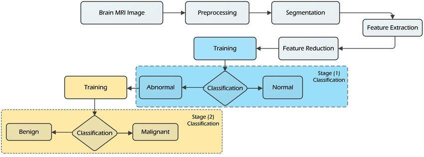
Figure-1: Methodology
International Research Journal of Engineering and Technology (IRJET) e ISSN: 2395 0056
Volume: 09 Issue: 07 | July 2022 www.irjet.net p ISSN: 2395 0072
Itisthefirstandforemoststepineveryprocess.Weneedto collect the available information’s from the relevant locations.Itistheprocessofcollectingrequiredinformation ordata,convertingitintomeaningfulstructureandgrouping itintotheproper order.Here wehavecollectedtheBrain MRIimagesasaninput.
Pre processing is the most basic action performed on the inputdata,inwhichboththesourceanddestinationarehave several. Suchpowerfulimagesareidenticaltothescanner's originalinformation,withapixelintensityoftenexpressed byanarrayofpicturevalues.Eventhoughspatialtransitions of images (example: rotation, scaling, translation) are labelledamongwhichwasbeforemethodologies.Themain goalofthispre processingistoremovetheunwanteddata andtoimprovethedataquality.
Thereare3stepsinpreprocessing:
1.
Itconvertsthenormalinputdataintoblackandwhiteimage. Grayscale display the images using only three colors i.e., White,BlackandGray.
Binarypictureshavingonlytwovaluesforeachandevery pixel i.e., 0 and 1. It will display the input data as binary valuesi.e.,0and1.
0representsblackcolorand1or255representswhitecolor.
3. Threshold:
Itwillhighlighttheinputdatasothatitwillbehelpfulfor thefurthersteps.
ImageSegmentationisamethodwhereadigitalpictureis split down into distinct groups small Sequence segments whichassistsinminimizing theintricacyoftheimageand makefutureprocessingorstudyoftheimageeasier.
Segmentation in basic words involves giving names on images.Asimilarlabelisgiventoeverypixelthatbelongsto thesamegroup.
Feature Extraction:
Itisdifficulttowork withlargeunwanteddatasetssoitis necessarytoselectthefunctionalfeaturesandtoremovethe undesiredfeatures.Thismodulemakesfurtherstepseasier.
Feature Comparison:
Inthissteptheextractedfeaturesaredetectedandclassify theBraintumordetectionbasedondifferentcharacteristics.
Result:
Thisisthefinalstep.Theoutcomesarethendisplayedasthe final stage. Deep learning algorithms will produce more preciseoutcomes.
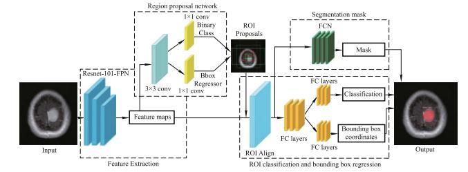
4. BRAIN TUMOR ARCHITECTURE USING MASK RCNN
InPre processingMRIimageswhicharetakenasinputare pre processed.Pre processingconsistsofthreestages:
1. Noise Filtering: It is nothing but the removal of unbothereddatatoobtainthecleandata.
2. OTSU Threshold: It is the process of calculating the measuresforobtainingthepixellevels.
International Research Journal of Engineering and Technology (IRJET) e ISSN: 2395 0056
Volume: 09 Issue: 07 | July 2022 www.irjet.net p ISSN: 2395 0072
In this stage it separates the foreground pixel from background.
This is the stage where raw data is compressed to high manageable form like differentiating in height, weight and patternoftumour.TheresultisfurtherenhancedbyRPNthat extractsbetterfeaturesforprocessing.
TheROI’scanbegeneratedbyfeedingthefeaturestoRPN.To representaboundingboxofvarioussizesthatisdistributed all over the images can be obtained by a 3*3 Convolution layer which scans the images using sliding window. To identify the anchor contains back ground or object binary classification of the image takes place. For setting IOU (Intersection Over Union) values the bounding box regressiongivesboundingboxes.IOUvaluegreaterthan0.7 canbeidentifiedaspositiveorelsenegative.
This stage takes the input from the ROI proposal and classifies the images deeper into tumour or non tumour. Later enhances the size of bounding box. To specifically identifythetumourregionbasedonlocationandsizecanbe donebyBBR.Toobtainthecorrectlengthvectorsoffeature fortheregionsofarbitrary sizeROIAlignlayerisused.
ThesegmentationmaskobtainstheinputfromtheROIAlign thatisofthetypeofpositive.Itidentifiesthemaskfromthe positiveimageduringthetrainingstage.Thepositiveinput obtained from the ROI Align is fed to the Fully Connected Network(FCN)andthenthemaskisidentified.Finally,the outputmaskisobtainedfromthebrainimage.
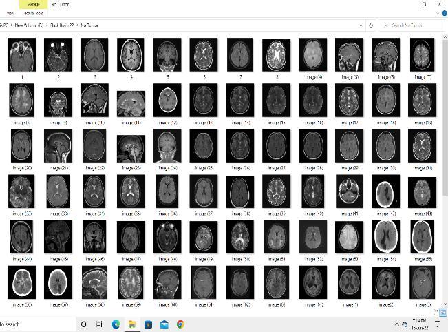
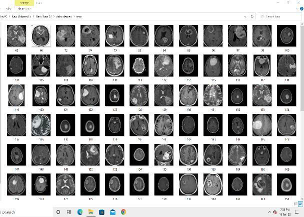
MRIimagesarethesourceofinputforourProject.Thebrain MRIimagesarecollectedfromvarioushospitals.Thedata required for the training is MRI images containing Tumor and Non Tumor MRI images. The MRI images pixel range from0 255.The0canbeconsideredaswhiteand255canbe considered as Black. Here we have attached some of the datasetsimagesconsistingofTumorandNon Tumor.
International Research Journal of Engineering and Technology (IRJET) e ISSN: 2395 0056
Volume: 09 Issue: 07 | July 2022 www.irjet.net p ISSN: 2395 0072
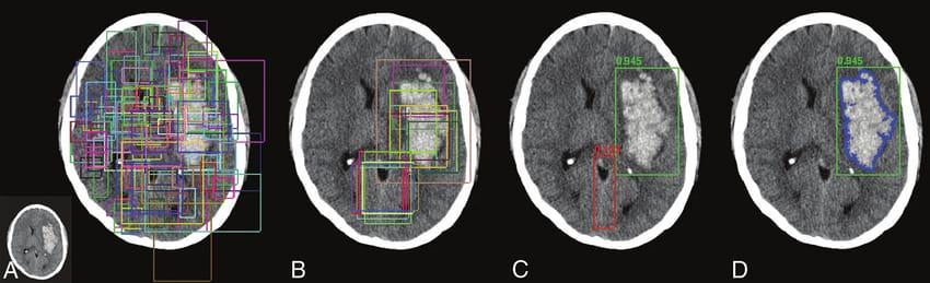
MaskRCNNProcessthebrainMRIimagesin4Steps. MRIimagesareprovidedasInputtothealgorithm
STEP A:InthisstepSemanticSegmentationofthepixelsin thebrainMRIimagestakesplace.
STEPB: In this step Classification and localization of the PreferredlocationoftheMRIpixelimagesoccurs.
STEP C:Inthisstepmoreaccuratepreferreddetectionofthe specifiedlocationtakesplace.
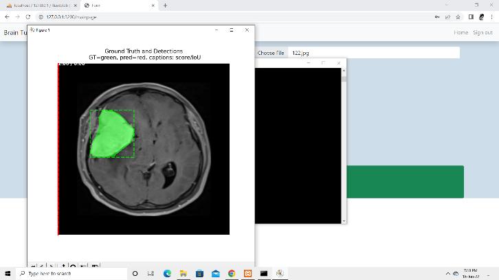
STEP D: In this step the specific location of the tumor is obtainedasresult.
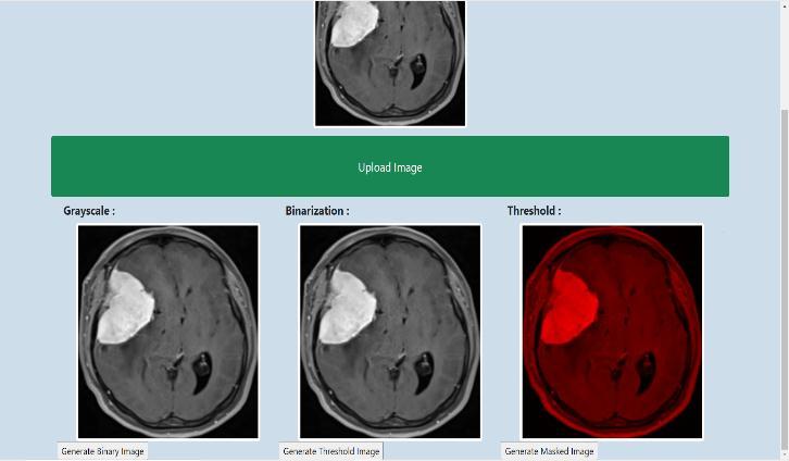
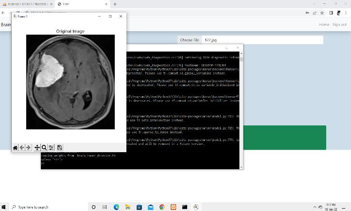
Figure-7: DisplayingOriginalImage
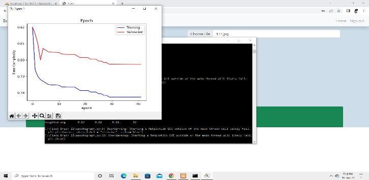
Figure 9: Epochv/sTimecomplexitygraph.
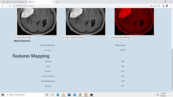
Figure 8: DisplayingMaskedImage.
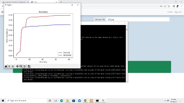
Figure 10: AccuracyvsTimecomplexitygraph.
Figure 11: DisplayingAccuracyandFeatures.
The tumour from the MRI images can be obtained from various techniques. One model gives higher accuracy and classification than the manual work. The MRI images are compared and then predicted the output based on the performance.Thetumourandnon tumourimagesareused and compared to obtain the output. The precise and the accurateoutputcanbeobtainedfromthesystem.Ourmodel
detects the tumour and classifies the tumour as Glioma, MeningiomaandPituitary.Themodeloutputsthefeatures likeorder,momentandentropy.Thesegmentationprocess extractstheRegionofInterestfortheprocess.Furtherany implementationorMethodologiescanbeusedtoincreasethe performance. We think our model will help the society in morepromisingwayforthefuturework.
[1]G.Raut,A.Raut,J.Bhagade, J.BhagadeandS.Gavhane, "Deep Learning Approach for Brain Tumor Detection and Segmentation," 2020 International Conference on Convergence to Digital World Quo Vadis (ICCDW),2020.
[2]R.Tamilselvi,A.Nagaraj,M.P.BehamandM.B.Sandhiya, "BRAMSIT: A Database for Brain Tumor Diagnosis and Detection," 2020 Sixth International Conference on Bio Signals, Images, and Instrumentation (ICBSII),2020.
[3]P. Ganasala, D. S. Kommana and B. Gurrapu, "SemiautomaticandAutomaticBrainTumorSegmentation Methods:PerformanceComparison," 2020IEEEIndiaCouncil International Subsections Conference(INDISCON),2020.
[4]S. K. Baranwal, K. Jaiswal, K. Vaibhav, A. Kumar and R.Srikantaswamy,"PerformanceanalysisofBrainTumour Image Classification using CNN and SVM," 2020 Second InternationalConferenceonInventiveResearchinComputing Applications (ICIRCA),2020.
[5]BKokila1 ,MSDevadharshini1 ,AAnitha1 andSAbisheak Sankar1, “Brain Tumor Detection and Classification Using DeepLearningTechniquesbasedonMRIImages”,Journalof Physics:ConferenceSeries,Volume1916,2021International Conference on Computing, Communication, Electrical and BiomedicalSystems(ICCCEBS)202125 26March2021
[6]Irmak,E.Multi ClassificationofBrainTumorMRIImages Using Deep Convolutional Neural Network with Fully Optimized Framework. Iran J Sci Technol Trans Electr Eng 45, 1015 1036(2021).
[7]C.Someswararao,R.S.Shankar,S.V.AppajiandV.Gupta, "Brain Tumor Detection Model from MR Images using Convolutional Neural Network," 2020 International Conference on System, Computation, Automation and Networking (ICSCAN),2020
[8]D. Divyamary, S. Gopika, S. Pradeeba and M. Bhuvaneswari, "Brain Tumor Detection from MRI Images usingNaiveClassifier," 2020 6thInternationalConferenceon Advanced Computing and Communication Systems (ICACCS), 2020.
[9]K.S.Rani,K.M.kumari,T.Nireekshna,D.V.Shobana,N. KavithaandB.B.Sri,"IdentificationofBrainTumorsusing Volume rendering Techniques," 2021 International
Conference on Artificial Intelligence and Smart Systems (ICAIS),2021.
[10]B.M,"AutomaticSegmentingTechniqueofBrainTumors withConvolutionalNeuralNetworksinMRIImages," 2021 6th International Conference on Inventive Computation Technologies (ICICT),2021.
[11]M.SwamiandD.Verma,"Analgorithmicdetection of brain tumour using image filtering and segmentation of variousradiographs," 2020 7th International Conference on Signal Processing and Integrated Networks (SPIN),2020.
[12] M. F. I. Soumik and M. A. Hossain, "Brain Tumor ClassificationWithInceptionNetworkBasedDeepLearning Model Using Transfer Learning," 2020 IEEE Region 10 Symposium (TENSYMP),2020.
[13] Z. Sobhaninia, S. Rezaei, N. Karimi, A. Emami and S. Samavi, "Brain Tumor Segmentation by Cascaded Deep Neural Networks Using Multiple Image Scales," 2020 28th Iranian Conference on Electrical Engineering (ICEE),2020.
[14] P. Wu and Q. Chang, "Brain Tumor Segmentation on Multimodal3D MRIusingDeepLearningMethod," 202013th International Congress on Image and Signal Processing, BioMedical Engineering and Informatics (CISP BMEI),2020.
[15] Somnath, S. Negi, P. C. Negi and N. Sharma, "Tumor Segmentation in Brain MRI using Fully Convolutional Network," 2020 International Conference on Advances in Computing, Communication & Materials (ICACCM),2020.
[16]Masood, Momina, et al. "Brain tumor localization and segmentationusingmaskRCNN." FrontiersComput.Sci. 15.6 (2021):156338.
International Research Journal of Engineering and Technology (IRJET) e ISSN: 2395 0056 Volume: 09 Issue: 07 | July 2022 www.irjet.net p ISSN: 2395 0072 © 2022, IRJET | Impact Factor value: 7.529 | ISO 9001:2008 Certified Journal