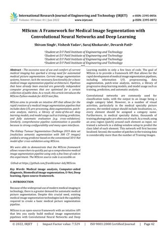
MIScnn aims to provide an intuitive API that allows for the rapid creation of a medical image segmentation pipeline that includes data I/O, pre processing, data augmentation, patch wise analysis, metrics, a library of state of the art deep learning models, and model usage such as training, prediction, and fully automatic evaluation (e.g. cross validation).
Similarly, comprehensive pipeline customization is possible because to strong configurability and many open interfaces.
Key Words: Medical image analysis, Computer aided diagnosis, Biomedical image segmentation, U Net, Deep learning, Open source framework.
International Research Journal of Engineering and Technology (IRJET) e ISSN: 2395 0056 Volume: 09 Issue: 01 | Jan 2022 www.irjet.net p ISSN: 2395 0072 MIScnn: A Framework for Medical Image Segmentation with Convolutional Neural Networks and Deep Learning Shivam Singh1 , Vishesh Yadav2 , Suraj Khokarale3, Devarsh Patil4 1Student at D.Y Patil Institute of Engineering and Technology 2Student at D.Y Patil Institute of Engineering and Technology 3Student at D.Y Patil Institute of Engineering and Technology 4Student at D.Y Patil Institute of Engineering and Technology *** Abstract
The Kidney Tumour Segmentation Challenge 2019 data set (multiclass semantic segmentation with 300 CT images) yielded a strong predictor based on the conventional3D U Net model after cross validation using MIScnn. We were able to demonstrate that the MIScnn framework allows researchers to quickly put up a comprehensive medical image segmentation pipeline using only a few lines of code in this experiment. The MIScnn source code is accessible on Github at https://github.com/frankkramer lab/MIScnn.
1. INTRODUCTION
islocalised.classtrainedantrainingFurthermore,everyprocess,activities,singleclassificationConvolutitraining,progressiveaugmentation,includingrapiddevelopmentofmedicalimagesegmentationpipelines,informationI/O,preprocessing,datapatchwiseanalysis,metrics,alibraryofdeeplearningmodels,andmodelusagesuchasprediction,andautomaticanalysis.onalnetworksarecommonlyusedfortasks,withtheoutputtoanimagebeingacategorylabel.However,inanumberofvisualparticularlyinthemedicalspecialtypicturetheneededoutputshouldincludelocalization,i.e.,elementshouldbeassignedacategoryname.inmedicalspecialtyduties,thousandsofphotographsareoftenoutofreach.Asaresult,usingarearegion(patch)aroundeachelementasinput,weanetworkinaslidingwindowsetuptopredictthelabelofeachelement.ThisnetworkwillfirstbecomeSecond,thenumberofpatchesinthetrainingdataconsiderablymorethanthenumberofTrainingImages. © 2022, IRJET | Impact Factor value: 7.529 | ISO 9001:2008 Certified Journal | Page42
The excessive ease of use and comfort of current medical imaging has sparked a strong need for automated medical picture segmentation. Current image segmentation systems, however, lack the necessary functionality for a basic medical image segmentation pipeline architecture. Pipelines that have already been enacted are generally independent computer programmes that are optimized for a certain collection of public data. As a result, this article introduces the MIScnn Python module for ASCII text files.
Becauseofthewidespreaduseofmodernmedicalimagingin technology,thereisagreaterdemandforautomaticmedical picture segmentation. Despite this crucial need, existing medicalimagesegmentationtechnologieslackthecapability required to create a basic medical picture segmentation MIScnnpipeline.isanopen sourceframeworkwithanintuitiveAPI that lets you easily build medical image segmentation pipelines with Convolutional Neural Networks and Deep Learning models in only a few lines of code. The goal of MIScnn is to provide a framework API that allows for the

Inconsistentsignalintensityrangesinphotoswillhavea significantimpactonsegmentationmethods'performance. Due to completely distinct picture formats, variable hardware/instruments(e.g.completelyseparatescanners), technicalinconsistencies,andeasilyabiologicalvariance,the signal rangesofmedical specialistimagingknowledgeare very varied amongst knowledge sets. Moreover, picture segmentation techniques that use machine learning often perform better on alternatives that follow a standard distribution.Scalingandstandardizingimaginginformation is advised to achieve dynamic signal intensity variation uniformity.
2. Data Input
FIG1.
Forloadingmagneticresonanceimagingandcomputed axialtomographydataintotheframework,MIScnnprovides a data I/O interface for the Neuroimaging Information Science Technology Initiative (NifTI) file format. This structurewasfirstdevisedtoexpeditetheeventwhilealso enhancingtheusabilityofinformationsciencetechnologies related to neuroimaging. Still, it's currently unremarkably usedforsharingpublicandanonymousmagneticresonance imagingandCTknowledgesets,notonlyforbrainimaging, but also for any other type of human 3D imaging.The 3D imagematrixandnumerous data,suchasthethicknessof themagneticresonanceimagingslices,arestoredinanNIfTI file.
International Research Journal of Engineering and Technology (IRJET) e ISSN: 2395 0056 Volume: 09 Issue: 01 | Jan 2022 www.irjet.net p ISSN: 2395 0072
2.2 Custom data I/O interface MIScnn allows the use of bespoke knowledge I/O interfacesforvariousimageknowledgeformatsinaddition to the mandated NIfTI I/O interface. This open interface enablesMIScnntohandlecertainmedicalspecialtyimaging choices(forexample,MRIslicethickness),withouttheloss of this information due to a format conversion demand. A specific I/O interface should be dedicated to the preprocessingprocess,anditshouldreturntothemedical picture as a second or 3D matrix for integration into the process.
3.2 Clipping Similar to component intensity standardization, it's conjointly common to clip component intensities to an explicit vary. Outside of this range, intensity costs will be reducedtothelowestormostvariablevalue.Eveninmany scanners,componentintensitylevelsforsimilarorgansor tissuetypesareassumedtobeequal,especiallyincomputer imagingimages.Organ specificcomponentintensityclipping mightbeusedtotakeadvantageofthis. FlowchartofMIScnnPipeline
3. Data Preprocessing
© 2022, IRJET | Impact Factor value: 7.529 | ISO 9001:2008 Certified Journal | Page43
2.1 NifTi data I/O interface
3.1 Pixel intensity normalization
The well known problem in medical images is large unbalancesbetweensignificantsegmentsandthebackdrop resultsinalargenumberofpartsthatarestrictlyclassified asbackgroundandhavenolearninginformation.There'sno use in multiplying these blank elements or patches, especiallywhenitcomestoknowledgeaugmentation.Asa result, any patches that are completely classified as backgroundareomittedfromthepatch wisemodeltraining tominimizeunnecessaryfittingdelays.
4.4 Multi CPU and GPU Support
InadditiontoGPUcomputing,MIScnnofferstheuseof several GPUs and simultaneous central processor batch loading.Thestoringofalreadypreparedbatchesonmemory, forexample,allowsforafastandparallelizableprocesswith centralprocessorsandGPUclustersbyremovingtheriskof batchpreparationbottlenecks.
5.1 Model Architecture
Sets of entire photos or patches are packaged into batches after the data preparation and hence the optional dataaugmentationfortraining.Onebatchcomprisesalarge numberofreadyimagesthatareprocessedbythemodeland GPU in a single phase. The neural network adjusts its internal weightsinaccordancewiththespecifiedlearning rateforeachbatchorprocessstep.Thenumberofimages thatmaybestoredinasinglebatchishighlydependenton theamountofGPUmemoryavailable,andthereforemustbe appropriately planned in MIScnn. Each batch is stored in memorysothatitmaybeaccessedatanytimeduringthe trainingprocess.Duetotheavoidanceofreservecontinuous batch preprocessing, this strategy dramatically decreases computation time. MIScnn also permits "on the fly" construction of the following batch in memory during the runtimetoovercomethislimitation.
4.3 Batch Shuffling
International Research Journal of Engineering and Technology (IRJET) e ISSN: 2395 0056 Volume: 09 Issue: 01 | Jan 2022 www.irjet.net p ISSN: 2395 0072 3.3 Resampling To change the width and/or height of images, the resamplingtechniqueisused.Thisendsupinabrand new imagewithachangingrangeofpixels.resonanceorlaptop pictorial representation scans will have different slice thicknesses. However, in order to train neural network models,thepicturesmusthavethesameslicethicknessor voxel spacing, this could be accomplished through resampling.Tobeginwith,downsamplingimageslimitsthe amountofGPUmemoryavailablefortrainingandprediction. 3.4 One hot encoding twomechanicallyvariablestyle,anlabelsdeepgenerated.depictionproblems.multiMIScnncanhandlebothbinary(background/cancer)andclass(background/kidney/liver/lungs)segmentationUsingavariablewithtwostates,zeroandone,theofabinarysegmentationmaybereadilyExceptinmachinelearningtechniques,suchaslearningmodels,wheremanycategorysegmentationareused,itisnecessarytotranslatethecategoriesintoextramathematicalpicture.UsingtheOneHotcodingthismightbeaccomplishedbycreatingasinglebinaryforeachsegmentationcategory.OneHotencodessegmentationlabelswithmorethancategoriesusingMIScnn.
Attheendofeachepoch,theorderofbatches,thatis,the areaunitintendedtobefittedandprocessedisshuffled.This strategy lessens the risk of overfitting by reducing the varianceoftheneuralnetworkduringfittingoveraperiodof time.Itshouldbeemphasized,however,thatjustthebatch processsequenceisshuffled,andtheknowledgeitselfisnot sortedintoanewbatchorder.
4. Sampling and Batch Generation 4.1 Skipping blank patches
The most important stage in a medical picture segmentation pipeline is choosing a deep learning or convolutional neural network model. There are many distinct model architectures, each with its own set of strengths and limitations. The MIScnn options provide an open model interface that allows you to load and switch between the several progressive convolutional neural network models available, such as the widely used U Net model.Keras,anASCIItextfileneuralnetworkframework that provides a straightforward API for commonly used neural network building blocks on top of TensorFlow, is
© 2022, IRJET | Impact Factor value: 7.529 | ISO 9001:2008 Certified Journal | Page44
In3Dsegmentationanalysis,theavailableGPUhardware plays an outsized role depending on the resolution of medicalimages.DuetothelargeGPUmemoryrequirements, itiscurrentlyunabletofullyintegratehigh resolutionMRIs with an associated example size of 400 512 512 into progressive convolutional neural network models. As a result,3Dmedicalimagingdataiseithersplitintosmaller cuboidpatchesorprocessedslicebyslice,similartoaseries ofsecondphotographs[10,11,23].MIScnncuts3Dmedical photosintopatcheswithacustomizablesize(e.g.128128 128) by default to fully use the data from all three dimensions.ThesepatcheswillfunctioninGPUswithRAM capacities of 4 24 GB, which are commonly used in the analysis, according to the model design. The slice by slice second analysis is supported and may be used in MIScnn, however,the3D patchanalysisisnot. It'salsopossible to combine the use of complete 3D images in the event that you're studyingunusuallysmall medical imagesorhavea largeGPUcluster.Secondmedicalimagesareautomatically incorporatedintoconvolutionalneuralnetworksanddeep learning models. Still, for images with high resolution, a secondpatch wisestrategyisused.
4.2 Batch Management
3.5 Patch wise and full image analysis
5. Deep Learning Model Creation
5.5
The model will now be utilized for training on the information to suit model weights or for prediction by leveraginganalreadyfittedmodelwiththeinitializeddeep learning model and hence the totally preprocessed information.Instead,themodelwilldoANanalysisbyusing cross validationwithnumeroustrainingandpredictioncalls, asanexample.ThemodelAPIallowsyoutosaveandload modelssothatyoumayreprocesspreviouslyfittedmodels forpredictionorsharepre trainedmodels.
knowledgefurthersegmentationusingthewithovpredictedmedicalwithinpredictions,thedegreepredictionpredictionpatchesknowledgediscreteduringafterprovidesknownholdingencodedrepresentedpanel'salgorithmfromnetworkOncetrained,anassociatedegreepreviouslyfittedneuralmodelwillbeutilizeddirectlyoritwillbeimportedafileforsegmentationprediction.Foreachpanel,theestimatesaSigmoidpriceforeachcategory.ThisprobabilityassessmentfortherelatedlabelisbytheSigmoidprice.TheargmaxoftheOneHotcategoryisthenbornagaintooneresultvariablethecategorywiththebestSigmoidpricing,whichisformulticlasssegmentationchallenges.MIScnntwoapproachesforpatchesinsidethepredictionusingtheoverlappingpatchwiseanalysismethodologythetraining.Eitherthepredictionapproachdevelopspatchesandregardsoverlappingpatchesassolelyenhancementduringthetraining,oroverlappingareproducedforprediction.Duetoalackofcapacityatpatchedges,generatingasecondforedgepixelsinpatchesbyvictimizingassociateoverlapmaybeanunderutilizedtechnique.Withinoverlappingsectionoftwopatchesandwithnumerousamergingmethodforthepixelsisneededthesubsequentmergeofpatchesbacktothebasicpictureform.MIScnnestimatesthemeanoftheSigmoidvaluesforeachcategoryineacherlappingpanelbydefault.Theresultingpicturematrixsegmentationprediction,whichhasthesameformatasoriginalmedicalimage,isstoredintoafilestructuretheknowledgeI/Ointerfacesupplied.ThepredictedmatrixisstoredinNIfTIformatwithnoinformationbydefaultwhenusingtheNIfTII/Ointerface. © 2022, IRJET | Impact Factor value: 7.529 | ISO 9001:2008 Certified Journal | Page45
5.4 Training Variousparametersshouldbeestablishedwhiletraining aconvolutionalneuralnetworkordeeplearningmodel.The information augmentation options of the data set, which haveasignificantimpactonmedicalpicturesegmentation training, should already be described in the pipeline. Followingthat,thebatchmanagementconfigurationlisted thebatchsizesettingsaswellasthebatchshufflingatthe endofeachepoch.Asaresult,simplythelearningrateand hence the number of epochs must be altered before the trainingtechniquecanbeused.Thetrainingrateofaneural networkmodelisdeterminedbytheamounttowhichthe neuralnetworkmodel'spriorweightsaremodifiedineach iterationorepoch.Thenumberofepochs,ontheotherhand, determinestheproportionoftimesthefullknowledgeset willbefittedintothemodel.Theresultingfittedmodelcan thenbestoredinmemory.Becauseoftheremainingfitting time,theunderlyingKerasframeworkprovidesinsightsinto the current model performance using specified metrics throughouttraining.MIScnnalsoprovidestheuseofafitting evaluation questioning practicality, in which the fitting scores and metrics are saved in a tab separated file or immediatelyplottedasagraph. Prediction
5.3 Model Utilization
5.2 Metrics
International Research Journal of Engineering and Technology (IRJET) e ISSN: 2395 0056 Volume: 09 Issue: 01 | Jan 2022 www.irjet.net p ISSN: 2395 0072 usedtodefinemodels.Thealreadyestablishedmodels,like the Optimized High Resolution Dense U Net model, are significantly flexible by a determinable range of neurons, custom input sizes, ex gratia dropout, and batch normalizationlayers,orenhanceddesignversions.MIScnn additionally provides 3D architectures, as well as 2D architectures for medical image segmentation. The open model interface enables for bespoke deep learning model implementations and simple integration of these custom modelsintotheMIScnnpipeline,inadditiontothefreedom inchangingbetweenpreviouslyimposedmodels.
MIScnnhasalargenumberofdifferentmetricsthatmay beutilizedforlossperformancetraining,figureanalysis,and manual performance analysis. One of the most often used metrics for medical picture segmentation is the Dice constant, also known as the Dice similarity index. It calculates the degree of similarity between the expected segmentationandthegroundtruth.Falsepositives,onthe otherhand,arepenalized.There'sasimpleandclass wise Diceconstantimplementationtoenjoytheprecisionmetric reckoningonthesegmentationcategories(binaryormulti class).Unlikethesimpleapproach,whichmerelycountsthe number of right and erroneous predictions, the category wisemethodaccountsforpredictionperformanceforeach segmentation class, which is strongly recommended for typicallyclass unbalancedmedicalimages.TheJaccardIndex isanotherwidelyacceptedstatistic.Eventhoughit'ssimilar to the Dice constant, it doesn't focus entirely on exact segmentation.Itdoes,however,punishover segmentationof understanding. MIScnn, on the other hand, employs the Tversky loss for training. The Tversky loss performance solves knowledge imbalance and is admired by the Dice constant.Evenso,itachievesafarbetterbalanceofprecision andrecall.Asa result,Tverskyloss performanceprovides smart binary as well as multi class segmentation performance. In addition, many common metrics used in Keras, such as accuracy and cross entropy, are used in MIScnn.MIScnnallowsyoutoblendadditionalmetricsfor training and analysis in addition to the already imposed metrics or loss functions. As described in Keras, a custom metricisenforcedandreadilysuppliedtothedeeplearning model.
© 2022, IRJET | Impact Factor value: 7.529 | ISO 9001:2008 Certified Journal | Page46
6. Real Life Use Cases
International Research Journal of Engineering and Technology (IRJET) e ISSN: 2395 0056 Volume: 09 Issue: 01 | Jan 2022 www.irjet.net p ISSN: 2395 0072 5.6 Evaluation
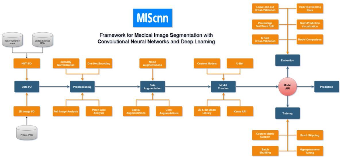
5.7 Convolutional Neural Network (U net) A contracting path (left side) and an expansive path (right side) make up the network design (right side). The convolutional network's contracting route follows the standardarchitecture.Itismadeupoftwo3x3convolutions (unpaddedconvolutions)thatareappliedrepeatedly,each followed by a rectified linear unit (ReLU) and a 2x2 max pooling operation with stride 2 for downsampling. We quadruple the number of feature channels with each downsampling step. An upsampling of the feature map is followedbya2x2convolution("up convolution")thathalves the number of feature channels, a concatenation with the proportionally cropped feature map from the contracting path,andtwo3x3convolutions,eachfollowedbyaReLUin the expanding path. Due to the loss of boundary pixels in everyconvolution,croppingisrequired.A1x1convolutionis employedatthefinallayertoconverteach64 component feature vector to the appropriate number of classes. The network comprises a total of 23 convolutional layers. To ensure that the output segmentation map tiles seamlessly (seeFigure2),theinputtilesizeshouldbechosensothatall 2x2max poolingoperationsareappliedtoalayerwithan evenx andy dimension.
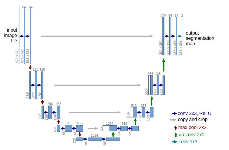
6.1 Kidney Tumor Segmentation
FIG2U NetArchitecture
ComputedTomography scansofurinaryorgantumors fromtheurinaryorgantumorSegmentationChallenge2019 information set showing the urinary organ (red) and tumor (blue) segmentation as overlays. The photographsshowthesegmentationvariationsbetweenthe bottom truth provided by the KiTS19 challengeandthereforethepredictionfromthequality3D U Netmodelsofourthree foldcross validation.
Multipleautomaticanalysistechniquesaresupportedby MIScnn to investigate medical image segmentation performance: k fold cross validation, leave one out cross validation, percentage split validation, hold out sets for checking(datasetsplitintotestandplaythingwithagiven percentage), and elaborate validation in which it is frequentlygivenwhichpicturesshouldbeusedfortraining and testing. Apart from extensive validation, sampling is usedtocreatetrainingandtestingdatasetsinallanalytical methodologies. The specified measurements and loss performance for the model are automatically planned in figuresduringthestudyandrecordedintab separatedfiles foreasyexamination.Inadditiontoperformancemeasures, the constituent worth changes, and the frequency of segmentation categories in medical images are frequently examinedaspartoftheMIScnnstudy.Byproducingpicture representations with segmentation overlays, the resulting forecastisfrequentlycompareddirectlytothebottomtruth. Thesliceswiththesegmentationoverlaysaremechanically depictedinsidetheGraphicsInterchange Format(GIF)for 3Dimages,suchasMRIs.
8. Results
Theuseofdilationtosightdominantoptionswithinthe image was explored. The authors planned a parallel expandedCNNmodel.Theexpandedmoduleconcernedthe skipping of pixels throughout the convolution method Parallel CNN branches square measure proposed with completely different dilation rates. The results of parallel brancheswerecombinedandsentintothenextconvolution layer.Theconcatenation convolution process wasused to investigatefeaturerelationshipsinenlargedconvolutions, resulting in visually prominent classification alternatives. The model conjointly used Grad CAM and Grad CAM++ to highlighttheregionsofclass discriminativenoticeablemaps. The performance metrics used were accuracy, precision, recall,F1 scorewithROC/AUC,andare96.58%,95%,91%, 93%,and99.1%severally
© 2022, IRJET | Impact Factor value: 7.529 | ISO 9001:2008 Certified Journal | Page47
7. Future Scope Multiple essential aspects are now the focus of active MIScnn development: Adding more knowledge I/O interfacesforthemostwidelyusedmedicalimageformats, such as DICOM, expanding preprocessing and knowledge augmentation procedures, and implementing a variety of cost effective patch skipping approaches rather than ignoring each blank patch (e.g. denoising patch skipping)
6.2 Coronavirus disease 2019(Covid 19) COVID 19mightbeaglobalpandemicvirusthatspreads quickly over the planet. RT PCR (Reverse Transcription enzyme Chain Reaction) is a commonly used test for detectingCOVID 19infection.RT PCRtestingisthatthegold standard for COVID 19 testing, RT PCR is an incredibly complicated, long, and labor intensive method, sparse availability,andnotcorrect.ChestX raymaybeusedforthe initialscreeningoftheCOVID 19inplaceshavingashortage ofRT PCRkitsandisadditionalcorrectatdiagnosing.Many researchershaveuseddeeplearningtoclassifyifthechest infectionisthankstoCOVID 19ordifferentailments.
Implementation of an open interface for bespoke preprocessingalgorithmsforcertainpicturetypes,suchas MRIs.TheMIScnnroadmap alsocontainsa model library expansionwithalargenumberofprogressivedeeplearning modelsformedicalimagesegmentation,inadditiontothe scheduled feature implementations. Furthermore, an objectivecomparisonoftheU Netmodelversionselection has been made available in order to encourage a lot of insightsonalternativemodelperformancesusingthesame pipeline. Contributions from the community in terms of implementationsorcritique areencouragedandmight be includedintheassessment.MIScnncurrentlyhasastrong pipeline for medical image segmentation; however, it will continuetobeupgradedandextendedinthefuture.
9. Conclusion
International Research Journal of Engineering and Technology (IRJET) e ISSN: 2395 0056 Volume: 09 Issue: 01 | Jan 2022 www.irjet.net p ISSN: 2395 0072
TheASCIItextfilePythonpackageMIScnn:Aframework formedicalimagesegmentationwithconvolutionalneural networks and deep learning was introduced in this publication.Theuser friendlyAPIenablesrapidcreationof medicalimagesegmentationpipeline,aswellasknowledge I/O, preprocessing, knowledge augmentation, patch wise analysis, metrics, a library of progressive deep learning models,andmodelusagessuchastraining,prediction,and completelyautomatedanalysis(e.g.cross validation).Users may entirely personalize the pipeline because of its high FIG3KidneyTumorSegmentation
6.2.1 PDCOVIDNET
CNNmodelsaredelineatedasblackboxesandthereisa greatdealofanalysishappeningintermsofanalyzingand understanding output at each layer. Since medical images square measure concerned, we'd like an associate degree responsibleandeconomicalpredictionsystemthatoughtto even be able to articulate about a call taken. Image captioning is being done by researchers (textual representationsoftheimage).Thiscanchangephysiciansto grasptheperceptionofthenetworkateachoutputlayerand intermediate levels. Researchers have tried theorem deep learning models that calculate the uncertainty estimates. Thiswouldfacilitatephysicianstoassessthemodel.Allof thesemighthelpcliniciansanalyzemedicalimagesfasterby employing CNNs. Here, we tend to analyze and measure information from the urinary organ tumor Segmentation Challenge2019exploitationMIScnn.Themaingoalofthis experiment is to demonstrate MIScnn's 'out of the box' performance without doing extensive and lengthy optimizationsonthedatasetorthemedicalanomaly.The scripts in the Appendix were used to acquire all of the findings.
International Research Journal of Engineering and Technology (IRJET) e ISSN: 2395 0056 Volume: 09 Issue: 01 | Jan 2022 www.irjet.net p ISSN: 2395 0072 configurabilityandmanyopeninterfaces.Researchersmay use this framework to replace a complete medical image segmentation process with just a few lines of code. We're going to test the MIScnn practicality by conducting automaticcross validationontheexcretoryorganneoplasm SegmentationChallenge2019CTknowledgeset,whichwill resultina robustpredictor. We expectthatit will make it easiertomovemedicalpicturesegmentationfromanalytical laboratoriestopracticalapplications. 10. Acknowledgement ThisPaperandsolutionitprovidewouldnotbepossible without the help our professor at D.Y Patil Institute of Engineering and Technology, ambi and the owner of the reviewed technology. We want to thank Profs. Rohini Hanchateforsharingtheirresourceswithuswhichwasused forthiswork.WealsowouldliketothankDominikMüller andFrankKramerfortheirtechnology. 11. References 1. Aggarwal P, Vig R, Bhadoria S, Dethe CG. Role of segmentationinmedicalimaging:acomparativestudy.IntJ ComputAppl.2011;29:54 61. 2. Gibelli D, Cellina M, Gibelli S, Oliva AG, Termine G, PucciarelliV,etal.Assessing symmetryofzygomaticbone through three dimensional segmentation on computed tomographyscanand“mirroring”procedure:acontribution forreconstructivemaxillofacialsurgery.JCranio Maxillofac Surg. 2018;46:600 4. https 3.://doi.org/10.1016/j.jcms.2018.02.012.CellinaM,GibelliD,CappanellaA,ToluianT, Pittino CV, CarloM,etal.Segmentationproceduresfortheassessment of paranasal sinuses volumes. Neuroradiol J. 2020. https ://doi.org/10.1177/1971400920946635. 4.HuX,LuoW,HuJ,GuoS,HuangW,ScottMR,etal.Brain SegNet: 3D local refinement network for brain lesion segmentation. BMC Med Imaging. 2020;20:17. https ://doi.org/10.1186/s12880 020 0409 2. 5.SunR,WangK,GuoL,YangC,ChenJ,TiY,etal.Apotential fieldsegmentationbasedmethodfortumorsegmentationon multi parametricMRIofgliomacancerpatients.BMCMed Imaging. 2019;19:48. https ://doi. org/10.1186/s1288 0 019 0348 y. 6. Claudia C, Farida C, GuyG, Marie Claude M, Carl Eric A. Quantitative evaluation of an automatic segmentation method for 3D reconstruction of intervertebral scoliotic disksfromMRimages.BMCMedImaging.2012;12:26.https ://doi.org/10.1186/1471 2342 12 26. 7.GuoY,LiuY,Georgiou T, LewMS.A reviewofsemantic segmentationusingdeepneuralnetworks.IntJMultimedInf Retr.2018;7:87 93.https://doi.org/10.1007/s13735 017 0141 z.
8. Anwar SM, Majid M, Qayyum A, Awais M, Alnowami M, KhanMK.Medicalimageanalysisusingconvolutionalneural networks: a review. J Med Syst. 2018;42:226. https ://doi.org/10.1007/s10916 018 1088 Wang G. A perspective on deep imaging. IEEE Access. 2016;4:8914 24. Litjens G, Kooi T, Bejnordi BE, Setio AAA, Ciompi F, Ghafoorian M, et al. A survey on deep learning in medical imageanalysis.MedImageAnal.2012;2017(42):60 88. ShenD,WuG,SukH I.Deeplearninginmedicalimage analysis. Annu Rev Biomed Eng. 2017;19:221 48. https ://doi.org/10.1146/annurev bioeng 071516 044442. RonnebergerO,FischerP,BroxT.U Net:convolutional networks for biomedical image segmentation. Lect Notes ComputSci
10.
12.
1. 9.
11.
.org/abs/1809.1048basedKohl14.2019.2018.nested13.Bioinformatics).(includingSubserLectNotesArtifIntellLectNotes2015;9351:23441.ZhouZ,SiddiqueeMMR,TajbakhshN,LiangJ.UNet++:aUNetarchitectureformedicalimagesegmentation.http://arxiv.org/abs/1807.10165.Accessed19JulIsenseeF,PetersenJ,KleinA,ZimmererD,JaegerPF,S,etal.nnUNet:selfadaptingframeworkforUNetmedicalimagesegmentation.2018.http://arxiv6.Accessed19Jul2019. 15.KolařikM,BurgetR,UherV,ŘihaK,DuttaM.Optimized highresolution3Ddense U Netnetworkforbrainandspine segmentation.ApplSci.2019;9:404. 16.CicekO,AbdulkadirA,LienkampSS,BroxT,Ronneberger O.3DU net:learningdensevolumetricsegmentationfrom sparseannotation.LectNotesComputSci.2016;9901:424 17.32. Lee K, Zung J, Li P, Jain V, Seung HS. Superhuman accuracy on the SNEMI3D connectomics challenge. 2017; Nips:1 11.http://arxiv.org/abs/1706.00120. © 2022, IRJET | Impact Factor value: 7.529 | ISO 9001:2008 Certified Journal | Page48