International Research Journal of Engineering and Technology (IRJET)
e-ISSN: 2395-0056
Volume: 08 Issue: 05 | May 2021
p-ISSN: 2395-0072
www.irjet.net
Classification of Brain MR Images into Malignant and Benign using Texture Features and Machine Learning Algorithm Priya K. Chiwande1, Prof. Jayant Adhikari2, Dr. Narendra Chaudhari3 1Student,
Dept. Of CSE, TGPCET Mohagaon, Nagpur, Maharashtra, India Jayant Adhikari, Dept. Of CSE, TGPCET Mohagaon, Nagpur, Maharashtra, India 3Dr. Narendra Chaudhari, Dept. of CSE, TGPCET Mohagaon, Nagpur, Maharashtra, India ---------------------------------------------------------------------***---------------------------------------------------------------------2Prof.
Abstract – Brain is the most important part of human. It is
a central nervous system of a human being. So, it is one of the leading causes of death among people is brain tumor. This research paper classifies the MRI brain tumor image as benign or malignant. In this research four methodologies involve are Preprocessing, Segmentation, Feature Extraction and Classification. The present work detects and classifies the tumor using SVM and KNN classifier. This method helps the doctor to analyze the tumor at earlier stages. Key Words: Benign, GLCM, high grade glioma, KNN, Low grade glioma, malignant, MRI, RBF kernel, SVM
1. INTRODUCTION Brain is a very complex organ since it contains more than 10 billion working brain cells. The damaged brain cells are diagnosed them by splitting to make more cells. This regeneration takes place in a controlled manner. If regeneration of the cells gets out of control the cells will continue to divide developing a lump which is called Tumor. Brain Tumor is a life threatening disease .The two major classification of tumor are Benign Tumor and Malignant Tumor. Benign Tumor is a non-cancerous cell. It does not cause death or serious injury. Moles are the example of benign tumor. Malignant Tumor is a cancerous cell. This malignant tumor tends to grow and spread in a rapid and uncontrolled way that can cause death and the Tumor are graded according to how aggressive.
The major drawback existing in this system of scan is misalignment may occur sometimes during locating the portion, as the image is rotated to 130 degree. Current clinical methods that are used to differentiate the tumor from normal Tissues, even after the injection of a contrast medium, may not detect the tumor in boundaries of the MRI brain image. The proposed system overcomes such location of misalignment during rotation. The goal of this paper is to classify the brain MRI into malignant and benign class. Algorithm used SVM, KNN algorithm. With the help of a web scrapping technology, website data could be collect in a format. It can used Machine learning method for collecting data. Then it is used data cleaning and data pre-processing method. So it will be showing more accuracy for Brain Tumor detection.
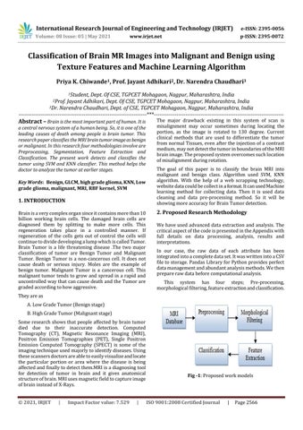
2. Proposed Research Methodology We have used advanced data extraction and analysis. The critical aspect of the code is presented in the Appendix with full details on data processing, analysis, results and interpretations. In our case, the raw data of each attribute has been integrated into a complete data set. It was written into a CSV file to storage. Pandas Library for Python provides perfect data management and abundant analysis methods. We then prepare raw data before computational analysis. This system has four steps; Pre-processing, morphological filtering, feature extraction and classification.
They are as A. Low Grade Tumor (Benign stage) B. High Grade Tumor (Malignant stage) Some research shows that people affected by brain tumor died due to their inaccurate detection. Computed Tomography (CT), Magnetic Resonance Imaging (MRI), Positron Emission Tomographies (PET), Single Positron Emission Computed Tomography (SPECT) is some of the imaging technique used majorly to identify diseases. Using these scanners doctors are able to easily visualize and locate the particular portion or area where the disease is being affected and finally to detect them.MRI is a diagnosing tool for detection of tumor in brain and it gives anatomical structure of brain. MRI uses magnetic field to capture image of brain instead of X-Rays. © 2021, IRJET
|
Impact Factor value: 7.529
|
Fig -1: Proposed work models
ISO 9001:2008 Certified Journal
|
Page 2566