International Research Journal of Engineering and Technology (IRJET)
e-ISSN: 2395-0056
Volume: 08 Issue: 03 | Mar 2021
p-ISSN: 2395-0072
www.irjet.net
Brain Tumor Detection using Infrared Thermography and Convolutional Neural Network Mrs. Archana A. Hatkar1, Megha Bhagat2, Sunita Karad3, Rushikesh Khade4 Bachelor of Engineering, Electronics and Telecommunication Engineering Savitribai Phule Pune University, Pune (MS) DEPARTMENT OF ELECTRONICS AND TELECOMMUNICATION SIR VISVESVARAYA INSTITUTE OF TECHNOLOGY CHINCHOLI, NASHIK (M. S.) INDIA. ----------------------------------------------------------------------***---------------------------------------------------------------------
Abstract - A human brain is centre of the nervous system; it
is a collection of white mass of cells. A tumor of brain is collection of uncontrolled increasing of these cells abnormally found in different part of the brain namely Glial cells, neurons, lymphatic tissues, blood vessels, pituitary glands and other part of brain which lead to the cancer. Cancer of Brain is of two types-1)Primary Brain Tumor- i)Benign which is not cancerous no danger at all, other one is ii) Malignant which is cancerous tumor; it grows abnormally by multiplying the cells rapidly, which leads to the death of the person if not detected. Manually it is not so easily possible to detect and identify the tumor. 2)Metastatic Brain Tumor - The metastatic brain tumor is the tumor that is formed elsewhere in the body and spread through the brain.The segmentation, detection, and extraction of infected tumor area from magnetic resonance (MR) images are a primary concern but a tedious and time taking task performed by radiologists or clinical experts, and their accuracy depends on their experience only. So, the use of computer aided technology becomes very necessary to overcome these limitations. We estimate the brain tumor severity using Convolutional Neural Network algorithm which gives us accurate results.
of the dense tissues and the thermography is not affected even due to hormonal changes. Thermography has 83% of sensitivity alone and 95% of sensitivity while combined with MRI. This also has high false-positive rate and false-negative rate but can be reduced further by using enhanced methods. Brain thermography works by finding an increase in surface temperature on brain. The approach leads to analyze the brain using various techniques like color analysis, asymmetric analysis, artificial neural networks, feature extraction, data mining techniques, segmentation approaches, sequential feature selection technique etc., Brain cancer detection using thermography begin with the screening of brain and analyzing thermal changes in obtaining thermogram. The image is observed, and further processing begins with the ordered sequence like preprocessing, segmentation, feature extraction, classification and post procession.
Keywords-Tumor Detection, Convolutional Neural Network, IRT Images, brain tumor, computer vision, bioinformatics, segmentation, medical images, review.
1. INTRODUCTION
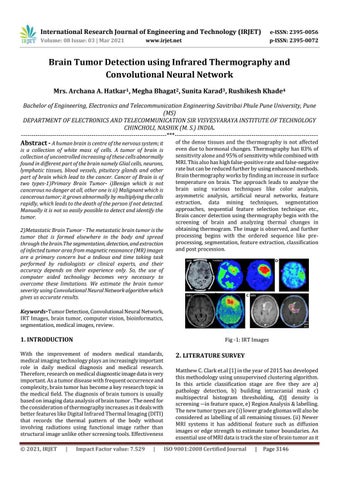
Fig -1: IRT Images
With the improvement of modern medical standards, medical imaging technology plays an increasingly important role in daily medical diagnosis and medical research. Therefore, research on medical diagnostic image data is very important. As a tumor disease with frequent occurrence and complexity, brain tumor has become a key research topic in the medical field. The diagnosis of brain tumors is usually based on imaging data analysis of brain tumor . The need for the consideration of thermography increases as it deals with better features like Digital Infrared Thermal Imaging (DITI) that records the thermal pattern of the body without involving radiations using functional image rather than structural image unlike other screening tools. Effectiveness © 2021, IRJET
|
Impact Factor value: 7.529
|
2. LITERATURE SURVEY Matthew C. Clark et.al [1] in the year of 2015 has developed this methodology using unsupervised clustering algorithm. In this article classification stage are five they are a) pathology detection, b) building intracranial mask c) multispectral histogram thresholding, d)‖ density is screening ―in feature space, e) Region Analysis & labelling. The new tumor types are (i) lower grade gliomas will also be considered as labelling of all remaining tissues. (ii) Newer MRI systems it has additional feature such as diffusion images or edge strength to estimate tumor boundaries. An essential use of MRI data is track the size of brain tumor as it ISO 9001:2008 Certified Journal
|
Page 3146