International Research Journal of Engineering and Technology (IRJET) e ISSN: 2395 0056 Volume: 08 Issue: 12 | Dec 2021 www.irjet.net p ISSN: 2395 0072 © 2021, IRJET | Impact Factor value: 7.529 | ISO 9001:2008 Certified Journal | Page1128 Deep learning based Automatic Diabetic Retinopathy Detection Using Fundus Images: A Survey Sugirtha K 1 , Menaka S R2 1Department of Information Technology, K.S.R College of Engineering, Tiruchengode, TamilNadu, India 2Department of Information Technology, K.S.R College of Engineering, Tiruchengode, TamilNadu, India *** Abstract DiabetesMellitus,oftenknownasDiabetes,isa condition in which a person's body either does not respond to insulin supplied by their pancreas or does not create enough insulin. Diabetics are at a significant risk of acquiring a variety of eye disorders throughout time. Early identification of diabetic eye illness via an automated communities,withmethodologies,thoroughperformancepreprocessingperspectives,diabeticprovideseyetechniques.asystemhassignificantadvantagesovermanualdetectionasconsequenceofdevelopmentsinmachinelearningAnumberofsophisticatedresearchondiabeticdiseasedetectionhavelatelybeenpublished.Thispaperathoroughreviewofautomatedapproachestoretinopathyidentificationfrommultipleincluding:accessibledatasets,imagetechniques,deeplearningmodels,assessmentcriteria.Thestudygivesaoverviewofdiabeticeyediseasedetectionincludingcuttingedgefieldapproaches,thegoalofprovidingimportantknowledgetoresearchhealthcareproviders,andpatients. Key Words: Diabetic eye disease, diabetic retinopathy, deepleaning,fundusimage,imageprocessing. 1. INTRODUCTION In many urbanised nations, diabetic retinopathy has been identified as one of the primary causes of blindness. According to the World Diabetes Foundation, by 2030, there will be more than 438 million individuals suffering with diabetes. As a result, early detection and diagnosis can help patients avoid losing their vision as a result of this symptomless diabetes condition [1]. It is a type of retinaltheydifficultdiabeticidentificationimportanthallmarkTheretinopathy,retinopathy:alterationsmalfunctionofmetabolicillnessthatdevelopsasaresultofahighamountglucoseintheblood,whichcausesaninsulinsecretionandvisualdifficulties.Smallbloodvesselintheretinageneratethreephasesofdiabeticpreproliferativeretinopathy,proliferativeandnonproliferativeretinopathy[2].developmentofexudatesontheretinaisanotherofdiabeticretinopathythathasbecomeanclinicalindicatorforautomateddiseaseanddiagnosis[3].Earlyidentificationofretinopathybyanophthalmologistismoreowingtothedifficultyindetectingthemsinceoccurinsmallersizes.Theearlysignsofdiabeticdiseasecanberecognisedutilisingmachine learning and deep learning algorithms in a real time automatedsystem.Asaresult,itcaneasilyreducetherisk of human mistake and the quantity of work that the Followingophthalmologisthastodo[4].that,theseverityphasesaredefinedtohelpthe ophthalmologist make suitable treatment and planning decisions following the diagnosis of non proliferative diabeticretinopathy.Thisdiagnosis issolelybased on the retinal fundus image's categorization of dark and bright lesions. Due of the restricted number of specialists availableinternationally,readingfundusphotographsmay be more costly. Because of the poor availability of ophthalmologists to patients, the prediction process may camerasHowever,unablearetinopathyofcanresearchfindingsdiseaserequiredcategorizationresolutionInsegmentationsystemcharacteristicsidentificationandcomplicatedretinopathysignificantDespiteinteresttakencharacteristics.retinopathytechniquesretinopathyandFurthermore,beslowedbythelackofahighlytrainedreader[5].theautomatedapproachismorereliableefficientthanhumanidentificationofdiabeticseveritystages.EffectivepreprocessingareappliedtotherecordeddiabeticpicturesinordertoeliminatenoisyThediscriminativecharacteristicsarethenfromthepicturesbysegmentingtheregionofinordertocreatetheappropriatepredictions.thefactthatseveralresearchpapershavemadecontributionstotheautomateddiabeticscreeningmethod.However,duetothestructureoflesions,theexistenceofnoise,thehighappearanceofinterclasssimilarity,lesionsremainsadifficulttask.Thesemaymakeitdifficultfortheautomatedtocreateaflexibleandreliablemodelforlesion[6].thepresenceoflesionsinthetargetpicture,imageisalsoimportantfordiseasepredictionandofseveritystages.Arobustsolutionisforlesionsrecognition,segmentation,andseveritygrading,accordingtotheresearchmentionedabove.Asaresult,thegoalofthisprojectistocreateaoneofakindplatformthatidentifyandanalyseretinalpicturesonboard.Ratherexposingtheafflictedpatienttolasersurgery,diabeticcanbeavoidedatanearlierstage.Becauseoflackofcompetenceandresources,manypatientsaretoreceivepromptscreeninganddiagnosis[7].asscanningophthalmoscopesanddigitalbecomemorecommon,telemedicinecanbeused

the example images of diabetic retinopathy. Fig 1:Examplesofdiabeticretinopathyimages Contribution To provide a structured and comprehensive overview ofthestateoftheartinDRdetectionsystemsusingDL,the proposed paper surveys the literature from the following perspectives:
International Research Journal of Engineering and Technology (IRJET) e ISSN: 2395 0056 Volume: 08 Issue: 12 | Dec 2021 www.irjet.net p ISSN: 2395 0072 © 2021, IRJET | Impact Factor value: 7.529 | ISO 9001:2008 Certified Journal | Page1129 to address on board diagnosis and prediction of diabetic retinopathy illness. As a result, tele ophthalmology is recognized as a critical component of the diabetic retinopathy screening procedure. The need for online eye care hospitals to treat diabetes patients is growing as feasible,practical,andclinicallyvalidatedoptionsbecome Furthermore,moredifficulttocomeby[8].theriskofdiabetic retinopathy can be reduced by providing good patient information and maintaining a healthy blood glucose level. There are numerous methods used in existing research studies to identify diabetic retinopathy, including dilated eye examination, fluorescein angiography, and optical coherencetomographyorfundusphotographytechniques Nowadays,[9].
algorithm evaluation.Thearrangement of this article is as follows. Section II analyses the papers based on the datasets used in their study. Section III addresses the image processing techniques used inthe priorwork. SectionIVanalyses the articles based on the classification methods employed. Section V discusses the findings and observations. Finally, SectionVIconcludesthepaper.
visual evaluation, which appears to be a more difficult process, need the use of a retina expert for diagnosis and grading. As a result, most patients would seek the help of a retina expert after losing their eyesight owingtoalackofaccesstoqualifieddoctors.Asaresult,a suitable and cost effective computer aided diagnosis systemisrequiredtotreat diabeticretinopathydiseaseat Figureanearlystage1shows
Kaggle EyePACS
The Kaggle EyePACS dataset, which contains over 80.000 fundus images and was provided by the EyePACS platform for the Diabetic Retinopathy Detection competition sponsored by the California Healthcare Foundation, is the most widely used and largest public datasetforDiabeticRetinopathyclassification.Itcomprises of a huge number of high resolution fundus pictures of both eyes taken with various technologies at several primary care facilities throughout California and worldwide under a range of imaging settings. Due to this unpredictability, both the data (e.g. artefacts, blurring, focusing,andexposureissues)andthegroundtruthlabels display noise, which was meant to better replicate a real world scenario. The photoswere rated using the ICDRDSS scale[10]byacertifiedprofessional Kaggle APTOS 2019 AravindEyeHospitalinIndia'sruralareasacquiredthe Kaggle APTOS 2019 Challenge [11] dataset in order to construct robust tools to automatically diagnose Diabetic Retinopathyandincreasethehospital'scapacitytoidentify futurepatients.Ithas5590photosandisthethirdbiggest collection. However,one of its flaws is the enormousclass imbalance, particularly in the Severe NPDR class, which only comprises 193 photos. APTOS dataset, like Kaggle
2)1)DatasetsavailableforDRPreprocessingtechniques applied to fundus images forDRdetection3)DLapproachesproposedforDRdetection4)PerformancemeasuresforDRdetection
2. DIABETIC RETINOPATHY DATASETS
The writers of the chosen publications make use of privateandpublicdatasetsthatareseparatedintotraining and testing scenarios. Kaggle and Messidor are the most popular datasets for DR detection. The Kaggle dataset contains 88,702 photos, 35,126 of which are utilised for training and 53,576 for testing. The Messidor dataset, which contains 1,200 fundus pictures, is the most extensively utilised. The DR phases are identified in the Kaggle and Messidor datasets. Table 2 lists the datasets used in the selected studies, organised by the different DEDs studied, i.e. DR, Gl, DME, and Ca. The table includes the DED's name, the dataset's name, a synopsis of the dataset, the sources of publications that utilised the dataset, and lastly, the URL where the dataset may be obtained(ifaccessiblepublicly).
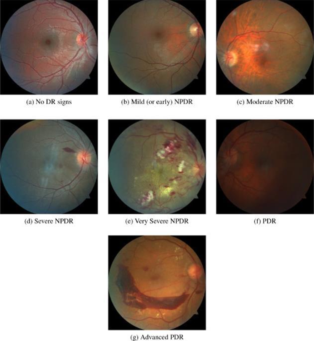
Table 1: Datasetsofretinafundusimages
The IDRiD [13] dataset contains 516 high quality photos taken with a Kowa VX 10 fundus camera at an ophthalmologyclinic inNanded,India.Priortothepicture capturing operation, all individuals' eyes were dilated in both directions. Forall 516photos, it includes image level grading on the severity of Diabetic Retinopathy according totheICDRSscale,aswellasgradingontheriskofDiabetic Macula Edema (DME). It also includes pixel by pixel annotationsonthepertinentlesions(suchasHardandSoft Exudates, Microaneurysms, and Haemorrhages) as well as theoptical discstructurefor81photosinthedataset
DDR With12522photos,theDDR[14]datasetisthesecond biggest when it comes to the classification challenge, although it is a relatively new dataset that hasn't been frequently utilised yet. The data was collected between 2016 and 2018 in 147 hospitals throughout China's 23 provinces and annotated by numerous specialists using a majority voting schema according to the ICDRDSS scale. Furthermore, a sixth grade was added to separate low quality photos into a distinct group. However, there is a significant disparity between the healthy/moderate DR classifications and the others, such as mild, severe, and proliferative DR, which might result in overfitting. Regarding the relevant DR lesions, 757 pictures from the dataset were labelled at the pixel level for lesion segmentationanddetectionpurposes,aswellas bounding boxessurroundingthem
or other tiny red lesions, and 47 images pertaining to exudates. It has been used to forecast DR in a binary job automatically(healthyvs diseased). However, because the dataset contains fewer pictures than larger datasets (Kaggle and Messidor), it is generally utilised in the research to create segmentation techniques rather than classificationsystems DiaRetDB1 DiaRetDB1[16]ismadeupof89fundusphotosthatwere obtained in a controlled setting at Kuopio University and rated byfourspecialists. However, because not onlyisthe datasamplesmallandfromasingleclinicallocation,butall ofthephotoswereobtainedinacontrolledsettingwithno Name Size nResolutio nAnnotatio Task [10]EyePACS 28870 Varying LevelImage GradingDR 2019[11]APTOS 5590 Varying LevelImage GradingDR [12]Messidor 1200 1440 × 2240960, × 23041488 × 1536 LevelImage GradingDR 2Messidor[12] 1748 Varying LevelImage GradingDR [13]IDRiD 576 4288 × 2848 Image & LevelPixel GradingDR DDR[14] 21252 Varying Image & LevelPixel GradingDR E [15]Ophtha 463 Varying LevelPixel DetectionaneurysmsandExudatesDiseasedHealthyvs.Micro 1DiaRetDB[16] 89 1500 × 1152 LevelPixel onSegmentatiLesion [17]DRiDB 50 768 × 584 LevelPixel ExtractionandDetectionMaculaODHEs,MAS,HMs,SEs,andVessel
Hospital'sadditionalphotostakenweretakenwithpupildilation,whereasthelatter400werewithoutit.TheMessidor2datasetincludes1058fromtheoriginalMessidordataset,aswellas690photographsgatheredintheBrestUniversityOphthalmologydepartmentbetween2009and
International Research Journal of Engineering and Technology (IRJET) e ISSN: 2395 0056 Volume: 08 Issue: 12 | Dec 2021 www.irjet.net p ISSN: 2395 0072 © 2021, IRJET | Impact Factor value: 7.529 | ISO 9001:2008 Certified Journal | Page1130 EyePACS dataset, displays variances owing to varying camerasettingsbetweencentresandnoisebothinthedata (i.e. artefacts, focus difficulties, being under/overexposed) and the labels (due to being gathered in a real world multicentrescenario) Messidor & Messidor 2
IDRiD
The E Ophtha [15] dataset contains 463 photos, including 268 images pertaining to healthy people, 148 images pertaining to patients with micro aneurysms
E Ophtha
The Messidor dataset [12] contains 1200 retina fundus pictures taken between 2005 and 2006 by three ophthalmologyinstitutionsinFrance.Thefirst800photos 2010. Unlike the Kaggle EyePACS dataset, both dataset’s picturesareofexcellentquality,withnodiscerniblenoise. The datasets provide an image level medical diagnostic of theseverityofDiabeticRetinopathyforeachimage,butno pixel level lesion segmentation information. Their own grading system, on the other hand, did not follow the widely recognized ICDRS procedure, limiting its validity andusefulness.
4. DL APPROACHES PROPOSED FOR DR DETECTION
Diabetic retinopathy is the major cause of blindness in peopleintheirworkingyears.Carsonetal[22]usedcolour fundus pictures to illustrate the application of convolutional neural networks (CNNs) for diabetic retinopathy stage detection. They also looked at multinomial classification models, demonstrating that due to the CNN's failure to recognize subtle illness signs, the majority of mistakes arise when mild disease is misclassifiedasnormal.Theyobservedthatusingcontrast limiting adaptive histogram equalization and assuring dataset consistency through expert class label verification improveddetectionofsubtlecharacteristics.Sheikhetal.[23]constructeda unique deep convolutional neural network that conducts early stage detectionbyrecognizingallmicroaneurysms(MAs),which are the earliest indicators of DR, as well as properly assigning labels to retinal fundus pictures that are graded into five categories. They used the largest publicly accessible Kaggle diabetic retinopathy dataset to test the network. In terms of both computing time and space, the suggesteddesignisbothsimpleandefficientWanetal.[24]figuredoutamechanismtocategorisea series of fundus photos automatically. They use convolutional neural networks (CNNs) to solve three fundamental problems in DR detection: classification, segmentation, and detection. We use AlexNet, VGGNet, GoogleNet,andResNet,togetherwithtransferlearningand hyper parameter tweaking, to see how well these models do with DR image categorization. They used the Kaggle classifierplatform,whichisopentothepublic,totrainthesemodelsAdiabeticretinopathydeeplearninginterpretablewasreportedbyDeetal[25].Ontheonehand,it
3. PREPROCESSING TECHNIQUES APPLIED TO FUNDUS IMAGES FOR DR DETECTION
DRiDB DRiDB [17] is a collection of 50 fundus photographs with comments on the anatomy of the retina's optic disc and vessels, as well as any existing diseases, neovascularizations, and disease grades, all of which were determinedbynumerousspecialists.Despitethefactthatit isatinydataset,itisthemostinstructive.
Contrast enhancement To begin, contrast enhancement is a popular preprocessing method used in any image processing or analysis pipeline to distinguish the foreground from the background. The histogram equalization [18] is a basic approachforcontrastenhancementinfunduspicturesthat raises the overall contrast of the image while ignoring the local changes throughout the image. Adaptive Histogram Equalization is a more advanced contrast adjustment method that takes into account local differences surrounding a specified region of each pixel. Contrast LimitedAdaptiveHistogramEqualization(CLAHE)ismore widely employed by the scientific community when it comestofundusimaging.
Color space transformation Apart from contrast improvement, normalisation, and noise reduction, the model's performance has been improved by converting the colour picture into another colourmodel orjustusingone of the RGBchannels.Linet al.[20]usedentropypicturestoconvertthedata,resulting in the DLS outperforming models trained on standard datasets. Furthermore, due to its rich information and strong contrast in compared to the other two colour channels,thegreenchannelofthefunduscolour pictureis frequentlyextracted Cropping and resizing
International Research Journal of Engineering and Technology (IRJET) e ISSN: 2395 0056 Volume: 08 Issue: 12 | Dec 2021 www.irjet.net p ISSN: 2395 0072 © 2021, IRJET | Impact Factor value: 7.529 | ISO 9001:2008 Certified Journal | Page1131 substantial changes in the collecting technique, their distributiondoesnotreflectatypicalpopulation.
Denoising & normalization [19] uses Non Local Means Denoising (NLMD) to reduce anypicturenoise.However,whilethebetterthedenoising algorithm is, the more noise it will remove, it will also impair the image's fine features (i. e. the image becomes blurry). Additionally, picture intensity normalization is used to reduce bias and long training durations in the network, as well as to standardize the data to a certain scale(e.g.,eachimagehasameanvalueof0andastandard deviationof1intermsofitspixels'intensity).
Additionally, the files may include photos with varying resolutions and aspect ratios. Uninformative dark space portionsmightalsobeseeninthephotos.Thephotographs may be cropped, rescaled, and enlarged to a certain resolution to standardise the image size and remove such dark space areas. Bravo et al. [21] used two distinct thecroppedcroppedcroppingapproachesintheirtrials.Inoneexperiment,theythepicturessothattheretinaencircledtheimage,butintheotherexperiment,theyclippedretina'sgreatestsquareimage.
Augmentation Although DL has been shown to operate effectively when raw data is put into a single model pipeline, it has also beenclaimedthatusingspecificpreprocessingapproaches, such as those described in this section, improves performance [22], particularly for fundus pictures. In addition, data augmentation techniques are utilised to improve the model's resilience and accuracy owing to a shortage of rich and balanced datasets. Rotating, shifting (translation), rescaling, shearing, and flipping pictures, as well as the usage of Generative Adversarial Networks for image synthesis, are examples of such approaches in the case of imaging datasets. The majority of the publications analysed employ some form of augmentation to enhance the number of available photos and hence speed up the model'straining.
International Research Journal of Engineering and Technology (IRJET) e ISSN: 2395 0056 Volume: 08 Issue: 12 | Dec 2021 www.irjet.net p ISSN: 2395 0072 © 2021, IRJET | Impact Factor value: 7.529 | ISO 9001:2008 Certified Journal | Page1132 accurately separates retina pictures into distinct severity degrees.Ontheotherhand,bygivingascoretoeachpoint in the hiddenand input spaces,thisclassifier iscapableof explaining the classification findings. The contribution of each pixel to the final categorization is indicated by these scores. They suggested a novel pixel wise score propagationmodeltogetthesescores,whichseparatesthe observed output score into two components for each neuron. The resulting visual maps can be easily comprehended by an ophthalmologist using this procedureRakhlin[26]usesdeepConvolutional NeuralNetworks (CNNs) to diagnose eye fundus pictures, which have proved innovative in a variety of disciplines of computer vision, including medical imaging. They utilised a publicly available Kaggle data set to train the models. They employed a subset of Kaggle data that had been omitted from training and the Messidor 2 reference standard for inexpensive,specificity.accuracy,proposedtheforapproachForclassification3alvisibilitydatasetISBIisoveralldiseasetwoachievements.linkdependentcharacteristicsspecificillnessesandattentionusingnetworknetworkautonomousconvolutionalusingoutcomestrainedcomparabletesting.Whendonebyskilledoptometrists,thefindingsaretocontemporarystateoftheartmodelsonfarlargerdatasetsandoutperformtheaverageofdiabeticretinopathyscreeningManojetal[27]developedthepredictionnetworkthepubliclyavailableKaggledatasetusinganeuralnetworkbasedtechniquefordetectionofdiabeticretinopathy.Thewastrainedusingaround35,000photos.Thewastrainedtoidentifythelateralityoftheeye8,810photos.Xiaomengetal.[28]proposedauniquecrossdiseasenetwork(CANet)tosimultaneouslyassessDRDMEbyexaminingtheintrinsiclinkbetweentheusingjustimagelevelsupervision.Thediseaseattentionmodule,whichselectivelylearnsrelevantforindividualdiseases,andthediseaseattentionmodule,whichcapturestheintrinsicbetweenthetwodiseases,aretwoofourmajorWeuseadeepnetworktocombinetheseattentionmodulestoprovidediseasespecificanddependentcharacteristics,aswellastomaximizeperformanceforgradingDRandDME.Ournetworktestedontwopubliclyavailablebenchmarkdatasets:the2018IDRiDchallengedatasetandtheMessidorAtechniquebasedontheRadontransform(RT)andthegraph(VG)wassuggestedbyMohammadpooryet[29]toautomaticallyidentifygrades0(normal),1,2,andoftheDRfromfunduspictures.Featureextractionandarethetwostepsoftheproposedtechnique.thefirsttimeintherealmofimageprocessing,theVGwasusedtoextractfeaturesinthiswork.Then,classificationpurposes,theseattributeswerehandedtoerrorcorrectingoutputcodes(ECOC)approach.Theapproachwassimpletouse,witha97.92percent95.83percentsensitivity,and98.61percentTheVGbasedapproachcanbeasimple,andeffectivetestforautomaticDRstage grading,and itcan also be used in other image processing applicationsAbdelsalam [30] developed a comprehensive and reliable automated system for detecting DR individuals early.Twostagesarenecessaryfortheprocesstowork:1) Employing created custom algorithms to reconstruct, improve, and re continue blood vessels, and 2) using an ArtificialNeuralNetwork(ANN)asanautomatedclassifier between diabetics without diabetic retinopathy (DR) and Mild to Moderate Non Proliferative Diabetic Retinopathy (NPDR) individuals. The overall accuracy of the method was 97 percent. The classification system's performance characteristics for normal vs diabetes were 97.5 percent sensitivity, 96.67 percent specificity, and 95.2 percent accuracy. The sensitivity, specificity, and precision scores for a diabetic without DR against non proliferative DR (mildtomoderate)were96.67percent,96.67percent,and 96.67percent,respectively.3.33percentofthetime,there wasamisclassificationmistakeXianglongetal[31]usedatransferlearningstrategyto buildauniqueconvolutionalneural network model witha Siamese like design. Unlike earlier studies, the proposed approach takes binocular fundus pictures as inputs and learns their association to aid prediction. The suggested binocular model achieves an area under the receiver operatingcurve of 0.951witha trainingsetofjust28104 pictures and a test set of 7024 images, which is 0.011 greater than the existing monocular model. A binocular model for five class DR detection is also trained and assessed on a 10% validation set to further validate the efficiency of the binocular architecture. The result reveals usingoperatinspecificityaccuracyTheaveragedcoefficientspropagationetconvolutionalnonthatitgetsakappascoreof0.829,whichisgreaterthantheensemblemodelcurrentlyinuse.MultipleweightedpathwayswereimplementedtoaneuralnetworktermedtheWPCNNbyLiual[32],whichwasdrivenbyensemblelearning.BackisusedtoimprovevariouspathweightinWPCNN,andtheoutputfeaturesareforredundancyreductionandquickconvergence.experimentfindingsrevealthatWPCNNobtainsanof94.23percentwithsensitivityof90.94percent,of95.74percent,areaunderthereceivergcurveof0.9823,andF1scoreof0.9087whileanefficienttrainingconvergencerate Ensemble learning approaches By integrating the benefits of different AI frameworks, forillnessdesignrepresemodelsoutperformcomplementarity,BecauserobustensemblelearninghasalsoplayedakeyroleinestablishingandstrongAIframeworksforDRcategorization.oftheknowledgegainproducedbytheirensemblelearninghasbeenshowntosolomodels.Thatmeansthatvariousbasemightacquiredifferentdegreesofsemanticntationsimplicitly,eitherdueofchangesintheirorbecauseofthetrainingtechnique.Zhangetal.[33]createdtwoensemblemodels,oneforidentification(binaryclassification)andtheotherdiseasegrading(quinaryclassification).Thevarious
J. Kaur and D. Mittal, Estimation of severity level of non proliferative diabetic retinopathy for clinical aid, BiocyberneticsandBiomedicalEngineering38 (2018) 708 732. R. O. Fite et al., Diabetic retinopathy in Ethiopia: A systematic review and meta analysis, Diabetology & MetabolicSyndrome13(2019)1885 1891. S. Joshi and P. T. Karule, A review on exudates detection methods for diabetic retinopathy, Biomedicine & Pharmacotherapy97(2018)1454 1460. U. Ishtiaq et al., Diabetic retinopathy detection through artificial intelligent techniques: A review and open issues, Multimedia Tools and Applications 79 (2020) 21 22. J. Lin et al., Retinal image quality assessment for diabetic retinopathy screening: A survey, Multimedia Tools andApplications79(2020)16173 16199. P. Porwal, et al, IDRiD: Diabetic retinopathy Segmentation and grading challenge ID, Medical ImageAnalysis59(2019). S. Stolte and R. Fang, A survey on medical image analysis in diabetic retinopathy, Medical Image Analysis 64 (2020)101742. D.Kalogeropoulosetal.,Theroleoftele ophthalmologyin diabetic retinopathy screening, Journal of Optometry 13(2020)262 268.
International Research Journal of Engineering and Technology (IRJET) e ISSN: 2395 0056 Volume: 08 Issue: 12 | Dec 2021 www.irjet.net p ISSN: 2395 0072 © 2021, IRJET | Impact Factor value: 7.529 | ISO 9001:2008 Certified Journal | Page1133 models were built using a combination of pre trained networks for feature extraction and a bespoke standard dense neural network for classification. In all tasks, the ensemblemodelsbeatindividualmodels,withasensitivity of 98.10 percent and a specificity of 98.56 percent. The authors also point out that the ‘stronger' the base learner (pre trained network), the better the overall performance. Furthermore, in several circumstances, a dual ensemble (ensembleofensembles)outperformedasingleensemble Because, according to the authors, each distinct lesion type is best recognised at various training iterations, Quellec et al. [34] built a CNN model that was exported at numerousiterationsduringthetrainingmethod.Theythen used ensemble learning (Random Forest Classifier) to Specificity,individualachievedpercent,algorithm.partnershipmodel.InceptionmodelsmergethestoredmodelstopredictDR'sseverityscore.Jiangetal.[35]usedtheAdaboostclassifieronthreebasedontheInceptionV3,ResNet152,andResnetV2architecturestocreateanensembleTheyusedaproprietarydatasetcreatedinwithBeijingTongrenEyeCentretotraintheSensitivity=85.57percent,Specificity=90.85Accuracy=88.21percent,andAUC=0.946werebytheensemblemodel,whichoutperformedthemodels.InceptionV3didbetterintermsofwithascoreof91.46percent. 5. PERFORMANCE MEASURES FOR DR DETECTION ALGORITHM EVALUATION Some specific measures, such as Sensitivity, Specificity, Accuracy, Precision, and F1 score, can be used to characterize the classification model's performance. Furthermore, the Receiver Operating Characteristic (ROC) curve depicts the classifier's performance by graphing its Sensitivity against its Specificity at various thresholds for theclassificationoutcome(i.e.atwhichprobabilityagiven sample is considered as a positive or negative outcome). Finally, AUC calculates the area beneath the whole ROC curve, providing an overall performance metric across all categorization criteria. The performance of the reviewed papersisshowninTable2. Table 2: Performanceofthereviewedclassificationmodels. erenceRefs Classification architectureBest acyAccur [22] 2 (EyePACS)class 5 class (EyePACS) GoogLeNet [23] 2 (EyePACS)(EyePACS)(EyePACS)Diseased2Risk5class CNNCustom [24] 5 (EyePACS)class VGGNet 95.68 % [25] 2 (Messidorclass2) 5 class(Messidor 2) CNNCustom 91% [26] 2 (EyePACS)class 2 class (Messidor) VGGNetModified [27] 5 (EyePACS)class CNNCustom [28] 2 (Messidor)class CNNCustom 92.6 % [29] 2 class transformRadon 97.92 % [30] 2 class ANN 97% [31] 2 class WP CNN 94.23% 6. CONCLUSIONS Diabetic retinopathy is a serious complication of diabetes that causes progressive retinal deterioration and, in severecases,blindness.Toavoiddegenerationandretinal damage, it is vital to recognize and treat it as soon as possible.Inrecentyears,therehasbeenasurgeininterest inemployingdeeplearningtodetectdiabeticretinopathy, andasvariousDLsystemsdevelopandareintegratedinto clinical practice, clinicians will be able to treat patients more successfully and efficiently. The current state of research on the application of deep learning in the diagnosis of diabetic retinopathy is examined in this article. Ophthalmologists still need to improve their performance, interpretability, and trustworthiness, despite the fact that deep learning has paved the way for moreaccuratediagnosisandtreatment
REFERENCES
International Research Journal of Engineering and Technology (IRJET) e ISSN: 2395 0056 Volume: 08 Issue: 12 | Dec 2021 www.irjet.net p ISSN: 2395 0072
M. Shaban et al., Low complexity computer aided diagnosisfordiabeticretinopathy(A.S.El BazandJ.S. Suri,Eds.UnitedStates:ElsevierBooks,2020).
P. A. C. S. Eye, “Diabetic retinopathy detection”. Available at: http://www.kaggle.com/c/diabetic retinopathy detection. Aravind Eye Hospital and APTOS, “2019 blindness detection”. Available at: https://www.kaggle, Commun.C/aptos2019 blindness detection.
E. Decenci’ere et al., B´eatrice Cochener, Caroline Trone, Philippe Gain, Richard Ordonez, Pascale Massin, Ali Erginay, B´eatrice Charton, Jean Claude Klein, Feedback on a publicly distributed database: the messidor database, Image Anal. Stereol. 33 (2014) 231 234. P.Porwal etal.,Indian diabetic retinopathyimage dataset (IDRiD):Adatabasefordiabeticretinopathyscreening research,Data3(2018).
T. Li et al., Diagnostic assessment of deep learning algorithms for diabetic retinopathy screening, InformationSciences501(2019)511 522. R. Gargeya and T. Leng, Automated identification of diabetic retinopathy using deep learning, Ophthalmology124(2017)962 969.
Z. Mohammadpoory et al., Automatic identification of diabetic retinopathy stages by using fundus images andvisibilitygraphmethod,Measurement140(2019) 133 141. M. M. Abdelsalam, Effective blood vessels reconstruction methodology for early detection and classification of diabetic retinopathy using OCTA images by artificial neural network, Informatics in Medicine Unlocked 20 (2020). X. Zeng et al., Automated diabetic retinopathy detection based on binocular Siamese like convolutional neural networks,IEEEAccess7(2019)30744 30753. Y.P.Liuetal.,Referablediabeticretinopathyidentification from eye fundus images with weighted path for convolutionalneuralnetwork,ArtificialIntelligencein Medicine99(2019)101694.
Z. Zhao et al., Bira net: Bilinear attention net for diabetic retinopathygrading,inIEEEInternationalConference onImageProcessing(2019)1385 1389. G. Quellec et al., Deep image mining for diabetic retinopathy screening, Medical Image Analysis 39 (2017)178 193. H. Jiang et al., An interpretable ensemble deep learning model for diabetic retinopathy disease classification, in 41st Annual International Conference of the IEEE Engineering in Medicine and Biology Society (EMBC) (2019)2045 2048.
P. Prentasic et al., Diabetic retinopathy image database(DRiDB): A new database for diabetic retinopathy screening programs research, IEEE, Sep. 8thIntl.Symp.OnImageAndSignalProcess.AndAnal. (ISPA),(2013)711 716.
© 2021, IRJET | Impact Factor value: 7.529 | ISO 9001:2008 Certified Journal | Page1134
“R.V.J.P.H. K¨ alviainen, ¨ H. Uusitalo, Diaretdb1 diabetic retinopathydatabaseandevaluationprotocol,Medical ImageUnderstandingandAnalysis(2007).
W. Zhang et al., Automated identification and grading system of diabetic retinopathy using deep neural networks, Knowledge Based Systems 175 (2019) 12 25. Q. Xiao et al., Improving lesion segmentation for diabetic retinopathy using adversarial learning, in Lecture Notes in Computer Science Intl. Conf. on Image AnalalysisAndRecognition.Springer(2019)333 344. G. M. Lin et al., Transforming retinal photographs to entropy images in deep learning to improve automated detection for diabetic retinopathy, Journal ofOphthalmology(2018)2159702. M. A. Bravo and A. Pablo, Arable´ aez, Automatic diabetic retinopathy classification in 13th Intl. Conf. on Med. Inf. Process. and Anal., 10572. International Society forOpticsandPhotonics,(2017)105721E. C. Lam et al., Automated detection of diabetic retinopathy using deep learning, AMIA AMIA Summits on TranslationalScienceProceedings(2018)147 155. S.M.SaifulIslametal.,BasedEarlyDetectionandGrading of Diabetic Retinopathy Using Retinal Fundus Images, 2018arXivpreprintarXiv:1812.10595. S. Wan et al., Deep convolutional neural networks for diabeticretinopathydetectionbyimageclassification, Computers & Electrical Engineering 72 (2018) 274 282. J.DelaTorreetal.,Adeeplearninginterpretableclassifier for diabetic retinopathy disease grading, Neurocomputing396(2020)465 476. R. Alexander, Diabetic retinopathy detection through integration of deep learning classification framework, bioRxiv,(2018)225508. M. Raju et al., Development of a deep learning algorithm for automatic diagnosis of diabetic retinopathy, Studies in Health Technology and Informatics 245 (2017)559 563.
X.Lietal.,Canet:Cross diseaseattentionnetworkforjoint diabetic retinopathy and diabetic macular edema grading, IEEE Transactions on Medical Imaging 39 (2020)1483 1493.