International Research Journal of Engineering and Technology (IRJET)
e-ISSN: 2395-0056
Volume: 08 Issue: 10 | Oct 2021
p-ISSN: 2395-0072
www.irjet.net
Divide and Conquer Approach to Diabetic Retinopathy Classification Sarthak Purohit1, Ujjwal Kumar2 1Student,
Department of Computer Science and Engineering, SRM Institute of Science & Technology, Chennai, India Department of Computer Science and Engineering, SRM Institute of Science & Technology, Chennai, India ---------------------------------------------------------------------***---------------------------------------------------------------------2Student,
Abstract - Medical image analysis using computer aided
No DR, Mild, Moderate, Severe and Proliferative. Proliferative DR here refers to neovascularization (abnormal growth of various blood vessels in the retina). DR without any abnormal growth vessels therefore fall under nonproliferative DR (Mild, Moderate and Severe). Table 1 gives a perspicuous overview of the different stages.
automation has been proven to be highly effective for classification of diseases like diabetic retinopathy. The present study reviews some of the recent AI driven solutions for diabetic retinopathy classification and gives a new solution to achieve state-of-the-art results on a retinal image dataset. A heuristic structure is used in the architecture of the proposed alternative that utilizes several binary classifiers to function as single multi-class classifier, thereby dividing the task amongst smaller groups. This new approach to diabetic retinopathy classification is described as ‘Divide & Conquer Network’ or ‘D&C Network’. After evaluation, the D&C network exhibited promising results and achieved the area under the receiver operating characteristic curve of 0.906, with 0.965 specificity and 0.853 sensitivity.
Key Words: Medical image analysis, Computer-aided diagnosis, Convolutional neural networks, Transfer learning, Diabetic retinopathy classification, Deep learning,
1.INTRODUCTION The complications that arise out of Diabetes Mellitus (DM) are unknown to none. World Health Organization reports that this chronic disease affects approximately 422 million people worldwide and is responsible for about 1.6 million deaths each year [1]. The most prevalent complication of DM is Diabetic Retinopathy (DR) [2] which is a vascular disease of the retina.
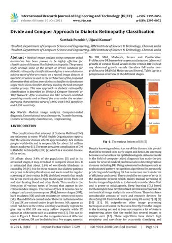
Fig -1: The various lesions of DR [5] Despite knowing such intricacies of this disease, it is pivotal that DR be treated in its early stages and hence, its screening becomes a crucial task for ophthalmologists. Advancements in the field of computer aided diagnosis has made the job easier for several medical professionals in detecting various diseases including DR. Using automated techniques such as sophisticated pattern recognition algorithms for accurately predicting and classifying DR has numerous merits in terms of efficiency and speed. There should be no scope of error in the diagnostic process which makes manual screening of fundus images disputable as it demands considerable effort and is prone to misdiagnosis. Deep learning (DL) based methodologies have revolutionized several aspects of our life and medical image analysis is one of those. There has been considerable amount of work and research devoted for classifying DR from fundus images using DL as in [7] [8] [9] [10] [11]. DL outperforms other image processing techniques as it learns the features directly from the images in the training set and it does not require a lot of feature engineering, given that the model has several images to sample over [12]. These algorithms have shown high sensitivity and specificity for accurately classifying and
DR affects about 3.4% of the population [3] and in its advanced stages, it may even lead to complete vision loss. It is estimated that about 2.6% of worldwide blindness is caused by DR [4]. It is for these reasons that diabetic patients are prone to develop this disease and are in need for regular screening of their retina. In DR, the blood vessels that reach the retina swell up due to high blood sugar levels from DM and start leaking blood and other fluids. This results in the formation of various types of lesions that appear in the retinal fundus images. The various types of lesions can be categorized as microaneurysms (MA), haemorrhages (HM), hard exudates (HX) and soft exudates or cotton wool spots (SX). MA and HM are coined under the term red lesions while HX and SX are coined under bright lesions. MA appear as small red dots in the retina, and they eventually rupture to give rise to HM. HX are waxy yellow deposits while SX appear as white spots such as a cotton wool [3]. This can be seen in Figure 1. Based on the categorizations of different types of lesions, DR can be divided into five stages, namely:
© 2021, IRJET
|
Impact Factor value: 7.529
|
ISO 9001:2008 Certified Journal
|
Page 491