International Research Journal of Engineering and Technology (IRJET)
e-ISSN: 2395-0056
Volume: 07 Issue: 09 | Sep 2020
p-ISSN: 2395-0072
www.irjet.net
Automatic Detection of Diabetic Retinopathy using Convolution Neural Network Nidhi Kamothi1, Dr. Rashmi Thakur2 1Student,
Dept. of Computer Engineering, Thakur College of Engineering and Technology, Maharashtra, India Dept. of Computer Engineering, Thakur College of Engineering and Technology, Maharashtra, India ---------------------------------------------------------------------***---------------------------------------------------------------------2Professor,
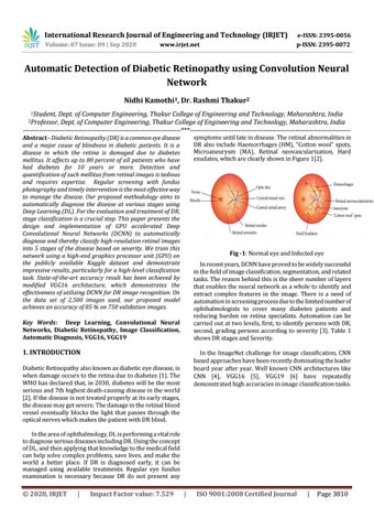
symptoms until late in disease. The retinal abnormalities in DR also include Haemorrhages (HM), “Cotton wool” spots, Microaneurysm (MA), Retinal neovascularization, Hard exudates, which are clearly shown in Figure 1[2].
Abstract - Diabetic Retinopathy (DR) is a common eye disease and a major cause of blindness in diabetic patients. It is a disease in which the retina is damaged due to diabetes mellitus. It affects up to 80 percent of all patients who have had diabetes for 10 years or more. Detection and quantification of such mellitus from retinal images is tedious and requires expertise. Regular screening with fundus photography and timely intervention is the most effective way to manage the disease. Our proposed methodology aims to automatically diagnose the disease at various stages using Deep Learning (DL). For the evaluation and treatment of DR, stage classification is a crucial step. This paper presents the design and implementation of GPU accelerated Deep Convolutional Neural Networks (DCNN) to automatically diagnose and thereby classify high-resolution retinal images into 5 stages of the disease based on severity. We train this network using a high-end graphics processor unit (GPU) on the publicly available Kaggle dataset and demonstrate impressive results, particularly for a high-level classification task. State-of-the-art accuracy result has been achieved by modified VGG16 architecture, which demonstrates the effectiveness of utilizing DCNN for DR image recognition. On the data set of 2,500 images used, our proposed model achieves an accuracy of 85 % on 750 validation images.
Fig -1: Normal eye and Infected eye In recent years, DCNN have proved to be widely successful in the field of image classification, segmentation, and related tasks. The reason behind this is the sheer number of layers that enables the neural network as a whole to identify and extract complex features in the image. There is a need of automation in screening process due to the limited number of ophthalmologists to cover many diabetes patients and reducing burden on retina specialists. Automation can be carried out at two levels, first, to identify persons with DR, second, grading persons according to severity [3]. Table 1 shows DR stages and Severity.
Key Words: Deep Learning, Convolutional Neural Networks, Diabetic Retinopathy, Image Classification, Automatic Diagnosis, VGG16, VGG19
1. INTRODUCTION
In the ImageNet challenge for image classification, CNN based approaches have been recently dominating the leader board year after year. Well known CNN architectures like CNN [4], VGG16 [5], VGG19 [6] have repeatedly demonstrated high accuracies in image classification tasks.
Diabetic Retinopathy also known as diabetic eye disease, is when damage occurs to the retina due to diabetes [1]. The WHO has declared that, in 2030, diabetes will be the most serious and 7th highest death-causing disease in the world [2]. If the disease is not treated properly at its early stages, the disease may get severe. The damage in the retinal blood vessel eventually blocks the light that passes through the optical nerves which makes the patient with DR blind. In the area of ophthalmology, DL is performing a vital role to diagnose serious diseases including DR. Using the concept of DL, and then applying that knowledge to the medical field can help solve complex problems, save lives, and make the world a better place. If DR is diagnosed early, it can be managed using available treatments. Regular eye fundus examination is necessary because DR do not present any
© 2020, IRJET
|
Impact Factor value: 7.529
|
ISO 9001:2008 Certified Journal
|
Page 3810