International Research Journal of Engineering and Technology (IRJET)
e-ISSN: 2395-0056
Volume: 07 Issue: 03 | Mar 2020
p-ISSN: 2395-0072
www.irjet.net
IDENTIFICATION OF MALARIAL PARASITES USING DEEP LEARNING Dr.S. Saravanan1, N Abinesh Kumar2, V A Ajith Kumar3 , R Joseph Patrick Soloman4 Department of Computer Science and Engineering, Agni College of Technology, Chennai-130 ----------------------------------------------------------------------***--------------------------------------------------------------------------
Abstract - The traditional system for detecting the infection of Malaria has been the manual process of diagnosing the stained slides under a microscope. This manual process might consume more time for producing the results and the availability of medical experts is not always assured. Considering this fact in mind we proposed a method which curbs the human error while detecting the presence of malarial parasite in the blood sample by using Image Processing. Hence by automating the diagnosis process, results can be acquired relatively quicker and more accuracy can be expected. The technologies and techniques to patently extract the required features and efficiently classify the infected samples are surveyed. Key Words: Malarial Parasite, Image Processing, Segmentation, Machine Learning, Detection. 1. INTRODUCTION
The mosquito-borne parasitic infection is spread by a female Anopheles mosquito by the Plasmodium parasite. It is a single-celled parasite that multiplies amongst the red blood cells of humans as well as the mosquito’s intestine. When the female feeds on an infected person, the parasites are ingested along with the human blood. The parasites multiply in the mosquito’s gut and these infectious worms are passed onto another human when the mosquito feeds on them. Humans can also get Malaria by blood transfusion and an offspring from it’s mother during childbirth from the placenta. To detect Malaria, the affected red blood cells are diagnosed first. Blood smear in analyzed under a microscope, which is the traditional process done by a medical expert. To automate the process, various Image Processing and Machine Learning methods were used. Various segmentation methods have been used to detect the presence of the parasite. The typical ring structure of the parasite that has held host in the red blood cells can be identified from the microscopic image. So, this image is segmented and classified to detect both infected and noninfected cells. The objective of this new system is to possibly increase the sensitivity, accuracy and the F-score of the previously existing systems to provide a more right diagnosis of the infectious disease. 1.1 Malaria Parasite Detection From Peripheral Blood Smear Images Using Deep Belief Networks This paper has introduced a trained model based on a DBN to classify 4100 peripheral blood smear images into the parasite or non-parasite class. The proposed DBN is pretrained by stacking restricted Boltzmann machines using the contrastive divergence method for pre-training. To train the
© 2020, IRJET
|
Impact Factor value: 7.34
|
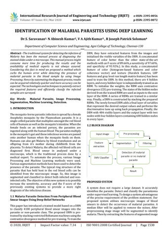
DBN, they have extracted features from the images and initialized the visible variables of the DBN. A concatenated feature of color better than the other state-of-the-art methods with an F-score of 89.66%,a sensitivity of 97.60%, and specificity of 95.92%.]. In this study, a concatenated feature of color (histogram-based features and color coherence vector) and texture (Haralick features, LBP features and gray level run length matrix feature) has been used to train the DBN. In this method, there are 4 hidden layers, and every hidden layer is independently trained as an RBM. To initialize the weights, the RBM uses contrastive divergence (CD) pre-training. The states of the hidden nodes derived from the trained RBM are used as inputs to the next layer of the RBM. A series of RBMs are trained in a similar way. Finally, a DBN is constructed by stacking the prepared RBMs. The newly formed DBN adds a final layer of variables that represent the desired output values and performs the discriminative tune up using back propagation. Finally, the DBN with 484 visible layers and the output layer with two nodes with four hidden layers containing 600 hidden nodes in every layer. 1.2 BLOCK DIAGRAM
PROPOSED SYSTEM A system does not require a large dataset. It accurately identifies the parasite. Detect and classify the parasitemia under supervised learning. To design a system advantageous of the precise requirement of the problem statement. Our proposed system utilizes microscopic images of blood smears to detect the occurrence of malarial parasites. A median filter will be applied to remove the noises. After preprocessing stage image will be segmented to identify malaria. Then by extracting the features of segmented image
ISO 9001:2008 Certified Journal
|
Page 1530