International Research Journal of Engineering and Technology (IRJET)
e-ISSN: 2395-0056
Volume: 07 Issue: 10 | Oct 2020
p-ISSN: 2395-0072
www.irjet.net
Convolutional Neural Networks for Automatic Detection of Pneumonia Anish Adnani1, Supriya Patil2 1,2Student,
Department of Computer Engineering, Vivekanand Education Society’s Institute of Technology, Mumbai, India ---------------------------------------------------------------------***---------------------------------------------------------------------Abstract: Pneumonia is a fatal disease found to cause infection in one or both the lungs. It can be caused by various types of microorganisms that are bacteria, viruses, fungi. This infection causes inflammation in the alveoli that allows fluid or pus to subsume in alveoli making it difficult to breathe. Moreover, the contagiousness of this infection makes it more harmful. Approximately 33% of the deaths in India are caused due to pneumonia reported by the World Health Organization (WHO). Pneumonia currently diagnosed using a Chest X-Ray evaluated by an expert radiotherapist. This process is exerting and travail and often leads to a difference in opinion among the experts. Thus developing a solitary automatic system would be beneficial for identification, preventing further transmission and treatment in remote areas. Due to the triumph of Convolutional Neural Networks (CNNs) in analyzing medical reports, attention to such medical problems is unambiguously incumbent. Also, due to the rise of pre-trained CNN models that trained on millions of images has made the work much easier one considered as inevitable. We analytically determine the optimal CNN model for the purpose.
lung cancer, fluid overloading, post-surgery changes. Due to the involvement of a high number of factors, there is an unambiguous need for an automatic system to detect pneumonia automatically with a low percentage of error. Over recent years, one of the major research domains in machine learning is Computer Aided Designs (CAD). Various Deep Learning models prove to be prime in the extraction of useful features in image classification tasks. The role of pre-trained CNN models is cardinal in the case of transfer learning as these pre-trained models are trained and tested on datasets that include millions of images that are almost impossible to replicate for a small group of people. Availability of pre-trained CNN models like ResNet[3], AlexNet[4], VGGNet[5], DenseNet[6].
Keywords: Deep Convolutional Neural Networks, SVM, Transfer Learning, Naive Bayes, Pneumonia detection, Computer-aided diagnostics, Medical Imaging. 1. INTRODUCTION Pneumonia is an illness that disturbs the alveoli of the lungs and is a mortal account for about 16% of the world deaths [1], being the world's leading cause for deaths among the children. Pneumonia is responsible for almost 127,000 deaths in India[2] the numbers are rising due to the spread of novel coronavirus. Pneumonia has killed over 1 million children in worldwide in 2018 and remains one of the most lifethreatening diseases if not detected at early stages, especially after the novel Covid-19 pandemic, which has shaken medical institutions around the globe due to increased spike in daily pneumonia cases far-flung the controlled limits that can be treated by medical professionals.
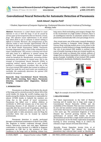
Fig 1: An example of normal CXR VS Pneumonia CXR 2. RELATED WORKS The latest improvements in the field of Machine Learning and AI mainly due to large scale usage of Convolutional Neural Networks (CNNs) and the availability of free datasets. That was once considered to be rare and has assisted various algorithms to perform much better that was not considered to be commonplace a few years ago. The automated diagnosis of varied diseases has received growing interest. The low performance of several CNN models on diverse abnormalities proved that a single model cannot be used for all purposes. So for the better exploration of machine learning in chest screening, Wang et al. (2017) [9] released a larger dataset of frontal chest X-Rays.
Pneumonia detection is usually performed through a professional examination of chest X-ray (CXR). The diagnosis is further confirmed through clinical history, laboratory exams, and vital signs. Additional confirmation is done because it is difficult to diagnose pneumonia on CXR due to the presence of other conditions in the lungs such as bleeding, volume loss,
Š 2020, IRJET
|
Impact Factor value: 7.529
Recently, Pranav Rajpurkar, Jeremy Irvin, et al. (2017)[10] explored this dataset for detecting pneumonia they referred their model as ChexNet that
|
ISO 9001:2008 Certified Journal
|
Page 117