International Research Journal of Engineering and Technology (IRJET)
e-ISSN: 2395-0056
Volume: 06 Issue: 03 | Mar 2019
p-ISSN: 2395-0072
www.irjet.net
Comparison of Preprocessing Methods for Diabetic Retinopathy Detection Using Fundus Images J. AashikathulZuberiya1, Dr. S. Shajun Nisha2, Dr. M. Mohamed Sathik3 1M.Phil
Research Scholar, PG & Research Department of Computer Science, Sadakathullah Appa College, Tirunelveli, Tamilnadu, India 2Assistant Professor & Head, PG & Research Department of Computer Science, Sadakathullah Appa College, Tirunelveli, Tamilnadu, India 3Principal, Sadakathullah Appa College, Tirunelveli, Tamilnadu, India ---------------------------------------------------------------------***----------------------------------------------------------------------
Abstract - Diabetic Retinopathy is eye disorder among
less growth of new blood vessels. PDR is an advanced stage in which the fluids sent by the retina for nourishment trigger the growth of new blood vessel that are abnormal and fragile. They grow along the retina and along the surface of vitreous gel which fills inside the eye. It might leak blood into retina which may result in severe vision loss and even blindness. Initial stage has no vision problem, but with time and severity of diabetes it may lead to vision loss[5][6].
people with diabetics which may lead to blindness. Diabetes is a chronic disorder caused by insulin deficiency in the body. Diabetes for a prolonged time damages the blood vessels of retina and affects the vision of a person and leads to Diabetic retinopathy. It is classifies into two categories, non proliferative diabetic retinopathy (NPDR) and proliferative diabetic retinopathy (PDR). Fundus photography involves capturing a photograph of the back of the eye. The raw retinal fundus images are hard to process. To enhance some features and to remove unwanted features Preprocessing is used. Preprocessing techniques like image enhancement, histogram equalization, Contrast Limited Adaptive Histogram Equalization (CLACHE) are performed. The results are evaluated by Mean Square Error (MSE), Peak Signal to Noise Ratio (PSNR) and Entropy Values. Key Words: Diabetic Retinopathy, fundus image, Preprocessing, clache.
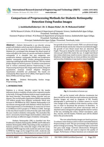
1. INTRODUCTION Diabetes is a chronic disorder caused by the insulin deficiency body or inability of body cell to respond to insulin in the body. Prolonged Diabetes leads to many complications like heart disorder, Neuropathy, kidney disorder and eye disease. The world health organization (WHO) reported that 135 million people have diabetes worldwide which may increase to 300 Million by 2025. Diabetic Retinopathy is an eye disease among people with diabetics which may lead to vision impairment or even blindness. It causes loss of vision in 1.8 Million in 2015 people to 37 Million in 2040. Damage to the tiny blood vessels in retina from the optic disk inside the eyes results in Diabetic retinopathy. The anomalies like micro- anourysms, hemorrhages, hard exudates, cotton wool spots develops at different stages of diabetic retinopathy.
Fig -1: Anomalies in human eye DR can be treated with effective treatments but there should be early detection and continuous monitoring. Fundus images are used to diagnosis of DR. Fundus photography is performed by a fundus camera to record color images of the condition of the interior surface of the eye, in order to document the presence of disorders . It consists of a specialized low power microscope with an attached camera designed to photograph the interior surface of the eye, including the retina, retinal vasculature, optic disc, macula, and the fundus. Patients eyes will be dilated before the procedure[10]. The raw retinal fundus images are hard to process. To enhance some features and to remove unwanted features Preprocessing is used .The pre processing is the important phase in image processing.. The acquired image is converted into gray scale image. Contrast enhancement, Histogram equalization, CLACHE are used to improve the quality of images. Performance of these functions are evaluated and
Diabetic Retinopathy (DR) is classified into Non Proliferative Diabetic Retinopathy (NPDR) and Proliferative Diabetic Retinopathy (PDR). Depending on the anomalies or features present in the retina the stages of the DR can be identified. NPDR stage has mild, moderate and severe stage. In NPDR the stage can be ranged from mild , moderate and severe by the presence of the features in various levels with
Š 2019, IRJET
|
Impact Factor value: 7.211
|
ISO 9001:2008 Certified Journal
|
Page 252