International Research Journal of Engineering and Technology (IRJET)
e-ISSN: 2395-0056
Volume: 06 Issue: 10 | Oct 2019
p-ISSN: 2395-0072
www.irjet.net
ANALYSIS OF AUTOMATED DETECTION OF WBC CANCER DISEASES IN BIOMEDICAL PROCESSING Vanitha I1, Ms. Sree Thayanandeswari C.S2, Mrs. Babimol T.M3 1PG
Scholar, PET ENGINEERING COLLEGE PROFESSOR, PET ENGINEERING COLLEGE -----------------------------------------------------------------------***-----------------------------------------------------------------------2Assistant
Abstract – Medical industry is one of the most prominant industry where deep learning can play a huge role especially when it comes to medical imaging.Automated diagnosis of WBC cancer diseases such as Leukemia and Myeloma having similar symptoms.Therefore, from the doctor’s side,they might be confused to diagnose those two diseases.In this paper,a new approach namedRandom forest classifier is applied for final decision.the system learning methodology is reduce the misdiagnosis cases.The proposed system will produce the parameters such as Mean, Accuracy, Texture features will be evaluated and the experimental result shows that the increase in the three parameter values will reduce the noise of an image.
develop an innovative approach for finding the optimal sequence alignment to reduce the time, space complexity and increase the efficiency of sequence alignment in the large data set and find the stages of CML. Here we are using innovative DSDPM algorithm, it is dynamic and recursive optimal distance calculation method to reduce the execution of time and making it cost effective for finding the mutation and optimal sequence alignment.
KeyWords: white blood cell, random forest, diseases INTRODUCTION: Leukemia is a type of cancer that affects the bone marrow causing increased production of white blood cells which influx the blood stream[1].White blood cells help the body to fight infections and other diseases. Red blood cells carry oxygen from the lungs to the body's tissues and take carbon dioxide from the tissues back to the lungs[2]. The images generated by digital microscopes are usually in RGB color space, which is difficult to segment[3]. The blood cells and image background varies greatly with respect to color and intensity[4]. The approach of leukocyte classification consists of two parts: (1) The segmentation of leukocyte, cytoplasm, and nucleus; (2) The classification of the segmented leukocytes using a set of features[5]. For the segmentation procedure the input image contains at least one leukocyte. Image pre-processing, feature extraction and classification plays major role in detection of white blood cell cancers[6]. The removal of noise is another main factor that yields accuracy to final result. The main goal is to de-noise the input image. Pre-processing also includes color relevance wheel RGB images are converted to Grey color space images[7]. When there is huge data, it is inefficient and consumes more time, and multiple comparisons of persons at a time are not possible and finding the stages is difficult. So here we are going to
© 2019, IRJET
|
Impact Factor value: 7.34
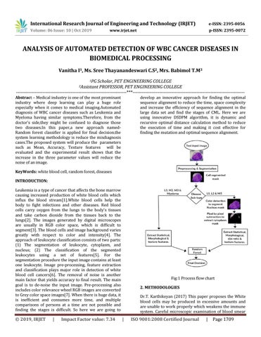
Fig:1 Process flow chart 2. METHODOLOGIES Dr.T. Karthikeyan (2017) This paper proposes the White blood cells may be produced in excessive amounts and are unable to work properly which weakens the immune system. Careful microscopic examination of blood smear
|
ISO 9001:2008 Certified Journal
|
Page 1709