International Research Journal of Engineering and Technology (IRJET)
e-ISSN: 2395-0056
Volume: 05 Issue: 10 | Oct 2018
p-ISSN: 2395-0072
www.irjet.net
Application of False Removal Algorithm Specially for Retinal Images with Exudates in Diabetic Retinopathy Detection Shreya Singh Chauhan1, Rekha Gupta2 1Department
2Associate
of Electronics and Communication, Madhav Institute of Technology and Science, Gwalior, India Professor, Department of Electronics and Communication, Madhav Institute of Technology and Science, Gwalior, India
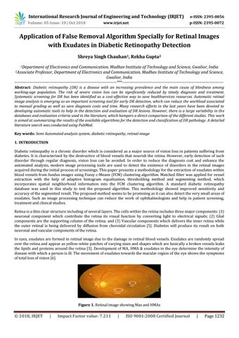
-----------------------------------------------------------------------------***---------------------------------------------------------------------------Abstract: Diabetic retinopathy (DR) is a disease with an increasing prevalence and the main cause of blindness among working-age population. The risk of severe vision loss can be significantly reduced by timely diagnosis and treatment. Systematic screening for DR has been identified as a cost-effective way to save healthservices resources. Automatic retinal image analysis is emerging as an important screening tool for early DR detection, which can reduce the workload associated to manual grading as well as save diagnosis costs and time. Many research efforts in the last years have been devoted to developing automatic tools to help in the detection and evaluation of DR lesions. However, there is a large variability in the databases and evaluation criteria used in the literature, which hampers a direct comparison of the different studies. This work is aimed at summarizing the results of the available algorithms for the detection and classification of DR pathology. A detailed literature search was conducted using PubMed. Key words: Semi Automated analysis system, diabetic retinopathy, retinal image 1. INTRODUCTION Diabetic retinopathy is a chronic disorder which is considered as a major source of vision loss in patients suffering from diabetes. It is characterized by the destructive of blood vessels that nourish the retina. However, early detection of such disorder through regular diagnosis, vision loss can be avoided. In order to reduce the diagnosis cost and enhance the automated analysis, modern image processing tools are used to detect the existence of disorders in the retinal images acquired during the initial process of screenings. This paper presents a methodology for the extraction of exudates within blood vessels from fundus images using Fuzzy c-Means (FCM) clustering algorithm. Matched filter was applied for vessel extraction with the help of adaptive histogram equalization, thresholding method and segmenting method, which incorporates spatial neighborhood information into the FCM clustering algorithm. A standard diabetic retinopathy database was used in this study to test the proposed algorithm. This methodology showed improved sensitivity and accuracy of the segmented result. The proposed method seems to be promising as it can also detect the very small areas of exudates. Such an image processing technique can reduce the work of ophthalmologists and help in patient screening, treatment and clinical studies. Retina is a thin clear structure including of several layers. The cells within the retina includes three major components: (1) neuronal component which contribute the retina its visual function by converting light to electrical signals; (2) Glial components are the supporting column of the retina; and (3) Vascular components which delivers the inner retina while the outer retinal is being delivered by diffusion from choroidal circulation [5]. Diabetes will produce its result on both neuronal and vascular components of the retina. In eyes, exudates are formed in retinal image due to the damage in retinal blood vessels. Exudates are randomly spread over the retina and appear as yellow-white patches of varying sizes and shapes which are basically a broken vessels leaks the lipids and proteins around the retina [3]. Development of MA, HMA & exudates in the eye determine the intensity of disease with which a person is ill. The movement of exudates towards the macular region of the eye shows the symptoms of total loss of vision [6].
Figure 1. Retinal image showing Mas and HMAs
Š 2018, IRJET
|
Impact Factor value: 7.211
|
ISO 9001:2008 Certified Journal
|
Page 1232