International Research Journal of Engineering and Technology (IRJET)
e-ISSN: 2395 -0056
Volume: 04 Issue: 01 | Jan -2017
p-ISSN: 2395-0072
www.irjet.net
An Ameliorate Technique for Brain Lumps Detection Using Fuzzy CMeans Clustering Pritam R. Dungarwal1, Prof. Dinesh D. Patil 1Research
Scholar Student, M.E (CSE), SSGBCOET,Bhusawal,Maharashtra,India Professor, Dept. of CSE, SSGBCOET, Bhusawal,Maharashtra,India ---------------------------------------------------------------------***-------------------------------------------------------------------2HOD,
ABSTRACT-Clustering is commonly used in biomedical applications particularly for brain lumps detection in Abnormal magnetic resonance images (MRI). In terms of segmentation efficiency Fuzzy clustering using fuzzy local information C-means algorithm proved to be greater over the other clustering methodologies. Most Research in developed countries show that the number of people who have brain cancer were died due to the fact of inaccurate detection and not in time. Generally, CT scan or MRI that is directed into intracranial cavity produces a complete image of brain. This image is visually examined by the physician for detection & diagnosis of brain cancer. However this method of detection resists the accurate determination of size of lump. In addition, it also reduces the time for analysis. At the end of the process the lump is extracted from the MR image and its exact position and the shape also determined. The graph based on pixel value is drawn taking the various points from the swelled cells lies in the original position from the affected region. Here the affected region is considered as ellipsoid shape and the volumes have been calculated from it. A fuzzy level set algorithm is proposed in this paper to facilitate medical image segmentation & on this performance of evaluation of the proposed algorithm was carried. Index Terms— introduction, clustering, Fuzzy clustering, image segmentation, Fuzzy C-Mean, result
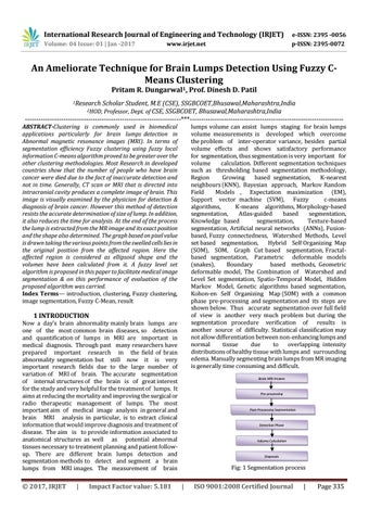
lumps volume can assist lumps staging for brain lumps volume measurements is developed which overcome the problem of inter-operator variance, besides partial volume effects and shows satisfactory performance for segmentation, thus segmentation is very important for volume calculation. Different segmentation techniques such as thresholding based segmentation methodology, Region Growing based segmentation, K-nearest neighbours (KNN), Bayesian approach, Markov Random Field Models , Expectation maximization (EM), Support vector machine (SVM), Fuzzy c-means algorithms, K-means algorithms, Morphology-based segmentation, Atlas-guided based segmentation, Knowledge based segmentation, Texture-based segmentation, Artificial neural networks (ANNs), Fusionbased, Fuzzy connectedness, Watershed Methods, Level set based segmentation, Hybrid Self Organizing Map (SOM), SOM, Graph Cut based segmentation, Fractalbased segmentation, Parametric deformable models (snakes), Boundary based methods, Geometric deformable model, The Combination of Watershed and Level Set segmentation, Spatio-Temporal Model, Hidden Markov Model, Genetic algorithms based segmentation, Kohon-en Self Organizing Map (SOM) with a common phase pre-processing and segmentation and its steps are shown below. Thus accurate segmentation over full field of view is another very much problem but during the segmentation procedure verification of results is another source of difficulty. Statistical classification may not allow differentiation between non-enhancing lumps and normal tissue due to overlapping intensity distributions of healthy tissue with lumps and surrounding edema. Manually segmenting brain lumps from MR imaging is generally time consuming and difficult.
1 INTRODUCTION
Now a day’s brain abnormality mainly brain lumps are one of the most common brain diseases, so detection and quantification of lumps in MRI are important in medical diagnosis. Through past many researchers have prepared important research in the field of brain abnormality segmentation but still now it is very important research fields due to the large number of variation of MRI of brain. The accurate segmentation of internal structures of the brain is of great interest for the study and very helpful for the treatment of lumps. It aims at reducing the mortality and improving the surgical or radio therapeutic management of lumps. The most important aim of medical image analysis in general and brain MRI analysis in particular, is to extract clinical information that would improve diagnosis and treatment of disease. The aim is to provide information associated to anatomical structures as well as potential abnormal tissues necessary to treatment planning and patient followup. There are different brain lumps detection and segmentation methods to detect and segment a brain lumps from MRI images. The measurement of brain
© 2017, IRJET
|
Impact Factor value: 5.181
Brain MRI Images
Pre-processing
Post-Processing Segmentation
Detection Phase
Volume Calculation
Diagnosis
Fig: 1 Segmentation process
|
ISO 9001:2008 Certified Journal
|
Page 335