
International Research Journal of Engineering and Technology (IRJET) e-ISSN: 2395-0056
Volume: 11 Issue: 06 | June 2024 www.irjet.net p-ISSN: 2395-0072
International Research Journal of Engineering and Technology (IRJET) e-ISSN: 2395-0056
Volume: 11 Issue: 06 | June 2024 www.irjet.net p-ISSN: 2395-0072
Gopika Subash Babu1 , Prof. Dr. Sheeja Agustin2
1UG Student, Dept. of Computer Science and Engineering, APJ Abdul Kalam Technological University, Kerala, India 2Professor, Dept. of Computer Science and Engineering, APJ Abdul Kalam Technological University, Kerala, India
Abstract - Detecting brain tumors plays a vital role in medical image analysis crucial for timely diagnosis and treatment planning. An inventive project is introduced here, merging Convolutional Neural Networks (CNNs) with machine learning to automate brain tumor detection. By utilizing Python libraries like TensorFlow, a comprehensive framework is built, integrating CNN-based feature extraction with conventional machine learning algorithms for binary tumor classification. The methodology includes pre-processing MRI scans, extracting significant features using a CNN structure, and inputting these features into machine learning classifiers to identify the presence or absence of a brain tumor. Extensive experimentation is conducted on a diverse dataset containing MRI images of brains with and without tumors to assess the performance of various CNN architectures and machine learning models. The results exhibit promising accuracy and efficiency in tumor detection tasks, with the developed framework achievingremarkable sensitivity and specificity rates. This system holds substantial potential in aiding healthcare professionals in precise diagnosis and treatment planning, ultimatelyenhancingpatient outcomes in neuro-oncology.
Key Words: Tumor,CNN,MRI,accuracy,detection
Among the various organs present in the human body, thebrain particularly stands out as being the most crucial. An issue commonly seen leading to dysfunction in the brain is the development of a brain tumor, characterized bytheuncontrolledgrowthofexcess cells. These abnormal cells tend to consume essential nutrients that healthy brain cellsand tissues require, ultimately resulting in brain failure. At present, doctors rely on manual examination of MRI scansto detect and evaluate brain tumors, a method known for its susceptibility to inaccuracies and inefficiencies. Brain cancer, being a severe and life-threatening condition, claims numerous lives due to delayed or incorrect diagnoses. The primary goal behind detecting brain tumors lies in enabling early diagnosis and treatment. This project aims to develop an automated system capable of identifying the presence of brain tumors in MRI scans. By utilizing CNNs, this system processes MRI images to determine the existence of a tumor, offering a reliable diagnostic tool for healthcare professionals. Such an automated system presents substantial advantages
withinclinicalsettings,assistingdoctorsinprovidingquick and precise diagnoses. By concentrating on the binary classification task determining whether a tumor is present or not this initiative strives to improve early detection, hence enhancing treatment outcomes and patient survival rates in neuro-oncology.
Current techniques for identifying brain tumors heavily depend on the manual analysis of MRI scans by radiologistsandhealthcareexperts.Whilethistraditional method is somewhat effective, it comes with its own set of. These include the risk of human mistakes, subjective judgment, and the considerable time needed for a thorough evaluation. The manual assessment process may result in inconsistent findings, especially consideringtheintricatenatureofbraintumorsandtheir subtle display in imaging. Furthermore, the growing number of MRI scans in medical setups can overwhelm healthcareproviders,causing delays in diagnosing and treating patients. Several current systems integrate basic image processing methods and computer-aided diagnosis (CAD) tools to support physicians. However, thesesystemsoftenlacktheadvancedfeaturesnecessary to precisely and effectively identify tumors.
One common method to detect brain tumors is Magnetic Resonance Imaging (MRI). However, the manual interpretation of MRI scans by radiologists can be time-consuming and subjective. This can lead to errors or delays in diagnosis. This project aims to develop an automated system using computer-based methods to identify brain tumors in MRI images. By utilizing Convolutional Neural Network (CNN) algorithms, the system can efficiently analyze MRI scans todeterminethepresenceofatumor.Theprocess involves several stages: image preprocessing, feature extraction, and classification. During image preprocessing, MRI scans areenhancedtomakeiteasier toidentifyrelevantfeatures.Featureextractionfocuseson identifying significant characteristics within the images thatindicatethepresenceofatumor.Intheclassification stage, neural network techniques are utilized to determineifatumorispresent.Thisinnovativeapproach aimsto streamline the detection process and improve diagnostic accuracy in identifying brain tumors using MRI technology.
International Research Journal of Engineering and Technology (IRJET) e-ISSN: 2395-0056
Volume: 11 Issue: 06 | June 2024 www.irjet.net p-ISSN: 2395-0072
The proposed system for detecting brain tumors aims to achieve several important goals to enhance the diagnosticprocess:
• Early Detection: Focus on finding brain tumors earlytoimprove patient outcomes.
• Enhanced Accuracy: Improve diagnostic accuracy using advanced machine learning techniques.
• Binary Classification: Develop a reliable system for distinguishing between tumor and non-tumor cases.
• Seamless Integration: Ensure smooth integration intoexisting clinical workflows.
• Ongoing Improvement: Keepperformancecurrent with the latest advancements in medical imaging andmachinelearning.
• Collaborative Decision Support: Provide comprehensive diagnostic insights and recommendations through collaborative decisionmaking.
• Multi-Modal Imaging Data: Combine multiple types of imaging data for a more thorough analysis.
• Scalability and Adaptability: Make sure the system canhandle large amounts of data and adapt totechnologicaladvancements.
In [1], image processing is crucial in identifying and diagnosing brain tumors through MRI and CT scans. The processstartswithpreprocessingstepslikereducingnoise, normalizing intensity, registering images, and enhancing them to make them suitable for analysis. Once the preprocessing is done, segmentation comes into play to outline tumor regions using various algorithms such as thresholding, region growing,level sets, active contours, andgraphcuts.Featuresarethenextractedtodescribethe tumors using statistical, shape, and spatial characteristics necessary for classification. Different machine learning algorithms like support vector machines (SVMs), random forests, and artificial neural networks (ANNs) are utilized for tumor classification. The performance of these algorithms is assessed using metrics like sensitivity, specificity,accuracy,andareaunderthecurve(AUC).
In [2], the article centers on the early classification of braintumorsusing MRI with the help of deep learning (DL)and transfer learning (TL) methods. By utilizing an automatedframework,thereisareductioninthenecessity forextensive humaninvolvement.Thisframework follows a structured process involving data preprocessing, data enhancement, feature extraction, and classification. VariousmodelslikeXception, NasNetLarge,DenseNet121, and InceptionResNetV2 are utilized, with their
performance being assessed using metrics such as accuracy, sensitivity, precision, specificity, and F1- score. The study accentuates the significance of timely detection and exhibits the remarkable efficiency of the Xception model when coupled with the ADAM optimizer. The research makes a valuable contribution to brain MRI image analysis by refining preprocessing techniques, optimizing data augmentation strategies, and employing cutting-edge DL models for the precise identification of brain tumors.
In [3], the proposed system offers a thorough examination ofstudiesonidentifyingandclassifyingbrain tumors from MRI scans using deep learning models. It underscores the significant impact of deep learning in medical imaging, especially with convolutional neural networks capturing key tumor features. The study delves into an extensive review of deep learning techniques, discussing their advantages and disadvantages. It explains how deep learning models leverage MRI data to improve tumor detection accuracy. This paper isavaluableguideto current trends and future possibilities in brain tumor diagnosis with deep learning models.
In [4], the proposed system introduces a dual-module approachfor detecting brain tumors. In the first module, MRI imagesare enhanced using adaptive Wiener filtering, neural networks,and independent component analysis to improve clarity and contrast. The second module utilizes Support Vector Machines (SVM) for segmenting and classifyingtumors,targetingvarioustypesofbraintumors. This method strives to generate clearer MRI images and enhance classification accuracy, contributing to improved clinical diagnoses’ reliability. The study validates these techniques on the CE-MRI image database, showcasing significantpotentialinenhancingthequalityofMRI images andthe precision of tumordetection endeavors.
In [5], the study discusses the complex challenge of categorizing brain tumors using computer-aided diagnostics, particularlyfocusing on magnetic resonance imaging (MRI). A method is proposed that emphasizes multi-level feature extraction and combination for early detection due to the diverse nature of tumor cells. By utilizingtwowell-knowndeeplearningmodels, Inceptionv3 and DenseNet201, two different scenarios were assessed for tumor detection and classification. Firstly, features from various Inception modules are extracted, combined, and classified using a SoftMax classifier. The second scenario entails extracting features from different DenseNet blocks, combining them, and employing a SoftMax classifier for classification as well. By applying these techniques toa publicly accessible three-class brain tumor dataset resultedin impressive testing accuracies of 99.34 percent and 99.51 percent with Inception-v3 and DenseNet201, correspondingly.These results highlight the superiorperformanceoftheproposedfeaturecombination approach comparedto existing deeplearningand machine
International Research Journal of Engineering and Technology (IRJET) e-ISSN: 2395-0056
Volume: 11 Issue: 06 | June 2024 www.irjet.net p-ISSN: 2395-0072
3.1
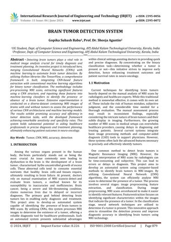
The system proposed comprises five main modules. Firstly, theDatasetmodule, then Pre-processing, followed bySplittingthedata,Buildingthe CNN model, training the DeepNeuralnetworkforepochs,andfinallyclassification.
Inthedataset module,multipleMRIimagescanbetaken with one as the input. During pre-processing, the image is encoded with labels and resized accordingly. When splitting the data, 80 percentisassignedto Training Data and 20 percent to Testing Data. Further steps involve building the CNN model followed by training the deep neural network for epochs. The images are then classified as either Tumor detected or no tumor. In cases where a tumorisdetected,itreturnsassuchwhileinthosewithout tumors, it returns as no tumor detected.
A Convolutional Neural Network (CNN) works with images and multiple layers to extract and understand features.FiltersinConvolution2Dlayershelpcreatemaps to display spatialpatterns. MAX Pooling2D layers simplify things by shrinking these maps, grabbing the highest values from various areas. Dropout layers help with generalization by turning off random neurons during training.
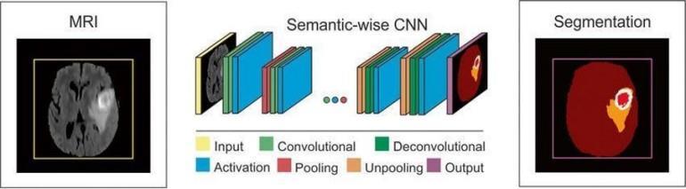
Flatten layers get the data ready for Dense layers, linking neurons through all levels for more complex feature learning Activation layers bring in non-linearity to assist in spotting intricate patterns. The model gets fine-tuned with binary cross-entropy loss and the Adamoptimizer for flexible learning rates and effective convergence.The SmallerVGGNet model is utilized in this implementation, which serves as a more compact version influencedby the VGGNet architecture. The primary goal is to decrease computational complexity while retaining essential structuralelements. The model is comprised of several crucial components:
1) InputLayer:Thislayerestablishestheshapeofthe input.
2) ConvolutionalLayers:Threesetsofconvolutional blocksare included:
a) Each block consists of Conv2D layers, followed by Activation (ReLU), Batch Normalization, Max-Pooling2D, and Dropout.
3) Fully Connected Layer: A Dense layer with 1024 unitsis present, followed by Activation (ReLU), BatchNormalization, and Dropout.
4) Output Layer: This layer features a Dense layer with a sigmoid activation function tailored for binaryclassification.
This design allows the SmallerVGGNet model to efficiently learn and extract image features, making it suitable for tasks such as binary classification of brain tumor images. The data undergoes preprocessing with various augmentations to enhance model robustness, and the learning rate adjusts dynamically using the ReduceLROnPlateau callback to improve model performance.
The specialized convolutional neural network, SmallerVGGNet, has been designed to handle 48x48 RGB images effectively Unlike VGG16, it opts for fewer convolutional layers and filters to reduce computational requirements while still maintaining spatial intricacies. Thisismadepossiblethroughconsistentconfigurationsof
International Research Journal of Engineering and Technology (IRJET) e-ISSN: 2395-0056
Volume: 11 Issue: 06 | June 2024 www.irjet.net p-ISSN: 2395-0072
stride and padding. Beginning with 3x3 convolutional filters, the network aims to uncover detailed spatial features such as edges and textures. Following each convolutionallayer,max-poolingstepsareimplementedto decreasethesizeoffeaturemapsusing2x2windowswith a stride of 2. This approach not only enhances computational efficiency but also helps in preventing overfitting
Following the convolutional and pooling layers, SmallerVGGNet combines simplified fully connected (FC) layers with ReLU activation functions. This integration aims to bring in non-linearity and support high-level feature learning. Each FC layer includes Batch Normalization to ensure stability andDropout to prevent overfitting. The network ends witha dense output layer employing sigmoidactivation, designed for precise binary classification tasks like identifying brain tumors. By excluding Local Response Normalization, SmallerVGGNet enhances memory usage and computational speed while maintaining classification accuracy. This characteristic makes it ideal for scenarios where computational resources are limited yet accurate image analysis is crucial.
4. SYSTEM DESIGN
4.1 System Overview
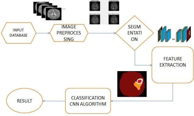
from Kaggle.com) to preprocessing images, segmenting, extracting features, using CNNs for classification, and showingresultsontheinterface.Thisvisualguidehelpsus understandhoweachpartworkstogether to detect brain tumors effectively andaccurately.
The Brain Tumor Detection interface is a big step forward in medical imaging tech to boost finding and treating brain tumors. With top-notch technology and easy-to-use design, it helps medical pros to spot and tackle brain tumors quickly. This technique can raise diagnostic accuracy, better patient results, and push forward medical research, ultimately improvingtreatment plans and patient results.
1) Input: The Brain Tumor Detection system relies on datasets from Kaggle.com, a well-known platform for accessing and working on datasets. These datasets are carefully selected to contain various medical imaging data essential for training and validating the brain tumordetection model. Through utilizing these datasets, the system guarantees that the model is strong and able to accurately assess a broad spectrum of brain tumor cases. This improves its dependabilityandefficiencyinclinicalsettings.
2) Image Processing: The preparation of images before analysis processing is important to enhance quality.Techniques like noise reduction, contrast enhancement, resizing, color correction, segmentation, and feature extraction are used in sequence. Noise reduction helps clarity while resizing color correction maintains consistency. Segmentation feature extraction isolates tumor featuresforaccurateanalysisbytheclassification model.
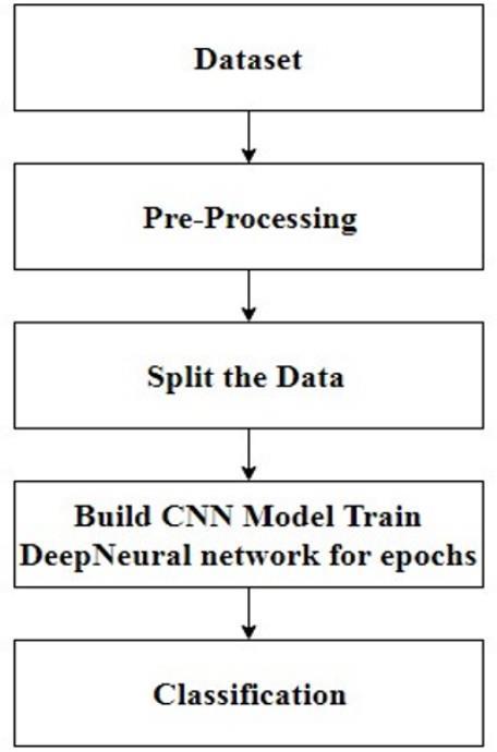
3) Segmentation: Segmentation represents a crucial stage in digital image processing. It involves dividinganimageintovariousdistinctregionsor segments, depending on pixel traits. In the realm of brain tumor identification, segmentationplays a key role in pinpointing and separating tumor areas from normal brain tissue. This methodologyallowsforthedetailedoutliningand examination of tumor attributes like dimensions, appearance, and position. Through precise segmentation of tumor zones, the system boosts diagnosticprecisionandstreamlinestheplanning of specialized treatments for individuals dealing with brain tumors.
The diagram of the system’s architecture shows how operations flow through the Brain Tumor Detection system. It mapsout the steps from getting data (datasets
4) Feature Extraction: The process of feature extraction entails capturing and transforming raw image data into numerical features that preserve vital information for analysis. This
International Research Journal of Engineering and Technology (IRJET) e-ISSN: 2395-0056
Volume: 11 Issue: 06 | June 2024 www.irjet.net p-ISSN: 2395-0072
conversionturnspixel-baseddataintosignificant features that machine learning algorithms, specifically Convolutional Neural Networks (CNNs), can process. Techniques for feature extraction encompass edge detection, texture analysis, and statistical measurements. These methods extract discriminative features that signalthe presence or absence of tumors. These features play a crucial role in training the classification model, allowing it to differentiate between images with tumors and thosewithout tumors accurately.
5) Classification: The task is to categorize data with Convolutional Neural Networks (CNNs), which is a custom deep learning design for image sorting duties. TheCNNstructure deciphersthegathered traits across many neuron layers, mastering hierarchylevelsofimagefeatures.Thiseducation processhelpsthedesigntosplitinputimagesinto two sections:” Tumor Spotted” or” No Tumor Spotted.” With the usage of CNNs, the solution attains strongandtrustworthy sortingoutcomes, aiding healthcare practitioners in forming wise judgments founded on precise brain tumor spotting.
6) Result Display: The Brain Tumor Detection interface features user-friendly components to showcase classification results instantly. By clickingthe”UploadImage” button,userskickstart theanalysisprocedure.Afterward,whenusersclick on the” Predict” button, the system processes the uploaded image using a trained CNN model. Followingthisstep,the interface promptly reveals theclassificationoutcome, determining if a brain tumorisidentifiedornot.Thissmoothintegration of image upload, processing, and result presentation enriches the user experience and streamlinesdecision-makingformedicalexperts.
7) User Interaction: The interface, designed with usability in mind, focuses on intuitive navigation and interaction for users. It includes easily understandable features like image upload and prediction buttons that streamline the process for medicalprofessionalsandresearchers.This design promotes efficiency in accessing and interpreting brain tumor detection results, aiding in timely clinicaldecisions and enhancing user satisfaction.
8) Impact and Benefits: Brain Tumor Detection interface offers significant benefits in clinical practice and medical research. By diagnostic accuracy, it allows for early detection and intervention in brain tumor patients, ultimately leading to better treatment outcomes and patient
care. Additionally, the interface supports progress in medical imaging technology and brain tumor research by providing data-driven insights and fosteringcollaborative research efforts.
Overall,theBrainTumorDetectioninterfacerepresents a transformative tool in neurology and oncology contributing to healthcare advancements through innovationandtechnologicalintegration.
To validate our proposed brain tumor classification model,weutilizestandardperformancemetrics:accuracy, precision,recall, and F1. These metrics offer a thorough evaluation ofthemodel’scapacitytodifferentiatebetween tumorandnon-tumor cases in medical imaging data.
• Accuracy: Reflects the overall correctness of the model’spredictions across all classes. It is the ratio ofcorrectlypredicted instances to total instances.
TP +TN
Accuracy=
TP +FN +FP +TN
True Positives (TP), True Negatives (TN), False Positives(FP),andFalseNegatives(FN)areobtained from the confusion matrix. A 97 percent accuracy impliesthatthemodelaccuratelyclassified97outof every100instances.
• Precision: Determines the proportion of correctly predicted positive cases (tumors) out of all predictedpositivecases.
Precision=TP/(TP+FP)
• Recall: Shows the proportion of correctly predictedpositive cases (tumors) out of all actual positive cases.
Recall=TP/(TP+TN)
• F1-score: A harmonic mean of precision and recall, providing a balanced metric between the two.
Precision × Recall
F1=2
Precision + Recall
The confusion matrix details the model’s predictions versus actual outcomes, aiding in comprehending the distributionofmisclassifications:
International Research Journal of
Volume: 11 Issue: 06 | June 2024 www.irjet.net
Table-1: ConfusionMatrix
This matrix reveals that the model correctly classified 326 ’no tumor’ cases and 269 ’tumor’ cases but also misclassified5’notumor’casesas’tumor’and11’tumor’ cases as’ no tumor’. The confusion matrix’s overall structure aids in visualizing themodel’s performance in specificclassesandidentifyingareasfor enhancement.
Thesummarizedperformancemetricsforourmodel are presented in the table below, offering a holistic evaluation of its ability to accurately classify both” tumor”and”notumor”instances.
Table-2: PerformanceMetricTable
The analysis of the performance metric table is summarizedbelow:
• Precision: Precision for” no tumor” is 0.97 and for” tumor” is 0.98, indicating correct predictions 97%and98%of the time respectively.
• Recall: Recall for” no tumor” is 0.98 and for” tumor”is0.96,signifyingaccurateidentificationof 98%and96%actual cases respectively.
• F1-Score: The F1-Score combines precision and recall into a single metric and delivers values of 0.98 for” no tumor” and 0.97 for” tumor”, showcasingabalancedhigh-performing model.
• Support: Reflecting the number of actual occurrences foreach class in the dataset with 331 instances of” no tumor” and 280 instances of” tumor”.

Fig-5: Resultshowingtumordetected
Visual results in Figure 5 exemplify the model’s efficacy in brain tumor detection from medical images, displaying correctidentifications or misses, illustrating its adeptness in analyzingcomplex medical imaging data and makingprecisepredictions.
Our proposed model for classifying brain tumors achieves an impressive accuracy rate of 97 percent, showcasing its strong performance in distinguishing between tumor and non-tumor cases. The high precision scores(0.97for’notumor’and0.98for’tumor’)alongwith recall rates (0.98 for’ no tumor’ and 0.96 for ’tumor’) emphasize the model’s effectiveness in minimizing both false positives and false negatives.
The detailed confusion matrix offers valuable insights into specific misclassifications, highlighting areas where the modelcould benefit from further enhancements. It’s worth noting thatdespite the complexity of the task, the model only makes a fewminor errors by misclassifying a small number of cases.
Further bolstering these results is Figure 5’s visual analysis,whichvividlyshowcasesthemodel’sperformance across various scenarios. The images provide clarity on how the model accurately identifies tumors based on distinct features while also highlighting potential challenges in detection.
The present Brain Tumor Detection System encounters several challenges, despite significant. These challengesinclude relying on the quality and variety of
International Research Journal of Engineering and Technology (IRJET) e-ISSN: 2395-0056
Volume: 11 Issue: 06 | June 2024 www.irjet.net p-ISSN: 2395-0072
training data,the complex nature of interpreting results from deep learning models, and limitations in computational resources that hinderreal-time processing capabilities.Anothercomplicatedfactoristhevariabilityin MRI acquisition protocols among medical institutions, affecting consistency and reliability.
Looking forward, there are significant opportunities forimproving the Brain Tumor Detection System.Future effortscould concentrate on refining neural network architectures,incorporating explainable AI techniques to enhance transparency and trust among healthcare providers, capitalizing on advancements in hardware technology for quicker processing speeds, standardizing MRI acquisition protocols for data consistency, and integrating multimodal data sources for more personalized diagnostics. These improvements aim to enhance overall accuracy, reliability, and userfriendliness in clinical settings, ultimately advancing early detection and treatment outcomes for patients with brain tumors.
Brain tumors present a significant challenge in medical diagnosis due to their complexity and potential impactonpatient health.Earlyandaccuratedetectionis crucial for improving treatment outcomes and survival rates. Magnetic Resonance Imaging(MRI)offersdetailed brain images. However, manual interpretation by radiologists is time-consuming and prone to errors. Automated systems utilizing machine learning and deep learning provide high accuracy, consistency, and efficiency, revolutionizing brain tumor detection and classification. The objective of this project is to develop an automated detection system that focuses on early detection, accuracy improvement, binary classification, andautomatedanalysis.Thesystemaimstoofferreliable and rapid diagnostic support. It emphasizes seamless integration into clinical workflows and continuous improvement to stay current with advancements in medicalimagingandmachinelearning.Furthermore,the project includes personalized risk assessments and collaborative decision support by integrating multimodal imaging data and patient-specific information. This ensures comprehensive diagnostic insights and recommendationsforpatients.
[1] PraveenGamage,” IdentificationofBrainTumorusing Image Processing Techniques,” ResearchGate, September 11,2017.
[2] SohaibAsifetal.,”ImprovingEffectivenessofDifferent Deep Transfer Learning-Based Models for Detecting Brain Tumors from MR Images,” IEEE International Conference on Recent Advances and Innovations in Engineering, December 23-25,2016.
[3] KarrarNeamahetal.,”Brain TumorClassificationand Detection Based DL Models: A Systematic Review,” IEEE.
[4] Abdullah A. Asiri et al.,” Optimized Brain Tumor Detection: A Dual-Module Approach for MRI Image Enhancement and Tumor Classification,” Deanship of Scientific ResearchatNajranUniversity.
[5] Neelum Noreen, Sellappan Palaniappan, Abdul Qayyum, Iftikhar Ah- mad, Muhammad Imran, and MuhammadShoaib,“ADeepLearningModelBasedon Concatenation Approach for the Diagnosis of Brain Tumor,”IEEE Journal, February 2020.