
International Research Journal of Engineering and Technology (IRJET) e-ISSN: 2395-0056
Volume: 11 Issue: 05 | May 2024 www.irjet.net p-ISSN: 2395-0072
International Research Journal of Engineering and Technology (IRJET) e-ISSN: 2395-0056
Volume: 11 Issue: 05 | May 2024 www.irjet.net p-ISSN: 2395-0072
P. Saranya 1 , Dr. C. Vennila2
1 Assistant Professor, Dept. of Computer Science & Engg., Government College of Engg. Srirangam, Tamilnadu, India
2 Professor, Dept of Electronics & Communication Engg., Saranathan college of Engineering, Tamilnadu, India
Abstract - In this study, weemployahyperparameter-tuned DenseNet121 architecture within a Convolutional Neural Network (CNN) framework to analyze fundus oculiimagesfor predicting the presence and severity of Diabetic Retinopathy (DR). Diabetes, a condition characterized by elevated blood sugar levels, can lead to DR, a significant cause of vision impairment and blindness, especiallyamongolderindividuals. Early detection of DR is crucial for timely intervention and treatment. Our model is trained and evaluatedusinga dataset comprising labeled fundus oculi images, each annotated with the severity of DR. Leveraging hyperparameter tuning, specifically optimizing the learning rate and dropout rate, we enhance the performance of the DenseNet121-based CNN model. Through rigorous experimentation, we demonstrate the effectiveness of our approach in accurately classifying DR severity levels, thus contributing to early diagnosis and management strategies for this sight-threatening condition.
Key Words: Diabetic Retinopathy, Deep Learning, ConvolutionalNeuralNetwork,TransferLearning.
Diabetic Retinopathy results from damage to the bloodvesselsintheretinacausedbydiabetes.Individuals with diabetes often experience some degree of retinal damage. The affected blood vessels can swell, leak, or promote the growth of new blood vessels. The loss of pericytes,whicharecontractilecellsthatenvelopcapillary endothelial cells in the body's venules, contributes to capillarydamage.Thisdamageoccursduetohighlevelsof glucoseintheblood,whichclumptogetherinthecapillaries and impede blood flow, a condition known as ischemia. Microaneurysms,resultingfromthediminishedbloodflow caused by the deterioration of these blood vessels, are saccular enlargements at the venous end of retinal capillaries. This process compromises the arteries' impermeability, leading to leaks such as bleeding or lipid exudation.
Ischemiaintheretinaleadstotwomajorcomplications.The first issue involves the synthesis of the cytokine protein VEGF,whichpromotestheformationofnewbloodvessels (neovascularization) from existing ones. This protein can cause problems by proliferating on the surface of the vitreous humor and retina. Due to insufficient blood flow,
these new vessels continue to grow until they rupture, leading to bleeding in the vitreous cavity or tearing the retina, ultimately resulting in vision loss due to tissue expansion. The second issue is plasma leakage, which involves lipid exudation that deposits fat in the macula, alteringitsstructureandleadingtovisionimpairment.
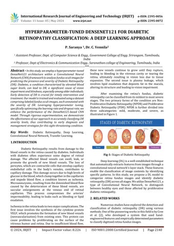
After examining the retina's fundus, diabetic retinopathycanbeclassifiedfromitsmildesttomostsevere stages. The two primary forms of the condition are NonProliferativeDiabeticRetinopathy(NPDR)andProliferative Diabetic Retinopathy (PDR). NPDR is further divided into three subcategories: mild, moderate, and severe, as illustratedinFigure1
Deeplearning(DL)isawell-establishedtechnique thatautomaticallyextractsfeaturesfromimagesthrougha convolutionalneuralnetwork'slayerstack.Thesefeatures enable the classification of image contents by identifying specific patterns. In this study, we propose a DL model to categorize retina fundus images and identify diabetic retinopathy(DR)acrossallstages.WeutilizeDenseNet121,a type of Convolutional Neural Network, to distinguish between healthy eyes and those affected by proliferative diabeticretinopathy.
Numerousstudieshaveexploredthedetectionand classification of diabetic retinopathy (DR) using various methods.OneofthepioneeringworksinthisfieldisbyCree et al. [2], who developed a system that used handengineeredfeaturesandempiricallydeterminedparameters toclassifydigitizedretinafundusimages.
International Research Journal of Engineering and Technology (IRJET) e-ISSN: 2395-0056
Volume: 11 Issue: 05 | May 2024 www.irjet.net p-ISSN: 2395-0072
Yun et al. [3] introduced a method for classifying retina fundus images into categories such as normal, moderate, severe, and proliferative diabetic retinopathy (DR). Their approach included preprocessing the images using morphological operations with disc and diamond structuringelements.Featureswerethenextracted,focusing on pixel area, perimeter, and RGB channel analysis. Classification was carried out using a single-layer feedforwardneuralnetwork.
Rosas et al. [4] concentrated on the detection of microaneurysmsinretinalimages.Theirapproachinvolved applying computer vision techniques to preprocess the images, extracting features relatedto nonuniform lighting and grayscale intensities. Principal component analysis (PCA) and the radon transform were employed to distinguish round-shaped candidate regions and quantify discreteanglevalues,respectively.
Gargeyaetal.[5]introducedanautomateddiabetic retinopathy (DR) screening system that achieved an impressive area under the curve (AUC) of 0.95 on the Messidordatasetusinga5-foldcross-validationtechnique. Theirapproachaimedtostreamlinethescreeningprocess byleveragingmachinelearningalgorithms.
Chetoui et al. [6] devised a system for detecting referablediabeticretinopathy(DR)andvision-threatening DRbyemployingEfficientNetwithtransferlearning.Their workyieldedpromisingoutcomes,achievinganimpressive areaunderthecurve(AUC)ofupto0.98onboththeAPTOS 2019datasetandtheEyePACSdataset.
Dondeti et al. [7] explored the use of pre-training modelNASNETandT-SNEspaceforfeatureextractioninDR classification. Their approach, based on the APTOS 2019 dataset,achievedanaccuracyrateof77.90%.
Qummaretal.[8]presentedanensemblemethod employingfivedeeplearningmodels,specificallycraftedto addressimbalanceddata.Theirstrategyyieldedanaccuracy rateof70%andunderscoredtheeffectivenessofensemble approachesinclassifyingdiabeticretinopathy(DR).
Inatraditionalfeed-forwardconvolutionalneural network (CNN), the initial convolutional layer, which receivestheinput,isthesolelayerthatdirectlyconnectsto theoutputoftheprecedingconvolutionallayer.Thislayer generatesanoutputfeaturemap,whichisthentransmitted tothesubsequentconvolutionallayer.Consequently,each layerhas"L"directconnectionstothenextlayer.However, as CNNs deepen or expand in levels, they encounter the challenge of the "vanishing gradient" problem. This issue implies that as the network's depth increases, some
informationmaydiminishorbecomelost,diminishingthe network'slearningcapacity.
DenseNetsaddressthisproblem bymodifyingthe conventionalCNNarchitectureandenhancingconnectivity betweenlayers.Theterm"DenselyConnectedConvolutional Network" denotes an architecture in which every layer is interconnected with all others. Consequently, there exist L(L+1)/2directlinksbetween"L"layers.
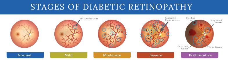
TheproposedmodelisbasedontheDenseNet121 architecture, which accepts an RGB image of 224 × 224 pixels. The model's weights were pre-trained using the ImageNet dataset, leveraging the learned features to optimize the fully connected layer's output weights adequately.Thefeatureextractionprocessoccurswithinthe convolutional layers, while the final layer handles classification.Asoftmaxactivationfunctionisappliedtothe model'soutputtoassignprobabilitiestoeachclass.Notably, eachoutputfromaconvolutionallayerisconcatenatedwith subsequent layers within the same block, as depicted in Figure2,showcasingadistinctivecharacteristicofDenseNet.
The features extracted from the image after processing throughtheconvolutionallayersaresubsequentlyfedinto the classification stage. The classifier comprises two fully connectedlayers.Followingadropoutlayerwithadropout probabilityof50%,thefirstlayerofthesystemconsistsof 1024unitswithaReLUactivationfunction.Thefinallayer comprises 5 units, representing the classes, and utilizes a softmaxactivationfunction.
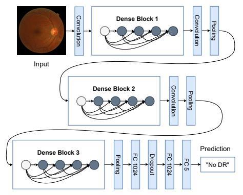
Inthisstudy,thedatasetissourcedfromKaggleand is knownas the APTOS dataset. Itcomprises 3662labeled images for trainingand 1928 unlabeled images for testing purposes.ThedatasetcanbeaccessedviatheKaggleDataset Download link: https://www.kaggle.com/c/aptos2019blindness-detection/data. Originally published as a
International Research Journal of Engineering and Technology (IRJET) e-ISSN: 2395-0056
Volume: 11 Issue: 05 | May 2024 www.irjet.net p-ISSN: 2395-0072
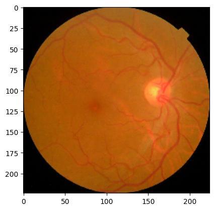
competition on the Kaggle platform, the APTOS dataset consists of fundus oculi images captured under various conditionsandofdifferentsizes.Sampleimagesareprovided belowasshowninFigure3.
4.1
As part of the preprocessing step, all images in the datasets underwent center cropping, resulting in only the retina'sfundusbeingretained,asitisthemostcriticalpart of the image for our task. Additionally, the images were resizedtodimensionsof224x224pixelsasinFigure4.This standardization ensures consistency in image sizes and focusesthemodel'sattentionontherelevantfeaturesofthe retinalimages.
4.2
Data augmentation involves generating new data samples from existing training data through various transformationssuchascropping,padding,flipping,rotating, andresizing.Thistechniqueiscommonlyusedtoincrease the diversity and quantity of training data, which in turn enhancestheperformanceofmachinelearningmodels.By exposing the model to a wider range of variations in the
inputdata,dataaugmentationhelpspreventoverfittingand improvesthegeneralizationabilityofthemodel.
TheCNNmodelemployedinthisstudyisbuiltupon the DenseNet121 architecture, renowned for its dense connectivity patterns. Leveraging pre-training on the ImageNet dataset, DenseNet121 serves as the feature extraction backbone, capturing rich visual features from retinal fundus images via transfer learning. To tailor the model for diabetic retinopathy classification, additional layersareaddedtotheDenseNet121base.Aglobalaverage poolinglayerreducesspatialdimensions,followedbyadense layer with ReLU activation to capture high-level representations. Dropout regularization is incorporatedto mitigateoverfittingbyrandomlydeactivatingneuronsduring training.
The output layer consists of a dense layer with softmaxactivation,producingaprobabilitydistributionover fiveclassesofretinaldiseases.Themodeliscompiledusing SGD optimizer with momentum, categorical cross-entropy loss,andaccuracymetric.Dataaugmentationtechniquesare employedtoenhanceperformanceandcombatoverfitting. Augmented images, generated using ImageDataGenerator, undergorandomtransformationslikerotationandflipping, enrichingthetrainingdatasetforbettergeneralization.
HyperparametertuningisconductedusingtheKeras Tunerlibrary,optimizingdropoutrateandlearningrate.The hyperband optimization algorithm efficiently explores parameter space, identifyingtheconfiguration maximizing validationaccuracy.Duringtraining,callbackssuchasreduce learningrateandearlystoppingmonitorperformanceand preventoverfitting.Themodelistrainedonaugmenteddata andevaluatedonavalidationsettoassesslossandaccuracy, ensuring robust performance in classifying diabetic retinopathy.
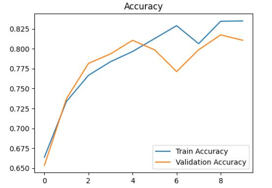
The trial consisted of training the model over 9 epochswhilemonitoringitsperformance.Attheendofthis trial,thevalidationaccuracyreachedapproximately76.45%. However, it's worth noting that the highest validation accuracy achieved during training was approximately 78.67%,indicatingapositivetrendofimprovementoverthe trainingperiod.
Optimal hyperparameters were identified to maximize validation accuracy, including a learning rate of 0.001 and a dropout rate of 0.5. These parameters significantlyinfluencedthemodel'sperformance,showcasing theirimportanceintrainingCNNseffectively.
Throughoutthetrainingprocess,themodelshowed consistent improvement, as seen in the steady increase in
International Research Journal of Engineering and Technology (IRJET) e-ISSN: 2395-0056
Volume: 11 Issue: 05 | May 2024 www.irjet.net p-ISSN: 2395-0072
accuracyanddecreaseinloss.Bythe9thepoch,themodel achievedanimpressiveaccuracyof84.29%,demonstrating itsabilitytolearnandrecognizepatternswithinthedataset effectively.
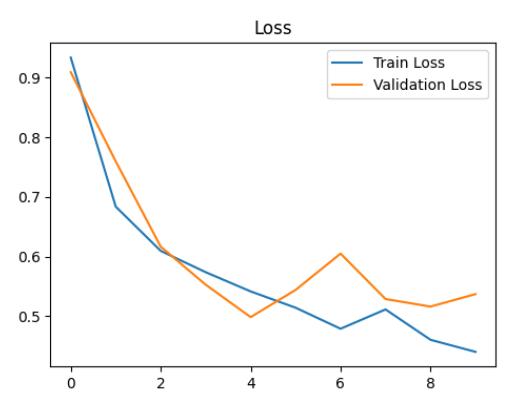
Dynamic adjustments to the learning rate were implementedduringtraining,withareductionto0.0001after the 8th epoch. This adaptive learning rate strategy likely contributedtothemodel'senhancedperformance,allowing for fine-tuning of parameters and convergence towards optimalsolutions.
After training, evaluation on the validation set revealedpromisingresults,withavalidationlossof0.5139 andavalidationaccuracyof80.38%asshowninChart-2&1 respectively. These metrics highlight the model's effectivenessinaccuratelyclassifyingdiabeticretinopathy, suggesting its potential application in clinical settings for diseasediagnosisandmanagementasshowninchart3
In conclusion, the trained convolutional neural network (CNN) model has demonstrated promising performance in effectively classifying diabetic retinopathy usingretinalfundusimages.Throughmeticuloustrainingand hyperparameter optimization, the model has achieved noteworthy results. Its capability to accurately classify diabetic retinopathy, along with its robustness in learning and identifying patterns within the dataset, highlights its potentialapplicabilityinclinicalsettings.
As a valuable tool for disease diagnosis and management, the CNN model holds promise in assisting healthcareprofessionalsinearlydetectionandintervention for diabetic retinopathy, ultimately leading to improved patient outcomes and enhanced quality of care. However, furtherresearchandvalidationonlargerdatasetsandrealworldclinicalscenariosareessentialtoascertainthemodel's efficacy and generalizability, ensuring its reliability and effectivenessinpracticalhealthcaresettings.
Future research directions aim to enhance the diabetic retinopathy classification model's efficacy and broaden its applicability in clinical practice. Firstly, the creationofabenchmarkdatasetwillestablishastandardfor evaluating model performance, enabling fair comparisons across different studies. Secondly, exploring alternative preprocessing functions and architectures, including multilabel approaches, holds promise for improving the model's accuracy and robustness. Thirdly, investigating a bounding box detection approach could provide valuable insightsintothelocalizationandextentofabnormalitiesin retinal images, facilitating more precise diagnosis and treatmentplanning.Theseproposedavenuesforfuturework
International Research Journal of Engineering and Technology (IRJET) e-ISSN: 2395-0056
Volume: 11 Issue: 05 | May 2024 www.irjet.net p-ISSN: 2395-0072
seektoadvancethestate-of-the-artindiabeticretinopathy classification,ultimatelybenefitingpatientsandhealthcare professionalsalike.
[1] Mellitus, D. Diagnosis and classification of diabetes mellitus.DiabetesCare2005,28,S5–S10.
[2] Cree, M.J.; Olson, J.A.; McHardy, K.C.; Sharp, P.F.; Forrester, J.V. A fully automated comparative microaneurysmdigitaldetectionsystem.Eye1997,11, 622–628.
[3] Yun,W.L.;RajendraAcharya,U.;Venkatesh,Y.;Chee,C.; Min, L.C.; Ng, E. Identification of different stages of diabeticretinopathyusingretinalopticalimages.Inf.Sci. 2008,178,106–121.
[4] Gargeya,R.;Leng,T.Automatedidentificationofdiabetic retinopathyusingdeeplearning.Ophthalmology2017, 124,962–969.
[5] Chetoui, M.; Akhloufi, M.A. Explainable Diabetic RetinopathyusingEfficientNET.Proceedingsofthe2020 42nd Annual International Conference of the IEEE Engineering in Medicine & Biology Society (EMBC), Montréal,QC,Canada,20–24July2020
[6] Dondeti,V.;Bodapati,J.D.;Shareef,S.N.;Veeranjaneyulu, N.DeepConvolutionFeaturesinNon-linearEmbedding SpaceforFundusImageClassification.Rev.d’Intell.Artif. 2020,34,307–313.
[7] Qummar,S.;Khan,F.G.;Shah,S.;Khan,A.;Shamshirband, S.;Rehman,Z.U.;Khan,I.A.;Jadoon,W.Adeeplearning ensembleapproachfordiabeticretinopathydetection. IEEEAccess2019,7,150530–150539.
[8] American Diabetes Association. Diagnosis and classification of diabetes mellitus. Diabetes Care, 28(Suppl. 1), S5–S10. doi:10.2337/diacare.28.suppl_1.S5.
[9] [2] Cree, M.J., Olson, J.A., McHardy, K.C., Sharp, P.F., & Forrester, J.V. (1997). A fully automated comparative microaneurysm digital detection system. Eye, 11(5), 622–628.doi:10.1038/eye.1997.160
[10] [3]Yun,W.L.,RajendraAcharya,U.,Venkatesh,Y.,Chee, C.,Min,L.C.,&Ng,E.(2008).Identificationofdifferent stages of diabetic retinopathy using retinal optical images. Information Sciences, 178(1), 106–121. doi:10.1016/j.ins.2007.07.012.
[11] [4] Gargeya, R., & Leng, T. (2017). Automated identification of diabetic retinopathy using deep learning. Ophthalmology, 124(7), 962–969. doi:10.1016/j.ophtha.2017.02.008
[12] [5] Ting, D.S.W., Cheung, C.Y.-L., Lim, G., Tan, G.S.W., Quang, N.D., Gan, A., ... & Wong, T.Y. (2017). Developmentandvalidationofadeeplearningsystem fordiabeticretinopathyandrelatedeyediseasesusing retinal images from multiethnic populations with diabetes. JAMA, 318(22), 2211–2223. doi:10.1001/jama.2017.18152.
[13] [6]Gulshan,V.,Peng,L.,Coram,M.,Stumpe,M.C.,Wu,D., Narayanaswamy, A., ... & Webster, D.R. (2016). Development and validation of a deep learning algorithmfordetectionofdiabeticretinopathyinretinal fundus photographs. JAMA, 316(22), 2402–2410. doi:10.1001/jama.2016.17216.
[14] [7] Abràmoff, M.D., Lou, Y., Erginay, A., Clarida, W., Amelon,R.,Folk,J.C.,&Niemeijer,M.(2016).Improved automated detection of diabetic retinopathy on a publicly available dataset through integration of deep learning.InvestigativeOphthalmology&VisualScience, 57(13),5200–5206.doi:10.1167/iovs.16-19964.
[15] [8] Ting, D.S.W., Cheung, G.C.M., Wong, T.Y. (2019). Diabetic retinopathy: global prevalence, major risk factors,screeningpracticesandpublichealthchallenges: areview.Clinical&ExperimentalOphthalmology,47(4), 536–547.doi:10.1111/ceo.13396.
[16] 9]Sahlsten,J.,Jaskari,J.,Kivinen,J.,Turunen,L.,Jaanio, E., & Pietilä, J. (2020). Deep learning fundus image analysis for diabetic retinopathy and macular edema grading. Scientific Reports, 10(1), 1-10. doi:10.1038/s41598-020-61905-7.
[17] [10] Natarajan, S., Jain, A., Krishnan, R., Rogye, A., Sivaprasad, S., & Rao, C. K. (2020). Evaluating performance of deep learning algorithms for diabetic retinopathy detection. Computers in Biology and Medicine, 122, 103801. doi:10.1016/j.compbiomed.2020.103801.
[18] [11]Costa,P.,Galdran,A.,Meyer,M.I.,Abràmoff,M.D., Niemeijer, M., & Mendonça, A. M. (2017). Towards adversarial retinal image synthesis. In International Workshop on Simulation and Synthesis in Medical Imaging(pp.42-51).Springer,Cham.doi:10.1007/9783-319-67561-9_5.
[19] [12] An, T., Doan, T., Cao, Q., & Jain, A. K. (2019). Adversarial representation learning for diabetic retinopathy analysis. In Proceedings of the IEEE ConferenceonComputerVisionandPatternRecognition Workshops(pp.0-0).doi:10.1109/CVPRW.2019.00169.