Trends and Techniques of Medical Image Analysis and Brain Tumor Detection
Rajan Chaturvedi11Research scholar Department of Physics and Electronics, Dr. Rammanohar Lohia Avadh University, Ayodhya, Uttar Pradesh, India, Dr.R.K.Tiwari2
2Corresponding Author: Professor Department of Physics and Electronics, Dr. Rammanohar Lohia Avadh University, Ayodhya, Uttar Pradesh, India. ***
Abstract
Image processing is adopted day by day by medical professionals for diagnostics and treatment of tumors. This application of engineering and technology is getting more and more space in the field of medical sciences. Various researchers across the world are working differently to explore the use of image processing in medical sciences and developed various algorithms and techniques to get more details like size, volume, and edges where tumors have been spread. In this paper, we have reviewed the various techniques which are carried out by different researchers in medical image analysis and brain tumor detection.
Keywords: medical image processing, brain tumor, diagnosis

INTRODUCTION:
Medical image processing is a rapidly growing and challenging field for researchers. The development of medical imaging techniques like x-ray MRC, CT scan, etc. led to a great advantage to diagnose and treat various internal diseases such as Tumor, Cancer, and internal deflects in various body parts. In medical images, the diagnosisofabnormalclusteringofcellsandtumorhighly depends upon the experience of a medical specialist. Medical image processing aims to develop a new and accuratealgorithmfor theautomaticdetectionof tumors and other abnormal entities. It aims to find not only the exact location of the tumor but also its size, shape, density, type, and boundaries. The development and progress of medical image processing from 1980 up to now,achievemanymilestonesbutstill,thisresearchfield has many unresolved difficulties and challenges. The image analysis community still needs to develop new, accurate algorithms and technologies to uncover information on the Molecular and cellular levels. No researcher has fully analyzed the variability in the requisition, equipment, used algorithm, and their interactionwithahumanoperatortocharacterizedwhole information Over the last20years,separateeffortshave been made by researchers to develop a new algorithm and new principle to design an appropriate model for practical uses. It needs the formation of a common database where algorithms can be compared with each
other. A major challenge is to develop appropriate validationandevaluationapproachesbetweentheoretical principles and practical utilization. Image segmentation has a vital role in image processing and the result of a proposed algorithm highly depends upon the segmentation process. There exist various preestablished segmentation techniques and their application for brain tumor detection. The development of new segmentation techniques in the last decade enhances the hidden information in medical images. A brain tumor is successfully and accurately segmented by these algorithms which help to auto-diagnose of brain tumors but for practical and real-time applications more research is required Medical image analysis involves, image pre-processing segmentation and post-processing. For pre-processing there are various filtering techniques available,themedianfilterismostcommonlyuseddueto its ease and efficiency in removing salt and paper noise. Gaussian filter is useful in smoothening Gaussian noise. Sobel filter is better for edge preservation. In the segmentation process,thresholdingis goodfor the initial stages but not useful for the extraction of much relevant information. Fuzzy C-means and K-means techniques required less human interaction and were useful in poor contrastimages
MATERIAL AND METHODS:
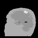
Michael R. Kaus et al. used automated segmentation of MRI of brain tumors with a template-driven segmentation-based algorithm. The automated segmentation of MRI of 20 patients with meningiomas and low-grade gliomas was tested and measure against manualsegmentation.Thefindingsofthisworkshowthat meningiomas and low-grade gliomas can be accurately and reproducibly auto-segmented. However, the validity of segmentation is difficult to assess in the absence of a standard automated segmentation method that has high accuracy within the maximum difference of 0.6% of manualsegmentation. Thesizeoftheabnormalstructure affects the accuracy of segmentation. Because segmentation error occurs on the boundary of the surface, therefore, larger surface led to more miss segmentation. Hence accuracy is less with large objects compared to smaller objects. The automated method has higher reproducibility hence compared to manual segmentation, it does not reduce inter-observer variability substantially. Under restriction to ICC, In the area of interest, the tissue which shows single intensity overlap with the meningioma was excluded and meningioma segmented successfully. So accurate segmentation is possible for low-grade gliomas and meningiomas with automated methods but further work isrequiredforpracticaluseandclinicaltesting[1].
Mathematical morphology and curvature evaluation based algorithm proposed by Frederic zona and JeanClaude Klein for detection vessel-like patterns in noisy image or environment, to separate vessels from the background, a cross-curvature evaluation is performed and these vessels have specific Gaussian-like profile. Morphological operators like erosion, dilation, opening, closing top hat, etc, are defined and utilized for the recognition of geometrical features of the image object. The evaluation of curvature is obtained by using Laplacian. The proposed algorithm was tested on a database of nearly 200 angiographies of patients with different abnormalities. The algorithm proposed by the scientist for the detection of the vessel-like pattern is usefulinawiderangeofretinalimagesanditalsocanbe usedinfieldslikesegmentationofreads[2]
Micheal R.kaus et al, Automated segmentation of MR Images of Brain Tumor”, Radiology 2001, 218: 586591[1].
In a paper by Zhao Yu-Qian et al., basic morphological operators were introduced and a novel morphological edge detection algorithm for detecting the edge of lungs inCTimageswithsaltandpeppernoise.Accordingtothe outcome results of the work, they concluded that the proposedalgorithmismoreefficientthantemplate-based edge detecting algorithms such as Sobel edge detector, morphologicaledgedefector,etc [3].
Mathematical morphology methods used by Akara Sopharak et al. for auto-detection of diabetic retinopathy exudates from non dilated retinal images. In a diabetic patient, exudates occur when lipid or fat leaks through abnormal blood vessels. The proposed algorithm was testedon60imagesandtheresultsofexudatesdetection weresuperimposed on original images.The results show that those exudates regions were visibly highlighted which are not clear before processing. The results were evaluated quantitatively against extractions of the ophthalmologist and it is found that the technique is sensitive and has accuracy. Therefore a practical system based on this technique can reduce ophthalmologist workload and also provide quick detection of exudates whichishelpfulforfastandrapidtreatment[4].
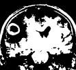
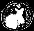
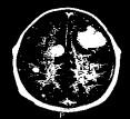
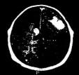
AhmedKharratetal.usesmathematicalmorphologyto increase contrast in MRI images, after that wavelet transform is utilized in segmentation and the K-means algorithm is implemented for detection and extraction of Brain tumors. The proposed methodology is practically usedfortheextractionofthetumorandtheresultofrealtime work is significantly concordance comparing with the expert’s result. However, the classification of the brain tumor as “benign” or malignant is the subject of futureresearch[5].
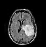
a) CerebralimageMRI
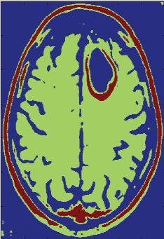
b) Contrast enhancement of image by mathematicalmorphologyalgorithm
c) Wavelettransformdecomposition
d) Tumor extraction by K-means unsupervised classificationmethod
e) Satisfyingclassification
f) Maling ORBenign
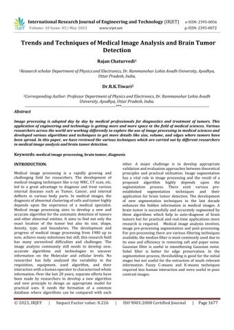
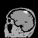
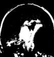
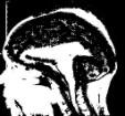
Fig.-1: Example of manual and automated segmentation of low-grade gliomas: SPGR image (a), Manual (b), and template moderated segmentation (c),
Fig.-2: Steps of the algorithm, Ahmed Kharrat et al, Detection of brain tumor medical images”, IEEE, international conference on signals, circuits and system 2009[5].
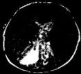
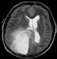
Fig.-3(a)
T. Logeswari and M. Karnan perform brain tumor detection by using a soft computing segmentation technique. In this work, a Hierarchical self-organizing map (HMOS) is used for brain image segmentation and target area (brain tumor) segmented and detected successfully[6].
An integrated approach of K-means clustering and fuzzy C-means clustering both integrated with the markercontrolled watershed algorithm for segmentation of medical images was proposed and compared by M.C. Jobin and R.M.S. Parvathi. K-means clustering with a controlled watershed algorithm gave a better result than Fuzzy C-means clustering with a controlled watershed algorithm.Thereforenosinglemethodissufficientfor all kindsofimages[7,13].
Inputimage Inputimage
Apply the K-means clusteringalgorithm
Clusteredimage
ApplySobelfilter
Apply Fuzzy C-means clusteringalgorithm
Clusteredimage
ApplySobelfilter
Edgemap Edgemap

Apply marker controlled watershedmethod
Finalsegmentationmap
Apply marker controlled watershed method
Final segmentation map
Fig.4: Flow chart of the used method, M.C.Jobin et al., Segmentation of medical image using clustering and watershed algorithm”. American Journal of applied sciences8(12):1349-1352,2011.[7].
The operators of mathematical morphology were discussed and used by Hu Jiang-Hua et al., to eliminate sharp angles and small spots in the dominant color images.Italsoenhancestheconsecutivenessofdominant colorspotsandexpandsthearea[8].
Subhranil Koley and Aurpan Majumder, work out to segmentbraintissuesandtumors byusingCSMbasedKmeans algorithm for detecting the location of the brain tumor in MRI. The proposed algorithm is efficient for segmentation with less computational complexity compared to other methods. The suggested method does not provide efficient results where the tumor is surroundedbyedemathereforetoremovesuchproblems morefutureworkisrequired[9].
Leela G.A. and H. Veena Kumari use K-mean and Fuzzy means clustering algorithm for segmentation to detect brain tumor and cancer cells in a medical image. The detectionoftheboundaryofthetumordependsuponthe segmentation algorithm. According to this study, both segmentation methods produce efficient results and use full for Artificial diagnosis of abnormalities like brain tumorsinmedicalimages[10].
A novel approach for generating AUHB-DW image by applying the concept of HCRF proposed by Mohammad Javed et al. In their study, they point out that the proposed AUHB-DWR algorithm has improved reconstructionqualityandimprovedintensitydelineation thanexistingAUHB-DWRmethods[11]
To enhance the contrast and quality of medical images, various morphological transform operations carried on medical images like top-hot and bottom-hot transform, generally, a disk-shaped mask used in these transforms. Raihan Firoz et al. takes a mask of arbitrary size and keep changing its size until an optimum enhanced image. The result of this work shows that the proposed method improves the contrast of medical imageswhichcanhelpinbetterdiagnosis[12].
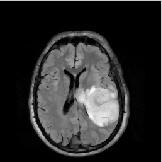
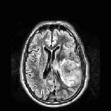
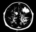
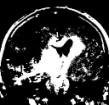
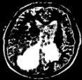
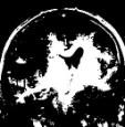
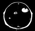
Devendra Somwanshi et al., compare and analyze various threshold entropy-based segmentation methods through simulation results. Entropy methods like Renvi, Vaida,Shannon,Kapar,and Havrda–Charvatareapplied forbraintumordetectionandresultsofvariousmethods compared with each other. Analysis and comparison of results show that Havrda – Charvat entropy method performsbetterthananyotherentropyalgorithm[13].
Fig.-5: Result of different images processed with various entropymethods,DevendraSomwanshietal.,Änefficient brain tumor detection from MRI images using entropy Measures”, IEEE International conference of recent advances and innovations in engineering Dec. 23-25. Jaipur,India,2016[13].
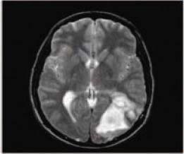
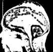

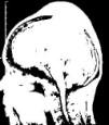
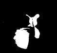

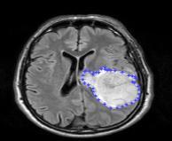
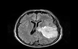
Swapnil R. Telrandhe et al. proposed a method for brain tumor detection using K-means segmentation with preprocessing of images and using object labeling for more detailed features of the tumor. Support vector machine (SVM) tool used for data analysis and classification.Theareaoftumorandtypeoftumorfindby this method and future work involves, determining the size,volume,andstageofthetumor[14].
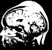
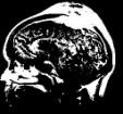
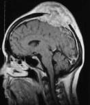
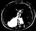
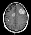
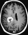
Fig.-6 :(a) Original image (b) segmentation resulted in the image, Swapnil et al., Detection of brain tumor from MRIimagesbyusingsegmentationandSVM”,IEEEworld conference on Futuristic Trends in research and innovationforsocialwelfare(WCFIR’-16),2016[14].
Hao Dong et al proposed a completely automatic methodforbraintumorsegmentationbasedontheU-net deed convolutional network. The proposed method was applied and tested on a multimodal dataset having 220 high-grade and 54 low-grade tumor cases. Results of the proposed method are comparable to other states of the artmethodsforalltumorregions,therefore,theproposed method is reliable for diagnosis of tumor and treatment planning[15].
Mohammad Havaei et al., introduce traditionally different CNN architecture which exploits local features aswellasglobalcontextualfeaturescommonly.A2-Phase training procedure was used which allows us to overcome difficulties related to the imbalance of tumor labels.Resultsoftheproposedmethodofevaluationfrom BRATS 2013 confirm that used method to improve accuracyandspeed.[16]
A method consisting of initial segmentation modeling of the energy function and optimization of the energy function, introduced by C.Hemasundara Rao et al. Conditionalrandomfield(CRF)basedframeworkutilized to combine information present in T1 and FLATR MRT images. A CRF-based framework is useful for modeling complex shapes easily. The proposed method for Brain Tumor detection and segmentation. Incorporates additional information present in T1 – weighted. Therefore its performance is finer in the presence of artifactsandimprovestheboundariesofthetumor.[17]
CNN Method of segmentation has a lack of imagespecific adjustment and ratiocination of previously unseen object classes. To address these problems Guotai et al. proposed a novel deep learning-based interactive segmentation framework of integrated CNNs into a bounding box and scribble-based segmentation pipeline. The result shows that the proposed method performs well on the previously unseen object and takes less user timethanthetraditionalsegmentationmethod.[18]
Ozan Oktay et al., propose a generic training policy that integrates anatomical prior knowledge into CNNs through a new regularization model. In the proposed method, autoencoder (AE) and T-L networks are used to train the neural network. The experimental result shows thattheproposedmodelbenefitincaseswheretheimage onecorruptedand contains artifacts,andhave thefuture scopeinotherimageanalysisfield.[19]
Mahesh Swam and Divya Verma proposed a method basedonamorphologicaloperationoferosion,denoising, and thresholding based tumor segmentation for the detection of a brain tumor in CT and MI images. The database was taken from google open-source and practical work carried on Mat LAB-2019 software. The proposed method achieved a sensitivity of 100%, the similarity of89.667.,anaccuracyof87.50%,andclaimed to be a cost-effective system accessible to medical practitionersoneverydaycomputers.[20]
Anoveltwo-stageframeworknamedspinepresided to execute automated spine parsing for volumetric MR images. The method consists of a 3D graph conventional segmentation network for 3D coarse segmentation along witha2DresidualU-Netfor2Dsegmentationrefinement. The practical results on Y2-weighted volumetric MR images of 215 subjects show an impressive performance with mean dice similarity coefficients of 87.32 ± 4.75%, 87.78 ± 4.64%, and 87.49 ± 3.81% for segmentation of vertebrae, 9 IVDs, and all 19 spinal structures respectively.[21]
InMRIimages,identificationofbraintumorsisdoneby using the GUI framework. Prewitt horizontal edge
strengthening filter used for filtering the image and “watershedpixels”toidentifythetumor.TheGUImethod helps one to adjust the parameters without updating the softwareandprovidesmuchbetterresultsthanstandard tumordetectiontechniques.[22]
CONCLUSION:
Medical image analysis is an emerging and challenging researchfieldthathasadirectsocialimpactandbenefits. It is very helpful for quick diagnosis and treatment, especially in developing countries where the doctorpatientratioispoor.
Medicalimageanalysisincludestheacquisitionofimages, pre-processing, segmentation, detection, analysis, and diagnosis. In the image, analysis segmentation plays a vital role and in recent years various segmentation algorithms like the K-Means method, Fuzzy logic, NeuroFuzzy, Neural network, Snake algorithm, Thersolding based algorithms have been developed. Each of these methods has some advantages and some limitations Different analysis techniques have a different degree of accuracy in different cases therefore practical sensitivity is also an issue. All these techniques and algorithms demand a common & universal stander or protocol so thattheaccuracy,validity&limitationofthesetechniques canbecompared.Acomparativestudyofallalgorithmsis also required to develop a standard practical model of real uses. This field requires more research work and a common approach to developing a standard model for auto-detectionanddiagnosis.
The proposed method of auto –segmentationofmeningiomaandlowgrade glioma has mean accuracy of 99.68% ±0.29(SI) for all 20. Cases. Intra observer variability is 0.10% - 3.57% and 0.14%4.70% Inter observer variability is 0.33%4.72%and0.99%-6.11%
The proposed algorithm is useful in a wide range of retinal images and also in the segmentationofroadsandotherfields.
The used algorithm is more efficient in medicalimagedenoisingandedgedetection compared to template-based edge detector, Gaussian operator, Sobel edge detector gradient operation, and dilation residue
4- Akaro Sopharak etal Mathematicalmorphology methods
edgedetector.
5- Ahmed Kharrat etal Mathematicalmorphology used to increase contrast in MRI image wavelet transform in segmentation and Kmeans algorithm for extractionofTumor
6- T, Logeswari andM.Karnan Soft computing based segmentation and Hsom Vectorquantization
7- M.C.JobinChrist and R.M.S. Parvathi
Integrated Fuzzy Cclustering and KClustering with marker controlled watershed segmentationalgorithm
8- HuJiang-Huaet al Mathematical morphological operator, erosiondilation,HMT
9- Subhranil Koley and Aurpan Majumder
10- Leela G.A. and H.M. Veena Kumari
11- Mohammad Javad Shafiee et al.
CMS based partitional KMeansalgorithm
Auto-detection of diabetic retinopathy exudates from nondilated retinal images
Detection of Brain Tumor in medical image
Theresultoftheusedalgorithmshowsthat those exudates regions were visibly highlighted which are not clear before processingandthetechniqueisfoundtobe accurate and sensitive against the extractionofophthalmologists
The introduced algorithm reduces the extractionsteps.Thefinalresultofthework is that it extracts the tumor region and the outcome significantly agrees with the expert’s result. However benign and malignantnatureofthetumoristhesubject offuturework.
Improved autodetection of Brain Tumor
Segmentation of medicalimages
The suspicious region or Brain Tumor is segmented and extracted successfully by usingaHierarchicalself-organizingmap.
IntegratedK-Meansclusteringwithmarkercontrolled watershed algorithm gave better segmentation compared to integrated Cmeans clustering with a marker-controlled watershedalgorithm.
K-means and Fuzzy C –meansClustering
To improve the multicolor image and eliminate sharp angles in camouflage.
Brain MRI segmentation for tumordetection
Detection of brain tumor and cancer cells.
Used morphological operator significantly improve multicolor image, reduced sharp angles, and small spots in a dominant color image.
The proposed method is simple and efficiently segment brain MRI however it doesnotprovideefficientresultsinthecase where brain tumors are surrounded by edema.
The tumor segmented with K-means clustering is faster and shows tumor boundaries more accurately compared to tumor segmented by fuzzy C- means clustering.
AUHB – DWR algorithm HCRFandHSFCRF.
To improve image quality and repairability betweentissue.
12- Raihan Firoz et al. Morphologicaltransforms Imagesenhancement
13- Devendra Somwanshi Threshold – Entropybased Segmentation methods
14- Swapnil R. Telrandhe K- means algorithm Labeling algorithm and SVM.
Brain Tumor detection in MRI images.
Proposed AUHB- DWR methods improve reconstruction quality and improve intensitydelineation.
The used method enhances the contrast of medicalimages.
Havrda CharvatEntropy methodis the best andaccurateamongotherentropymethods.
Extraction of brain tumorfromMRI Tumor Area and its type extracted successfully.Thetumorsizeandstageofthe tumoristhefieldoffuturework.
15- HaoDongetal Li- Net-based fully convolutionalnetwork
16- Mohammad Havaeietal DeepNeuralNetwork
17- C. Hema Sundara Rao et al
Automatic brain tumor detection and segmentation
Brain Tumor segmentation
Conditionalrandomfield Brain Tumor detection and segmentation
18- Guotai Wang et al. Deep Learning with image-specificfine-tuning
19- Ozan Oktay et al Anatomically constrained NeuralNetworks
20- Mahesh Swami DivyaVerma
A method based on the morphologicaloperatorof erosion, denoising, and thresholding based tumor segmentationmethod
21- Shuma pang et all The spine parse net consisting of GCSN and ResUNetbasedmethod
22- G Bhargavi, J.agril Robinson, etal.
REFERENCES:
Brain tumor detection fromMRIimagesbyusing Prewitt horizontal edgeemphasizing filtering technique
Accurate and robust imagesegmentation
Cardiac image enhancement and segmentation
To detect brain tumors in CT & MRI by using image filtering and segmentation method
To achieve spine parsing for volumetric magnetic resonanceimages.
Brain tumor detection in MRI images
Theproposed methodproduces anefficient and robust segmentation of tumors comparedtomanualmethods.
High performance was achieved with the helpofanoveltwopathwayarchitecture.
The proposed method shows promising results and improves the boundaries of the tumor.
The used method performs well on the previously unseen object in an image and takeslesstime.
Theproposedmodel isusefulforcorrupted imagesandcontainsartifacts.
An accuracy of 87.50% sensitivity of 100% and 89.66% similarity was achieved by the proposedmethod.
An accurate spine parsing for volumetric MR images achieved by spine parse net method and has area + potential in diagnosisandtreatmentofspinaldisease.
Theproposedmethodachievesmuchbetter results than standard tumor detection techniques.
1- Michael R. Kaus, Simon K. Warfield, et al., Automated segmentation of MR Images of Brain Tumor”,[images],Radiology2001,218:586-591.
2- Frederic Zana and Jean–Claude Klein, “Segmentation of vessel-like patterns using mathematical morphology and curvature evaluation” IEEE Trans image processing, vol-10 No-7July2001.
3- Zhao –Yu-Qian, Gui Wei-Hua, Chen Zhen –Cheng Tang Jing-tion and Li Ling-Yum, “Medical images edge detection based on Mathematical morphology?2005IEEEEngineeringinMedicine and Biology 27th annual conference shanghai, China,Sep.1-4,2005.
4- Akara Sopharak, Bunyarit Uyyanonvar, Sarah Barman and Thomas H. Williamson, Automatic
detection of diabetic retinopathy exudates from non dilated retinal images using mathematical morphological methods,” ELSEVIER, Science direct, computerized medical imaging and graphics32(2008),720-727
5- AhmedKharrat,MohamedBenMessaoud,Nacera Benamrane, and Mohamed Abid, “Detection of brain tumor medical images”[images & chart], IEEE,internationalconferenceonsignals,circuits andsystem2009.
6- T. Logeswari and M. Karnan An improve implementation of brain tumor detection using segmentation based on soft computing”, Journal of Cancer Research and Experimental Oncology vol-2(2),PP-006-014,March2010.
7- M.C. Jobin and R.M.S. Parvathi, “Segmentation of medical image using clustering and watershed
algorithm”[flow chart], American Journal of appliedsciences8(12):1349-1352,2011.
8- Hu Jiang-Hua, Qin Lei and Fu Tian-qi, “Image processingbasedonmathematicalmorphologyin camouflage” IEEE, seventh international conferenceonimageandgraphics,2013.
9- Subhranil Koley and Aurpan Majumder, “Brain MRI Segmentation for tumor detection using cohesion based self merging algorithm,” IEEE 2011.
10- Leela G.A. and H.M. Veena Kumari, “Morphological approach for the detection of brain Tumor and cancer cell”, Journal of electronics and communication engineering research,vol.-2,PP:07-12,2014.
11- MohammadJavedShafiee,ShahidA.Haider,etal., “Apparent Ultra-high b-Value diffusion-weighted image reconstruction Via Hidden Conditional Random Fields”, IEEE Tran. Medical imaging, Vol.-3444,No.-5,May2015.
12- Raihan Firoz, Md. Shahjahan Ali et al. “Medical image enhancement using morphological transformation”, Journal of data analysis and informationprocession,vol.-4,PP:1-12,2016.
13- Devendra Somwanshi, Ashutosh Kumar Pratima Sharma and Deepika Joshi, Än efficient brain tumor detection from MRI images using entropy Measures”,[images], IEEE International conferenceofrecentadvancesandinnovationsin engineeringDec.23-25.Jaipur,India,2016.
14- Swapnil R. Telrandhe, Amit Pimpalkar, and Ankita Kendhe, “Detection of brain tumor from MRI images by using segmentation and SVM”,[image], IEEE world conference on Futuristic Trends in research and innovation for socialwelfare(WCFIR’-16),2016.
15- HaoDong,Guang Yang, FangdeLiu, Yuanhan Mo, and Yike Guo, Automatic brain tumor detection and segmentation using U-Net based fully Convolutional networks”, Springer-Verlag Berlin Heidelberg.MIUA2017.
16- Mohammad Havaei, Axel Davy et al. “Brain Tumor Segmentation with deep neural networks”,ELSEVIER,Medicalimageanalysis35, PP:18-31,2017.
17- C.Hemasundara Rao, Dr. P.V. Naganjaneyulu, and Dr. K. Satya Prasad, “Brain tumor detection and segmentation using the conditional random field”, IEEE, 7th international advance computing conference,2017.
18- GuotaiWang,WenqiLi,etal.,“Interactivemedical image segmentation using deep learning with image-specific fine-tuning” IEEE, Trans. Medical imaging,Vol.-37,No.-7,July2018.
19- OzanOktay,EnzoFerrantem,etal.,“Anatomically constrainedneuralnetwork(ACNNs):application to cardiac image Enhancement and Segmentation”, IEEE Medical Imaging, Vol.-37, No.-2,February2018.
20- MaheshswamiandDivyaVerma,“Analgorithmic detection of brain tumor using image filtering and segmentation of various radiographs”, 7th international conference on signal processing and integrated networks (SPIN), 978-1-72815475-6/20,2020
21- Shumao pang, chunlan Pang, et al, “Spineparse Net: spine parsing for volumetric MR Image by a two-stage segmentationframework wit semantic image representation” cornell university Library, Transactions o medical Imaging, September 28 2020,IEEE-2020
22- G. Bhargavi, J. Cyril Robinson Azariah and S. Sivasak thiselvan, “Integrated report on brain tumor detection from MRI Images using pewit horizontaledgeemphasizingfilteringtechnique”, International conference on communication and signal processing, July 28-30, 2020, India, IEEE 2020