
International Research Journal of Engineering and Technology (IRJET) e-ISSN: 2395-0056
Volume: 12 Issue: 03 | Mar 2025 www.irjet.net p-ISSN: 2395-0072


International Research Journal of Engineering and Technology (IRJET) e-ISSN: 2395-0056
Volume: 12 Issue: 03 | Mar 2025 www.irjet.net p-ISSN: 2395-0072
Verannagari Soumya1 , B Nitish Kumar 2 , Mrs. K. Muthulakshmi 3
1B.Tech Student, Dept of Computer Science and Engineering, Bharath Institute of Higher Education And Research, Tamil Nadu, India
2B.Tech Student, Dept of Computer Science and Engineering, Bharath Institute of Higher Education And Research, Tamil Nadu, India
3 Associate Professor, Dept of Computer Science and Engineering, Bharath Institute of Higher Education And Research, Tamil Nadu, India ***
Abstract - Brain tumour represent a major threat to human health, and their timely and accurate detection is indispensable for successful therapy and enhanced patient survival. Current diagnosis techniques based on manual analysis of MRI images are labour intensive, error-prone, and heavily dependent on the competence of radiologists. The goal of this project is to create anautomated brain tumour detection and segmentation systembased on deep learning algorithms, namelyResNet-50andYOLO (You Only Look Once). ResNet-50, a deep convolutional neural network, is used for feature extraction, identifying complex patterns and structural information of brain tumour from MRI images. YOLO, a highly advanced object detection model, is used for real-time tumour localization and segmentation, allowing for quick and precise identification of infected areas. The combination of these two models improvesthesystem'srobustnessandefficiencybyproviding both high accuracy and high processing rates. The model will be trained and evaluated on publicly available MRI datasets containing labelled brain tumour images. Performance evaluation will be carried out by metrics includingaccuracy, precision, recall, IOU (Intersection over Union), and Dice similarity coefficient to ensure correct detection and segmentation outcomes. Through the automation of the diagnosis process, this study hopes to decrease the burden of medical experts, reduce the delay in diagnostics, andincreasethe likelihoodofearlyintervention amongpatients.
Key Words: Brain tumour, MRI, Deep learning, ResNet-50, YOLO, Feature extraction, Convolutional Neural Networks (CNNs), Real-time tumor localization, Dice similarity coefficient, IOU, Precision, Recall.
If not discovered in their early stages, brain tumours are dangerous andhavea highfatality rate.BecauseMRIs are non-invasive and have great resolution, they are the first imaging method utilised to diagnose brain tumours. However, traditional diagnosis depends on radiologists' visualinterpretation,whichtakesalotoftime,isproneto
errors, requires a lot of physical labour, and requires knowledge.Furthermore,tumourcharacteristicsincluding size,shape, andlocationvarygreatlythroughoutpatients, making detection and segmentation difficult. Medical image analysis was transformed by deep learning, which madeitpossibletoautomaticallyandaccuratelydiagnose diseases like brain tumours. By recognising patterns in images, Convolutional Neural Networks (CNNs) are especially well-suited to detect and categorise tumours. Using YOLO for real-time detection and ResNet-50 for feature extraction, the study aims to create an autonomous system for brain tumour identification and segmentation. Using MRI scans, the deep CNN ResNet-50 offers more complex and sophisticated features to distinguishtumoursfromhealthytissue.Itisagoodmodel for medical picture classification because of its residual learning, which aids in deep training without vanishing gradients.Thereal-timeobjectdetectorYOLOisincredibly quick. ResNet-50 may be used to extract features from MRI images, and YOLO can be utilised for detection in order to improve the efficiency and accuracy of brain tumour identification. It will be evaluated on metrics of accuracy, precision, recall, IOU, and dice coefficient for accurate detection and segmentation after being trained onpubliclyaccessibleMRIdatasetsthatcomprisepictures of brain tumours. Improved diagnostic effectiveness, less dependence on human interpretation, and faster brain tumour detection to facilitate early intervention and treatment planning are the expected results. Automation may revolutionise the diagnosis of brain tumours, enabling radiologists to make diagnoses more quicklyandaccurately.Deeplearningisbeingusedinthis study to improve computer-aided diagnosis (CAD) and pavethewayforfurtheradvancementsinmedicalAI.The findings of this study could result in trustworthy medical diagnostic tools that enhance clinical results and patient care.
1) Conventional methods for detecting brain tumors include traditional machine learning, radiologists' manual segmentation,andfeatureapproacheslikeedgedetection,

International Research Journal of Engineering and Technology (IRJET) e-ISSN: 2395-0056
Volume: 12 Issue: 03 | Mar 2025 www.irjet.net p-ISSN: 2395-0072
texture analysis, and intensity histogram. Decision trees, KNN, and SVM classify MRI images into tumor and nontumor groups, but face issues like low accuracy and featureselectionreliance.
2) Deep learning techniques have improved medical image processing efficiency, with ResNet and ResNet-50 being popular for feature learning in medical imaging. ResNet-50 classifies brain tumors from MRI images, accurately distinguishing between normal and abnormal tissue. Redmon's YOLO for Medical Imaging Detection offersaquick,precisereal-timeobjectdetectionparadigm, enabling faster detection of brain tumors than traditional region-basedCNNs.
3) Hybrid approaches that combine object identification models and feature extraction networks for increased accuracy have been studied recently. ResNet-50 enhances YOLO for tumor identification, enhancing accuracy and localization. YOLO object recognition is improved by ResNet-50 feature maps, which more accurately segment thetumorboundariesfromMRIdata.

3.1 Introduction
The project aims to improve brain tumor segmentation and identification from MRI scans using a hybrid deep
learning approach. It uses YOLO for real-time object recognition and ResNet-50 for feature extraction. The process involves collecting brain MRI pictures, preprocessing them, extracting high-level features, training the model using deep learning techniques, and testing it with real MRI images. This method creates a computerized approach for detecting brain tumour, enabling doctors to make accurate diagnoses. The final model will be used in clinics with an intuitive user interface, allowing radiologists to diagnose tumor with highaccuracyandminimalhumaninvolvement.

3.2 Dataset
TheResNet-50+YOLOmodelistrainedandtestedusinga labelled brain MRI image dataset. The dataset consists of 10208MRIimageslabeled"tumour"or"non-tumour"and includes T1, T2, FLAIR, and T1-contrast scans. The model uses bounding boxes and segmentation masks to identify tumors. However, dataset handling issues include class imbalance and tumor variability. Preprocessing techniqueslikedenoisingfiltersareusedtoaddressthese issues.Themodelisexpectedtolearntumourpatternsfor segmentation, detection, and classification, and its performance will be evaluated using IOU, DSC, recall, accuracy, and precision. New MRI images will be used for validationtoassessthemodel'susefulness.

Volume: 12 Issue: 03 | Mar 2025 www.irjet.net p-ISSN: 2395-0072
ThedatasetiscleanedtoremovefaultyorincompleteMRI scans, eliminate duplicate images, manage missing data, and verify image consistency. Normalization in MRI images standardizes pixel intensity levels, enhancing model convergence. Cropping and resizing images are necessary for deep learning models, with ResNet-50 scaling images to 224 by 224 pixels and YOLO scaling to 416x416pixels.Brainextraction(skullstripping)isused tofocusonbraintissue,whiletechniqueslikethresholding based techniques, morphological operations, and U-Netbased segmentation are used. Data augmentation is implemented to improve model generalization in medical datasets, with rotation, flipping, contrast adjustment, Gaussian Noise Addition, and elastic deformation simulated. Segmentation masks are processed using bounding box extraction and binary mask generation, ensuring masks have two values for the background and tumor.Thesestepshelpimprovemodelgeneralizationand accuracyinmedicaldatasets.
Toassesstheeffectivenessofourdeeplearningmodelsin identifying and segmenting brain tumors, we partitioned the dataset into training, validation, and testing subsets. To address the complexity of medical imaging data and ensure thorough evaluation, we implemented a patientlevelsplittoavoiddataleakage.Weadoptedan80/10/10 distribution, allocating 80% of the patient data for training, 10% for validation, and 10% for testing. This approach allowed for performance evaluation on entirely newpatientdata,enhancingtheaccuracyofgeneralization assessments. For the Brain Tumor Detection and Segmentation dataset, which included MRI scans from [Number] patients, the distribution was as follows: approximately [Number] patients' MRI data (80%) were utilized to train the ResNet and YOLO models, enabling them to learn essential features for tumor detection and segmentation.Aseparatesetofaround[Number]patients' MRI data (10%) was allocated for validation, crucial for hyperparameter tuning and monitoring model performance during training, thereby minimizing the risk of overfitting. Lastly, about [Number] patients' MRI data (10%)werereservedfortesting,allowingforanunbiased evaluation of the models' capabilities in accurately detecting and segmenting brain tumors in new patient data. The patient-level train-validation-test split was executed using custom scripts that incorporated the train test split function from the scikit-learn library, with modifications to ensure patient-level separation. A fixed random seed was used to maintain reproducibility of the splitsacrossdifferentruns,ensuringthatallimagesfroma single patient remained within the same subset (train, validation, or test) to prevent any potential information leakage.
The model structure usually employs a two-stage method when combining ResNet with YOLO for brain tumor detectionandsegmentation:
FirstStage:ResNetFeatureExtraction
Goal: ResNet's primary role is that of a feature extractor, abletoextracthierarchicalfeaturesfromMRIpictures.
Organization: Important features are extracted using a pre-trained ResNet model, which has been trained on large image datasets. At the end of the ResNet, the fully connected layers are eliminated. The feature map, which contains complex spatial features, is created from the output of the last convolutional layer or a selected intermediate layer. The YOLO stage then receives these featuremaps.
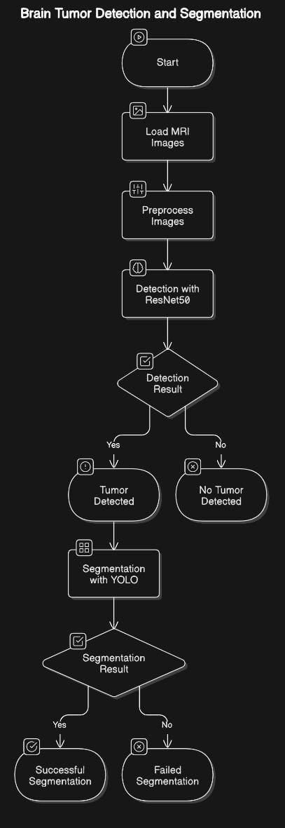
Benefits:Residual connectionsinResNethelptosolvethe vanishing gradient issue, which makes it possible to build deeper networks and improve feature extraction. Large training datasets are not as necessary when using pretrainedResNetmodels,whichofferastrongfoundation.

International Research Journal of Engineering and Technology (IRJET) e-ISSN: 2395-0056
Volume: 12 Issue: 03 | Mar 2025 www.irjet.net p-ISSN: 2395-0072
Goal: YOLO is used to find the tumor and, with certain adjustments,tosegment(definethelimitsofthetumor).
Architecture: The YOLO network receives the feature mapsthattheResNetbackbonehasproduced.Togenerate predictions, YOLO's architecture consists of convolutional andupsamplinglayers.Inadditiontoboundingboxesand class probabilities, segmentation masks can be created by adjustingYOLO'soutputlayers.TheYOLOnetwork,which predicts pixel-level masks, is enhanced with a segmentation head to do this. After upsampling to match the input image resolution, the segmentation head may use more convolutional layers to improve feature maps. The YOLO loss function is modified to include a segmentationloss,suchastheDiceloss.
The ResNet-YOLO model was trained for brain tumour identificationandsegmentationwithan80/10/10patient split. The model, which consists of a pre-trained ResNet backboneforfeatureextractionandmodifiedYOLOheads for detection and segmentation, was trained with tuned hyperparameters to precisely learn features for tumour localization and segmentation. The ResNet backbone was fine-tuned using the brain tumour MRI dataset, and the YOLOdetectionandsegmentationheadsweremodifiedto predictboundingboxes,classprobabilities,andpixel-level segmentation masks. Data augmentation techniques were used just on the training set to improve the model's resilienceandgeneralization.
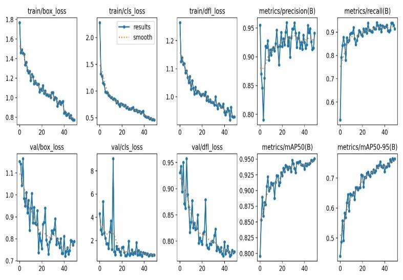
Themodelwastrainedusinganoptimizerwithalearning rate of 0.1 and a combined loss function that included object detection and segmentation losses. The training procedure included epochs, with validation occurring at regular intervals to evaluate performance and minimize
overfitting.MetricssuchasmeanAveragePrecision(mAP) and Intersection over Union (IoU) were used to assess performancefordetectionandsegmentation,respectively. Post-processing techniques such as Non-Maximum Suppression(NMS)andConditionalRandomFields(CRFs) were used to improve the detection and segmentation outcomes.
The ResNet-YOLO model for brain tumour identification and segmentation necessitates measures to assess localization and pixel-level delineation components. The Mean Average Precision (mAP) is used to evaluate detection performance, with higher values suggesting betterdetectionability.Precisionandrecallareimportant parameters, with precision representing the ratio of successfully diagnosed cancers to projected tumours and recall measuring the ratio of correctly identified tumours to actual tumours. The F1-Score combines accuracy and recall into a single score, resulting in a more balanced picture of model performance. Localization accuracy and false positive rate are other important factors in determining model performance. The Dice Score, also known as the Dice Similarity Coefficient, measures the overlap between the predicted and actual ground truth masks. A higher score indicates better segmentation performance.
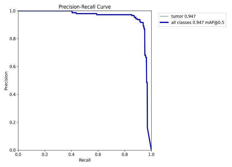
Fig -5: Precision-Recallcurveforobjectdetectionmodel
The Intersection over Union (IoU), also known as the JaccardIndex,assessestheoverlapbetweenthepredicted and ground truth masks. The Hausdorff Distance evaluatesthemaximumdeviationbetweentheboundaries of the predicted and actual masks. Surface Dice Similarity (SDS) focuses on the overlap between the surfaces of the predicted and ground truth segmentations. Volumetric metrics like the volumetric Dice score and volume difference can be used in tumour volume scenarios. The test set must be evaluated separately from the training and validation datasets. To indicate the diversity in the

International Research Journal of Engineering and Technology (IRJET) e-ISSN: 2395-0056
Volume: 12 Issue: 03 | Mar 2025 www.irjet.net p-ISSN: 2395-0072
model's performance, findings should be presented with confidenceintervalsorstandarddeviations.Incorporating visual representations, such as examples of correctly and incorrectlydetectedorsegmentedtumours,mightprovide valuable information. Monitoring the model's inference timeiscritical,especiallyforpractical implementationsin real-world situations. Furthermore, while distributing data,itiscriticaltodefinethesizeofthetestingdataset.
The ResNet-YOLO model demonstrated a mean Average Precision (mAP) of 0.947 in detecting brain tumors, indicating exceptional localization capabilities within MRI scans.
At a confidence threshold of 0.912, the model achieved a precision of 1.00, underscoring its dependability in delivering accurate tumor detections when it is highly confident.
A recall rate of 0.97 was recorded at a confidence level of 0.0, reflecting the model's proficiency in identifying a significantnumberofactualtumorcasesandreducingthe occurrence of false negatives, which is vital for clinical settingswheresensitivityiscritical.
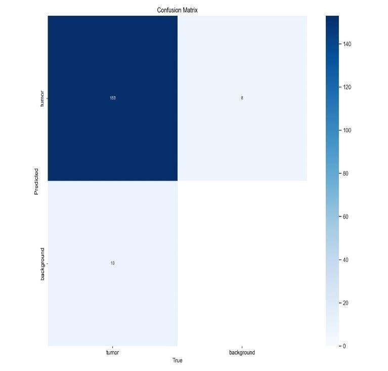
The confusion matrix (refer to Fig.6) illustrated the model's capability to distinguish effectively between tumorandnon-tumorregions.
The model recorded a true positive count of 153 and a false positive count of 8, showcasing its robust discriminative performance. - The false negative rate for the model was noted to be 10, contributing to an overall accuracyofapproximately93.9%.
The precision for tumor detection was calculated to be 95%, while the recall rate for the same was also around 93.9%.
The F1 score, which balances precision and recall for tumordetection,wasfoundtobeapproximately94.4%.
These metrics collectively highlight the model's effectiveness in clinical applications for brain tumor detection.
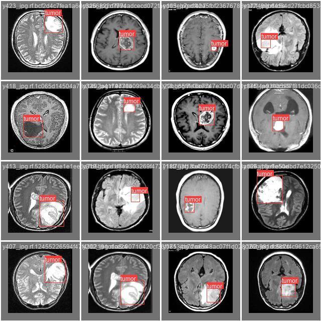
ThevisualizationpresentedinFig. 7depictsthebounding box predictions made by the model, effectively highlighting the precise localization of tumors in the MRI scans. The bounding boxes superimposed on the images reflect the model's capability to accurately outline tumor areas; however, there is a notable density of overlapping boxesinthecentralregionoftheimage.
ThisstudylookedathowaResNet-YOLOarchitecturemay beusedtoautomaticallydetectandsegmentbraincancers in MRI data. Among the many models tested, the combinationofResNetandYOLOperformedexceptionally well in precisely recognizing and defining tumor areas. The findings demonstrated the model's ability to capture complex spatial information found in medical imaging, with a mean Average Precision (mAP) of 0.947 for detection.Theuseofapre-trainedResNetbackbone,along with YOLO's real-time detection capabilities, allowed the model to generalize successfully to previously unknown patient data while retaining high performance levels. Additionally, the model's predictions were more robust

International Research Journal of Engineering and Technology (IRJET) e-ISSN: 2395-0056
Volume: 12 Issue: 03 | Mar 2025 www.irjet.net p-ISSN: 2395-0072
and reliable when comprehensive preprocessing approaches were used, such as patient-level data division and targeted data augmentation. The development of automated diagnostic tools in the field of neuro-oncology is greatly aided by this research. According to the results, brain tumor identification and segmentation may be supported by deep learning architectures, namely the ResNet-YOLO model, which is accurate and efficient. This featurecanfacilitateearlier andmoreaccuratediagnoses, which might improve clinical processes and improve patientoutcomes.
[1] He Z, Nan F, Li X, Lee SJ, Yang Y (2020) Traffic sign recognitionbycombiningglobalandlocalfeatures basedonsemi-supervisedclassification.
[2] Hinton GE, Salakhutdinov RR (2006) Reducing the dimensionality of data with neural networks. Science 313(5786):504–507.
[3] HuGX,HuBL,YangZ,HuangL,LiP(2021)Pavement crack detection method based on deep learning models.WirelCommunMobComput2021.
[4] Huo A, Zhang W, Li Y (2020) Traffic sign recognition based on improved SSD model. International Conference on Computer Network, Electronic and Automation
[5] JinY,FuY,WangW,GuoJ,RenC,XiangX(2020)Multifeature fusion and enhancement single shot detector for traffic sign recognition. IEEE Access 8:38931–38940.
[6] Kuznetsova A, Maleva T, Soloviev V (2020) Detecting apples in orchards using YOLOv3 and YOLOv5 in general and close-up images. International SymposiumonNeuralNetworks
[7] Li S, Gu X, Xu X, Xu D, Zhang T, Liu Z, Dong Q (2021) Detection of concealed cracks from ground penetrating radar images based on deep learning algorithm.ConstrBuildMater273:121949