Shaping Tomorrow's Care: New Therapies, Hidden Risks and Predictive Technology
[ pages 4, 8, 12 ]
News from the Harvard Medical School
Department of Otolaryngology–Head and Neck Surgery
Spring 2025 | Vol. 21, No. 1
Published twice per year.
For any inquiries or comments regarding this issue, please contact:
Nicole Feldman
Communications Manager nfeldman1@meei.harvard.edu
Department of Otolaryngology–Head and Neck Surgery
Mass Eye and Ear
243 Charles Street, Boston, MA 02114
CONTRIBUTORS
Editor-in-Chief
Mark A. Varvares, MD, FACS
William W. Montgomery and John W. Merriam Professor and Department Chair of Otolaryngology–Head and Neck Surgery
Harvard Medical School
Chair of Otolaryngology–Head and Neck Surgery
Mass Eye and Ear
Massachusetts General Hospital
Managing Editor | Writer
Nicole Feldman
Design | Layout | Photography
Garyfallia Pagonis
Beth Israel Deaconess Medical
Boston Children’s Hospital
Brigham and Women’s Hospital
Mass Eye and Ear
Massachusetts General Hospital
CONTENTS
1 Letter From the Chair
Mark A. Varvares, MD, FACS
2 News: Faculty Retreat
16 Alumni Profile
Jennifer Kim, MD, 2000
18 Donor Profile
Atchinson Family: Advancing head and neck cancer research
19 Alumni Giving Society
20 Highlights
22 Research Advances
FEATURES
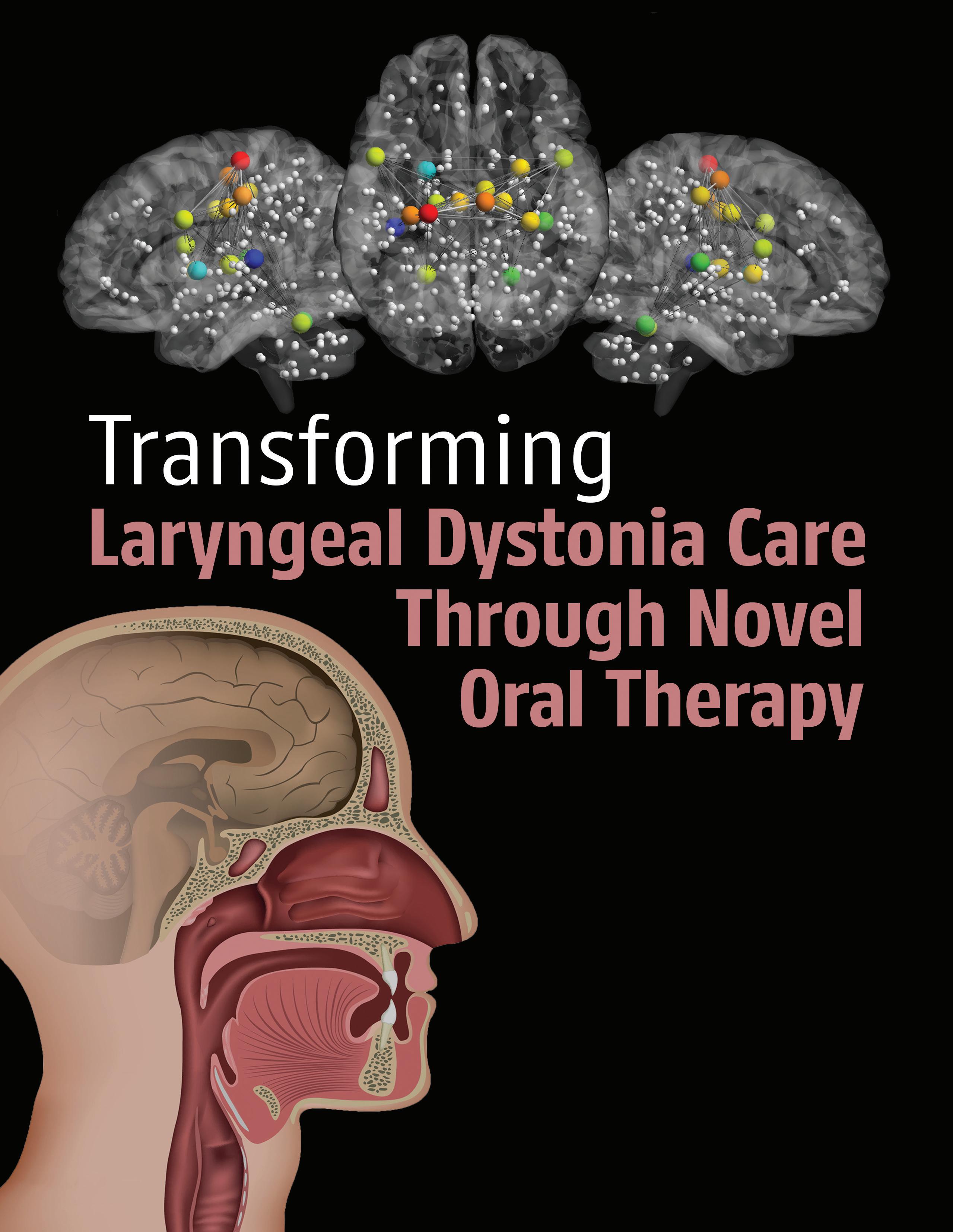
4 Transforming Laryngeal Dystonia Care Through Novel Oral Therapy
A clinical trial conducted at Mass Eye and Ear identified sodium oxybate as the first oral treatment for laryngeal dystonia to demonstrate efficacy in alleviating challenging symptoms that impact speaking.
8 PM2.5 and Head and Neck Cancer: The Unexplored Link
A collaborative first-of-its-kind study correlates higher levels of pollutant particulate matter to higher occurrences of head and neck aerodigestive cancer.
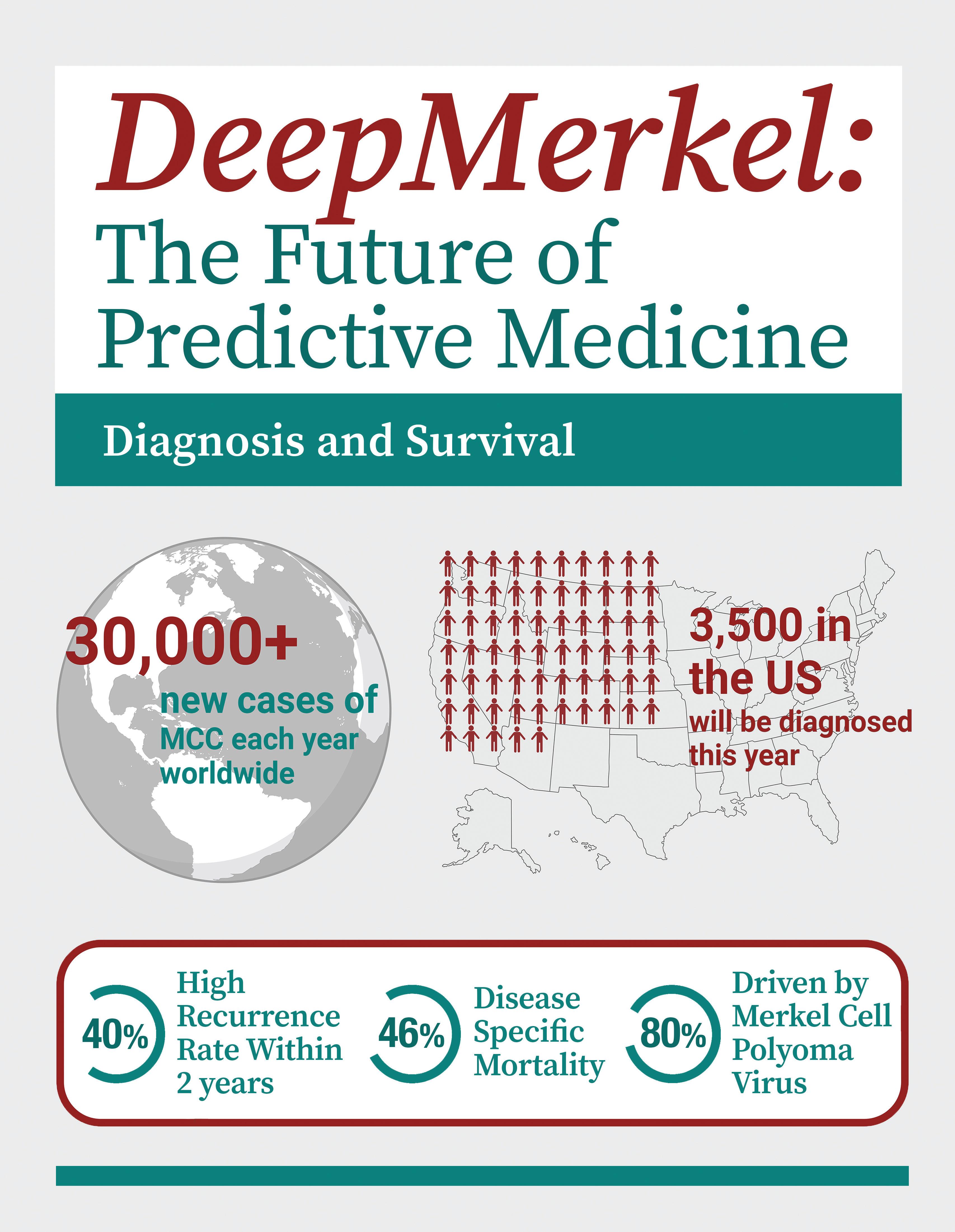
12 DeepMerkel: The Future of Predictive Medicine
A groundbreaking artificial intelligence model, DeepMerkel, is setting a new standard for personalized, time-dependent survival predictions in Merkel cell carcinoma.
Dear colleagues and friends,
The field of otolaryngology–head and neck surgery has made exceptional strides in recent decades. Driven by landmark research and practice changing clinical innovation, we’ve developed treatments and diagnostics once considered impossible. These achievements reflect the creativity, dedication and expertise of our department members. As new technologies emerge and our understanding of disease deepens, our faculty is ready to redefine standards of care and bring innovations to more patients, more effectively than ever before.
As Chair, I’ve seen years of work and research come to fruition and remain incredibly impressed by our faculty’s collaboration and commitment to forward-thinking, research-infused care. In this edition of Harvard Otolaryngology, I’m proud to share novel therapies and findings that advance our patient-centered mission.
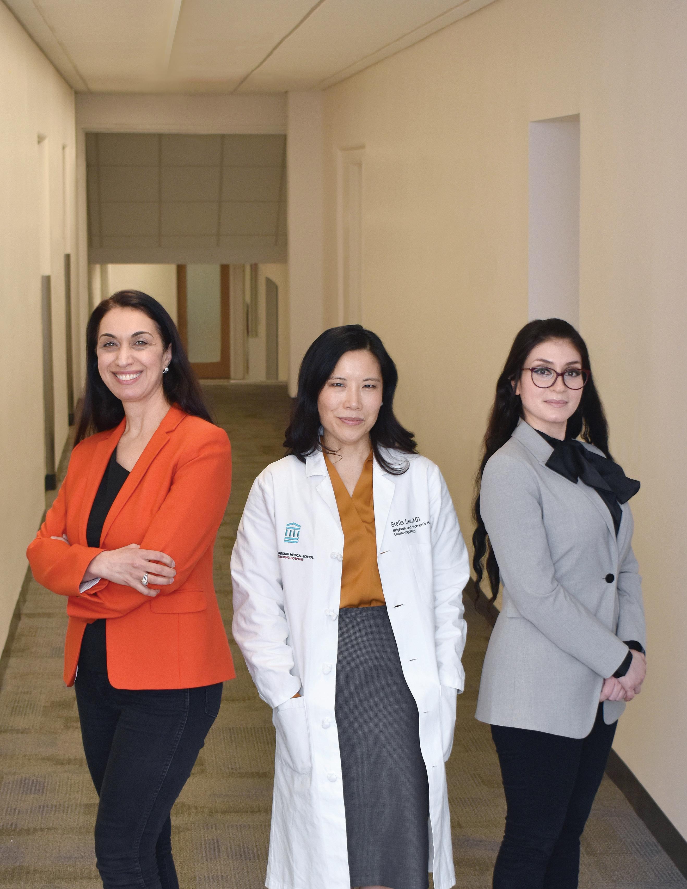
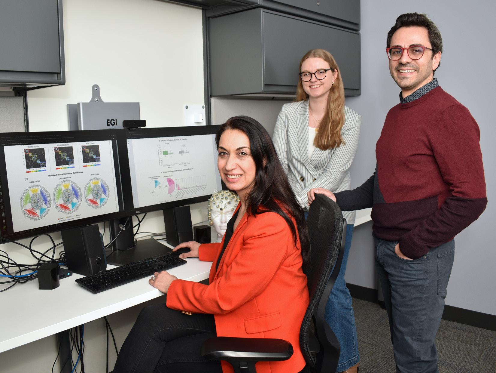
Kristina Simonyan, MD, PhD, DrMed, led a clinical trial at Mass Eye and Ear that identified sodium oxybate as the first oral treatment to show efficacy in alleviating the speech-related symptoms of laryngeal dystonia. These findings are particularly exciting because they suggest that sodium oxybate can be taken as needed, giving patients the flexibility to tailor treatment to their daily needs and manage symptoms more effectively.
Another remarkable study, led by Stella Lee, MD, explored the link between environmental exposure and cancer risk. This firstof-its-kind research found a strong correlation between elevated levels of pollutant particulate matter and higher occurrences of head and neck aerodigestive cancers. It’s a powerful example of how uncovering overlooked determinants of health could have real implications for our patients.
Finally, I’m thrilled to highlight a breakthrough in skin cancer care led by Sophia Z. Shalhout, PhD. Through an international collaboration, she developed DeepMerkel, an advanced artificial
intelligence model that sets a new benchmark for personalized, time-dependent survival predictions in patients with Merkel cell carcinoma, a rare but aggressive skin cancer. In a disease where treatment decisions are complex and outcomes are uncertain, this kind of precision prognostication could be life-changing.
While we continue to reach these major milestones, we also recognize that we are currently navigating unprecedented times in academic healthcare. Now more than ever, we are remaining laser focused on our four-part mission: providing the world’s best patient care, driving discoveries through research, training tomorrow’s leaders in our specialty and ensuring the needs of our entire community are met. I hope this edition leaves you as encouraged as I am by our progress in delivering better outcomes for patients.
As always, I thank you for your interest and support in our department’s research, initiatives and activities. And as we come into the summer months, I hope you are all able to find some time to spend with family and friends in your favorite vacation spot, to unwind and recharge as we prepare to kick off the next academic year.
Wishing you the best in health and fulfillment in everything you do.
Sincerely,
Mark A. Varvares, MD, FACS
William W. Montgomery and John W. Merriam Professor and Chair Department of Otolaryngology–Head and Neck Surgery, Harvard Medical School
Chair, Departments of Otolaryngology–Head and Neck Surgery, Mass Eye and Ear
Massachusetts General Hospital
Faculty Retreat
Department of Otolaryngology–Head and Neck Surgery at Harvard Medical School.
On Saturday, February 8th, 2025, the Department of Otolaryngology–Head and Neck Surgery (OHNS) at Harvard Medical School (HMS) hosted its second annual faculty retreat at the Boston Museum of Science.
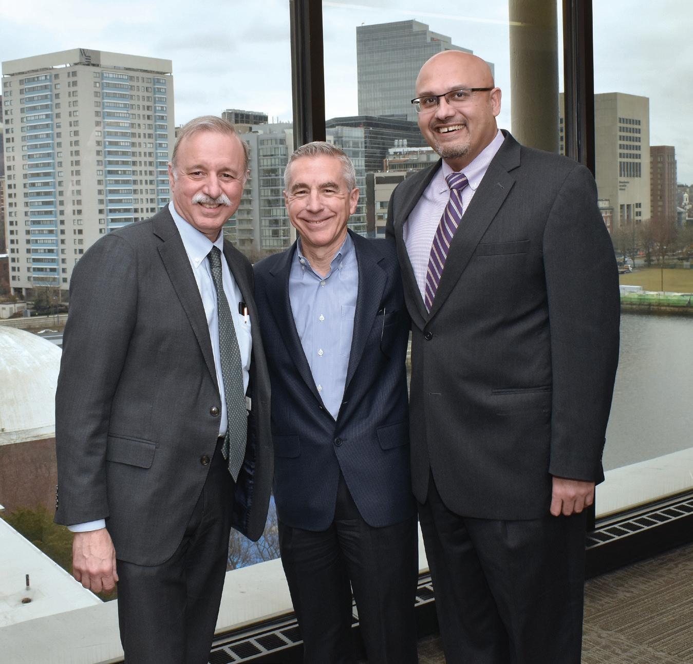
Mark A. Varvares, MD, FACS, William W. Montgomery and John W. Merriam Professor and Chair of Otolaryngology–Head and Neck Surgery (OHNS) at Harvard Medical School and Chair of the Department of OHNS at Mass Eye and Ear, led the retreat alongside the department chiefs from each affiliate hospital: Michael J. Cunningham, MD, FACS, Professor of OHNS at Harvard Medical School and Chief of OHNS at Boston Children’s Hospital; Ravindra Uppaluri, MD, PhD, Associate Professor of OHNS at Harvard Medical School and Chief of OHNS at Brigham and Women’s Hospital; and Scharukh M.
Jalisi, MD, Associate Professor of OHNS at Harvard Medical School and Chief of OHNS at Beth Israel Deaconess Medical Center.
“The goal of the Harvard OHNS faculty retreat is to foster connection across departments, share important updates and offer guidance on key professional challenges,” shared Dr. Varvares. “We were delighted by the turnout of 70 faculty members joining, and I am pleased by the collaborative insights and conversations sparked from the day.”
The day featured five riveting panel discussions and concluded with a thoughtful lunch lecture. Each panel included a moderator and four panelists representing the Harvard OHNS affiliate hospitals.
The first panel focused on innovations in research, both basic and translational. Panelists highlighted surgical simulation in head and neck surgery; advances in gene therapy for hearing loss; ongoing work on circulating tumor DNA and a neoadjuvant immunotherapy trial; and the use of lifetime fluorescence imaging in solid tumor surgery.
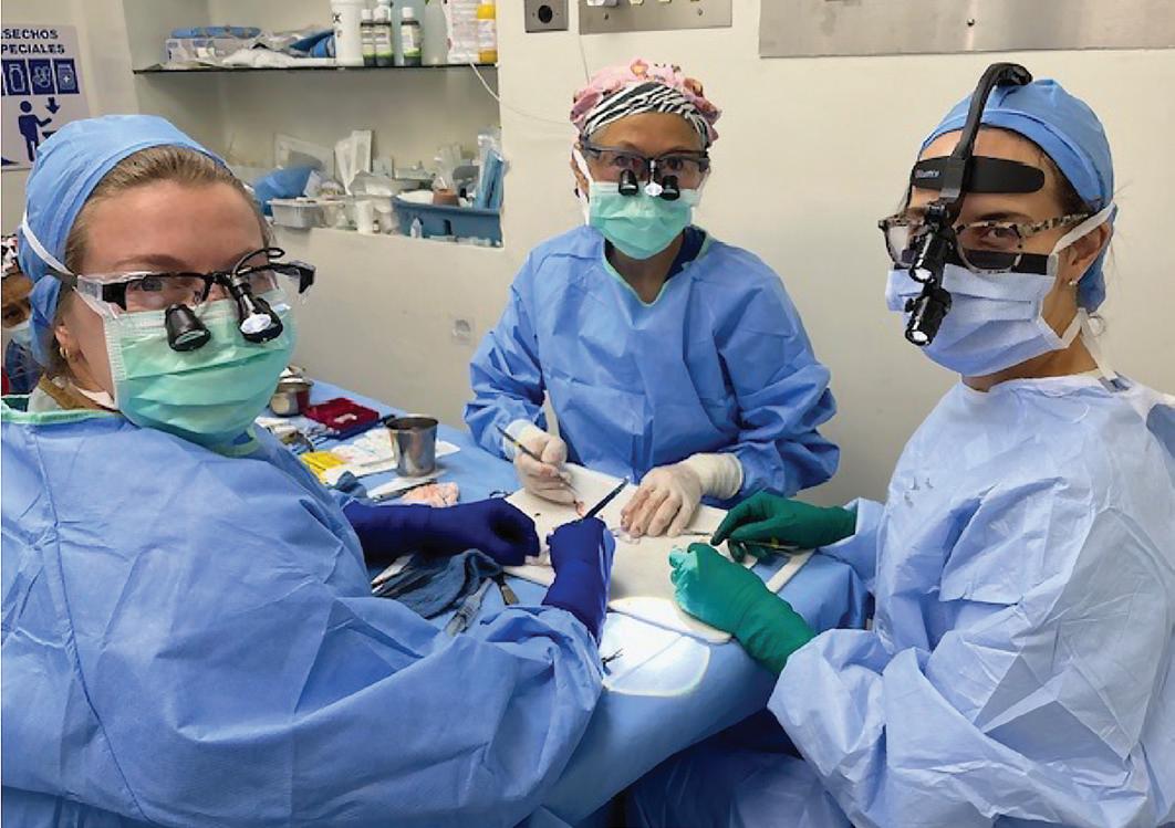
The next panel focused on global surgery. Speakers discussed long-standing efforts in surgical education abroad, including work in Pakistan, East Africa and Vietnam, as well as initiatives closer to home.
There was an emphasis that global health also means addressing local needs, highlighting the importance of language-competent care.
From left to right: Mark A. Varvares, MD, FACS; Michael J. Cunningham, MD, FACS; Scharukh M. Jalisi, MD; Inset: Ravindra Uppaluri, MD, PhD, who was not able to attend.
A dynamic panel on HMS promotions followed. After an overview of the process, recently promoted faculty at various ranks shared their experiences and insights. Throughout the conversation, panelists reflected on their promotions, sharing which parts of their dossiers they believed made the greatest impact, the most challenging aspects of assembling their dossiers, and the key lessons they learned—whether about the process itself, unexpected obstacles or valuable advice they received along the way.
The session also included a practical discussion on crafting narrative statements for the Harvard Medical School CV, with panelists providing guidance on both the clinical and educational innovations and investigator narrative formats.
The final panel explored clinical innovation and the journey from ideas to market. Speakers shared
Moderator and Panelist List
Innovations in Research: Basic and Clinical
• Moderators: Kristina Simonyan, MD, PhD, DrMed, John W. Merriam/William W. Montgomery Professor of OHNS at HMS and Rosh K. Sethi, MD, MPH, Assistant Professor of OHNS at HMS
• Panelists:
–Ernest D. Gomez, MD, MTR, Instructor in OHNS at HMS
–A. Eliot Shearer, MD, PhD, Assistant Professor of OHNS at HMS
–Regan W. Bergmark, MD, Assistant Professor of OHNS at HMS
–Anand T.N. Kumar, Associate Professor of OHNS at HMS
Global Surgery
• Moderator: Jeremy D. Richmon, MD, Associate Professor of OHNS at HMS
• Panelists:
–Dr. Jalisi
–Roger C. Nuss, MD, Assistant Professor of OHNS at HMS
–Dr. Bergmark
–Jennifer Kim, MD, Associate Professor of OHNS at HMS
My Path to Promotion
• Moderator: Jennifer J. Shin, MD, SM, Associate Professor of OHNS at HMS
lessons learned from engaging with venture capital groups, completing business trainings and pursuing entrepreneurial ventures.
To close the retreat, the lunch lecture explored how the morbidity and mortality conference format can be adapted to include all factors that impact patients’ outcomes, beyond what current morbidity and mortality conferences include as a consideration in surgical care.
“One of the greatest strengths of our department is the ability to collaborate with leading institutions across the country to address the challenges of conditions affecting the ears, nose, throat, head and neck,” said Dr. Varvares. “Thank you to our moderators and panelists for their dedication to making this retreat possible. I look forward to another productive and successful year ahead.” n
• Panelists:
–George A. Scangas, MD, Assistant Professor of OHNS at HMS
–Karl R. Koehler, PhD, Associate Professor OHNS and Aaron K. Remenschneider, MD, MPH, Associate Professor of OHNS
–Benjamin S. Bleier, MD, FACS, Professor of OHNS at HMS and Dr. Simonyan
Writing Your Narrative
• Moderator: Dr. Shin
• Panelists:
–Clinical Expertise and Innovations: Dr. Cunningham and Daniel G. Deschler, MD, FACS, Professor of OHNS at HMS
–Investigation: Margaret A. Kenna, MD, MPH, FACS, FAAP, Professor of OHNS at HMS
Clinical Innovations: How to Get an Idea to Market?
• Moderators: Ramon A. Franco, Jr., MD, Associate Professor of OHNS at HMS and Dr. Bleier
• Panelists
–Dr. Jalisi
–Jeffrey R. Holt, PhD, Professor of OHNS at HMS
–Carleton E. Corrales, MD, Assistant Professor in OHNS at HMS
–Alicia M. Quesnel, MD, Assistant Professor of OHNS at HMS
A clinical trial conducted at Mass Eye and Ear identified sodium oxybate as the first oral treatment for laryngeal dystonia to demonstrate efficacy in alleviating challenging symptoms that impact speaking.
Brain views (top row) show a functional neural network of speech-controlling regions. The color of the spheres represents the relative strength of the connection of each region, ranging from the weakest in dark blue to the strongest in red (adapted from Simonyan and Fuertinger, 2015, Journal of Neurophysiology).
Laryngeal dystonia, or spasmodic dysphonia, is a rare neurological disorder that causes uncontrollable vocal cord spasms, severely impacting a person’s ability to speak. These communication challenges can significantly affect social life, employment and mental health. Laryngeal dystonia affects more women than men, and the average onset ranges between the ages of 30 and 50 years old.
Most patients with laryngeal dystonia experience voice breaks while speaking. Additionally, approximately one-third develop dystonic tremor of the voice, which further complicates communication.
For years, the only available treatment for symptom management was botulinum neurotoxin (BoNT) injections. These injections temporarily paralyze the laryngeal muscles to prevent spasms but do not address the underlying neurological cause. Additionally, nearly 40 percent of patients with laryngeal dystonia do not benefit from BoNT, leaving them without effective symptom management options.
To develop additional treatments, researchers needed a deeper understanding of the disorder. Since
laryngeal dystonia is a neurological disease, the key question became how it affects the brain.
Kristina Simonyan, MD, PhD, DrMed, John W. Merriam and William W. Montgomery Professor of Otolaryngology–Head and Neck Surgery at Harvard Medical School, has extensively studied this question, revealing abnormalities in specific brain regions that disrupt neural processes and likely lead to the development of dystonic voice. Among these was the discovery of abnormal decreases of the major inhibitory neurotransmitter, GABA, leading to neural hyperfunction in brain regions controlling speech production and contributing to the dystonic neural network.
In parallel with identifying the pathophysiological loss of GABAergic transmission, Dr. Simonyan, who also serves as Director of Laryngology Research and Vice Chair of Clinical Research at Mass Eye and Ear, noticed a common patient report: alcohol temporarily relieved their symptoms. Since alcohol enhances the effects of GABA, this led to the realization that sodium oxybate—an FDA-approved oral medication for narcolepsy, which mimics the effects of alcohol—might be beneficial. Case reports and preliminary studies in other movement disorders, such as alcohol-responsive myoclonus, further supported this potential approach.
“We were extremely excited to explore a novel oral drug that, for the first time, might target the pathophysiology of laryngeal dystonia for its symptom relief,” shared Dr. Simonyan.
[ continued p. 6 ]
Kristina Simonyan, MD, PhD, DrMed, (seated) with members of her laboratory from left to right: Lena C. O’Flynn and Giovanni Battistella, PhD.
Sodium oxybate efficacy
To test the efficacy and safety of sodium oxybate as a potential treatment for isolated laryngeal dystonia, Dr. Simonyan led a phase 2b double-blind randomized clinical trial, following positive results of a previously published open-label trial in The Laryngoscope
The investigators enrolled 106 participants at the hospital, including individuals from the United States, United Kingdom and Canada who traveled to Mass Eye and Ear to take part in the study. “We had an overwhelming response for our study participation, which further proves the vital need of the treatment options other than botulinum toxin,” Dr. Simonyan explained.
To identify the alcohol-responsive patients, the team conducted a standardized alcohol challenge test using a controlled amount of vodka. Researchers measured symptoms associated with laryngeal dystonia, including the number of characteristic voice breaks in sentences, as well as voice quality such as harshness, breathiness and tremor.
These symptoms were measured using a visual analog scale ranging from zero (no symptoms) to 10 (most severe or profound symptoms). Measurements
that patients with alcohol-responsive laryngeal dystonia would have reduced voice symptoms after the treatment with sodium oxybate but not placebo. It was, therefore, absolutely necessary to establish whether patient’s voice symptoms respond or not to alcohol using objective measures, such as the standardized alcohol challenge test,” Dr. Simonyan added.
Subsequent to deciphering alcohol-responsiveness, the group of 106 patients was split evenly, where 53 patients received single doses of 1.5g sodium oxybate first and 53 patients a placebo first over the span of two days.
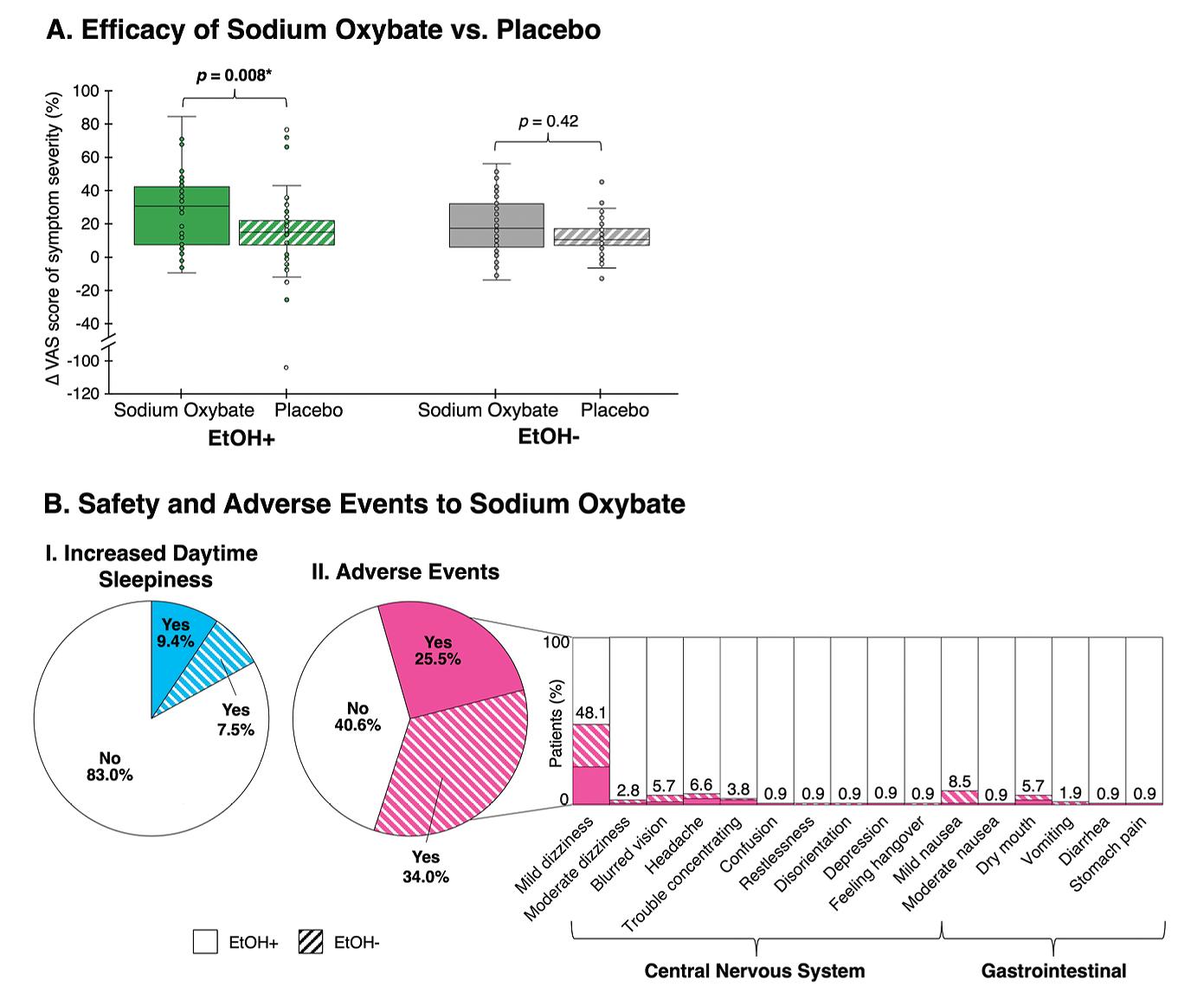
The results, published in Annals of Neurology, demonstrated that sodium oxybate significantly reduced symptoms in alcohol-responsive laryngeal dystonia patients within 40 minutes, with effects lasting up to five hours. The average efficacy of the drug was 41 percent of voice improvement in patients with alcohol-responsive laryngeal dystonia.
“Due to the formulation's effects lasting four to five hours, patients may take another dose after that time frame, if needed. Sodium oxybate may be consumed on an as-needed basis, allowing patients to monitor their own symptoms and actively make treatment decisions together with their clinicians, which is revolutionary,” said Dr. Simonyan.
“Sodium oxybate may be consumed on an as-needed basis, allowing patients to monitor their own symptoms and actively make treatment decisions together with their clinicians, which is revolutionary.”
—Kristina Simonyan, MD, PhD, DrMed
were taken before alcohol consumption, which was the baseline, and then at 15, 30, 45 and 120 minutes after alcohol intake. If the patient had 10 percent or greater symptom improvement, they were categorized as alcohol-responsive, while those with less than 10 percent improvement were classified as having little to no response to alcohol.
According to Dr. Simonyan, conducting this standardized alcohol challenge test was critical, as some patients may experience relief with alcohol but may not show noticeable vocal or tremor improvements, or they may not even know their symptoms respond to alcohol. “Our hypothesis for the trial was
Lena C. O’Flynn, a graduate student in the Speech Hearing Bioscience and Technology Program at Harvard University and co-author of the published study, began working on the project as a research assistant in the Dystonia and Speech Motor Control Lab, where she contributed to patient recruitment and data collection. Now, as part of her PhD program, she is focused on data analysis and has described seeing the paper published as a fullcircle moment.
“What’s been most incredible is witnessing the tangible impact our research has had on patients,” said O’Flynn. “I’ve seen patients regain their voices after being unable to communicate effectively, often for decades. At the end of the day, that’s what truly motivates us in this work.”
(A) Efficacy of sodium oxybate versus placebo in patients with alcoholresponsive (EtOH+) and alcohol-nonresponsive (EtOH—) laryngeal dystonia (LD). The asterisk indicates the statistically significant difference between sodium oxybate and placebo based on the change in the visual analog scale (ΔVAS) score of voice symptom severity.
(B) Safety and adverse events of sodium oxybate in EtOH+ and EtOH—patients, including (I) increased daytime sleepiness and (II) other side effects
What’s next?
According to Dr. Simonyan, the team works to advance sodium oxybate to a Phase 3 clinical trial, a necessary step for the ultimate FDA approval. Currently, no FDAapproved drugs exist for laryngeal dystonia or any other form of dystonia.
Clinicians occasionally prescribe medications such as propranolol, which may help in some cases, but treatment largely relies on trial and error. Among other forms of dystonia, voice-related symptoms present a unique challenge—not only in dystonia but also in Parkinson’s disease and other neurological conditions. Improving voice function remains particularly difficult, even when other motor symptoms respond to treatment.
Sodium oxybate is not a cure, but it treats symptoms when taken. The key difference between sodium oxybate and BoNT is that sodium oxybate, by modulating brain activity that is abnormal in patients with laryngeal dystonia, directly affects dystonia pathophysiology. Phillip C. Song, MD, Associate Professor
of Otolaryngology–Head and Neck Surgery at Harvard Medical School and Director of the Laryngology Division at Mass Eye and Ear, who played a critical role in drug administration to study participants, emphasized that the study’s most important finding is not just that the medication reduces voice symptoms, but that the brain changes associated with treatment are measurable. He explained, “The study provides significant insight into the pathogenesis of focal dystonia and uncovers how the brain coordinates voice and speech production.”
Several patients who participated in the study have since followed up with their clinicians, who have prescribed them sodium oxybate as part of their ongoing treatment.
“We are thrilled about this first successful clinical trial of a novel oral drug for patients with laryngeal dystonia and eager to move forward to the next phase,” emphasized Dr. Simonyan. n
PM2.5 and Head and Neck Cancer: The Unexplored Link
A collaborative first-of-its-kind study correlates higher levels of pollutant particulate matter to higher occurrences of head and neck aerodigestive cancer.
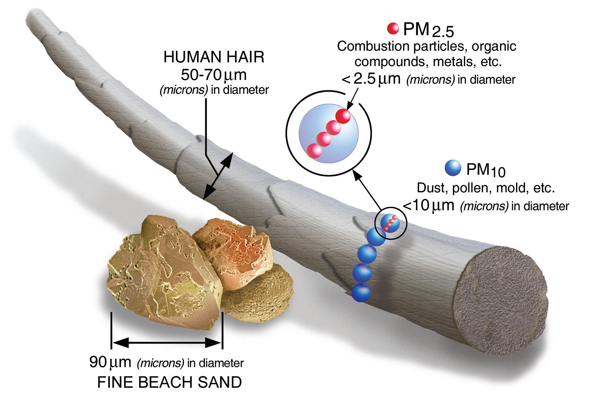
According to the World Health Organization (WHO), air pollution occurs when chemical, physical or biological agents alter the natural composition of the atmosphere in indoor or outdoor environments. In 2013, the International Agency for Research on Cancer, an intergovernmental agency which is part of WHO, classified air pollution—specifically particulate matter smaller than 2.5 microns in diameter (PM2.5)— as a causal agent (Group 1 carcinogen) for lung cancer.
Similarly, tobacco smoke is a well-established carcinogen for both lung cancer and head and neck cancers. According to the Centers for Disease Control and Prevention (CDC), cigarette smoking and secondhand smoke exposure account for nearly nine out of 10 lung cancer deaths, and smoking is also a leading risk factor for cancers of the head and neck.
While significant research has explored the effects of climate variables, such as air pollution and heat, on lower respiratory diseases, there has been limited research on how this affects upper respiratory diseases
and specifically otolaryngology patients. Given the established link between air pollution and lung cancer, as well as the known role of smoking in both lung and head and neck cancers, a research team led by Stella Lee, MD, Associate Professor of Otolaryngology–Head and Neck Surgery at Harvard Medical School and Director of the Brigham Sinus Center at Brigham and Women’s Hospital, sought to investigate the association between fine particulate matter exposure and the incidence of head and neck cancer.
“As a rhinologist, I can’t help but be interested in the air we breathe and its components,” said Dr. Lee. “Pollutants like PM2.5, volatile organic compounds and other common indoor contaminants often go unnoticed but may have a detrimental impact on health.”
Dr. Lee added, “In this collaborative investigation, we hypothesized that tissues in the head and neck may be especially susceptible to harm from air pollution, as they come into direct contact with airborne pollutants.”
[ continued p. 10 ]
Understanding particulate matter
The United States Environmental Protection Agency (EPA) explains that PM2.5 consists of hundreds of different chemicals from both indoor and outdoor sources, including construction sites, unpaved roads, fields, smokestacks, fires, certain cleaning and combustion activities, burning candles, fireplaces, unvented space heaters, kerosene heaters and biological contaminants such as mold. Analyzing PM2.5 is particularly important because its tiny particles can bypass the nose and throat’s natural filtration system, reaching deep into the upper airways.
For more than a decade, Dr. Lee has studied the impact of environmental pollution on upper airway diseases. Before joining the faculty at Brigham and
[“After years devoted to uncovering the relationship between PM2.5 and rhinology, the connection to cancer then emerged naturally, considering chronic inflammation as a broader disease paradigm,” Dr. Mady added.
To examine the role of air pollution in head and neck cancer, Dr. Lee and her team conducted an epidemiological cohort analysis using data from the Surveillance, Epidemiology, and End Results (SEER) Program of the National Cancer Institute database from 2002 to 2012. This data was linked with publicly available county-level air pollution data (specifically PM2.5) from the EPA for all 48 contiguous U.S. states.
“As a rhinologist, I can’t help but be interested in the air we breathe and its components. Pollutants like PM2.5, volatile organic compounds and other common indoor contaminants often go unnoticed but may have a detrimental impact on health.”
– Stella Lee, MD
Women’s Hospital, she served as a clinician-researcher at the University of Pittsburgh, where she and her collaborator, Leila J. Mady, MD, PhD, MPH, now an Assistant Professor of Otolaryngology-Head and Neck Surgery at Johns Hopkins School of Medicine, established a link between air pollution and chronic sinus disease.
“At the time in Pittsburgh, we saw many patients with upper respiratory and allergy symptoms who underwent extensive evaluation but with negative testing. Similarly, it became apparent that patients with chronic sinus disease with environmental exposures still suffered from poorly controlled inflammation despite complete endoscopic sinus surgery and good adherence to topical therapy,” explained Dr. Mady. In response, the duo analyzed public EPA PM2.5 data to better understand the relationship between air pollution and patient outcomes including the need for repeat surgery. For example, for every unit increase of PM2.5 exposure there was almost a two-fold increase in risk of requiring further sinus surgery.
Additionally, this study is particularly unique because it links SEER, one of the few large epidemiologic cancer databases, with exposure risk factors that are not traditionally available in such databases. By integrating data from the CDC’s Behavioral Risk Factor Surveillance System, the study captures behavioral factors such as smoking cigarettes and drinking alcohol, which are often missing from cancer research relying solely on big data.
“The unique opportunity to couple county-level data with patient data made this study robust. We were able to exclude patients who actively smoked or consumed alcohol, allowing for a more accurate analysis of whether air pollution contributed to cancer diagnoses,” Dr. Lee shared.
The results, published in Scientific Reports, included a significant connection between PM2.5 levels and head and neck cancer incidence, with the strongest link seen when looking at a five-year lag between exposure and cancer development.
The team analyzed 600 counties across 11 states from 2002 to 2012. The findings demonstrated that certain types of aerodigestive head and neck cancer, such as oral cavity and laryngeal cancers, were most common.
Interestingly, the data revealed that viral-mediated cancers, such as Epstein-Barr virus-related nasopharyngeal and human papillomavirus-related oropharyngeal cancers, were not significantly linked to particulate matter air pollution, unlike other cancer subtypes. This suggests a different pathway for cancer development, particularly for non-viral cancers, as
viral-mediated types were no longer linked to air pollution after adjusting for variables.
“Conducting this study wouldn’t have been possible without collaboration across multiple institutions, including my colleagues from Wayne State University School of Medicine, who helped lead the study, my peers in the Department of Otolaryngology-Head and Neck Surgery at Harvard Medical School, and our exceptional biostatisticians from the Harvard School of Public Health and the Brigham Center for Surgery and Public Health,” said Dr. Lee. “Their expertise enabled us to develop the analytic methods needed to process and analyze large datasets like SEER. While we developed the concepts, their collaboration allowed us to fully explore and accurately interpret the data, ultimately providing key insights into air pollution exposure and its harmful effects on patients.”
Next steps
Evidence linking geographic areas with high PM2.5 levels to increased rates of head and neck cancer, with a five-year lag time, raises important considerations for prevention and screening.
“For example, if wildfire exposure increases cancer risk, those affected may be most vulnerable within five to ten years. Identifying this window enables targeted screening, replacing a one-size-fits-all approach. Datadriven insights can refine screening timelines and strengthen prevention efforts,” explained Dr. Lee.
Looking ahead, Dr. Lee and her team hope this study serves as a foundation for developing wearable devices to monitor individual PM2.5 exposure levels and associate them with disease outcomes and severity at the patient level. While general air quality data is available, there is no personalized profile that tracks an individual’s PM2.5 exposure, identifies sources and assesses risk levels.
These findings not only highlight the need for personalized exposure monitoring and continued research but also align with broader efforts to reduce air pollution through sustainability and advocacy.
Regan Bergmark, MD, MPH, Assistant Professor of Otolaryngology–Head and Neck Surgery at Harvard Medical School and sinus and endoscopic skull base surgeon at Brigham and Women’s Hospital, chairs the Harvard Medical School Otolaryngology–Head and Neck Surgery Sustainability Committee that has been focused on reducing emissions and waste associated with patient care.
The committee has been tracking emissions at Mass Eye and Ear’s Boston Main Campus for nearly four years and developing targeted sustainability initiatives, including partnering with anesthesia colleagues to reduce the use of anesthetic gases that have environmental impact. Other initiatives include surgical tray streamlining projects at Brigham and Women’s Hospital and Mass Eye and Ear, and to include sustainability in the otolaryngology core curriculum.
“As a department, we continue to lead initiatives that cultivate a culture of environmental responsibility and sustainability through advocacy, education, research, public service, operations and clinical care,” shared Mark A. Varvares, MD, FACS, William W. Montgomery and John W. Merriam Professor and Chair of the Department of Otolaryngology–Head and Neck Surgery at Harvard Medical School. “Our faculty’s work continues to uncover overlooked determinants of health, with Dr. Lee’s study serving as a promising first step.” n
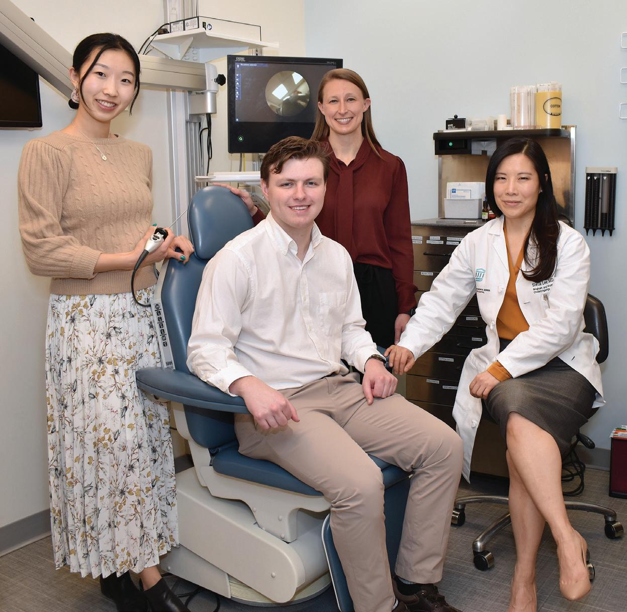
Stella Lee, MD, (seated far right) with members of her laboratory, from left to right: Xiaofei (Sophie) Yu; William B. Thorley; and Margaret (Megan) Mitchell MD, MS-HPEd.
A groundbreaking artificial intelligence model, DeepMerkel, is setting a new standard for personalized, time-dependent survival predictions in Merkel cell carcinoma.
Merkel cell carcinoma (MCC) is a rare, aggressive skin cancer driven by the Merkel cell polyomavirus (MCPyV) or UV-induced DNA damage, with risk factors including advanced age, chronic sun exposure, fair skin and immunosuppression. According to the American Cancer Society, MCC is significantly less common than other skin cancers such as squamous and basal cell carcinoma. Unfortunately, MCC is frequently misdiagnosed as benign conditions like cysts or lipomas, since its rarity contributes to a lack of awareness, often leading to delayed diagnosis. MCC is also more likely to spread, making treatment more challenging. The fatality rate of MCC is more than double that of melanoma, and poor outcomes are often attributed to its tendency to appear in elderly patients at advanced stages.
MCC prognosis is currently best assessed using the American Joint Cancer Committee (AJCC) staging system, which primarily considers tumor size, nodal involvement and spread. However, this system has limitations, including its reliance on overall survival endpoints without accounting for competing risks like death from other causes. For example, since most MCC diagnoses occur in individuals over the age of 70, all-cause mortality may overestimate MCC-specific death rates.
oncology to develop a novel, adaptive, prognostic tool that enhances personalized risk stratification for MCC.
“We knew we had to do more for patients with this disease. Albeit rare, MCC is highly invasive,” said Dr. Shalhout. “If we could create a personalized AI tool to accurately predict a patient's survival with MCC, it would be a significant breakthrough for clinicians who treat this aggressive cancer.”
Together, in collaboration with an international consortium of clinicians, MCC experts and data scientists, Drs. Shalhout and Andrew developed DeepMerkel, a novel web-based prognostic tool leveraging a hybrid AI deep learning framework.
Discovering DeepMerkel
Accurate prognostication is crucial to guiding optimal clinical management in skin cancer, particularly in aggressive forms like MCC. DeepMerkel, a web-based, time-dependent survival prediction tool, was developed using a deep learning feature selection method and a modified XGBoost framework, which is a machine
“The goal of our study was to develop a hybrid machine learning model specifically for MCC, test it using real-world patient data, and create an online tool to assist clinicians in making more precise, personalized treatment decisions.”
–
[To address these constraints and improve prognostication, an artificial intelligence (AI)–driven, personalized diseasespecific survival model is needed to better support complex clinical decision-making. Sophia Z. Shalhout, PhD, Assistant Professor of Otolaryngology–Head and Neck Surgery at Harvard Medical School and Principal Investigator in the Mike Toth Head and Neck Cancer Research Center at Mass Eye and Ear, partnered with Tom W. Andrew, MD, MSc, MRCS, an Academic Plastic and Reconstructive Surgeon at Newcastle University, United Kingdom, and Visiting Research Fellow in Dr. Shalhout’s lab, to leverage their expertise in AI and
Sophia Z. Shalhout, PhD
learning algorithm that builds ensembles of decision trees to optimize predictive accuracy. This tool offers a significant advancement by providing personalized and accurate survival predictions from readily available clinical information. Dr. Shalhout noted that this approach helps to maximize limited but readily available clinical data, capturing complex disease biology and providing personalized risk predictions to enhance patient care.
[ continued p. 14 ]
“The goal of our study was to develop a hybrid machine learning model specifically for MCC, test it using real-world patient data, and create an online tool to assist clinicians in making more precise, personalized treatment decisions,” added Dr. Shalhout. “We are thrilled to have delivered on every front.”
The study leveraged data from the National Institutes of Health Surveillance, Epidemiology, and End Results (SEER) database, which included 11,342 patients with confirmed MCC. Given that only 3,000 cases are diagnosed annually in the United States, this dataset provides a robust foundation for analysis.
To enhance predictive accuracy, a hybrid AI model that combined deep learning and machine learning, including TabNet and XGBoost, was employed for optimal predictions and performed better than other AI model architectures in comparison. A standard AI approach was followed where 80 percent of the U.S. data
was used for training, and 20 percent was used as a holdout test set, ensuring the model was tested on unseen data. The model was then further validated on an independent National Health Service U.K. patient cohort of 121 MCC patients to confirm its generalizability.
Explainable AI techniques were applied to uncover key mortality risk factors specific to MCC, providing transparency into the new model’s decision-making process. The goal with explainable AI is to one day enable the clinicians to understand, trust and act on the AI’s predictions. This is particularly important in rare and aggressive cancers like MCC, where insights into individualized risk can guide more precise treatment and follow-up strategies.
A Shapley Additive Explanations (SHAP) analysis pinpointed the strongest predictors of survival, including tumor spread, metastasis, tumor size, age, sex, ethnicity, marital status and income. For example, the number of
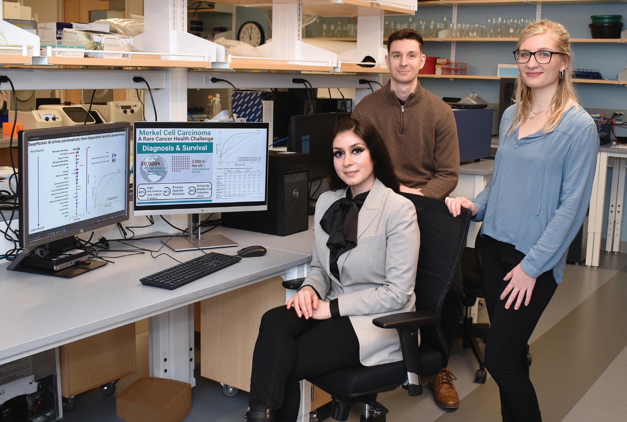
Sophia Z. Shalhout, PhD, (seated) with members of her lab from left to right: Ryan Byrne, MS, and Angela Fragano. Inset photo: Tom Andrew, MD, MSc, MRCS.
lymph nodes evaluated correlated with better survival rates, likely due to early detection and intervention. Additionally, an unexpected but significant finding was that married patients had improved survival, possibly because spouses encourage early medical attention for suspicious skin lesions.
Results, published in npj Digital Medicine, demonstrated that DeepMerkel outperformed other machine learning models in both test cohorts: Decision-Tree Classifier, Random Forest Classifier and Logistic Regression. DeepMerkel also achieved a significantly higher Area Under the Receiver Operating Characteristic curve (AUROC) score in the U.S. test cohort and U.K. test cohort when compared to the eighth edition of the AJCC Staging system.
DeepMerkel has been developed into a web-based tool that provides survival predictions for patients diagnosed with MCC. The platform offers both an average survival estimate based on disease stage and a more personalized prediction that incorporates individual patient features, tumor characteristics, and the extent of tumor spread. This dual approach allows clinicians to go beyond broad staging categories and understand how specific factors influence an individual patient’s prognosis. By offering transparent, tailored risk assessments, DeepMerkel can support shared decision-making, help identify patients who may benefit from more aggressive treatment or closer follow-up, and ultimately improve personalized care planning.
Clinical setting implementation
The web-based platform is currently available for research purposes only. “The next step is to move from retrospective to prospective testing,” emphasized Dr. Shalhout.
Since DeepMerkel is trained on data from patients who have been in the system for years, continuing the model's growth and achieving positive clinical results will require testing on patients with active MCC.
At Mass Eye and Ear, Kevin Emerick, MD, Associate Professor of Otolaryngology–Head and Neck Surgery at Harvard Medical School, is the Co-Director of the Non-Melanoma Skin Cancer Multidisciplinary Clinic and Program. Dr. Emerick and his team see and treat patients with this rare and aggressive cancer. Dr. Shalhout’s goal, upon obtaining Institutional Review
Board (IRB) approval for the study, is to work with Dr. Emerick to begin testing the platform prospectively at Mass Eye and Ear to assess the model’s robustness in the real-world setting.
During in-office visits, the study would require entry of the consented patient’s important data points that the model found correlate with survival, into the second-generation version of the AI tool to predict the patient’s MCC-specific survival. Notably, these predications would not be shared with the patient or alter intervention. At this stage, the results would be utilized to optimize the model and inform next steps for potential clinical integration if acceptable accuracy is demonstrated. For example, in future applications, this model may support more informed clinical decisionmaking for patients identified as high risk. It may help guide treatment intensity such as considering adjuvant therapies or closer surveillance. For patients identified as lower risk, this tool may help them avoid unnecessary imaging and interventions. “DeepMerkel will need to be tested prospectively and continuously updated with new data, as medicine evolves rapidly, and new treatments may impact survival rates,” shared Dr. Emerick. “Implementing this in the clinic will be a groundbreaking and powerful tool for the future.”
Dr. Shalhout acknowledges that it is too early to determine whether MCC patients should be informed of their predicted survival. A patient advocacy group will eventually be consulted to ensure the model’s output benefits patients or remains a tool for clinician use only.
“It’s clear that predicting patient outcomes with precision is vital for managing aggressive skin cancers like MCC, where treatment decisions can be complex and life-altering. With AI, we have the potential to support more precise and informed prognostication,” shared Dr. Shalhout.
She added, “This international collaboration underscores the potential of AI-driven solutions in addressing the challenges of rare and deadly diseases, paving the way for precision medicine breakthroughs on a global scale. The team aims to make the tool widely available online and validate prospectively.”
“Our advancements in DeepMerkel enable us to deliver personalized survival predictions, potentially helping medical teams identify the most effective treatment options for each patient, which is remarkable,” Dr. Andrew emphasized. n
Jennifer Kim, MD, 2000
Jennifer Kim, MD, has always enjoyed working with her hands. Growing up, she had a passion for drawing, sculpting and painting—but, raised in a family of physicians, including her father, she knew early on that she wanted to follow in their footsteps.
In medical school, Dr. Kim discovered a way to merge her artistic talents with her medical expertise through facial plastic and reconstructive surgery. She subsequently joined the Harvard Combined Residency Program in Otolaryngology–Head and Neck Surgery, confident that she had found not just her subspecialty but her true calling.
Now, nearly 25 years after completing her residency, she returns to her alma mater as Director of the Facial Plastic and Reconstructive Surgery Division at Mass Eye and Ear.
“Returning to Boston feels like coming home. I’m reunited with both my personal and professional families, working once again with colleagues from my residency and fellowship,” said Dr. Kim. “It’s a true privilege to lead alongside the mentors who shaped my career.”
The last 25 years…
During her residency, Dr. Kim trained under trailblazers in the field, including William W. Montgomery, MD, Mack L. Cheney, MD, and Mark A. Varvares, MD, FACS, where she had the opportunity to develop concentrated skills in both microvascular surgery and aesthetics. “As a specialized hospital, Mass Eye and Ear has the tools to teach trainees incredibly unique surgeries as we see some of the most rare and complex patients,” said Dr. Kim.
She added, “That’s why I stayed at Mass Eye and Ear for my fellowship in Facial Plastic and Reconstructive Surgery—to be a mentee to the most world-renowned head and neck surgeons. If you think about the major leaders in otolaryngology, the majority of them have a tie to our Department of Otolaryngology–Head and Neck Surgery in some way, shape or form.”
As a trainee, Dr. Kim gained extensive experience in facial reanimation, microvascular reconstruction and cosmetic surgery. Her mentors, Drs. Montgomery, Cheney and Varvares, encouraged a hands-on approach to learning. Dr. Kim emphasized that the trust her mentors placed in her
“While Mass Eye and Ear already has an excellent reputation, I’m committed to maintaining that standard and making it a sought-after destination for residents and fellows in facial plastics training.”
—Jennifer Kim, MD
strengthened her confidence and profoundly shaped the teaching style she has carried throughout her career.
After completing her fellowship, Dr. Kim began her academic career in the Department of Otorhinolaryngology at the University of Michigan (UM) in Ann Arbor. Her exceptional contributions led to her promotion to professor in 2021.
A dedicated surgical educator, Dr. Kim has earned widespread recognition for her commitment to teaching and mentorship. She has received numerous accolades from trainees in UM’s Department of Otorhinolaryngology, including the prestigious Frank Ritter “Teacher of the Year” Award in 2004, 2009 and 2014. She was also honored with the Facial Plastic Teaching Award in 2018 and the Resident Mentorship Award in 2022.
“Learning from my mentors has been invaluable,” Dr. Kim reflected. “Their support and guidance shaped my approach to education and patient care, and I’m committed to passing that same level of dedication on to the next generation of surgeons.”
Dr. Kim’s passion for furthering education is deeply rooted in her commitment to humanitarian work, which serves as one of her greatest motivators. “Dr. Cheney introduced me to mission work. In fact, the reason I got started was one year he couldn’t attend his annual trip through Medical Missions for Children, and he asked if I would go in his place. The rest is history,” Dr. Kim said.
Since 2007, Dr. Kim has been greatly committed to medical missions, traveling three to four weeks annually to developing countries including but not limited to Guatemala, Peru, Vietnam, Ecuador and Haiti.
In regions lacking local expertise, Dr. Kim focuses on performing as many surgeries as possible, particularly concentrating on microtia cases. However, in countries like Vietnam, for example, where local surgeons are more available, she prioritizes training to encourage long-term sustainability by providing clinicians with critical surgical education.
On a recent trip to Vietnam, Dr. Kim, alongside her colleague Allen L. Feng, MD, and resident Hoang Bui-Nguyen, MD, PhD, led comprehensive training programs that included lectures, anatomy labs, cadaver dissections and surgical workshops. During these courses, local surgeons and trainees practice alongside Dr. Kim and the team, developing expertise in advanced procedures such as microvascular and microtia surgeries. This approach ensures that local surgeons can continue providing care long after the mission ends.
During her time at UM, Dr. Kim served as a Co-Director of the Facial Plastic and Reconstructive Fellowship and frequently brought trainees on mission trips to provide hands-on experience in challenging settings. In her new role at Mass Eye and Ear, she remains committed to these trips and plans to continue bringing residents and fellows, expanding access to care and setting an example for the importance of humanitarian work.
Plans for the Facial Plastic and Reconstructive Surgery Division
When offered to lead the Facial Plastic and Reconstructive Surgery Division, Dr. Kim embraced the challenge to direct a renowned division at one of the nation’s top otolaryngology hospitals. “This division is already among the top in the world;
my goal is not to establish its reputation, but to build on it and make it even stronger.”
Dr. Kim is eager to continue growing the Non-Melanoma Skin Cancer Multidisciplinary Clinic and Program, led by Kevin S. Emerick, MD, and Jessica L. Fewkes, MD, as well as the Mohs Surgical Program, in collaboration with Dr. Fewkes and Sameer G. Gupta, MD, MPhil. She explained that these programs offer the unique advantage of having multidisciplinary experts together in one location, creating a one-stop shop for patients seeking Mohs surgery, Mohs reconstruction, and laser treatment for scarring. With skin cancer primarily affecting an elderly population, this benefit is especially important, ensuring a positive patient experience and easy access to renowned care.
Additionally, the Microtia and Aural Atresia Center, though niche, plays a critical role for patients. Dr. Kim’s goal is to establish it as a center of excellence and a destination for microtia care, supported by the well-equipped and talented surgeons already within the department.
Dr. Kim also plans to continue developing the Facial Nerve Center. When she first arrived at UM, there was no focus on facial nerve disorders, so she established and grew its Facial Nerve Clinic. She now hopes to preserve and expand the foundation already in place at Mass Eye and Ear.
Finally, Dr. Kim plans to host annual meetings or courses on facial nerve reconstruction and other reconstructive procedures, further enhancing the center’s reputation and attracting local, regional and national attendees.
“While Mass Eye and Ear already has an excellent reputation, I’m committed to maintaining that standard and making it a sought-after destination for residents and fellows in facial plastics training,” emphasized Dr. Kim. n
Jennifer Kim, MD, and former fellows carve a rib during a microtia-focused mission trip in Guatemala.
Atchinson Family Advancing head and neck cancer research
In 2019, Michelle and Bob Atchinson established the Mike Toth Head and Neck Cancer Research Center in honor of their late friend, Mike Toth, to increase survival rates from head and neck cancer and improve quality of life after treatment.
Since then, the Toth Center has been at the forefront of developing innovative methods to diagnose, monitor, and treat head and neck cancer. There is likely no other place in the country with such a unique concentration of experts across diverse scientific fields—physics, biochemistry, biology, computer science, biomedical engineering, and clinical surgical practice—all dedicated to addressing head and neck cancer.
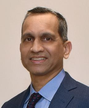
Anand T.N. Kumar, PhD, was appointed Director of the Toth Center in 2024. His pioneering work addresses a critical challenge in cancer surgery: accurately distinguishing between tumor tissue and healthy tissue to ensure 100 percent clean margins while minimizing the removal of normal tissue, which comes at the expense of function. The current standard approach relies on a surgeon’s sight and touch to identify and remove cancerous tissues, which lacks precision.
Previously, Dr. Kumar’s team made a groundbreaking advance in demonstrating the potential of fluorescence lifetime imaging in animal models. This innovative technique uses short laser pulses, high-speed cameras and an FDA-approved dye called indocyanine green to differentiate cancerous tissue from healthy tissue. By measuring the dye’s fluorescence duration within tissues, the technique accurately identified tumors. Dr. Kumar then tested it in specimens from over 70 patients enrolled at Mass Eye and Ear, Massachusetts General Hospital and other institutions in the United States, United Kingdom and Europe, achieving 97 percent accuracy for tumor detection.
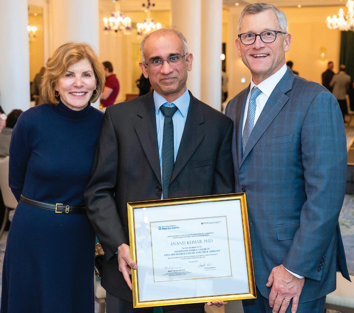
As champions of the Toth Center, Michelle and Bob Atchinson were inspired to sustain its profound leadership in the decades ahead, driving them to endow the Atchinson Family Chair in Otolaryngology–Head and Neck Surgery. Dr. Kumar was named the inaugural incumbent.
“Giving surgeons the capability to nearly guarantee complete tumor removal would be remarkable and exemplifies why we founded the Toth Center—to provide the brightest head and
neck cancer researchers with the tools they need to achieve tangible life-changing results,” said Bob Atchinson, who serves as the Board Chair at Mass Eye and Ear. “Dr. Kumar, among the other outstanding researchers in the Toth Center, are doing just that.”
According to Dr. Kumar, these initial successes serve as proof of concept, and the next major step is developing this technology for live imaging during surgery.
“The studies we will perform over the next few months will lay the foundation for decades of work, with a goal for the technique to be optimized, implemented in various forms, and eventually integrated into clinical practice for all types of solid cancers,” said Dr. Kumar who also serves as an Associate Professor in Otolaryngology–Head and Neck Surgery at Harvard Medical School. “I am confident that this will become standard of care one day, and Mass Eye and Ear and the Atchinson Family would have played a crucial part in this.” n
From left to right: Michelle Atchinson, Anand T.N. Kumar, PhD, and Bob Atchinson at the Atchinson Family Chair in Otolaryngology–Head and Neck Surgery celebration.
ALUMNI GIVING SOCIETY
The Alumni Giving Society of the Department of Otolaryngology–Head and Neck Surgery at Harvard Medical School
The Department of Otolaryngology–Head and Neck Surgery at Mass Eye and Ear/Harvard Medical School established the Alumni Giving Society in 2015 to recognize faculty and alumni who contribute $1,000 or more during the fiscal year (October 1–September 30). Participation not only strengthens your connection to the department but also provides critical resources, training tools and mentorship for the next generation of physicians, ensuring they are well-prepared to become future leaders in otolaryngology–head and neck surgery
Our alumni understand firsthand how essential philanthropic support is in driving excellence and innovation in education, research and patient care. In fiscal year 2024, we proudly recognized 81 dedicated members whose generosity helped advance our department’s goals and institutional mission.
We are thrilled to share that exciting changes are coming to the Alumni Giving Society! As we look ahead, we want to ensure that this community is as meaningful and impactful as possible—and we need your help to make it great. Your input and support will shape the future of the society, strengthening its role in advancing our field.
If you’re not already a member, now is the perfect time to join. Your gift can be designated in a way that is most meaningful to you. Together, we can continue to build a vibrant, engaged alumni community that supports the next generation of physicians, researchers and patients.
Stay tuned for more details and thank you for being a valued part of our alumni network!
To learn more, please contact Elizabeth Vitello in the Development Office at evitello@meei.harvard.edu
sCurrent Alumni Giving Society members for fiscal year 2024, from October 1, 2023, to September 30, 2024, are listed below. With your gift of $1,000 or more, you will be included in the 2025 Alumni Giving Society.
CHAMPION
(Gifts of $25,000 and more)
Jeffrey P. Harris, MD, PhD
Ralph B. Metson, MD
Eugene N. Myers, MD, FACS, FRCS Edin (Hon)
Laxmeesh M. Nayak, MD
Michael M. Paparella, MD
Herbert Silverstein, MD, FACS
VISIONARY
(Gifts of $10,000 - $24,999)
Michael S. Cohen, MD
Paul M. Konowitz, MD, FACS
David A. Lewis, MD
Michael J. McKenna, MD
Joseph B. Nadol, Jr., MD
Michael B. Rho, MD, FACS
Jeremy D. Richmon, MD
Josef Shargorodsky, MD
Mark A. Varvares, MD, FACS
INNOVATOR
(Gifts of $5,000 - $9,999)
Fred G. Arrigg, Jr., MD
Carlos Ayala, MD
Daryl G. Colden, MD, FACS
Richard E. Gliklich, MD
Michael G. Moore, MD
Jay T. Rubinstein, MD, PhD
Joshua B. Silverman, MD
PIONEER
(Gifts of $2,500 - $4,999)
Barry J. Benjamin, MD
Samir M. Bhatt, MD
Neil Bhattacharyya, MD
Bertrand Delgutte, PhD*
Daniel G. Deschler, MD, FACS
Wade W. Han, MD
Jeffrey Hoffman, MD
Elliott D. Kozin, MD
John B. Lazor, MD, MBA, FACS
Leila A. Mankarious, MD
Hideko H. Nakajima, MD, PhD
Greg G. Ota, MD
Alicia M. Quesnel, MD
Mark F. Rounds, MD
Phillip C. Song, MD
Department of Otolaryngolog y Head and Neck Surger y
FRIEND
(Gifts of $1,000 - $2,499)
Dunia E. Abdul-Aziz, MD
John F. Ansley, MD
Babak Azizzadeh, MD
Wayne Barber, MD, FACS
Benjamin S. Bleier, MD, FACS and Chia A. Haddad, MD
David M. Bowling, MD
Nicolas Y. BuSaba, MD
Michael J. Cunningham, MD, FACS
Matthew Dedmon, MD, PhD
Ruth Eatock, PhD
Kevin S. Emerick, MD
Jordan T. Glicksman, MD, MPH
Stacey T. Gray, MD
Warren L. Griffin, Jr., MD
Paul E. Hammerschlag, MD
Eric H. Holbrook, MD
Craig A. Jones, MD
David H. Jung, MD, PhD, FACS
Donald G. Keamy, Jr., MD, MPH
Ely A. Kirschner, MD
Thomas R. Klein, MD
Sharon G. Kujawa, PhD
Derrick T. Lin, MD, FACS
Giant C. Lin, MD
William W. McClerkin, MD
Cliff A. Megerian, MD
Steven A. Metzger, MD
David E. Nash, MD
Joseph H. Oyer, MD
Didier L. Peron, MD
Dennis S. Poe, MD
Sunil Puria, PhD
Steven D. Rauch, MD
Edward J. Reardon, MD
Vicente A. Resto, MD, PhD
John J. Rosowski, PhD
George A. Scangas, MD
Andrew R. Scott, MD
Earl F. Singleton, MD
Joseph H. Traxler, MD
Feodor Ung, MD
Robert L. Witt, MD
Richard J. Wong, MD
Michael Zoller, MD
*Deceased
HIGHLIGHTS s
News from every corner of the Department of Otolaryngology–Head and Neck Surgery at Harvard Medical School.
New Faculty
Introducing the newest clinicians, clinician-scientists, researchers and educators in the Department of Otolaryngology–Head and Neck Surgery.
Kameron Clayton, PhD, is an investigator in the Polley lab within the Eaton-Peabody Laboratories at Mass Eye and Ear. He received his PhD in the Speech and Hearing Biotechnology program at Harvard University before completing his postdoctoral training under the mentorship of Daniel B. Polley, PhD, at Mass Eye and Ear. Dr. Clayton’s research seeks to understand how mysterious perceptual disorders, like hyperacusis and tinnitus, emerge from compensatory plasticity in neural circuits throughout the auditory central pathway following hearing loss.
Kaiser San Bernardino Service Area. Dr. Paulson sees patients with general ear, nose, and throat concerns. He also has clinical interests in thyroid surgery, parotid gland surgery and the surgical treatment of nasal obstruction.
Awards, Grants and Honors
Margaret Kenna, MD, MPH, delivered the keynote Marion Downs Lecture at the American Academy of Audiology Conference. Her presentation was titled “Genetics of Pediatric Hearing Loss in the Age of Precision Medicine: From Newborn Screen to Novel Gene.”
At the 2024 Fall New England Otolaryngology Society Meeting (NEOS) PGY-4 residents Allen Zhou, MD, and Kathyrn Marcus, MD, both won second place for their presentations. Dr. Zhou’s was titled “Improving Language Access for Otolaryngology Patients” and Dr. Marcus' was titled “Ears of Experience: Evolution of Microtia Management.”
Eric P. Paulson, MD, is a board-certified otolaryngologist who practices adult and pediatric otolaryngology at the Mass Eye and Ear, Stoneham location. For the past 19 years, Dr. Paulson has practiced with Kaiser Permanente in California.
Over the last four years, he has served as Chief of Head and Neck Surgery for the
Recent Promotions
David H. Jung, MD, PhD, FACS, Associate Professor of Otolaryngology–Head and Neck Surgery
Hae-Young Kim, DrPH, Assistant Professor of Otolaryngology–Head and Neck Surgery
Stella Lee, MD, Associate Professor of Otolaryngology–Head and Neck Surgery
David A. Shaye, MD, MPH, FACS, Associate Professor of Otolaryngology Head and Neck Surgery
Phillip C. Song, MD, Associate Professor of Otolaryngology–Head and Neck Surgery
Anne E. Takesian, PhD, received the prestigious Presidential Early Career Award for Scientists and Engineers (PECASE). This honor, awarded by President Biden to only 400 scientists and engineers nationwide, is the highest recognition given by the U.S. government to outstanding early-career professionals in these fields.
Sunil Puria, PhD, was named specialty chief editor of Frontiers in Audiology and Otology, Auditory Science. Specialty chief editors, in collaboration with Frontiers, define their section’s scope, recruit editors, and lead, supervise, and support their editorial board.
Barbara S. Herrmann, PhD, received the 2024 Frank R. Kleffner Lifetime Clinical Career Award from the American SpeechLanguage-Hearing Foundation. This award honors an individual for exceptional contributions to communication sciences and disorders spanning at least 20 years.
The National Institute of Biomedical Imaging and Bioengineering Bioimaging awarded Anand T.N. Kumar, PhD, a fouryear, $2.16 million R01 grant to develop a new intraoperative cancer surgical guidance system based on cutting edge fluorescence lifetime imaging technology. The technology developed in this project will initially be tested in patients undergoing surgery of various bone and soft tissue sarcomas at Massachusetts General Hospital but will also be widely applicable to other cancers including head and neck cancers.
Thomas L. Carroll, MD, has been named an Honorary Lifetime Member of the Association of Phono Surgeons of India.
Ernest D. Gomez, MD, was appointed chair-elect of the Simulation Education Committee of the American Academy of Otolaryngology–Head and Neck Surgery.
Richard F. Lewis, MD, was named Principal Investigator of a five-year grant from the National Institute on Deafness and Other Communication Disorders titled “Vestibular Dysfunction in Patients with Sporadic Vestibular Schwannomas and with Neurofibromatosis Type 2–Related Schwannomatosis.” The research team will investigate how vestibular schwannomas
National Academy of Sciences Elected Member
Jeffrey R. Holt, PhD, Professor of Otolaryngology–Head and Neck Surgery and Neurology at Harvard Medical School, was elected as a member of the National Academy of Sciences (NAS) in 2025.
The NAS, established by President Abraham Lincoln in 1863, is a private, nonprofit institution that provides independent, objective advice to the nation on matters related to science and technology. Election to the NAS is considered one of the highest honors in science. Dr. Holt is one of 120 scientists nationwide elected this year in recognition of their distinguished achievements in original research. He is the first member of the Harvard Medical School Department of Otolaryngology–Head and Neck Surgery ever elected to this prestigious society, which currently includes 2,662 active U.S. members.
and related therapies affect vestibular function by analyzing changes in vestibular signals and noise, associated behavioral responses, and the relationships between vestibular dysfunction, clinical symptoms, and biological or imaging markers.
Daniel B. Polley, PhD, was named Head of the Innovation Board for the nonprofit Tinnitus Quest. The Innovation Board maximizes the impact of its grants program by shaping the research funding framework, connecting research and patient communities, and setting the agenda for the annual research meeting.
Mark A. Varvares, MD, FACS, delivered the annual Morley Binstock Distinguished Visiting Professor Lecture at the University of Toronto and Mount Sinai Hospital. His presentation was titled “Lessons of a Journey from Cancer Surgeon to Cancer Patient.”
Phase III KEYNOTE 689 Clinical Trial
Ravindra Uppaluri, MD, PhD, Associate Professor of Otolaryngology–Head and Neck Surgery at Harvard Medical School, presented the Phase III KEYNOTE 689 clinical trial at the American Association for Cancer Research annual meeting. The talk was titled “Adding Perioperative Pembrolizumab to Standard of Care Improves Outcomes in Patients with Newly Diagnosed Head and Neck Cancer.”
Dr. Uppaluri served as the principal investigator for this practicechanging randomized study of 714 patients with stage 3–4 head and neck squamous cell carcinoma. The trial found that adding perioperative pembrolizumab to standard care significantly reduced recurrence risk by at least 27 percent and increased major pathologic response, especially in patients with PD-L1–positive tumors.
This is the first positive immunotherapy trial to definitively manage head and neck cancers and may lead to the first change in the standard of care in more than two decades.
Daniel J. Lee, MD, and Mass Eye and Ear Neurotology Fellow, Abel David, MD, traveled to Dubai, United Arab Emirates in collaboration with the TarabichiStammberger Ear and Sinus Institute, to lead an advanced surgical workshop on cutting-edge techniques in otologic surgery. The event focused on endoscopic ear surgery, cochlear implant procedures and robotic cochlear implant insertion, and had surgeons from 10 different countries attend.
New Leadership
Benjamin S. Bleier, MD, was named Director of Innovation and Commercialization at Mass Eye and Ear
Marika D. Russell, MD, FACS, was appointed Director of Education for the Division of Thyroid and Parathyroid Surgery at Mass Eye and Ear.
New Society Inductions
David H. Jung, MD, PhD, FACS, was formally inducted into the American Otological Society.
Anne E. Takesian, PhD, received a collaborative Kavli Exploration Award titled “Brain-wide cortical circuits for sensoryguided behavior.” The team will combine cutting-edge techniques in holographic activation, two-photon imaging and computational modeling to determine how sound is encoded within the auditory cortex and signaled to diverse brain regions to guide complex behavioral outcomes.
Matthew R. Naunheim, MD, MBA, became an elected member of the American Laryngological Association.
RESEARCH ADVANCES s
The following are select research advances from the Harvard Medical School Department of Otolaryngology–Head and Neck Surgery.
Basic Research
Dual role of Sox2 in Atoh1 transcription and degradation during hair cell development
Sox2, a stem cell pluripotency gene, stimulates the expression of Atoh1, a transcription factor necessary for hair cell development in the embryo. Sox2 is essential for the development of cochlear hair cells and binds to the Atoh1 3’ enhancer to stimulate Atoh1 expression. Previous work determined that destabilizing Atoh1 is crucial for tightly regulating the development of the cochlea’s sensory hair cells. It was found that deleting Huwe1, the ligase responsible for Atoh1 degradation in the cochlea, led to increased Atoh1 levels and the formation of extra hair cells. Additionally, disrupting the Huwe1-Atoh1 pathway by stabilizing Atoh1 promoted the generation of more hair cells from supporting cells in the developing cochlea.
Albert Edge, PhD, of Mass Eye and Ear, led a study on the regulation of Atoh1. The team discovered a dual role for Sox2 in determining Atoh1 expression levels in the cochlea. Sox2 initiates Atoh1 expression while also limiting Atoh1 protein levels by activating the proteasome pathway. This mechanism of Atoh1 regulation explains the variable phenotypes induced by Sox2 deletion: a lack of hair cells when Sox2 is deleted early in embryogenesis is due to the absence of Sox2’s stimulation of Atoh1 expression, while Sox2 deletion later in development leads to an overproduction of hair cells due to reduced degradation of Atoh1 at the lowered levels of the E3 ubiquitin ligase Huwe1. This contradictory signaling enables Sox2 to direct the fate of inner ear progenitor cells through a brief burst of Atoh1 expression, followed immediately by degradation of the protein.
These findings, published in PLOS Genetics, conclude that Sox2 initiates Atoh1 expression while also limiting its continued activity. This inhibition represents a novel
mechanism for regulating the activity of Atoh1, a key initiator of hair cell development.
Cheng YF, Kempfle JS, Chiang H, et al. “Sox2 interacts with Atoh1 and Huwe1 loci to regulate Atoh1 transcription and stability during hair cell differentiation.” PLoS Genetics 2025; Vol 21(1) Doi: 10.1371/journal.pgen.1011573
Mammalian TMC1 or 2 are required for scramblase activity in auditory hair cells
Sensory transduction in auditory hair cells converts mechanical sound vibrations into electrical signals through opening of mechanosensitive ion channels. Previous work identified the transmembrane channel-like proteins TMC1 and TMC2 as key components forming the pore of these transduction channels. Structural studies of TMC proteins in C. elegans, along with predicted structures of mammalian TMC proteins, revealed similarities to the TMEM16 proteins, which include proteins known to function as Ca2+-activated ion channels and lipid scramblases.
Jeffrey R. Holt, PhD, of Boston Children’s Hospital, led a study that investigated lipid scramblase activity in live auditory hair cells following pharmacologic or genetic disruption of TMC1. Using annexin-V to label phosphatidylserine (PS) in the outer leaflet of hair cell stereocilia membranes, the team found that PS externalization was triggered by disrupting sensory transduction with the blocker benzamil or by introducing genetic mutations that alter TMC1 permeation properties. Expression of either TMC1 or TMC2 was essential for PS externalization, as Tmc1/Tmc2 knockout and Tmie mutant mice lacked this activity entirely. Notably, expression of exogenous human TMC1 or TMC2 in Tmc1/Tmc2 knockout mice restored PS externalization. Additionally, a dominant Tmc1 mutation evoked constitutive PS externalization, while a recessive mutation eliminated it.
These findings, published in Hearing Research, suggest that disruption of sensory transduction destabilizes membrane lipid homeostasis in hair cells, which may contribute to auditory dysfunction in both mice and humans.
Peineau T, Marcovich I, Rodriguez CVM, et al. “Mammalian TMC1 or 2 are necessary for scramblase activity in auditory hair cells.” Hearing Research. 2025; Vol 460 Doi: 10.1016/j. heares.2025.109229
Translational Research
Soft auditory brainstem implants enable high-resolution prosthetic hearing in macaques
Cochlear implants (CIs), surgically implanted electronic devices that deliver electrical stimulation directly to the auditory nerve fibers within the cochlea, have been successful in providing sound and speech perception for the majority of children and adults with sensorineural deafness. However, patients with neurofibromatosis type 2 (NF2), a condition that often results in bilateral vestibular schwannomas and cochlear nerve damage, typically do not benefit from CIs. In addition, some children who are born deaf from severe inner ear abnormalities cannot receive the CI. For these specialized cases, auditory brainstem implants (ABIs) were developed to bypass the auditory periphery and directly stimulate the surface of the cochlear nucleus using a multichannel electrode array. While most ABI users gain sound awareness that can assist with lip reading, speech comprehension remains limited.
In collaborative translational studies that now span more than a decade, Daniel J. Lee, MD, FACS, of Mass Eye and Ear and researchers from École Polytechnique Fédérale de Lausanne–Geneva engineered a dual-site implantable system targeting both the brainstem and cortex, scaled to macaque anatomy, to investigate auditory perception evoked by electrical stimulation of the cochlear nucleus. The team developed a soft, multichannel ABI by using thin-film processing, which enabled highresolution auditory percepts, with distinct
stimulation sites producing cortical responses consistent with frequency-specific tuning. Over several months, behavioral testing showed that subjects could reliably distinguish stimulations from adjacent channels.
Published in Nature Biomedical Engineering, these findings suggest that soft, multichannel ABIs hold promise for improving auditory rehabilitation in individuals with profound hearing loss who are not candidates for CIs. This work lays the foundation for a future clinical ABI designed to enhance hearing outcomes and reduce side effects in patients with severe inner ear abnormalities present at birth or in those who develop bilateral vestibular schwannomas, particularly in older children and adults with NF2.
Trouillet A, Revol E, Coen FV, et al. “High-resolution prosthetic hearing with a soft auditory brainstem implant in macaques.” Nature Biomedical Engineering. 2025; Doi: 10.1038/s41551-025-01378-9
The Completion of Surgery Index: A tool for assessing surgical extent in CRSwNP
Chronic rhinosinusitis with nasal polyps (CRSwNP) and aspirin-exacerbated respiratory disease (AERD) are sinus conditions that often improve with endoscopic sinus surgery (ESS) followed by steroid rinses. However, some patients may have overly limited initial surgery and require revision procedures or additional medical therapy. While biologic therapies are available for severe, unresponsive cases, they are costly and require longterm use, making it important to ensure adequate surgical treatment remains the first-line standard of care for most patients.
Defining what constitutes an adequate sinus surgery before moving on to biologic treatments is critical to determine. To address this, Alan D. Workman, MD, MTR, of Mass Eye and Ear, led a study to develop a Completion of Surgery Index (CoSI) to assess extent of surgical intervention in polyp patients using preand postoperative CT scans. The CoSI was then applied and tested in a group of consecutive chronic rhinosinusitis with nasal polyps (CRSwNP) patients and evaluated in relation to quality-of-life improvements.
The CoSI is a 0-100 scale used to measure the extent of sinus surgery, with a score of 100 representing the maximal surgical intervention that can be performed. In a study of 100 consecutive patients with chronic rhinosinusitis with CRSwNP undergoing ESS with postoperative steroid irrigations, SNOT-22 symptom scores significantly improved postoperatively and remained low at two years. Among 75 revision surgery patients, average CoSI scores improved from 49.4 preoperatively to 91.0 postoperatively. Primary ESS patients and those with a preoperative CoSI score below 70 experienced significant greater symptom improvement than patients with a CoSI of 70 or higher, identifying a clear group of patients who derive tremendous benefit from revision surgery. These findings, published in the International Forum of Allergy & Rhinology, suggest that defining the extent of surgery with the CoSI score in CRSwNP is important for stratifying postsurgical patients based on their likelihood of improvement with revision surgery or alternative medications.
Workman AD, Kuppusamy K, Lerner DK, et al. “Assessing adequacy of surgical extent in CRSwNP: The Completion of Surgery Index.” International Forum of Allergy & Rhinology. 2025; Vol 15(1) Doi: 10.1002/alr.23450
Model-Based inference of electrode distance and neuronal density in cochlear implant patients
among CI users. There has long been an assumption that one source of this variability is how effectively the electrodes of a cochlear implant stimulate the target auditory neurons, referred to as the electrode-neuron interface. The team created a simplified cochlear model to infer the two main components of this interface: electrode distance from auditory neurons and auditory neuron density. They applied the model to patients with postoperative CT imaging, using electrode distance measurements to validate its accuracy. With a general and simplified set of parameters, the model showed good fits to electrode distance in many patients.
Published in the Journal of the Association for Research in Otolaryngology, the inverted model shows as a simple, practical tool to better assess and understand the electrode-neuron interface. Through a collaboration with Alicia M. Quesnel, MD, of Mass Eye and Ear’s Otopathology Laboratory, future studies will further validate the model in human temporal bones from patients who had cochlear implants during life. Once validated, the model will be applied to living cochlear implant patients to improve programming and performance outcomes.
Perkel DJ, Giardina CK, Goldwyn JH, et al. “Model-based inference of electrode distance and neuronal density from measured detection thresholds in cochlear implant listeners.” Journal of the Association for Research in Otolaryngology. 2025; Doi: 10.1007/s10162-025-00978-1
Cochlear implants (CI) are a highly successful neural prosthesis that can restore hearing in individuals with sensorineural hearing loss. However, the extent of hearing restoration varies widely. Two key factors believed to contribute to this variability are the distance between electrodes and surviving spiral ganglion neurons, and the density of those neurons. Reprogramming the CI at a poor electrodeneuron interface, using focused tripolar stimulation or remapping the electrodes, would benefit from a clearer understanding of the underlying causes of poor interfaces.
Julie G. Arenberg, MS, PhD, of Mass Eye and Ear, led a study to develop a computational model aimed at explaining variability in speech perception outcomes
Clinical Research
Near-infrared autofluorescence vs visual identification in total thyroidectomy
Total thyroidectomy is a common surgery in the United States where the entire thyroid gland is removed. One of the most frequent complications after this operation is hypocalcemia, or low blood calcium levels. This occurs when the function of the nearby parathyroid glands is disrupted during surgery. Transient hypocalcemia affects about 30 percent of patients, while permanent hypocalcemia occurs in up to 4 percent of cases. Near-infrared autofluorescence (NIRAF) technology is a surgical adjunct that has been increasingly
RESEARCH ADVANCES s
utilized with the aim of preventing post-operative hypocalcemia, but its clinical benefits have not yet been firmly established.
Gregory W. Randolph, MD, FACS, FACE, of Mass Eye and Ear, led a study to assess the clinical benefit of utilizing NIRAF technology in patients undergoing total thyroidectomy. Dr. Randolph and team conducted a systematic review and meta-analysis of randomized clinical trials. Seven randomized clinical trials were analyzed, including a total of 1,437 patients who underwent total thyroidectomy. The studies found that using NIRAF during surgery reduced the risk of postoperative hypocalcemia. Specifically, patients in the NIRAF group were 35 percent less likely to develop hypocalcemia compared to those without it. Additionally, using NIRAF lowered the risk of permanent parathyroid dysfunction by 54 percent and reduced the risk of inadvertent parathyroid gland resection during surgery by 60 percent.
These results, published in Head and Neck, suggest that use of camera-based NIRAF technology reduces the risk of postoperative hypocalcemia, permanent parathyroid dysfunction and inadvertent parathyroid gland resection.
Canali L, Russell MD, Sistovaris A, et al. “Camera-based near-infrared autofluorescence versus visual identification in total thyroidectomy for parathyroid function preservation: Systematic review and meta-analysis of randomized clinical trials.” Head & Neck. 2025; Vol 47(1) Doi: 10.1002/ hed.27900
Thyroglossal duct cyst recurrence predictors and complications post Sistrunk procedure
Thyroglossal duct cysts (TGDC) are the most common midline congenital neck mass. The standard treatment is the Sistrunk procedure, which involves removing the cyst along with the central portion of the hyoid bone. Recurrence is the primary complication, and identifying patient-related factors that contribute to recurrence despite a well-performed procedure is essential for improving surgical outcomes.
To better delineate risk factors associated with recurrence of TGDC after Sistrunk, Sukgi S. Choi, MD, of Boston Children's Hospital, led a study that aimed to more confidently identify patients who may be predisposed to recurrence or other postoperative complications based on their clinical presentation and/or perioperative course. The team reviewed 357 patients treated at Boston Children’s Hospital over 23 years. The average patient age was 5.75 years, with a mean follow-up of 26.5 months. Cutaneous tracts were present in 13.2 percent of patients, and 32.5 percent had tongue base findings. Sixteen patients had procedures before their Sistrunk operation. Intraoperative drains were used in 67.8 percent of cases, most often a Penrose drain. The primary recurrence rate was 4.5 percent; most recurrences (93.8 percent) were managed with revision surgery, with 12.5 percent of those requiring a second revision.
Their findings, published in The Laryngoscope, demonstrate that age, gender, race, cutaneous tracts, tongue base findings, preoperative infection, number of preoperative infections, history of incision and drainage, needle aspiration, placement of a postoperative drain, and the type of drain placed were not associated with recurrence. The two factors associated with recurrence were postoperative infection and postoperative seroma. The team concluded that recurrences were likely due to inadequate surgical technique and/or histological factors, such as remnant arborizing microscopic ductules. While postoperative infection and seroma do not cause recurrence, they may serve as predictors of residual disease resulting from one of these factors and warrant closer surveillance.
10.1002/lary.32082
Evaluating home sleep devices for hypoglossal nerve stimulation patients
Obstructive sleep apnea (OSA) is a common chronic condition in which an individual’s upper airway repeatedly becomes blocked during sleep. Hypoglossal nerve stimulation (HGNS) is an implantable therapy for OSA and offers an effective treatment option with daily compliance rates exceeding 80 percent. Therapy efficacy is currently confirmed through a formal sleep study after the patient adjusts the device at home based on their subjective experience. However, longitudinal objective measures of improvement from home-based sleep technologies could provide more objective data to help refine HGNS during this period of self-titration.
Phillip A. Huyett, MD, of Mass Eye and Ear, and Phoebe Yu, MD, MPH, recent graduate of the Harvard Combined Residency Program in Otolaryngology–Head and Neck Surgery, led recruitment at the primary site for a multicenter study comparing apnea-hypopnea index (AHI) measurements from standard sleep studies to those from two different home sleep devices in patients recently implanted with HGNS. For this study, 60 patients with HGNS therapy were provided a Sleep Tracking Mat and a NightOwl peripheral arterial tonometry (PAT) sensor to monitor AHI at home over one to six months after initial implant. Therapy efficacy was assessed at three-and six-months post-implant using either in-lab polysomnography (PSG) or a home sleep apnea test (HSAT). AHI measurements from the sleep mat and PAT sensor were then compared to PSG and HSAT results to evaluate their accuracy in identifying OSA severity.
The findings, published in The Laryngoscope, concluded that the sleep mat and PAT sensor demonstrated high sensitivity and specificity for detection of mild and moderate OSA in patients with HGNS therapy. These devices may enable objective monitoring of HGNS in the home setting to fine tune therapy before formal sleep testing and/or confirm continue efficacy.
Huyett P, Yu PK, Kent D, et al. “Comparison of home sleep devices and sleep study testing in hypoglossal nerve stimulation patients.” The Laryngoscope. 2025; Vol 135(1) Doi: 10.1002/lary.31710
Campbell BA, Kelly Z, Kim HY, et al. “Predictors of thyroglossal duct cyst recurrence and complications following Sistrunk procedure.” The Laryngoscope. 2025; Doi:
Connect with us on LinkedIn: All former residents and fellows are invited to join the Harvard Otolaryngology–Head and Neck Surgery Alumni Group.
by
Photo
Garyfallia Pagonis
News from the Harvard Medical School
Department of Otolaryngology–Head and Neck Surgery