Department of Otolaryngolog y Head and Neck Surger y Spring 2024, vol. 20, no. 1 The Toth Center: New Leadership, Novel Innovations [ page 4 ]

News from the Harvard Medical School
Department of Otolaryngology–Head and Neck Surgery
Spring 2024 | Vol. 20, No. 1
Published twice per year.
For any inquiries or comments regarding this issue, please contact:
Nicole Feldman
Communications Manager Department of Otolaryngology–Head and Neck Surgery
Mass Eye and Ear
243 Charles Street, Boston, MA 02114 nfeldman1@meei.harvard.edu
CONTRIBUTORS
Editor-in-Chief
Mark A. Varvares, MD, FACS
John W. Merriam and William W. Montgomery
Professor and Department Chair of Otolaryngology–Head and Neck Surgery
Harvard Medical School
Chair of Otolaryngology–Head and Neck Surgery
Mass Eye and Ear
Massachusetts General Hospital
Managing Editor | Writer
Nicole Feldman
Design | Layout | Photography
Garyfallia Pagonis
Department of Otolaryngolog y Head and Neck Surger y
Beth Israel Deaconess Medical Center
Boston Children’s Hospital
Brigham and Women’s Hospital
Mass Eye and Ear
Massachusetts General Hospital
©2024, Mass Eye and Ear
CONTENTS
1 Letter From the Chair
Mark A. Varvares, MD, FACS
2 News
Partnering with Boston Health Care for the Homeless Program
18 Donor Profile
Ansin Foundation
19 Alumni Giving Society
20 Alumni Profile
Steven D. Rauch, MD, ’84
22 Highlights
25 Research Advances
FEATURES
4 The Toth Center: New Leadership, Novel Innovations
Scientists in the Mike Toth Head and Neck Cancer Research Center are well underway towards new, innovative approaches to faster and more accurate diagnoses, less invasive treatments, predictive medicine and more. With new leadership and collaborations in place, the Toth Center is making great headway for the future of head and neck cancer care.
10 Establishing a Nonsyndromic Hearing Loss Gene
A robust collaboration between Boston Children's Hospital and Mass Eye and Ear resulted in the discovery of a novel gene for nonsyndromic hearing loss called PKHD1L1.
14 Global Surgery: Reaching the Unreachable
Mass Eye and Ear leaders in Global Surgery launched a new program to help address the global surgical workforce crisis.
Dear colleagues and friends,
The Department of Otolaryngology–Head and Neck Surgery at Harvard Medical School has a longstanding history of bench to bedside medical and surgical innovations, all aimed to improve the lives of patients with conditions of the ear, nose, throat, head and neck. To achieve the best patientcentered outcomes, our faculty continuously adapts to changing times, eagerly optimizes new opportunities and utilizes collaborative efforts interdepartmentally and beyond.
Since being appointed Chair in 2021, it has been a pleasure to witness the department’s forward-thinking approach to advancing patient care, research, education and community initiatives. In the latest edition of Harvard Otolaryngology, we emphasize inaugural partnerships, new leadership and innovative ideas—all intended to profoundly impact the field of otolaryngology–head and neck surgery.
Our cover story highlights the Mike Toth Head and Neck Cancer Research Center at Mass Eye and Ear and the remarkable research that has been accomplished to date and that is in progress. The work includes novel approaches to faster and more accurate diagnoses, less invasive treatments and predictive medicine for head, neck and skin cancer. In the arena of hearing loss and preservation, I am delighted to report on the work of a collaborative team spanning multiple investigators and labs across the Harvard department and the world, resulting in the establishment of a nonsyndromic hearing loss gene. This type of team science is necessary to tackle difficult genetic problems and I am proud of our faculty for taking this approach to help serve a population of children with hearing loss. Finally, I am pleased to highlight the ongoing global surgery efforts within our department, which include addressing the global
surgical workforce shortage and implementing an educational program for junior surgeons from developing countries—with a goal to introduce advanced surgical techniques and equipment for the surgeons to bring back to their home countries.
Furthermore, I am thrilled to share that 2024 is Mass Eye and Ear’s bicentennial year. This year is an exciting opportunity to reflect on the hospital’s compelling history, commemorate 200 years of groundbreaking innovation, be in awe of the tremendous evolution of our department and set the stage for the next 200 years.
The achievements of the past have shaped our present, yet it is the continuous efforts of our committed faculty that are happening every day in the Department of Otolaryngology–Head and Neck Surgery at Harvard Medical School that assures better patient care for the future.
As always, thank you for your interest and support of our department’s research, initiatives and activities. I hope you enjoy this edition and want to wish you a wonderful summer!
Sincerely,

Mark A. Varvares, MD, FACS
The John W. Merriam and William W. Montgomery Professor and Chair Department of Otolaryngology–Head and Neck Surgery, Harvard Medical School
Chair, Departments of Otolaryngology–Head and Neck Surgery, Mass Eye and Ear Massachusetts General Hospital

1 HARVARD Otolaryngology
LETTER FROM THE CHAIR
Partnering with Boston Health Care for the Homeless Program
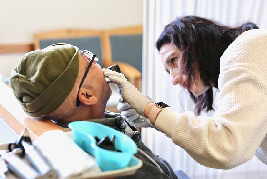
On February 28, 2024, the Department of Otolaryngology–Head and Neck Surgery (OHNS) at Mass Eye and Ear celebrated one year of partnership with the Boston Health Care for the Homeless Program (BHCHP).
Since its establishment in 1985, BHCHP has evolved into a leading nonprofit that ensures individuals who experience homelessness have equitable access to health care in Boston. Under the leadership of James O’Connell, MD, alumni of Harvard Medical School and Massachusetts General Hospital, the program serves a patient population of nearly 10,000 and has a team of more than 600 medical and behavioral health staff, social service providers and support staff committed to fulfilling BHCHP’s mission.
In response to an ongoing need to expand ear, nose, throat, head and neck care to an underserved population, the collaboration between BHCHP and the Department of OHNS at Mass Eye and Ear was spearheaded by Mark A. Varvares, MD, FACS, John W. Merriam/William W. Montgomery Professor and Chair of OHNS at Harvard Medical School and Chair of the Departments of OHNS at Mass Eye and Ear and Massachusetts General Hospital, and Gregory W. Randolph, MD, FACS, FACE, Claire and John Bertucci Endowed Chair in Thyroid Surgical Oncology and Professor of OHNS at Harvard Medical School and Director of the Thyroid and Parathyroid Surgical Division at Mass Eye and Ear.
Primary care providers through BHCHP evaluate patients and place a referral for anyone who may benefit from seeing an otolaryngologist. According to Dr. Suresh, there are consistently new referrals to the clinic, confirming the significant need for this care.
Now, over a year later, the Mass Eye and Ear otolaryngology clinic is located at the Barbara McInnis House in Boston’s South End, where BHCHP’s inpatient respite facility and primary outpatient clinics are located. Every fourth Tuesday of each month, up to three residents and an attending from Mass Eye and Ear travel there with necessary medical equipment to provide renowned otolaryngology care to individuals in need.
“What drew me to this program was bringing our subspecialty expertise directly to patients who otherwise might not have access to otolaryngology care,” shared Krish Suresh, MD, fifth-year resident in the Harvard Combined Residency Program in OHNS and BHCHP resident lead.
To obtain funding for the clinic, Dr. Varvares connected with Regan W. Bergmark, MD, MPH, Assistant Professor of OHNS at Harvard Medical School and sinus and endoscopic skull base surgeon at Brigham and Women's Hospital, who serves as the Principal Investigator on the United Against Racism grant for the Department of OHNS. “A primary focus of the United Against Racism grant is to address equitable access to care for otolaryngology services,” said Dr. Bergmark. “When patients do not have access to otolaryngology care, they may have significantly worse outcomes—such as increased morbidity and mortality for patients with head and neck cancer.”
Department leadership allocated thousands of dollars from the United Against Racism grant to purchase necessary, safe and sterile equipment for this monthly clinic. Further, 2
2 HARVARD Otolaryngology NEWS
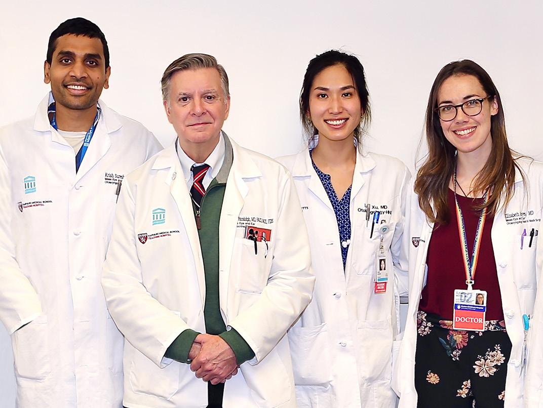
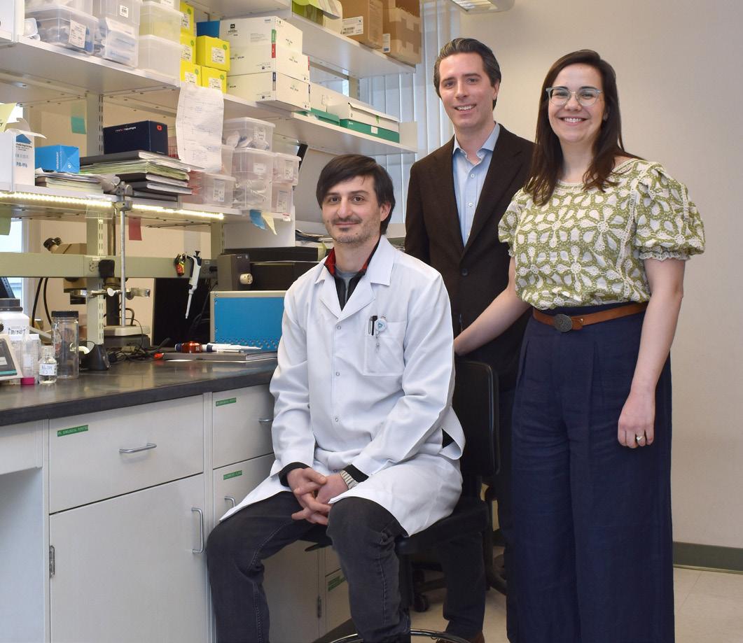
BHCHP resident and faculty leads and active volunteers pictured from left to right: Krish Suresh, MD; Gregory Randolph, MD, FACS, FACE; Lucy Xu, MD; Elizabeth Noyes, MD.
the department has opened its doors to patients who may need follow-up care after the clinic visit at Mass Eye and Ear’s main Boston campus for free. “This program would not be possible without the institutional support provided by Dr. Varvares. His advocacy and commitment to this work propels our mission to help the sickest and most vulnerable patients,” Dr. Bergmark remarked.
The partnership between BHCHP and the Department of OHNS at Mass Eye and Ear is constantly evolving. When the program began in 2023, there was a handful of resident volunteers, but due to increased interest and the aim of longterm clinic sustainability, BHCHP has now become part of the resident rotation.
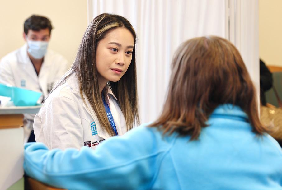
“As clinicians and trainees, we benefit greatly when we learn to listen and understand the many biopsychosocial forces shaping our patients' health, allowing us to provide more patient-centered care both in the BHCHP clinic and in our daily practice,” said Elizabeth Noyes, MD, second-year resident in the Harvard Combined Residency Program in OHNS and active BHCHP volunteer. “I am thrilled that fellow trainees and attendings were eager and willing to participate in this critical program.”
Additionally, the faculty volunteer portion of the program has continued to expand over the past year, now at five attending volunteers who each specialize in different areas. Dr. Randolph, BHCHP faculty lead, shared that one of their first patients had ear canal stenosis, or narrowing of the ear canal that results in backed-up earwax, and was beaten by her parents because she was unable to hear. “It was a devastating story, but I knew if we could perform a canal widening surgery to expand the ear canal, her hearing would be fully restored.”
Dr. Randolph reached out to his colleague Felipe Santos, MD, Assistant Professor of OHNS at Harvard Medical School and Director of the Otology and Neurotology Division at Mass Eye and Ear, who not only immediately agreed to perform the surgery pro bono, but also offered to volunteer for the program. “I already knew that our department attracted quality faculty, but stories like these reaffirm our shared mission-driven approach,” emphasized Dr. Randolph.
“The partnership with BHCHP has been an extraordinary opportunity to further expand our care to the Boston community, while continuing to address inequities in access to the specialty of otolaryngology,” said Dr. Varvares. “This work has also helped the Department of OHNS identify the need for a homeless medicine specialty, an academic evolution that is underway.” n
Eye,
Ear,
Eat–
Community Health Screening
On Saturday, April 6, 2024, Mass Eye and Ear hosted its second “Eye, Ear, Eat” event. In partnership with the nonprofit Roxbury Tenants of Harvard, the four-hour community health screening was held at Mass Eye and Ear’s Longwood location. Attendings and residents from the Department of Otolaryngology–Head and Neck Surgery at Mass Eye and Ear offered hearing and head and neck screenings to more than 200 residents of Mission Hill and other neighboring communities. If a clinician identified a need for follow up otolaryngology care, the patient received assistance in scheduling a further consultation. In tandem, the Department of Ophthalmology performed eye screenings, and the event offered patients access to financial counselors and education on health care delivery options.
3 HARVARD Otolaryngology
The Toth Center: New Leadership, Novel Innovations
Scientists in the Mike Toth Head and Neck Cancer Research Center are well underway towards new, innovative approaches to faster and more accurate diagnoses, less invasive treatments, predictive medicine and more. With new leadership and collaborations in place, the Toth Center is making great headway for the future of head and neck cancer care.
4
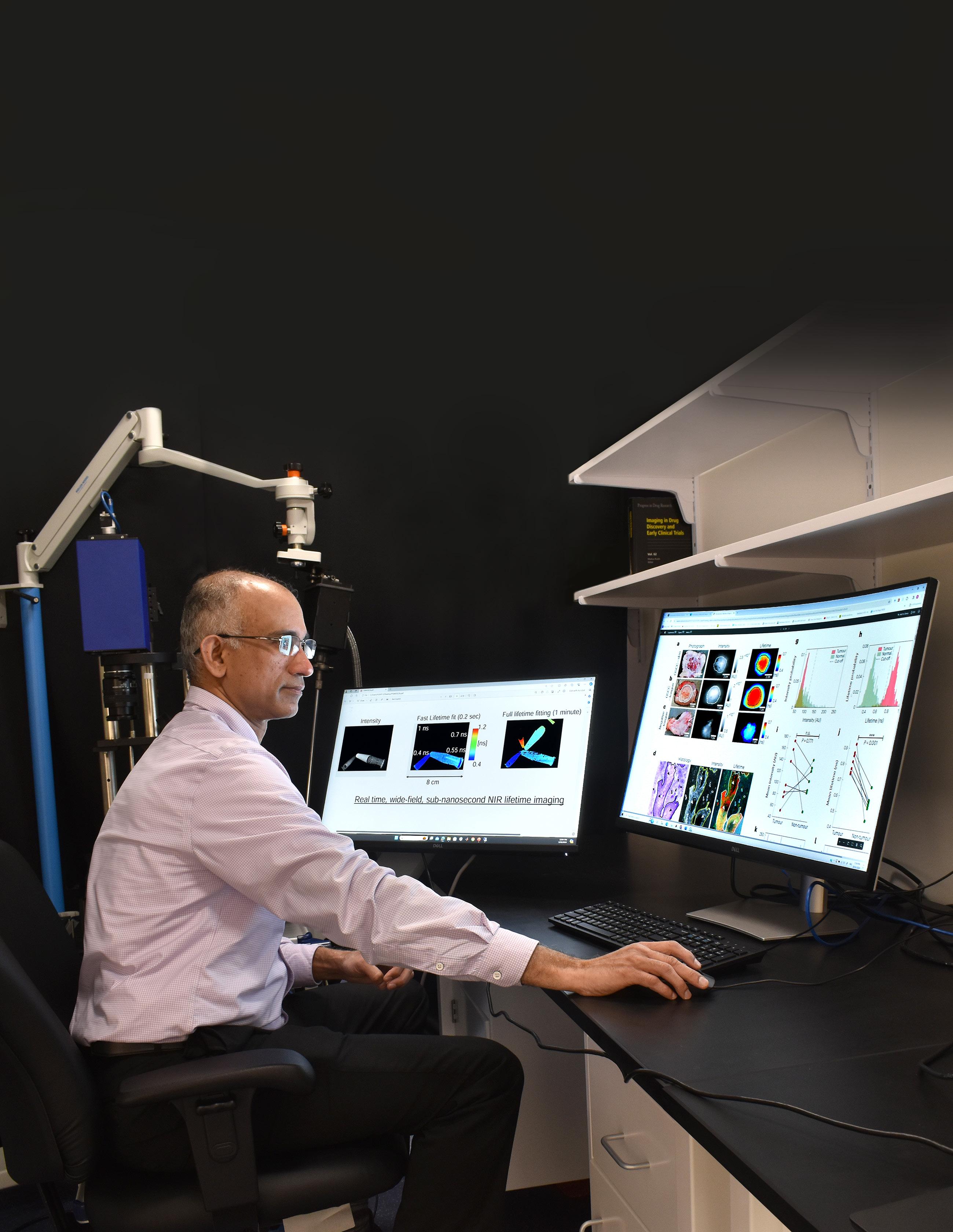
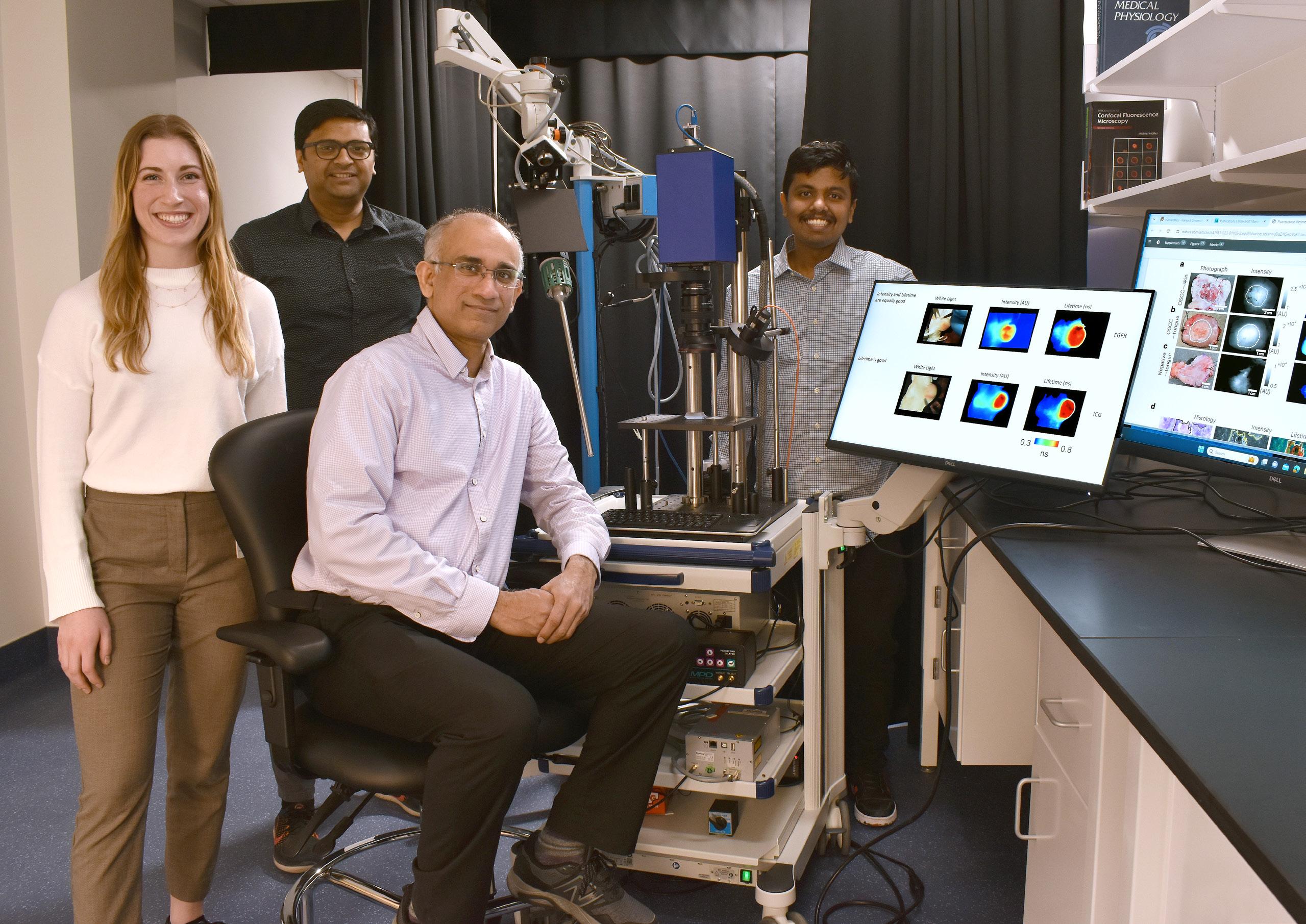
Members of the Kumar lab pictured from left to right standing: Hannah Collins; Rahul Pal, PhD; Murali Krishnamoorthy, PhD; seated, Anand T. N. Kumar, PhD.
The Mike Toth Head and Neck Cancer Research Center—named in memory of Mike Toth, a patient and friend of Mass Eye and Ear who courageously fought head and neck cancer but sadly succumbed to the disease—has been integral in pioneering innovative approaches to diagnosing, monitoring and treating cancers of the head, neck and skin since 2019.
Under the new leadership of Anand T. N. Kumar, PhD, Associate Professor of Otolaryngology–Head and Neck Surgery at Harvard Medical School and Director
of the Toth Center at Mass Eye and Ear, the team continues to leverage the brightest minds to challenge current treatments and improve future outcomes for patients with these diseases.
“The Toth Center is compiled of an extraordinary group of interdisciplinary investigators, all with the same goal to eradicate head and neck cancer,” shared Dr. Kumar. “We have a lot of areas to continue to explore, and as the new director I look forward to building and growing a collaborative center.”
[ continued p. 6 ]
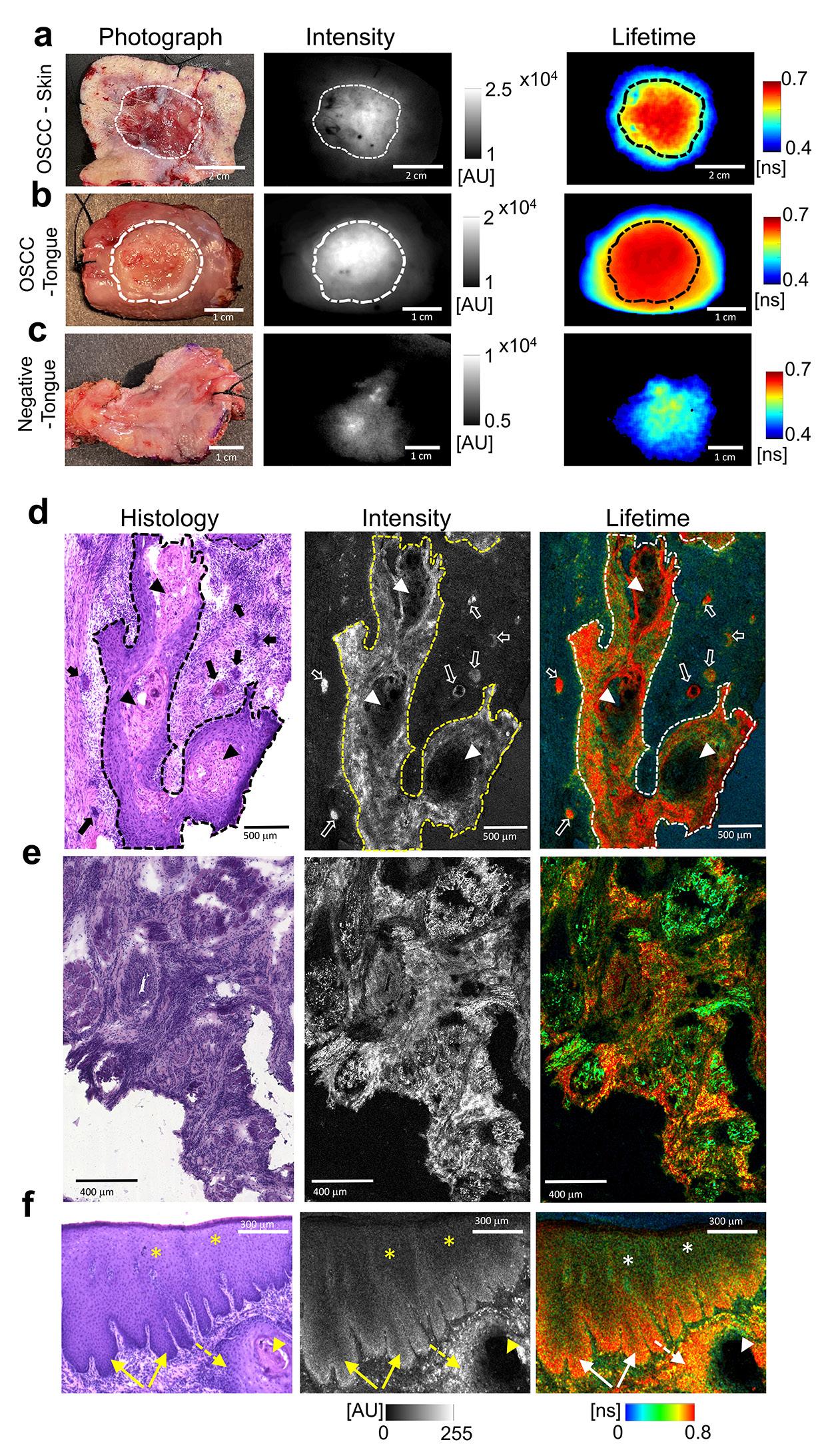
Fluorescence lifetime (FLT) enhancement and microscopic cancer specificity in head and neck cancers. (a–c), Color photographs (left), wide-field fluorescence intensity (center) and FLT maps (right) of whole resected specimens from patients H8, H6 and H5 in Supplementary Table 12, respectively, with cutaneous squamous-cell carcinoma (SCC) in the left ear (a), oral SCC in the tongue (b) and a histologically tumor-free tongue specimen (c), injected with indocyanine green (ICG) 20–47 h before surgery. Dashed lines indicate visually identified tumor boundaries based on color photographs. (d,e), Histology, microscopic fluorescence intensity, and fluorescence-lifetime imaging microscopy (FLIM) of an oral squamous-cell carcinoma (OSCC) specimen from the left jaw (d) and a tongue OSCC specimen (e). Confocal FLIM was performed on frozen (OCTembedded) sections (15 µm) of the specimen using a 10×, 0.4 NA objective. Solid lines in d indicate histologically identified boundaries of infiltrative tumor nests that also enclose necrotic cores and keratinized regions (arrowheads) with shorter FLT than tumor cells. The FLTs within individual microscopic cancer nests (open arrows on FLIM image and solid arrows on histology) are longer than the FLTs of non-tumor cells in the tumor micro-environment (TME), including the stroma and muscle (outside the tumors boundaries). (e), Invasive tumor cell clusters displayed longer FLTs compared with the surrounding desmoplastic stroma or muscle despite a comparable ICG fluorescence intensity. (f), Long FLT values comparable to tumor cells are also observed in dysplastic epithelium adjacent to invasive tumors in an OSCC specimen from the tongue. Solid arrows: dysplasia at the basal epithelium (long FLT); dashed arrow: invasive tumor nest (long FLT); arrowhead: hyper-keratinized necrotic core (no ICG uptake); asterisk: mature and metabolically less active superficial epithelium (short FLT).
5 HARVARD Otolaryngology
FEATURE
INNOVATIONS
continued
Introducing the Kumar Laboratory
As a physicist and biomedical engineer by training, it is only fitting that Dr. Kumar’s lifelong dream is to create a medical device that physicians use directly to benefit patients. After joining Massachusetts General Hospital in 2002, Dr. Kumar quickly found his research niche—combining medical imaging devices and the power of light to diagnose disease, specifically cancer.
He carefully studied fluorescence imaging, a technique that uses fluorescent markers to visualize specific molecules within cells and tissues. Fluorescence imaging can use dyes to target cancer-specific
“A tumor is clearly visible after a CT or MRI is performed, but the physician cannot see this in real time as they are operating to remove tumors. There is an urgent need to see the exact margin of a tumor when patients are open in surgery.”
– Anand T. N. Kumar, PhD
molecules, however, standard imaging methods seem to have limited accuracy for detecting tumor margins due to varying levels of these molecules among different tumors.
The standard approach to differentiating between tumor tissue and normal tissue is to rely on sight and the physical touch of the tumor. However, this process often causes physicians to either leave some cancer behind or to remove more tissue than is needed during surgery, which particularly in the case of head and neck cancer, can result in drastically changing a patient’s appearance.
“A tumor is clearly visible after a CT or MRI is performed, but the physician cannot see this in real time as they are operating to remove tumors,” said Dr. Kumar. “There is an urgent need to see the exact margin of a tumor in real time when patients are open in surgery.”
He and his team further studied a different imaging technique, called fluorescence lifetime (FLT) imaging, that looks at the duration of time that fluorescent molecules remain in their excited state before emitting light.
FLT refers to the average time it takes for
fluorescent molecules to return to their ground state once they are excited by a short laser pulse. Similar to a half-life measure of radioactivity, FLT is the time it takes for fluorescence to decrease by half from its initial value. The difference is, while the half-life of radioactive substances can be years to thousands of years, the halflife of fluorescence is often a fraction of a billionth of a second. Imaging cameras can capture these rapid changes in fluorescence decay times. FLT has many benefits compared to standard fluorescence imaging, which is widely used in medicine but is limited in its ability to distinguish disease from normal tissues. While FLT imaging has been widely used in research settings, its application for clinical studies in humans had been limited.
With this in mind, in 2016, Dr. Kumar’s team evaluated a Food and Drug Administration (FDA)-approved fluorescent dye, known as indocyanine green (ICG), in cancer mouse models to evaluate whether FLT can help distinguish cancer better than standard fluorescence imaging. ICG typically accumulates in tumor tissue, but also settles in healthy tissue. Through multiple preclinical studies, Dr. Kumar’s team found that the FLT of ICG in tumors is longer than the FLT in the normal tissue. This finding allowed the researchers to accurately distinguish between tumor tissue and normal tissue—a breakthrough, since most dyes currently being evaluated for tumor detection have non-specific uptake, like ICG, which limits their practical use.
After making this initial discovery in animal models, Dr. Kumar approached Kevin S. Emerick, MD, Associate Professor of Otolaryngology–Head and Neck Surgery at Harvard Medical School and Division Director of Head and Neck Surgical Oncology at Mass Eye and Ear, and Mark A. Varvares, MD, FACS, John W. Merriam and William W. Montgomery Professor and Chair of the Department of Otolaryngology–Head and Neck Surgery at Harvard Medical School and Chair of Otolaryngology–Head and Neck Surgery at Mass Eye and Ear and Mass General Hospital, about moving forward with patient trials. “Both Drs. Emerick and Varvares were extremely enthusiastic and confident
6 HARVARD Otolaryngology
THE TOTH CENTER: NEW LEADERSHIP, NOVEL
[
]
that my laboratory was onto something monumental, said Dr. Kumar. “Their support was invaluable in the next steps of our study.”
Initially, their human study analyzed tissue samples from patients who underwent liver surgery at Mass General Hospital and head and neck surgery from Mass Eye and Ear. Patients received an ICG injection one to three days before surgery.
In 2021, the first head and neck cancer patient (for this study) at Mass Eye and Ear was injected with ICG, and the tissue was analyzed with FLT imaging. Dr. Kumar and his team found that not only did the tumor tissue have a longer lifetime—or FLT—but zooming in at high resolution, they found that each individual cancer cell had a longer FLT as well.
Expanding on this research, the team collaborated with multiple institutions and assessed samples from more than 60 ICG injected patients with various cancer types, expanding the trial sites to include the University of Pennsylvania, University of Newcastle in the United Kingdom and Leiden University in the Netherlands.
Dr. Kumar and colleagues were able to detect an FLT shift at the cellular level that was consistent across
tumor types and in multiple patients. FLT imaging was also able to distinguish benign from metastatic lymph nodes. Overall, this novel technique demonstrated 97 percent accuracy in distinguishing tumor tissue from healthy tissue. These results were published in Nature Biomedical Engineering.
Although ICG is approved by the FDA for other uses, it is not yet approved for clinical use as a tumor marking agent. The researchers’ next step is to conduct a large-scale clinical trial to evaluate the safety and efficacy of FLT imaging with ICG for tumor identification during surgery.
Dr. Kumar received a R01 grant from the National Institutes of Health for $2.2 million over five years to further develop and test an imaging device based on their technology to help surgeons visualize tumors better during surgery. Drs. Emerick and Varvares will serve as co-investigators on this grant.
The Kumar laboratory, which includes Rahul Pal, PhD, Instructor of Otolaryngology–Head and Neck Surgery, Harvard Medical School, is now situated at Mass Eye and Ear, and the Mike Toth Head and Neck Cancer Research Center is well-positioned for upcoming research initiatives.
Advances in the Toth Center
Other exciting developments in the Toth Center come from the Shalhout and Faden laboratories. Sophia Z. Shalhout, PhD, Assistant Professor of
[ continued p. 8 ]
7 HARVARD Otolaryngology
Top photo: Sophia Z. Shalhout, PhD.
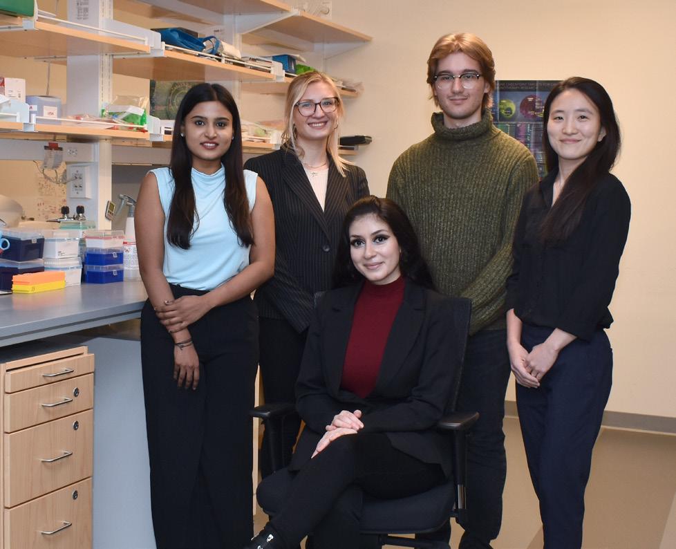
Right photo: Members of the Shalhout lab pictured from left to right standing: Dharani Balakrishnan, MPH; Angela Fragano; Daniel Panariti; Minkyung Kim, PhD; seated, Sophia Z. Shalhout, PhD.
THE TOTH CENTER: NEW LEADERSHIP, NOVEL INNOVATIONS
Otolaryngology–Head and Neck Surgery at Harvard Medical School and Assistant Scientist in the Toth Center, focuses on using artificial intelligence (AI) to develop more effective therapies, enhance the understanding of tumor biology, and improve how physicians diagnose, treat and manage cancer.
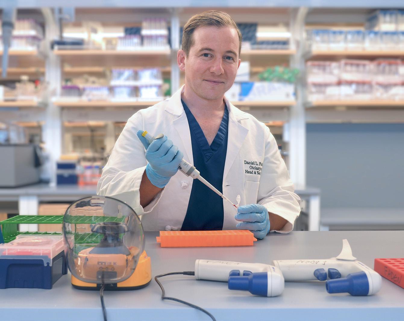
Daniel L. Faden, MD, FACS, Assistant Professor of Otolaryngology–Head and Neck Surgery at Harvard Medical School and Investigator in the Toth Center, concentrates on the development and use of liquid biopsies to detect human papillomavirus (HPV)-associated head and neck cancers.
In the Shalhout laboratory, a primary focus is to use AI to accelerate and enhance the drug discovery process. Notably, most patients do not respond to the current available therapies for advanced cancers of the head, neck and skin, and those who respond at first often develop resistance or recurrence despite initial success. Dr. Shalhout recognizes that there is a dire need for new therapies and strategies to treat advanced cancers; currently, the drug development in oncology is labor-intensive, costly and largely depends on trial and error.
Dr. Shalhout’s team is developing deep learning and generative AI models, using combined expertise in computer science and chemistry to develop new drugs through algorithms capable of analyzing vast
amounts of biological, chemical and pharmacological data. The AI models consider various factors such as protein structures and target selection, while interrogating a huge chemical space to identify novel candidate molecules that are structurally diverse yet likely to interact with the target. AI offers a much faster and more comprehensive approach to drug discovery than current traditional pathways allow. By leveraging AI, the lab aims to expedite the initial stages of drug development, potentially reducing the current timeline of seven to 12 years to a scale of months.
Another major project in the Shalhout laboratory involves the design and development of novel AI software capable of creating computer simulations of an individual patient’s head, neck or skin cancer. These models can be used to predict the best treatment to offer a specific patient. The patient simulations have the ability to ‘test’ outcomes virtually, before treating a patient. Using computational models, doctors can simulate a tumor’s response to various treatment options before implementing them in the clinical setting.
8 HARVARD Otolaryngology
continued
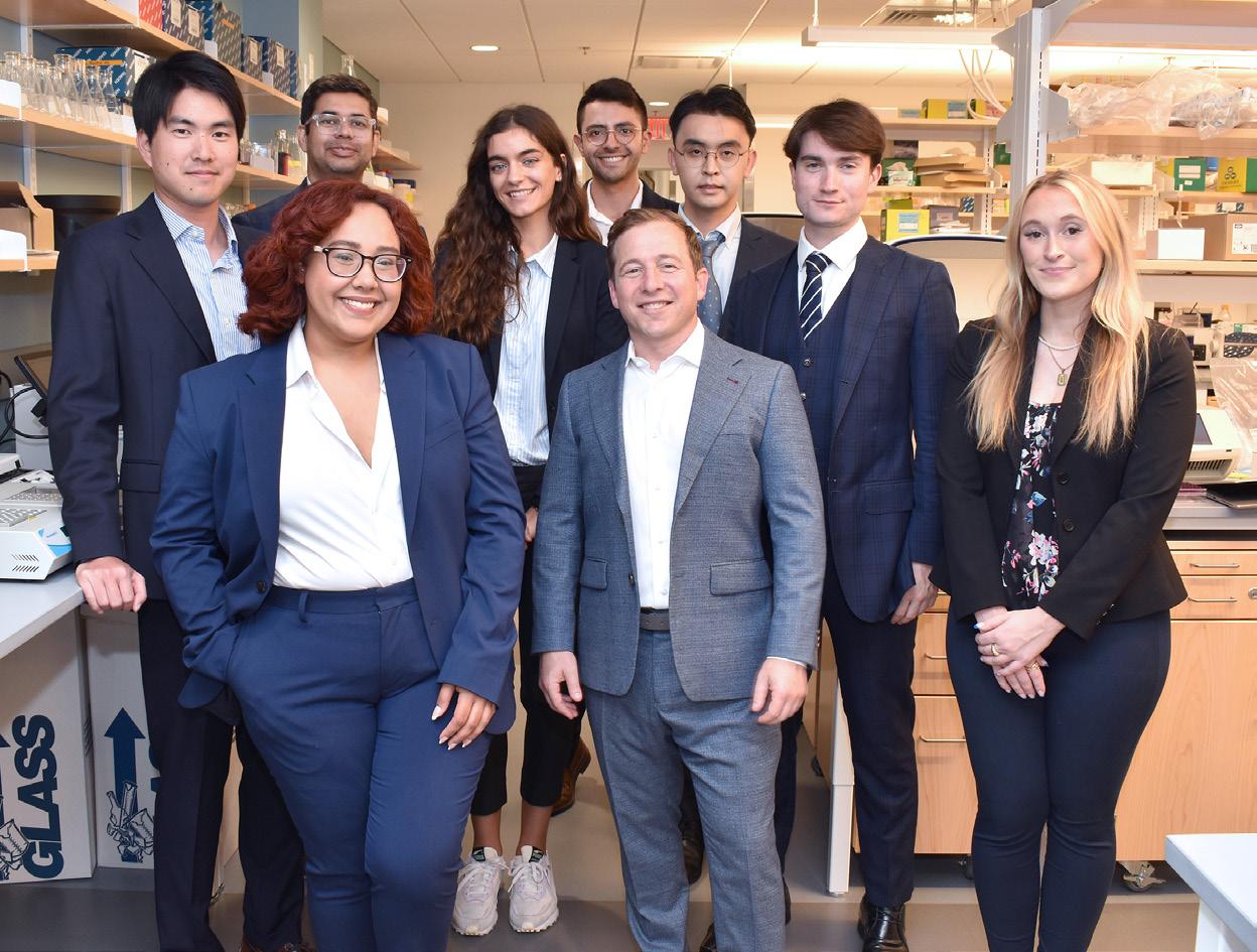
Left photo: Daniel L. Faden, MD, FACS.
Right photo: Members of the Faden lab pictured from left to right. Back row: Shun Hirayama, MD, PhD; Dipon Das, PhD; Yana Al-Inaya; Daniel Ruiz; Alan Ling; Michael Bryan; Julia Mendel. Front row: Elba Marie González and Daniel L. Faden, MD, FACS.
The computer models learn from various data sources, including genetic information, tumor characteristics and medical history. The Shalhout laboratory relies on a multidisciplinary team, including clinical coordinators, computer scientists, bioinformaticians, imaging experts and pathologists, to drive this research forward.
“The end goal is for physicians to use these simulations to help devise treatment plans tailored to the individual cancer patient,” shared Dr. Shalhout.
The Faden laboratory is one of the largest surgeon-run laboratories at Mass Eye and Ear. Comprised of eight scientists and trainees from all over the world, one of the primary focuses of the lab is developing and applying blood-based cancer diagnostics, or liquid biopsies. The goal of the lab is to detect, diagnose and monitor cancers more accurately than current approaches, and to do so in a noninvasive way.
Currently, the scientists in the lab are most heavily focused on HPV-associated head and neck cancers. The team has developed several liquid biopsies for HPV-associated cancer which have been shown to outperform standard-of-care approaches in various clinical settings—all through a single blood sample.
Most recently, the researchers in the Faden laboratory developed a next generation liquid biopsy
screening and minimal residual disease detection after surgery.
“Our goal in developing, validating and applying liquid biopsy diagnostics like HPV-DeepSeek is to study large unmet needs in cancer care, such as blood-based cancer screening in high-risk patient populations, with the hope of increasing early cancer detection and thereby, decreasing cancer-related mortality,” said Dr. Faden. “Our research is leading us into novel, and often unexpected directions, which is really quite exciting.”
New leadership, new collaborations
According to Dr. Kumar, his vision for the future of the Toth Center begins with collaboration. Each laboratory has its specific focus, but all of the research is translational with the common goal of better identifying and curing cancer. With Dr. Kumar at the helm, the Toth Center scientists will have regular meetings to discuss ideas for collaborations that combine each research program’s expertise, ultimately leading to new grant opportunities and publications.
Additional Toth Center transformational tactics include hosting a monthly seminar series with invited speakers, both within and outside the Mass General Brigham system, to present on innovative topics in relation to head and neck cancer research.
“A common theme at the Toth Center is leveraging our diverse backgrounds and expertise–from physics, chemistry and medicine–to advance our shared goal of improving diagnostic testing and treatment options for patients with head, neck and skin cancers.”
– Anand T. N. Kumar, PhD
called HPV-DeepSeek; this test is more than 25 times more sensitive than the original tests and can also look at many other cancer-related features, which are then analyzed using machine learning. HPVDeepSeek is specifically designed for addressing low tumor burden settings such as asymptomatic cancer
They also will host an annual workshop to showcase the cuttingedge imaging and computational tools for head and neck cancer imaging and treatment that are being developed at the Toth Center.
“A common theme at the Toth Center is leveraging our diverse backgrounds and expertise—from physics, engineering, chemistry and medicine—to advance our shared goal of improving diagnostic testing and treatment options for patients with head, neck and skin cancers,” emphasized Dr. Kumar. “I am thrilled to have the opportunity to lead this renowned Center.” n
9 HARVARD Otolaryngology
[ ]
Establishing a Nonsyndromic Hearing Loss Gene
A robust collaboration between Boston Children's Hospital and Mass Eye and Ear resulted in the discovery of a novel gene for nonsyndromic hearing loss called PKHD1L1.
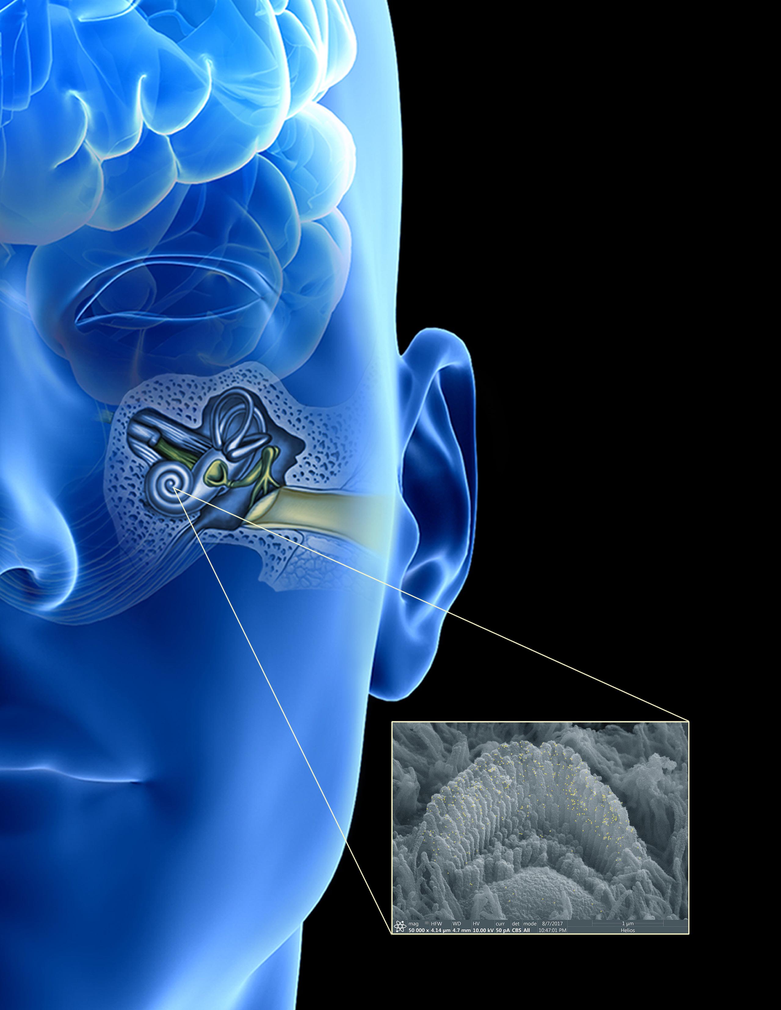
Scanning Electron Microscopy photomicrographs of early postnatal mouse outer hair cell stereocilia bundle, labeled with an antibody against PKHD1L1. Multiple 12-nm gold beads (pseudo-colored as yellow dots) are detected on the surface of stereocilia, localizing the PKHD1L1 protein to the bundle.
10
Conventionally, children who have hearing loss are treated with hearing aids or cochlear implants, depending on the severity. However, understanding the underlying cause of hearing loss is critical for patients and families as well as their treating physicians. Only about a decade ago, most children with hearing loss had no concrete diagnosis. Thanks to new advances including genetic testing and the discoveries of more hearing loss genes, many more children with hearing loss can receive a diagnosis today.
According to the Hereditary Hearing Loss Homepage, 152 genes are known to cause nonsyndromic hearing loss, and within those genes, there are thousands of genetic variants. Unfortunately, there are still many deafness-causing genes not confirmed, making final diagnoses challenging.
“In my practice, I primarily see pediatric patients with hearing loss, and for most of my patients—but not all—we are able to determine a diagnosis,” said Eliot Shearer, MD, PhD, FACS, Assistant Professor of Otolaryngology–Head and Neck Surgery at Harvard Medical School and Pediatric Otolaryngologist at Boston Children’s Hospital. “Identifying the underlying cause of hearing loss, the diagnosis, is critical as it provides prognostic information, reveals associated medical conditions and provides a sense of closure
and empowerment to families. My goal is to use the best clinical and research tools so that one day all children with hearing loss have a diagnosis.”
Dr. Shearer and his mentor, Margaret A. Kenna MD, MPH, FACS, FAAP, Professor of Otolaryngology–Head and Neck Surgery at Harvard Medical School and Director of Clinical Research in the Department of Otolaryngology and Communication Enhancement at Boston Children’s Hospital, manage an extensive hearing loss research program at Boston Children’s Hospital. When they work with a pediatric patient presenting with hearing loss, they initially conduct a standard diagnostic workup, which often includes imaging and genetic testing. If results are inconclusive, and the family is interested, the team enrolls the patient in their research study. During the study, advanced genetic diagnostic tools, such as exome sequencing and genome sequencing, are used to discover new hearing loss genes.
In 2020, Dr. Kenna evaluated a 10-year-old female patient presenting with moderate hearing loss and found no cause of genetic hearing loss within the known hearing loss genes. The patient then enrolled in the large genetic hearing loss study run by Dr. Kenna at Boston Children’s Hospital, supported by the Children’s Rare Disease Research Cohort Initiative.
[ continued p. 12]
11 HARVARD Otolaryngology
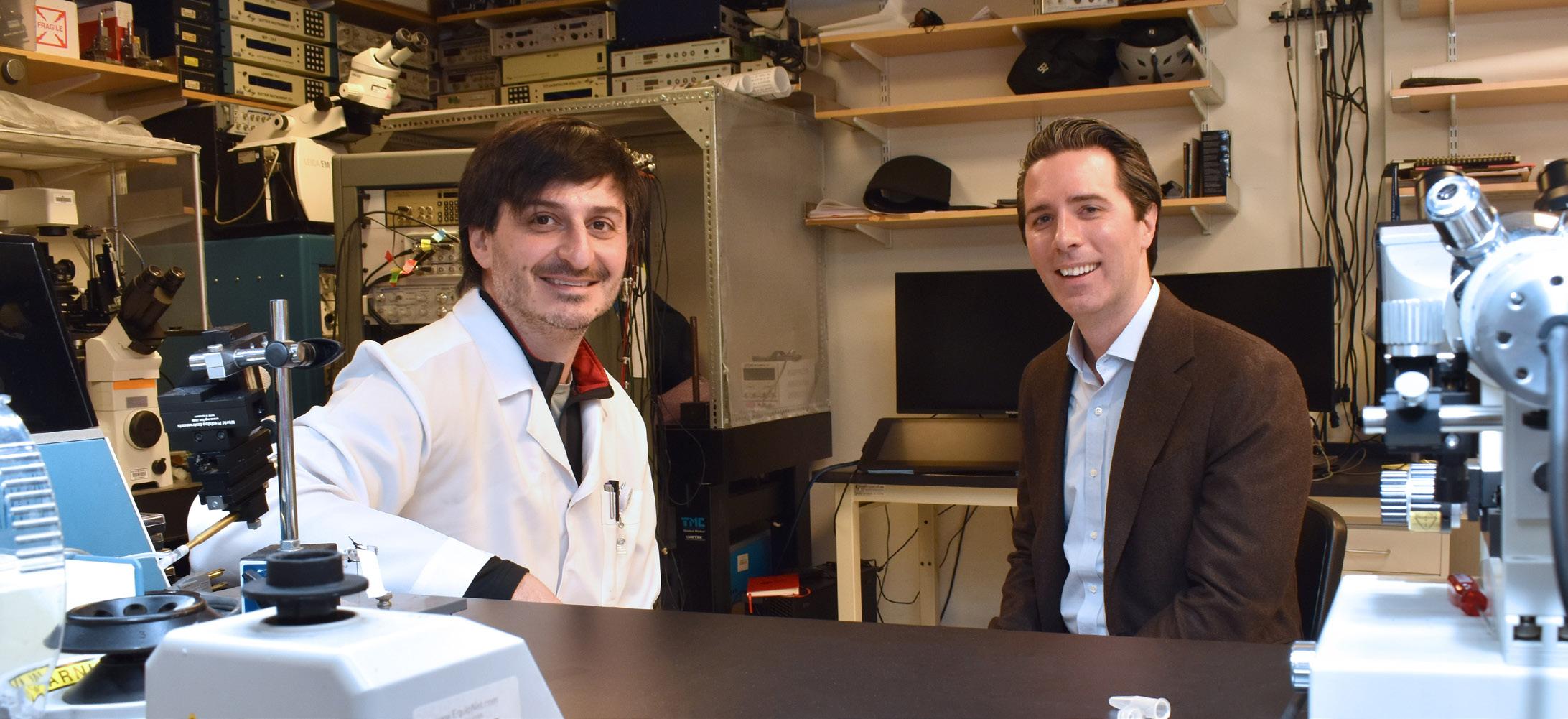
FEATURE
From left to right: Artur A. Indzhykulian, MD, PhD, and Eliot Shearer, MD, PhD, FACS, collaborating in the Indzhykulian lab at Mass Eye and Ear.
ESTABLISHING NONSYNDROMIC HEARING LOSS GENE,
Dr. Shearer, a co-investigator on the study, then looked outside the already established hearing loss genes and discovered some interesting genetic variants in a gene called polycystic kidney and hepatic disease 1-like 1 (PKHD1L1).
“PKHD1L1 rang a bell for me, but I couldn’t pinpoint where, until I came across the Harvard Medical School publication in Nature Communications titled ‘PKHD1L1 is a coat protein of hair-cell stereocilia and is required for normal hearing,’” said Dr. Shearer. “My colleague Artur Indzhykulian, MD, PhD, was one of the corresponding authors, and we quickly connected.”
Discovery of PKHD1L1 in mouse cochlea
Over a decade ago, Artur A. Indzhykulian, MD, PhD, Assistant Professor of Otolaryngology–Head and Neck Surgery at Harvard Medical School and Assistant Scientist in the Eaton-Peabody Laboratories (EPL) at Mass Eye and Ear, was a postdoctoral researcher in the Harvard Medical School Department of Neurobiology, conducting research under the direction of David Corey, PhD, Bertarelli Professor of Translational Medical Science at Harvard Medical School.
Scientists in the Corey Laboratory investigated different proteins that might be at the surface of stereocilia carried by the hair cells—the sensory cells
of our inner ear. Using a database of proteins found in hair cells, they identified PKHD1L1, which is highly enriched in the inner ear sensory cells, particularly in the high-frequency region of the cochlea.
Following this observation, Dr. Corey’s team created a mouse model lacking the PKHD1L1 protein specifically within the sensory cells of the inner ear. This enabled the team to investigate how the absence of PKHD1L1 affected hearing in mice.
According to Dr. Indzhykulian, the team quickly discovered that the absence of PKHD1L1 in sensory cells led to progressive hearing loss in the mice. They also found that this protein was located on the tips of stereocilia, which respond to sound stimulation.
The results of this study, published in 2019 in Nature Communications, indicate that the PKHD1L1deficient mice lack the surface coat of stereocilia. This deficiency resulted in progressive hearing loss in mice, demonstrating the critical role of PKHD1L1 for normal hearing in mice.
“At the time our study was published, I had already established my own laboratory in the EPL and was awarded a major grant through the National Institutes of Health to further investigate the role of PKHD1L1 in this mouse model,” said Dr. Indzhykulian. “However, I had not heard of any patients with this protein deficit until Dr. Shearer connected with me, which was a game-changer for the next phase of this research.”
A cross-institutional effort
After the genetic screening was conducted for the 10-year-old patient from Boston Children's Hospital, it was determined that she had two different amino acid changes within the PKHD1L1 protein. Based on the previous study implicating the gene, there was strong reason to believe that her hearing loss could be attributed to this protein deficit.
Dr. Indzhykulian’s team studied PKHD1L1’s protein structure, aiming to understand how a single amino acid change could affect the protein’s overall folding architecture, stability and its function.
Pedro De-la-Torre, PhD, a research scientist in the Indzhykulian laboratory, designed experiments to examine, at the atomic level, both the normal fragment of the protein and the fragment carrying this pathogenic variant. His goal was to understand
12 HARVARD Otolaryngology
PKHD1L1 continued
From left to right: Artur A. Indzhykulian, MD, PhD, Eliot Shearer, MD, PhD, FACS, and Shelby Redfield, MS, CGC.
Inset photo: Pedro De-la-Torre, PhD.
whether this protein, with the single point pathogenic variant, could be deleterious.
The hypothesis was that the pathogenic variant protein fragment was less stable compared to the normal protein sequence. To test it, the team cloned and purified both proteins, and gradually increased the temperature to determine the protein melt temperature, at which the protein undergoes structural failure and collapses, rendering non-functional proteins.
These findings, complemented by computer simulations, led the team to conclude that the pathogenic variant carried by the patient yields a protein of lower stability, which could be the cause of her hearing loss.
To further bolster evidence that a mutation in PKHD1L1 is a cause of human hearing loss, Drs. Shearer and Indzhykulian had reached out to human genetics groups world-wide to learn if any additional groups had patients with the PKHD1L1 deficit.
Barbara Vona, PhD, Group Leader at University Medical Center Göttingen-Institute of Human Genetics and Institute of Auditory Neuroscience and InnerEarLab in Göttingen, Germany, was immediately interested in this collaboration and used her connections to spread the word about the study. Through collaborations of her own, Dr. Vona helped identify three additional pediatric patients—from Iran, Pakistan and China—who have symptoms of hearing loss and carry plausible variants in the gene PKHD1L1.
“A lot of these pathogenic variants are extremely rare, so it’s critical to be connected with as many groups as possible to stay on top of which genes are being discovered in different parts of the world,” said Dr. Indzhykulian. “Dr. Vona played a significant role in this study, because without confirming additional pediatric patients with deleterious PKHD1L1 variants who present symptoms of hearing loss, this wouldn’t have been nearly the study it is today.”
Clinicians from Iran, Pakistan and China examined these patients and conducted genetic testing on each, analyzed that information and compared it to the findings the Boston Children's Hospital team reported. The results indicated that each pediatric patient has pathogenic variants within the PKHD1L1 gene, and each patient demonstrates hearing loss. Therefore,
there is enough evidence between mouse models, in vitro experimental data in the lab, computational models and observations in patients to indicate that PKHD1L1 is a nonsyndromic hearing loss gene.
On March 9, 2024, these results were published in Human Genetics in a paper titled, “PKHD1L1, a gene involved in the stereocilia coat, causes autosomal recessive nonsyndromic hearing loss.” Drs. Shearer, Indzhykulian and Vona were co-senior authors and Dr. De-la-Torre and Shelby Redfield, MS, CGC, genetic counselor and clinical research coordinator in the Shearer Lab, were co-first authors.
Looking into the future
This research suggests that going forward, clinicians should test hearing loss patients for the PKHD1L1 gene in addition to the other genes that are known to cause hearing loss.
As a next step, Dr. Shearer plans to study the prevalence of pathogenic variants in PKHD1L1 worldwide, starting by combing through the database of approximately 450 patients who are enrolled in his study with Dr. Kenna.
Dr. Indzhykulian will take a deeper dive into understanding how the disease manifests in humans— considering environmental factors such as exposure to loud noise— as the hearing loss phenotype varies among patients. Their most recent findings, reported in a preprint, suggest that noise exposure may contribute to hearing loss presenting differently across the patient population, as mice lacking PKHD1L1 were more susceptible to noise insult.
Knowing the number of patients and the timeframe to address this deficit to either maintain or potentially reverse hearing loss will enable the team to explore different gene therapeutic options.
“Establishing PKHD1L1 as a nonsyndromic hearing loss gene is a remarkable first step in directly helping patients who have this protein deficit. The goal is to provide all patients with a concrete diagnosis, to truly improve their quality of life,” said Dr. Shearer. “And now with clinical trials for gene therapy for hearing loss beginning, new avenues of treatment may soon be available for these children.” n
13 HARVARD Otolaryngology

Global Surgery: Reaching the Unreachable
Mass Eye and Ear leaders in Global Surgery launched a new program to help address the global surgical workforce crisis.
14
Perla Villamor (Colombia)
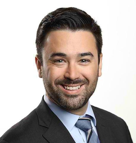
Wayne Manana (Zimbabwe)
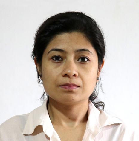
Pramila Shakya (Nepal)
In 2015, the Lancet Commission on Global Surgery established six indicators to measure surgical access, marking a pivotal turning point in better understanding worldwide disparities in surgical accessibility. The indicators include geographic accessibility, density of surgical providers, number of procedures performed, perioperative mortality, impoverishing expenditure and catastrophic expenditure.
To further understand the scope of measuring surgical access in otolaryngology–head and neck surgery, Blake C. Alkire, MD, MPH, Assistant Professor of Otolaryngology–Head and Neck Surgery at Harvard Medical School and a comprehensive otolaryngologist at Mass Eye and Ear, co-founded the Global Otolaryngology–Head and Neck Surgery Initiative, a research collaborative with a focus on identifying and prioritizing gaps in otolaryngology–head and neck surgical care worldwide. The collaborative recruited globally, resulting in a membership of
[“The goal of our investigations is to benefit countries and regions interested in expanding access to high-quality surgical care.”
– Blake C. Alkire, MD, MPH
more than 350 otolaryngologists, with representation from every World Bank region and 50 percent from low- and middle-income countries.
“We are investigating several different facets, including essential procedures and conditions, workforce population, training and education, infrastructure and equipment and barriers to care,” explained Dr. Alkire. “The goal of our investigations is to benefit countries and regions interested in expanding access to high-quality surgical care.”
The initiative launched a major survey sent to ministries of health and professional societies around the world, with a goal of learning the number of
otolaryngology–head and neck surgery care clinicians per capita worldwide. Responses came from 114 countries or territories representing 84 percent of the world’s population.
The survey results, published in August 2023 in JAMA Otolaryngology–Head & Neck Surgery, revealed variations in clinician density based on income and region. Higher-income countries generally had more clinicians per capita, with Europe having the highest density and low-income countries in Africa and Southeast Asia having the lowest. These findings can steer focused investments in training and policy making to address inequalities in the availability of otolaryngology–head and neck surgery clinicians.
Global Surgery Scholars Program
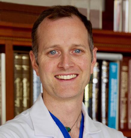
The surgical workforce is in crisis across low-tomiddle income countries. For over a decade, David A. Shaye, MD, MPH, FACS, Assistant Professor of Otolaryngology–Head and Neck Surgery at Harvard Medical School and facial plastic and reconstructive surgeon at Mass Eye and Ear, has travelled to countries in Africa, Asia and South America to teach facial reconstructive surgery for treating trauma, head and neck cancer and congenital anomalies.
“The shortage of surgeons across Africa has reached a critical state, such that a billion people on the continent do not have access to surgical care,” said Dr. Shaye, who serves as Director of Global [ continued p. 16 ]
15 HARVARD Otolaryngology
FEATURE
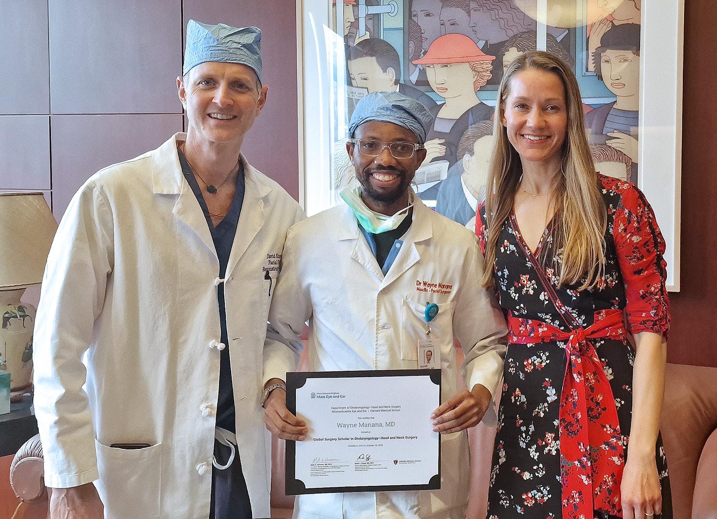
From left to right: David A. Shaye, MD, MPH, FACS, Wayne Manana, MD, and Caroline A. Banks, MD.
]
Surgery, Otolaryngology–Head and Neck Surgery. “The intention of our international surgical work is to bring what academic surgery does best—teaching—to places that need it most. Teaching surgery is a winning investment every time.”
Dr. Shaye spends many months each year working directly with surgeons in countries affected by the surgical workforce crisis. Through firsthand experience, he has identified a critical need for further educational and training opportunities, outside of what he can offer on his own.
Since joining Mass Eye and Ear in 2014, Dr. Shaye has seen the organization’s unique position as a hub of subspecialty surgical activity. With up to 15 operative rooms in action daily, and a multitude of talented surgeons, visiting surgeons have a unique opportunity to experience in just a few weeks what would amount to years’ worth of surgeries in their home countries.
In 2023, Dr. Shaye founded the Global Surgery Scholars Program at Mass Eye and Ear. The program offers educational opportunities for junior surgeons from Africa, Asia and South America to spend one month at Mass Eye and Ear observing surgeries, attending lectures and immersing themselves in a subspecialty head and neck hospital. The program is fully funded by philanthropy, from visa
“The intention of our international surgical work is to bring what academic surgery does best—teaching—to places that need it most. Teaching surgery is a winning investment every time.”
– David A. Shaye, MD, MPH, FACS
fees to flights to meals. Visiting surgeons are hosted by hospital faculty and other generous volunteers to create a welcoming and educational experience.
“For surgeons already armed with foundational skills from their residency training, this program offers a unique opportunity to see the vast array of surgeries performed at Mass Eye and Ear. Surgical skills, safety
systems and other ideas can then be tailored and replicated at university hospitals in Africa, Asia and South America,” said Dr. Shaye. “From novel surgical techniques, to new equipment, to building lifelong relationships, we are excited to see this program have global reach.”
Inaugural program launch
In the summer of 2023, the Department of Otolaryngology–Head and Neck Surgery hosted three surgeons: Pramila Shakya, MD, an oral and maxillofacial surgeon from Kirtipur Hospital in Nepal; Wayne Manana, MD, a maxillofacial and cleft surgeon from Parirenyatwa Hospital in Zimbabwe; and Perla Villamor, MD, a pediatric otolaryngologist from Children’s Hospital “La Casa del Niño” in Colombia.
Dr. Shakya is the leading cleft and craniofacial surgeon in Nepal with vast experience in her field; however, she identified a gap for her Nepalese patients in nasal reconstruction and rhinoplasty. In Boston, Dr. Shakya was able to observe dozens of nasal reconstruction surgeries. She also trained under Caroline A. Banks, MD, Instructor in Otolaryngology–Head and Neck Surgery at Harvard Medical School and a facial plastic and reconstructive surgeon at Mass Eye and Ear, which helped Dr. Shakya recognize the unmet need for facial paralysis care in Nepal.
“I feel more confident than I ever have, and I believe that I can better help and serve people of my country with the knowledge and experience gathered from Mass Eye and Ear,” said Dr. Shakya.
Following Dr. Shakya’s visit, Dr. Wayne Manana arrived from Zimbabwe. Dr. Manana is a lead facial reconstructive surgeon; one of the few in the entire country of 16 million people. During his month-long visit at Mass Eye and Ear, Dr. Manana relished the opportunity to learn techniques in free tissue transfer, skin cancer reconstruction, nasal reconstruction, facial reanimation surgery, hypoglossal nerve stimulators and periocular surgery.
“This experience was arguably the biggest turning point in my career,” said Dr. Manana. “It has challenged and redefined my scope of practice. I believe that this could mark the birth of facial plastic and reconstructive surgery in Zimbabwe.” He added that the biggest winner in the Global
16 HARVARD Otolaryngology
GLOBAL SURGERY: REACHING THE UNREACHABLE continued
[
]
Surgery Scholars Program are the patients that they save and serve.
“It was such a privilege to be a mentor in the Global Surgery Scholars Program and work with incredibly talented surgeons like Drs. Manana and Shakya,” said Dr. Banks. “Exchanging ideas and learning about each other's practices has been a very rewarding experience.”
The third surgeon, Dr. Villamor, trained under Christopher J. Hartnick, MD, MS, Professor of Otolaryngology–Head and Neck Surgery at Harvard Medical School and Division Director of Pediatric Otolaryngology at Mass Eye and Ear, to expand her learning in pediatric airway reconstruction.
She shared that her experience at Mass Eye and Ear was transformative. “The opportunity to observe cutting-edge medical and surgical practices, as well as advanced technologies, fills me with enthusiasm to replicate these advances as much as possible at our hospital,” said Dr. Villamor.
Dr. Hartnick remarked, “The Global Surgery Scholars Program allowed a wonderful pediatric otolaryngology surgeon to learn and experience our team culture of care and bring what she saw in Boston back home with her to Colombia. She is building her own program and creating a true multidisciplinary effort to help children in need.”
In addition to returning home with new knowledge and fresh skillsets, the scholars were given much needed surgical instruments to perform the surgeries they observed at Mass Eye and Ear. For example, Dr. Manana returned to Zimbabwe with a new batterypowered surgical headlight, enabling him to perform surgeries of the head and neck even when the electricity goes out, which is a common occurrence. “We want our visiting surgeons to not only leave with knowledge, but also with the equipment to perform and teach these surgeries at home,” said Dr. Shaye.
The Global Surgery Scholars Program is now approaching year two of hosting global surgeons, with the intention to expand the program with each year. “Every surgeon in this program has a shared goal to improve surgical care through teaching. I am extremely proud that Mass Eye and Ear has a program with global reach where it matters most,” emphasized Dr. Shaye. n
17 HARVARD Otolaryngology
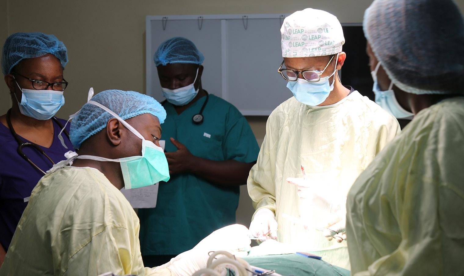
[1] Wayne Manana, MD, and surgical team perform facial reconstructive surgery on a patient in the far northern district of Zimbabwe.
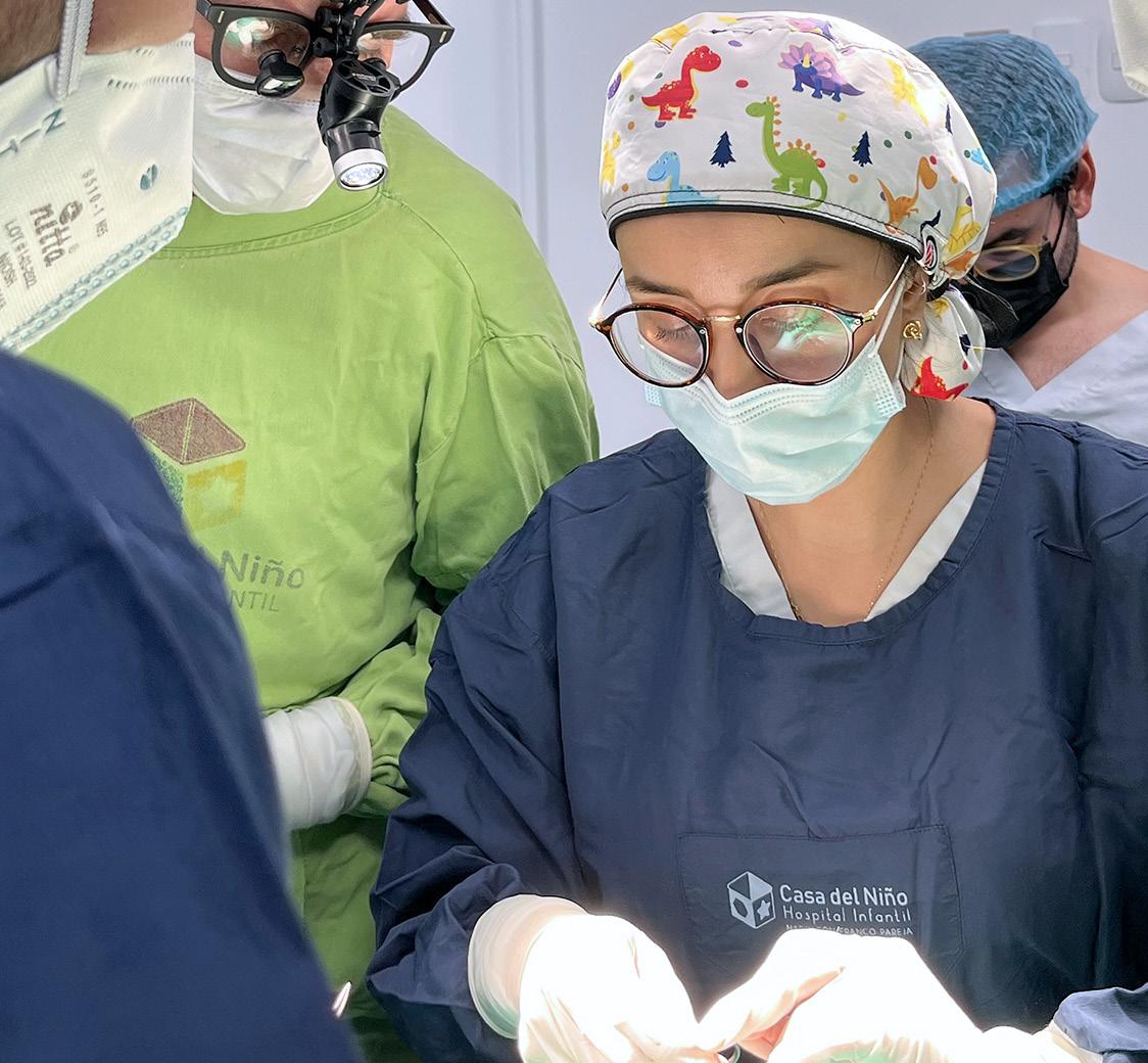
[2] Perla Villamor, MD, and surgical team operate on a pediatric patient at “La Casa del Niño” hospital in Cartagena, Colombia.
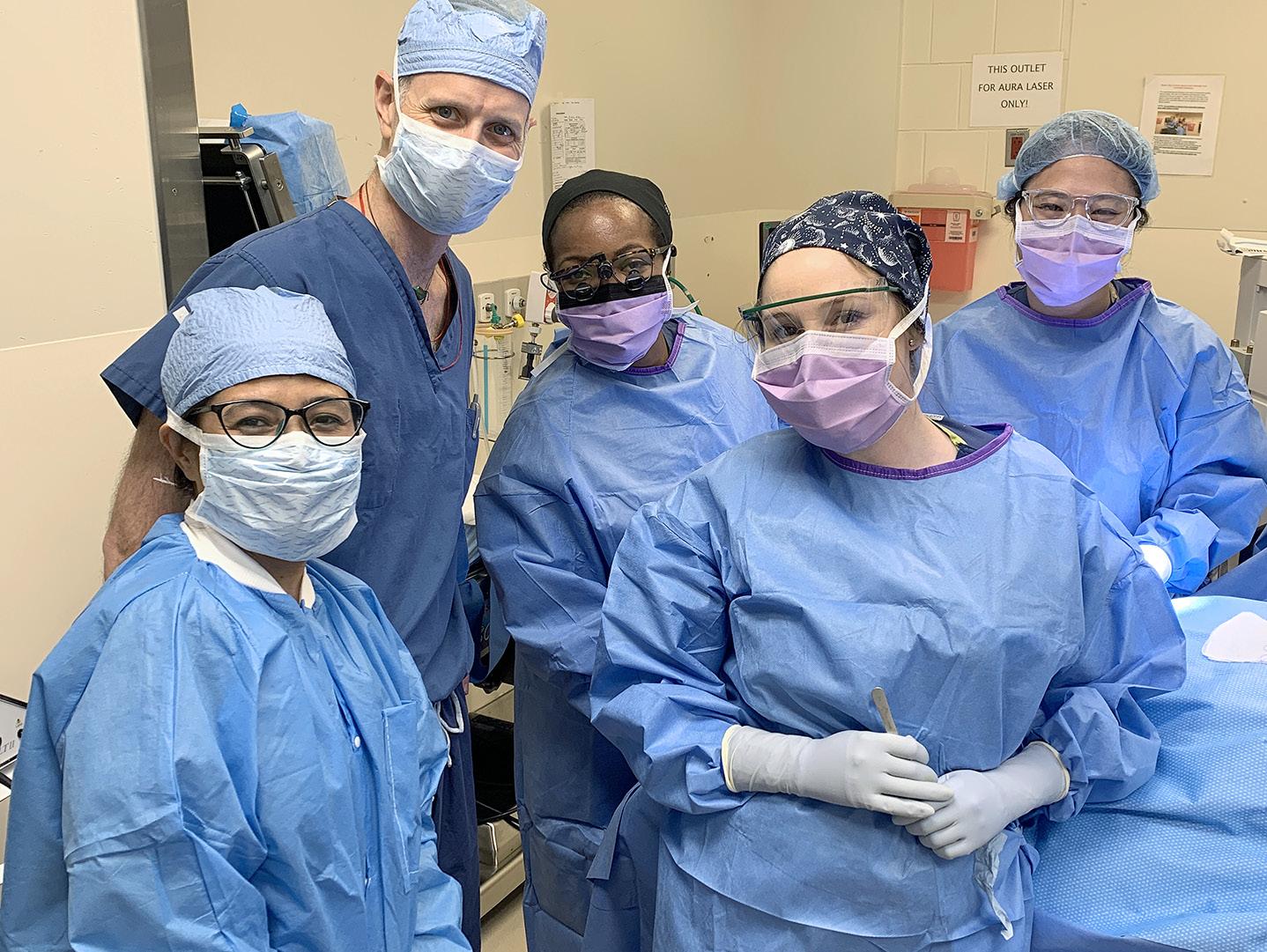
[ 1 ] [ 2 ] [ 3 ]
[3] Pramila Shakya, MD, David A. Shaye, MD, MPH, FACS, and surgical team in the operating room at Mass Eye and Ear, Boston, MA.
Ansin Foundation
Exceptional care inspires endowed chair
When Andy Ansin first noticed hearing loss, he chalked it up to his freediving hobby or exposure to loud environments. He became concerned, however, when it became more difficult to hear, and he scheduled a consultation with Dr. Fred Telischi, a neurotologist in his hometown of Miami.
After confirming significant hearing loss in Andy’s right ear, the doctor ordered an MRI that revealed an unexpected diagnosis. Andy had a vestibular schwannoma, a rare, noncancerous tumor of the balance nerves that can interfere with hearing. Turning to his network for help, Andy discovered Daniel J. Lee, MD, FACS, a leading otology and neurotology specialist at Mass Eye and Ear.
Dr. Lee confirmed Andy’s diagnosis and presented his options: surgery, radiation therapy, or monitoring for potential tumor growth and worsening symptoms. Considering the risks, Andy chose a “wait and watch” approach, visiting Dr. Lee annually for checkups.
Andy was impressed with Dr. Lee’s expertise and became interested in learning more about Dr. Lee’s passion: auditory brainstem implant (ABI) research.
The ABI is a hearing rehabilitative option that was originally designed for patients with Neurofibromatosis Type 2 (NF2). Children and adults with NF2 develop tumors called vestibular schwannomas in both ears that result in deafness. Due to injury to the hearing nerves, cochlear implants are not an option. The current ABI provides basic sound awareness but only very few users can understand spoken language. Dr. Lee’s research is aimed at refining ABI technology to improve hearing outcomes for these specialized patients.
As co-trustee of his family’s charitable foundation, Andy was no stranger to the power of philanthropy in solving problems. He knew right away that he wanted to support Dr. Lee’s
“My deep trust in the team at Mass Eye and Ear comes from the organization's devotion to their patients' well-being. I am confident that with Dr. Lee at the helm, the Ansin Foundation Chair will make a critical impact on generations of people affected by hearing loss. ”
– Andy Ansin
research, not only to thank Dr. Lee for all he has done for him, but to offer hope to others with profound hearing loss.
Andy and his family generously established the Ansin Foundation Chair in Otolaryngology, which will accelerate research to advance the care of patients who have otologic and lateral skull base tumors.
“My deep trust in the team at Mass Eye and Ear comes from the organization’s devotion to their patients’ well-being,” said Andy. “I am confident that with Dr. Lee at the helm, the Ansin Foundation Chair will make a critical impact on generations of people affected by hearing loss.” n
18 HARVARD Otolaryngology s DONOR PROFILE
Daniel J. Lee, MD, FACS
ALUMNI GIVING SOCIETY
The Alumni Giving Society of the Department of Otolaryngology–Head and Neck Surgery at Harvard Medical School
The Department of Otolaryngology–Head and Neck Surgery at Mass Eye and Ear/ Harvard Medical School established the Alumni Giving Society in 2015 to recognize faculty and alumni who make gifts of $1,000 or more during the fiscal year (October 1–September 30). Participation is a way to stay connected and to deliver critical resources, training tools and mentorship to the next generation of physicians, preparing them to become tomorrow’s clinicians, researchers and leaders in otolaryngology and head and neck surgery.
Our alumni know from firsthand experience that support of the vital work of our students and faculty in the Department of Otolaryngology–Head and Neck Surgery helps drive continued excellence and innovation across education, research and patient care. In the 2023 fiscal year, we had 45 members who we thanked for their generosity and partnership. Their collective support helped us achieve our department goals and institutional mission.
The 200th Anniversary is a wonderful opportunity to reflect on what Mass Eye and Ear means to each of us and give back to support future generations of physicians, researchers and patients. If you are not already a member, please consider giving in honor of this historic occasion through your annual gift, named endowed gift or estate gift. As a member, you may designate your gift in the way that is most meaningful to you.
To learn more, please contact Julie Dutcher in the Development Office at 617-573-3350

Department of Otolaryngolog y Head and Neck Surger y
sAlumni Giving Society members for fiscal year 2023, from October 1, 2022 to September 30, 2023, are listed below. With your gift of $1,000 or more, you will be included in the 2024 Alumni Giving Society.
VISIONARY

Gifts of $10,000 to $24,999
Michael S. Cohen, MD
INNOVATOR

Gifts of $5,000 to $9,999
Neil Bhattacharyya, MD, FACS
Nathan Jowett, MD, FRCSC
Leila Mankarious, MD
PIONEER

Gifts of $2,500 to $4,999
Catherine Banks, MD
Nicolas Y. BuSaba, MD, FACS
Richard E. Gliklich, MD
John B. Lazor, MD, MBA, FACS
Kasey K. Li, MD, DDS
David E. Nash, MD
H. Gregory Ota, MD
Gregory W. Randolph, MD, FACS, FACE
John J. Rosowski, PhD
Mark F. Rounds, MD
Jonathan Y. Ting, MD, MS, MBA
Mark A. Varvares, MD, FACS
William M. White, MD
FRIEND

Gifts of $1,000 to $2,499
Megan E. Abbott, MD
Barry J. Benjamin, MD
Ruth Anne Eatock, PhD
Bruce Feldman, MD
Robert A. Gryboski, MD
Paul E. Hammerschlag, MD, FACS
Earl Harley, Jr., MD
Martin L. Hopp, MD, PhD
Nelson Y.S. Kiang, PhD #
William McClerkin, MD
Cliff A. Megerian, MD, FACS
Ralph B. Metson, MD
Eugene N. Myers, MD, FACS, FRCS
Didier Peron, MD
Adrian J. Priesol, MD
Sunil Puria, PhD
Edward J. Reardon, MD
Michael B. Rho, MD, FACS
Jeremy D. Richmon, MD
George A. Scangas, MD
Theodoros N. Teknos, MD
Isaac Wasserman, MD
#Deceased
19 HARVARD Otolaryngology
Steven D. Rauch, MD, ’84
Steven D. Rauch, MD, wanted a challenging surgical career with complex anatomy, intraoperative decision-making and objective outcomes as he embarked on his professional journey in medicine. “During my early years in medical school, I was interested in becoming a hand surgeon. However, I soon realized that I had little control of the long-term outcome. If a patient didn’t push their rehab, results suffered,” remembered Dr. Rauch.
With a smirk he shared, “As a young, proud, aspiring surgeon, I wanted to see results that directly reflected my surgical skill and judgement. That’s when hand surgery lost its luster, and I was drawn to otolaryngology–head and neck surgery.”
In his fourth year of medical school, Dr. Rauch had the opportunity to complete an otolaryngology rotation at Mass Eye and Ear, training under renowned surgeons William W. Montgomery, MD, and Harold F. Schuknecht, MD.
“I fell in love with the place. When it came time to apply for residency, I was lucky enough to land at Harvard Medical School/Mass Eye and Ear. I trained here, and never reached escape velocity,” Dr. Rauch teased.
Now, 45 years later, Dr. Rauch is retiring from his alma mater, the institution where he had grown to be a world-leading otologist, with clinical and research expertise focused on the management of disorders of hearing and balance, with special emphasis on Meniere’s disease, autoimmune inner ear disease, sudden deafness and migraine-associated dizziness.
The ear became Dr. Rauch’s primary interest during his residency. At the time, under Dr. Schuknecht's department leadership, the aural aspect of otolaryngology had the most robust scientific underpinnings, in his opinion. He was also drawn to the ear as the surgery was particularly complex.
“The ear is tiny. Every procedure is completed through a microscope. Having the manual dexterity to perform this surgery was both a challenge and a joy,” he said.
After completing his residency, Joeseph B. Nadol Jr., MD, former Chief of Otology and Laryngology at Mass Eye and Ear, invited Dr. Rauch to join the faculty. He assumed the only position available, which was in general otolaryngology. According to Dr. Rauch, his scope of work was wide-ranging, including cancer operations, sinus surgeries and analyzing balance tests.

Five years later, Dr. Nadol identified a critical need to expand the capacity to treat patients with dizziness and balance disorders. He hired Conrad Wall, PhD, a biomedical engineer who specialized in vestibular diagnostic testing, to address this need.
“Dr. Wall built us an extraordinarily comprehensive vestibular lab, and he recruited me as a young doctor interested in the ear, to work with him. That was a major turning point in my career,” shared Dr. Rauch.
Becoming focused on balance was a natural career evolution, Dr. Rauch recalled. Over the years, his passion for surgery was overtaken by the interest in patient medical management. Many patients with balance disorders need ongoing treatment, and the time spent examining and working with patients had become immensely gratifying, and the most enjoyable part of his practice.
Working at the same organization for decades, Dr. Rauch emphasized that he greatly cared about the “ecosystem” at his place of work and has always been an advocate for community service. He said, “There are many aspects of my life at Mass Eye and Ear that have not been in the operating room or in the clinic taking care of patients.”
20 HARVARD Otolaryngology
s ALUMNI PROFILE
The committee membership list on Dr. Rauch’s CV exceeds a page, and he shared that these efforts were a personal decision to contribute to the culture Mass Eye and Ear has today. From the Animal Care Committee to the Human Studies Committee— which he chaired for 15 years—to search committees for new faculty to the institutional review board, Dr. Rauch had his hand in several advocacy projects and decision-making initiatives within the hospital.
Dr. Rauch brought this same commitment to service to his professional activities outside of Mass Eye and Ear. He served in leadership roles in numerous national and international professional societies, participated in grant review, planning and oversight committees at the National Institute for Deafness and Communication Disorders, held editorial and review positions for professional journals and traveled widely to speak on hearing and balance topics.
Dr. Rauch’s interest in medicine dates back to his childhood growing up in Western Massachusetts; however, another lifelong passion of his was music. Since he was a young boy, he played many different instruments—the clarinet becoming his primary instrument. It is a passion he continues to pursue with great enthusiasm.
“For nearly 45 years, this institution offered me countless opportunities to develop my skills and interests, both as an investigator and a clinician.
I will be forever grateful for the experiences I’ve had and for the relationships I’ve made with my patients, colleagues, mentors and mentees. I could not have imagined a richer or more satisfying career.”
– Steven D. Rauch, MD
One of his three sons is now a professional musician and composer. He was rising through the ranks of youth symphony around the same time remarkable breakthroughs in acoustic trauma of noise injury were happening at the Eaton-Peabody Laboratories—led by Sharon G. Kujawa, MS, PhD, and M. Charles Liberman, PhD.
“I started to think about how musicians, even at a young age, are exposed to a tremendous amount of noise and are at major risk. Especially with my son entering this profession, I was bothered that there wasn't greater awareness and greater care being taken,” said Dr. Rauch.
He soon approached the New England Conservatory of Music, Berklee College of Music, and several high school level music
classes, asking if he could give lectures about hearing health and hearing injury.
Not long after, Dr. Rauch was offered a position to teach a health and wellness course at Berklee. “I had only ever taught ENT; this was definitely out of my comfort zone. But I was given carte blanche and was asked to try it for one semester. I loved it and taught this course for the next 10 years.”
Reflecting on his professional career, Dr. Rauch takes great pride in his ability to juggle his work and other interests, while successfully parenting three children and remaining a devoted partner to his wife Lisa Geller, an accomplished scientist and intellectual property attorney, who he looks forward to spending his retirement with. They are in the final stages of renovating an old historic house in Southern California, in the town of Claremont where she grew up.
“As a California native, she has been supportive staying in cold, gray and snowy New England for 30 plus years, but now we are ready for some well-deserved sunshine,” he said.
According to Dr. Rauch, his time at Mass Eye and Ear has been nothing short of extraordinary and memorable. He loved being surrounded by incredibly intelligent, creative and committed people every day, from fellow faculty to the trainees. Due to the reputation of the hospital, he emphasized, the scope of rare and exotic cases was exceptional, greatly expanding his knowledge and training.
“For nearly 45 years, this institution offered me countless opportunities to develop my skills and interests, both as an investigator and a clinician. I will be forever grateful for the experiences I’ve had and for the relationships I’ve made with my patients, colleagues, mentors and mentees,” reflected Dr. Rauch. “I could not have imagined a richer or more satisfying career.” n
21 HARVARD Otolaryngology
HIGHLIGHTS
News from every corner of the Department of Otolaryngology–Head and Neck Surgery at Harvard Medical School.
New Faculty
Introducing the newest clinicians, clinician-scientists, researchers and educators in the Department of Otolaryngology–Head and Neck Surgery.
Anand T. N. Kumar, PhD, serves as the Director of the Mike Toth Head and Neck Cancer Research Center and the Atchinson Family Chair in Otolaryngology–Head and Neck Surgery at Mass Eye and Ear. He earned his PhD in physics from Northeastern University and subsequently received postdoctoral training in biomedical optics at Massachusetts General Hospital/Harvard Medical School. The Kumar lab is focused on the development and translation of novel biomedical optical techniques for preclinical and clinical applications.
Rahul Pal, PhD, is an investigator in the Kumar lab at the Mike Toth Head and Neck Cancer Research Center at Mass Eye and Ear. He received his graduate training at the University of Texas Medical Branch, where his research focused on the applications of optical microscopy and fluorescence imaging in cancer biology. He subsequently obtained his postdoctoral training in biomedical optics in the Kumar lab at Massachusetts General Hospital/Harvard Medical School.
Jorge Serrador, PhD, is an associate scientist in the Jenks Vestibular Lab at Mass Eye and Ear. He earned his PhD in Kinesiology from the Western University in London, Ontario, Canada and subsequently completed his postdoctoral training at Harvard Medical School. His research focuses on the role of the vestibular system in cardiovascular control, enhancement of vestibular functions using stochastic resonance and the regulation of cerebral blood flow.
Andrew R. Scott, MD, is a fellowship-trained pediatric otolaryngologist and head and neck surgeon at Mass Eye and Ear. He earned his medical degree from Harvard Medical School prior to completing his residency in otolaryngology–head and neck surgery at Mass Eye and Ear/Harvard Medical School. After his residency, he pursued a fellowship in pediatric otolaryngology and pediatric facial plastic surgery from the University of Minnesota. Dr. Scott’s clinical interests include all aspects of craniofacial surgery such as cleft lip and palate repair, infant and pediatric mandibular distraction, microtia repair/auricular reconstruction, vascular anomalies and functional adolescent rhinoplasty.
Awards, Grants and Honors
Karl R. Koehler, PhD, was named Principal Investigator on a R01 grant from the National Institutes of Health to build and test a microphysiological system that entirely recapitulates the development of human skin, including accessory cells, such as immune cells and neurons, and hair. The goal is to use the model to develop new therapies for congenital skin wounding disorders.
Phillip A. Huyett, MD, completed his 500th implantation of the Inspire hypoglossal nerve stimulator device, becoming one of only three surgeons worldwide to reach this milestone. Additionally, Dr. Huyett and his team performed the most implantations in the world during 2023.
Julie G. Arenberg, PhD, CCC-A and Alicia M. Quesnel, MD, were named Co-Principal Investigators on a R01 grant from the National Institutes of Health to improve cochlear implant outcomes through modeling and programming strategies based on human inner ear pathology. They will aim to improve their understanding of the
effects of hearing loss and cochlear implantation on the histopathology of the auditory nerve, the relation of those details to clinical measures, and using the information for a prospective clinical trial.

Mark A. Varvares, MD, FACS, was invited to present the 2024 Spector Lectureship at the Washington University School of Medicine and deliver the keynote address at the inaugural Washington University Head and Neck Cancer Survivorship Symposium.
Matthew R. Naunheim, MD, MBA, was appointed as a board member and treasurer of the Alliance for Voice, Airway and Swallowing Education, a group dedicated to the promotion of research, advocacy, collaboration and education in Laryngology and Speech-Language Pathology.
Thibault Peineau, PhD, postdoctoral research fellow in the Holt/Géléoc lab, was awarded a fellowship grant from the American Otological Society. He will use this grant to investigate how deficits in sensory transduction affect development of ribbon synapses.
Recent Promotions
Brikha Shrestha, PhD, Assistant Professor of Otolaryngology–Head and Neck Surgery
Aaron K. Remenschneider, MD, MPH, Associate Professor of Otolaryngology–Head and Neck Surgery
Rahmatullah Wais Rahmati, MD, MPH, FACS, Assistant Professor of Otolaryngology–Head and Neck Surgery
Sophia Z. Shalhout, PhD, Assistant Professor of Otolaryngology–Head and Neck Surgery
Karl R. Koehler, PhD, Associate Professor of Otolaryngology–Head and Neck Surgery
22 HARVARD Otolaryngology
s
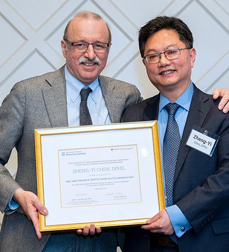
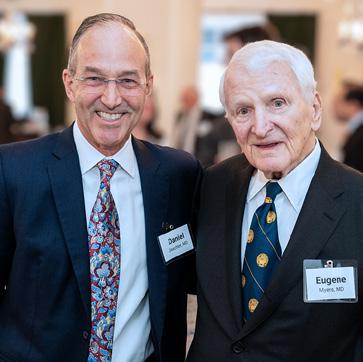
In the winter of 2024, Mass Eye and Ear hosted celebrations for Zheng-Yi Chen, DPhil, and Daniel G. Deschler, MD, FACS, in honor of being named inaugural incumbents of endowed chairs. Dr. Chen now serves as the Ines and Fredrick Yeatts Chair in Otolaryngology and Dr. Deschler now serves as the Dr. Eugene N. and Barbara L. Myers Chair in Head and Neck Surgery.
Heidi Nakajima, MD, PhD, was selected to present the 2024 Carhart Memorial Lecture at the Annual Scientific and Technology Conference hosted by the American Auditory Society. Her presentation was titled, “The Ear: In Search of Solutions for Engineers and Clinicians."
Pavan S. Mallur, MD, was inducted into the American Laryngological Association as an Active Fellow. Invited by peers, this honor recognizes specialists in the management of diseases or dysfunctions related to voice production, swallowing and breathing.
Artur A. Indzhykulian, PhD, was awarded a collaborative Department of Defense grant to develop treatments for noiseinduced hearing loss. The team will explore the use of FDA-approved drugs, employing a drug repurposing approach, to target the mitochondrial calcium uniporter (MCU) complex. Dr. Indzhykulian also received the Spark Grant through the Mass General Brigham Cell and Gene Therapy Institute for his work on the evaluation of human mini-PCDH15 in human inner ear organoids to treat USH1F.
Reza Rahbar, MD, DMD, was the 2024 recipient of the Gabriel Tucker Award from the American Laryngological Association. The award is a prestigious honor offered on
an annual basis to recognize an individual’s contributions to the field of Pediatric Laryngology for outstanding services to the Association.
Sukgi Choi, MD, Michael J. Cunningham, MD, and Anne F. Hseu, MD, authored a published manuscript titled “Diagnostic Accuracy of Ultrasound for Evaluating Vocal Fold Impairment in Children,” along with Boston Children’s Hospital surgical and radiology colleagues, that won three prestigious awards, including: Best Abstract Award at the 12th Annual Harvard Surgery Research Day; M. Judah Folkman Memorial Award for Outstanding Presentation in Clinical and Basic Science at the Annual Meeting of the American Pediatric Surgical Association; and Best Clinical Presentation Award at the New England Surgical Society Annual Meeting.
Stacey T. Gray, MD, was awarded the American Rhinologic Society Golden Head Mirror Award at the Annual Meeting of the American Rhinologic Society. This esteemed award honors meritorious teaching and mentorship in the field of Rhinology.
Ernest D. Gomez, MD, MTR, was named Multiple Principal Investigator on a R01 grant from the National Institutes of Health to develop a virtual reality-based training
New Leadership
Kristina Simonyan, MD, PhD, Dr med, was named Vice Chair of Clinical Research in the Department of Otolaryngology–Head and Neck Surgery at Mass Eye and Ear.
Amanda E. Dilger, MD, was appointed Chair of the Environmental Sustainability Task Force for the American Academy of Otolaryngology–Head and Neck Surgery.
Alex Grilli, MD, was named the Medical Director of the Quincy and Bridgewater practices at Mass Eye and Ear.
Matthew G. Crowson, MD, was named Director of Clinical Informatics and Artificial Intelligence in the Department of Otolaryngology–Head and Neck Surgery at Mass Eye and Ear.
simulator for transoral robotic surgery and to study the effects of transcranial direct current stimulation on acquisition of skill by surgeons.
Nicole Jiam, MD, and Divya Chari, MD, were awarded the Hearing Health Foundation Emerging Research Grant. This is a prestigious grant earned by promising investigators working on groundbreaking hearing research. Dr. Jiam will use the grant to study agespecific differences in electrical hearing neuroplasticity and hearing health and to develop personalized cochlear implant programs to improve music perception, and Dr. Chari will explore a possible link between a subtype of Meniere's disease and X-linked hypophosphatemia, a rare form of hereditary rickets.
As Principal Investigator, Karl R. Koehler, PhD, earned a R01 renewal grant from the National Institutes of Health to study mechanisms of human inner ear development to build a novel microphysiological system for gene therapy testing. Together, Dr. Koehler and Gwenaelle S. Géléoc, PhD, who serves as a collaborator on this grant, aim to demonstrate the utility of this model for tissue patterning to control the development of cochlear ducts and sensory cells.
The Sandy and Herb Pollack Young Investigator Award was awarded to Andreas Eckhard, MD, at the 2023 Mass Eye and Ear Annual Meeting of Trustees and Medical Staff. The award, which is endowed by the late Herb Pollack, a former Mass Eye and Ear Board member and trustee, will help expand an initiative by Dr. Eckhard to pinpoint the most critical molecular pathways implicated in the development of Meniere's disease in the Otopathology Laboratory at Mass Eye and Ear.

Aaron K. Remenschneider, MD, MPH, was invited to co-deliver the Presidential Symposium at the MidWinter Meeting of the Association for Research in Otolaryngology and received a Presidential Citation for his contributions to the success of the meeting.
23 HARVARD Otolaryngology
1 2
[1] Mark A. Varvares, MD, FACS, and Zheng-Yi Chen, DPhil. [2] Daniel G. Deschler, MD, FACS, and Eugene N. Myers, MD, FACS.
HIGHLIGHTS
The first edition of Otologic and Lateral Skull Base Trauma textbook was published in the fall of 2023. Elliott D. Kozin, MD, was the editor, with several other faculty from the Harvard Department of Otolaryngology–Head and Neck Surgery as contributors. The textbook is the first of its kind to explore in depth the many aspects of temporal bone trauma, including its epidemiology, diagnosis, and medical and surgical management, and contemporary research.

Drs.
Heidi Nakajima and Stacey Gray Named Next
Incumbents of Harvard Professorships
Ravindra Uppaluri, MD, PhD, was invited to deliver the keynote address at the 2024 Multidisciplinary Head and Neck Cancers Symposium in Scottsdale, AZ. His lecture was titled, “Evolving Role of Neoadjuvant Immunotherapy in Curative-Intent Head and Neck Cancer.”
Cochlear Americas awarded Julie G. Arenberg, PhD, CCC-A, and Rachel Petrie, AuD, CCC-A, a research grant to investigate the possibility to reduce the number and duration of appointments for patients who recently received cochlear implants to treat their severe-to profound hearing loss. Specifically, the duo is assessing the need for one of their standard follow-up visits in the first month post cochlear implantation, with and without the use of an automated programming approach.
The Harvard Department of Otolaryngology–Head and Neck Surgery launched the inaugural continuing medical education (CME) course “Technology and Innovation in Endocrine Head and Neck Surgery,” directed by Gregory W. Randolph, MD, FACS, FACE, and Marika Russell, MD.
The “Endoscopic Surgery of the Sinuses, Eustachian Tube and Ear,” CME course has been offered by the Harvard Department of Otolaryngology–Head and Neck Surgery for the past two decades. This year, the course was directed by Ralph B. Metson, MD, Dennis S. Poe, MD, PhD, and Daniel J. Lee, MD, FACS, and co-directed by Stacey T. Gray, MD, Eric H. Holbrook, MD, and Elliott D. Kozin, MD.
Two Mass Eye and Ear faculty have been awarded endowed Harvard professorships. Heidi Nakajima, MD, PhD, was named the Gudrun Larsen Eliasen and Nels Kristian Eliasen Associate Professor of Otolaryngology–Head and Neck Surgery and Stacey T. Gray, MD, was named the Walter Augustus Lecompte Associate Professor of Otolaryngology–Head and Neck Surgery.
The Gudrun Larsen Eliasen and Nels Kristian Eliasen Professorship was established in 2003, with the initial incumbent Saumil N. Merchant, MD, a pioneering surgeon and scientist who specialized in human otomechanics. Dr. Merchant’s professorship was succeeded by the renowned middle-ear mechanics researcher, John J. Rosowski, PhD, who held this title from 2012-2023.
The Walter Augustus Lecompte Professorship was originally established in 1907, the first ever endowed chair for the Department of Otolaryngology–Head and Neck Surgery. That same year, otology great Clarence Blake, MD, was named the first incumbent. Since then, incredible pioneers in the field of otolaryngology have bestowed this honor, including Joseph B. Nadol, MD, and D. Bradley Welling, MD, PhD, FACS.
Both professorships include endowments that will support Drs. Nakajima’s and Gray’s respective academic research.
Harvard Medical School Celebrates Drs. Daniel Polley’s and Kristina Simonyan’s Promotion to Professor
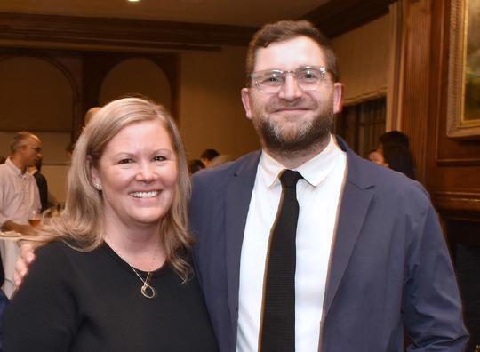
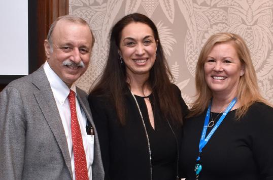
Harvard Medical School faculty and Mass Eye and Ear leadership gathered on two separate occasions at the Harvard Faculty Club in Cambridge, MA to celebrate the promotions of Daniel B. Polley, PhD, Director of the Eaton-Peabody Laboratories at Mass Eye and Ear and Kristina Simonyan, MD, PhD, Dr med, Vice Chair of Clinical Research at Mass Eye and Ear, to Professor of Otolaryngology–Head and Neck Surgery at Harvard Medical School.
Throughout the evenings, we heard from several colleagues of both Drs. Polley’s and Simonyan’s who shared memories and anecdotes of their professional and personal experiences. The rooms were filled with leadership, faculty and trainees, along with family members.
24 HARVARD Otolaryngology
s
[1] CarolAnn Williams, Mass Eye and Ear President and Daniel B. Polley, PhD.
1
[2] Mark A. Varvares, MD, FACS, Kristina Simonyan, MD, PhD, Dr med, and CarolAnn Williams
2
RESEARCH ADVANCES s
The following are select research advances from the Harvard Medical School Department of Otolaryngology–Head and Neck Surgery.
Gene therapy restores hearing in children with inherited hearing loss
Hearing loss affects more than 1.5 billion people worldwide, with congenital deafness making up about 34 million of those individuals. For hearing loss specifically in children, 60 percent emerge from genetic reasons. Autosomal recessive deafness 9 (DFNB9), caused by mutations of the OTOF gene, is manifested by severe-to-complete bilateral hearing loss. Currently, there is no drug medication available for congenital deafness.

Zheng-Yi Chen, DPhil, of Mass Eye and Ear, and colleagues at the Eye & ENT Hospital of Fudan University in Shanghai, China, published results of a clinical trial in The Lancet, reporting the safety and efficacy of gene therapy with an adeno-associated virus (AAV) serotype 1 carrying a human OTOF transgene (AAV1-hOTOF) as a treatment for children with DFNB9.
The research team screened 425 participants for eligibility and enrolled six eligible participants in the clinical trial, three girls and three boys of Han ethnicity all between two and five-years old. A single injection of AAV1-hOTOF was administered into the cochlea through the round window and all six participants had to follow up 26 weeks later. The results revealed five of the six children had recoveries in their hearing, and dramatic improvements in speech perception and conversational abilities.
Lv J, Wang H, Cheng X, Chen Y, Wang D, Zhang L, Cao Q, Tang H, Hu S, Gao K, Xun M, Wang J, Wang Z, Zhu B, Cui C, Gao Z, Guo L, Yu S, Jiang L, Yin Y, Zhang J, Chen B, Wang W, Chai R, Chen ZY, Li H, Shu Y. AAV1-hOTOF gene therapy for autosomal recessive deafness 9: a single-arm trial. The Lancet. 2024. doi:10.1016/S0140-6736(23)02874-X.
A new approach to measure hearing abilities in laboratory animals
Involuntary behaviors occur less than one second after intense sound reaches the ear, including but not limited to pupil dilation, muscle contractions and eardrum tension. These rapid responses are mediated by independent neural pathways that protect the inner ear from acoustic injury.
insights into complex sound processing that should extend the reach of hearing research in laboratory animals.
Clayton K., Stecyk K., Guo A., Chambers A., Chen K., Hancock K., Polley D. Sound elicits stereotyped facial movements that provide a sensitive index of hearing abilities in mice. Current Biology. 2024; Vol 34(8):1605-1620. doi.10.1016/j. cub.2024.02.057
Conventional auditory reflexes are often insensitive to low and moderate sound levels within the audible range and do not reflect the involvement of neural circuits beyond the brainstem, limiting their use to assess overall hearing ability. Daniel B. Polley, PhD, and Kameron Clayton, PhD, both of Mass Eye and Ear, led a study that reports a new way to measure hearing abilities in laboratory animals— requiring nothing beyond a high-resolution video camera and a computer.
The findings, published in Current Biology, report that sound elicits rapid facial twitches that provide a more reliable and sensitive index of hearing than other involuntary reflexes. Facial twitches synchronized to the spectrotemporal envelope of ongoing sounds, allowed accurate reconstruction of complex sounds such as phoneme sequences presented in background noise. Further, recordings of mice with mutations in autism risk genes or noise-induced sensorineural hearing loss identified behavioral signatures of auditory hypersensitivity, demonstrating that facial videography provides a measure of cochlear function as well as plasticity in central auditory processing. This measure is complementary to methods like the auditory brainstem response and otoacoustic emissions and provides new
Vocal fold paralysis function regained and recurrent laryngeal nerve preserved in advanced thyroid cancer patients subsequent to neoadjuvant therapy, the “Lazarus Effect”
Advanced thyroid cancers may invade recurrent laryngeal nerve (RLN) which can result in vocal fold paralysis (VFP). VFP not only impacts the patient’s quality of life but also affects the ability to successfully remove the tumor due to a significant risk of bilateral VFP during surgery that would result in a tracheotomy.
Gregory W. Randolph, MD, FACS, FACE, of Mass Eye and Ear, led a multicenter study that identified and reported the return of vocal fold motion in addition to the surgical preservation of the invaded recurrent laryngeal nerves after the use of neoadjuvant therapy in patients presenting with advanced thyroid cancer and vocal fold paralysis. This is the first report dedicated to studying the return of laryngeal function as a standalone outcome in patients diagnosed with advanced thyroid cancers with invaded RLNs regardless of pathology, underlying genetic alterations or type of neoadjuvant therapy used. This challenges the prior assumption that carcinomatous neural invasion is always permanent and suggests a more dynamic invasive pathophysiology than previously understood.
The findings, published in Thyroid, revealed that neoadjuvant therapy in the setting of advanced thyroid cancer not only was
25 HARVARD Otolaryngology
RESEARCH ADVANCES s
associated with return of function but allows the ultimate thyroid cancer surgery to spare the nerve in the majority of cases. The majority of patients who had VFP before neoadjuvant therapy experienced return of function of the nerves that were paralyzed within three months and had a sufficient clinical response to allow surgical preservation of most RLNs.
Silver Karcioglu A, Abdelhamid Ahmed AH, Feng Z, Russell M, Shonka DC Jr, Iwata A, Cabanillas M, Shin JJ, Kyriazidis N, Park JC, Wirth LJ, Zafereo ME, Randolph GW. Return of vocal fold motion and surgical preservation of invaded recurrent laryngeal nerves after the use of neoadjuvant therapy in patients presenting with advanced thyroid cancer and vocal fold paralysis: The lazarus effect. Thyroid. 2023; Vol 33(10). Doi: 10.1089/thy.2023.0136.
TMC function, dysfunction and restoration in vestibular organs
Inner ear abnormalities affect more than 400 million individuals globally. One debilitating symptom of these abnormalities are vestibular disorders, affecting approximately 15 percent of adults in the United States. Vestibular hair cells are critical in detecting and transmitting mechanical signals related to balance and when these cells fail to properly function, symptoms of dizziness, imbalance and nausea may arise. Therefore, clarifying when and where the mechanosensory transduction components are expressed is deemed essential for optimizing genetic diagnostics and gene therapy interventions for vestibular dysfunction.
Jeffrey R. Holt, PhD, of Boston Children's Hospital, Evan Ratzan, PhD, a postdoctoral research fellow in the Holt laboratory, and colleagues investigated the expression and function of Tmc1 and Tmc2 in vestibular organs using various techniques including qPCR, FISH-HCR, FM1-43 and measuring vestibular evoked potentials (VsEPs) and vestibular ocular reflexes (VORs). Their findings, published in Frontiers in Neurology, revealed that Tmc1 knockout (KO) mice lack VsEPs, which are normally activated during head movements.
The Tmc1 gene encodes for the TMC1 protein, the pore-forming subunit of the sensory transduction complex in hair cells of the inner ear. TMC1 is critical for hair cell function and mutations in the TMC1 gene lead to hearing loss in human patients. In mice, Tmc1 KOs lack this gene and as a result, lose cochlear hair cells in early adolescence, but, surprisingly, their vestibular hair cells remain into adulthood. The study demonstrated that gene replacement of Tmc1 through utricle injection of an AAV encoding Tmc1 is sufficient to restore VsEP signal in mice for up to six months.
Ratzan,
15. Doi: 10.3389/fneur.2024.1356614
Circulating
tumor-tissue modified HPV DNA testing in patients at risk for HPV-positive oropharynx cancer
Human papillomavirus (HPV)-positive oropharyngeal squamous cell carcinoma (OPSCC) has surpassed cervical cancer as the most prevalent HPV-related cancer in the United States, primarily affecting middle to older-aged men. Although survival outcomes are favorable for HPV-positive OPSCC, there is a need for improved diagnostic tools to detect the disease earlier to decrease treatmentrelated morbidity and mortality for this growing patient population.
In a prospective cohort study led by Eleni M. Rettig, MD, of Brigham and Women’s Hospital, the team evaluated circulating tumor tissuemodified viral (TTMV)-HPV DNA plasma testing to facilitate early diagnosis of HPVpositive OPSCCs. There were 39 patients enrolled in the study, all presenting with either tonsil asymmetry, a neck mass and or throat pain.
The results, published in Oral Oncology, determined that two of the three subjects diagnosed with HPV-positive OPSCC, had
detectable TTMV-HPV DNA. The findings indicated that the test was feasible and was perceived as useful by physicians, holding the potential as a diagnostic aid for HPV-positive OPSCC alongside standard clinical workup. However, the researchers noted that clinicians should be cognizant of its limitations, as a negative test does not necessarily indicate the absence of disease.
Batool S, Sethi RKV, Wang A, Dabekaussen K, Egloff AM, Del Vecchio Fitz C, Kuperwasser C, Uppaluri R, Shin J, Rettig EM. Circulating tumor-tissue modified HPV DNA testing in the clinical evaluation of patients at risk for HPV-positive oropharynx cancer: The IDEA-HPV study. Oral Oncology. 2023. Vol 147. doi: 10.1016/j.oraloncology.2023;106584.
Auditory neuron loss in normalhearing patients with tinnitus
Tinnitus, which is perceived as a ringing or other sounds in the ears, is estimated to affect more than one out of 10 adults worldwide. These symptoms are taxing, often resulting in sleep deprivation, social isolation, anxiety, depression, decline in work performance and an overall negative impact on quality of life. For years, scientists hypothesized that the cause of tinnitus was a result of maladaptive plasticity of the brain, thought to occur as a result of peripheral damage. However, this idea was challenged due to patients with normal hearing tests reporting tinnitus.
A research team led by Stéphane F. Maison, PhD, CCC-A, CH-TM, of Mass Eye and Ear, conducted a study aiming to search for peripheral hearing loss in patients with normal audiograms reporting tinnitus. The study involved 294 normal-hearing participants, 18-years-old to 72-years-old, split into three groups: 201 adults never experiencing tinnitus, 64 who have had temporary tinnitus and 29 suffering from chronic tinnitus.
The findings, published in Scientific Reports, revealed that chronic tinnitus is associated with various changes in auditory processing including reduced cochlear nerve responses, weaker middle-ear muscle reflexes, stronger medial olivocochlear efferent reflexes and hyperactivity in the central auditory
26 HARVARD Otolaryngology
E, Lee, J, Madison, M, Zhu, H, Zhou, W., Géléoc, G, & Holt, J. TMC function, dysfunction, and restoration in mouse vestibular organs. Frontiers in Neurology. 2024; Vol
pathways. The results suggest that tinnitus may be triggered by a loss of auditory neurons, including individuals with normal audiograms.
Vasilkov V, Caswell-Midwinter B, Zhao Y, de Gruttola V, Jung DH, Liberman MC, Maison SF. Evidence of cochlear neural degeneration in normal-hearing subjects with tinnitus. Scientific Reports. 2023. doi: 10.1038/s41598-023-46741-5
Voice
disorders prevalence increasing in post-COVID
era
Following the COVID-19 pandemic, there have been speculations that voice disorders are on the rise due to dramatic changes in lifestyle. Wearing facial masks for protection, conducting teleconferences or phone meetings and utilizing virtual assistants all require different levels of voice strain that individuals were not familiar with previously.
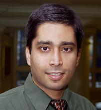
In 2012, the National Health Interview Survey (NHIS), a cross-sectional survey administered by the U.S. Census Bureau that assesses the prevalence of many health conditions, included a section specifically discussing adult voice, speech and language disorders. In a recent study published in The Laryngoscope, Neil Bhattacharyya, MD, FACS, and Matthew R. Naunheim, MD, MBA, both of Mass Eye and Ear, and colleagues analyzed the 2022 NHIS for changes in the incidence of voice problems over the past 10 years.
Their findings concluded that nearly one in eight adults are affected by a voice issue annually, a notable increase since 2012, which was approximately one in 13 adults. Patients with long-COVID were significantly more likely to be diagnosed with dysphonia.
Naunheim MR, DeVore EK, Huston MN, Song PC, Franco RA Jr, Bhattacharyya N. “Increasing prevalence of voice disorders in the USA: Updates in the COVID era.” The Laryngoscope. 2024. doi: 10.1002/lary.31409.
Vestibular information affects navigation in virtual environments differently in men and women
Navigation is a complex function, which involves tracking an individual’s position within a spatial environment while moving towards a particular destination. Vestibular information is accessible to the brain during navigation, as are other idiothetic, or self-generated, and allothetic, or external, sensorimotor cues. Previous studies conducted in rodents have shown that vestibular information contributes to navigation, but human studies have been significantly less conclusive. Similarly, there is no evidence on whether men and women utilize vestibular information differently during navigation.
In a study published in Brain Communications, Richard F. Lewis, MD, of Mass Eye and Ear, and a team of researchers studied navigation in a virtual reality environment in patients with bilateral vestibular damage and in normal subjects. The team used two paradigms to investigate— a static paradigm where the subject was stationary and navigated with key presses and a dynamic paradigm where subjects walked and turned on a platform, thereby activating the vestibular sensors in the inner ear.
The findings uncovered that men, both with vestibular damage and normal subjects, used vestibular information to navigate in the virtual environment and women did not. This is a first of its kind study to suggest that men and women utilize vestibular information differently when they navigate their surroundings.
Chari DA, Ahmad M, King S,
A,
C, Panic AS, Karmali F, Lewis RF.
damage affects the precision and accuracy of navigation in a virtual visual environment. Brain Communications. 2023; Vol 5(6) doi: 10.1093/ braincomms/fcad345.
Neural circuits that assess whether sounds are threatening
To survive, all mammals must evaluate and respond to potential auditory threats in the environment. The amygdala, cholinergic
basal forebrain and higher-order auditory cortex play a significant role in monitoring the brain’s ability to change in response to auditory threats. Threat evaluation has been modeled using Pavlovian auditory fear conditioning paradigm, where a neutral sound paired with an aversive stimulus is used to evoke defensive behaviors and profound autonomic responses.
Daniel B. Polley, PhD, of Mass Eye and Ear, led a study that explores the connection between how the acoustic and emotional features of sound are processed by the brain. The researchers used cuttingedge neurophysiological methods to observe changes in the auditory cortex and amygdala as mice learn to treat an otherwise neutral sound as a potential threat.
The findings, published in Cell Reports, indicated that responses were heightened in the lateral amygdala (LA), but not in the higher-order auditory cortex, suggesting a distributed and cooperative plasticity in LA inputs as mice learn to reassess neutral stimuli as possible threats. The dynamics of these circuits studied in the context of natural learning might identify a biological basis for hearing disorders such as hyperacusis, tinnitus and misophonia, which can feature an intense aversion to innocuous sounds.
Asokan MM, Watanabe Y, Kimchi EY, Polley DB. Potentiation of cholinergic and corticofugal inputs to the lateral amygdala in threat learning. Cell Reports. 2023. Vol 42(10) doi: 10.1016/j.celrep.2023;113167.
The relationship between personal social networks and seeking care for head and neck cancer
Head and neck cancer accounted for 3.4 percent of malignancies in 2023, with approximately 15,000 deaths annually. Finding symptoms early coupled with a prompt referral dramatically helps the prognosis, while treatment delays are
27 HARVARD Otolaryngology
Boutabla
Fattahi
Vestibular
RESEARCH ADVANCES s
associated with lower survival rate or higher rate of recurrence. Studies have shown that the size of an individual’s social network— including family, friends, acquaintances, coworkers etc.—has impacted hospital arrival times for medical emergencies, but there had been limited research on social network influence on care-seeking among head and neck cancer patients.
A collaborative study led by Regan W. Bergmark, MD, MPH, of Brigham and Women’s Hospital, was conducted to understand how patients' personal social networks influence their access to care for head and neck cancer from the perspectives of both patients and healthcare workers. The study involved interviews with 42 patients newly diagnosed with head and neck cancer and 30 health care workers providing care for such patients, conducted at two academic medical centers.
The findings, published in Otolaryngology–Head and Neck Surgery revealed that patients' social networks are crucial in prompting early care-seeking among head and neck cancer patients. Additionally, implementing social network-based interventions could enhance care and improve outcomes for this patient population.
Batool S, Hansen EE, Sethi RKV, Rettig EM, Goguen LA, Annino D, Uppaluri R, Edwards HA, Faden DL, Dohan D, Dhand A, Reich AJ, Bergmark RW. Personal social networks and care-seeking for head and neck cancer: A qualitative study. Otolaryngology–Head and Neck Surgery. 2024; Vol 170(2):457-467. doi: 10.1002/ohn.602.
Costs of facial fracture fixation materials remain out of reach for majority of the world
Trauma accounts for more global deaths than HIV/AIDS, malaria and tuberculosis combined, yet trauma garners little worldwide attention. Facial fractures make up a significant portion of traumatic injuries, the majority of which occur in lowand middle-income countries such as those across Africa and Asia. The costs of plate and screw fixation materials are a limitation
to fracture fixation as patients bear the majority of these costs out of pocket.
David A. Shaye, MD, MPH, FACS, of Mass Eye and Ear, and PGY-3 resident Obinna Nwosu, MD, along with a group of international facial trauma surgeons analyzed standard plate and screw fixation costs in 14 countries across Africa and Asia. The cost burden of these materials was presented as a percentage of gross domestic product (GDP) per capita to assess affordability. These findings, published in OTO Open, determined the costs of a single six hole and six screws range from 0.6 – 23.2 percent of GDP per capita, with an average cost representing 10 percent of GDP per capita.
The authors concluded that these costs are significantly out of reach for the majority of facial trauma patients in countries in Africa and Asia. Future efforts to adopt low-cost alternatives to facial trauma repair have the potential to expand care to more patients globally.
Shaye DA, Nwosu O, Ncogoza I, et al. Cost Burden of Rigid Internal Fixation in Craniomaxillofacial Trauma Care in Low- and Middle-Income Countries. OTO Open. 2023; Vol 7(4) doi:10.1002/oto2.92
Cochlear implants and the vestibular system in children
Cochlear implants have offered many children with profound hearing loss the opportunity to hear and to gain improved access to spoken language, even when the hearing loss is too severe for hearing aids to be beneficial. However, concerns have long been expressed about the risks of the surgical insertion of a cochlear implant electrode array on the integrity of the vestibular organs in the inner ear. A number of studies have previously shown that cochlear implant surgery can often cause impairment of the vestibular organs, which can negatively impact balance and motor development in children. Recently, cochlear
implant electrode array designs and surgical approaches have increasingly focused on minimizing trauma to the inner ear with the goal of preserving residual hearing in patients with partial hearing loss.
In a study published in The Laryngoscope, Greg R. Licameli MD, Jacob R. Brodsky MD, FACS, FAAP, and their colleagues in the Cochlear Implant Program at Boston Children’s Hospital set out to assess the impact of hearing preservation approaches on vestibular function in pediatric cochlear implant recipients. They performed a retrospective review of pre- and postoperative vestibular testing results on children under-going cochlear implantation using slim, non-styletted electrode arrays and a round window insertion approach. The team then compared the results to patients implanted with less atraumatic approaches from data in a previous study by the same group performed 15 years ago before hearing preservation approaches were commonplace.
The findings from the study demonstrated that the risk of a decrease in vestibular function following cochlear implant surgery in children is much lower using hearing preservation type approaches compared to more traditional approaches. This suggests that patients may benefit from such atraumatic approaches to cochlear implant surgery even in the absence of significant residual hearing, particularly in settings where concerns for balance or motor impairment are high, such as with a pre-existing unilateral vestibular loss or other concurrent developmental concerns.
Licameli GR, Wang A, Zhou G, Faller D, Kenna M, Poe D, Shearer E, Oster L, Brodsky JR. Vestibular preservation in pediatric cochlear implantation. The Laryngoscope. 2024; Vol 134(4):1913-1918. doi: 10.1002/lary.30978.
28 HARVARD Otolaryngology
Join us in July for Mass Eye and Ear’s 200th Anniversary Celebration!
On Friday, July 19, 2024, all current and former Department of Otolaryngology–Head and Neck Surgery faculty and trainees are invited to return to Boston for the hospital’s bicentennial celebration. Festivities include lectures by distinguished alumni, tours of the hospital and a gala dinner.
Please RSVP by July 1.
For more information use the QR code below or contact Dana Grider, Director of Development for the Department of Otolaryngology–Head and Neck Surgery at Mass Eye and Ear, at dgrider@meei.harvard.edu or 617-573-4223.
We hope to see you there!


Connect with us on LinkedIn: All former residents and fellows are invited to join the Harvard Otolaryngology–Head and Neck Surgery Alumni Group.
Photo by Garyfallia Pagonis
News from the Harvard Medical School
Department of Otolaryngology–Head and Neck Surgery
243 Charles Street, Boston, MA 02114
Beth Israel Deaconess Medical Center
Boston Children’s Hospital
Brigham and Women’s Hospital
Mass Eye and Ear
Massachusetts General Hospital
