[ page 4 ]

Department of Otolaryngolog y Head and Neck Surger y Spring 2023, vol. 19, no. 1
Inside
A Line of Defense Hidden
the Nose
CONTENTS
1 Letter From the Chair
Mark A. Varvares, MD, FACS
News from the Harvard Medical School
Department of Otolaryngology–Head and Neck Surgery
Spring 2023 | Vol. 19, No. 1
Published twice per year.
Please send comments, requests for additional copies and other inquiries regarding this issue to:
Michael Kotsopoulos
Senior Communications Manager
Department of Otolaryngology–Head and Neck Surgery

Massachusetts Eye and Ear
243 Charles Street, Boston, MA 02114
mkotsopoulos@meei.harvard.edu
CONTRIBUTORS
Editor-in-Chief
Mark A. Varvares, MD, FACS
John W. Merriam and William W. Montgomery
Professor and Department Chair of Otolaryngology–Head and Neck Surgery
Harvard Medical School
Chief of Otolaryngology–Head and Neck Surgery
Massachusetts Eye and Ear
Massachusetts General Hospital
Managing Editor | Writer
Michael Kotsopoulos
Design | Layout | Photography
Garyfallia Pagonis
Department of Otolaryngolog y Head and Neck Surger y
Beth Israel Deaconess Medical Center
Boston Children’s Hospital
Brigham and Women’s Hospital
Massachusetts Eye and Ear
Massachusetts General Hospital
©2023, Massachusetts Eye and Ear
2 COSM 2023
Celebrating Two Centuries of Otolaryngology at Mass Eye and Ear
16 In Memoriam
Nelson Y.S. Kiang, PhD
18 Donor Profile
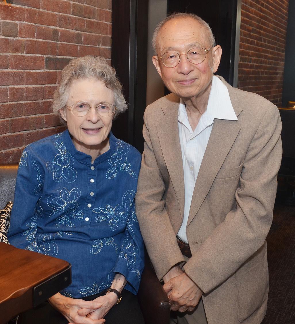
Ines and Fredrick Yeatts, PhD
19 Alumni Giving Society
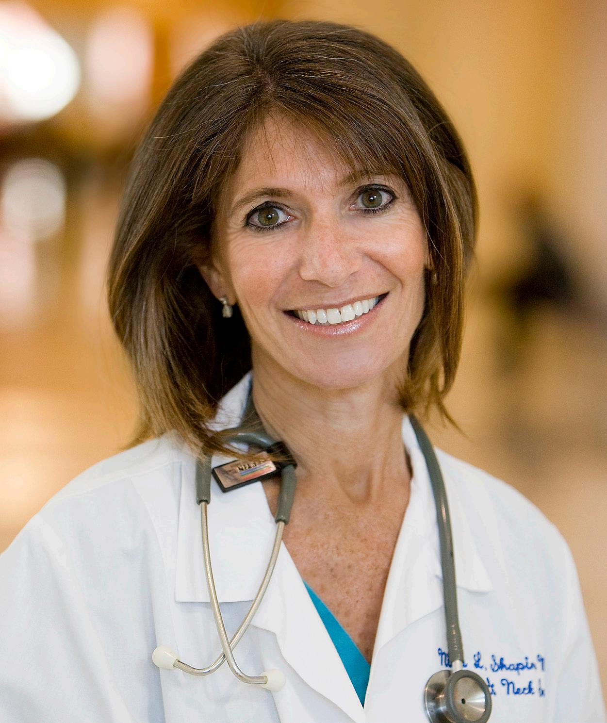
20 Alumni Profile
Nina Shapiro, MD, ’96
22 Highlights
25 Research Advances
FEATURES
4 A Line of Defense Hidden Inside the Nose
The discovery of an innate immune response at the front of the nose has provided an explanation for why colder temperatures correlate to upper respiratory viruses. It could also break ground on a new generation of therapeutics.
8 Artificial Intelligence Enters the Ear
Surgeons at Mass Eye and Ear have engineered an artificial intelligence device that could diagnose a middle ear infection with higher accuracy than a clinician.
12 Expanding Immunotherapy to Fight Head and Neck Cancer
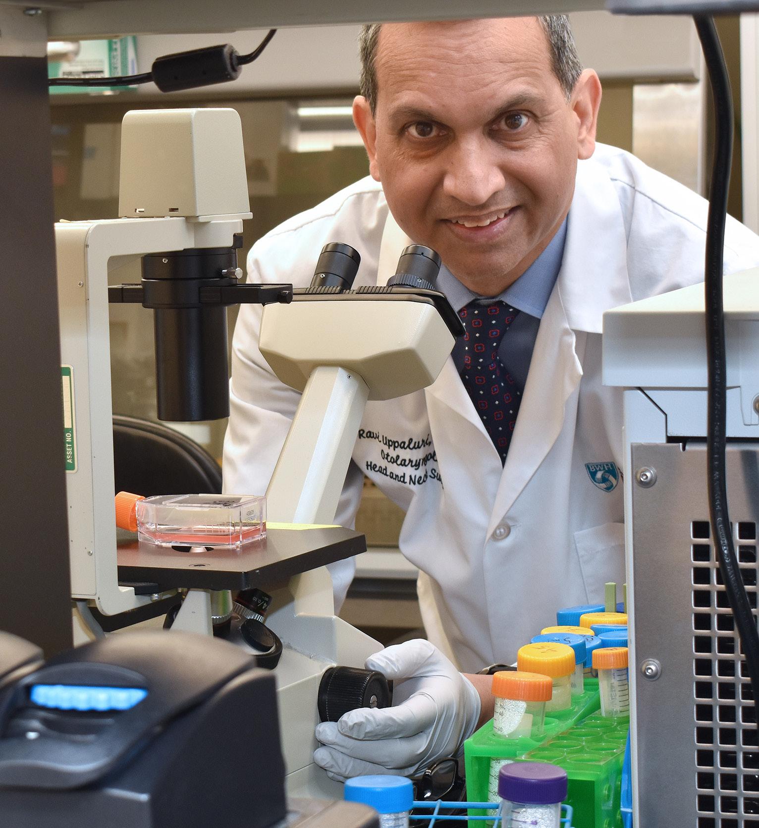
At Brigham and Women’s Hospital, Ravindra Uppaluri, MD, PhD, is working to make immunotherapy a reality for surgical patients with head and neck cancer.
Dear colleagues and friends,
In January 2023, it was my pleasure to welcome faculty from the entire Harvard Medical School (HMS) Department of Otolaryngology–Head and Neck Surgery to the Museum of Science in Boston for our first department-wide meeting since the COVID-19 pandemic.
It was a cold Saturday morning, and I was grateful that— after a busy week of cases in the clinic, operating room and laboratory—our faculty were willing to give up a part of their weekend to engage in a series of meaningful discussions relevant to our department and the medical school.
The meeting was well received by our faculty, who began the day with an address by George Daley, MD, PhD, Dean of the Faculty of Medicine at HMS, on the state of the Harvard medical program. Dr. Daley was joined by fellow HMS leaders Grace C. Huang, MD, Dean for Faculty Affairs, and Anne E. Becker, MD, PhD, ScM, Dean for Clinical and Academic Affairs, both of whom shared Dr. Daley’s united vision of the school’s future. Leaders from our affiliate hospitals also had the opportunity to share goals from their respective institutions, as well as highlight the most recent accomplishments of faculty from each location.
Personally, I was most inspired by how our faculty responded to our panel on implicit bias. The Department of Otolaryngology–Head and Neck Surgery upholds diversity, equity and inclusion as three of its core values, and identifying and managing bias in our everyday work can help derive equity in the care of patients, the quality of our research and the training we provide the next generation of otolaryngologists.
Led by David Brown, MD, a graduate of HMS who now serves as Associate Vice President and Associate Professor of Otolaryngology–Head and Neck Surgery at the University of Michigan Medical School, the panel made way for several powerful moments between panel members and faculty, many of whom shared personal experiences dealing with bias against them. In the subsequent weeks, I was proud to witness several faculty conscientiously evaluating research through the lens of these discussions.
In this latest edition of Harvard Otolaryngology, I hope you feel as inspired by our faculty as I have been since the January meeting. Their findings on head and neck cancer immunotherapy, artificial intelligence diagnostics and a new line of defense inside the nose are just the beginning of our department’s extraordinarily bright future.
As always, I thank you for your continued support of our community.
Sincerely,
Mark A. Varvares, MD, FACS

The John W. Merriam and William W. Montgomery Professor and Chair Department of Otolaryngology–Head and Neck Surgery, Harvard Medical School
Chief, Departments of Otolaryngology–Head and Neck Surgery, Massachusetts Eye and Ear Massachusetts General Hospital
1 HARVARD Otolaryngology
FROM THE CHAIR
LETTER
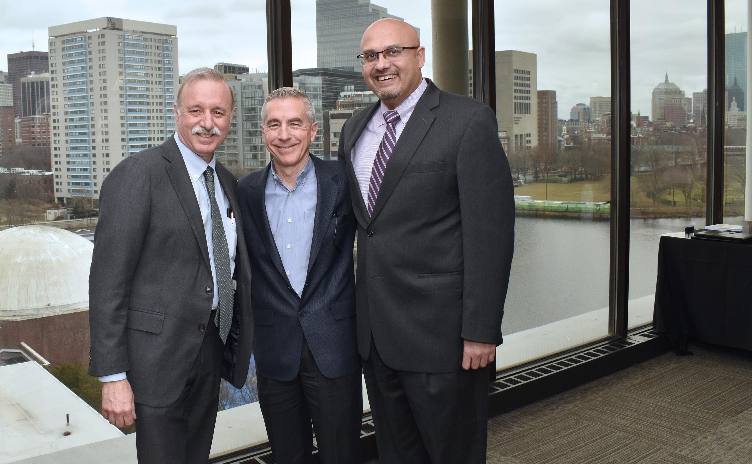
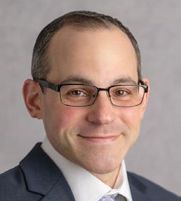
Left: Mark A. Varvares, MD, FACS; Michael J. Cunningham, MD, FACS; and Scharukh M. Jalisi, MD, (pictured left to right) at the Museum of Science.
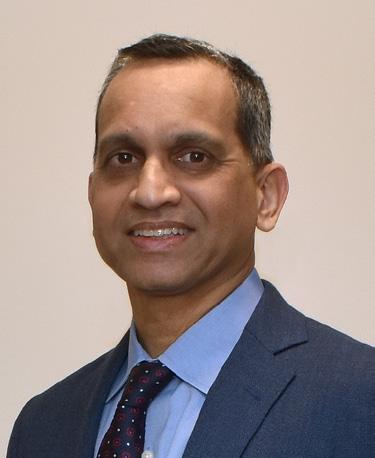
Above: Ravindra Uppaluri, MD, PhD, who was unable to attend the January meeting for personal reasons.
Mass Eye and Ear Bicentennial Celebration Launched at COSM
Former and current Harvard Medical School faculty and trainees worldwide gathered in Boston to celebrate the beginning of the hospital’s year-long celebration.
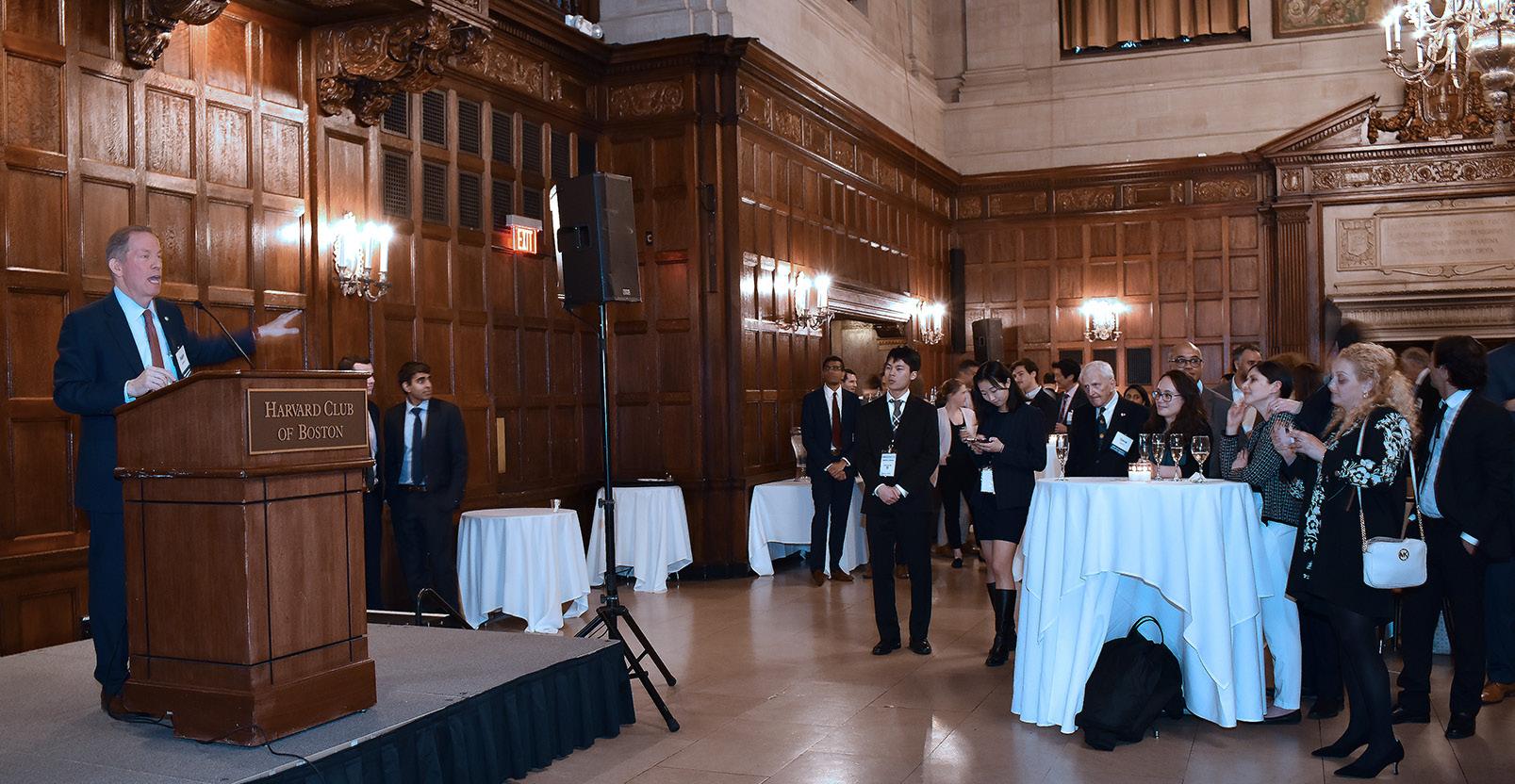
On the evening of Friday, May 5, the Department of Otolaryngology–Head and Neck Surgery (OHNS) at Mass Eye and Ear launched its yearlong celebration of the hospital’s 200th anniversary at the Harvard Club in Boston. The celebration occurred against the backdrop of the Combined Otolaryngology Spring Meetings (COSM) at the nearby Hynes Convention Center, drawing hundreds of trainees and faculty from the Department of OHNS at Harvard Medical School.
Ralph B. Metson, MD, Professor of OHNS at Harvard Medical School and Surgeon and Physician at Mass Eye and Ear, began the evening with a toast to every OHNS faculty member and trainee—past and present—who helped build Mass Eye and Ear into one of the world’s premier centers for otolaryngological care, research and training. To symbolize the longevity of their commitment to the hospital, Dr. Metson brought with him a piece of the cornerstone from Mass Eye and Ear’s current location, which was constructed in 1900.
“There is no Mass Eye and Ear without the extraordinary individuals who have contributed so much to this institution throughout these past 200 years,” Dr.
Metson said. “Their steadfast commitment will forever serve as the cornerstone for a brighter future in our specialty.”
The reception culminated in the naming of three Honorary Chairs for the department’s year-long celebration. Alumni Celebration Co-Chairs Michael Rho, MD, FACS, President of the OHNS Alumni Association at Mass Eye and Ear, and Gregory W. Randolph, MD, FACS, FACE, the Claire and John Bertucci
Chair in Thyroid Surgical Oncology at Harvard Medical School, bestowed the much-anticipated honor to Charles W. Cummings, MD; Eugene Myers, MD, FACS; and Herbert Silverstein, MD, FACS, each of whom completed their residency training at the Harvard Otolaryngology Residency Program.

2 HARVARD Otolaryngology NEWS
Endowed
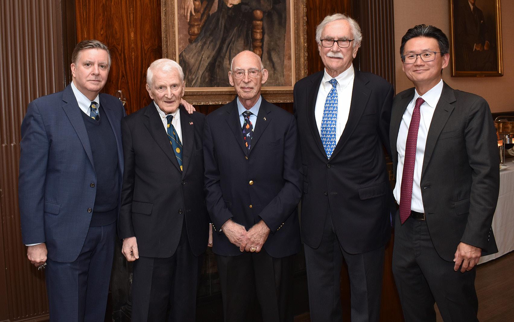
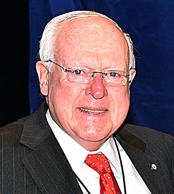
[ 1 ] Alumni Celebration Co-Chairs posing with the three Honorary Chairs. From left to right: Gregory W. Randolph, MD, FACS, FACE; Eugene Myers, MD, FACS; Herbert Silverstein, MD, FACS; Charles W. Cummings, MD; Michael Rho, MD, FACS.
[ 2 ] Ralph B. Metson, MD, welcoming guests to the Harvard Club for a night of celebration.
1 2
[ 3 ] Current and former faculty and trainees enjoying the first of many festivities in honor of Mass Eye and Ear’s 200th Anniversary.
The Honorary Chairs are luminaries in the field of otolaryngology–head and neck surgery. Dr. Myers served as Chair of the Department of OHNS at the University of Pittsburgh School of Medicine for more than 30 years, and Dr. Cummings served as Chair of the Department of OHNS at the University of Washington and The Johns Hopkins School of Medicine for more than 10 years at both institutions. Both have authored hundreds of publications, served in innumerable leadership positions within the specialty and began their medical careers serving in the United States Military. Dr. Silverstein is President and Founder of the internationally acclaimed Silverstein Institute and the Ear Research Foundation in Sarasota, FL, where he has pioneered treatments for Meniere’s disease. He has authored more than 250 scientific publications, written six books and trained more than 50 fellows in otology and neurotology.
Each Honorary Chair will assist with the planning of a day-long 200th Anniversary Alumni Event and Gala in Boston on Friday, July 19, 2024. According to Mark A. Varvares, MD, FACS, Chair of the Department of OHNS at Harvard Medical School and Chief of OHNS at Mass Eye and Ear and Massachusetts General Hospital, the Alumni Event and Gala will not only commemorate the storied history of Mass Eye and Ear, but the faculty and trainees whose tireless efforts have allowed the hospital’s founding mission to stand the test of time.
“It’s awe-inspiring to think that 200 years ago, the bold vision, courage and compassion of our founding physicians would help forever change the fields of ophthalmology and otolaryngology as they knew it,” said Dr. Varvares, who also serves as an Alumni Celebration Co-Chair. “Were they alive today, they would be astonished by the monumental advances achieved in specialty care, the boundless promise of today’s research and the far-reaching impact of our training programs.” n
Harvard Medical School Faculty Awarded Presidential Citations from Triological Society
Three distinguished Harvard Otolaryngology Head and Neck Surgery (OHNS) faculty members were awarded Presidential Citations by the Triological Society at the Combined Otolaryngology Spring Meetings (COSM).
Mark A. Varvares, MD, FACS, Chief of OHNS at Mass Eye and Ear; Stacey T. Gray, MD, Vice Chair of Otolaryngology Education at Mass Eye and Ear; and Gerald B. Healy, MD, emeritus Surgeon-in-Chief at Boston Children’s Hospital, were each bestowed citations by Ralph B. Metson, MD, President of the Triological Society, inside the Hynes Convention Center at the beginning of the society’s 125th Anniversary Annual Meeting.
The Triological Society— widely considered the most prestigious society in otolaryngology—considers the Presidential Citation its highest honor. The honor is bestowed to a select group of otolaryngologists whose careers have had a profound impact on mentees, colleagues, patients and their field as a whole.
Dr. Gray headlined the list of honorees as the Triological Society’s Guest of Honor. As a former mentee of Dr. Metson during her rhinology fellowship training at Mass Eye and Ear, she delivered a keynote address on the importance of building relationships among fellow physicians and surgeons. n

3 HARVARD Otolaryngology
3
Photo by Frank Bordonaro
Hidden Inside the Nose A Line of Defense
Scientists had long tried to understand why colder temperatures correlate with colds, flus and other upper respiratory viruses. The discovery of an innate immune response at the front of the nose has provided a missing link to the phenomenon and could help break ground on a new generation of therapeutics.

4
The common cold disrupts millions of lives every winter. According to the Centers for Disease Control and Prevention, an American adult, on average, contracts a cold two-to-three times a year, and American children are infected at a rate even higher. The sore throat, runny nose, coughing, aches and pains of a typical cold can last for days and, in particularly bad cases, are enough to keep adults and children home from work and school, respectively.
For years, the viruses responsible for colds followed a trend that had bewildered clinicians and epidemiologists alike. Upper respiratory infections (URIs) from these viruses, as well as those from the flu and COVID-19, would occur in significantly higher frequencies during cold seasons.
Researchers suspected that cold weather played a significant role in the seasonal variation of URIs. However, they could never point to an exact reason why. Many speculated that colder weather forced people indoors for longer periods of time, allowing viruses to spread from person to person with great ease. Even so, such an explanation overlooked a critical trend of modern, first-world nations.
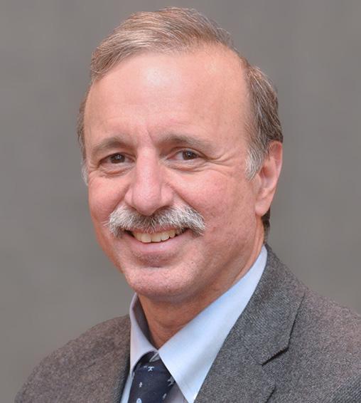
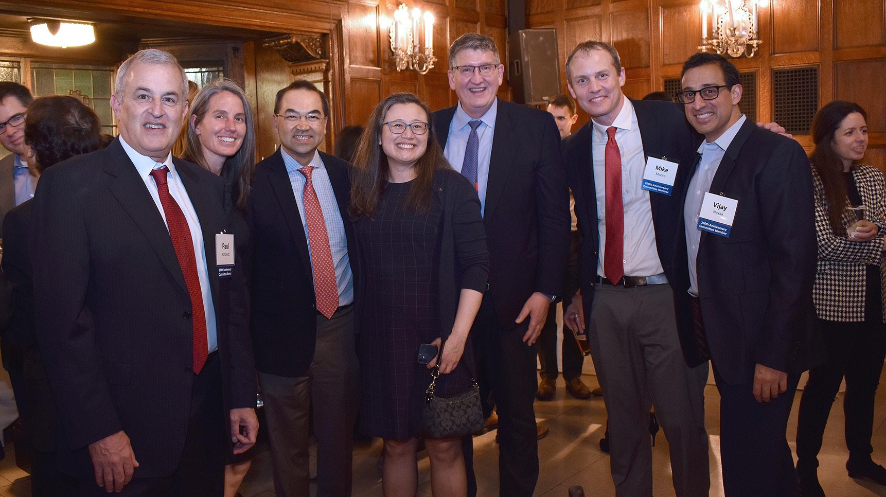
“Regardless of the season, people in America and other developed nations are spending more time than ever indoors, whether it’s at work, playing sports or relaxing with family,” said Benjamin S. Bleier, MD, FACS, Professor of Otolaryngology–Head and Neck Surgery at Harvard Medical School and Director of Otolaryngology Translational Research at Mass Eye and Ear. “If we’re spending so much more time indoors year-round, then why haven’t we seen similar spikes in URIs during warmer months year-to-year?”
The correlation between URIs and cold weather remained a mystery until 2022, when Dr. Bleier, Mass Eye and Ear Investigator Di Huang, PhD, and collaborators from the laboratory of Mansoor Amiji, PhD, at Northeastern University uncovered a previously unidentified immune response to viruses at the front of the nose. Published in The Journal of Allergy and Clinical Immunology, their findings are the first to quantitatively demonstrate how cold temperatures impair the ability of the nose to defend against seasonal respiratory viruses.
Since then, the uncovered missing link has galvanized the worldwide scientific community, captivating scientists with the possibility of harnessing the
newfound immune response to fight a wide array of airborne diseases.
Setting a trap
The immune system relies on two types of immunity to fight disease: adaptive and innate. Adaptive immunity is a trained response to a new group of pathogens. Through vaccination or natural exposure, the body gradually builds the antibodies necessary to defend itself from pathogens. Innate immunity, on the other hand, is an evolutionary, built-in response to a broad group of pathogens. During such a response, the body immediately recognizes a pathogen and uses preexisting antibodies to activate pathways which generate an instantaneous reaction.
The earliest signs of an innate immune response inside the nose were uncovered by Dr. Bleier and Dr. Amiji, Distinguished Professor of Pharmaceutical Sciences at Northeastern University, in 2018. Together, the researchers introduced bacteria to live nasal tissue samples and observed millions of tiny particles— referred to as extracellular vesicles (EVs)—swarming around the pathogen. Published in The Journal of Allergy
[ continued p. 6 ]

5 HARVARD Otolaryngology
FEATURE
From left to right: Benjamin S. Bleier, MD, FACS, and Di Huang, PhD
A LINE OF DEFENSE HIDDEN INSIDE THE NOSE continued
and Clinical Immunology, their early observations revealed that nasal epithelial cells from the front of the nose had detected the bacteria and released the EVs, each of which carried molecules specially designed to neutralize the bacteria.
“Imagine swarms of angry hornets attacking someone who hits their nest,” said Dr. Bleier. “The immune system rarely, if ever, releases a response outside its own body to attack a pathogen, and we immediately wondered if a similar response might occur against viruses.”
In 2022, Drs. Bleier, Huang and Amiji tested the uncovered immune response against three of the most common seasonal respiratory viruses—a single coronavirus and two rhinoviruses. According to their most recent findings published in The Journal of Allergy and Clinical Immunology, the viruses triggered similar swarms of EVs when introduced to live human tissue samples in vitro. But, not without a unique twist. The EVs had met the viruses with a second line of defense unseen against bacteria: decoys.
Each EV carried special receptors resembling those on the surface of nasal epithelial cells. Unlike bacteria, viruses can only invade a cell if spike proteins on its exterior are attached to unique receptors on a cell’s surface. By confronting the viruses with millions of receptors indiscernible from those on the surface of the targeted cells, the EVs set an elaborate trap. The viruses mistook the receptors on the EVs for their intended targets and, once the viruses latched onto the decoys, the EVs used packaged strands of microRNA to disable the pathogens.
“Think of the viruses like burglars trying to break into a home,” explained Dr. Huang. “Except, instead of arriving at their destination, the viruses encounter millions of booby-trapped homes identical to their target.”
Freezing the defense
Few areas of the body are more sensitive to their environment than the front of the nose. According to Dr. Bleier, approximately 10,000 liters of air are inhaled through the nose each day. With each breath inhaled, countless airborne pathogens are caught by mucus at the front of the nose, where only a few minutes of direct exposure to cold air can decrease its internal temperature by several degrees. Unlike the deeper nasal cavity, where specialized structures provide higher amounts of surface area to humidify air, the front of the nose cannot regulate abrupt changes to air temperature on its own, making it especially susceptible to colder air.
To determine how temperature might affect the observed immune response to airborne viruses, Dr. Bleier and his team decided to quantify temperature reductions at the front of the nose in cold, ambient environments. The researchers began by exposing healthy volunteers at room temperature to temperatures at 4.4° C. Then, after 15 minutes, the researchers used an endoscope and thermocouple to determine a temperature drop of about 5° C in their anterior nasal cavities. The temperature difference was then applied to in vitro samples of nasal tissues, which were
[A] Confocal microscopic images of fluorescent dye-labelled extracellular vesicle uptake in human nasal epithelial cells in 60 minutes; scale bar = 100 μm.
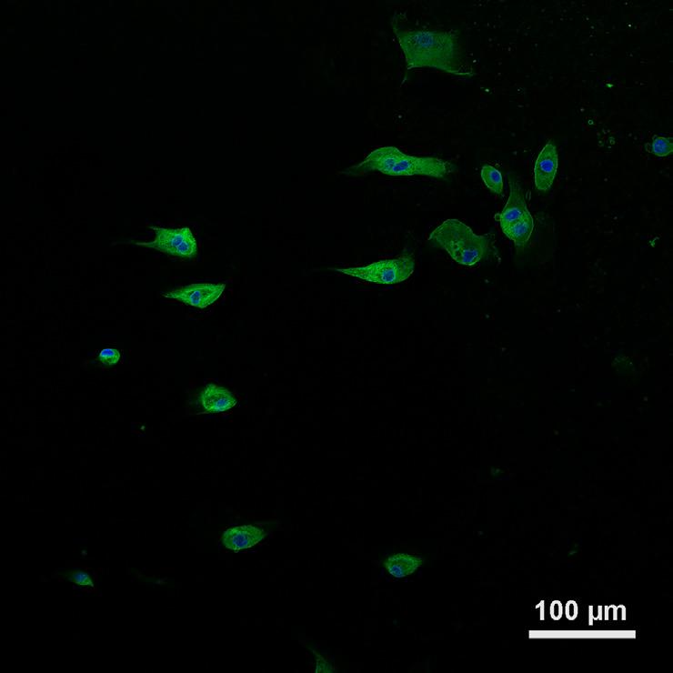
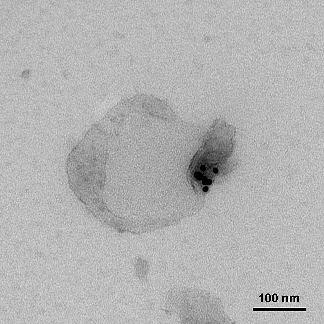
[B] TEM image demonstrating the binding between human rhinovirus RV-16 and nasal epithelium-derived extracellular vesicles; RV-16 was labelled with a rhinovirus marker VP3 conjugated with 10 nm nanogold particles and extracellular vesicle membranes were co-labelled with CD81 conjugated with 15 nm nanogold particles; scale bar = 100 nm.
6 HARVARD Otolaryngology
A B
subsequently exposed to the researchers’ original trio of respiratory viruses.
Observations from the preclinical study revealed that the total amount of EVs had decreased by at least 40 percent at colder temperatures in comparison to the total released at room temperature. Each released EV appeared less capable of defending the nose, too. The number of special receptors used as decoys against the viruses had decreased by 77 percent, and the amount of microRNA strands used to neutralize the viruses had decreased by at least a quarter.
While the observations still required the validation of a definitive clinical trial, Dr. Bleier knew the study had unveiled something extraordinary.
“It was as if a switch had been flipped,” he said. “Just a five-degree drop in temperature was enough to double the likelihood of viruses infecting nasal epithelial cells, replicating inside those cells and infecting neighboring tissue deeper in the body.”
Augmenting optimal responses
For each new part of the immune system uncovered, an intriguing opportunity can present itself. Discovering how temperature affects the immune response has encouraged Drs. Huang and Bleier to imagine a wide range of therapeutics capable of recreating and augmenting the nose’s innate defense mechanism. Ideally, therapeutics might strengthen response potency by increasing the amount of EVs released from nasal epithelial cells or increasing the amount of microRNAs and
special protein receptors carried by each EV.
Existing drugs delivered to the front of the nose, such as topical nasal sprays, have made the prospect of future therapeutics even more tangible. The complexity of any therapeutic research, however, hinges on whether scientists can learn how to initiate or maintain the desired innate immune response over an extended period of time without jeopardizing its safety or effectiveness.
At Mass Eye and Ear, Drs. Huang and Bleier are currently researching the specific microRNA strands found inside antiviral EVs. The researchers would like to determine whether individual microRNA strands possess the antiviral properties sufficient for disabling viruses, or if combinations of different microRNAs are required. Future studies will also include testing immune responses for additional families of airborne viruses aside from those responsible for URIs.
“If we can learn how to manipulate the immune system into doing what it does best, the possibilities are endless,” said Dr. Huang. “Identifying how it responds to temperature is just the first step.”
Until then, the best way to avoid nasty colds, flus and other URIs is to keep the immune system healthy during colder seasons. Masks worn over the mouth and nose not only protect from airborne pathogens but keep air inside the nose warm. And, if anything else, maintaining good hygiene, a healthy diet and plenty of rest should help keep bothersome infections at bay. n
7 HARVARD Otolaryngology
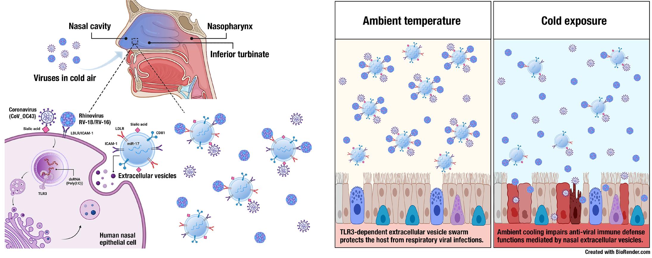
Left diagram: Extracellular vesicles produced by nasal epithelial cells confront coronaviruses and rhinoviruses entering the nose. Right diagram: At room temperature, extracellular vesicles use receptors to capture intruding viruses. At colder temperatures, fewer extracellular vesicles are produced. Those that are produced possess fewer receptors to capture viruses.
Artificial Intelligence Enters the EAR
Surgeons at Mass Eye and Ear have engineered an artificial intelligence device that could diagnose a middle ear infection with higher accuracy than a clinician.

8
Few public health issues affect children more than middle ear infections. In the United States alone, many parents cite ear infections as a leading reason for a visit to the pediatrician. Ear infections extend far beyond national borders, too. In fact, approximately three of every four children worldwide will contract an ear infection before the age of three.1,2,3
Most infections produce mild symptoms such as ear pain, hearing loss and a low-grade fever, yet are easily treated with antibiotics. Inadequate timing and accessibility of treatment, however, carry serious consequences. Untreated children can develop chronic hearing loss that may result in delayed language development. In underdeveloped nations where antibiotics are scarce, the most severe infections can result in meningitis, as well as tens of thousands of deaths per year.4 Conversely, antibiotic resistance can occur among children overtreated with antibiotics, rendering medications ineffective against future infections.
An inaccurate diagnosis further complicates treatment. Despite innovations to technology and clinical practice guidelines, studies suggest the conventional diagnostic accuracy of ear infections in children from a physical exam remains below 70 percent.
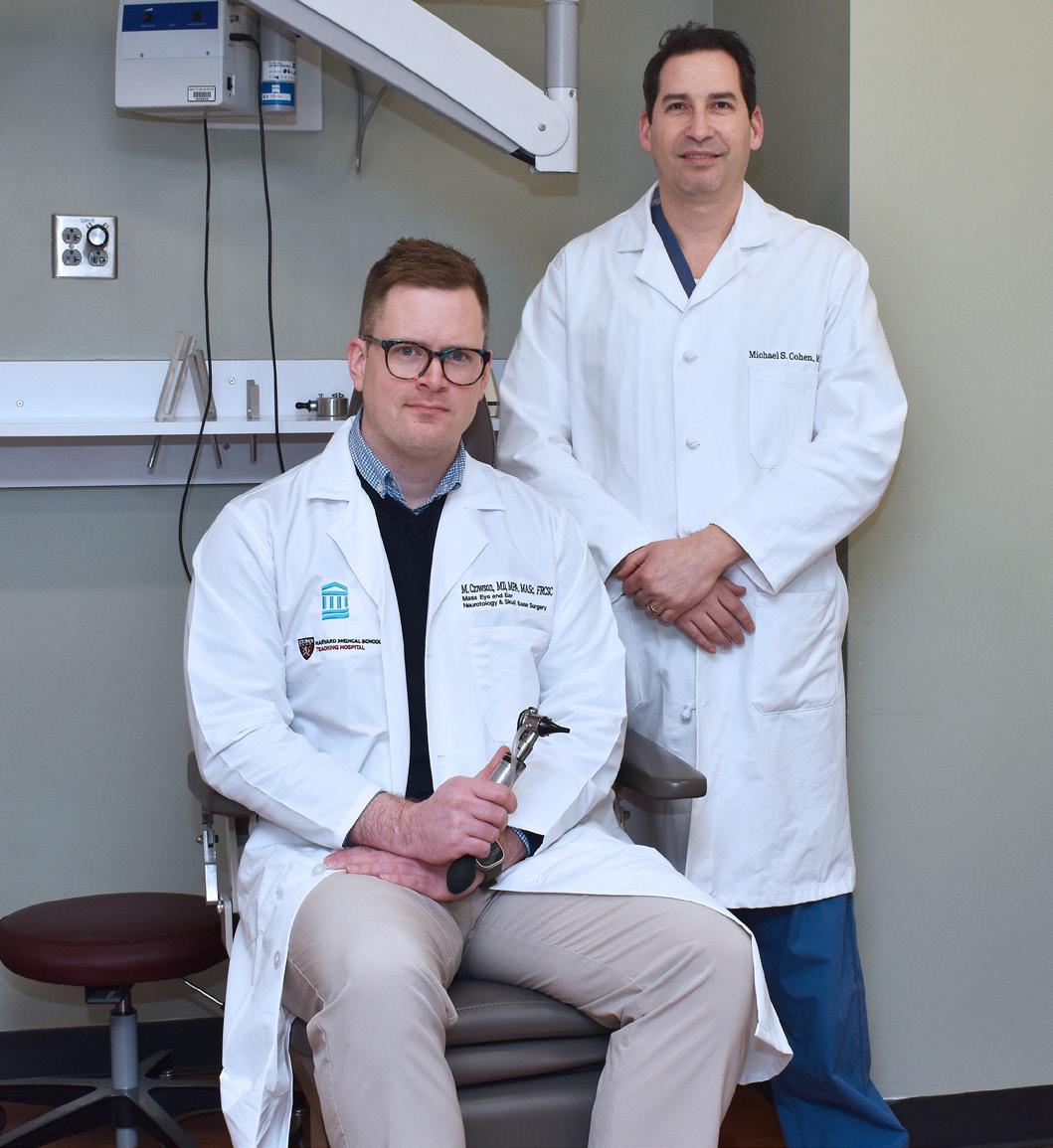
“During these evaluations, you’re asking a cranky, sick child to hold still while trying to look through a handheld device with a window lens less than three millimeters in diameter,” explained Matthew G. Crowson, MD, Assistant Professor of Otolaryngology–Head and Neck Surgery at Harvard Medical School. “Even if you’re lucky enough to have a child cooperate, the small view and—in some cases—the clinicians’ relative inexperience in diagnosing middle ear disease are enough for an incorrect diagnosis.”
A heavy burden ultimately falls on parents who, without any easy way of looking inside the ear, must decide whether to seek urgent care for their child in the first place or wait and see if symptoms abate.
At Mass Eye and Ear, Dr. Crowson has collaborated with surgeons Michael S. Cohen, MD, Assistant Professor of Otolaryngology–Head and Neck Surgery at Harvard Medical School, and Christopher J. Hartnick, MD, MS, Professor of Otolaryngology–Head and Neck Surgery at Harvard Medical School, to build an artificial intelligence (AI) model capable of diagnosing ear infections from photos taken on a handheld device. The team’s most recent success has been very promising. In a 2022 study published in Otolaryngology–
“You’re asking a cranky, sick child to hold still while trying to look through a handheld device with a window lens less than three millimeters in diameter. Even if you’re lucky enough to have a child cooperate, the small view and—in some cases—the clinicians’ relative inexperience in diagnosing middle ear disease are enough for an incorrect diagnosis.”
– Matthew G. Crowson, MD (pictured below)
Head and Neck Surgery, their AI model outperformed doctors in diagnosing ear infections, offering a glimpse of what peace of mind might look like for countless parents and clinicians alike.

Looking past the eardrum
The majority of ear infections occur in the middle ear, where the smallest bones in the body relay sound from the eardrum to the inner ear. The bones form a delicate chain confined to an empty space: an optimal environment for bacteria to fester.
Children are more susceptible to ear infections than adults. When air pressure spikes inside the [ continued p. 10 ]
9 HARVARD Otolaryngology FEATURE
[ ]
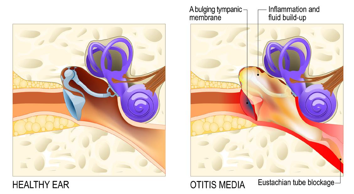
middle ear, a special valve opens a narrow tube to the nose. The tube—known as the Eustachian tube— helps equalize pressure between the middle ear and external environment. It can also inadvertently serve as a passageway for bacteria-laden fluid to flow into the middle ear. In children, the Eustachian tubes are shorter and at more of a horizontal angle than those found in adults, which allows fluid to pool into the ear with great ease. Weaker muscles surrounding the tubes can also prevent the valves from opening wide enough to drain the accumulated liquid.
When evaluating the inside of the ear for an infection, some front-line clinicians do not have the tools or experience needed to definitively determine if fluid exists behind the eardrum. In most settings, these clinicians use a handheld otoscope to view the outside of the eardrum. A definitive diagnosis, however, requires a less routine practice: testing the infected fluid behind the eardrum by extracting it from an incision.
At Mass Eye and Ear, Drs. Cohen and Hartnick have the opportunity to extract such samples during ordinary ear tube insertions for children experiencing frequent ear infections.
In 2018, the surgeons sought to make the most of the
routine procedure. By taking high-resolution, up-close photos of the eardrum before and after the incision and testing fluid extracted from the inner ear, the surgeons figured they could build a unique dataset that would match the external appearance of an eardrum with its respective fluid samples. A large enough collection of photos labeled “infected” or “healthy,” could then train an AI model to detect infections from images captured by a regular otoscope.
“AI tools are only as good as the data you feed them, and most tools have only been trained with images pulled from online search engines,” said Dr. Cohen, who serves as Director of the Multidisciplinary Pediatric Hearing Loss Clinic at Mass Eye and Ear. “We wanted to go straight to the source and gather the gold standard, ‘ground truth’ data that few other models— if any—had ever accessed.”
Unprecedented accuracy
Over the span of a few years, Drs. Cohen and Hartnick had gathered hundreds of photos from children undergoing surgery for tube insertions. Unfortunately, neither surgeon knew how to build the machine-learning algorithm needed to put the accumulated data to good use. Few surgeons did, which prompted Drs. Cohen and Hartnick to explore possible collaborations with AI specialists outside Mass Eye and Ear. The trajectory of their work changed when Dr. Crowson arrived at Mass Eye and Ear in 2020. His research and expertise on data analytics and AI caught their attention within days of his arrival.
“AI is in its infancy within otolaryngology, let alone the entire medical community, and here was an ear, nose and throat surgeon who knew how to build elaborate AI models,” said Dr. Hartnick, who also serves as Director of the Division of Pediatric Otolaryngology at Mass Eye and Ear. “It was as if we had hit the lottery. Everything we needed to create a diagnostic model was here under one roof.”
The collaboration between Dr. Crowson and Drs. Cohen and Hartnick paid immediate dividends. Within months, Dr. Crowson would train an artificial neural network to diagnose infections using the surgeons’ photographs. In a 2021 proof-of-concept study published in Pediatrics, the model was found to be 84 percent accurate in detecting “normal” versus “abnormal” middle ears.
10 HARVARD Otolaryngology
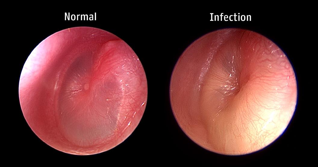
ARTIFICIAL INTELLIGENCE ENTERS THE EAR continued
A B C
[A] Side-by-side image of a normal eardrum (left) and an acute middle ear infection (right). [B] A healthy middle ear. [C] An infected middle ear.
Before comparing the accuracy of their model against clinicians, the team would need to refine its dataset. According to their most recent paper, the surgeons updated the model with more than 639 surgical images of eardrums from children aged 18 years or younger. The images were tagged as either “normal,” “infected,” or having “fluid,” as opposed to the “normal” or “abnormal” classifications used in their earlier model. The added label differentiated between infected fluid, which requires antibiotics, and non-infected fluid, which does not. Even with the more complex analysis, the model achieved a mean diagnostic accuracy of 80.8 percent.
A survey was then created asking clinicians and trainees of various medical specialties to view 22 images of eardrums and to diagnose the ear as one of the three tagged categories. While the machine learning model correctly categorized more than 95 percent of the sample images, the average diagnostic score among 39 clinicians who responded to the survey was 65 percent.
Forging a new reality for public health
In collaboration with Mass General Brigham Innovation, Drs. Cohen and Hartnick are currently engineering a handheld tool* that would use the algorithm to help clinicians diagnose ear infections in a matter of seconds. The machine learning algorithm is currently employed in a prototype device paired with a smartphone app. Acting as a “mini otoscope,” the device would fit over the phone’s camera and allow clinicians and parents to take photos of the inside of a child’s ear, upload them directly to the app and receive an estimated diagnosis in seconds.
A final product could have major implications on global health, Dr. Cohen insisted.
“One interesting thing about international medicine is that you can go to a country with no medical infrastructure, but everyone there has a cell phone,” he said. “Developing diagnostic tools into smartphone apps could extend medical care to people living in some of the most medically inaccessible parts of the world.”
Advances in synthetic data could help refine the prototype even further.
In 2023, Dr. Crowson worked with Krish Suresh, MD, a resident in the Harvard Combined Residency Program in Otolaryngology–Head and Neck Surgery,
to develop a separate machine learning model capable of producing new images of eardrums augmented from preexisting, real images. Findings published in PLOS Digital Health revealed that the augmented images were nearly indiscernible from real ones. A study in JAMA Otolaryngology–Head & Neck Surgery went another step further, revealing that augmented images could actually help increase the accuracy of a diagnostic model.
“AI will never replace the time-tested expertise of trained clinicians,” Dr. Crowson said. “Instead, if we can learn how to validate and harness these tools, we can transform the way we treat tomorrow’s patients and train new clinicians.” n
*This tool is based on technology Drs. Cohen and Hartnick have developed at Mass Eye and Ear. The technology is currently being licensed by Mass Eye and Ear to a company that Drs. Cohen and Hartnick are forming and in which they have a financial interest.
1Teele DW, Klein JO, Rosner B. Epidemiology of otitis media during the first seven years of life in children in greater Boston: a prospective, cohort study. J Infect Dis. 1989;160:83–94. doi: 10.1093/infdis/160.1.83
2Gunasekera, H., Haysom, L., Morris, P. and Craig, J., 2008. The global burden of childhood otitis media and hearing impairment: a systematic review. Pediatrics, 121(Supplement_2), pp.S 107-S107
3Danishyar A, Ashurst JV. Acute Otitis Media. [Updated 2022 Dec 11]. In: StatPearls [Internet]. Treasure Island (FL): StatPearls Publishing; 2023 Jan-. Available from: https:// www.ncbi.nlm.nih.gov/books/NBK470332/
11 HARVARD Otolaryngology
4Monasta, L., Ronfani, L., Marchetti, F., Montico, M., Vecchi Brumatti, L., Bavcar, A., Grasso, D., Barbiero, C. and Tamburlini, G., 2012. Burden of disease caused by otitis media: systematic review and global estimates. PloS one, 7(4), p.e36226.
From left to right: Matthew G. Crowson, MD, and Michael S. Cohen, MD
EXPANDING IMMUNOTHERAPY to Fight Head and Neck Cancer
Immunotherapy has dramatically changed the way physicians treat patients with cancer. At Brigham and Women’s Hospital, Ravindra Uppaluri, MD, PhD, is working to make this treatment a reality for surgical patients with head and neck cancer.
12
Surgeons have long relied on a two-step approach for treating head and neck cancer. In many cases, treatment begins with surgery to remove the tumor. Then, any remaining cancer cells are treated with a course of radiation and, in some cases, chemotherapy. Together, both steps can take up to three months to complete, not to mention several years of follow-up.
Yet, in spite of the provision of standard of care treatment, many tumors return. According to experts, 35 percent of patients with head and neck squamous cell carcinoma—the most common head and neck cancer histology in the world—relapse. And, despite advances in surgery such as robotics and more advanced methods of radiation delivery, long-term clinical outcomes for many patients have plateaued over the past few decades.
“We’re using many of the same procedures I was taught years ago during my residency training and expecting different outcomes,” said Ravindra Uppaluri, MD, PhD, Associate Professor of Otolaryngology–Head and Neck Surgery at Harvard Medical School and Division Chief of Otolaryngology–Head and Neck Surgery at Brigham and Women’s Hospital. “The time to explore new therapeutic options is now.”
A newer class of drugs offers hope. Referred to as immunotherapy, these drugs help the immune system successfully target cancer cells, as opposed to chemotherapy and radiation, which kill cancer cells at the expense of surrounding healthy tissue. The first immunotherapy drugs were approved more than 10 years ago for melanoma and, since then, outcomes observed in clinical trials have provided hope for providers and patients alike.
“Patients once thought to be out of treatment options or too frail for chemotherapy were suddenly seeing responses in their cancers, some with durable or lasting benefit that were previously not seen,” said Glenn J. Hanna, MD, Medical Oncologist at the DanaFarber Cancer Institute. “These drugs showed us that a cure might be possible for some patients with widely metastatic melanoma.”
The U.S. Food and Drug Administration has since approved immunotherapy for treating recurrent and metastatic head and neck cancers. However, whether these drugs could help improve the outcomes of patients undergoing surgery remained a mystery
until 2020. In a multicenter Phase II clinical trial, Dr. Uppaluri and a team of researchers became the first to prove the safety and therapeutic impact of one such drug among these patients undergoing surgery.
But, despite its landmark findings, the study could not account for why some patients did not respond to the new class of drugs at all. Why they had resisted treatment, Dr. Uppaluri said, was the million-dollar question that, if answered, could significantly impact head and neck cancer care.
Taking the breaks off the immune system
The immune system relies on T cells as its final line of defense against cancer. These cells exist throughout the body and are potent enough to damage healthy cells, too. However, a special mechanism keeps healthy cells out of harm’s way. On the surface of T cells are checkpoint proteins that allow healthy cells to “turn off” the potency of the T cell. When a T cell latches onto a healthy cell, complementary proteins on the surface of the healthy cell activate the checkpoint and render the T cell ineffective.
“The body never wants to keep its immune system firing on all cylinders, just as someone wouldn’t want to accelerate a car without having breaks,” said Dr. Uppaluri, who also serves as the Brigham and Women’s Hospital Distinguished Chair in Otolaryngology. “Unfortunately, cancer cells have found a way to slam the breaks through the proverbial car floor.”
[ continued p. 14 ]
13 HARVARD Otolaryngology
FEATURE
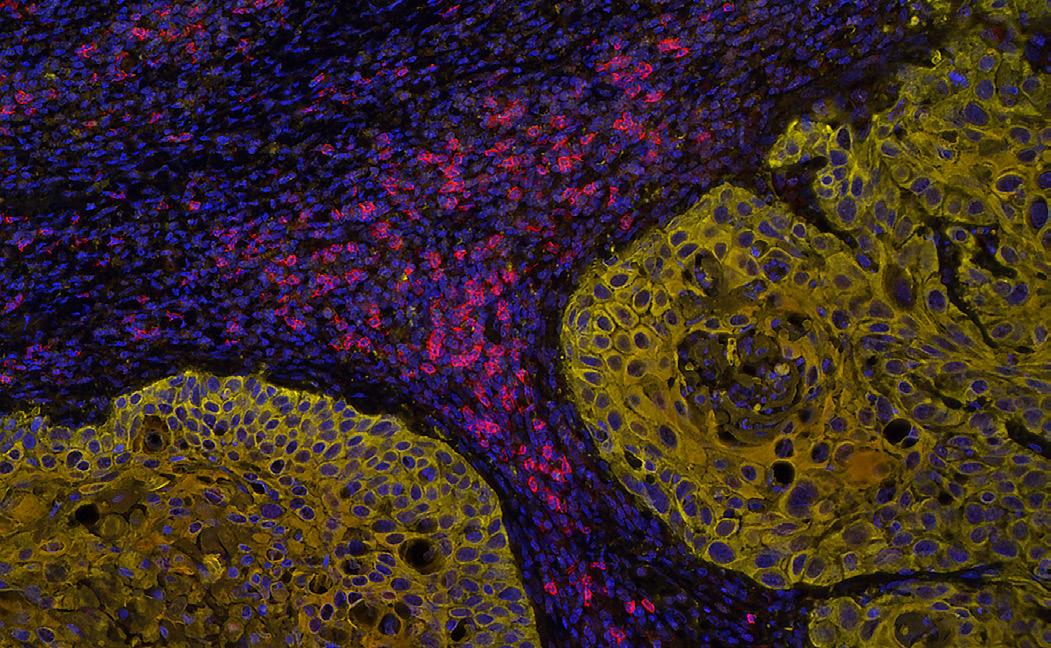
The above image is from a collaboration between Ravindra Uppaluri, MD, PhD, and Scott Rodig, MD, PhD, Director of the Tissue Biomarker Laboratory of the Center for Immuno-Oncology at Dana-Farber Cancer Institute and Professor of Pathology at Harvard Medical School.
The red cells in the picture are T cells infiltrating nests of head and neck cancer cells. The cancer cells are pictured in yellow
EXPANDING IMMUNOTHERAPY TO FIGHT HEAD AND NECK CANCER continued
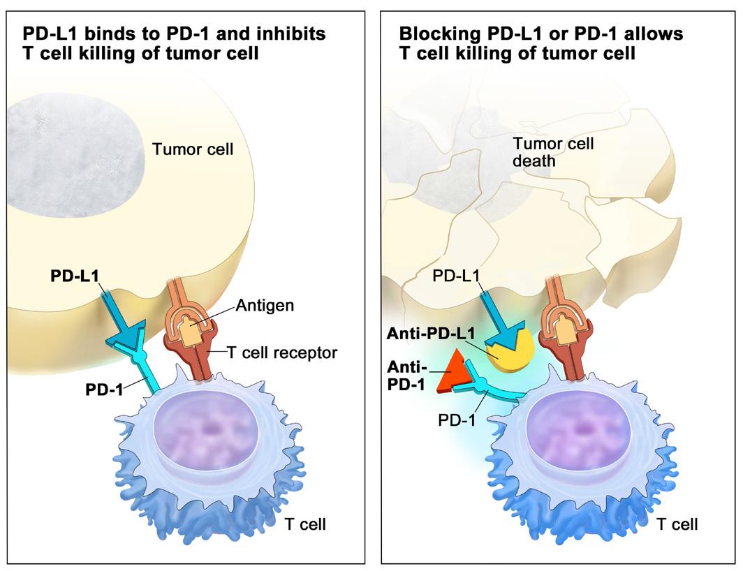
Years ago, scientists discovered several checkpoints generally responsible for allowing a wide range of cancers to evade immune attacks. One of these checkpoints proved crucial for immunotherapy: programmed cell death 1 (PD-1) protein. Cancer cells activate PD-1 using a special protein called programmed death cell ligand (PDL-1), which binds to PD-1 whenever a T cell attempts to attack a cancer cell.
Immunotherapy drugs such as nivolumab and pembrolizumab block cancer cells from activating the PD-1 checkpoint, allowing T cells to retain their potency against the detected threat. In 2015, Dr. Uppaluri began evaluating how patients with head and neck cancer responded to pembrolizumab in a multicenter Phase II clinical trial. Thirty-six patients with locally advanced head and neck squamous cell carcinoma enrolled in the study, and each patient received a single dose of neoadjuvant pembrolizumab two-to-three weeks prior to undergoing surgery.
As reported in Clinical Cancer Research, none of the patients experienced adverse effects or surgical complications from the medication, which had been a major concern of surgeons who sought to expedite surgical care for their patients. Instead, 44 percent of participants demonstrated a change in the pathology of their tumor, and the one-year relapse rate of the cohort was less than half the rate exhibited in previous studies. Remarkably, one patient who presented with Stage IV buccal squamous cell carcinoma had their diagnosis downgraded to Stage I.
One plausible answer, he suspected, was that cancer cells “hid” from the immune system in plain sight. When identifying a cancer cell, T cells must have the ability to bind to the surface of their target. This “lock and key” interaction requires a perfect match between a T cell receptor and a specific molecule on the surface of a cell called human leukocyte antigen (HLA). Ultimately, if a T cell cannot detect HLA and latch itself onto a cancer cell, the tumor will no longer need to rely on a T cell checkpoint to escape the immune system.
According to Dr. Uppaluri, previous studies had confirmed that tumors regularly disguised their HLA to hide from T cells.
“Immunotherapy won’t work if there are no HLA molecules for T cells to target; you’re asking a T cell to look for cancer cells that have become invisible,” said
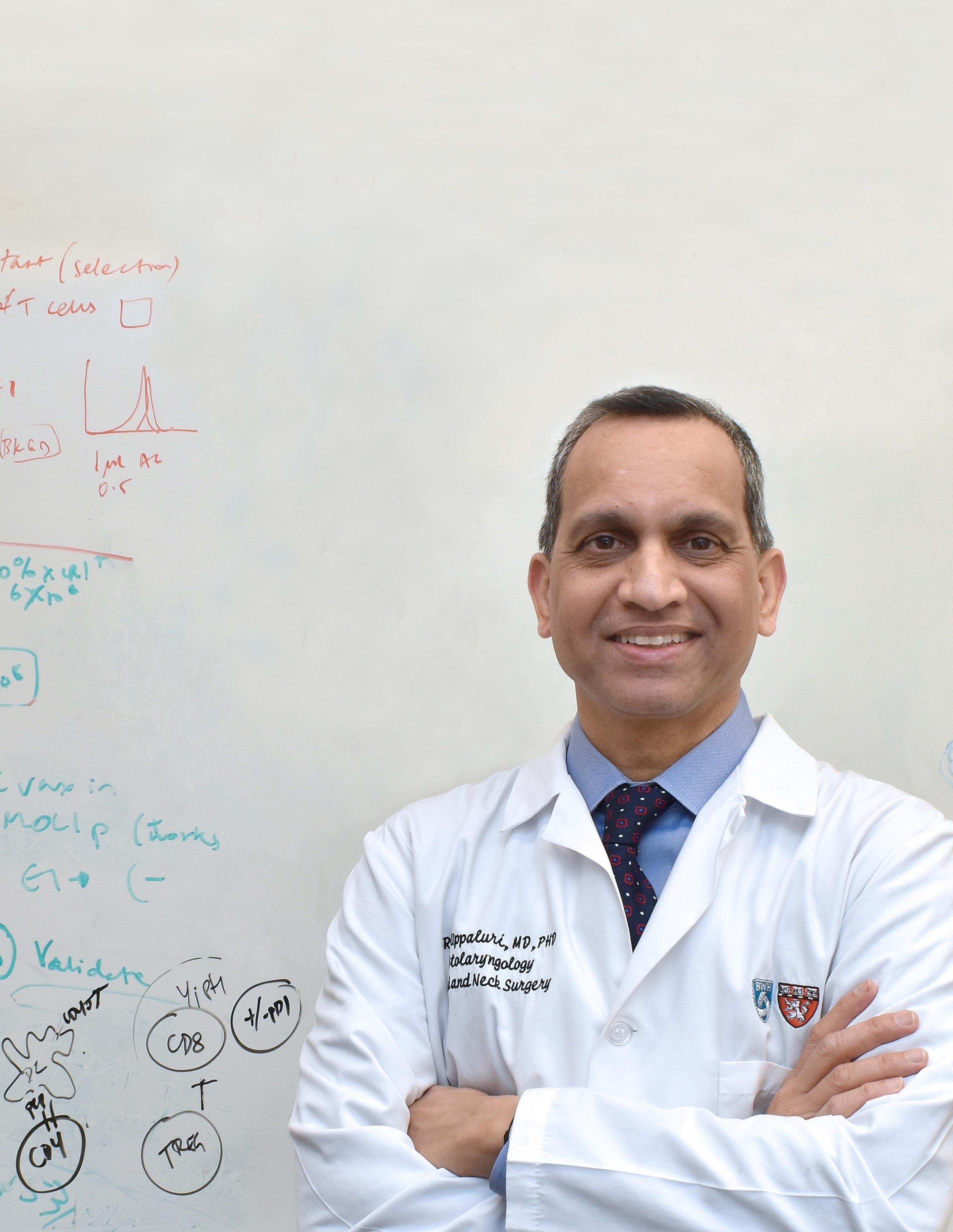
“Immunotherapy won’t work if there are no HLA molecules for T cells to target; you’re asking a T cell to look for cancer cells that have become invisible. We knew we needed to look for another treatment that could ‘uncloak’ a cancer cell for the T cell to see its target.”
– Ravindra Uppaluri, MD, PhD (pictured below)
“On one hand, a single dose had achieved a near cure in this patient,” Dr. Uppaluri added. “But our attention immediately turned to why some of the other patients had not responded at all.”
Cancers hiding in plain sight
In 2019, the National Cancer Institute and National Institute of Dental and Craniofacial Research awarded Dr. Uppaluri a $4.3 million Cancer Moonshot Grant to take his research one step further. With the support of the grant, Dr. Uppaluri launched several studies aimed at better understanding why blocking PD-1 from cancer cells had not improved outcomes among certain patients.
14 HARVARD Otolaryngology
[ ]
Dr. Uppaluri. “We knew we needed to look for another treatment that could ‘uncloak’ a cancer cell for the T cell to see its target.”
So, rather than replacing pembrolizumab, Dr. Uppaluri turned his attention towards uncovering the mechanisms used by cancer cells to hide their HLA molecules. In a separate study published in Clinical Cancer Research, Dr. Uppaluri and a team of researchers from Brigham and Women’s Hospital and the DanaFarber Cancer Institute used CRISPR-Cas9 mediated genome editing and pharmacological inhibition to identify the gene EZH2 as one possible mechanism for hiding HLA.
Higher levels of EZH2, they found, correlated with decreased antigen presentation among head and neck cancer samples collected in The Cancer Genome Atlas. The researchers then applied their findings to a mouse model of head and neck cancer cells resistant to PD-1 blocking immunotherapy. When treated with a drug designed to inhibit EZH2, the mouse model exhibited heightened T cell activity and sensitivity to immunotherapy.
Combining therapies; changing future outcomes
EZH2 is one of a multitude of pathways cancer cells use to prevent patients from responding to immunotherapy. As more pathways are identified, researchers can design therapeutics capable of combating resistance to treatment. Combining such a therapeutic with standard immunotherapy could narrow the gap between extraordinary outcomes and poor responses.
It could also drastically improve quality of life in the years following treatment.
According to the National Cancer Institute, head and neck cancers account for nearly four percent of reported cancer cases in the Unites States. These cancers impact organs crucial for breathing, speaking, chewing and tasting. All therapies—including surgery, chemotherapy and radiation—can irreparably damage these organs and lead to long-term toxicity. Ideally, a patient may be able to forgo chemotherapy and radiation by undergoing a course of immunotherapy paired with another therapeutic prior to surgery.
“Patients shouldn’t have to consider whether they might taste food or communicate the same way they did prior to treatment,” said Rosh K. Sethi, MD, MPH,
Assistant Professor of Otolaryngology–Head and Neck Surgery at Harvard Medical School and Associate Surgeon at Brigham and Women’s Hospital. “It’s one thing to beat cancer, but it’s another to enjoy life the way it was before.”
Further studies are needed to make this alternative treatment a reality. Under his Moonshot Grant, Dr. Uppaluri has partnered with Dana-Farber Cancer Institute Investigators David Barbie, MD, Associate Professor of Medicine at Harvard Medical School, and Robert Haddad, MD, Professor of Medicine at Harvard Medical School, to accelerate his research on resistance pathways into clinical trials for combined therapeutic interventions.
Meanwhile, Dr. Uppaluri has sought to validate the safety and efficacy of pembrolizumab in a much larger, randomized trial. In partnership with Merck & Co. Inc.,* he has helped develop KEYNOTE-689, the first international Phase III immunotherapy clinical trial for surgical patients with head and neck cancer. The trial aims to recruit 700 patients—more than 20 times the number of participants reported in the 2020 study—results of which will help investigators better understand whether this approach can truly benefit head and neck cancer patients in a surgical setting.
“We’re on the verge of revolutionizing head and neck cancer treatment,” Dr. Uppaluri said. “This definitive study has the potential to show whether using immunotherapy can impact thousands of patients worldwide who suffer from this disease.” n
15 HARVARD Otolaryngology
*Ravindra Uppaluri, MD, PhD, serves on a Merck Scientific Advisory Board.
©2015 Terese Winslow LLC, U.S. Govt. has certain rights
Nelson Y.S. Kiang, PhD
A pioneer of otolaryngology.
On March 19, 2023, Nelson Y.S. Kiang, PhD, Founding Director of the Eaton-Peabody Laboratories (EPL) and Professor Emeritus of Physiology at Harvard Medical School, passed away at his Beacon Hill home in Boston.
Dr. Kiang, aged 93, earned international fame in the 1960s for his pioneering neurophysiological recordings, which systematically described how the inner ear translates sound into patterns of electrical discharge in the fibers of the auditory nerve.
Born in Wuxi, China, Dr. Kiang arrived in the United States in 1934 at the age of five. He lived in New York City and Seattle before earning the first-ever PhD in biopsychology from the University of Chicago. He arrived at the Massachusetts Institute of Technology (MIT) in 1955 and later added faculty appointments at Harvard Medical School, Mass Eye and Ear and Massachusetts General Hospital.
In 1958, Dr. Kiang partnered with Miss Amelia Peabody, a philanthropist and former Mass Eye and Ear Board Member, to establish the EPL at Mass Eye and Ear. Dr. Kiang served as Laboratory Director until 1996 and oversaw its rapid growth into the largest research group in the world dedicated to the study of hearing and deafness.
“[Dr. Kiang’s] commitment to ensuring that his students adhere to a rigorous scientific pathway will forever guide the clinicians and investigators who carry on the mission of his trailblazing work.”
Leveraging his close ties to MIT, Dr. Kiang was also a pioneer in the application of computers to neuroscience, installing one of the original LINC computers in the EPL in 1962 and using it in data acquisition for his 1965 monograph on auditory-nerve response.

He was a mentor, both formally and informally, to dozens of students and young investigators who came to know his fierce dedication to scientific rigor and clarity of thought. His passion for education was further realized in 1992, when he spearheaded the creation of a PhD program in Speech and Hearing Biosciences and Technology (SHBT), which is now a

16 HARVARD Otolaryngology IN MEMORIAM s
[ ] 1 2
– Mark A. Varvares, MD, FACS
part of the Division of Medical Sciences at Harvard Medical School. Since its inception, SHBT has awarded more than 150 PhDs, and its graduates have become leaders in neuroscience, medicine, biotech and other fields.
“Nelson Kiang’s vision and life-long dedication to discovery in the hearing sciences—including training the next generation of scientists with the goal of improving human health—has left a lasting impression on the hearing research communities at Mass Eye and Ear, Harvard and MIT,” said Mark A. Varvares, MD, FACS, the John W. Merriam and William W. Montgomery Professor and Chair of Otolaryngology–Head and Neck Surgery at Harvard Medical School. “His commitment to ensuring that his students adhere to a rigorous scientific pathway will forever guide the clinicians and investigators who carry on the mission of his trailblazing work.” n
Remembering Joseph A. Moretti, MD
Joseph A. Moretti, MD, was a former trainee of the Harvard Otolaryngology Residency Program and a former physician at South Shore Hospital who passed away on November 4, 2022 in Naples, FL.
Dr. Moretti, 80, is remembered for his myriad of interests, which encompassed world travel, aviation, cycling, linguistics and theology. He grew up in Elmwood Park, IL, and attended Providence College in Rhode Island. Upon graduating from the University of Chicago Medical School, he returned to New England and began his residency training in otolaryngology at Mass Eye and Ear.

In 1971, Dr. Moretti graduated from the residency program and was subsequently stationed as a Lieutenant Commander at the U.S. Naval Hospital in Philadelphia. He later opened a private practice for ear, nose and throat care in South Weymouth, MA—right across the street from South Shore Hospital—and was elected President of the Massachusetts Medical Society.
His sense of humor, smile, willingness to help those around him and dedication to resident teaching will endure forever. n
17 HARVARD Otolaryngology
[ 1 ] Nelson Y.S. Kiang, PhD, in the Eaton-Peabody Laboratories (EPL), circa 1960s.
[ 2 ] Portrait painting of Dr. Kiang in the EPL library.
3
[ 3 ] Dr. Kiang with his wife Barbara at the 2019 Harold F. Schuknecht Society reception at the Liberty Hotel.
Ines and Fredrick Yeatts, PhD
Advancing research on hearing loss.
Ines and Fredrick Yeatts, PhD, are two of Mass Eye and Ear’s closest and most ardent supporters.
In the nearly 200-year history of Mass Eye and Ear, few donors have matched their outstanding support. The Yeatts have donated more than $12 million to the hospital, half of which has helped support the work of Zheng-Yi Chen, DPhil, a hearing scientist in the Eaton-Peabody Laboratories committed to treating deafness and hearing loss.
In 2020, the hospital formally renamed Dr. Chen’s laboratory the “Ines and Fredrick Yeatts Inner Ear Research Laboratory” in recognition of the Yeatts’ unprecedented generosity.
“We’ve been alongside Zheng-Yi every step of the way and it has been exciting and rewarding to watch his progress unfold,” said Dr. Yeatts. “It is just a matter of time before treatments will be available to help people save or even regain their hearing.”
Dr. and Mrs. Yeatts first learned of Dr. Chen’s research from reading a 2005 Boston Globe article. Intrigued, Dr. Yeatts called Dr. Chen, asking to learn more. They met and, together, their partnership has since transformed Dr. Chen’s career and accelerated the pace of hearing science.
Thanks to the Yeatts’ philanthropic partnership, Dr. Chen has moved his research closer than ever to developing treatments for hearing loss and deafness. He and his team have used a novel gene editing technique called CRISPR/Cas9 to successfully halt progression of genetic hearing loss in mice and have discovered a new way to regenerate hair cells, a critical step in developing a treatment for hearing loss. Some of Dr. Chen’s work has already been licensed to companies currently developing treatments for different forms of hearing loss.

“We’ve been alongside Zheng-Yi every step of the way and it has been exciting and rewarding to watch his progress unfold. It is just a matter of time before treatments will be available to help people save or even regain their hearing.”
Fredrick Yeatts, PhD
Having experienced hearing loss in more recent years, Dr. and Mrs. Yeatts couldn’t be more thrilled by Dr. Chen’s progress.
“My work isn’t possible without the exceptional commitment of the Yeatts,” said Dr. Chen, who also serves as Associate Professor of Otolaryngology–Head and Neck Surgery at Harvard Medical School. “Year after year, it’s their support that allows my research to explore new areas, such as genome editing, and reach levels of inquiry never seen before. I have never met anyone like them with the singular goal of helping us develop hearing loss treatments for countless patients worldwide.” n
18 HARVARD Otolaryngology s DONOR PROFILE
–
Zheng-Yi Chen, DPhil
ALUMNI GIVING SOCIETY

The Alumni Giving Society of the Department of Otolaryngology–Head and Neck Surgery at Harvard Medical School
The Department of Otolaryngology–Head and Neck Surgery at Mass Eye and Ear/ Harvard Medical School established the Alumni Giving Society in 2015 to recognize faculty and alumni who make gifts of $1,000 or more during the fiscal year (October 1–September 30). Participation is a way to stay connected and to help deliver the finest teaching experience for today’s otolaryngology trainees.

Our alumni know from firsthand experience that support of the vital work of our students and faculty in the Department of Otolaryngology–Head and Neck Surgery helps drive continued achievement across all areas of education, research and patient care. In the 2022 fiscal year, we had 39 members who we thanked for their generosity and partnership. Their collective support helped us achieve our department goals and institutional mission.
If you are not a member, please consider joining your colleagues today by making a gift with the enclosed envelope. As a member, you may designate your gift in the way that is most meaningful to you.
To learn more, please contact Julie Dutcher in the Development Office at 617-573-3350.
sCurrent Alumni Giving Society members for fiscal year 2022, from October 1, 2021, to September 30, 2022, are listed below. With your gift of $1,000 or more, you will be included in the 2023 Alumni Giving Society.


VISIONARY
Gifts of $10,000 to $24,999
Michael S. Cohen, MD
INNOVATOR
Gifts of $5,000 to $9,999
Neil Bhattacharyya, MD, FACS
Nathan Jowett, MD, FRCSC
Leila Mankarious, MD
PIONEER
Gifts of $2,500 to $4,999
Catherine Banks, MD
Nicolas Y. BuSaba, MD, FACS
Richard E. Gliklich, MD
John B. Lazor, MD, MBA, FACS
Kasey K. Li, MD, DDS
David E. Nash, MD
H. Gregory Ota, MD
Gregory W. Randolph, MD, FACS, FACE
John J. Rosowski, PhD
Mark F. Rounds, MD
Jonathan Y. Ting, MD, MS, MBA
Mark A. Varvares, MD, FACS
William M. White, MD
FRIEND
Gifts of $1,000 to $2,499
Megan E. Abbott, MD
Barry J. Benjamin, MD
Ruth Anne Eatock, PhD
Bruce Feldman, MD
Robert A. Gryboski, MD
Paul E. Hammerschlag, MD, FACS
Earl Harley, Jr., MD
Martin L. Hopp, MD, PhD
Nelson Y.S. Kiang, PhD #
William McClerkin, MD
Cliff A. Megerian, MD, FACS
Ralph B. Metson, MD
Eugene N. Myers, MD, FACS, FRCS
Didier Peron, MD
Adrian J. Priesol, MD
Sunil Puria, PhD
Edward J. Reardon, MD
Michael B. Rho, MD, FACS
Jeremy D. Richmon, MD
George A. Scangas, MD
Theodoros N. Teknos, MD
Isaac Wasserman, MD
#Deceased
19 HARVARD Otolaryngology
Department of Otolaryngolog y Head and Neck Surger y
Nina Shapiro, MD, ’96
Dr. Shapiro blazed a trail for female surgeons from Boston to Los Angeles and beyond.
Male surgeons had always seemed to dominate the operating room (OR).
As a student at Harvard Medical School in the 1990s, this was a reality Nina Shapiro, MD, ’96, the former Director of Pediatric Otolaryngology and Professor Emerita of Head and Neck Surgery at the David Geffen School of Medicine at the University of California, Los Angeles (UCLA), confronted every day. During her clinical rotations, she could count on one hand the number of female residents and attendings she saw in the OR.
However, she never accepted the status quo. Dr. Shapiro knew she belonged shoulder-to-shoulder with her male counterparts.
Originally from New York, Dr. Shapiro grew up with several doctors in her family; her father a psychiatrist, and her brother an otolaryngologist. After graduating from Cornell University, she entered medical school determined to study ophthalmology. The complex anatomy of the eye had always fascinated her, as had the intricate procedures used to treat its delicate structures.
Had it not been for Marshall Strome, MD, the former Chief of Otolaryngology Services at the Beth Israel Deaconess Medical Center and Brigham and Women’s Hospital, she just might have stayed that course.
“I remember Dr. Strome saying to me, ‘Nina, I’ve been doing this for 30 years, and I could do it for another 30, no problem,’” said Dr. Shapiro, who marveled at her mentor’s love of otolaryngology during her first few rotations at Brigham and Women’s Hospital. “I remember thinking to myself, ‘How lucky would I be to love a specialty like that?’ I knew I had to give it a chance.”
Dr. Shapiro never looked back. Over the past two decades, she has emerged as a trailblazer for female surgeons nationwide and remains one of the most sought-after medical experts by the media. What’s more, she has authored nearly 100 journal articles, presented at countless national and international meetings
“You need to establish a sense of mutual respect with each patient and family member. And, when caring for children, it’s important to include them in the conversation in ageappropriate ways that they can understand.”
– Nina Shapiro, MD
and—upon leaving UCLA—started her own practice as a pediatric otolaryngologist at Westside Head and Neck in Santa Monica, CA. Her surgical career began after medical school when she joined the Harvard Otolaryngology Residency Program. Despite the few women in her program, Dr. Shapiro always felt respected by the male residents and fellows. Her mentors treated her just as they would any other resident, and she quickly developed an interest in pediatric otolaryngology under the influences of former Mass Eye and Ear Pediatric Otolaryngology Division Chiefs Michael J. Cunningham, MD, FACS, and Roland Eavey, MD.
20 HARVARD Otolaryngology
s ALUMNI PROFILE
The breadth of their cases interested her most. According to Dr. Shapiro, an ear tube insertion may have taken less than three minutes to complete but could forever change the way a child communicated. On the other hand, a multi-hour airway reconstruction surgery could improve the life of a child in critical condition.
The renowned bedside manner of William W. Montgomery, MD, the former John W. Merriam Professor of Otology and Laryngology at Harvard Medical School, proved just as influential. To this day, she recalls him explaining why he treated patients like a neighbor—not necessarily a family member, but not as a complete stranger either.
“You need to establish a sense of mutual respect with each patient and family member,” Dr. Shapiro said. “And, when caring for children, it’s important to include them in the conversation in age-appropriate ways that they can understand.”
By the time Dr. Shapiro completed her residency and fellowship training in 1997, she had spent 10 years living in Boston. It was time for a change, and a job opening for a pediatric otolaryngologist at UCLA caught her attention.
While she accepted the role thinking she would eventually return to the East Coast, the opportunity paved the way to an unexpected passion. One day, the Discovery Channel contacted Dr. Shapiro and a few of her colleagues at UCLA for insight on a breathing problem affecting a young boy. Then, in 2008, she
appeared as a guest on the television show The Doctors, an Emmy award-winning broadcast featuring health and medical issues. The show was in its first season, and she immediately took to its style of helping lay audiences understand complex medical subjects.


“As physicians, we have this gift of knowledge and the language of medicine that we learn to communicate to one another like some kind of ‘secret handshake,’” said Dr. Shapiro. “But there’s no reason why we can’t communicate that information with other people outside of our fields.”
Within a few years, she would regularly appear as a guest on the show and, soon thereafter, NPR and CNN. Her success motivated her to find new outlets for her expertise, including a regular column in Forbes. She then began writing books and, in 2018, her book Hype was translated into five languages and recognized by Publisher’s Weekly as a “Best Book of 2018.”
Wherever she practices, Dr. Shapiro emphasizes the importance of communication. Even with her trainees, she’ll encourage each one to try and present their research in a way a third grader might understand.
After all, if just one more person can better understand a condition or treatment, then who knows who it might inspire. It might even help another young woman—much like herself— realize that the world of medicine isn’t excluded to a select few, but open to everyone. n
21 HARVARD Otolaryngology
[ 1 ] Dr. Shapiro’s book Hype was named a “Best Book of 2018” by Publisher’s Weekly
1 2
[ 2 ] Dr. Shapiro (pictured right of center) on the set of The Doctors
HIGHLIGHTS s
News from every corner of the Department of Otolaryngology–Head and Neck Surgery at Harvard Medical School.

New Faculty
Introducing the newest clinicians, clinician-scientists, researchers and educators in the Department of Otolaryngology–Head and Neck Surgery.
Mark T. Brown, MD, MBA, FACS, is a boardcertified comprehensive ear, nose and throat surgeon at Mass Eye and Ear, where he serves as Medical Director of the hospital’s Concord location. Upon graduating from Baylor College of Medicine, he completed his residency training in otolaryngology–head and neck surgery at Harvard Medical School and subsequent fellowship training in head and neck reconstructive surgery and microvascular surgery at Mass Eye and Ear. He also earned his MBA from George Washington University. His interests include public health equity, business operations and general otolaryngology in adults and children.
Sameer G. Gupta, MD, DPhil, is a Mohs micrographic and reconstructive surgeon at Mass Eye and Ear. Dr. Gupta graduated from Yale College and the University of Cambridge as a Churchill Scholar, and he subsequently earned his medical degree from Harvard Medical School with honors. He completed his residency training at the Harvard Combined Dermatology Residency Training Program and his surgical fellowship training at Massachusetts General Hospital. Dr. Gupta’s clinical and research interests focus on better management of skin cancer. He has authored more than 20 peer-reviewed publications, reviews and book chapters.

Sophia Z. Shalhout, PhD, is an assistant scientist in the Mike Toth Head and Neck Cancer Research Center at Mass Eye and Ear. Dr. Shalhout earned her PhD in biochemistry from Wayne State University and completed wet and dry lab postdoctoral training as a research fellow at Boston

Children’s Hospital, the Massachusetts Institute of Technology, Harvard University and, most recently, as a data scientist and the Cutaneous Oncology Research Fellow at Mass General Cancer Center. Her lab specializes in applying big data analytics and novel machine learning approaches to multiscale biomedical clinical data.
Hae-Young Kim, DrPH, is the Senior Biostatistician for the Biostatistics and Research Design Center in the Institutional Centers of Clinical and Translational Research at Boston Children’s Hospital. Dr. Kim earned her doctorate in biostatistics from the University of North Carolina at Chapel Hill and has most recently served as Associate Professor in Biostatistics in the Department of Public Health for New York Medical College. Her research interests include designing and analyzing data from clinical trials and observational studies, as well

as statistical methodologies for clustered correlated data, longitudinal data and survival data.
Awards, Grants and Honors
The British Cochlear Implant Group awarded Julie G. Arenberg, PhD, CCC-A, the Annual Graham Fraser Memorial Lectureship, which she delivered at the group’s annual meeting in Cambridge, England. The lectureship is awarded to those most qualified to contribute to the advancement of cochlear implant research and practice. The Division of Otolaryngology–Head and Neck Surgery at Brigham and Women’s Hospital became an official site for the WAYPOINT trial, becoming the only site in Massachusetts and surrounding states to offer the medication tezepelumab—a noninvasive monoclonal antibody treatment— to patients with severe chronic rhinosinusitis with nasal polyps.
[
[ 3
[ 4
[ 5 ]
[ 6
22 HARVARD Otolaryngology
[ 1 ] James R. Brodsky, MD, FAAP, Associate Professor of Otolaryngology–Head and Neck Surgery
2 ] Thomas L. Carroll, MD, Associate Professor of Otolaryngology–Head and Neck Surgery
] Andreas Eckhard, MD, Assistant Professor of Otolaryngology–Head and Neck Surgery
] Judith Kempfle, MD, Lecturer of Otolaryngology–Head and Neck Surgery, Part-Time
Rachel E. Roditi, MD, Assistant Professor of Otolaryngology–Head and Neck Surgery
Recent Promotions 1 2 3 4 5 6
] Kristina Simonyan, MD, PhD, Dr med, Professor of Otolaryngology–Head and Neck Surgery
Matthew G. Crowson, MD, earned an Oracle for Research Project Award for the development of an “autopilot” system for ear surgery. The award provides financial backing, computational resources and hands-on technology consultations to researchers seeking solutions to complex, disruptive and high-impact challenges to humankind.
Bertrand Delgutte, PhD, was awarded the William and Christine Hartmann Prize in Auditory Neuroscience by the Acoustical Society of America. The prize recognizes a researcher whose work has linked auditory physiology with auditory perception or behavior in humans or other animals.
Mass General Brigham named Stacey T. Gray, MD, the recipient of its Outstanding Program Director Award. The award recognizes a program director who has improved the quality of a training program and enhanced the overall experience of its residents and fellows.
Cochlear implant guidelines co-authored by Amanda M. Griffin, PhD, CCC-A, were recently selected for an Ear and Hearing Readers’ Choice Award by the American Auditory Society.

Christopher J. Hartnick, MD, MS, was awarded a collaborative, multicenter $5.6 million grant from the Patient-Centered Outcomes Research Institute to study interventions that could help improve the coordination of at-home care for children with tracheostomies.
The Harvard Otolaryngology Combined
Grand Rounds Lecture Series welcomed two-time Olympic gymnast Aly Raisman to Brigham and Women’s Hospital as one of
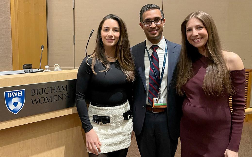
its distinguished visiting speakers. Raisman was joined by fellow speaker Abbey Bergman, a cancer survivor, for a lecture titled, “The Cancer I Never Should Have Had.”
Iván Coto Hernández, PhD, earned a K25 Mentored Quantitative Research Development Award from the National Institute of Biomedical Imaging and Bioengineering. The grant will support a project he undertakes with the mentorship of internationally-recognized experts from Mass General Brigham and Boston University.
Eric H. Holbrook, MD, helped launch a clinical trial for a nasal stent developed by Lyra Therapeutics, a Boston-based biotech company committed to treating chronic rhinosinusitis. When inserted into the nasal cavity of a patient with chronic rhinosinusitis, the stent helps reduce symptoms by eluting steroids. Alongside Stacey T. Gray, MD, Dr. Holbrook will help lead a Phase III clinical trial for approval of the device by the U.S. Food and Drug Administration.
Nicole Jiam, MD, was awarded the Physician Scientist Fellowship Award from the Doris Duke Charitable Foundation,
New Leadership
Nicolas Bu-Saba, MD, was named Division Chief of Comprehensive Otolaryngology–Head and Neck Surgery and Director of Otolaryngology–Head and Neck Surgery Emergency Services at Mass Eye and Ear.
Sunil Puria, PhD, was elected President of the Association for Research in Otolaryngology.
Stacey T. Gray, MD, was appointed President of the New England Otolaryngological Society.
Amanda Dilger, MD, and Regan Bergmark, MD, were named co-Chairs of the Harvard Otolaryngology–Head and Neck Surgery Committee on Climate Change.
which seeks to develop careers of physicianscientists by providing researchers with the protected time and resources necessary to build their future research programs. Her proposed research will be mentored by Julie Arenberg, PhD, CCC-A, and is titled “AgeSpecific Cochlear Implant Programming for Optimal Hearing Performance.”

David H. Jung, MD, PhD, FACS, was awarded a Department of Defense grant to research the development of a gene therapy treatment for neurofibromatosis type 2, a rare hereditary disorder affecting the balance and hearing systems. Also named to the grant were Mass Eye and Ear researchers D. Bradley Welling, MD, PhD, FACS, and Judith Kempfle, MD, as well as Massachusetts General Hospital researcher Xandra Breakefield, PhD.
Daniel J. Lee, MD, was named Principal Investigator of a Department of Defense grant awarded for a multicenter study on auditory brainstem implants (ABIs). Through his grant, Dr. Lee aims to improve ABI performance among patients with neurofibromatosis type 2.
Gi-Soo Lee, MD, EdM, and Mark S. Volk, MD, directed the annual Children’s Otolaryngology Residents Emergency Skills Course, which provides PGY-1 residents throughout New England with emergency otolaryngology care simulation training.
Stella Lee, MD, and Emily Moldoff, NP, were awarded a $10,000 grant from the American Academy of Otolaryngic Allergy Foundation. The duo will study the effects of sublingual immunotherapy on Eustachian tube dysfunction.
M. Charles Liberman, PhD, was awarded the Scientific Grand Prize from the Francebased La Fondation Pour l’Audition for his seminal contributions to hearing loss research. Dr. Liberman, who previously served as Director of the Eaton-Peabody Laboratories at Mass Eye and Ear for more than 25 years, co-discovered cochlear synaptopathy or “hidden hearing loss” alongside Sharon Kujawa, PhD, in 2009.
23 HARVARD Otolaryngology [ continued p. 24 ]
From left to right: Aly Raisman; Rosh K. Sethi, MD, MPH; Abbey Bergman.
HIGHLIGHTS s

Pavan S. Mallur, MD, was formally accepted into the Triological Society as an Active Fellow. His accepted candidate thesis examined the effects of Botox injections on patients with idiopathic chronic cough.
The Harvard Department of Otolaryngology–Head and Neck Surgery relaunched its annual New England Sinus Course after a two-year hiatus amid the COVID-19 pandemic. The course, which was led by Alice Z. Maxfield, MD, and George A. Scangas, MD, was hosted inside the Joseph B. Nadol, Jr., MD, Surgical Training Laboratory at Mass Eye and Ear.
The Society of Academic Emergency Medicine (SAEM) invited James G. Naples, MD, to join its Guideline Writing Committee for Acute Dizziness in the Emergency Department, which culminated in the publication of several manuscripts used to help create the SAEM Guidelines for Reasonable and Appropriate Care in the Emergency Department (GRACE-3). Dr. Naples also received a $50,000 grant from the Harvard Catalyst Five Senses Program to research possible biomarkers for balance and dizziness.
Matthew Naunheim, MD, MBA, was named recipient of the 2023 Young Faculty/ Practitioner Award by the American Laryngological Association. The award recognized his paper, “Do You Like Your Voice? A Population-Based Survey of Voice Satisfaction and Cosmetic Enhancement.”
Gregory W. Randolph, MD, FACS, FACE, was awarded the 2023 Gold Medal from the International Federation of ORL Societies during a ceremony held in the United Arab Emirates. He was also awarded the 2023 Distinguished Member Award from the New England Endocrine Alliance (NEEA) during the NEEA 2023 Endocrine-Surgical Collaborative Meeting in Boston.
Congratulations on Your Retirement, Dr. Samir Bhatt!
In May 2023, Samir Bhatt, MD, the former Otolaryngology Medical Director of Mass Eye and Ear, Newton-Wellesley, and Chief of Otolaryngology at Newton-Wellesley Hospital, officially retired from his clinical practice.
Dr. Bhatt, a part-time Instructor in Otolaryngology–Head and Neck Surgery at Harvard Medical School and an alumnus of the Harvard Otolaryngology Residency Program, helped provide world-class ear, nose and throat care to suburban communities in Greater Boston for more than 30 years. During that time, Boston magazine regularly recognized Dr. Bhatt as a “Top Doctor” in otolaryngology, as did countless colleagues, mentees and patients across New England.
Dr. Bhatt also helped establish an ear, nose and throat service at a charity hospital in India. While enjoying his retirement, he will continue to teach at Harvard Medical School and will begin a faculty position at the Berklee College of Music teaching, “The Science of Health.”
Harvard Medical School and Mass Eye and Ear
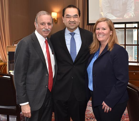
Celebrate Dr. Derrick Lin’s Promotion to Professor
Faculty and staff from Harvard Medical School and Mass Eye and Ear gathered at the Harvard Faculty Club in Cambridge, MA to celebrate the promotion of Derrick T. Lin, MD, FACS, Associate Chief of Otolaryngology–Head and Neck Surgery at Mass Eye and Ear, to Professor of Otolaryngology–Head and Neck Surgery at Harvard Medical School.
The promotion celebration was the first by the Harvard Department of Otolaryngology–Head and Neck Surgery since in-person gathering resumed after the COVID-19 pandemic. Joining faculty and leadership were Dr. Lin’s wife, children and brother, as well as several mentors and colleagues who travelled across the country to attend.
Among those mentors and colleagues were James L. Netterville, MD, the Mark C. Smith Professor of Head and Neck Oncology at Vanderbilt University Medical Center, and James Rocco, MD, PhD, Chair of the Department of Otolaryngology–Head and Neck Surgery at The Ohio State University Wexner Medical Center.
24 HARVARD Otolaryngology
Derrick T. Lin, MD, FACS, (center) stands between Mark A. Varvares, MD, FACS, (left) and CarolAnn Williams (right), interim President of Mass Eye and Ear.
RESEARCH ADVANCES s
A new look at the reticular lamina
The outer hair cells (OHCs) are sensory cells that help amplify speech and music. Deep inside the inner ear, the cells protrude from the cochlea and are supported by the reticular lamina, a membrane that anchors each OHC in place. The membrane keeps OHCs suspended in an extracellular fluid, allowing them to move in response to sound vibrations entering the cochlea.
For years, researchers had assumed the reticular lamina was a stiff, platelike structure. In a study published in Scientific Reports, Sunil Puria, PhD, and postdoctoral research fellow Nam Hyun Cho, PhD, both of Mass Eye and Ear, have upended this notion. Using optical coherence tomography, Drs. Puria and Cho measured in vivo the postmortem displacements of individual OHC rows through the gerbil round-window membrane. Recorded measurements ultimately provided a high spatial resolution view of the reticular lamina that was not possible before.

Findings from the study revealed varying degrees of motion along the reticular lamina, implying that the membrane is more flexible than researchers initially believed. The membrane appeared most similar to a mosaic, with different supporting structures enabling it to bend and stretch.
Cho NH, Puria S. “Cochlear Motion Across the Reticular Lamina Implies That It Is Not a Stiff Plate.” Scientific Reports 2022; Vol. 12(1): 18715. Doi: 10.1038/s41598-022-23525-x
Building an automated hair cell analysis toolbox
Sensory cells inside the inner ear are responsible for detecting sound vibrations and relaying them to the brain. These cells, referred to as hair cells, are highly specialized and arranged in a precise order. While researchers often use light microscopy to image hair cells along the entirety of the
cochlea, no widely applicable tools exist for fast, comprehensive analysis of such imaging data.
Artur A. Indzhykulian, MD, PhD, of Mass Eye and Ear, and Christopher Buswinka, a graduate student in his laboratory, have created a highly accurate machine learning-based model capable of quantifying cochlear hair cells of the inner ear. According to a study published in PLOS Biology, Dr. Indzhykulian’s team developed a software called the Hair Cell Analysis Toolbox (HCAT), which can use light microscopy images to automate common image analysis tasks. These tasks not only include counting hair cells, but also differentiating between outer and inner hair cell types and determining the best sound frequency for a given cochlear region.
The automated tools removed a considerable barrier in cochlear image analysis, allowing for faster, unbiased and more comprehensive data analysis practices. HCAT can also serve as a template for deep learning-based detection tasks in other types of biological tissue. Its core codebase could be used to develop a custom deep learning detection model for any object on an image.
Buswinka CJ, Osgood RT, Simikyan RG, Rosenberg DB, Indzhykulian AA. “The Hair Cell Analysis Toolbox Is a Precise and Fully Automated Pipeline for Whole Cochlea Hair Cell Quantification.” PLOS Biology. 2023; 21 (3). Doi: 10.1371/ journal.pbio.3002041
Drug-like cocktail regenerates hair cells in preclinical study
Hair cells are sensory cells in the inner ear that help relay sound to the brain. Once damaged or destroyed, these cells cannot regenerate, which has made effective hearing loss treatments elusive for years. Aging, noise exposure and other factors can harm hair cells and, in turn, render hearing loss permanent.
A research team led by Zheng-Yi Chen, DPhil, of Mass Eye and Ear, has developed a potential solution to this longstanding limitation. According to a study published in Proceedings of the National Academy of Sciences, a drug-like cocktail of different molecules successfully regenerated hair cells in a mouse model by reprogramming a series of genetic pathways in the inner ear. Previous research by Dr. Chen had identified two molecular signaling pathways—Myc and Notch—responsible for inducing the cell division required for hair cell regeneration. However, these pathways cannot be readily activated in humans. And, while chemical compounds have been shown to activate Notch, scientists had yet to find one that could activate Myc.
Using single-cell RNA sequencing, Dr. Chen and his team discovered that small biological molecules called small interfering RNAs (siRNA) could remove genes downstream that suppressed the activation of the Myc pathway. They then combined these siRNAs with the existing compounds into a single therapeutic and applied it to an adult mouse model with damaged hair cells. Results from the study indicated that the mouse regenerated new, functional hair cells.


Quan Y, Wei W, Ergin W, et al. “Reprogramming by Druglike Molecules Leads to Regeneration of Cochlear Hair Cell–Like Cells in Adult Mice.” Proceedings of the National Academy of Sciences. 2023; Vol. 120 (17). Doi: 10.1073/ pnas.2215253120
Effects of neurofibromatosis type 2 on vestibular dysfunction
Neurofibromatosis type 2 (NF 2) is a rare genetic disorder often manifesting as benign tumor growths on the vestibular nerve. Referred to as vestibular schwannomas, these tumors can grow large enough to disable a patient’s ability to hear and balance. However, the vestibular functions of patients with the condition have never been carefully analyzed, nor have the vestibular functions of those who have had
25 HARVARD Otolaryngology
The following are select research advances from the Harvard Medical School Department of Otolaryngology–Head and Neck Surgery.
RESEARCH ADVANCES s
their tumors treated with chemotherapy.
In a study published in Brain Communications, Richard F. Lewis, MD, of Mass Eye and Ear, and a team of researchers studied the primary vestibular-mediated behaviors, clinical vestibular disability and imaging and hearing of eight untreated patients with NF 2. After comparing their results with healthy patients and those who experienced sporadic, unilateral vestibular schwannomas, the team determined that the central vestibular signal-to-noise ratio had deteriorated in these patients, thereby degrading vestibular precision.
Findings from the study included observations of patients with NF 2 who were treated with the chemotherapy drug bevacizumab. Patients who received treatment experienced improved vestibular precision without affecting vestibular accuracy, which suggests the tumors had generated afferent neural noise along the vestibular nerve while bevacizumab had suppressed its harmful effects.
Madhani AS, King S, Zhu J, et al. “Vestibular Dysfunction in Neurofibromatosis Type 2-Related Schwannomatosis.” Brain Communications. 2023; Vol. 5 (2). Doi: 10.1093/ braincomms/fcad089
Establishing an objective method for diagnosing cochlear nerve degeneration
Since the discovery of cochlear synaptopathy in 2009, researchers have sought to establish its differential diagnosis in patients. Current hearing evaluations fail to unveil this condition, which can only be directly measured by quantifying the missing synapses between cochlear nerve fibers and hair cells within the inner ear of harvested temporal bones from cadavers.

Stéphane F. Maison, AuD, PhD, CCC-A, and colleagues at Mass Eye and Ear have established an objective method for detecting the condition in a study published in JASA Express Letters. They developed a
computer-controlled algorithm capable of automatically isolating the auditory neural responses from patients without the interference of other potential generators from the inner ear. When applied on human participants with normal hearing thresholds, their technique showed that participants with the smaller electric signals experienced the most difficulty hearing in noisy environments, a trademark of cochlear synaptopathy.
The objective methodology could help investigators uncover the first human biomarkers for cochlear synaptopathy.
First national perspective published on juvenile nasopharyngeal angiofibroma management and outcomes
Juvenile nasopharyngeal angiofibroma (JNA) are highly vascularized, fibrous benign growths known to be locally aggressive. The growths account for less than one percent of nasopharyngeal tumors and are primarily present in adolescent males. Surgeons have often managed JNA using a transnasal endoscopic resection and, given the rarity of the condition, surgeons seldom have had the opportunity to publish large studies on treatment outcomes.
A team of researchers led by Eelam Adil, MD, of Boston Children’s Hospital, has produced the first national perspective on JNA management and outcomes from the Pediatric Health Information System (PHIS) database, a national database containing billing and clinical data from 49 tertiary care centers. Dr. Adil identified 287 JNA patients over a 10-year period and analyzed data from these patients based on whether they were managed at high- or low-volume institutions.
Findings from the study, published in The Laryngoscope, suggested that better surgical outcomes occurred when JNA patients are managed at high-volume institutions. According to the study, nine high-volume institutions managed 42.2 percent of JNA patients while 36 low-volume hospitals accounted for the remaining 57.8 percent of patients. Furthermore, JNA patients managed at high-volume institutions were less likely to require postoperative mechanical ventilation or return for revision surgery due to residual disease than patients who underwent surgery at low-volume hospitals.
How delayed care impacts patients with facial trauma in Rwanda
Trauma accounts for more deaths worldwide than malaria, HIV/AIDS and tuberculosis combined. Yet it is often neglected, especially in low- and middle-income countries (LMICs) where delayed care contributes significantly to morbidity and mortality among young people.
David Shaye, MD, FACS, MPH, of Mass Eye and Ear, was part of an international group of colleagues to lead a one-year prospective study at the University Teaching Hospital of Kigali, investigating the reasons why patients with facial trauma in Rwanda received delayed treatment. According to the study, which was published in OTO Open, 78 percent of patients waited more than four days at the hospital before receiving care. Delays were associated with a four times greater likelihood of complication and were more likely to occur among patients from rural settings and lower socioeconomic classes. A significant number of patients with facial fractures were treated with wiring techniques under local anesthesia due to inadequate operating room times.
The most common reported reasons for delays included a lack of surgical materials, a lack of surgeons and financial constraints. To improve surgical training and overall access
Otolaryngology
26 HARVARD
Vasilkov V, Liberman MC, Maison SF. “Isolating AuditoryNerve Contributions to Electrocochleography by HighPass Filtering: A Better Biomarker for Cochlear Nerve Degeneration?” JASA Express Letters. 2023; Vol. 3 (2). Doi: 10.1121/10.0017328
Adil EA, Francisco S, Morgan E, Kawai K, Cunningham MJ. “Juvenile Nasopharyngeal Angiofibroma: Outcomes Analysis Based Upon Hospital Volume.” The Laryngoscope. Published online March 10, 2023. Doi: 10.1002/lary.30640
to quality care, the Global Surgery Program at Mass Eye and Ear is working with the AO Foundation to provide hands-on facial trauma education programs in LMICs throughout Africa and Asia.
Loss of Pex1 in mice inner ears leads to deterioration of neural synapses
Peroxisomal Biogenesis and Zelleweger Spectrum Disorders (PBD-ZSD) are rare hereditary disorders associated with severe neuro-physiological impairments along with sensorineural hearing loss. PBD-ZSD are caused by mutations in PEX genes, which encode for proteins involved in peroxisomal assembly and function. Peroxisomes play an important role in maintaining vital cellular and metabolic functions. While sensory cells in the inner ears of mammals are particularly vulnerable to metabolic changes, few studies have investigated the impact of mutated PEX genes on auditory function.
To test whether mutated PEX genes can affect auditory function, a team of researchers led by Gwenaelle Géléoc, PhD, of Boston Children’s Hospital, has developed conditional knockout mice to study the effects of Pex1 gene deletion in sensory cells of the inner ear. The study, which was published in Cells, revealed that removal of the Pex1 gene from inner ear hair cells leads to progressive hearing loss associated with alteration in sensory hair cell synapse morphology and function and significant decrease in auditory brainstem responses. Their findings demonstrate that Pex1 plays a critical function in the development and maturation of the afferent synapses connecting sensory inner hair cells with auditory nerve fibers. This work suggests that hearing loss in PBD-ZSD patients may be the result of oxidative stress imbalance, an increase in pexophagy and hair cell
dysfunction. As such, treatments aimed to preserve the inner hair cell synapse may help protect auditory sensitivity in PBD-ZSD patients.
New artificial intelligence tool developed to predict efficacy of botulinum toxin injections for patients with dystonia
Dystonia is a chronic movement disorder that causes abnormal postures and repetitive movements from involuntary muscle contractions. Dystonic symptoms are often painful and negatively impact an individual’s quality of life. Botulinum toxin injections are a standard treatment to temporarily manage dystonic symptoms, yet a significant cohort of these patients receive no benefit from these injections. There are no objective biomarkers of treatment response to determine who might benefit from injections, which limits the proper utilization of botulinum toxin for dystonia treatment.
In a study published in Annals of Neurology, a team of researchers led by Kristina Simonyan, MD, PhD, Dr med, of Mass Eye and Ear, has developed a novel deep learning platform that can help determine which patients with dystonia would benefit from botulinum toxin injections. According to the study, the platform, referred to as DystoniaBoTXNet, used a 3D convolutional neural network and raw structural brain MRIs to discover the neural biomarker of botulinum toxin efficacy in 284 patients with four different types of dystonia.
The platform achieved an overall accuracy of 96.3 percent in predicting treatment efficacy in 19.2 seconds per case.
Reviewing parathyroid gland identification technologies in thyroid surgery
Identifying and preserving parathyroid glands remains a challenge for surgeons despite recent advances in surgical techniques. If these glands are inadvertently removed or injured during an operation, the production of circulating parathyroid hormone decreases, ultimately leading to low calcium levels and higher post-operative morbidity rates.
In an effort to help surgeons better identify the parathyroid glands, an international group of surgeons has produced a comprehensive review of emerging imaging techniques for these glands. Published in JAMA Otolaryngology–Head & Neck Surgery, the review was led by Gregory W. Randolph, MD, FACS, FACE, of Mass Eye and Ear, and provided a detailed manual with step-by-step guidance on how to possibly implement emerging technologies in thyroid surgery.

From their findings, the surgeons have reason to believe emerging imaging technologies offer great promise and that future research should address a plethora of variables for determining the most optimal technique. These variables include postoperative outcomes among patients, costs associated with imaging and the training required to operate the new technology.
Silver Karcioglu AL, Triponez F, Solórzano CC, et al. “Emerging Imaging Technologies for Parathyroid Gland Identification and Vascular Assessment in Thyroid Surgery: A Review from the American Head and Neck Society Endocrine Surgery Section.” JAMA Otolaryngology–Head & Neck Surgery. 2023; 1; 149 (3): 253-260. Doi: 10.1001/ jamaoto.2022.4421.
Synthetic eardrum images found capable of improving AI diagnostic model accuracy
Yao D, O’Flynn LC,
K.
Novel Neural Network Biomarker of Botulinum Toxin Efficacy in Isolated Dystonia.” Annals of Neurology. 2023; Vol. 93 (3): 460-471. Doi: 10.1002/ana.26558
Over the past several years, researchers have used artificial intelligence (AI) to diagnose a variety of diseases with surprisingly high accuracy. The accuracy of these AI models depends on the quality and quantity of the datasets used to train
27 HARVARD Otolaryngology
Stanford-Moore GB, Niyigaba G, Tuyishimire G, et al. “Effect of Delay of Care for Patients with Craniomaxillofacial Trauma in Rwanda.” OTO Open. 2022; Vol. 6 (2). Doi: 10.1177/2473974X221096032
Mauriac SA, Peineau T, Zuberi A, Lutz C, Geleoc GSG. “Loss of Pex1 in Inner Ear Hair Cells Contributes to Cochlear Synaptopathy and Hearing Loss.” Cells. 2022; Vol. 11 (24): 3982. Doi: 10.3390/cells11243982
Simonyan
“DystoniaBoTXNet:
RESEARCH ADVANCES s
them. Without large, diverse datasets, a model would struggle to make an accurate diagnosis, which puts diagnostic models built for rarer diseases with fewer available datasets at a significant disadvantage. Creating synthetic images could help address this data gap. In a study published in PLOS Digital Health, Krish Suresh, MD, and Matthew Crowson, MD, both of Mass Eye and Ear, developed synthetic images of a tympanic membrane using a generative machine learning model. The generative model was used to create thousands of synthetic images and—when validated by human reviewers—the synthetic images were nearly indiscernible from the originals.
Drs. Suresh and Crowson moved their findings one step further in a subsequent study published in JAMA Otolaryngology–Head & Neck Surgery. To determine whether the synthetic images could improve the diagnostic accuracy of an AI model, Drs. Suresh and Crowson trained one model with synthetic images and a separate model with real images. They then compared how each distinguished between normal and abnormal tympanic membranes. According to their study, the model trained with synthetic images outperformed its counterpart, showing how synthetic data could be used to develop clinical tools and models.
Suresh K, Cohen MS, Hartnick CJ, Bartholomew RA, Lee DJ, Crowson MG. “Generation of Synthetic Tympanic Membrane Images: Development, Human Validation, and Clinical Implications of Synthetic Data.” PLOS Digital Health. 2023; 2(2):e0000202. Doi: 10.1371/journal.pdig.0000202
Suresh K, Cohen MS, Hartnick CJ, Bartholomew RA, DJ Lee, Crowson MG. “Making Use of Artificial Intelligence–Generated Synthetic Tympanic Membrane Images.” JAMA Otolaryngology–Head & Neck Surgery. Published online March 30, 2023. Doi: 10.1001/jamaoto.2023.0218
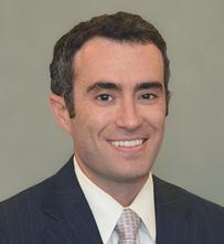

Tympanostomy tubes designed with liquid-infused properties to improve clinical performance
The most common ambulatory pediatric surgery in the United States is the
placement of tympanostomy tubes for recurrent middle ear infections. Contemporary tubes are typically small cylindrical devices made of plastic or silicone that are placed through the eardrum and ‘straddle’ the middle ear and outer ear canal. Upon placement, the device helps equalize air pressure across the tympanic membrane and drain fluid from the middle ear. However, the simple geometry and basic material properties of the traditional tube can lead to a number of unwanted clinical issues, including persistent ear discharge from bacterial biofilms, tube blockage due to cellular adhesion and poor delivery of medications through the tube to the site of infection.
In collaboration with the Wyss Institute for Biologically Inspired Engineering at Harvard University and the Harvard John A. Paulson School of Engineering and Applied Sciences, Aaron K. Remenschneider, MD, MPH, and Elliott D. Kozin, MD, both of Mass Eye and Ear, have overhauled the current design and materials of the device alongside a multidisciplinary team of researchers. According to a study published on the cover of Science Translational Medicine, the team redesigned the tube with an optimized geometry and a modified, liquid-infused surface. Despite its smaller size, the device was shown to be capable of preventing bacteria from growing on its surface, resisting cell adhesion and impeding water from flowing into the ear.
The new liquid-infused tympanostomy tube, which features a novel geometry, not only maintained the essential functions of its original design when tested in animal models, but it improved the reliability of passively administering ear drops into the middle ear. Findings from the study, which Dr. Remenschneider senior co-authored, have encouraged researchers to apply their patented design to other implantable
medical devices, such as catheters for the eye and brain.
Uncovering the identity of olfactory neuroblastoma
Olfactory neuroblastoma (ONB) is a rare type of cancer in the nasal cavity thought to originate from the specialized neuroepithelial lining responsible for detecting odors. Existing biomarkers of the disease can be ineffective for diagnosing the cancer with implications for proper treatment. A deeper understanding of the cellular identity of the tumor could provide better diagnosis and treatment strategies, as well as developmental avenues into future mechanistic studies.
For the first time, a team of researchers led by Eric H. Holbrook, MD, of Mass Eye and Ear, and collaborators at Tufts University School of Medicine, have linked ONB to a specific, analogous normal cell type. According to a study published in Modern Pathology, the research team identified shared transcriptomic and immunohistochemical characteristics between ONB and a specific population of stem/progenitor cells within the olfactory epithelium known as globose basal cells (GBCs). Furthermore, the team identified a cellular hierarchy within the tumor that mirrors developmental processes observed in GBCs. Among these was an EZH2-expressing proliferative compartment, suggesting one possible pathway for epigenetic treatment.
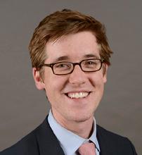
Findings from the study could help identify additional tumor markers for improving diagnosis and prognosis of the disease, while the added developmental insight may inform future treatment strategies.
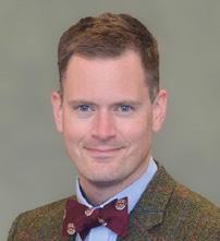
28 HARVARD Otolaryngology
Patel H, Pavlichenko I, Grinthal A, et al. “Design of medical tympanostomy conduits with selective fluid transport properties.” Science Translational Medicine. 2023; Vol. 15 (690). Doi: 10.1126/scitranslmed.add9779
Zunitch MJ, Fisch AS, Lin B, et al. “Molecular Evidence for Olfactory Neuroblastoma as a Tumor of Malignant Globose Basal Cells.” Modern Pathology. 2023; Vol. 25 (5). Doi: 10.1016/j.modpat.2023.100122
Save the Date for Mass Eye and Ear’s 200th Anniversary Celebration!
On Friday, July 19, 2024, all current and former Department of Otolaryngology–Head and Neck Surgery faculty and trainees are invited to return to Boston for the hospital’s bicentennial celebration. Festivities include lectures by distinguished alumni, tours of the hospital and a gala dinner.

For more information, please contact Dana Grider, Director of Development for the Department of Otolaryngology–Head and Neck Surgery at Mass Eye and Ear, at dgrider@meei.harvard.edu or 617-573-4223. Learn more:
Connect with us on LinkedIn: All former residents and fellows are invited to join the Harvard Otolaryngology–Head and Neck Surgery Alumni Group.

 Photo by Mary Leach
News from the Harvard Medical School

Department of Otolaryngology–Head and Neck Surgery
243 Charles Street, Boston, MA 02114
Beth Israel Deaconess Medical Center
Boston Children’s Hospital
Brigham and Women’s Hospital
Massachusetts Eye and Ear
Massachusetts General Hospital
NONPROFIT U.S. Postage PAID N. READING, MA Permit NO. 121
