18 minute read
Clinical

Orthodontic treatment
In this case, the patient’s main concerns where the uneven smile and the shortened upper central incisor teeth.
The characteristics of the malocclusion were as follows: • Class I molar and canine occlusion • 5mm overjet • Proclination of the upper and lower incisor teeth • Mild lower anterior crowding • Upper incisor irregularity.
After a new patient consultation and a treatment planning discussion where all the options were discussed with the patient, she elected to have an orthodontic assessment to explore the options to move and align the teeth.
She was presented with two options, that of fixed braces or aligning treatment.
She requested that aligning treatment was undertaken. PVS impressions were sent to Align Technology for conversion into 3D study models using the company’s software.
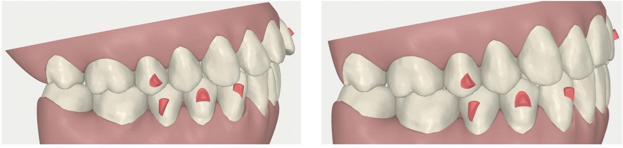
The Clincheck Pro software was used to modify the initial set-up (Figures 10 and 11). When finalised, 20 upper and lower aligners were prescribed, giving a treatment duration of about 10 months (Figures 12 and 13). IPR, totalling 1.2mm in the lower arch and 2.4mm in the upper arch, was prescribed. IPR was necessary to make space to correct the crowding the decrease the incisor protrusion.
Treatment proceeded as expected with no complications, or need for refinement. The patient was compliant and wore the aligners 22 hours per day.
When treatment was finished and the restorative treatment completed, fixed retainers were bonded upper (palatal) and lower (lingual) on the incisors and canines.
The retention phase
Normally, the final aligner acts as the retainer in most cases of Invisalign treatment.
Despite extensive research, the various elements leading to relapse of treated malocclusions are not completely understood, which makes retention one of the most challenging aspects of orthodontic treatment (Kuncio et al, 2007).
However, in this case, immediately after composite bonding, the upper retainer was cut in the incisal area to allow for the increased incisal length. New impressions were taken after completion of the restorative treatment and new retainers were made to ensure that the occlusion was well maintained. It is essential to stress that patients should wear their Invisalign retainers as prescribed by the orthodontist to ensure stability of the occlusion and correct alignment of the teeth.
In a comparative study of the retention of fixed braces versus Invisalign retainers, Kuncio et al (2007) found that, in many cases, aligner treatment can relapse more than fixed braces, and so patients are instructed to continue to wear their retainers for maintenance treatment.
However, the total number of patients in each group was 22, which is a very small number and further research is necessary.
Whitening in Invisalign aligners
Using the Invisalign aligners as whitening trays has become a viable treatment option for patients.
The recommendation is to wait for a month after the first aligners are placed and after the initial discomfort has settled down. The patient applies the whitening gel directly into the aligners.
A similar Van Haywood protocol can be adopted, namely to whiten the upper teeth first followed by the lower teeth.
The upper teeth whiten quicker and have fewer side effects and so the first stage of whitening is relatively simple. The lower whitening normally takes longer. It is thought that this is due to the wash-out effect of the salivary glands.
Due to the rigid nature of the aligner, it seems that whitening may occur quicker than a normal bleaching tray, but this has not been studied significantly.
Most aligners do not reach over the gingivae, so the gingivae may be less irritated during whitening.
There is no effect of whitening around the orthodontic attachments, and the whitening occurs in a multidirectional way and can whitening around the attachments.
Whitening and composite bonding
It is essential after completion of home whitening treatment
Figure 9: Shades of Aura composite.
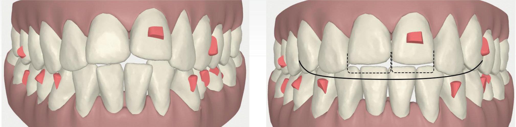
Figure 10: Initial centre view using Clincheck software. Figure 11: Final centre view.
Figure 12: Initial right buccal view. Figure 13: Final right buccal view.
to wait for a period of two weeks to allow all the oxygen to be dissipated from the tooth and for the shade to settle to the actual shade.
After tooth whitening, there is maximum saturation of oxygen inside the enamel. This causes a reduction in bond strength of 20%. It is thus essential to wait for two weeks for the enamel bond strength to recover back to the normal levels prior to commencing direct bonding onto the surface of the tooth.
Stratified layering technique
With the introduction of multiple component composite systems, it is now possible to create beautiful natural restorations using multiple layers of composite using their different optical and material properties. The Aura composite system is ideal as it contains both enamel and dentine shades (Figure 9).
The enamel shades are a microfill composite, which give the properties of a glass-like appearance of natural enamel (Figure 5). The dentine shades are a nanohybrid material (Figure 6), which gives extra strength for occlusal buildups and they can be used as a bulk-fill material. There are separate bulk-fill syringes available for this purpose.
Shade selection of composite resin
According to Vanini (1996), it is essential to undertake a detailed evaluation of hue, chroma, opalescence, and fluorescence of the tooth in order to simplify the composite stratification technique.
This is done early in the clinical procedure to ensure that there is no dehydration of the tooth when the tooth is fully isolated (Figure 5).
Once the tooth is fully isolated, it dehydrates and lightens and this can result in the selection of a shade that is too light. Blends of composite colours are normally used and, after selection of the transluscent enamel shade (Dietchi, 2008), the dentine shade is used (Figure 6).
The hue is given by the dentine. The hue remains the same, although the greater thickness of the enamel interferes in its perception, giving it a less saturated aspect. Therefore, the hue of the tooth is given by the dentine and influenced by the enamel. The enamel does not change the hue, but only
confers a greater or lesser saturation or chroma according to its thickness (Franco et al, 2007).
This is applied from darkest to lightest to give the restoration a lifelike appearance. Different translucencies may be selected for the mesial corners as, often, these are more translucent than the distal corners (Figure 7).
Used with an understanding of tooth morphology, restorative material selection, colour options, and the physical properties of light, these layering techniques allow optimally aesthetic restorations to be predictably achieved (Terry, 2003).
Applying the composite resin
A test run is undertaken first using a variety of composite shades. A clinical photograph is taken after the first test to review how the shades appear on a digital photograph. A polarised light photograph can also be taken to understand the nuances of the existing anatomy of the tooth, which needs to be copied. As the teeth have been bleached, the bleaching composite shades can be used. In Aura composite, the DB shade blends very well to the bleached enamel (Figure 8). The enamel shades are tested first and light-cured followed by the dentine shades.
Placing the layers
Normally, the tooth is built-up from the palatal part first. A clear matrix is adapted to help form the shape of the missing incisal edge.
A wax-up can be used and a silicone stent can be made for ease of placement and for patient and dentist visualisation of the final outcome. The clear matrix is curved and rolled in the operator’s gloved hand to form a curve. This is placed on the missing incisal edge and bonded into place using a dentine bonding agent. This helps to keep the hands free so that the layering of the composite can commence.
The layers are placed into the area with a flat plastic tool and then sculpted into place and refined with a fine haired brush dipped into bonding liquid or a rubber sculpting instrument.
The tooth is built-up in layers and light-cured (Figures 6 and 7). Each layer is checked after placement and modifications made as the restoration takes shape. Once the layering is completed, the final form is created with finishing instruments.
Sof-lex polishing discs (3M Espe) are used first to remove any bulk excess and then fine flame finishing burs are used.
After the form is completed, the patient is asked to sit up so that the incisal edges can be checked from the front of the patient. When the dentist is in a working position behind the patient, there is tendency to build the incisal edge restorations too long. This final length of the incisal edge is checked when the patient is supine in the chair.
The occlusion is checked and the final polishing can commence with the use of rubber wheels and a felt tip rubber wheel and polishing paste. The final glossy layer is created with a special rotary rubber wheel and polishing paste.
Conclusion
A multidisciplinary case such as this requires essential communication between the specialists undertaking the treatment, and also between the dentist and the patient, so that the patient is fully aware of the risks and the benefits of the different treatment procedures, and what is involved in each treatment so that an ideal outcome can be achieved. The patient also needs to be fully aware of the retention phase needed to maintain the teeth in the same position, and to maintain a beautiful smile and when further whitening may be needed. In addition, any repairs to composite need to be detailed and occlusal checks need to be made regularly to maintain a beautiful smile. From time to time, during recall appointments, further polishing of the composite may be required, including retention recall evaluations.
References
Bernabe E, Sheiham A, de Oliveira CM (2008) Impacts on daily performances related to wearing orthodontic appliances. The Angle Orthodontist 78(3): 482-486
Blank JT (2006) Creating translucent edge effects and maverick internal tints using microhybrid resin. Pract Proced Aesthet Dent 18(2): 131-136
Dietschi D (2008) Optimising aesthetics and facilitating clinical application of free-hand bonding using the ‘natural layering concept’. Br Dent J 204(4): 181-185
Franco EB, Francischone CE, Medina-Valdivia JR, Baseggio W (2007) Reproducing the natural aspects of dental tissues with resin composites in proximoincisal restorations. Quint Int 38(6): 505-510
Houle JP, Piedade L, Todescan L Jr, Pinheiro FHL (2017) The predictability of transverse changes with Invisalign. The Angle Orthodontist 87(1): 19-24 Joffe L (2003) Invisalign: early experiences. J Orthod 30(4): 348-352
Kravitz ND, Kusnoto B, BeGole E, Obrez A, Agran B (2009) How well does Invisalign work? A prospective
clinical study evaluating the efficacy of tooth movement with Invisalign. Am J Orthod Dentofacial Orthop 135(1): 27-35
Krieger E, Seiferth J, Marinello I, Jung BA, Wriedt S, Jacobs C, Wehrbein H (2012) Invisalign treatment in the anterior region: were the predicted tooth movements achieved? J Orofac Orthop 73(5): 365-376
Kuncio D, Maganzini A, Shelton C and Freeman K (2007) Invisalign and Traditional Orthodontic Treatment Postretention Outcomes Compared Using the American Board of Orthodontics Objective Grading System. The Angle Orthodontist 77(5): 864-869
Lagravere MO, Flores-Mir C (2005) The treatment effects of Invisalign orthodontic appliances: a systematic review. J Am Dent Assoc 136(12): 1724-1729
Malik OH, McMullin A, Waring DT (2013) Invisible orthodontics part 1: Invisalign. Dent Update 40: 203-204, 207-210, 213-215
Terry DA (2003) Dimensions of color: creating highdiffusion layers with composite resin. Compend Contin Educ Dent 24(2 Suppl): 3-13
Tuncay OC, Orhan C (2006) The Invisalign System. Quintessence, Chicago
Vanini L (1996) Light and color in anterior composite restorations. Pract Periodontics Aesthet Dent 8(7): 673-682
Vlaskalic V, Boyd R (2001) Orthodontic treatment of a mildly crowded malocclusion using the Invisalign system. Aust Orthod J 17(1): 41-46
Reprinted with permission by Aesthetic Dentistry Today April 2017
COVID-19 risk management in dental practice: The infection chain pathway of SARSCoV-2
Johan Hartshorne1 and Andre van Zyl2
Keywords: aerobiology, aerosols, airborne, droplets, coronavirus, co-morbidities, COVID-19, SARSCoV-2, dentistry, risk management, reservoir, transmission, infection chain, susceptible host
1 Johan Hartshorne B.Sc., B.Ch.D., M.Ch.D., M.P.A., Ph.D., (Stell), FFPH.RCP (UK) General Dental Practitioner, Intercare Medical and Dental Centre, Tyger Valley, Bellville, 7530 South Africa Email: jhartshorne@kanonberg.co.za
2 Andre van Zyl B.Ch.D., M.Ch.D. (Stell) Specialist in Oral Medicine and Periodontics Honorary Professor: Department of Oral Medicine and Periodontology University of Witwatersrand Johannesburg Private practice: 9 College Road, Hermanus Email: info@andrevanzyl.co.za
Executive Summary
Rationale
Understanding the coronavirus (SARS-CoV-2) and its pathways from its reservoir to host can help us understand how to fight the virus, and is critical in developing effective and sustainable infection prevention and control measures.
Key points The pathogen – SARS-CoV-2
• SARS-CoV-2 is the most contagious of all the respiratory viral infections. • The main sources of SARS-CoV-2 are asymptomatic, pre-symptomatic, symptomatic
COVID-19 individuals in the population. • SARS-CoV-2 transmission from asymptomatic and pre-symptomatic hosts are a fundamental fault- line in the spread of COVID-19 because we do not know who they are. • The average incubation period (infectious period) is 6.4 days (range 2-24 days) • SARS-CoV-2 is very stable at room temperature, wide range of pH and on smooth surfaces (including glass, plastic, and stainless steel). • SARS-CoV-2 is stable on stainless steel and plastic for up to 9 days. • Detectable levels of infectious virus is still present on the outer layer of a surgical mask at 7 days. • SARS-CoV-2 is very susceptible to standard disinfection materials, including 70% ethanol, household bleach and 7.5% povidone-iodine.
Reservoir
• SARS-CoV-2 infectious cycle can start in the lungs, naso-pharynx, oral mucosa, tongue and salivary glands. • During the first 10 days of incubation the virus mainly accumulates in the pharyngeal, oral and nasal areas. • SARS-CoV-2 is consistently detected in saliva.
Portal of exit (leaving the host reservoir)
• Individuals with infection produce respiratory droplets or aerosol particles from breathing, talking, singing, coughing and sneezing
• Common exit portals for SARS-CoV-2 are the mouth, nose, respiratory tract and faecal route.
Mode of transmission
• Transmission of SARS-CoV-2 can occur from asymptomatic and symptomatic individuals. • SARS-CoV-2 is mainly transmitted through close physical contact and respiratory droplets. • Airborne transmission is possible during aerosol generating procedures. • Large droplets (>5 µm) settle faster due to gravity, thus contaminating surrounding surfaces. • Smaller droplets (<5 µm) evaporate faster forming droplet nuclei that can stay airborne for hours. • Aerosolized viral droplet nuclei particles can travel great distances and remain airborne and viable for up to 3 hours and can infect dental health care workers, patients and contaminate surfaces.
Portal of entry and replication
• Portal of entry is through mouth, nose, respiratory tract. • SARS-CoV-2 S-spike protein invades host target cells by using ACE2 as its receptor.
Susceptible host and disease pathogenicity
• Infected hosts can present with clinically inapparent (asymptomatic) or mild (80%), moderate severe (14%) or critical illness requiring hospitalization (6%). • Individuals with co-morbidities presented with increased
COVID-19 severity and higher case fatality rates. • Hypertension and hyperlipidaemia were the most frequent co-morbidities. • Elderly and immune-compromised individuals are most vulnerable • Individuals with periodontitis may be linked to more severe
COVID-19
Practice implications
• The disturbing reality is that we have no idea who among us is spreading the disease. • Early recognition of an infected person (source of infection) and cutting off the route of transmission are key points to control COVID-19. • A decrease in the oral viral load would diminish the amount of virus expelled and reduce the risk of transmission. • Pre-procedural mouth rinse is a critical prophylactic measure for reducing oral viral load and risk of spreading
SARS-CoV-2. • The weight of combined evidence supports airborne precautions for occupational health and safety of health workers treating asymptomatic or suspected patients with
COVID-19. • Ventilation is of critical importance to control airborne transmission of SARS-CoV-2
Introduction
The global outbreak of coronavirus disease (COVID-19) is caused by the novel severe acute respiratory syndrome coronavirus 2 (SARS-CoV-2). It is the most contagious of all the viral respiratory pandemics, spreading fast with an increasing number of infected patients world-wide. The current global statistics on March 22, 2021, show the total number of confirmed cases 123,868,982, total deaths 2,727,738 and total recovered /discharged 99,795,152 individuals. Current global active infected cases 21,346,092 (17,2 %) of which 21,255,883 (99,6%) have a mild condition and 90,209 (0,4%) are serious / critical.1 The COVID-19 statistics for Australia on March 22, 2021 showed a total number of 29,205 and New Zealand 2,462 cases; total deaths: Australia 909, New Zealand 26 and total recovered: Australia 26,243, New Zealand 2,373 cases. Current active COVID-19 cases on March 22, 2021 were: Australia 2,053, New Zealand 63 of which none are serious/critical.
Outbreaks of newly emerging infectious viral diseases present unique challenges and threats to health care providers due to lack of immunity, absence of specific, effective, and safe antiviral drugs, and a limited understanding of the emerging threat and reliance on infection prevention and control measures.
A series of events has to happen to enable a pathogen such as SARS-CoV-2 to cause an infection (COVID-19). This series of events is referred to as the ‘chain of infection’. The links of the infection chain consist of : (i) the pathogen (infectious agent), (ii) reservoir or source, (iii) portal of exit, (iv) mode of transmission, (v) portal of entry, and (vi) a susceptible host.2,3 The chain of infection model holds that infectious diseases result when an agent (pathogen) leaves its reservoir or host through a portal of exit, is conveyed by some mode of transmission, and enters through an appropriate portal of entry to infect a susceptible host.
The spread of infection can be mitigated by breaking the infection chain at any of its links. Therefore understanding the setting and characteristics of each link of the infection chain and how SARS-CoV-2 spreads to a susceptible host is critical in developing effective and sustainable infection prevention and control measures in the absence of a vaccine and anti-viral drugs.
Methodology and Purpose
The literature search methodology used for the data assimilation and knowledge synthesis in this series is described in Part 1. The epidemiological ‘infection chain’ model was also used to enhance data assimilation and knowledge synthesis. (Table 1)
Part 2 of this series will focus on the key parameters of the infection chain that impact directly on risk management in the dental practice (Table 1)2,3 In addition, Part 2 also provides a review of the current knowledge and understanding of aerobiology and flow physics implicated in the generation, expulsion, evolution and transmission of virus-laden droplets and aerosols generated during expiratory activities such as breathing, talking, coughing and sneezing and during aerosol generating procedures.
This knowledge will enhance dental practitioners understanding the what, the why, and the how, underpinning SARS-CoV-2 infection control and prevention.
Pathways of the infection chain – What is our knowledge and understanding of SARS-CoV-2 (virus) and susceptible host characteristics?
1. The pathogen – How virulent and infectious is SARS-CoV-2?
• The origin and identification of the coronavirus SARS-CoV-2 In December 2019, a cluster of fatal pneumonia outbreaks originated in Wuhan City, China.4 All patients had been associated with the Wuhan Wholefood Market, where seafood and live animals are sold. The disease spread rapidly to most provinces in China and subsequently the rest of the world.5,6
Chinese researchers quickly isolated a new virus from a patient and sequenced its genome (29,903 nucleotides).7 The infectious agent of this viral pneumonia that originated in Wuhan was finally identified as a novel coronavirus (2019nCoV), the seventh member in a family of coronaviruses that affect humans.8 After analysis of respiratory samples, the Peoples Republic of China Centers for Disease Control declared that the pneumonia was caused by a novel coronavirus now referred to as SARS-CoV-2.9
Coronaviruses (CoV) are respiratory pathogens. They belong to the Coronaviridae family. Currently there are four genera of coronaviruses: α-CoV, β-CoV, λ-CoV, and δ-CoV.10 SARS-CoV-2 is an enveloped single stranded RNA virus. Six corona viruses were previously known to cause disease in humans, SARS-CoV-2 is the seventh member of the coronavirus family that infects humans after SARS-CoV, MER-CoV.4 and the Middle East respiratory syndrome coronavirus (MERS-CoV)12 that occurred in 2002-2003 and in 2012 respectively. SARS-CoV, MERS-CoV and SARSCoV-2 belong to β-CoV.7.13
• The morphologic and genetic structure, replication and pathogenic mechanisms The morphologic structure of SARS-CoV-2 is similar to SARSCoV, with virion size ranging from (60-140 nm) (0.060.14μm).14 The virus has distinctive spikes of 9-12 nm that give the appearance of “coronas” around the sun. Spike, membrane and envelope surface viral proteins of the coronavirus are embedded in its host-derived lipid bilayer membrane encapsulating the helical nucleocapsid comprising viral RNA.15
SARS-CoV-2 presents with two notable genomic features: (i) it is optimized for binding to human receptor angiotensinconverting enzyme 2 (ACE) and, (ii) the receptor binding domain in the spike S-protein has a site at the S1-S2 boundaries (the two subunits of the spike) through which insertion of nucleotides takes place. The cleavage site on the S-protein allows effective cleavage by furin (protease) and other proteases that allows nucleotides of SARS-CoV-2 to enter host cells through human cell receptor ACE2, to allow viral entry, duplication, infectivity and host range.16-18
ACE2 is an important receptor for SARS-CoV-2 19 and found in many target cells including type II alveolar cells of lung20-22, epithelial cells of the oesophagus, adsorptive enterocytes from the ileum and colon22, cholangiocytes23 , myocardial cells, kidney proximal tubule cells, bladder and urothelial cells20, salivary gland epithelial cells24,25 and oral mucosa.26
Viral entry and cell infection trigger the host’s immune response, and the inflammatory cascade is initiated by antigen presenting cells.27 It is suggested that the severity of the virus infection is closely related to the maturity and binding capacity of ACE2.28 Gao and co-workers29 have suggested that a lower level of ACE2 and weaker binding could be a major contributing factor that leads to the absence of any clinical manifestations in asymptomatic cases.
• What is known about the incubation period of SARS-CoV-19? Incubation period is the time from moment of exposure to the corona virus until signs and symptoms appear. The best current estimates of the incubation of SARS-CoV-2 range from 2-14 days with an average of 6.4 days.27,30,31