
DENTAL SOLUTIONS
The Magic Wand of Painless Dentistry
I have been using the STA wand for over 19 years having been introduced to the STA Wand at a course in Ballarat where I volunteered to have the STA. The only time I previously had intraoral local anaesthetic (LA) was more than ten years earlier as a third-year dental student. I did not feel the LA and subsequently bought a STA unit.
As a Specialist Periodontist, I generally see an older demographics of patients for the management of periodontitis, implant placements, extractions and periodontal surgeries. However, I treat a number of teenagers who are either undergoing or recently completed orthodontics. A local orthodontic practice sends a large number of his patients to my practice as we have the “Magic Wand”.
I most frequently do infiltrations on the buccal aspects of the teeth when I do initial stage non-surgical periodontal treatment and the occasion supportive periodontal maintenance patient. However, I also use the STA wand to do palatal injections for implants, extractions, periodontal surgeries and rarely for non-surgical root surface debridement. With the STA Wand, the palatal injections are painless unlike conventional palatal injections which can be very sore. I don’t do single-tooth anaesthesia as I do not do restorative treatment or endodontics.
On average, it takes me 2-4 minutes to use the STA Wand. I count 25 beeps when I do an infiltration and then do the next injection a few teeth away for non-surgical periodontal
treatment. For implants, extractions and periodontal surgeries, I use the entire carpule of LA, giving the LA both buccally and lingually or palatally.
A couple of tips for using the STA Wand include dispensing a couple of drops of LA on the mucosa before inserting the needle, holding the Wand with a pen grip and asking your patients to count the beeps. This is a fantastic distraction.
My patients love the Wand. They ask, “aren’t you going to give me a needle” and “why don’t all dentists have the Wand?” My answer to the first question is “I already have” and the patients look at me incredulously.
My answer to the second question is” I honestly don’t know but they should”.
The STA Wand is an invaluable part of my practice. There are two pieces of equipment that I won’t work without – my loupes and the STA Wand.
Author: Dr Susan Wise is a Specialist Periodontist in the South Eastern suburbs of Melbourne. Susan graduated as a dentist from the University of Melbourne in 1994 and as a periodontist at the University of Queensland in 2002. Susan is an Australian Dental Association Board Director. She is a Past President of the Royal Australasian College of Dental Surgeons, Australian Dental Association Victorian Branch and the Australian Society of Periodontology Victorian Branch. Susan has been a demonstrator to and examined undergraduate and postgraduate students in periodontics.

Dr Susan Wise Specialist Periodontist
View Product
Glass-Fibre-Reinforced Composites as a Core Build-Up in Minimally Invasive Endodontics
Even though dentistry is nowadays focusing more and more on prevention, there is still a considerable amount of patients with extensive caries lesions in need of endodontic treatment. When the damage has already occurred, it’s important to treat the lesion in a minimally invasive way. Preserving the cervical dentine is of utmost importance here, as maintaining a ferrule is necessary for a good prognosis of the restorative treatment.
Since the introduction of microscopes and NiTi files in dentistry, preservation of the cervical dentine has become more simple and predictable.
Several options exist for the post-endodontic treatment1: the treatment plan depends on the remaining tooth structure, wall thickness and the total cavity size.
Heavily damaged teeth often end up in a restorative cycle, with increasingly larger restorations after the original fractured and endodontic retreatments, the prognosis of the tooth becoming more and more challenging each time. Hence, it’s important to implement a correct treatment plan to preserve the teeth not only in the short term, but to avoid catastrophic failures that compromise the long-term survival.
The first step is to preserve the tooth tissue, especially in the cervical part of the crown. Preparation designs based on root canal orientation allow for very conservative access cavities and give a better restorable prognosis in fracture cases2
At the restorative side, fibre-reinforced composites (FRC) can be used to reinforce the cavity. Several studies have highlighted that the fracture pattern and load-bearing capacities of large cavities restored with FRC was more favourable compared to those restored with conventional direct composites3,4 Hence, FRC become a promising solution for post-endodontic treatment, mostly in cases with conservative access.
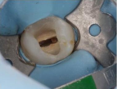
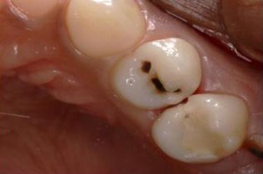
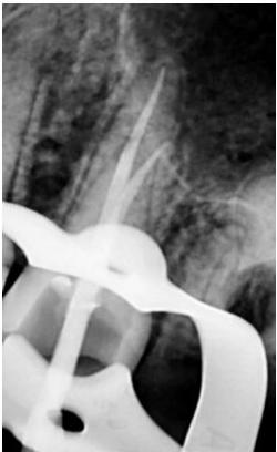
One of the suitable indications for FRC is the endodontically treated premolar with one big orifice and one large oval canal or deep furcation (Figs. 1-4). In such cases, an FRC can be placed as a Nayyar’s core modification5 without preparing the orifice part with Gates or Largos. The restoration can be finished as usual, directly with a conventional composite.
Endodontically treated molars with conservative access do not require a post. Of course, the final treatment does not only depend on the access type, but also on the size and depth of the cavity. Here, FRC have also proven their worth in complicated cases with internal resorptions (Figs. 5-8).
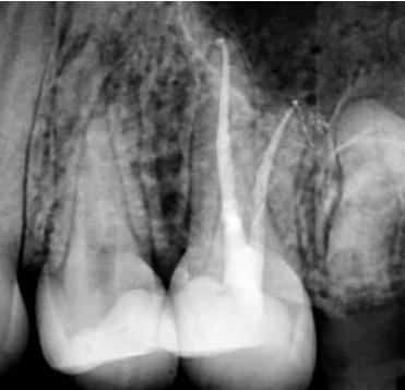
Even when the endodontic treatment is finished with an indirect restoration (a crown, onlay or overlay), FRC can be used for core build-ups such as a Nayyar’s core. This will be mostly indicated in retreatment cases, where the orifice part of the canal had already been prepared (Figs. 9-13).
EverX Posterior™ and everX Flow™ are excellent options to restore the core of endodontically treated teeth. Together with the preservation of cervical dentine, they are part of a strategy to increase the longevity of the post-endodontic restorative treatment.
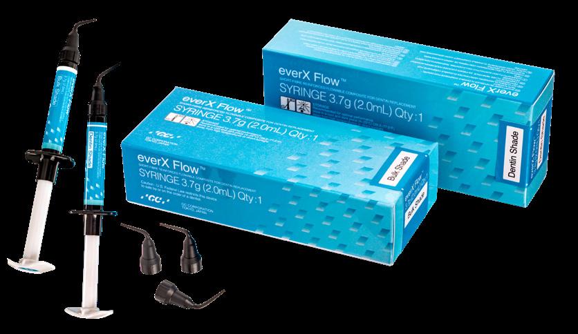
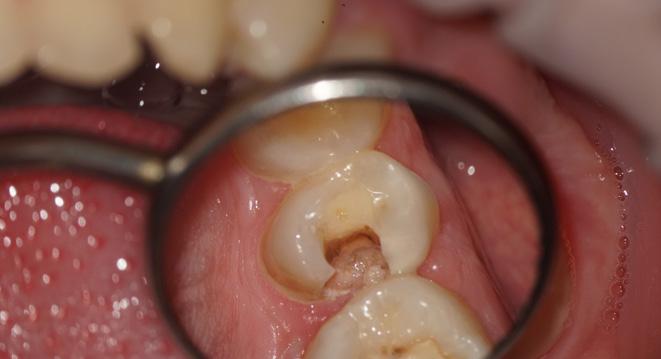
Fig. 1: Pre-operative situation with large orifice.
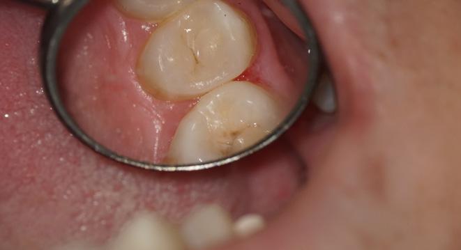
4: Final result.
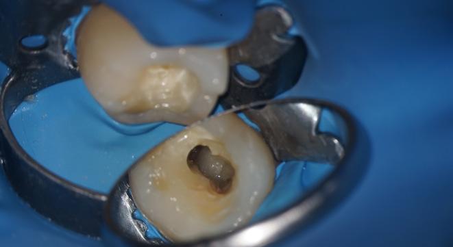
6: (a) Cavity and (b) access view after obturation.
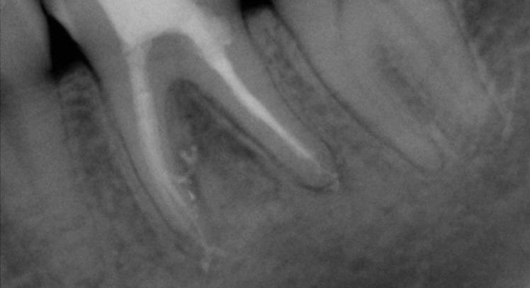
Fig. 8: X-ray at 6-months follow-up, after placement of a full ceramic crown.
11: Tooth prepared for the adhesive luting of an all-ceramic restoration.
By DR. Kaplan B. Sheudzhen, Russia

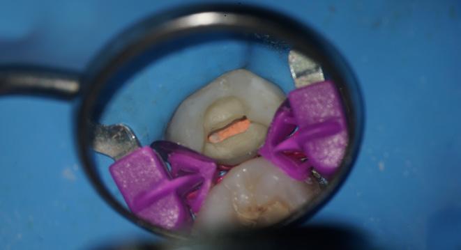
Fig. 2: A free space with a depth of 3-4 mm was created in the orifice part with a heated plugger.
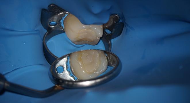
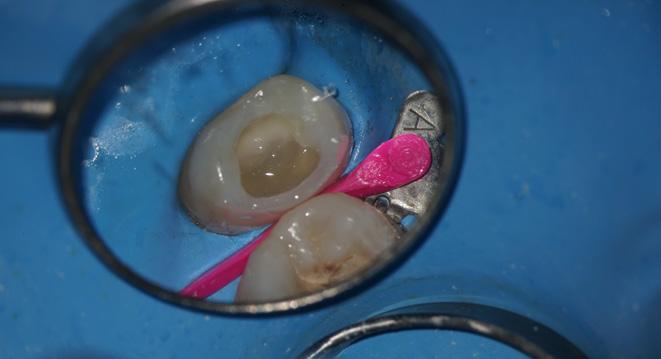
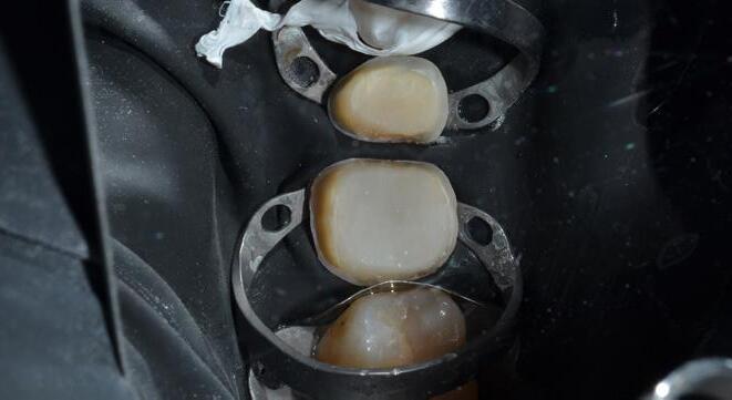
Fig. 3: The proximal wall was created first using a conventional composite. The core was built up with everX Flow (GC), a flowable fibre-reinforced composite
5: Pre-operative situation with large defect and internal resorption (a) Intraoral view (b) X-ray
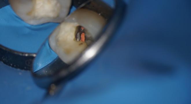
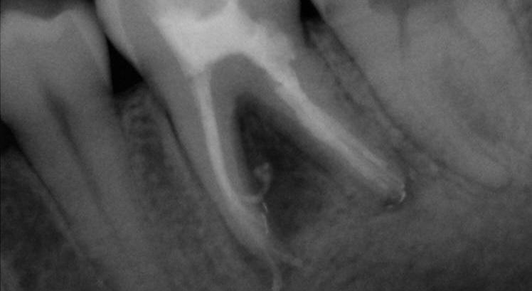
Fig. 7: Final X-ray.
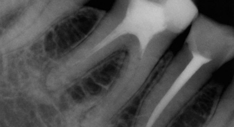
Fig. 9: Pre-operative X-ray.
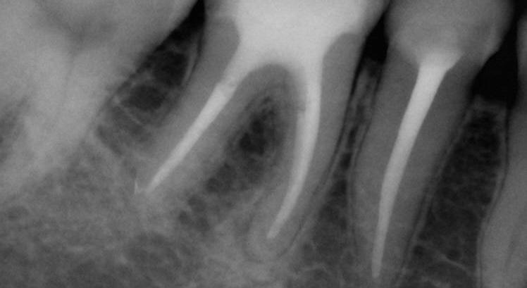
Fig. 12: Final X-ray.
References
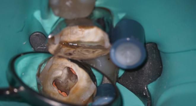
Fig. 10: Cavity before placement of everX Posterior (GC), a fibre-reinforced composite in paste consistency.
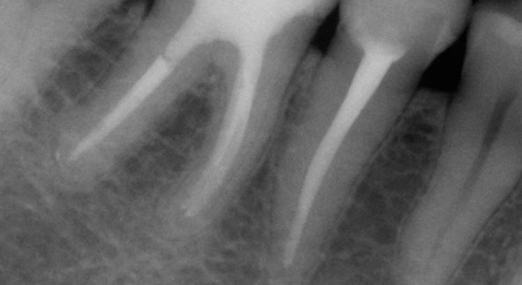
Fig. 13: Follow-up after three years.
1. Zarow M, Ramírez-Sebastià A, Paolone G, de Ribot Porta J, Mora J, Espona J, Durán-Sindreu F, Roig M. A new classification system for the restoration of root filled teeth. Int Endod J. 2018; 51(3):318-334.
2. Özyürek T, Ülker Ö, Özsezer Demiryürek E, Yılmaz F. The Effects of Endodontic Access Cavity Preparation Design on the Fracture Strength of Endodontically Treated Teeth: Traditional Versus Conservative Preparation. J Endod. 2018; 44(5):800-805.
3. Garoushi S, Sungur S, Boz Y, Ozkan P, Vallittu PK, Uctasli S, Lassila L. Influence of short-fiber composite base on fracture behavior of direct and indirect restorations. Clin Oral Investig. 2021 Jan 8 (Online ahead of print).
4. Geerts G, Pitout E, Visser H. Fracture resistance of endodontically treated premolars with fibre-reinforced composite restorations. Eur J Prosthodont Restor Dent. 2011; 19(1):25-31.
5. Nayyar A, Walton RE, Leonard LA. An amalgam coronal-radicular dowel and core technique for endodontically treated posterior teeth. J Prosthet Dent. 1980; 43(5):511-5.
Fig.
Fig.
Fig.
Fig.
EQUIA Forte HT, a Glass Hybrid System
Combining Bulk Fill Strength with Advanced Resin Coating
EQUIA Forte HT is a glass hybrid restorative system that combines a self-cure bulk fill restorative (EQUIA Forte HT Fil) with a highly filled, light-cured resin coating agent EQUIA Forte Coat. EQUIA Forte Coat is a unique self-adhesive surface treatment material which protects and optimises the physical properties of the underneath EQUIA Forte HT Fil restoration.
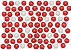
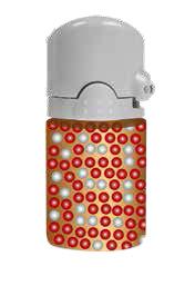
The coat is highly filled with 40 nm silica fillers and a filler dispersion technology (Fig.1) that ensures the uniform repartition of the fillers in the material is used. Thanks to this filler dispersion, a high wear resistance can be expected.
Figure 1: Uniform dispersion of nanofillers in EQUIA Forte Coat

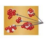
Agglutination
Using conventional filler technology, nanofillers would easily agglutinate, forming filler clusters.
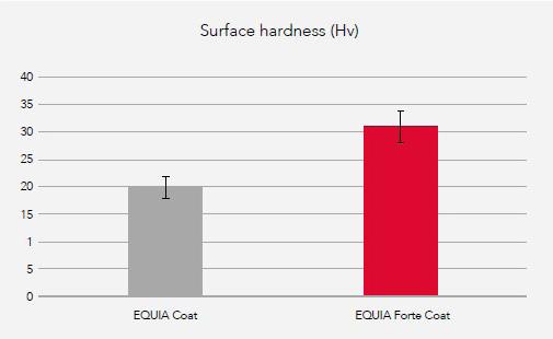
It also contains a new, highly reactive multifunctional monomer. This innovation is responsible for an increase of around 35% in surface hardness and more than 40% in wear resistance as compared to its predecessor, i.e. G Coat Plus (Fig. 2).
Figure 2: Vicker’s hardness of EQUIA Forte Coat in comparison to EQUIA Coat. Source: GC Corporation R&D, Japan. Data on file.
The film thickness of EQUIA Forte Coat is as low as 35 to 40 μm and the resin is able to penetrate the surface of the EQUIA Forte HT restoration, filling pores and micro fissures and rendering the final restoration much stronger (Fig. 3).
Even more important, the coating is able to protect EQUIA Forte HT during its initial setting period, when it is mostly susceptible to water uptake or dehydration.
Resin Matrix
Nanofillers
Conventional Technology
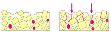
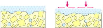
4. Mechanical stress is dispersed on the smooth and toughcoating layer.
The formulation of EQUIA Forte Coat has been designed to allow evaporation of some specific components (monomers) during light-curing, thereby limiting the contact of the coating with oxygen from the air (Fig. 4).
As a result, no air inhibition layer is formed and the surface is kept smooth and glossy.
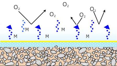
Figure 3: Synergy between EQUIA Forte HT Fil and EQUIA Forte Coat
Figure 4: Vapor layer preventing the inhibition of polymerisation by oxygen
1. EQUIA Fil Restoration
2. Mechanical stress concentrate on surface voids and gaps.Then, cracks are formed.
3. EQUIA Coat bonds to the surface and fills in the voids
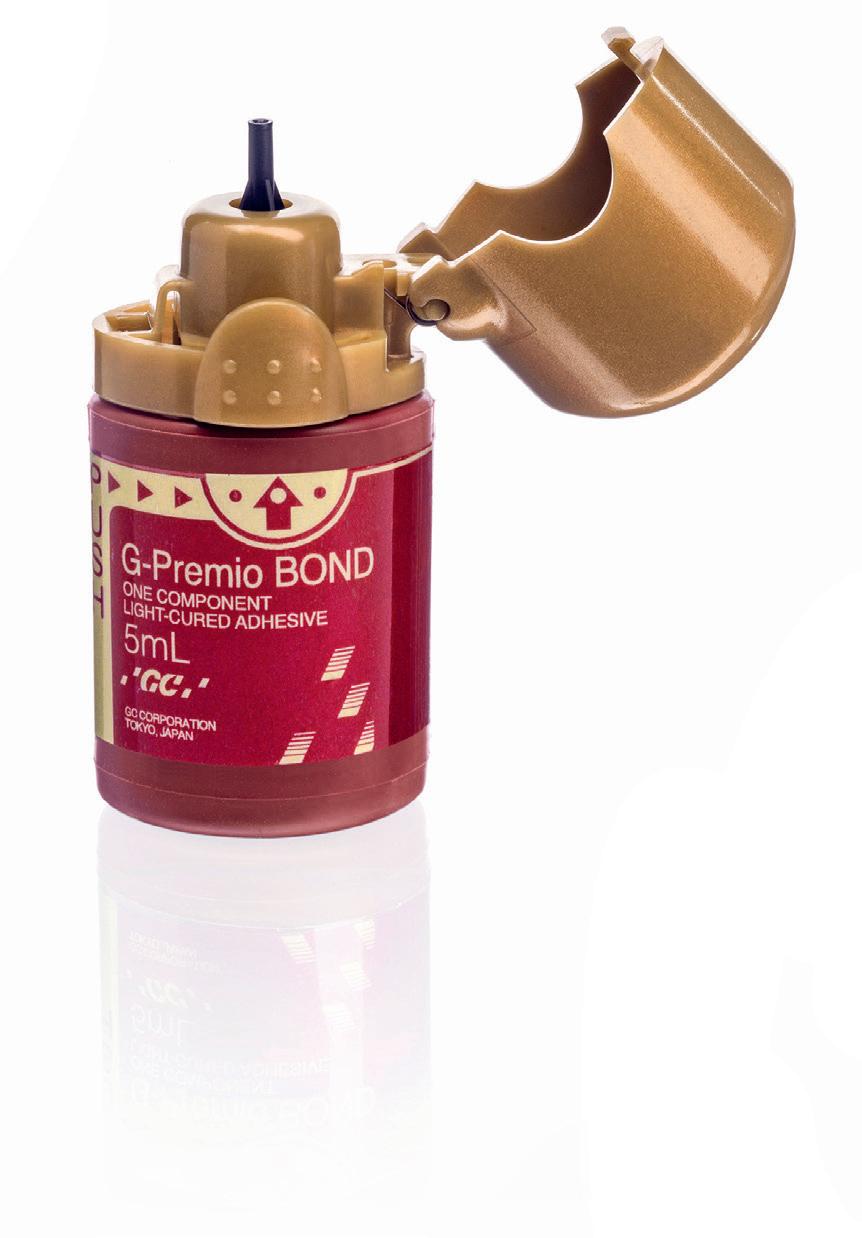
G-Premio Bond
One-Component Light-Cured Universal Adhesive
G-Premio BOND
Universal one-bottle adhesive
G-Premio BOND is a no compromise, one-component, light-cured universal adhesive that delivers a bonding experience like no other. It offers outstanding performance in all etching modes and all clinical situations.
• Thin Nozzle for clean and precise dispensing
• Ergonomic flip-top cap creating easy onehand opening, no spilling
• Clinically proven performance benefiting from the excellent long-term experience ^
• HEMA-free and therefore less prone to water sorption and to causing contact allergy
• Universal workflow for all your procedures, can be used in all etching modes
Q&A
Can G-Premio Bond be used for Indirect repair cases?
G-Premio BOND enables excellent adhesion to all substrates after thermocycling,3 recognising that the use of a separate primer (G-Multi Primer) together with G-Premio BOND is essential on glass ceramics to ensure durable adhesion.
How can G-Premio BOND achieve good bond strength values even with a very short application time? 1
Thin nozzle Clean and precise dispensing
Ergonomic flip-top cap Easy one-hand opening, no spilling
Clinically proven performance Benefit from the excellent long-term experience 541258-GC-G-Premio Bond-ADV-EN-210x297+5-OK.indd 1
HEMA-free Less prone to water sorption and to causing contact allergy
Thanks to a new formulation, G-Premio BOND displays a faster dissolution of the smear layer and improved infiltration. This enables the bonding agent to achieve sufficient bond strength to both enamel and dentin even with a very short application time, regardless of the etching mode.
What is the effect of G-Premio BOND on hypersensitive surfaces?
G-Premio BOND fills and covers the dentinal tubules, which prevents dentin hypersensitivity.
Does G-Premio BOND perform well in all etching modes?
Yes, G-Premio BOND presents very good bond strengths to both dentin and enamel in self-etch, selective etch and totaletch modes. 3 It is fully compatible with all techniques.
Universal workflow For all your procedures, can be used in all etching modes
How many drops are there per bottle of G-Premio Bond?
GC EUROPE N.V. info.gce@gc.dental https://www.gc.dental/europe
Each bottle of G-Premio Bond contains approximately 300 drops, offering a highly economical dispensing system that supports cost-effective clinical use
25/10/2023 10:03
References
^Clinical effectiveness of contemporary adhesives for the restoration of non-carious cervical lesions. A systematic review. M. Peumans, J.De Munck, A. Mine, B. Van Meerbeek. Dental Materials 30 (2014) 1089-1103
1 K. Sai et al. (2016). Influence of degradation conditions on dentin bonding durability of three universal adhesives. Journal of Dentistry 54 (2016) 56–61.
2. Oz, F.D., Kutuk, Z.B., Ozturk, C. et al. (2019). An 18-month clinical evaluation of three different universal adhesives used with a universal flowable composite resin in the restoration of non-carious cervical lesions. Clin Oral Invest 23(3), 1443–1452. https:// pubmed.ncbi.nlm.nih.gov/30109443
3. GCC R&D, Japan (2024). Data on file (test condition: in-house test partially refer to ISO 29022: 2013) available from: info.australasia@gc.dental
Case Study
Class IV controlled layer concept utilising G-ænial™ A’CHORD in 3 shades
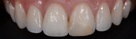
This case study presents the clinical management of a patient who presented with an existing failed anterior composite, requiring a Class IV direct composite replacement. The restoration aimed to achieve seamless integration with the natural dentition while restoring form and function.
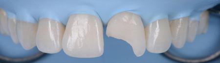
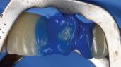
3 - Completion of the Palatal Shelf and interproximal wall with the application of G-ænial™ A’CHORD composite, shade JE. The Interproximal wall was formed with the use of a plastic mylar strip and pull through technique to help develop an anatomical contour.
5 - A chromatic body shade, G-ænial™ A’CHORD shade A2 was then applied and extended beyond the bevel to mask the transition line. Internal anatomy in the incisal third was also sculpted and formed in this increment of composite resin. White tints, Essentia White Modifier (WM) was then utilised to accentuate the mamelons and to replicate similar characteristics present in the adjacent right central incisor.

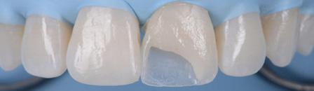
4 - The dentine layer was then completed by the application of an opaque shade of G-ænial™ A’CHORD Shade AO2. This is to provide the correct opacity and “block out” effect of the final restoration.
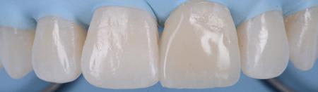
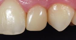
Figure 1 - Pre-Operative presentation, exhibiting a failed direct composite restoration on the upper left central incisor (Tooth 21).
Figure
Figure
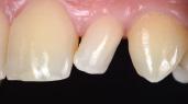
Figure 7 – 2-week review demonstrating the complete optical and functional G-ænial™ A’CHORD restoration on the tooth 21.
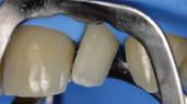
Figure 2 - Tooth surface cleaned and prepared with 37% Phosphoric Acid Etch prior to application of the adhesive with G-Premio BOND.
Figure
Figure 6 - A final translucent shade of G-ænial™ A’CHORD shade JE was then placed to bring the anatomy to full contour.
Clinical images courtesy of Dr Anthony Mak Sydney
quality. ROEKO is now HySolate.

HySolate Flexi Dam
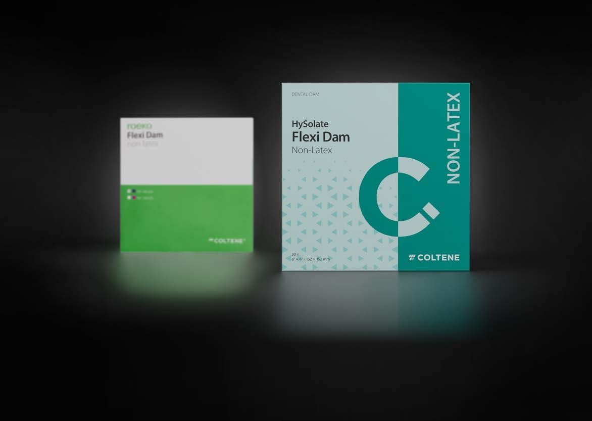
ROEKO Flexi Dam is now Hysolite Flexi Dam
Composite Restorations in the Anterior Region
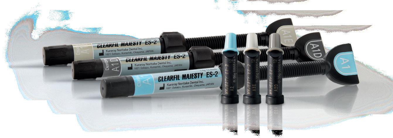
How Many Shades Do We Need? With CLEARFIL MAJESTY™ ES-2 Premium
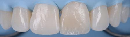
Restoring anterior teeth with large defects using composite seems to be quite challenging. With high-performance materials at hand and a systematic layering concept in mind, however, it is possible to produce highly aesthetic results in a reproducible way. The clinical case below is used to illustrate a dual-shade layering technique with CLEARFIL MAJESTY™ ES-2 Premium, a composite system with pre-defined colour combinations.
Case Example
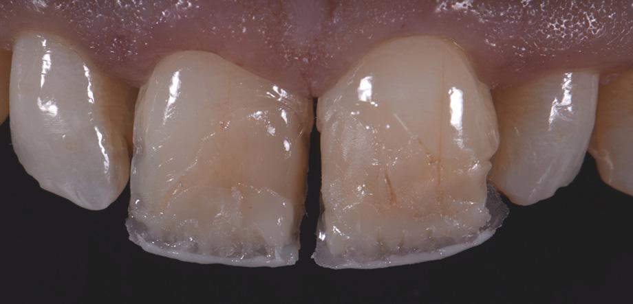
The patient, a young male, was unhappy with the appearance of his maxillary anterior teeth. Several years ago, his central incisors had been restored with composite. These existing restorations had defective and heavily discoloured margins, while their shade did not match the adjacent natural tooth structure. The maxillary lateral incisors were peg-shaped (microdontia). Economic considerations and the desire to save as much natural tooth structure as possible made the team decide to restore all four maxillary incisors with composite. CLEARFIL MAJESTY™ ES-2 Premium became the material of choice as it eliminates the need for complicated shade combination formulas and supports predictable outcomes.
Restoring The Central Incisors
We decided to restore the central incisors first and then focus on the lateral incisors. The tooth shade was determined using the VITA™ classical A1-D4 shade guide, while composite buttons were applied to the teeth to verify the determined shade combination. In order to simplify the restoration procedure, a palatal silicon index was produced before removing the existing restorations.
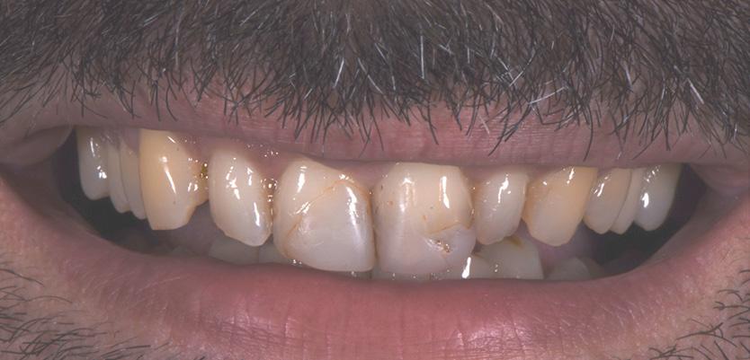
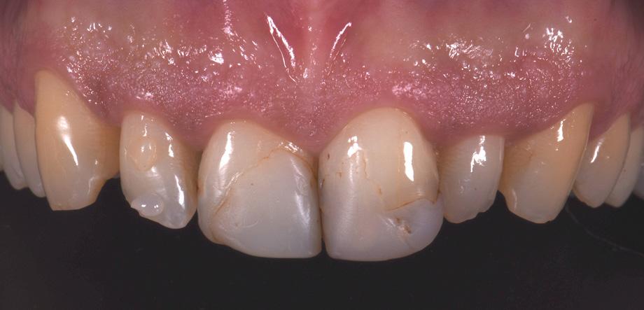
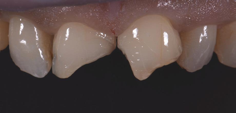
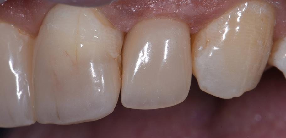
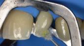
Fig. 2 Intraoral image of the initial situation with defective composite restorations and microdonts. Two composite buttons on the right lateral incisor are used to verify the determined shade combination.
Fig. 3 Central incisors after removal of the old restorations and the beveling of the enamel.
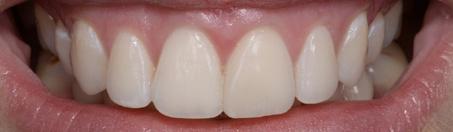
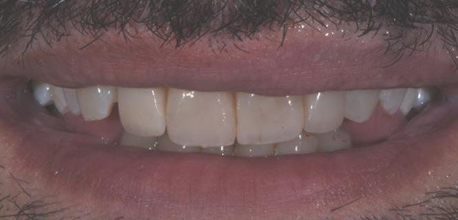
Fig. 1 The patient’s initial smile.
During minimally invasive tooth preparation, bevels were created at the margins to provide for a smooth optical transition from the natural tooth structure to the composite.
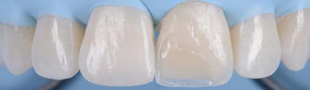
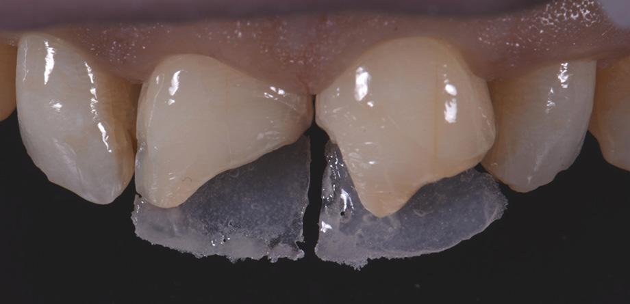
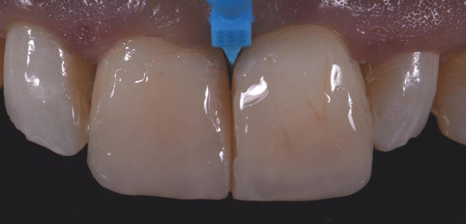
An adhesive (CLEARFIL™ Universal Bond Quick) was applied after selective etching of the enamel to achieve a strong bond. With the aid of the silicon index, it was easy to create the palatal shells of the restorations with CLEARFIL MAJESTY™ ES-2 Premium in the shade A3E (enamel), which matches the determined tooth shade A3. The dentin core was built up with the same composite in the recommended shade A3D (dentin), mamelons were modelled and some CLEARFIL MAJESTY™ ES-2 Premium in the shade WD added for the incisal halo, while some individual effects (like enamel cracks) were imitated with brown stain. The build-up was finalized in the interproximal and labial areas with composite in the shade A3E. Between the central incisors, a wedge was used to retract the papilla and facilitate the designing of the interproximal contact area. The finished and pre-polished restorations already had a natural appearance.
Restoring The Lateral Incisors
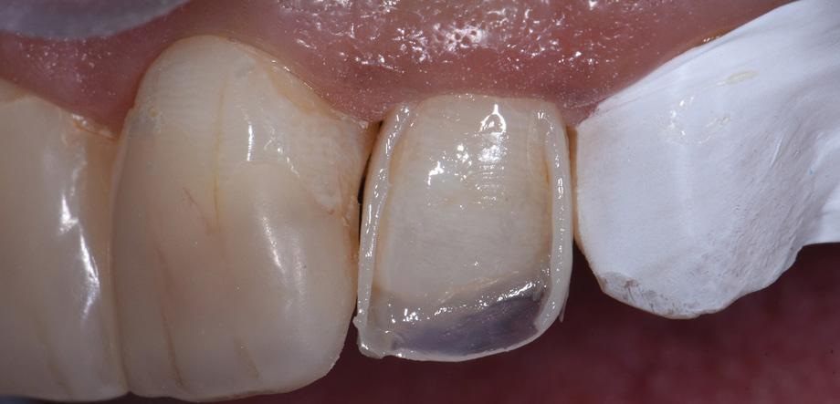
Tooth preparation was not required on the lateral incisors. Instead, they were merely cleaned after a slight roughening of the enamel surfaces. The build-up procedure was similar to the one used for the central incisors. The adjacent tooth was protected with PTFE tape, and the palatal shell was created with the aid of a finger instead of a silicone index. Afterwards, we focused on the build-up of the interproximal walls before a small amount of dentin was placed and the shape was finalized by applying the labial enamel layer.
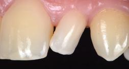
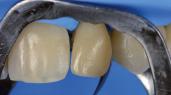
Fig. 6 Situation after finalization of the central incisor restorations with composite in the enamel opacity.
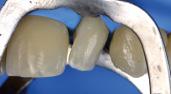
Fig. 4 Light-cured palatal shells made of CLEARFIL MAJESTY™ ES-2 Premium in the shade A3E.
Fig. 7 Central incisor restorations after finishing and initial polishing.
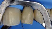
Fig. 5 Build-up of the dentin core with mamelons individualized with the shade WD and brown stain.
Fig. 8 Build-up of the left lateral incisor.
Conclusion
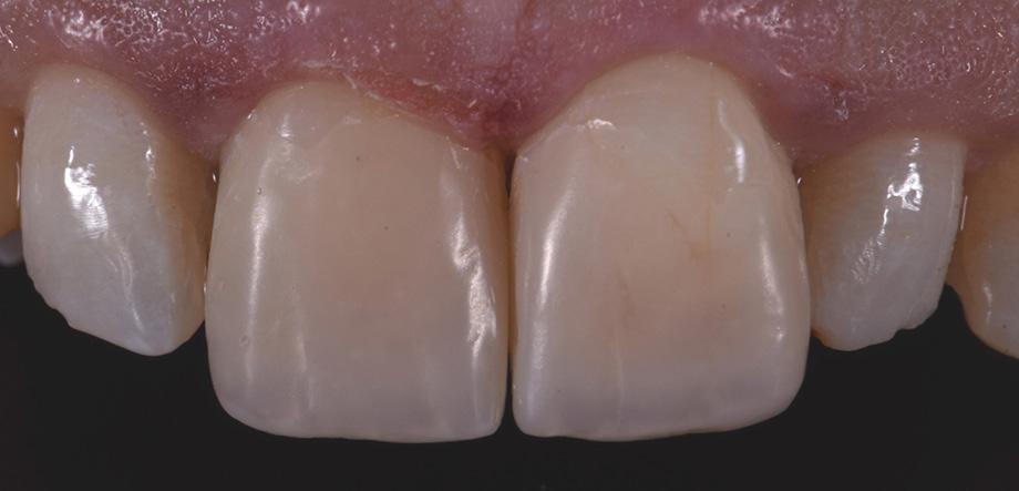
Two different opacities, a single shade combination and some bleached shade plus stain for special effects – in the present patient case, a simple formula allowed us to create lifelike anterior restorations. With one enamel and one dentin paste used, it is possible to simply rebuild the natural anatomy without the risk of ending up with a bulky core that – once reduced – will lose its special optical structure. It is also easy to control the thickness of the final enamel layer with its huge impact on the light-optical properties of the whole restoration. For most patients and teeth with a simple or medium-to-complex internal colour structure, the selected concept is very well suited and will lead to pleasing outcomes.
Case by:
Postgraduate student in Restorative Dentistry program, Faculty of Dentistry, National and Kapodistrian University of Athens, Greece
Fig. 10 Final smile of the patient demands.
Fig. 9 Situation after finishing and polishing.
View Product

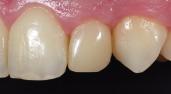
Simple peg-shaped lateral restoration
3M Health Care is now Solventum
About the case
What could be simple about restoring a peg shaped lateral?
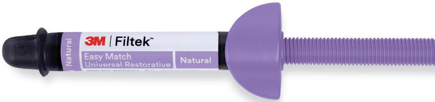
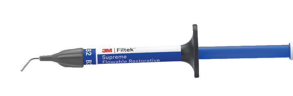
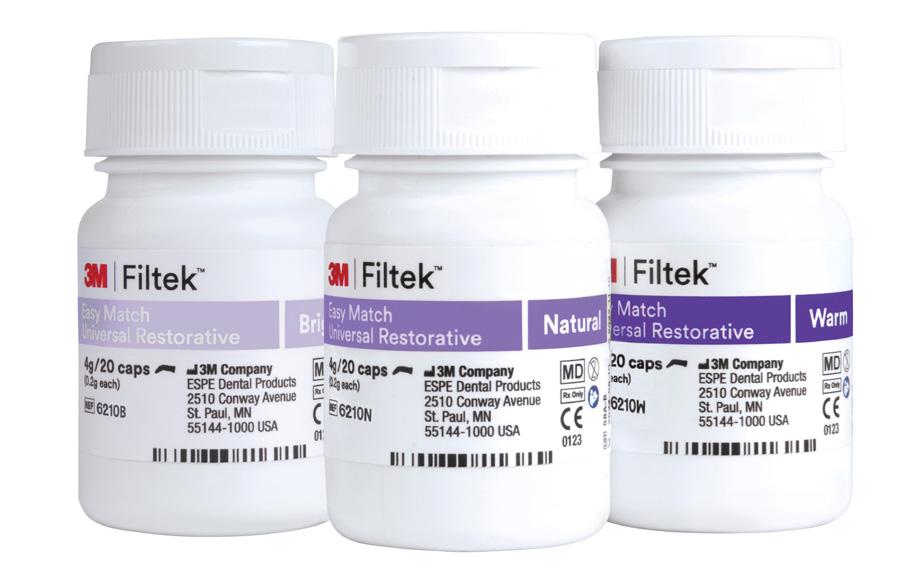
With 3M™ Filtek™ Easy Match Universal Restorative, it is easy to focus on shape, contour and anatomy, while achieving a natural-looking, aesthetic result — without worrying about multiple layers and shades of composite.
Dental restorative used:
3M™ Filtek™ Easy Match Universal Restorative, Natural shade. with oxygen from the air (Fig. 4).
Dr. Walter Devoto graduated with honors in dentistry and dental prosthesis in 1991 from the University of Genoa, Italy. He is particularly interested in the fields of conservative dentistry and aesthetic dentistry and runs his own private practices in Sestri Levante and Portofino. In addition, he is collaborating with diverse prestigious dental offices throughout Europe that specialise in aesthetic dentistry.

He has worked as a teacher and demonstrator at the University of Genoa and as a lecturer at the universities of Siena and Madrid. Now, he is a lecturer at the International University of Catalonia, Barcelona, and visiting professor at the Universitè de la Mediterranee, Marseille.
Dr Walter Devoto
Before
After
Henry Schein
and proximal aspects
5.
8.
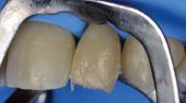
6. An increment of Filtek Easy Match Universal Restorative is placed on the buccal aspect and contoured to provide a scaffold for the mesial and distal contours of the lateral
9. After the general contours are defined, and contact points established, the restoration is ready for finishing and polishing
1. Initial situation
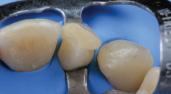
2. After isolation and retraction, the tooth’s enamel is minimally prepped with a fine diamond bur, to enhance adhesion
3. The preparation
of the adjacent teeth are etched with phosphoric acid
4. 3M™ Scotchbond™ Universal Plus Adhesive is applied to the prep and adjacent teeth, dried and cured
3M™ Filtek™ Easy Match Universal Restorative is used to contour the distal aspect of the central incisor, as well as the mesial aspect of the canine
7. Using a sectional matrix, the distal wall and contact point with the canine is created
The same procedure is repeated for the mesial aspect of the lateral, ensuring a contact point
10. Final appearance, after finishing and polishing, as well as gingival healing
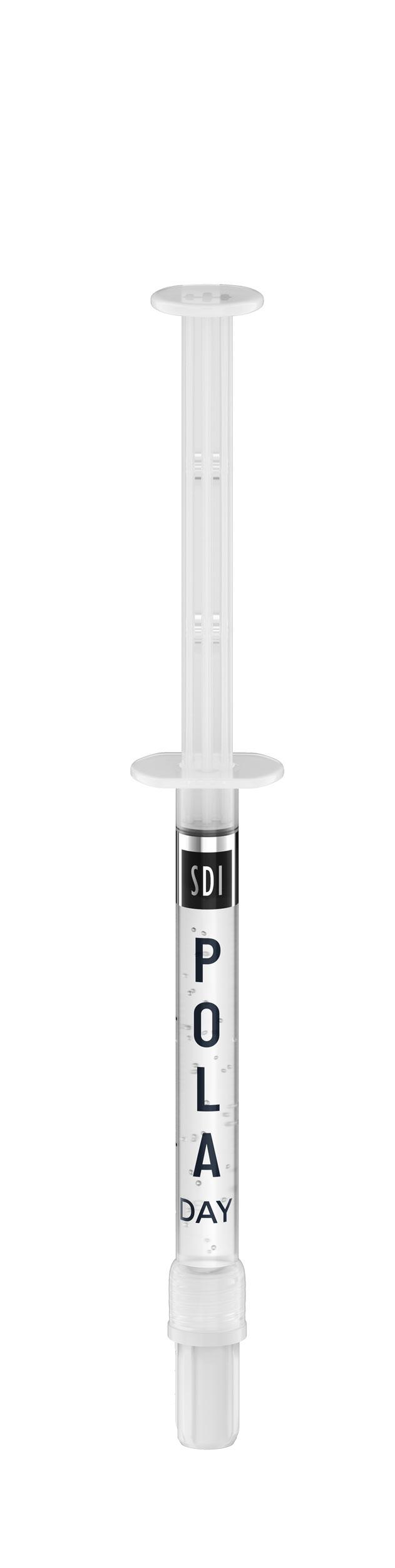
High viscosity gel ensures it can be easily and securely placed into the aligner tray, staying in place
Advanced Tooth Whitening System
In-office whitening
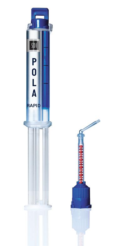
POLA RAPID
A unique 38% hydrogen peroxide formulation that releases active ingredients immediately.
38% HYDROGEN PEROXIDE
Fast and simple to use: From 3 x 8 minute applications
• Non-stick gel for faster application and removal
• Enhanced blue gel for superior application visibility
Key features
• In-office whitening system 38% Hydrogen Peroxide
• Dual-barrel syringe dispenser with brush-tip appl icator
Description
In only one visit, Pola Rapid initiates a powerful whitening treatment that is designed to whiten teeth in 24 minutes. No pre-desensitizer step needed.
Evaluator’s comments
“Instructions were easy to follow and time for the procedure was excellent.”
“I really liked the brush tip and simplicity of the packaging.”
“Easy application.”
Pola Rapid is an advanced in-office tooth whitening system:
• Features built-in desensitisers and fluoride.
• Uses a 38% hydrogen peroxide formulation.
• Simple and precise application.
Unique Attributes
• Blue gel makes for easy application and visibility.
• Faster system - this material only requires 24 minutes of treatment, significantly reducing patient time in the chair.
• The brush tip allows you to place very easily.
• Light is not necessary; however, the Radii Xpert light can be used with the whitening attachment and the Pola Stand.
“Worked nicely and didn’t drive sensitivity on root exposure patientable to control application.” •
Clinical Tips
• Use a surgical suction to remove the whitening gel between applications for fast and precise removal.
• Double check the barrier between each application and make sure there are no areas of leakage.
• Make the time to take before and after photos. It really shows the patient the difference. Even I did not realise there was such a big difference for a couple of patients until I looked at the photos later
“The quick chair time is amazing. I had patients who have had other brand name in-office whitening comment they this was so much faster and they got better results.”
“Color of the gel allowed easy visibility in placing.”
“I feel that it worked well both with and without the Radii Xpert light.”
“The brush applicator takes a little getting used to. It makes the applicator tip overall wider than you may be used to.”
Clinical Case Studies


Dr. Sam Koh
BDSc Melb (Hons) Melbourne, Australia
"I find Pola Rapid a beautiful, easy-to-use product. It is simple to apply and remove with its non-stick and enhanced blue gel formula. A great product to use by clinicians for in-chair whitening with minimal chair time and patient sensitivity, but reliable and immediate results.”

Dr. Miles Cone
Fellow American College of Prosthodontists Diplomate American Board of Prosthodontics Nuance Dental Specialist Portland, Maine, USA
“Pola Rapid has exceptional colour saturation for enhanced visibility during intraoral application. The new non-stick bleaching gel stays where you apply it, and easily wipes away clean with no mess. I found the new system fast, reliable to use with minimal sensitivity for patients”

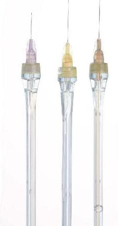
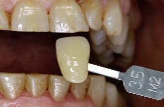
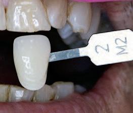
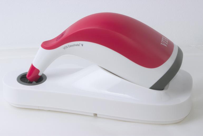
Tooth Shade Determination & Workflow
With VITA EasyShade V
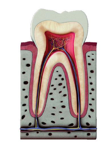
Standardising visual tooth shade determination and precise shade reproduction are some of the everyday challenges faced in dental practices. The perception of colour is based on subjective, visual sensory impressions and is influenced by a number of factors. For example, lighting conditions have a significant impact on the shade chosen during tooth shade determination. The basic shade of the tooth is mainly defined by the dentine, while the layers of enamel on top produce a variety of optical effects, depending on the thickness and translucency. In the following case, dentist Dr. José Gabriel Martínez demonstrates how easy and reliable tooth shade determination and material selection can be using the VITA Easyshade V digital spectrophotometer (VITA Zahnfabrik, Bad Säckingen, Germany).
Clinical situation
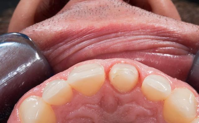
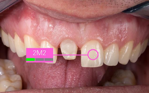
independently from the ambient conditions and the tooth shade was determined. This procedure was used to determine the 2M2 tooth shade for the patient and to select the systemrecommended VITABLOCS RealLife blank (VITA Zahnfabrik, Bad Säckingen, Germany). In order to provide the dental technician with detailed information on the results, additional digital photos were created in RAW format. The photographs were taken using the shade guide and a polarizing filter in order to reduce reflections.
CAD/CAM Workflow
A patient’s upper middle incisor was reconstructed using a direct composite restoration, which fractured. In addition to the material deficiency, the patient was also not satisfied with the tooth shade and aesthetic appearance. For that reason, the decision was made to reconstruct the tooth using an efficient, digital workflow with a monolithic, tooth-coloured feldspar ceramic crown. In order to treat the patient in a single session, a full crown restoration was performed and the composite structure was almost entirely removed. Following that, a temporary crown was made from the CAD/CAM VITA CAD-Temp multiColor composite material using the 2M2 shade, in order to stabilize the tooth and soft tissue.
Tooth shade determination
The shade was digitally determined using the VITA Easyshade V, which achieved a perfect colour match between the new restoration and the neighbouring left incisor tooth. A spectrophotometer was used to transmit defined light into the dentine core and the reflected light spectrum was recorded by a measuring probe. The spectral data was then analysed
After an analogue method was used for the impression and model fabrication, the situation was scanned using the inEos X5 and the crown was constructed using the inLab-CAD- Software (both Dentsply Sirona, Bensheim, Germany). The applied VITABLOCS RealLife replicated the tooth’s natural arched shade gradient between the dentine and anterior, thanks to 3D layered structure. In order to reproduce the shade and translucency of the neighbouring anterior teeth with a natural appearance, the virtual restoration was positioned individually within this three-dimensional layer structure. The monolithic restoration was then fabricated using the inLab MC XL milling unit (Denstply Sirona, Bensheim, Germany). Finally, the restoration was finished with fine diamond and polishing tools.
Treatment result
During the clinical try-in, the patient and technician were both highly satisfied with the results, as the feldspar ceramic crown was very well-integrated into the natural tooth structure. After conditioning the lumen with hydrofluoric acid and silane, the crown was fixed with adhesive. Precise digital tooth shade determination, the correct blank choice based on the VITA Easyshade V block mode and the additional information provided by digital photography were the success factors in the efficient production of an aesthetic, monolithic restoration with natural shade effects and light dynamics.
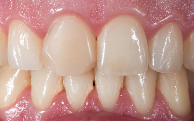
Fig. 1: The insufficient, fractured composite filling on tooth 11 was to be restored using a CAD/CAM-supported feldspar ceramic crown.
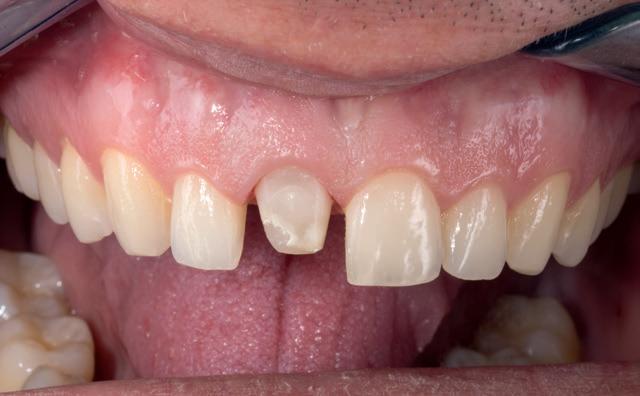
Fig. 3: During the preparation of tooth 11, the composite filling was almost completely removed.
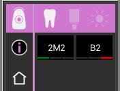
Fig. 5: The basic shade 2M2 was determined using the VITA Easyshade V.
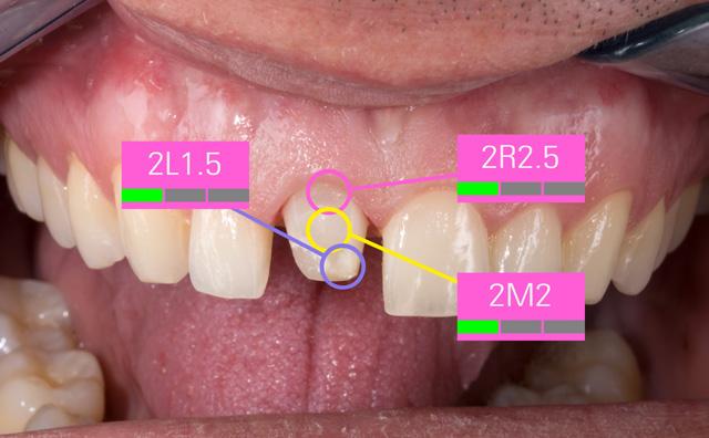
Fig. 7: Since the shade of the prepared tooth affects the shade of the restoration, a three-point measurement was conducted.
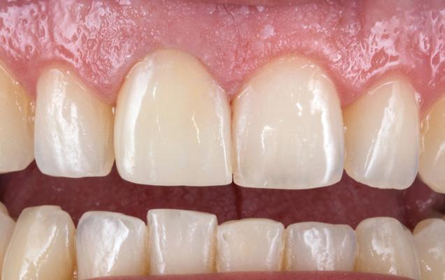
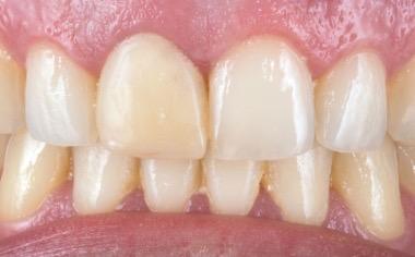
Fig. 2: As a quick solution, a temporary composite crown was fabricated using CAD/ CAM-based VITA CAD-Temp multiColor.
4: During the preparation, the minimum layer thickness of the restoration was observed.
6: The shade of the adjacent tooth was integrated into a digital photo and sent to the laboratory.
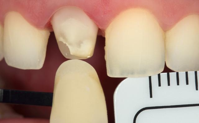
Fig. 8: A digital photograph with polarizing filter and corresponding shade tabs provided the dental technician with individualized information.
Barcelona, Spain

Fig.
Fig.
Fig. 9: The highly aesthetic monolithic crown naturally integrated into the dental arch.
Dr. José Gabriel Martínez

Welcome to EdgeBioceramic Sealers
EdgeEndo’s Bioceramic products are premixed bioceramics with varying viscosities, designed for specific clinical applications. These bioceramics are pure calcium silicate, calcium phosphate - based bioceramics that have been developed to repair or replace the presence of moisture naturally found in dentin.
Clinical Applications
• EdgeBioCeramic Sealer and EdgeBioCeramic ThermalFlow are used for obturation (ortho or retrograde as a root end filling capped with a plug of putty).
• EdgeBioCeramic ThermalFlow - optimised for warm condensation methods EdgeBioCeramic Sealeroptimised for cold hydraulic condensation.
WELCOME TO EDGEBIOCERAMIC™ SEALERS
Edgeendo’s pure pre-mixed Bioceramic materials
• EdgeBioCeramic™ Sealer,
• EdgeBioCeramic™ ThermalFlow™ Root Canal Sealer
EdgeEndo’s Bioceramic products are premixed bioceramics with varying viscosities, designed for specific clinical applications. These bioceramics are pure calcium silicate, calcium phosphate - based bioceramics that have been developed to repair or replace the presence of moisture naturally found in dentin.
• EdgeBioCeramic RetroFill™ and Perforation Repair.
EDGEENDO’S PURE PREMIXED BIOCERAMIC MATERIALS
• EdgeBioCeramic™ Sealer,
• EdgeBioCeramic™ ThermalFlow™ Root Canal Sealer
• EdgeBioCeramic RetroFill™ and Perforation Repair.
• EdgeBioCeramic RetroFill and Perforation Repair (moldable putty) - used for all repair procedures when and where condense and strong resistance material is required to prevent washout. EdgeBioCeramic RetroFill and Perforation Repair is:
Approved for retro filling
Approved for pulp capping
Patented premixed calcium silicate. calcium phosphate bioceramics designed for maximum biocompatibility, healing and optimal handling. They are also void of any non-bioceramic components such as metal and resins.
Patented premixed calcium silicate. calcium phosphate bioceramics designed for maximum biocompatibility, healing and optimal handling. They are also void of any non-bioceramic components such as metal and resins.
CLINICAL APPLICATIONS


Approved for pulpotomies - Adult and Paediatric Approved for resorptive defect repair (internal and external), apexification and apexogenis
EdgeEndo’s comprehensive range of bioceramic products have been developed to cover endodontic material needs whilst providing superior healing and handling characteristics.
How does your current Sealer compare?
See graph below
HOW DOES YOUR CURRENT SEALER COMPARE?
• EdgeBioCeramic Sealer and EdgeBioCeramic ThermalFlow are used for obturation (ortho or retrograde as a root end filling capped with a plug of putty).
EdgeBioCeramic ThermalFlow - optimised for warm condensation methods
EdgeBioCeramic Sealer - optimised for cold hydraulic condensation.
• EdgeBioCeramic RetroFill and Perforation Repair (moldable putty) - used for all repair procedures when and where condense and strong resistance material is required to prevent washout.
EdgeBioCeramic RetroFill and Perforation Repair is:
Approved for retro filling
Approved for pulp capping,
Approved for pulpotomies - Adult and Paediatric
Approved for resorptive defect repair (internal and external), apexification and apexogenis.
EdgeEndo’s comprehensive range of bioceramic products have been developed to cover endodontic material needs whilst providing superior healing and handling characteristics.
EdgeBioCeramic truly shines with its exceptional composition and clinical efficacy. Clinically, its handling is both intriguing and ideal for daily use, consistently delivering excellent results in follow-up procedures. I am confident in its effectiveness and highly recommend it for endodontic applications.”
Dr. Biraj Patel American Trained Endodontist, Diplomate of the American Board of Endodontics (ABE)
Case 1: 46 years old Male patient, ASA 1, presented for the evaluation and the treatment of tooth #36. On clinical examination the tooth was sensitive to percussion and palpation. The radiographic examination showed a very large radiolucency on both mesial and distal roots, a separated instrument in the mesio-lingual canal. The diagnosis of previously initiated with symptomatic apical periodontitis was made, and the endodontic treatment was indicated.
Following aesthesia and rubber dam placement, access was performed, and canals were located. The coreonal fragment was removed and the apical fragment was bypassed. The canals were instrumented using Edge endo X7 files (EdgeEndo) to size 35.04 in the mesial canals and 40.04 in the distal canal.
Canals were obturated using hydraulic condensation using EdgeBioCeramic Sealer (EdgeEndo). Bioceramic cement was used for its antibacterial and bioactive properties. The one year follow up shows a complete healing of the lesion.


“I am confident that EdgeBioCeramic stands out not just for its composition but also for its clinical efficacy. From a clinical perspective, its handling proves to be both intriguing and suitable for daily use, yielding excellent results in follow-up procedures.” Maya Feghali DDS, France
Case 2: A 52-year-old female referred to the clinic for the treatment of tooth 21. Intra-oral examination showed a poor restoration on tooth 21. Percussion and palpation were negative with mobility grade 1. Radiographic examination showed an unsatisfactory restauration on tooth 21 with an apical radiolucency. After obtaining the written consent, the treatment was carried out. The area was anesthetized. Tooth was isolated using the rubber dam and access cavity was opened.
The root canal used for shaping was the EdgeTaper Blaze Utopia to size F2 #25. The final irrigation protocol was performed by a continuous delivery of EDTA and NaOCl. Solutions were activated using ultrasonic activation - 1 minute per solution. Canals were dried and obturated using hydraulic condensation with EdgeBioCeramic Sealer (EdgeEndo).
Lateral and secondary canals are visible on the post operative radiograph.


View product
Pre-op
Post op
One year follow up
Endodontic Treatment #25 on ZenFlex Simplified Technique
A patient came to the practice after a prolonged period without visiting a dentist. The patient reported intense pain in the upper left premolar, particularly in response to cold and heat, and also experienced spontaneous pain. After a comprehensive examination, various treatment options were discussed with the patient. Due to the severe pain, the patient opted to begin with the treatment for the second premolar.
Clinical Examination
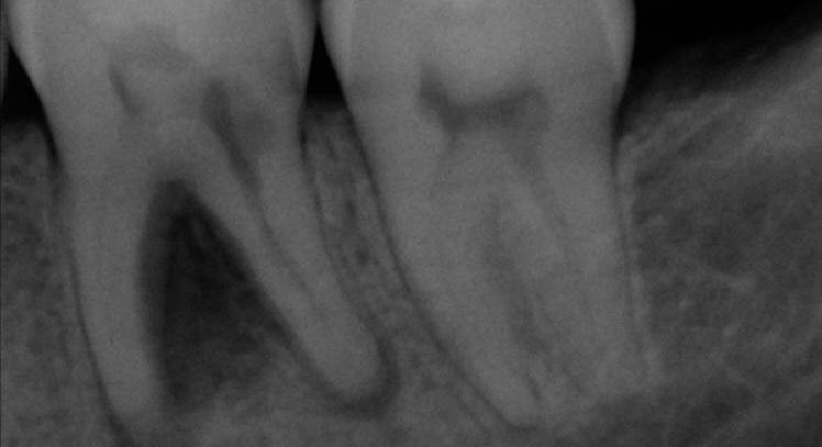
Upon clinical examination, caries were diagnosed in the distal area of tooth #24, and a deep caries lesion in the mesial area of tooth #25 was found to be causing irreversible pulpitis. In tooth #26, only a root remnant was present, and an extraction was planned. (Fig. 1 & 2)
Treatment
Local anesthesia with articaine hydrocholride 40mg / epinephrine 0,012mg was administered, followed by rubber dam isolation. The caries in teeth #24 and #25 were completely removed. For tooth #24, a sectional matrix was used for the reconstruction. Selective etching of the enamel was performed, followed by bonding application. The cavity was filled using SimpliShade™composite in Medium shade.
In tooth #25, the mesial wall was first reconstructed using the same technique, leaving the central portion of the cavity open to facilitate the root canal treatment (Fig. 3). Root canal treatment was then performed using the elements™ Connect and Apex Connect systems. The initial file used was Traverse Glide path file size 13/.06 (21 mm), followed by ZenFlex files 20/.06 and 25/.06 (21 mm). Sodium hypochlorite 5.25% was used as an irrigant between files, with ultrasonic irrigation and a final rinse with EDTA 17%. For canal obturation, the vertical condensation technique was employed using an epoxy-amine resin-based sealer with the elements™ IC system (Fig. 4).
Then, the cavity was cleaned with alcohol and filled with a SimpliShade composite in Medium shade.
Outcome
The root canal treatment was successfully completed,and the patient reported relief from the spontaneous pain. (Fig. 5&6).
Dr. Atenea Baena Urueña
Graduated in Dentistry from Universidad Cardenal Herrera-CEU, Valencia, in 2008. In 2012, I completed a Master’s in Endodontology at Universitat de Valencia, where I developed a profound interest in the complexity of root canal treatments and advanced endodontic techniques.
Since then, I have been practicing in the Netherlands, focusing exclusively on endodontics and continuously enhancing my skills through numerous specialised courses and international conferences.
I am also passionate about sharing my knowledge with colleagues through lectures and training sessions, promoting evidence-based techniques and innovation in endodontics, while staying at the forefront of advancements to provide optimal care and contribute to the professional growth of the dental community.
Dr. Atenea Baena Urueña Netherlands

About
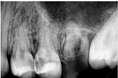
Fig. 1 The X-ray initially reveals the following findings: caries on the distal sur-face of tooth #24, mesial caries on tooth #25, and a root remnant associated with tooth #26.
3 Reconstruction of the mesial wall using SimpliShade prior to endodon-tic treatment.
4 Canal shaping performed with Traverse and ZenFlex files, followed by obturation using the 3D continuous wave technique.
Simplified Procedure View Product
Fig. 2 Pre-treatment image captured during an intraoral examination.
Fig.
Fig.
Fig. 5 Final radiographic assessment following the composite restoration of tooth #24 (occluso-distal) and tooth #25 (occluso-mesial) using SimpliShade Medium.
Fig. 6 Post-treatment image taken following the polishing of the composite restoration. View

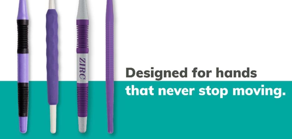
The Mirror, Remade.
A sharper tool for cutting-edge dentistry
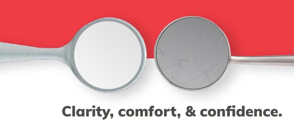
In dentistry, precision isn’t just preferred—it’s essential. Every dental procedure, from routine check-ups to complex restorations, relies heavily on clear visual assessment. At the center of achieving optimal clarity is the dental mirror, an essential instrument used in every patient interaction. One major concern dental professionals often face is the durability of mirrors, particularly the susceptibility to scratching that can compromise performance and reliability. Addressing this directly, the new Crystal HD® Mirror line demonstrated a 269% improvement in resistance to initial surface scratching, significantly enhancing long-term performance and helping ensure consistent visual clarity across procedures.
Why clarity, comfort, and confidence start with your mirror
Dental practitioners understand the impact of mirror quality on clinical outcomes. Even minor imperfections or lens scratches
can complicate procedures and lead to inefficiencies or inaccuracies. This is why significant effort has been invested into developing mirrors that resist scratching and remain consistently clear. By significantly improving mirror durability, dental professionals can rely on exceptional visual clarity, streamlining their workflow and enhancing patient care quality. Ergonomic considerations and patient comfort have also been central to the mirror’s redesign, making every procedure smoother and less physically taxing for practitioners and more comfortable for patients.
Building tools professionals believe in
Trust is foundational to successful dental practices, encompassing patient interactions and the reliability of the tools clinicians use daily. Understanding this, the enhanced Crystal HD Mirror line by Zirc represents a continuous commitment to excellence and innovation. Close collaboration and ongoing dialogue with dental professionals have driven targeted improvements in mirror durability, usability, and performance. This collaborative approach ensures that product enhancements directly address the real-world challenges dental teams face.
Transparency is also vital, with dental professionals regularly informed about product enhancements, rigorous testing processes, and validation methods. Practitioners receive comprehensive updates that instill confidence, allowing seamless integration of these thoroughly tested mirrors into their practices.
Furthermore, the commitment to professional success extends beyond the product itself. Educational resources such as webinars, demonstrations, detailed usage guides, and accessible customer support ensure dental professionals fully leverage the potential of their tools, ultimately supporting consistent, high-quality patient care.
Mirror durability directly affects clinical efficiency and patient outcomes. Recognizing that mirrors often degrade over time, extensive research and development have resulted in a significantly more robust product. The Crystal HD Mirrors feature an innovative flush lens design within a specialized resin frame, greatly reducing debris accumulation and potential corrosion points. The advanced resin composition itself provides superior resistance to scratching, maintaining pristine clarity even after repeated sterilization cycles. Real-world testing with direct input from dental practitioners has confirmed the mirror’s enhanced durability and practical effectiveness. Feedback on handling, cleaning, sterilization, and daily wear was integral in refining the mirror design, ensuring its resilience matches clinical demands precisely. The improved durability directly contributes to greater procedural efficiency, reduced downtime for instrument maintenance, and consistently superior clinical results. With these mirrors, dental professionals can confidently perform their best work, knowing they have tools specifically engineered for longevity, reliability, and outstanding performance.
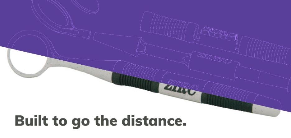
Understanding the ergonomic demands placed on dental professionals, the Crystal HD Mirror line prioritizes user comfort. Poorly designed instruments can lead to chronic strain and fatigue, impacting both practitioner health and procedural outcomes. Each handle within this mirror line is thoughtfully designed to significantly minimize hand fatigue, optimizing comfort and ease of use throughout prolonged dental procedures.
The Soft-Grip Handle weighs only 2.3 grams, markedly reducing pinch force and hand fatigue during extensive use. The Thin-Grip Handle, even lighter at 1.7 grams, allows for effortless maneuverability without compromising precise control. Additionally, the Ergo-Grip Handle features a unique 360-degree elastomer grip with strategically placed dimples, offering exceptional comfort and secure handling to minimize repetitive strain injuries.
These ergonomic designs are developed through detailed feedback from dental practitioners, ensuring each handle meets real-world needs. The result is tools that improve procedural efficiency, practitioner comfort, and overall patient care quality.
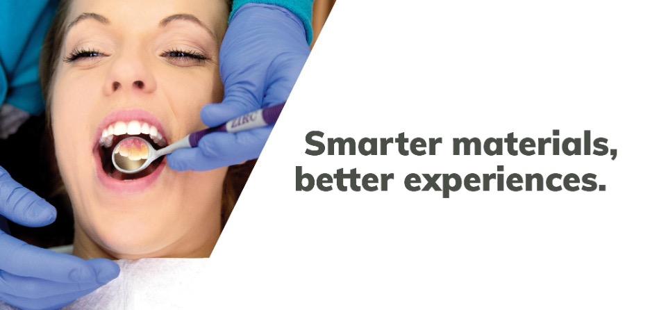
Choosing advanced resin materials for the Crystal HD Mirrors provides multiple benefits, including chemical resistance, durability, and patient safety. Resin construction eliminates galvanic shock, a common discomfort associated with traditional metal mirrors, significantly enhancing patient comfort.
The flush lens integration minimizes soft tissue trauma and enhances procedural comfort, reducing patient anxiety and discomfort during visits. These mirrors also come in vibrant color options that help practices reduce patient anxiety and reinforce branding. In addition, they are exceptionally easy to clean, resist discoloration, and provide a consistently bright, crisp reflection—even after repeated sterilization cycles. These practical advantages allow dental professionals to spend less time on instrument upkeep and more time focused on patient care.
One mirror doesn’t fit all—so we built options
Dental practices vary widely in their procedures and preferences, making customization essential. The Crystal HD Mirror line addresses this need with various sizes, ranging from specialized 14mm mirrors for detailed procedures to versatile 24mm mirrors suitable for general dentistry.
Innovative double-sided mirror heads further enhance procedural efficiency by enabling simultaneous visualization and tissue retraction. Compatibility with Cone Socket and Simple Stem threading allows easy interchangeability of mirror heads and handles, facilitating tailored setups for specific clinical requirements.
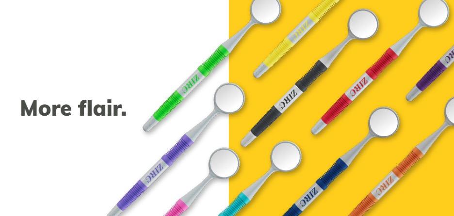
Vibrant color choices allow dental practices to personalize their tools, creating calming, patient-friendly environments that reduce anxiety and reinforce practice branding.
This customization enhances patient comfort and loyalty while meeting diverse clinical and ergonomic needs comprehensively.
A mirror that calms and comforts
Patient comfort and a welcoming clinical environment play crucial roles in successful dental practice. Zirc’s Crystal HD Mirror line emphasizes both functionality and aesthetics, featuring vibrant colors and thoughtful designs intended to soothe patient anxiety and create a more positive experience. By making dental instruments less intimidating, practices can significantly improve patient cooperation, reduce stress, and enhance overall satisfaction.
Detailed attention to patient comfort is evident in the smooth, flush integration of the mirror lens, minimizing soft tissue trauma and enhancing procedural comfort. Such thoughtful design ensures not only clinical efficiency but also patientfocused care, strengthening the dentist-patient relationship and promoting long-term loyalty.
More than a mirror: A partner in your practice
Effective dental tools extend beyond their immediate functionality; they actively support practitioners in their broader clinical and professional objectives.
What dentists are saying—and seeing
The true measure of a dental tool’s success is reflected in its real-world performance.
Feedback from dental practitioners who have incorporated the Crystal HD Mirrors into their practices consistently highlights improved durability, superior ergonomics, enhanced procedural efficiency, and greater patient comfort.
Clinicians report noticeable improvements in handling, reduced hand fatigue, and significant patient satisfaction. Testimonials often emphasize how these mirrors streamline daily procedures, enhance visibility, and maintain clarity through repeated sterilization cycles, underscoring the mirrors’ exceptional resilience. Such real-world validation confirms that these mirrors effectively address and exceed practitioners’ needs and expectations.
Better mirrors. Better outcomes. Full stop.
When we set out to make the best mirror better, we didn’t just aim for incremental improvements. We aimed to reimagine what a dental mirror could be—smarter, stronger, more comfortable, and designed with both clinician and patient in mind.
The new Crystal HD Mirror is more than just an upgrade. It’s the result of listening, thoughtful engineering, and a genuine commitment to the dental professionals who rely on our tools day in and day out. With dramatically improved durability, lightweight ergonomic options, colorful personalization, and comfort-first features, we’ve created a tool that truly reflects the future of dental care.At Zirc, we know your time, your hands, and your patients are all incredibly valuable. So we’ll keep innovating, responding, and making products that help you do what you do best - deliver exceptional care with confidence, comfort, and clarity.
My favourite product is their HD mouth mirrors, seeing is truly believing with these.
It’s like night and day compared to our other old ones. (Plus), it’s way less strain on my eyes since it reflects the light so well!
Madison Collins
The Crystal HD Mirror line symbolizes more than just an advanced dental tool—it represents a comprehensive partnership aimed at helping dental practices succeed. This partnership is manifested through continuous engagement with dental professionals, providing valuable resources such as training sessions, webinars, detailed guides, and responsive customer support. This robust support system empowers dental teams, enhancing their ability to fully utilize their tools, streamline procedures, and improve patient outcomes. Through ongoing collaboration and support, dental professionals gain the confidence and resources necessary to deliver exceptional care consistently.
Because when you see better, you do better. And when your mirror keeps up with your excellence, that’s when dentistry really shines.
Shop Crystal HD Mirrors
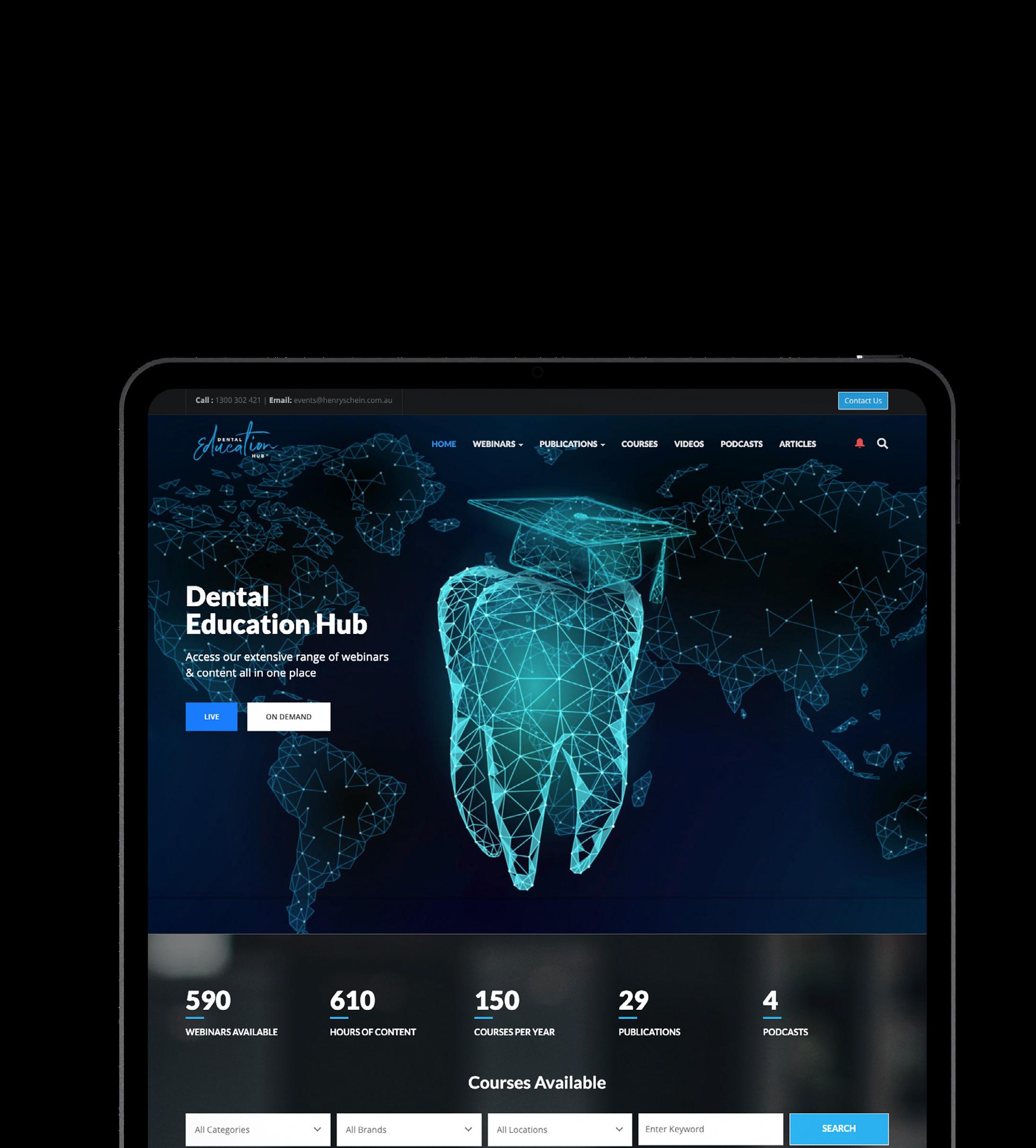
Explore over 600 hours of clinical and business related content all in one place with access to courses, webinars, podcasts and articles.
Precision Sandblasting
Focus on efficiency, cost-effectiveness and perfect visibility
From
Heike Möllenberg | 14th May 2025
The challenge: Sandblasting is not just sandblasting
A precise fine sandblasting unit is essential for dental technology, as selecting the right unit does not only have an impact on quality, but also on cost-effectiveness and overall work efficiency. However, not all devices can provide the ideal combination of visibility, precision and control of abrasive consumption. High abrasive consumption, a lack of precise control and inadequate visibility are often underestimated challenges in daily work.
The solution: Renfert Basic fine blasting units
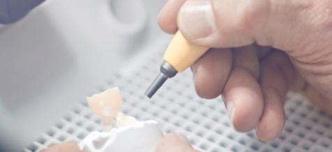
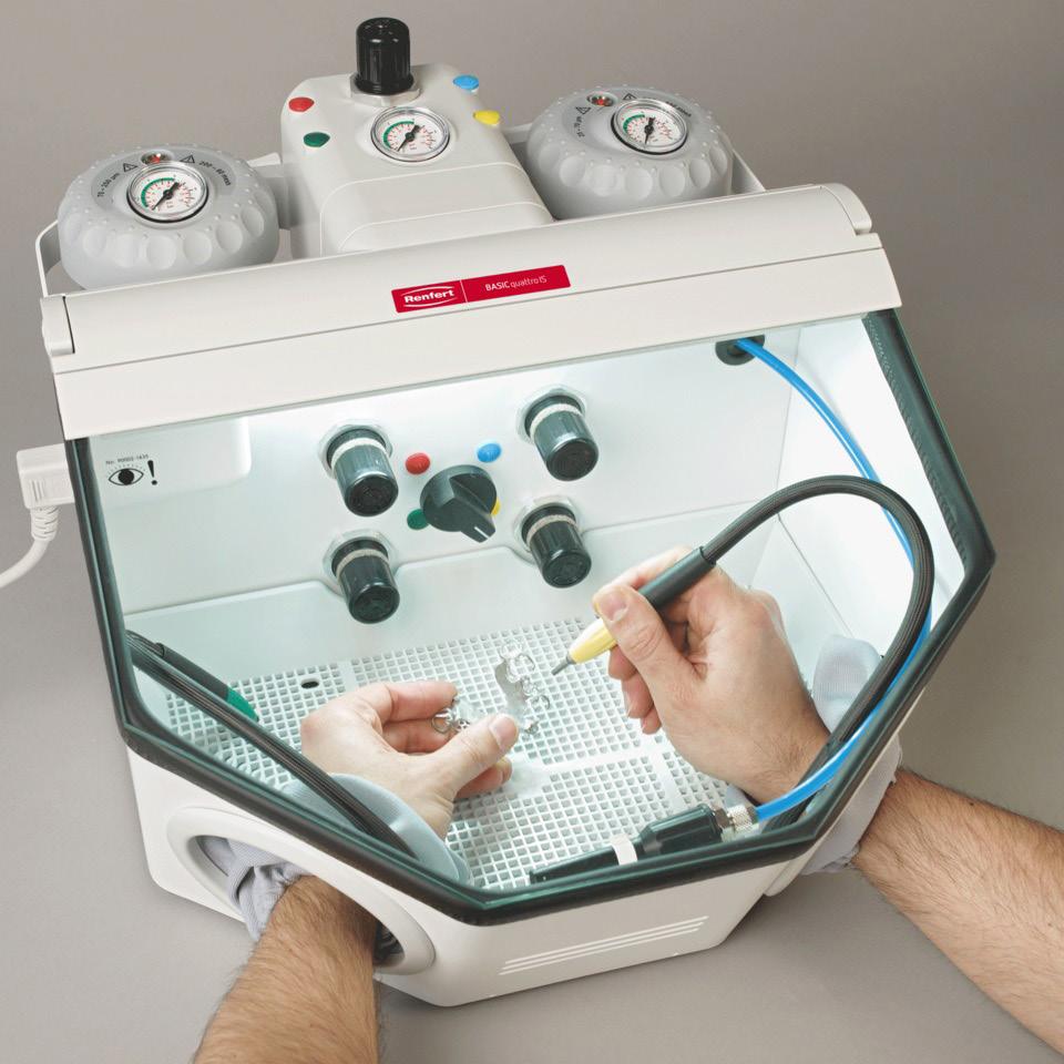
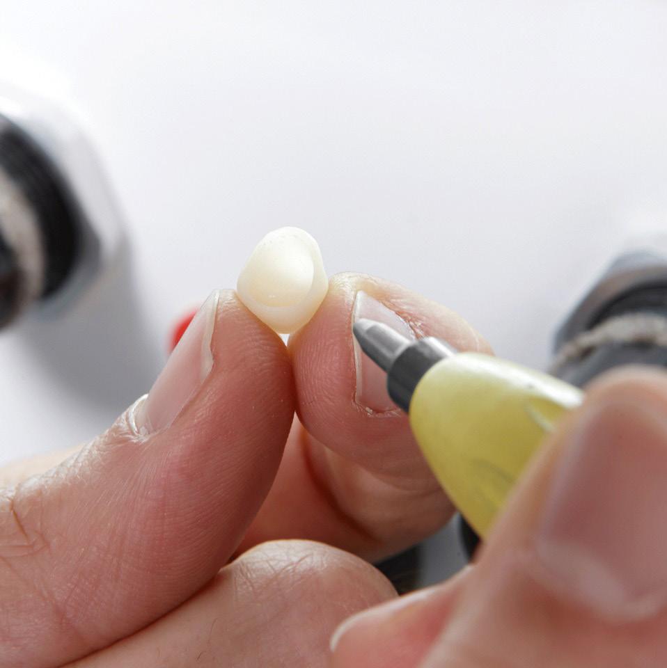
Perfect visibility and precision sandblasting Renfert’s Basic fine blasting units offer a remarkably clear view of your hands and objects to be sandblasted and ensure maximum precision when sandblasting.
The PERFECT view system guarantees optimum lighting with a brightly illuminated blasting chamber (4800 lux). The large glass panel provides a perfect overview, while the spacious blasting chamber ensures that even larger objects can be processed with ease.
A range for every requirement
The fine blasting units are available in different versions - from a compact 1-tank version to the complex 4-tank version with immediate stop technology. In this way, dental technicians can find the right solution for their individual requirements.
Optimized abrasive performance thanks to innovative technology
The special mixing chamber technology and the ingenious nozzle geometry ensure a constant, precise flow of abrasive. This drastically reduces consumption and maximizes the efficiency of the work process. The high-precision nozzle enables pinpoint accuracy without unnecessary material waste.
The advantages at a glance
• Drastic reduction in abrasive consumption: thanks to low forerun and overrun flow rates
• Constant abrasive flow: Uniform sandblasting for perfect surface quality.
• Consistently bright blasting chamber (4800 lux): Maximum visibility during work.
• Extremely precise nozzle: High accuracy for the finest details.
• Large view panel: Perfect overview of the blasting process.
• Ergonomic operation: Spacious blasting chambers from 11-20L
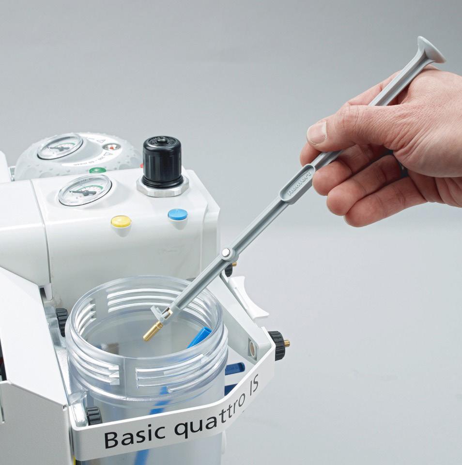
Tip: Easy-care system for a long service life
All Renfert fine blasting units come with a tool to easily remove the dosing nozzle from the tank for cleaning and then reinsert it. This ensures that the blasting performance always remains at the highest level.
Conclusion: A smart investment
Renfert sandblasting units combine precision, efficiency and cost-effectiveness with clever technology. If you want to optimize your work processes, reduce material consumption while achieving excellent results, these units are the right choice.
Customer testimonial:
Proven technology in everyday use
An experienced dental technician reports:
“After 15 years with old devices, I was skeptical as to whether Basic quattro IS would really be better. But it didn’t take long for me to be convinced. The huge, crystal-clear viewing panel and the excellent lighting are impressive. And precise control and low consumption have completely persuaded me. Now I can work faster, with greater precision and more relaxed.”
Author Senior Marketing Manager | Dental Technician
With 14 years of experience at Renfert, Heike Möllenberg combines deep technical expertise as a certified dental technician with strategic marketing know-how. She currently leads the areas of Content Management and Customer Communication, shaping the company’s voice across channels and ensuring consistent, valuedriven messaging for dental professionals worldwide.


Heike Möllenberg Senior Marketing Manager | Dental Technician
Learn more
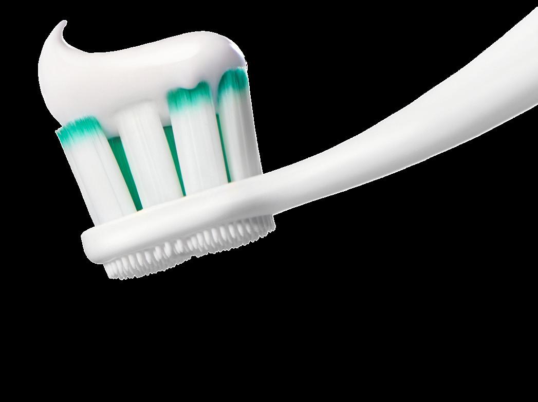
The Total Package
Active Prevention, Advancing Oral Health Protection For Your Patients
As dental professionals, one of the most frequent questions we will be asked by our patients is which toothpaste should I use? As a general rule, we will recommend a toothpaste that contains fluoride. However, what I have recently gained a greater appreciation of is that not all toothpastes are made equal. When it comes to actively helping prevent oral care issues, we as dental professionals can be more proactive for our patients’ oral health by recommending a multibenefit toothpaste, as well as a more systematic approach or oral hygiene regimen.
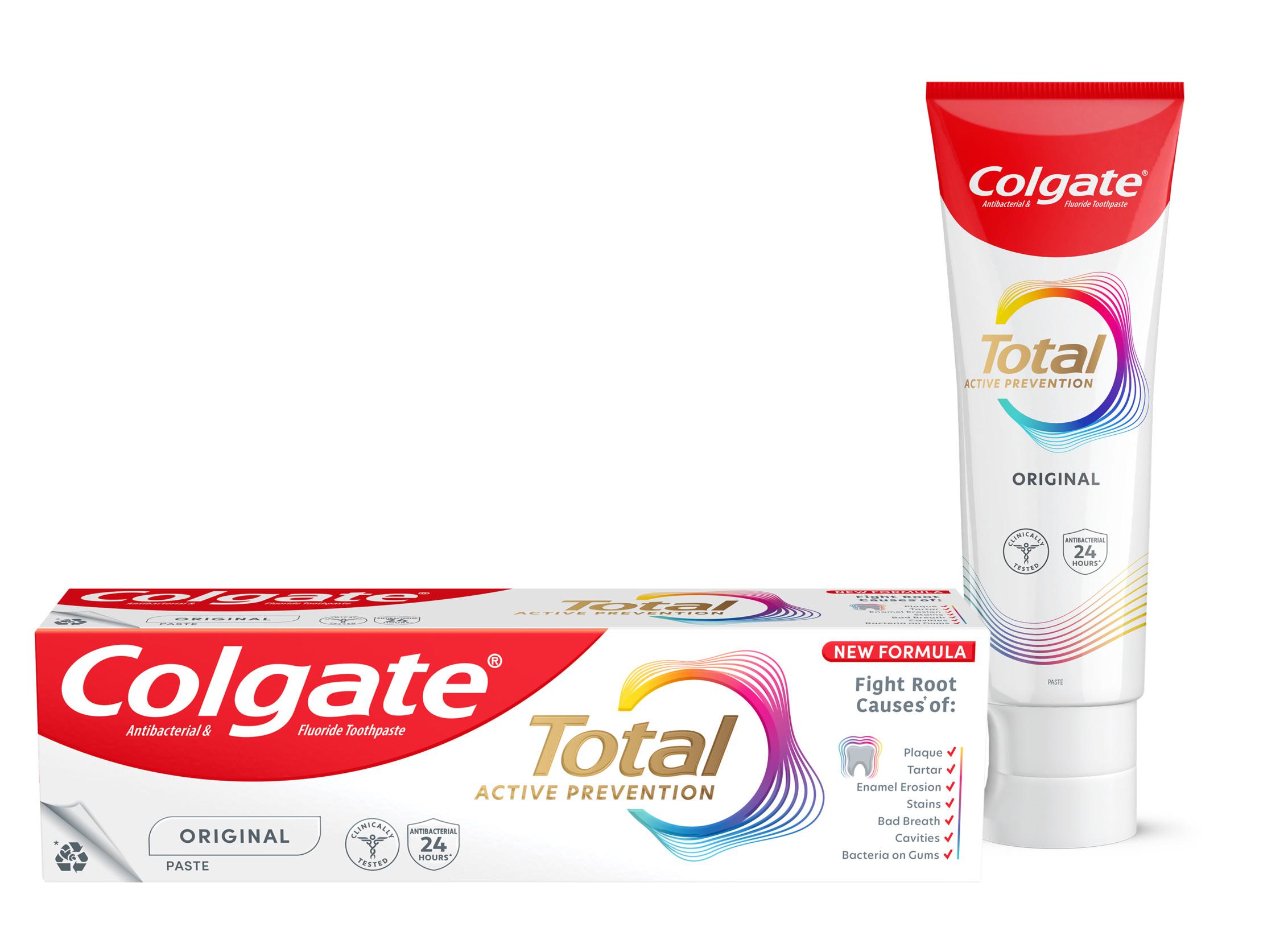
In this article for Colgate-Palmolive, I will be sharing with you the science behind the Colgate Total Active Prevention toothpaste, which contains stannous fluoride, and the unique way in which the toothpaste is stabilized to ensure high bioavailability of the stannous ions. I will also share Colgate’s new approach to active prevention, which really aims to empower patients in their oral hygiene and improve oral health outcomes1
Active Prevention
We know that twice daily use of a fluoride toothpaste delivers fluoride to the dental surfaces and is very much a cornerstone to caries prevention2. However, we also know that caries isn’t the only oral care issue we want to help our patients prevent. Erosion, staining and bad breath are also key issues when it comes to oral health. This is where a multibenefit toothpaste should be part of our recommendation. An antibacterial and fluoridated toothpaste can play a key role in active prevention for our patients. It is also important to understand that not all toothpastes are equally effective in preventing oral care issues, and not all toothpastes have the clinical studies and research to support their use. Therefore, we should routinely ask ourselves, what benefits does this toothpaste offer my patients? What does it actively prevent? What is the science and evidence behind this toothpaste that I am recommending?
Let’s take a deep dive into Stannous Fluoride
Stannous fluoride (SnF2) is a tin-based fluoride compound known to have multiple benefits for oral health. These include anticaries and antibacterial effects. It is also able to modulate the microbial composition of the dental biofilm3. However, stannous fluoride toothpastes are challenging to develop as SnF2 is difficult to keep stable in a toothpaste, which can limit effectiveness. Exposing stannous ions to air, water, or heat can lead to oxidation of these ions from their active form (Sn2+) to stannic ions (Sn4+); this is a form which is no longer effective against bacteria. The key to an effective antibacterial toothpaste is keeping the stannous ions in their active Sn2+ form throughout the shelflife of the toothpaste4. These bioactive ions interfere with bacterial growth and metabolism pathways, disrupting and reducing growth, and inhibiting bacterial acid production5
Historically, drawbacks of toothpastes containing stannous fluoride have been grittiness, enamel staining, and a disagreeable taste. This has meant stannous fluoride toothpastes in the past have not been patient friendly and, as a result, this discourages our patients from wanting to use them. Many of these drawbacks are a result of trying to ensure the toothpaste can deliver the clinical efficacy we need and keep the Sn2+ ions in their active form. For example, low water or no water formulations can be useful to prevent hydrolysis and subsequent oxidation to the Sn4+ species, however these formulations can compromise the taste and feel of the paste. Some toothpastes may add stannous salts, such as stannous chloride, to replenish lost stannous ions, but this has proven ineffective as a high proportion of inactive stannic ions still forms.
The Science behind Colgate Total Active Prevention
The Stannous Fluoride in the new Colgate Total Active Prevention toothpaste has been stabilised by Nitrates and Phosphates. Pyrophosphate-chelated Sn2+ ions are able to maintain stability in water, and the addition of nitrates has been shown to block the oxidation of bioactive Sn2+ ions to inactive Sn4+ ions6. This maintains a high concentration of bioavailable stannous ions in the toothpaste. This unique stabilisation technology addresses many formulation challenges toothpastes containing stannous fluoride face, to deliver an enjoyable brushing experience. This is a really important standout point for me too: if I am going to recommend a toothpaste, I want it to be something my patients will want to use.
Oral Biofilm Control
The key to a healthy mouth and prevention of many oral care issues is a healthy oral biofilm. Mechanical plaque removal alone is not sufficient to disrupt the oral biofilm7,8. This is where the use of antibacterial agents comes in.
Stannous fluoride has multiple modes of action which promote its antibacterial effects. Stannous fluoride is able to alter and breakdown cell walls of bacteria, including those implicated in caries, and oral malodour9,10,11,12. A single-centre, randomised, controlled, double-blinded clinical investigation involving 98 participants demonstrated that brushing twice daily with Colgate Total Active Prevention toothpaste with SNaP technology provided significantly greater and long-lasting antibacterial effects versus using a non-antibacterial fluoride toothpaste.
Compared to baseline, the SNaP-containing toothpaste led to a significant reduction in bacterial load from the teeth (-39%), cheeks (-42%), gums (-62%), tongue (-24%) and even saliva (-30%) after 4 weeks of twice daily use13
Colgate Total Active Prevention Regime
We know that typically patients only brush for 45 seconds, rather than the recommended 2 minutes7, and that mechanical plaque removal alone only removes plaque from 42% of oral surfaces8. An additional way to improve the oral health of our patients includes the addition of a mouthrinse to their daily routine. One well-established antibacterial agent for mouthwash formulation that has been shown to reduce plaque formation is Cetylpyridinium Chloride (CPC). CPC works by cationically attracting negatively-charged bacteria and penetrating the bacterial cell membrane by the hydrophobic portion of the molecule. The subsequent solubilisation of the bacterial membrane and altered permeability for the bacteria ultimately leads to bacterial cell death14
The Colgate Total Active Prevention Mouthrinse contains CPC and Zinc which, when used alongside the Colgate Total Active Prevention Toothpaste and Colgate Total Active Prevention Foaming Clean toothbrush, exhibited fifteen times more plaque reduction when compared with non-antibacterial fluoride toothpaste and regular brush with 1 week of continued use14. In an additional clinical study, the test group used a full regimen of Total Active Prevention products, including Colgate Total Active Prevention Toothpaste, Colgate Total Active Prevention Foaming Clean toothbrush and Colgate Total Active Prevention Mouthrinse, versus a non-antibacterial toothpaste and an ordinary flat trim toothbrush. 100% of people had a healthier mouth in just 3 weeks14.
Dental professionals now have a clinically tested, comprehensive oral care solution for patients through the Colgate Total Active Prevention system. The combination of a toothbrush design complemented by a multibenefit toothpaste with SNaP technology, and clinically tested antibacterial, alcohol-free, CPC-containing mouthrinse, provides a complete oral care regimen system to help patients fight many common oral care issues and to help maintain optimum dental health.
References
1. Colgate. NEW Colgate Total® Active Prevention System. Colgate Professional. Accessed May 31, 2025. https://www.colgateprofessional. com/products/colgate-total
2. Walsh T, Worthington HV, Glenny AM, Marinho VC, Jeroncic A. Fluoride toothpastes of different concentrations for preventing dental caries. Cochrane Database Syst Rev. 2019;3(3):CD007868. Published 2019 Mar 4. doi:10.1002/14651858.CD007868.pub3
3. National Center for Biotechnology Information. PubChem Compound Summary for CID 24550, Stannous Fluoride. https://pubchem.ncbi.nlm. nih.gov/compound/Stannous-Fluoride. Accessed May 31, 2025.
4. Myers CP, Pappas I, Makwana E, et al. Solving the problem with stannous fluoride: Formulation, stabilization, and antimicrobial action. J Am Dent Assoc. 2019;150:S5-S13.
5. Chakraborty B, Seriwatanachai D, Triratana T, et al. Antibacterial effects of a novel stannous fluoride toothpaste stabilized with nitrate and phosphates (snap): in vitro study and randomized controlled trial. Compend Contin Educ Dent. 2024;45(Suppl 3):12-19.
6. Manus L et al., 2024: The Evolution of Colgate Total®: A New Era Stabilized by Nitrate and Phosphates. 45(3). https://compendiumlive. com/2024/active-prevention/the-evolution-of-colgate-total-a-new-era-stabilized-by-nitrate-and-phosphates/
7. Creeth JE, Gallagher A, Sowinski J, et al. The effect of brushing time and dentifrice on dental plaque removal in vivo. J Dent Hyg. 2009;83:111116.
8. Chapple IL, et al. Primary prevention of periodontitis: managing gingivitis. J Clin Periodontol. 2015;42 Suppl 16:S71-6.
9. Gumber HK, Louyakis AS, Sarma T, et al. Effects of a stannous fluoride dentifrice on biofilm composition, gene expression and biomechanical properties. Microorganisms. 2022;10:1691.
10. Xie S, Iberi V, Boissy Y, et al. Stannous fluoride forms aggregates between outer and inner membranes leading to membrane rupture of Porphyromonas gingivalis and Prevotella pallens. Front Oral Health. 2024;5:1427008.
11. Johannsen A, Emilson CG, Johannsen G, Konradsson K, Lingström P, Ramberg P. Effects of stabilized stannous fluoride dentifrice on dental calculus, dental plaque, gingivitis, halitosis and stain: A systematic review. Heliyon. 2019;5:e02850.
12. Fine N, Barbour A, Kaura K, et al. Effects of a stabilized stannous fluoride dentifrice on clinical, immunomodulatory, and microbial outcomes in a human experimental gingivitis model. J Periodontol. 2024;95:421-431
13. Brinta, C. et al. 2024. Antibacterial Effects of a Novel Stannous Fluoride Toothpaste Stabilized With Nitrate and Phosphates (SNaP): In Vitro Study and Randomized Controlled Trial. 45(3)
14. Ayad F, Hetata M, Ahmed R, et al. Antiplaque and antigingivitis effects of a novel oral care regimen featuring stannous fluoride toothpaste, a unique toothbrush, and cetylpyridinium chloride mouthwash: A randomized clinical trial. J Evid Based Dent Pract. 2025;25(2):1 02123. doi:10.1016/j.jebdp.2025.102123 2025;25(2):102123. doi:10.1016/j.jebdp.2025.102123
Dr Kaejenn Tchia is a recent graduate working in a corporate private practice in Darwin, Northern Territory. He is the current President of the Australian Dental Association NT Branch Inc.
He has also served leadership positions for Bupa Dental Corporation including the Clinical Advisory Panel, Clinical Procurement Committee and currently the Graduate Committee. He is passionate about helping and collaborating with fellow dental colleagues, recently embarking on a new journey to help recent graduates eliminate burnout through a 6-step B.E.L.I.E.F System through his motivational coaching platform, The Limitless Dentist.

Kaejenn is a member of the Colgate Advocates for Oral Health: Content Community and hopes to use this platform to raise awareness of the importance of mental health in dentistry and provide mindset tools, which can help his colleagues unlock their next level of growth and success.
Dr Kaejenn Tchia
Pro Toothpaste Range with Nano-Hydroxyapatite
As patients become more discerning about the ingredients in their oral care products, dental professionals are increasingly fielding questions about fluoride alternatives. At Grin, we’ve responded to this shift with our elevated Grin Pro toothpaste range, launched in 2025. These advanced formulations are built on a foundation of clinical science - most notably, nanohydroxyapatite (n-HAp), a biomimetic mineral that’s redefining preventive dentistry.
What Is Hydroxyapatite?
Hydroxyapatite (HAp) is the naturally occurring mineral that comprises 97% of enamel and 70% of dentin. Its structural similarity to tooth material makes it ideal for remineralisation, enamel repair, and sensitivity reduction.
Nano vs. Micro Hydroxyapatite: Why Size and Shape Matter
Grin began its journey with micro-hydroxyapatite in our Strengthening Toothpaste, which quickly gained popularity for its gentle remineralizing effects. In our Grin Pro range, we’ve advanced this approach by incorporating nano-hydroxyapatite
- particles under 100 nm in size and rod-shaped to mimic the natural orientation of enamel prisms.
Why nano?
• Higher surface area → better adhesion to enamel
• Deeper penetration → effective repair of micro-lesions
• Rod-shaped particles → optimal crystal alignment for enamel regeneration
• Low abrasivity (RDA < 10) → safe for daily use, even on sensitive teeth
What the Literature Says
The clinical evidence supporting nano-hydroxyapatite (n-HAp) is robust and growing:
• A comprehensive review concluded that “nHAp is a versatile, biocompatible, and clinically relevant biomaterial with proven efficacy in preventive and restorative dentistry” (Pushpalatha et al., 2023).
• Its applications span enamel and dentin remineralization, sensitivity reduction, and whitening support.
• In a randomized clinical trial, toothpaste containing n-HAp was shown to “significantly reduce dentin hypersensitivity through effective tubule occlusion” (Polyakova et al., 2022), validating its role in managing sensitivity.
• Most notably, an 18-month double-blinded clinical trial found that “hydroxyapatite toothpaste was proven to be safe and non-inferior to fluoride toothpaste in preventing caries in adults,” with 89.3% of participants showing no increase in DMFS scores—compared to 87.4% in the fluoride group (Paszynska et al., 2023).
• Additional studies have highlighted n-HAp’s role in “supporting whitening treatments without compromising enamel integrity” (Pushpalatha et al., 2023), reinforcing its suitability for cosmetic and therapeutic use. These findings validate Grin’s decision to incorporate n-HAp into our newest formulations.
Grin’s Nano-Hydroxyapatite: Pure, Safe, and Powerful
We’re proud to use the only nano-hydroxyapatite approved by the Scientific Committee on Consumer Safety (SCCS) under European authorities. Our n-HAp is:
• 15.5 wt% concentration
• Rod-shaped particles < 100 nm
• RDA < 10
• High purity, biocompatible, paraben-free, vegan This ensures both safety and efficacy for daily use across a wide range of patient profiles.
The new Grin Pro Range is more than just toothpaste - it’s a clinically supported, patient-centric solution for modern oral care. With nano-hydroxyapatite at its core, our range reflects the future of dentistry: smart, safe, and inspired by nature.
References
• Paszynska E, et al. (2023) Caries-preventing effect of a hydroxyapatite-toothpaste in adults: an 18-month double-blinded randomized clinical trial. Frontiers in Public Health, 11:1199728. https://doi.org/10.3389/fpubh.2023.1199728
• Pushpalatha C, et al. (2023) Nanohydroxyapatite in dentistry: A comprehensive review. The Saudi Dental Journal, 35(8), 741–752. https://doi.org/10.1016/j.sdentj.2023.05.018
• Polyakova M, et al. (2022) The effect of toothpastes containing hydroxyapatite, fluoroapatite, and Zn-Mg-hydroxyapatite nanocrystals on dentin hypersensitivity: A randomized clinical trial. Journal of International Society of Preventive and Community Dentistry, 12(2), 252–259. https://doi.org/10.4103/jispcd.JISPCD_333_21
The Grin Pro Range: Tailored for Clinical Needs
Our Grin Pro collection includes four elevated formulas designed to meet diverse oral health needs:
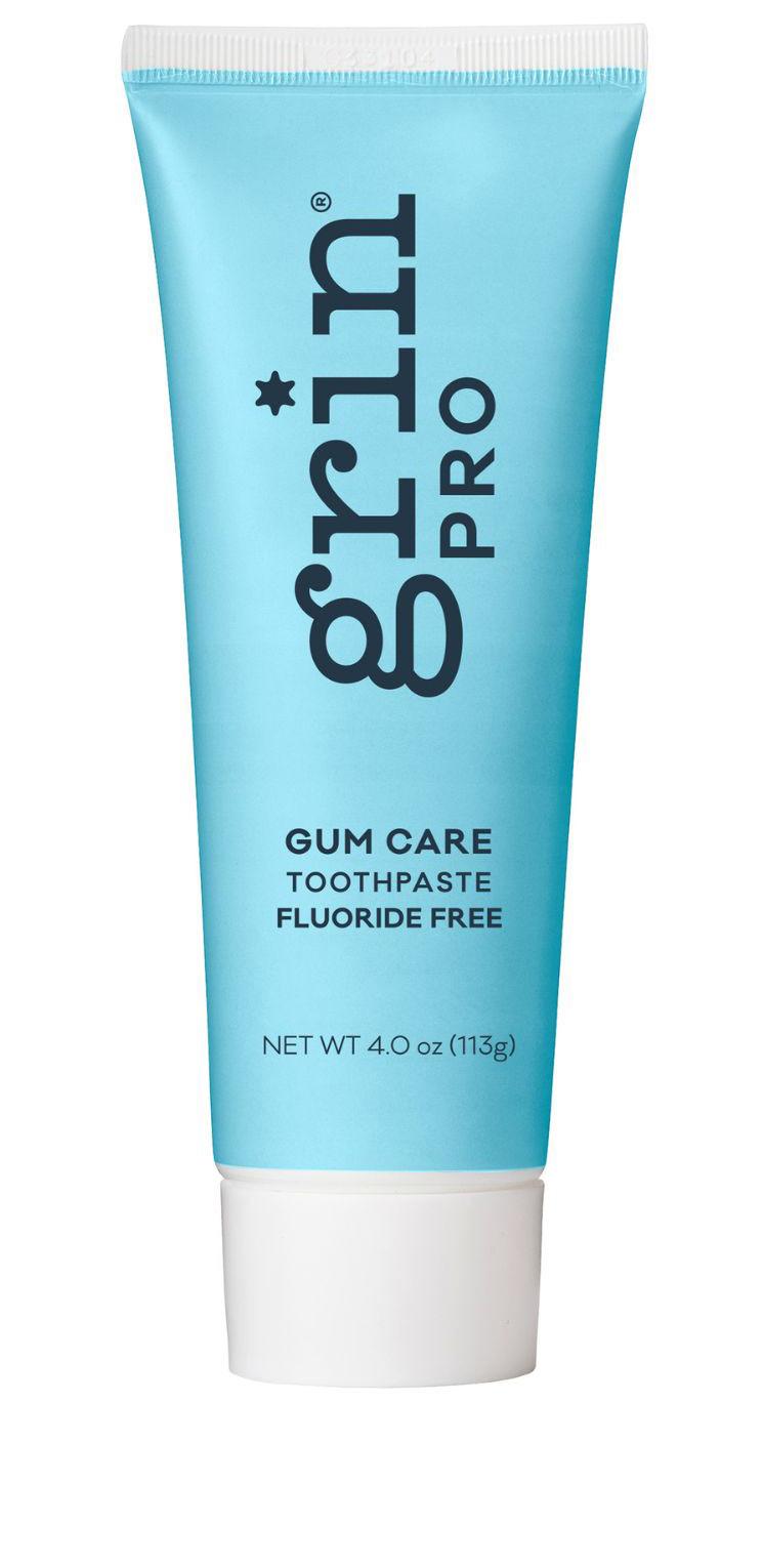
Grin Pro Gum Care Toothpaste
Formulated with New Zealand Manuka Oil and Propolis to help control common oral bacteria and support gum health. A naturepowered option for patients focused on periodontal wellness.

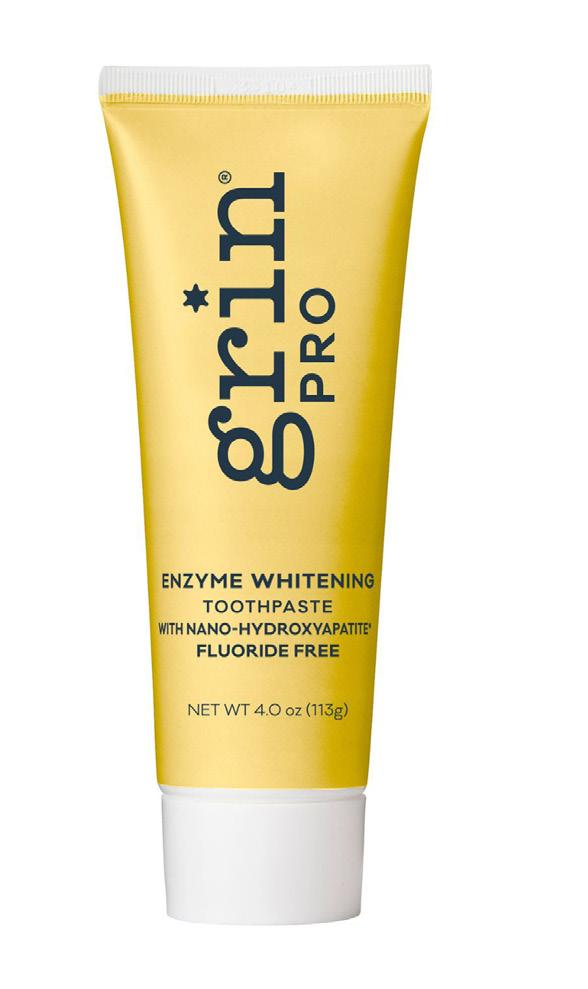

Grin Pro Enzyme Whitening with Nano-Hydroxyapatite
A triple-action formula powered by fruit enzymes - bromelain (pineapple) and papain (papaya)to dissolve and lift extrinsic stains. Clinically proven to whiten teeth in 14 days, while strengthening enamel with n-HAp.


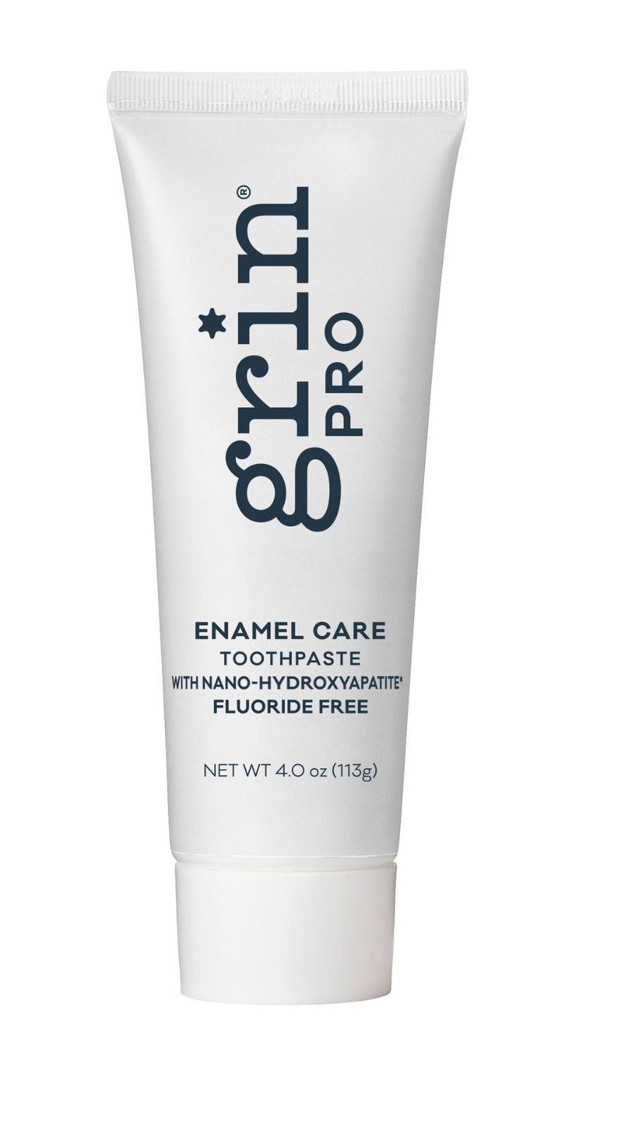
Grin Pro Enamel Care Toothpaste with Nano-Hydroxyapatite
An advanced version of our original Strengthening Toothpaste, combining nano and micro hydro-xyapatite to soothe sensitivity and reinforce enamel. Ideal for patients with mild erosion or post-whitening sensitivity.
Micro + Nano-Hydroxyapatite
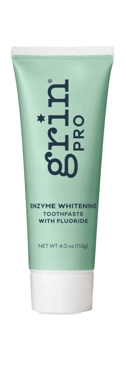

Grin Pro Enzyme Whitening with Fluoride
For patients who prefer fluoridebased protection, this formula offers the same enzyme-powered whitening - plus fluoride for enhanced caries prevention.
New Zealand Manuka Oil
New Zealand Propolis
Nano-Hydroxyapatite
Triple Whitening Formula Enzyme Boosted
Triple Whitening Formula Enzyme Boosted
Fluoride 1000ppm
Magnolia Bark Extract (Organic)

Clinicians around the world choose 3M™ Filtek™ Dental Restoratives for their performance and handling, and for their ability to create natural-looking smiles.
Meet the new Family member, 3M™ Filtek™ Easy Match Universal Restorative: an intuitive 3-shade system featuring naturallyadaptive opacity. Built on the same TRUE nanotechnology behind the global success of Filtek Dental Restoratives, it offers brilliant polish retention, strength and wear resistance in both anterior and posterior restorations.