J. HrdinovĂĄ et al.
by APCs from a DRB1*0401/DRB1*1301 positive donor which were pulsed with 500 nM ADAMTS13.66 In a recent study, we explored the repertoire of ADAMTS13derived peptides that were presented on HLA-DQ.69 In 4 of 9 donors analyzed, ADAMTS13-derived peptides presented on HLA-DQ were identified. Three HLADQB1*0301 positive donors were included in this study, and ADAMTS13-derived peptides were only found in one donor.69 The identified HLA-DQ-presented ADAMTS13 peptides originated from different domains; a CUB1 domain-derived peptide was presented in 3 of 8 donors. In the same study, the re-evaluation of HLA-DR-presented peptide repertoires confirmed the presentation of FINVAPHAR-derived peptides on HLA-DRB1*11.69 A large collection of ADAMTS13 derived peptides appeared to be presented on HLA-DR when compared to a previous study.66 Overall, the presented peptides were derived from multiple domains of ADAMTS13, with, however, a dominant contribution of CUB1/2-derived peptides to the total repertoire.69 To summarize, the specific peptides of ADAMTS13 recognized preferentially by HLA-DRB1*11 and DQB1*03 have now been identified. However, we need to understand how the recognition of these peptides by specific HLA molecules leads to the production of pathogenic antiADAMTS13 antibodies. Comparison of autoantibody profiles in animal models possessing/lacking risk MHC II alleles for iTTP and immunized with ADAMTS13 might help address this question.
Patients with iTTP are prone to develop autoimmunity Immune-mediated thrombotic thrombocytopenic purpura is associated with another autoimmune disease in up to 20% of cases. Other autoimmune diseases can occur years before iTTP, during long-term follow up, or con-
comitantly with iTTP. SLE can be observed in up to 10% of iTTP patients and represents the most common associated autoimmune disease, followed by Sjogren’s syndrome, presented by 3% of iTTP patients. Antinuclear Abs can be identified in 50% of cases. The presence of additional autoimmune diseases has no impact on the outcome of an acute iTTP episode. The presence of anti-double stranded DNA Abs or anti-SSA Abs at iTTP diagnosis is significantly associated with the development of an additional autoimmune disease during follow up.25,61,62,70
Modifying factors: role of ethnicity, sex, obesity, and others The over-representation of women and blacks in most iTTP registries in Western countries highlighted sex and ethnic disparities in this disease, further suggesting the involvement of specific genetic risk factors.23,71,72 In black patients, iTTP is typically associated with an increased risk of exacerbations at the acute phase but with less fatal outcomes (death rate 2.7% vs. 11.6% white patients in our experience), although initial presentation and prognosis is comparable to that of white patients,72,73 raising the hypothesis of a differential tolerance and/or adaptation to tissue ischemia between both ethnic groups.74 The increased prevalence of iTTP in blacks could at least in part result from the naturally low prevalence of the protective allele DRB1*04 in this ethnic group.72 The striking predominance of iTTP in women of childbearing age and during pregnancy raises the hypothesis that estrogen may favor the occurrence of iTTP. As reported in SLE, it is likely that estrogen increases the risk of iTTP in genetically predisposed women by elevating type1 interferon production and favoring the survival of autoreactive B cells.75 Similarly, obesity could represent a risk factor for iTTP as a result of increased peripheral
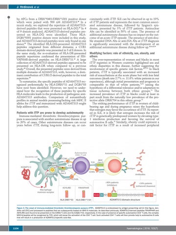
Figure 3. The onset of immune-mediated thrombotic thrombocytopenic purpura (iTTP). ADAMTS13 is endocytosed by antigen-presenting cell [in this figure, dendritic cell (DC)] and processed to peptides that are subsequently loaded on MHC-II molecules. As described previously, ADAMTS13-derived peptides FINVAPHAR and ASYILIRD were found to be presented on HLA-DRB1*1101 and HLA-DQB1*03, respectively. In the case of presence of specific autoreactive CD4+ T cells, the complex MHC-II/peptide will be recognized by TCR, which will cause the activation of the CD4+ T cell. Such activated CD4+ T cells will then provide help to autoreactive B cells that will result in a production of ADAMTS13-specific auto-Abs.
1104
haematologica | 2018; 103(7)