miR-7977 modulates PCBP1 level in BM MSCs
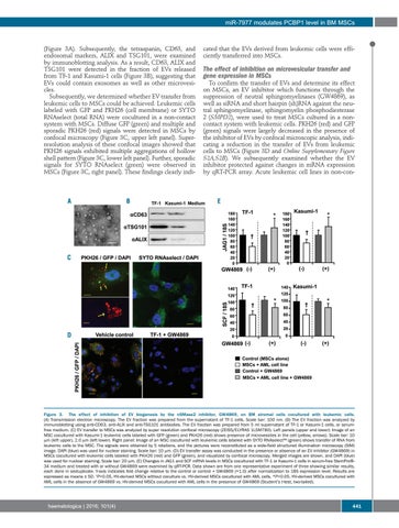
(Figure 3A). Subsequently, the tetraspanin, CD63, and endosomal markers, ALIX and TSG101, were examined by immunoblotting analysis. As a result, CD63, ALIX and TSG101 were detected in the fraction of EVs released from TF-1 and Kasumi-1 cells (Figure 3B), suggesting that EVs could contain exosomes as well as other microvesicles. Subsequently, we determined whether EV transfer from leukemic cells to MSCs could be achieved. Leukemic cells labeled with GFP and PKH26 (cell membrane) or SYTO RNAselect (total RNA) were cocultured in a non-contact system with MSCs. Diffuse GFP (green) and multiple and sporadic PKH26 (red) signals were detected in MSCs by confocal microscopy (Figure 3C, upper left panel). Superresolution analysis of these confocal images showed that PKH26 signals exhibited multiple aggregations of hollow shell pattern (Figure 3C, lower left panel). Further, sporadic signals for SYTO RNAselect (green) were observed in MSCs (Figure 3C, right panel). These findings clearly indi-
A
B
cated that the EVs derived from leukemic cells were efficiently transferred into MSCs.
The effect of inhibition on microvesicular transfer and gene expression in MSCs To confirm the transfer of EVs and determine its effect on MSCs, an EV inhibitor which functions through the suppression of neutral sphingomyelinases (GW4869), as well as siRNA and short hairpin (sh)RNA against the neutral sphingomyelinase, sphingomyelin phosphodiesterase 2 (SMPD2), were used to treat MSCs cultured in a noncontact system with leukemic cells. PKH26 (red) and GFP (green) signals were largely decreased in the presence of the inhibitor of EVs by confocal microscopic analysis, indicating a reduction in the transfer of EVs from leukemic cells to MSCs (Figure 3D and Online Supplementary Figure S2A,S2B). We subsequently examined whether the EV inhibitor protected against changes in mRNA expression by qRT-PCR array. Acute leukemic cell lines in non-con-
E
C
D
Figure 3. The effect of inhibition of EV biogenesis by the nSMase2 inhibitor, GW4869, on BM stromal cells cocultured with leukemic cells. (A) Transmission electron microscopy. The EV fraction was prepared from the supernatant of TF-1 cells. Scale bar: 100 nm. (B) The EV fraction was analyzed by immunoblotting using anti-CD63, anti-ALIX and anti-TSG101 antibodies. The EV fraction was prepared from 5 ml supernatant of TF-1 or Kasumi-1 cells, or serumfree medium. (C) EV transfer to MSCs was analyzed by super resolution confocal microscopy (ZEISS/ELYRAS 1LSM780). Left panels (upper and lower): Image of an MSC cocultured with Kasumi-1 leukemic cells labeled with GFP (green) and PKH26 (red) shows presence of microvesicles in the cell (yellow, arrows). Scale bar: 10 mm (left upper), 2.0 mm (left lower). Right panel: Image of an MSC cocultured with leukemic cells labeled with SYTO RNAselect™ (green) shows transfer of RNA from leukemic cells to the MSC. The signals were obtained by 5 rotations, and the pictures were reconstituted as a wide-field structured illumination microscopy (SIM) image. DAPI (blue) was used for nuclear staining. Scale bar: 10 mm. (D) EV transfer assay was conducted in the presence or absence of an EV inhibitor (GW4869) in MSCs cocultured with leukemic cells labeled with PKH26 (red) and GFP (green), and visualized by confocal microscopy. Merged images are shown, and DAPI (blue) was used for nuclear staining. Scale bar: 20 mm. (E) Changes in JAG1 and SCF mRNA levels in MSCs cocultured with TF-1 or Kasumi-1 cells in serum-free StemPro®34 medium and treated with or without GW4869 were examined by qRT-PCR. Data shown are from one representative experiment of three showing similar results, each done in sextuplicate. Y-axis indicates fold change relative to the control or control + GW4869 (=1.0) after normalization to 18S expression level. Results are expressed as means ± SD. †P<0.05, HV-derived MSCs without coculture vs. HV-derived MSCs cocultured with AML cells. *P<0.05, HV-derived MSCs cocultured with AML cells in the absence of GW4869 vs. HV-derived MSCs cocultured with AML cells in the presence of GW4869 (Student’s t-test, two-tailed).
haematologica | 2016; 101(4)
441