Summary
Currently, most cases of organ failure or severe injury are treated with organ transplants. However, considering both the lack of available donors and occurrences of organ injury and transplant failure, there is a need for alternative solutions.Tissueengineeringattemptsabettersolution.Therearetwomain types of tissue engineering: seeding a scaffold with human cells or implanting healthy cells into failing tissue. This study explores the newly expanding method of creatingscaffoldsusingbiomaterials,analyzingthegrowthofauniquemycelium strain-growthmediumcombination.
Mycelium, the vegetative root of fungi, is emerging as a promising alternative to synthetic materials. Mycelium, which is composed of well-organized interconnected fibers, has been shown to be a cost-effective, all-natural bio-scaffold whose properties are tunable based on the strain-substrate combination. Recent research suggests that the entire fibrous structure can be used as a bioscaffold with just the one-step process of inactivation with an autoclave.
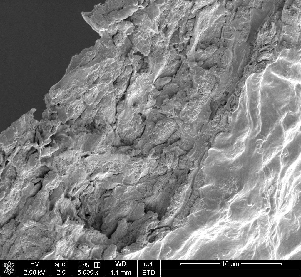
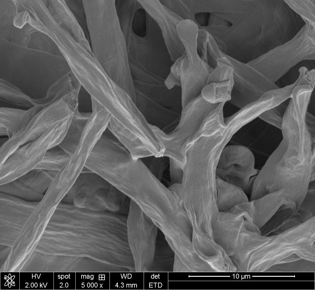
This study uses the one-step process of inactivation with autoclave. The viability of combinations of Lentinula edodes and Pholiota nameko cultured in either Potato Dextrose Broth (PDB) or d-glucose enriched PDB was assessed by using scanning electron microscope imaging (SEM) and attenuated total reflection(ATR)Fouriertransforminfrared(FTIR)spectroscopy.Analysisofthe SEM images revealed a wide distribution of pore sizes, but the majority of these strain-medium combinations demonstrated a porosity range with some potential to facilitate cell migration, adhesion, and ECM production. Additionally, the SEM imaging revealed a substance that the mycelium secreted which might provide an explanation for why some samples were much denser or more porous than others. The spectra gathered from the ATR-FTIR spectrometer was almost exactly identical to the spectra of two well documented strains of mycelium, and one of which has been shown to be a viable bio-scaffold. Given the tunable properties of mycelium and the cheap cost of growth, mycelium has the potential to become the next go-to material for creating bio scaffolds for tissue engineering.
ProjectPurpose
To analyze the properties of P. nameko and L. edodes when cultured in PDB or d-glucoseenrichedPDBtodeterminethesample’spotentialasabioscaffoldafter only inactivation. These data will be compared with data from other strain-medium combinations to provide a more comprehensive prediction for the viabilityofmyceliumasabioscaffold.
Hypothesis
P. nameko and L. edodes cultured in either PDB or d-glucose enriched PDB will have similar hydrodynamic properties, morphologies, and chemical compositions tothoseofhumanextracellularmatrices(ECMs).
Strain,Media,andGrowth Conditions
Pholiota nameko NSPN1 and Lentinula edodes NSLE2 plate cultures were purchased from North Spore and were
WaterAbsorption Measurements
Tomeasurethewaterretentioncapabilitiesofthesecombinationsofmyceliumand growthmedia,drysamplesofmyceliumwereweighedonasensitiveelectronic balanceandthenplacedintoaincubatorwithbowlsfilledwithwater.After24hof airdrying,sampleswereweighed,andthentransferredto86%humidityconditions forapproximately21h,beforebeingweighedagain.Theamountofadsorbedwater wascalculatedbasedontheinitialdryweight.Thepercentbymassofwaterinthe hydratedsamplesofmyceliumwasmeasuredusingtheformulaMW/MM where MWisthemassofthewaterandMMisthemassofthehydratedmycelium.Only onestrain-mediumcombination--P. nameko culturedind-glucoseenriched PDB--hadconsistentresults,whichwas5.54%waterbymassafter21hin86% RH.Futureresearchonthisarea
Discussion
measured.SpectraanalysisisperformedwiththeOMNIC™SpectraAnalysissoftwarethatcomeswiththeNicolet™SummitSpectrometer.

Thesespectraalignalmostentirelywiththoseof Pleurotus ostreatus and Ganoderma lucidum cultured inPDB. P. ostreatus wasreportedtobebiocompatible withhumankeratinocytes,whichprovidesafavorable outlookonthepotentialofthesamplespresentedin thisstudy.Figure4displayshow P. na culturedin PDBhasthehighestabsorbanceatallpointsalongthe spectrum,followedby L. ed culturedind-glucose enrichedPDB, L. ed culturedinPDB,andthen P. na culturedind-glucoseenrichedPDB.Theonemain differencebetweenthesamples’spectraisthatevery samplebut P. na culturedinPDBhasapeakat1550 cm-1(amideIIstretchingregion).Previousliterature hasobservedthatdifferencesinthisregionarelikely relatedtovariationsinchitincontent,whichcan influencemycelialpropertiessuchashydrophobicity andmechanicalresistance.Thepresenceofallof thesefunctionalgroupsintheATR-FTIRanalysis showspromiseforthepotentialofthesesamplestobe abio-scaffold.
waspredominantlyinagel-likeform,whilethemoredeveloped P. nameko strain appeared to be composed mostly of its hyphae (the elongated cells that compose the mycelium). Therefore, the substance that gave L. edodes its volume could have potentially evaporated while it was in the autoclave or while it was air drying. In addition to the high pressure environment inside the autoclave, this potential evaporation would leave condensed layers of what remained. Further research needs to be done on the gel-like substance that is dominant in L. edodes cultured in d-glucose enriched PDB, and somewhat present in all other combinations. Figure 1 (e) and (h) show the outlines of fibers emerging in the gel-like substance, suggesting that this substance could potentially be undeveloped fibers. In culture prior to imaging, the substance was significantly lighter in color and more gelatinous than the fibrous structure present in P. nameko. Additionally, L. edodes takes longer to grow than P. namiko, so one hypothesis could be that this substance is simply premature hyphae, pooling nutrients and preparing to expand into the area the premature part occupies. Furthermore, because of the shrinkage displayed in Figure 1 (b) and (d), more work will need to be done to ensure that the structure of the mycelium remains unchanged during and post-autoclave. Finally, Figure 2 (k) shows a crystal attachedtothestructureofthemycelium,whichmeansthatthesampleswerenot rinsed thoroughly enough prior to autoclaving and SEM imaging. Measures shouldbetakeninthefuturetocorrectthiserror.
Applications. On average, 16 people die every day because of organ failure, and the gap between the amount of organs available for transplant and the amountoforgansneededgrowssteadilyeachyear.Tissueengineeringhasbeena robust field for the past 30 years, attempting to solve exactly this problem. However, many synthetic polymers that have been created to date (e.g. PGA or PLA) require follow-up treatment to display all the necessary hydrodynamical and chemical properties of a bio-scaffold, not simply just the proper structure. Furthermore, because these compounds rely on synthetic compounds, they are not sustainable. With the recent mycelium research suggesting that it can potentially be a biomedical scaffold after a simple one-step autoclaving process, there now exists a sustainable, low-cost, and effective solution to the problem. Mycelium has already been shown to be a successful and scalable alternative to meat and animal clothing products, potentially alleviating two significant contributors to global warming.These paths serve to show the promise mycelium has for scaffolding animal cellular growth and its many applications. Future research in the field should work towards a predictive model, in which one can request what tissue they want to replicate, and the model outputs what substrate-strain.
Conclusions
-Analysis of the SEM images revealed a wide distribution of pore sizes, but the majority of these strain-medium combinations demonstrated a porosity range with potentialtofacilitatecellmigration,adhesion,andECMproduction.
-All combinations aside from L. ed cultured in d-glucose enriched PDB have pore sizesbigenoughthatare“optimal”forcellgrowth(>150��m2)
-Anunknown,self-repairingsubstancewasdiscoveredinthescaffold -ATR-FTIR revealed that these combinations are almost exactly identical to the spectra of two well documented strains of mycelium, and one of which has been showntobeaviablebio-scaffold.
FutureWork
-Determinethebestproceduretousewhenautoclavingthemyceliatopreserve theirstructure.
-Gathermoredataonaselectionofstrain-mediumcombinationsandanalyzetheir viabilityasabioscaffold.
-Inquirefurtherintotheunknown,self-repairinggel-likesubstancethatisvery presentinL.edodesandsomewhatpresentinP.nameko.
-Identifywhichbodilytissuesandorgansaremostsupportedbythepresented scaffoldingcombinations.