— Nandini Chatterjee MD, FRCP (Glasgow), FICP Professor, Department of Medicine, IPGME&R and SSKM Hospital, Kolkata 700020 and Hony Editor, JIMA
Snake bite has been categorized as a Neglected Tropical Disease by the World Health Organization (2009) despite an estimated number of 5.4 million incidents happening every year in tropical and subtropical regions. Annual Snakebite death rate in India is around 4.1/100,000, with increased figures in rural areas even though this can be easily prevented by timely and accurate intervention.
Unfortunately only 22.19% of the snakebite victims are taken to healthcare facilities and 20.25% of them seek medical help after consulting the traditional healer.
Though it is a life threatening medical emergency, management of snake bite is hindered and often delayed due to misconceptions, myths and beliefs in age old traditional therapies
There are some clinical pearls to be borne in mind —
(1) All snake bites are not venomous
Snake bites cause unprecedented panic as all bites are initially thought to be poisonous.
However, it should be kept in mind that out of the 300 species of snakes found in India only 52 are venomous. Only 30% bites are by venomous snakes. Sometimes venom may not even be injected into bites, a phenomenon called called dry bite. (50%). Sore-assurance of the victim is warranted in most cases, that most snake bites are harmless.
(2) Fang Marks do not say it all
Though we look for obvious evidence of a snakebite by fang puncture marks, Fang mark or their patterns give no clue whether the species was venomous or not, the amount of venom injected, severity of systemic poisoning or nature of envenomaton – hematotoxic or neurotoxic.
Kraitbite does not produce fangmarks.
(3) All symptoms are not due to envenomation
Sometimes panic and sympathetic overactivity can manifest as palpitations, sweating, tremulousness, tachycardia, tachypnoea, elevated blood pressure, cold extremities dilated pupils and paraesthesia.
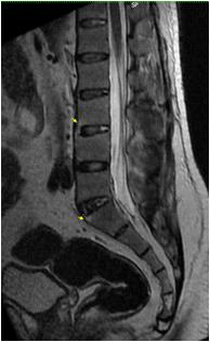
Kraitbites are common at night, biting a person asleep on the ground. Maximum Viper and Cobra bites occur during the day or dawn, while
walking bare foot in overgrown grass or crops.
(4) The markers of envenomation should not be missed –
Localsigns–
These are commonest in Russel’s viper bite but may be present in other viperine bites.
Swelling, bleeding, blisters, and necrosis are the hallmarks. It is considered significant when it involves more than half of the bitten limb or show rapid progression in severity.
Pain at bite site and on passive movement with severe swelling along with absence of peripheral pulses and hypoesthesia helps to diagnose compartment syndrome.
Tender enlargement of local draining lymph node may accompany.
Systemicsigns–
(a) neuroparalytic features – ptosis, diplopia, dysphonia, dysarthria, dyspnoea, dysphagia. Ptosis (drooping of eyelids) occurs first while Dyspnoea (breathlessness) and Dysphagia occur last. This is followed by descending muscular paralysis.
Neuroparalytic snakebite patients present within 6 hours in case of Cobra bite and upto 24 hours for Krait bite; however, ptosis in Krait bite has been documented after 36 hours even.
(b) Hematotoxic effects – Visible bleeding from gums or orifices, subconjunctival hemorrhages, continuous bleeding from the bite site. Intra-cranial bleeding with unequal pupils and gastro-intestinal or retroperitoneal bleeding with abdominal tenderness should not be missed.
(5) Impending respiratory failure should be meticulously excluded -
(a) Single breath count – number of digits counted in one exhalation - >30 is considered normal
(b) Breath holding time – breath held in inspiration – normal > 45 sec
(c) Completion of a sentence in one breath.
(d) Diminished or absent deep tendon reflexes
(e) Head lag due to neck muscle weakness.
(6) Management IssuesTourniquets should NOT be tied
Blood supply may get obstructed leading to gangrene. Any constricting clothing or jewellery should be removed for the same reason when edema increases.
The limb may be immobilized with bandages or cloth to hold a splint, but not to apply too much pressure.
Care must be taken when removing tight tourniquets as sudden removal can lead to a massive influx of venom leading to paralysis and hypotension due to vasodilatation. Before removal of the tourniquet, the presence of a pulse distal to the tourniquet is felt. If it is occluded, then a blood pressure cuff can be applied to reduce the pressure slowly.
Sucking / washing / electrocuting the wound of snakebite does not prevent envenomation.
Wound is not to be washed or interfered with by any procedures or chemicals as this may lead to infection, bleeding or increased absorption of the venom.
(7) Traditional first aid methods and herbal therapy have no role and do more harm.
Immediately after providing first aid, patient is to be transferred to a health facility where Anti-snake Venom (ASV) is available with provision for close observation, basic laboratory investigation and definite treatment.
Urgent transportation of the patient to medical facility by carrying is of utmost importance.
Any vehicle, ambulance, boat, bicycle, motorbike, stretcher is suitable.
(8) Species Identification is not mandatory.
No, the clinical manifestations of the patient may not correlate with the species of snake brought –attempt to kill or catch the snake may be dangerous. Treatment with ASV depends on signs of envenomation and WBCT 20.
(9) The Differential diagnosis of snakebite like krait
Early morning symptoms of acute pain abdomen without neuroparalysis can be mistaken for acute abdomen due to appendicitis, cholecystitis pancreatitis etc. If neuroparalysis is present stroke, GB syndrome, myasthenia gravis and hysteria are the differentials. Neurotoxicity leads to descending paralysis in contrast to GB syndrome where it is ascending.
(10) ASV is not effective against all venomous snakes
In India only polyvalent ASV is available, it is effective against all the four common species; Russells viper, Common Cobra, Common Krait and Saw Scaled viper. It is documented that known species such as the Hump-nosed pitviper, Malabar pit viper, also Sochurek’s Saw Scaled Viper in Rajasthan, and Kalach in West Bengal polyvalent ASV is not very effective.
121, No 9, September 2023Journal
(11) Follow Protocol on admission
All victims of snakebite confirmed or suspected are to be kept under observation for 24 hours. Observation for signs of envenomation and Consumptive coagulopathy detectable by 20 minutes Whole Blood clotting test (20WBCT). ASV therapy is to be instituted where there is evidence of envenomation. NOT to apply or inject Anti-snake Venom (ASV) locally.
(12) Monitoring the patient is of prime significance
– To check for the following: Pulse rate, respiratory rate, blood pressure and 20 WBCT hourly for initial 3 hours and every 4 hours for remaining 24 hours.
(13) An ASV test dose is not to be given Skin/conjunctival hypersensitivity testing is not recommended. These reactions cannot predict future adverse events. They activate Complement to presensitize the patient. They may predispose to future hypersensitivity events.
(14) Dosage of ASV in children and pregnant women -
ASV dosage is identical for Children, pregnant women and adults. As snakes inject similar quantity of venom into adults and children the neutralizing dose is same.
In this issue a peripheral hospital based study has been published comprising 201 patients with signs of
envenomation. Majority of victims hailed from rural areas (88.6%), active males in the 31-50 years age group. There was a seasonal preponderance and local pain (82.1%) and swelling (81.1%) were the commonest presentations far exceeding ptosis (22.4%) and hematuria (18.4%). The mortality rate was 4.5% and mean bite to hospital admission time was the most important determinant for survival. Thus, snake bite is a life threatening health hazard that needs public awareness and prompt intervention in a healthcare facility. If properly managed without prejudice morbidity and mortality can be significantly reduced.
2Bawaskar HS — Snake Venoms and Antivenoms: Critical Supply Issues. J Assoc Physicians India 2004; 52: 11-3.
3Paul V, Pratibha S, Prahaald KA — High-Dose Anti- Snake Venom Versus Low- Dose Anti Snake Venom in the treatment of Poisonous Snake Bites- A Critical Study. JAssocPhysicians India 2004; 52: 14-7.
4Menon JC, Joseph JK, Jose MP — Clinical Profile and Laboratory Parameters in 1051 Victims of Snakebite from a Single Centre in Kerala, South India. J Assoc Physicians India 2016; 63: 22-9.
5Pandey PC, Bajaj S, Srivastava A — A Clinico-Epidemiological Profile of Neuroparalytic Snake Bite: Using Low Dose ASV in a Tertiary Care Centre from North India. J Assoc Physicians India 2016; 63: 16-20.
Original Article
Effects of Neo-adjuvant Chemotherapy on Locally Advanced Breast Cancer (LABC) and Its Correlation with Hormone Profile
Neo-Adjuvant Chemotherapy (NACT) has been the mainstay in the treatment of Locally Advanced Breast Cancer (LABC). The clinico-radiological response of LABC to NACT is dependent on various variables, including the hormonal profile.
This observational study was aimed to study the response of LABC to NACT and its correlation with the hormonal profile of the patient.
Thirty patients with LABC were included in the study and were assessed clinically and radiologically after predetermined intervals. Each case was classified according to the RECIST 1.1 criteria for the response to NACT. Eventually, 24 patients showed a satisfactory partial response to NACT and underwent curative surgical treatment Modified Radical Mastectomy (MRM). While the remaining six patients were found to have stable disease and continued receiving further cycles of Chemotherapy.
The Triple-negative tumours (ER/PR/Herceptin negative) showed an excellent response to NACT and all of them eventually underwent MRM. The Lumina B tumours exhibited the worst response to Chemotherapy.
Key words :LABC, NACT, Response Evaluation.
Carcinoma breast is the most common malignancy in women 1 . It is the second most common malignancy after cervical cancer in India. Around 90,000 new cases of breast cancer are diagnosed every year in India. Locally Advanced Breast Cancer (LABC) accounts for 10-20% of newly diagnosed cases of breast cancers2
LABC includes operable disease (Stage T3 N1), inoperable disease (Stage T4 N2-3) and Inflammatory Breast Cancer (IBC) which is the clinical stage (T4d N0-3, also inoperable). This includes patients with •T3 (>5cm) or T4 tumours (chest wall fixation or skin ulceration and/ or satellite nodules)
•Supraclavicular nodes in the absence of any evidence of distant metastases3
Subdividing patients into these 3 broad group facilitates clinical management.
Locoregional control following the Breast Conservation Surgery (BCS) approach appears to be excellent except in patients with one or more of the features including clinical N2-3 disease, lymphovascular
Department of General Surgery and Breast Diseases, Topiwala
National Medical College, Mumbai, Maharashtra 400008
1MBBS, MS, FICS, Professor and Head and Corresponding Author
2MBBS, MS (General Surgery), FMAS, FIOS, Assistant Professor 3MS, Senior Resident
Received on : 19/07/2022
Accepted on : 21/01/2023
[J Indian Med Assoc 2023; 121(9): 15-9]
Editor's Comment :
Neo-adjuvant Chemotherapy should be used upfront before surgery in the management of LABC. The hormonal profile and the concurrent chemotherapy has given results as effective as PCR, leading to probable BCS.
invasion, residual primary pathological size >2 cm and multifocal disease. Patients presenting with IBC cannot be taken for BCS even after complete resolution and is an absolute contraindication for BCS4
Neo-adjuvant Chemotherapy (NACT) has been the mainstay in the management of LABC. The main aim of NACT is to downstage and prevent early systemic micrometastasis. The effectiveness of NACT depends on the combination of used drugs5. Studies show that pathologic complete response is a crucial factor in long term survival 6 . Factors which affects the Pathological Complete Response (PCR) to NACT are age, size of the tumour, pre/peri/post-menopausal status, chemotherapy regimen given, number of chemotherapy cycles, IHC status, lymphovascular and perineural invasion7. A lot of trials have been done reporting the effectiveness of anthracycline-based chemotherapy regimens over conventional regimens6 In one of the clinical study conducted in India for evaluation of the efficacy of NACT for LABC it was revealed that taxane-based Chemotherapy was more effective as compared to Anthracycline-based Chemotherapy8. Taxanes (docetaxel or paclitaxel) have been added in the Chemotherapy regimen to get a better pathological response since then9-11
121, No 9, September 2023Journal
Patients who achieve a pathologic Complete Response (pCR) following NACT have shown improved survival compared to patients who do not achieve pCR12-14. Several quantitative and categorical methods have been developed to characterise the pathological response to NACT, including Residual Cancer Burden Index (RCBI)15 the Chevallier score16, and the MillerPayne score17. Radiologic assessment of tumour size changes based on the Response Evaluation Criteria in Solid Tumours (RECIST) guidelines issued by the World Health Organisation (WHO) 18 correlates radiological and pathologic response of solid tumours to NACT19,20. Good local response of the primary tumour to NACT correlates well with improved survival19, 21
The determination of Estrogen (Er), Progesterone receptor (Pr) and Herceptin (Hr) content is routine in the management of Carcinoma of the Breast. Such data are commonly used to predict responses to endocrine therapy. However, little attention has been focused upon the effect of NACT on hormonal receptor expression. This is an observational retrospective and prospective analysis to see the outcome of NACT in LABC patients and to correlate the hormonal receptor status to the response of LABC to NACT.
MATERIALS AND METHODS
This was a retrospective and prospective observational study done at a Tertiary Care Centre. The study aimed to study the clinico-radiological response of LABC tumours to NACT and to correlate the hormonal profile of the patients to the clinicoradiological response. Institutional Ethics Committee approval was obtained with a waiver of consent.
All-female patients above the age of 18 with LABC who were subjected to NACT, were included in the study over five years from June, 2014 to June, 2019. Total of 30 patients were included in the study. Their details such as age, co-morbidities, clinical size and stage of the tumour before and after NACT, details of the NACT regimen and hormonal profile (Er, Pr, Hr) of the patient were recorded.
According to the Institutional Protocol for the treatment of LABC, each patient received three cycles of CEF (Cyclophosphamide + Epirubicin or Adriamycin + 5 Fluorouracil) regimen of NACT. This protocol mandated the patient to be seen by a clinician for response assessment after the initial three cycles. Response assessment included clinical examination and USG mammography or X-ray mammography. Clinical evaluation included the comparison of clinical notes for the size of the tumour before the inception of Chemotherapy. Percentage reduction in the size of the tumour was calculated by comparing the breast to
tumour ratio pre and postchemotherapy. Mean reduction in tumour size was calculated by the combination of clinical and radiological assessment. These patients were then categorised according to the RECIST 1.1 criteria 22 into the following categories.
•Complete Response (CR) : Disappearance of all target lesions.
•Partial Response (PR) : At least a 30% decrease in the sum of diameters of target lesions, taking as reference the baseline sum diameters.
• Progressive Disease (PD) : At least a 20% increase in the sum of diameters of target lesions, taking as reference the smallest sum on study.
·Stable Disease (SD) : Neither sufficient shrinkage to qualify for PR nor sufficient increase to qualify for PD, taking as reference the smallest sum diameters while on the study22
There were three instances at which response assessment was done in this study.
The first response assessment was done after three cycles of NACT. Patients with CR or satisfactory PR underwent surgery. Those patients who underwent surgery were excluded from the follow-up. Those patients who did not have a satisfactory response to chemotherapy ie, unsatisfactory PR or SD were subjected to one more cycle of the same initial Chemotherapy Regimen (CEF). Second response assessment was done after the fourth cycle. Those with satisfactory response underwent surgery and the rest of the patients received four cycles of Taxens (a combination of drugs with Taxanes- docetaxel or paclitaxel) regimen. After completion of the second regimen, third response assessment was done. Those with satisfactory response underwent surgery and those with SD or PD received further cycles of Chemotherapy and radiation.
Statistical Analysis :
Qualitative data was represented in form of frequency and percentage. Association between qualitative variables was assessed by the Chi-Square test with continuity correction for all 2 X 2 tables and by fisher’s exact test for all 2 X 2 tables, where the Chi-Square test not valid due to small counts. Percentage reduction in the size of the tumour was calculated by comparing the breast to tumour ratio pre and postchemotherapy. Mean reduction in tumour size was calculated by the combination of clinical and radiological assessment.
Mean reduction in tumour size was obtained by taking an arithmetic mean of the clinical and radiological response. Tumour reduction in centimetres
121, No 9, September 2023Journal
on clinical response was obtained by comparison of clinical notes before and after NACT. The radiological response was obtained by taking into account the reduction in tumour size on Sonomammography or X-ray mammography.
RESULTS
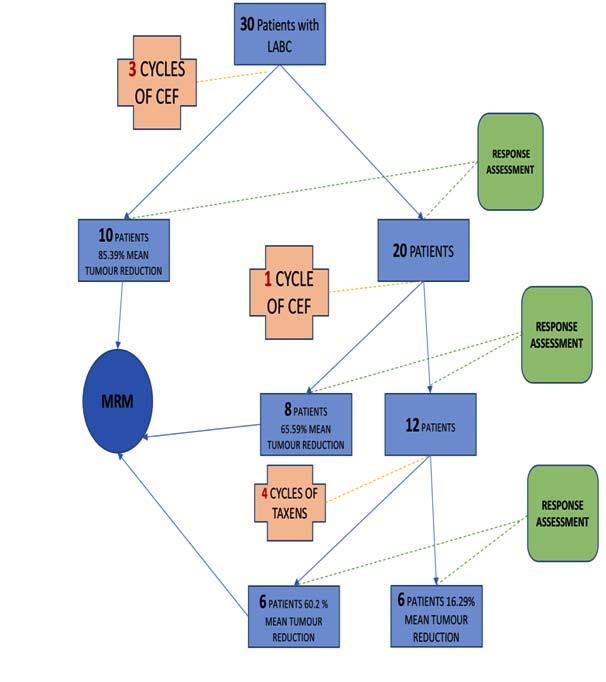
The follow-up module and the study design is mentioned in figure 1.1. At the first response assessment after three cycles of NACT, out of 30, ten patients were found to have a mean reduction of 85.39% in the size of the tumour. Satisfactory Partial Response (PR) and then were selected for surgical management. These ten patients were offered a choice of surgical treatment of either undergoing BCS or Modified Radical Mastectomy (MRM). All of the patients chose to undergo MRM.
Table 1 — Age distribution amongst the study participants
The remaining 20 patients received one additional cycle of CEF Chemotherapy and were assessed after the fourth cycle. Amongst these 20 patients, eight patients were found to have had satisfactory Partial Response in terms of tumour reduction. Mean reduction was found to be 65.59%. These patients then underwent MRM. The rest of the 12 patients had mean reduction in the size of 38.24% after four cycles of CEF. These patients were then given four cycles of taxen chemotherapy regimen. After these four cycles, out of 12, six patients showed a mean reduction in the tumour size of 60.20% (Satisfactory PR) and then they underwent MRM.
The remaining six patients had a mean reduction in tumour size of 16.29% (Stable disease). They underwent further cycles of Chemotherapy (Fig 1).
Most of the patients were more than 40 years of age. (73.3%)(Table 1).
Twenty-four out of the 30 patients taking part in the study exhibited a partial response after various number of cycles of NACT (Table 2).
On comparison of the correlation between the hormonal profile and the Mean reduction in tumour size, the triple-negative (ER -ve/ PRve/ Hr – negative) patients had the best response to NACT. There were 15 patients with triplenegative hormone profile in the study. All of them eventually had a satisfactory partial response to NACT and all were subjected to MRM. On the comparison between the mean reduction in tumour size exhibited by triple-negative
patients to other hormone profiles, there was a statistically significantly superior response to NACT. (P-value 0.003 chi-square test with Bonferroni correction)
LUMINAL B (ER +PR+/- Hr positive) exhibited the worst response and none of the 4 patients were subjected to MRM (Table 3).
On follow up of the patients who underwent MRM and adjuvant chemo radiation, 3cases showed distant recurrence at 1 year follow-up, The subtype we observed 2 cases were TNBC and 1 case was HR+ HER. On 6 months, there was no recurrence on PET scan.
A study done by Harris, et al29 showed that TNBC
Table 2 — Mean Reduction in tumour reduction at each response assessment step
MRMMRMMRM after ProlongedTotal afterafter4 Additionalchemo3 cycles4 cycles cycles of therapy of CEFof CEFPACLITAXEL cycles RegimenRegimenRegimen Number of1086630 patients
Response toPartialPartialPartialStable chemotherapy responseresponseresponse disease according to RECIST 1.1 Mean reduction
in tumour size
Fig 1 — A schematic diagram depicting the observations made as per the study design
Vol 121, No 9, September 2023Journal
has absolute15-20% of cases who will benefit from chemotherap and for HR+ HER2- it was 2-3%. This study also showed distant recurrence risk in TNBC was 50-60%, and for HR+ HER2- it was10-15%.
Different subtype of breast cancer have different risk of recurrence. This findings was also observed in our study on 1 year follow up of breast cancer cases (Table 4).
DISCUSSION
Table 3 — Hormonal Profile and Mean Reduction in tumour size
HormonalMRMMRM MRM after ProlongedTotal Profileafterafter4 Additionalchemo3 cycles4 cycles cycles of therapy of CEFof CEFPACLITAXEL cycles
RegimenRegimenRegimen
Number of1086630 patients
LUMINAL A22127 (ER +/PR +/HERCEPTIN -ve)
LUMINAL B00044 (ER +/PR +/HERCEPTIN +)
WHO Response guidelines were updated in 2009 in the European Journal of Cancer (RECIST 1.1) 22 . The revised guidelines incorporated major changes to the original RECIST criteria, including a reduction in the number of lesions to be assessed, a new measurement method to classify lymph nodes as pathologic or normal, the clarification of the requirement to confirm a Complete Response (CR) or Partial Response (PR) and new recommendations for the assessment of disease progression. NACT is currently established as a standard therapeutic approach for patients with large (>2 cm) and locally advanced breast cancer. However, standard guidelines for pathologic evaluation of breast specimens after NAT have not been established. Assessment of the therapeutic response and measurement of residual disease in the breast and/or axillary lymph node is important because it may predict survival and provide guidelines for further therapy22
Herceptin Positive12104 (ER -/PRHERCEPTIN +)
Triple Negative744015 (ER -PRHERCEPTIN -)
Mean reduction 85.39%65.59%60.20%16.39% in tumour size
Table 4 — Different subtype on follow up distant recurrence rate
Elghazaly, et al studied 125 patients with stage-II and stage-III non-inflammatory breast cancer treated with six cycles chemotherapy. Among them 20% of patients achieved PCR28
In this study, amongst the six patients with stable disease, four patients were found to have Luminal B hormone profile and two were Luminal A. Those who had a satisfactory partial response early in the chemotherapy regimen 61% of them were TripleNegative Breast Cancer (TNBC).
Luminal B patients had the worst response to NACT while those with TNBC had the best response to chemotherapy. All of the TNBC patients in the study eventually showed a satisfactory partial response in tumour reduction. All of these patients underwent MRM. Although TNBCs carry higher mortality as compared with luminal-type breast cancers NACTis more likely to result in a pathologic complete response (pCR) in patients with TNBC primary tumours than in those with luminal tumours23
Patients with TNBC were more likely to get PCR26 Rodenhuis, et al studied the neoadjuvant response in 267 patients: 55 patients (21%) had a pathological complete response in both breast and axilla. Pathological complete responses were more frequently seen in patients with triple negative tumor27.
In our study 14 (14%) of patients had pathological complete response following NACT. The majority (57%) of these had triple negative hormone receptor status.
PCR is an important endpoint because patients who attain this status after surgery have improved survival and this improved prognosis is greatest in the more aggressive subtypes of TNBC and HER2-positiveonly tumours24,25
On follow up of the patients who underwent MRM and adjuvant chemo radiation, 3 cases showed distant recurrence at 1 year follow up, The subtype we observed 2 cases were TNBC and 1 case was HR+ HER. On 6 months, there was no recurrence on PET scan.
A study done by Harris, et al29 showed that TNBC has absolute 15-20% of cases who will benefit from Chemotherapy and for HR+ HER2- it was 2-3%. This study also showed distant recurrence risk in TNBC was 50-60% and for HR+ HER2- it was 10-15%.
Different subtype of breast cancer have different risk of recurrence. This findings was also observed in our study on 1 year follow up of breast cancer cases.
121, No 9, September 2023Journal
CONCLUSIONS
Hormonal profile is an important variable in the determination of the response of LABC to NACT
Poor response should be expected in triple-positive cases.
Overall response with Triple negative cases was found to be excellent. Different subtype of breast cancer have different risk of recurrence.
REFERENCES
1Kereena CH, Vardhan ZV, Krishna TV — Importance of awareness, specific knowledge and screening behavior of rural women with breast cancer at Government General Hospital, Guntur, AP. Int J Bio-pharma Res 2012; 1: 7-10.
2Akhtar M, Akulwar V, Gandhi D, Chandak K — Is locally advanced breast cancer a neglected disease? Indian J Cancer 2011; 48: 403-5.
3DeVita VT, Lawrence TS, Rosenberg SA, editors. DeVita, Hellman, and Rosenberg’s cancer: principles & practice of oncology. 11th edition.
4Tewari M, Krishnamurthy A, Shukla HS. Predictive markers of response to neoadjuvant chemotherapy in breast cancer. Surg Oncol 2008; 17(4): 301-11.
5Mathew J, Asgeirsson KS, Agrawal A, Mukherjee A, Ellis IO, Cheung KL — Neoadjuvant chemotherapy in locally advanced primary breast cancers: The Nottingham experience. Euro J Surg Oncol 2007; 33: 972-6.
6Yao X, Hosenpud J, Chitambar CR, Charlson J, Cheng YC — A Phase II study of concurrent docetaxel, epirubicin and cyclophosphamide as a neoadjuvant chemotherapy regimen in patients with locally advanced breast cancer 2012; 3: 145-51.
7Parmar V, Nair NS, Badwe RA, Hawaldar R, Shet T, Desai S— Pathological complete response in locally advanced breast cancer: Determinants and predictive significance. Natl Med J India 2012; 25: 132-6.
8Polychemotherapy for early breast cancer: An overview of randomised trials. Early Breast Cancer Trialists 2 Collaborative Group. 1998; 352: 930-42.
9Bull JM, Tormey DC, Li SH, Carbone PP, Falkson G, Blom J, et al — A randomised comparative trial of adriamycin versus methotrexate in combination drug therapy. Cancer 1978; 41: 1649-57.
10Falkson G, Tormey DC, Carey P, Witte R, Falkson HC — Longterm survival of patients treated with combination chemotherapy for metastatic breast cancer. Eur J Cancer 1991; 27: 973-7.
11Gupta D, Raina V, Rath GK, Shukla NK, Mohanti BK, Sharma DN — Clinical and pathological response rates of docetaxelbased neoadjuvant chemotherapy in locally advanced breast cancer and comparison with anthracycline-based chemotherapies: Eightyear experience from single centre. Indian J Cancer 2011; 48: 410-4
12Rastogi P, Anderson SJ, Bear HD, Geyer CE, Kahlenberg MS, Robidoux A, et al — Preoperative chemotherapy: updates of National Surgical Adjuvant Breast and bowel project protocols B-18 and B-27. J Clin Oncol 2008; 26(5): 7780-85.
13Kuerer HM, Newman LA, Smith TL, Ames FC, Hunt KK, Dhingra K, et al — Clinical course of breast cancer patients with complete pathologic primary tumor and axillary lymph node response to doxorubicin-based neoadjuvant chemotherapy. J Clin Oncol 1999; 17(2): 460-9.
14Guarneri V, Broglio K, Kau SW, Cristofanilli M, Buzdar AU, Valero V, et al— Prognostic value of pathologic complete
response after primary chemotherapy in relation to hormone receptor status and other factors. J Clin Oncol 2006; 24(7): 1037-44.
15Symmans WF, Peintinger F, Hatzis C, Rajan R, Kuerer H, Valero V, et al — Measurement of residual breast cancer burden to predict survival after neoadjuvant chemotherapy. J Clin Oncol 2007; 25(28): 4414-22.
16Chevallier B, Roche H, Olivier JP, Chollet P, Hurteloup P— Inflammatory breast cancer. Pilot study of intensive induction chemotherapy (FEC-HD) results in a high histologic response rate. Am J Clin Oncol 1993; 16(3): 223-8.
17Ogston KN, Miller ID, Payne S, Hutcheon AW, Sarkar TK, Smith I, et al —A new histological grading system to assess response of breast cancers to primary chemotherapy: prognostic significance and survival. Breast 2003; 12(5): 320-7.
18Eisenhauer EA, Therasse P, Bogaerts J, Schwartz LH, Sargent D, Ford R, et al — New response evaluation criteria in solid tumours: revised RECIST guideline (version 1.1). Eur J Cancer 2009; 45(2): 228-47.
19Romero A, García-Sáenz JA, Fuentes-Ferrer M, López GarciaAsenjo JA, Furió V, Román JM, et al — Correlation between response to neoadjuvant chemotherapy and survival in locally advanced breast cancer patients. Ann Oncol 2013; 24(3): 655-61.
20Marinovich ML, Macaskill P, Irwig L, Sardanelli F, Mamounas E, von Minckwitz G, et al — Agreement between MRI and pathologic breast tumor size after neoadjuvant chemotherapy, and comparison with alternative tests: individual patient data meta-analysis. BMC Cancer 2015; 15: 662.
21Penault-Llorca D, Abrial C, Raoelfils I, Cayre A, Mouret-Reynier MA, Leheurter M, et al — Comparison of the prognostic significance of Chevallier and Sataloff’s pathologic classifications after neoadjuvant chemotherapy of operable breast cancer. Hum Pathol 2008; 39(8): 1221-8.
22Eisenhauer EA, Therasse P, Bogaerts J, Schwartz LH, Sargent D, Ford R — I New response evaluation criteria in solid tumours: revised RECIST guideline (version 1.1). Eur J Cancer 2009; 45(2): 228-47. doi: 10.1016/j.ejca.2008.10.026. PMID: 19097774.
23Carey LA, Dees EC, Sawyer L — The triple negative paradox: primary tumorchemosensitivity of breast cancer subtypes. Clin Cancer Res 2007; 13(8): 2329-34. [PubMed: 17438091]
24N M, A D. Neoadjuvant Chemotherapy Considerations in TripleNegative Breast Cancer [Internet]. PubMed. 2020 [cited 16 December 2020]. Available from: https:// pubmed.ncbi.nlm.nih.gov/29577076/
25Cortazar P, Zhang L, Untch M — Pathological complete response and long-term clinical benefit in breast cancer: the CTNeoBC pooled analysis. Lancet 2014; 384(9938): 164-72. DOI: 10.1016/ S0140-6736(13)62422-8 [PubMed: 24529560]
26Zhao Y, Dong X, Li R, Ma X, Song J, Li Y, Zhang D — Evaluation of the pathological response and prognosis following neoadjuvant chemotherapy in molecular subtypes of breast cancer. Onco Targets Ther 2015; 8: 1511-21. doi: 10.2147/ OTT.S83243. eCollection 2015
27Rodenhuis S, Richel DJ, van der WE, Schornagel JH, Baars JW, Koning CC, et al — Randomised trial of high-dose chemotherapy and haemopoietic progenitor-cell support in operable breast cancer with extensive axillary lymph-node involvement (see comments). Lancet 1998; 352: 515-21. [PubMed]
28Elghazaly H, Razek NA, Anies E, Elia S, Youssef O — Correlation of Pathological Complete Response with Radiological Evaluation after Neoadjuvant Chemotherapy of Breast Carcinoma. J Cell Sci Ther 2013; 4: 149. doi: 10.4172/ 2157-7013.100014
Changes in Serum Levels of IL-6, TNF-α α α α and CRP in People Living with HIV Following Initiation of Antiretroviral Therapy — A Longitudinal Cohort Study from a Tertiary Hospital In Eastern India
Background and Objectives : Combination Antiretroviral Therapy (cART) is routinely used in HIV/AIDS patient care. The effect of Non-nucleoside Reverse Transcriptase Inhibitors (NNRTI) based first-line cART drug regime is observed with changes in immune activation. This study aimed to examine the effect of cART drugs (Tenofovir, Lamivudine and Efavirenz) on serum cytokine levels for HIV disease progression.
Methodology : 130 treatment-naïve HIV-infected patients were enrolled and their ART eligibility was confirmed as per National AIDS Control Organization (NACO) guidelines. Blood samples were collected for quantification of CD4+ cell count (by flow cytometry), HIV-1 Plasma Viral Load (PVL) by RT-PCR and serum cytokines (IL-6, TNF-α and CRP) by ELISA at 0, 24 and 48 weeks after treatment initiation. The treatment-naïve subjects were similarly followed up. Baseline values served as the control for both arms for statistical analysis.
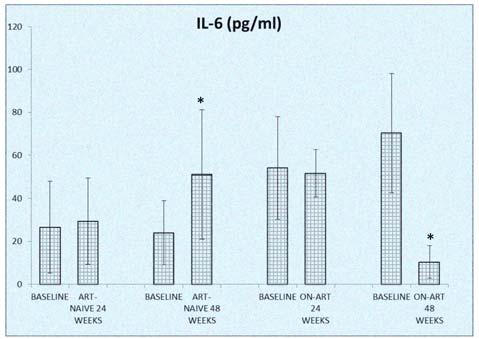
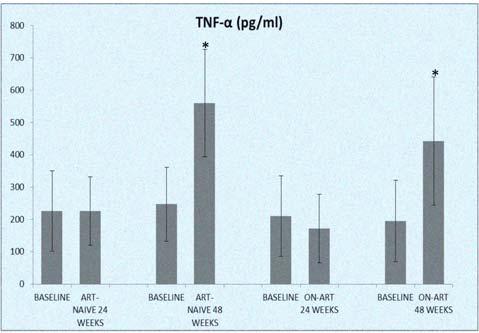
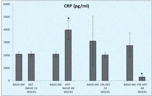
Results : Patients on ART reported a significant decrease in IL-6 and CRP serum levels after 24 weeks of treatment initiation, TNF-α level showed minimal changes after 24 weeks which proceeded to increase significantly at the end of 48 weeks. The on-ART group also exhibited a substantial decrease in HIV Plasma Viral Load. CD4+ cell population showed a significant rise after 24 weeks of cART initiation.
Interpretation and Conclusion : This study on a patient population showed a negative correlation between the CD4+ cell population and serum cytokine concentration (after the administration of cART drugs). A decline in PVL supports the use of NNRTI-based cART in the management of HIV/AIDS.
nitial detection of HIV through sero-surveillance in 1986 prompted a massive organized response from National AIDS Control Organization (NACO) to collaborate on HIV prevention and treatment plans with affiliate hospitals, medical centres, and NGOs1. The stabilising decline in HIV adult prevalence rate has been attributed to accessible free diagnostic services, integrated health care facilities such as Anti-Retroviral Therapy (ART), Parent-to Child Transmission of HIV (PPTCT) services, management of ensuing Opportunistic Infections (OIs), nutritional and psychological counselling2
Department of Tropical Medicine, Calcutta School of Tropical Medicine, Kolkata 700073
1MD, Assistant Professor
2MSc Molecular Biology and Human Genetics, Research Scholar, Department of Laboratory Medicine,
3MSc Biotechnology, Research Assistant, Department of Laboratory Medicine
4PhD, Demonstrator, Department of Laboratory Medicine and Corresponding Author
5MD, Director
Received on : 16/02/2023
Accepted on : 06/04/2023
Editor's Comment : Extrapolation of broader spectrum of cytokine analysis, may help us to understand newer aspects of inflammatory response in HIV infection and subsequent changes following ART administration.
HIV progresses by disrupting the immune machinery and introducing its viral DNA into the selfmaintaining CD4+ memory cell population. Depletion of CD4+ and CD8+ reservoirs initiates immunomodulation and triggers cytokine release3. Altering levels of circulating cytokines work synergistically to directly impact HIV proliferation and disease progression. The reorganization of the cytokine network may lead to newer and stronger cytokine bonds whose rigidity can quickly become fatal to the host body. Some researchers observed profound changes in the cytokine profile very early upon HIV infection and have drawn attention to the alteration in the co-ordination of complex immune functions. These changes are retained throughout the infection’s chronic phase.
121, No 9, September 2023Journal
The introduction of combination antiretroviral therapy (cART) in HIV-infected individuals helps improve health by inhibiting viral replication and decreasing viremia levels in the patient4. It promotes immune integrity by suppressing viral replication thus improving CD4+ and CD8+ T cell proliferation and activity. The improved activities of these immune cells prolong survival and quality of life among HIV-infected persons while keeping opportunistic infections under control. Both HIV infection and antiretroviral treatment deeply influence levels of circulating inflammatory cytokines.
Our team from West Bengal carried out an observational pilot study from our Tertiary Care Hospital (Calcutta School of Tropical Medicine) to determine the baseline levels of HIV-infected individuals in comparison to age and sex-matched healthy individuals. We observed the base level elevations in IL-6, TNF-α (pro-inflammatory cytokines) and CRP in serum, after a viral attack5. The study was followed up to 48 weeks post-initiation of ART as per NACO guidelines.
METHODS AND MATERIALS
Study Participants :
130 newly detected treatment naïve HIV-1 reactive patients were enrolled from the ART center of CSTM, Kolkata, between June-August, 2015 under NACO guidelines. The study design was approved by the Clinical Research Ethics Committee and informed consent was obtained from patients.
The study enrolled patients of both sexes, above 18 years while excluding severely ill or pregnant patients. A standardized proforma was filled up with background information of patients such as medical history, diagnosed opportunistic infections and WHO clinical staging6 at the time of detection. The ARTtreated group served as the ‘case’ arm and the treatment-naive group was taken as the ‘control’ arm for comparative studies. Both groups were followed up at 24 and 48 week-interval. Study parameters such as serum cytokine levels, CD4+ measurements, plasma viral load and incidence of opportunistic infection were recorded during admission and follow-up.
Sample Collection :
8ml of blood was collected by trained phlebotomists for measurement of cytokines, CD4+ cell count, HIV1 RNA and biochemical analysis from all subjects. Serum and plasma were separated by centrifugation (using REMI Centrifuge- R8C, at 3000 rpm, 10 minutes). Samples were labelled and stored in cryovials at -20°C, limited to a single freeze-thaw cycle.
CD4+ T Cell Count :
CD4+ T cell count was done in BD-FACS Calibre (Serial No-E97300192) in absolute numbers and percentages and was analyzed using BD Cell Quest software.
Detection of Plasma HIV-1 Viral Load (PVL) :
HIV-1 RNA Viral Load from each study participant was recorded by Cobas TaqMan 48 by following the Abbott Real-time HIV-1 assay7 (Abbott Molecular Inc, USA). The viral load represents the presence of viral nucleic acids in plasma and is analogous to the HIV-1 disease progression.
Serum Cytokine Estimation :
Cytokine Sandwich ELISA was conducted to specifically detect and quantify the concentration of soluble cytokines in the sample. The cytokines (IL-6, TNF-α) and CRP were measured from the serum of all subjects using ELISA kits (Ray Biotech, USA).
Laboratory Investigations :
Routine biochemical laboratory tests were performed on collected plasma supernates for estimation of lipid profile, liver function, urea, creatinine and fasting blood glucose. These tests were conducted by an Autoanalyzer machine (ERBA-EM-360) according to the manual (TransAsia Biochemical Ltd).
Statistical Analysis :
Baseline data was converted into a Microsoft Office 2007 Excel spreadsheet and revised for outliers/errors. This data was then imported into Graph Pad Prism (version 6.0, San Diego, CA, USA) for statistical analysis. Parametric data were shown as mean ±SD and compared using the unpaired t-test. In all the analyses, p-values <0.05 were considered statistically significant.
RESULTS
Socio-demographic Characteristics :
The enrolled subjects comprise females (n=68, 52.3%), males (n=59, 45.3%) and transgender (n=3,2.3%), where the mean age was 34±10 years. The majority of the participants belonged to the Kolkata district (66.1%) and self-reported addictions to tobacco and alcohol were found in a majority of male patients (n=96, 73.8%). Their marital status revealed 81(62.3%) were married, 35 (26.9%) were unmarried and 14 (10.7%) were widows/widowers (Table 1).
Clinical Observations :
Out of the total enrolled patients, 77(59.2%) were assigned WHO clinical staging with Stages 1 & 2, and 53 patients (40.8%) were in Stages 3 & 4 (Table 1). Following NACO guidelines, 63 enrolled patients were 21
121, No 9, September 2023Journal
immediately initiated on an ART regimen and were given fixed-dose combinations of tenofovir (300mg), lamivudine (300mg) and efavirenz (600mg). Follow-up of this cohort was performed at 24- and 48 weeks posttreatment initiation.The treatment naïve group (51) was similarly followed up every 24 weeks. By the end of our study, 69 patients enrolled on the ART regimen and 27 remained treatment naïve.
Serum Cytokines and their associated CD4+ and Patient Viral Load :
The baseline cytokines (IL-6, TNF- α, and CRP) were recorded and treatment naïve participants did not show statistically significant improvement in any of the parameters (CD4+ T cell count, PVL, IL-6, TNF- α and CRP) when compared to their baseline values. Conversely, the on-ART patients reported an increase in their CD4+ T cell count and decreased PVL after 24 weeks and a significant decrease in plasma CRP, IL-6 and TNF-α (p<0.05) in comparison to their baseline levels (Figs 1-3).
Table 1 — Baseline particularities of subjects(treatment naïve) at point of enrolment (n=130)
At 48 weeks, elevated levels of TNF-α in serum (pg/ml) marked a significant increase as compared to their baseline data. The patients receiving ART treatment had increased CD4+ cell count and decreased levels of serum IL-6, CRP and PVL upon follow-up at 48 weeks. Plasma viral load of 3 subjects were ‘belowdetection’ level after our study enrolment and ART initiation. At 24 weeks follow-up, 32.6% (30 out of 92), had undetectable PVL which increased to 41 % (33 out of 80) at 48 weeks follow-up (assuming good drug compliance).
Opportunistic Co-Infections :
Our baseline observations indicated the presence of co-infections (Pulmonary/ extra-pulmonary TB) in
Fig 1 — Changes in serum IL-6 level in subjects, who continued to be ART- naïve after 24/48 weeks of enrolment. Also, changes in serum IL-6 level in subjects after 24/48 weeks of ART initiation (as per NACO guidelines)
*P value according to unpaired t-test (significance level <0.05).
At 24 weeks follow-up1106 At 48 weeks follow-up1403 Total2509
Fig 2 — Changes in serum TNF-α level in subjects, who continued to be ART- naïve after 24/48 weeks of enrolment. Also, changes in serum TNF-α level in subjects after 24/48 weeks of ART initiation (as per NACO guidelines)
*P value according to unpaired t-test (significance level <0.05).
** Values are in mean±SD
Fig 3 — Changes in serum CRP level in subjects, who continued to be ART- naïve after 24/48 weeks of enrolment. Also, changes in serum CRP level in subjects after 24/48 weeks of ART initiation (as per NACO guidelines)
*P value according to unpaired t-test (significance leveld”0.05).
** Values are in mean±SD
36(28%) of the enrolled. After 48 weeks of follow-up of the HIV/TB (Tuberculosis) on ART and anti-TB drugs, the CD4+ cell count increased significantly compared to their treatment naïve counterparts. PVL, IL-6, TNFα and CRP levels were also significantly diminished in this cohort (Table 3). The presence of candidiasis (oropharyngeal and esophageal) in 13 patients were also noted.
Deaths and LFU :
25 patients were lost in follow-up within 48 weeks of enrolment along with 9 cases of mortality. Their deaths were augmented by the presence of O.Is (n=7, 77.8%). They also presented with low CD4 count (<±50 cells/mm3 ) and high detectable PVL (>106 copies/ml). One outlier case was registered with a higher CD4+ count (>700 cells/ mm3).
DISCUSSION
Surveillance efforts undertaken by National AIDS Control Programme (NACP) have revealed a consistently declining trend in the disease prevalence,
incidence of new infections and mortality due to AIDS since 20108. A similar falling trend is presented by epidemiological analyses in West Bengal, with 62,000 AIDS-related deaths; contributing 6% to the nation’s HIV burden9. Our study was carried out to record changes in serum levels of selected inflammatory cytokines with attention to classic HIV detection and prognosis parameters after 24 and 48 weeks, to draw baseline comparisons.
IL-6, a well-known indicator of persistent inflammation as a result of HIV infection has been linked to morbidity and mortality events. Active studies have demonstrated consistently low levels of plasma IL-6 in virologically suppressed individuals undergoing treatment. Our study recorded a slight decline in IL-6 in on-ART group ((with CD4+ cell count <200 cells/ mm3) after 24 weeks as opposed to elevated IL-6 levels observed in the treatment naïve group (with CD4+ cell count >500 cells/mm3). However, after 48 weeks, the on-ART group displayed a considerable decline in their IL-6 population compared to their baseline levels with CD4+ cell count restoring up to >350 cells/mm3 Anomalies were noted in two separate cases where IL-6 levels declined before ART initiation10 (CD4+ cell count -240/mm³ and 315/mm³. Elevated IL-6 levels can serve as an early prognostic marker for HIV and timely ART intervention can minimize inflammation11
TNF-α, a pro-inflammatory cytokine in the TNFR pathway is a hallmark promoter of HIV-1 infection12, where levels of plasmic TNF-α were observed to be much higher in ART-naïve patients than in patients’ on-ART13. Our study recorded minute changes in TNFα levels of the on-ART group even after 20 weeks of treatment as opposed to a significant jump after 48 weeks. Controlled viremia aided by ART did not effectively reduce the TNF-α level even after a rise in CD4+ levels, as per our surveillance data. The treatment-naive group also exhibited a significant elevation in TNF- α level (higher than the on-ART group) in comparison to its baseline level.
Table 3 — Changes in cytokines, immunological and virological parameters in HIV/TB co-infected patients who started on ART and ATD (baseline versus 48 weeks FU)
VariableBaseline values of48 weeks following treatment- naiveATD and ARTp- value* HIV/TB patients(n=19) ** (n=19)**
*P value according to unpaired t-test (significance level (S)
** Values are in mean ±SD
CRP levels of the untreated group display a marked increase at the end of 48 weeks compared to the on-ART group which records a drastic fall in their levels at 48 weeks post-treatment initiation. Low levels of CRP in HIV-infected patients can potentially serve as a predictor for longevity and a prognostic marker for the prevalence of opportunistic co-infections and cardiovascular morbidities14
Our study exposed extrapulmonary tuberculosis as the primary co-infection and
Vol 121, No 9, September 2023Journal of the Indian Medical Association
mucocutaneous candidiasis and extrapulmonary TB as common OIs. Of the 9 subjects who succumbed, 7 were on-ART and had acquired OIs, with 4 of them infected with TB (TB meningitis/Lymph Node TB) and were receiving CAT 1 ATD (Category 1 Anti Tubercular Drug). 6 deaths were reported before the completion of 24 weeks of our study, making extrapulmonary TB a bad prognostic factor.
We inferred that the coordinated regimen of both ART and TB drugs worked in tandem to suppress inflammation. Our study detected a few cases of Elite Controllers (ECs)15 (n=23) where despite maintaining low viremia levels, elevation in inflammatory biomarkers continued, signaling the presence of Long-term NonProgressors (LTNPs).
CONCLUSION
Being one of the few regional studies drawing on associations between the influence of ART and the altering levels of inflammatory markers16, our study was supported by clinical monitoring of treatment adherence and frequency of co-infections. However, a further extrapolation on a wider population of the Indian gene pool, with a broader spectrum of cytokines analysis, may bring forward and unwind newer aspects of inflammatory networking in hosts after HIV infection and subsequent ART administration. Besides ART, a careful individualized approach supported with strategies that focus on minimizing the impact of comorbidities17 can further strengthen the study.
REFERENCES
1Kumar S — AIDS and COVID-19 infections: impact on vulnerable Indian population. New Microbes and New Infections 2021; 42: 100903.
2National AIDS Control Organization -Sankalak: Status of National AIDS Response (Third edition, 2021)http:// naco.gov.in/sites/default/files/Sankalak_Status_of_National_ AIDS_Response_Third_Edition_2021.pdf (accessed Sep 25, 2022): 23-32.
3Deeks SG, Overbaugh J, Phillips A, Buchbinder S— HIV infection. Nature Reviews Disease Primers 2015; 1(1): 122.
4Pau AK, George JM — Antiretroviral therapy: current drugs. Infectious Disease Clinics 2014; 28(3): 371-402.
5Debnath A, Mandal M, Choudhuri S, Saha MK, Guha SK— Baseline Levels of Interleukine-6, Tumor-Necrosis Factor-
Alpha and C-Reactive Protein in Treatment Naive Human Immunodeficiency Virus Infected Patients-A Study from a Tertiary-Care Hospital in Eastern India. Current Biomarkers (Formerly: Recent Patents on Biomarkers) 2016; 6(2): 12432.
6UNAIDS — Report on the global AIDS epidemic, 2013. https:/ /www .unaids.org/en/resources/documents/2013/ 20130923_UNAIDS_Global_Report_2013 (accessed Sep 20,2022).
7Scott LE, Crump JA, Msuya E, Morrissey AB, Venter WF, Stevens WS — Abbott RealTime HIV-1 m2000rt viral load testing: manual extraction versus the automated m2000sp extraction. Journal of Virological Methods 2011; 172(1-2): 78-80.
8National AIDS Control Organization -Sankalak: Status of National AIDS Response (Third edition, 2021)http:// naco.gov.in/sites/default/files/Sankalak_Status_of_National_ AIDS_Response_Third_Edition_2021.pdf (accessed Sep 25,2022): 23-32
9Ganguly S, Chakraborty D, Goswami DN — HIV/AIDS epidemic in West Bengal: An overview. Journal of Family Medicine and Primary Care 2018; 7(5): 898.
10Hattab S, Guihot A, Guiguet M, Fourati S, Carcelain G, Caby F, et al — Comparative impact of antiretroviral drugs on markers of inflammation and immune activation during the first two years of effective therapy for HIV-1 infection: an observational study. BMC Infectious Diseases 2014; 14(1): 1-9.
11Trovato M, Ruggeri RM, Sciacchitano S, Vicchio TM, Picerno I, Pellicanò G, et al — Serum interleukin-6 levels are increased in HIV-infected patients that develop autoimmune disease during long-term follow-up. Immunobiology 2018; 223(3): 264-8.
12Pasquereau S, Kumar A, Herbein G — Targeting TNF and TNF receptor pathway in HIV-1 infection: from immune activation to viral reservoirs. Viruses 2017; 9(4): 64.
13Behrens NE, Wertheimer A, Love MB, Klotz SA, Ahmad N — Evaluation of HIV-specific T-cell responses in HIV-infected older patients with controlled viremia on long-term antiretroviral therapy. PLoS ONE 2020; 15(9): e0236320.
14Chenciner L, Symonds M, Dissanayake O, Hunter A, Burns F, Miller RF — Lymphocyte-CRP-ratio and CRP-albumin-ratio as potential inflammation markers in adults with HIV. Journal of Acquired Immune Deficiency Syndromes 2022; 13: 10-97.
15Gurdasani D, Iles L, Dillon DG, Young EH, Olson AD, Naranbhai V, et al — A systematic review of definitions of extreme phenotypes of HIV control and progression. AIDS 2014; 28(2): 149-62.
16Amoani B, Sakyi SA, Barnie PA, Karen Pomeyie K, Aniagyei W, et al — Effect of ART on Cytokine Profile amongst HIV Patients: A Systematic Review and Meta-Analysis. Focus Med Sci J 2021; 7(3)
17Kumbale CM, Voit EO — Toward Personalized Medicine for HIV/AIDS. Journal of AIDS and HIV Treatment 2021; 3(2): 37.
Original Article
Diagnostic Utility of Percutaneous FNAC in the Evaluation of Hepatic Masses
Gopalam Vashista Saikumar1, Kandibanda Sai Sri Ram Rao2, Arijit Roy3, Jayashree Gurudatta Pawar4
Background : The clinical assessment and management of focal Hepatic Masses is a difficult task.The role of percutaneous liver Fine Needle Aspiration Cytology (FNAC) and cytological assessment is of utility and can enhance diagnostic value in such cases.
Objectives : The purpose of the study was to evaluate the cytological features of hepatic masses and to establish the diagnostic utility of hepatic FNAC in a limited resource set-up.
Materials and Methods : A retrospective study of 2 years duration was conducted in the Department of Pathology of a Tertiary Care Hospital which included 56 patients with clinically and radiologically detected Hepatic Masses. Cytohistopathological correlation was done in 20 cases.
Statistical analysis : The statistical values of correlation such as Sensitivity, Specificity, Positive Predictive Value (PPV), Negative Predictive Value (NPV) and Diagnostic efficacy were calculated using Epi info 7.0 statistical software.
Results : The number of male patients were more than females. The distribution of cyto-diagnoses cases were; Pyogenic Abscess (1 Case), Amoebic Liver Abscess (1 Case), Fungal Liver Abscess(2 Cases), Chronic Hepatitis (1 Case), Cirrhosis (1 Case), Hydatid Cyst (1 Case), Large Cell Dysplasia (3 Cases), Hepatic Adenoma (1 Case), Hepatocellular Carcinoma (30 Cases), Cholangiocarcinoma (1 Case), Metastatic Adenocarcinoma (9 Cases), Malignant Lymphoma (2 cases) and (1 case) each of metastatic deposits of Malignant melanoma, Neuroendocrine carcinoma and poorly differentiated carcinoma. Statistical analysis showed a Sensitivity-94%, Specificity-100%, PPV100%, NPV-75% and Diagnostic efficacy was 95%.
Conclusion : FNAC is a quick,feasible and reliable procedure with high accuracy for cytological diagnoses of Hepatic Masses.
Liver lesions are frequently encountered in clinical scenarios and requires a variety of tests to understand their nature and plan management accordingly. While Liver Function Test’s (LFT’s) and Ultrasonography (USG) are the usual initial investigation modalities, the role of liver FNAC has proved to be useful in understanding the pathological process underlying these lesions. It is a simple, rapid and sensitive technique allowing the clinician to plan for the treatment by avoiding unnecessary surgical procedures1 . The role of liver FNAC in a limited resource setup is useful where biopsy technique is
Department of Pathology, Mamata Medical College, Khammam, Andhra Pradesh 507002
1MD, Assistant Professor and Corresponding Author
2MD, Assistant Professor
3DCP, DNB, Consultant Pathologist, Department of Pathology, Chikitsa Medicare Centre Pvt Ltd, Kolkata 700034
4MD, Professor, Department of Pathology, PES Institute of Medical Sciences and Research Kuppam, Andhra Pradesh 517425
Received on : 24/07/2022
Accepted on : 21/01/2023
Editor's Comment : The present study emphasizes the diagnostic utility of Liver FNAC as a cost effective procedure in a low resource setup by rendering rapid diagnosis. It reduces the reliance on liver biopsy.
not readily available. It can be employed as an outpatient procedure and therefore offers the clinician a faster diagnostic aid 2,3 . The advent of newer modalities of imaging such as Computed Tomography (CT) and USG have revolutionized the guided FNAC approach4-6. The procedure has limited application in identifying diffuse Liver Disorders, such as Hepatitis, Cirrhosis and Necrotic lesions as these are associated with reactive changes in hepatocytes mimicking malignancies and therefore it is very difficult to suggest an etiology based on FNAC alone unless correlated with clinical, biochemical, radiological and histopathological findings7-10. Hence the aim of the study was to utilise USG-guided Liver FNAC as an effective, economical procedure to identify the various Liver lesions.
121, No 9, September 2023Journal
MATERIALS AND METHODS
A retrospective study of image guided FNAC cases was conducted in the Department of Pathology of a Tertiary Care Hospital for a duration of 2 years after obtaining an approval from the Ethical Clearance Committee.
The study included a total of 56 cases.All of them underwent USG-guided FNAC with prior written consent.Patients presenting with clinical and radiological evidence of known contraindications such as haemangioma, hydatid cyst, massive ascites or deranged coagulation profile were excluded. The procedure was performed under aseptic conditions in the supine position by employing a 23-gauge 90 mm lumbar puncture needle fitted to a 10ml syringe.
The stains employed to assess the cytological features were air-dried Giemsa, alcohol based Papanicolaou and Haematoxylin and Eosin (H&E).
Histopathological correlation was obtained in 20 cases by liver biopsy and cell block along with FNAC.Clinical correlation was done with laboratory investigations such as (LFT’s), Hepatitis B and C viral markers, HIV antibody and tumor markers like Alfa-fetoprotein (AFP) and USG findings.
The statistical analysis was done using Epi info 7.0 statistical software to analyse the data and diagnostic efficacy obtained by Sensitivity, Specificity, Positive predictive value and Negative predictive values.
RESULTS
In the present study, male patients were 36 (64.28%) and female patients 20 (35.71%) with a male to female ratio of 1.8:1.The age of the patients were in the range of 5 months to 80 years with a mean age of 55.2 years. The youngest patient was 5 months old and was diagnosed as Pyogenic Liver Abscess and the oldest was 80 years old and diagnosed as moderately differentiated Hepatocellular Carcinoma (HCC).
The commonest mode of presentation was pain abdomen in the right Hypochondrium. Other main clinical features were mass per abdomen, Jaundice, Anorexia, Fever, Easy Fatiguability and Respiratory Symptoms. Majority of clinical diagnosis for Space Occupying Lesions (SOL’s) was HCC. Serological studies included viral markers for Hepatitis B,C and
HIV. Hepatitis B surface antigen (HBs Ag) and anti HCV antibody positivity was seen in 6 cases each of HCC respectively and 1 case of fungal abscess was positive for HIV antibody. AFP values were available in 6 cases of clinically suspected HCC. The patients had high values of AFP minimally increased by 2 to 4 folds.
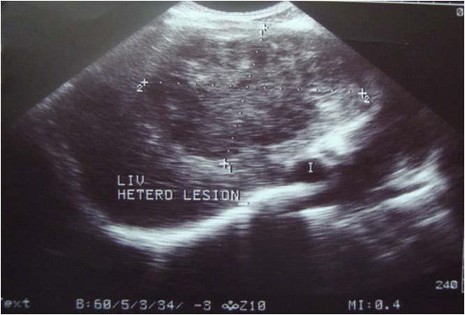
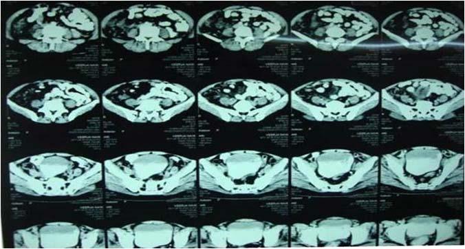
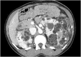
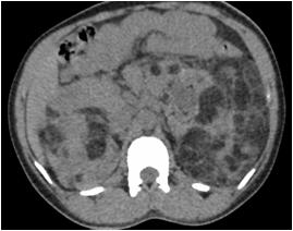
Radiological imaging was done in all the cases. USG findings revealed solitary masses in 45 cases (80.35%) and diffuse masses in 11 (19.64%). Solitary masses were around 45 in number out of which 30 were diagnosed as HCC. Right lobe had around 30 (66.7%) whereas left lobe had 15 (33.33%) masses respectively. Maximum (SOL’s) observed showed hyper-echogenicity present in 43 cases (76.78%). Heterogenous echotexture was observed in 6 cases (10.71%) (Fig 1).
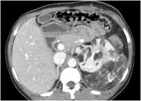
CT scan was done in two patients which showed multiple well defined hyperdense lesions and hypodense lesions in another patient. Cyto-radiological correlation of hepatic masses was seen in 94% cases (Fig 2).
Fig 1 — USG liver showing well defined heteroechoic lesion in left lobe
Cytological evaluation of the smears obtained from FNAC of Hepatic Masses was done and were categorized into: Non-neoplastic, pre-neoplastic and neoplastic lesions (Table 1).
Out of 7 cases categorized as non-neoplastic lesions,4 were of infectious etiology which included 1 case each of Pyogenic and Amoebic Liver Abscess (3.56%). Two cases of fungal liver abscesses (3.56%)were also seen. Other lesions included 1 case of chronic hepatitis (1.78%),1 case of Hepatic Cirrhosis (1.78%) and 1 solitary case of Hydatid Cyst (1.78%).
In the pre-malignant category, 3 cases of large cell dysplasia (5.35%) were obtained.In the neoplastic category, 1 case of benign hepatic adenoma(1.78%) was diagnosed cytologically. There were 31 cases (55.35%) of primary hepatic malignancy and 14 cases (25%) cases of metastatic malignancies. HCC was the most common and predominant neoplastic lesion numbering to 30(53.57%) from around 46 neoplastic liver lesions. Among them 21(70%) were males and 9(30%) were females with a male to female ratio of 2.3:1. Majority of the cases were in the 50-60 years age group.
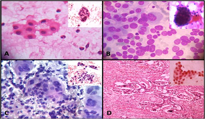
Cytologically, HCC was graded into well differentiated HCC (13), Moderately differentiated HCC (12) and Poorly differentiated HCC (5). Histopathological correlation was obtained in 9 cases. The most consistent cytological findings in HCC were increased nuclear-cytoplasmic ratio, pleomorphism, hyperchromasia, macro nucleoli, bare nuclei,intracytoplasmic bile and trabecular pattern of arrangement followed by traversing blood vessels,
Table 1 — Final cytological diagnosis in 56 patients
peripheral endothelial rimming, hypercellularity and intracytoplasmic inclusions. The least common findings were multinucleation, intranuclear inclusions, bile duct epithelium and tumor giant cells (Table 2).
Our study also revealed a case of Cholangio Carcinoma (CC)(1.78%) and cellular features included occasional clusters of atypical cells exhibiting hyperdense nuclei with scant cytoplasm along with a few foci of fibrosis which was confirmed by liver biopsy.
Metastatic Tumors constituted 14 (25%) among 56 cases and metastatic adenocarcinoma (9 cases) was the commonest type (16.07%). Known primary sites of adenocarcinoma were: Colon and rectum (1), Ovary (1), Breast (1), Pancreas (1) and unknown primary (5). Cytological features revealed dense chromatin and increased nuclear-cytoplasmic ratio followed by hypercellularity, benign hepatocytes, eccentric nucleus, pleomorphism, vacuolated cytoplasm, conspicuous nucleoli and inflammatory cells. In all the previously known cases of primary adenocarcinoma, the cytological features were simulating the primary tumor and even the radiological investigations like USG and CT features confirmed the primary in respective cases. Histopathological correlation was obtained in 3 cases of metastatic adenocarcinoma.
Metastatic tumors included 2 cases of (3.57%) malignant lymphomas and one case each of (5.34%) of metastatic malignant melanoma, metastatic neuroendocrine carcinoma and metastatic poorly differentiated carcinoma. Histopathological correlation was done in 1 case each of malignant lymphoma and metastatic malignant melanoma.
Cytohistological correlation was done in 20/56 cases. In 4 non-neoplastic lesions, cytological diagnoses were concordant with histopathology. One discordant case (diagnosed as cirrhosis on cytology)
Table 2 — Cytological features of hepatocellular carcinoma in the present study
Cytological featuresHCC(n=30)(%)
Hypercellularity 18(60%)
Trabecular pattern 20(66.7%)
Peripheral endothelium rimming 18(60%)
Transgressing endothelium 20(66.7%)
Intracytoplasmic inclusions 17(56.7%)
Intracytoplasmic bile 22(73.3%)
Bile duct epithelium 5(16.6%)
Pleomorphism 27(90%)
Increased N/C ratio 30(100%)
Hyperchromasia 27(90%)
Intranuclear inclusions 6(20%)
Multiple nuclei 6(20%) Bare nuclei 24(80%)
Macro nucleoli 24(80%)
Tumor giant cells 3(10%)
121, No 9, September 2023Journal
was later on confirmed as HCC on histopathology. All malignant neoplasms were accurately diagnosed cytologically. Remaining 16 neoplastic lesions diagnosed cytologically were concordant with histopathology. The statistical values of USG-guided FNAC in the diagnosis of liver SOL’s were sensitivity and specificity of 94% and 100% respectively, with a positive predictive value of 100% and negative predictive value of 75% and overall diagnostic efficacy of 95%.
DISCUSSION
FNAC is widely practiced, quick, precise, costeffective and efficient method that can be employed to differentiate benign from malignant lesions of the liver by offering a definite diagnosis. SOL’s of the liver include cysts and abscesses of infectious etiologyas well as tumors of benign and malignant nature. This group is frequently targeted by FNAC performed under imaging guidance USG or CT11-14. Numerous studies have reported sensitivity in the range of 67% to 100% and accuracy rates upto 96% 9,15,16 . Severe complications like bleeding, biliary peritonitis, pneumothorax and sepsis are hardly encountered. The risk of malignancy spreading along the needle tract is insignificant and the incidence of needle track seeding in a recent study involving primary liver tumors was 0.6% when compared to wide bore biopsies which may depend on diameter of the needle used17,16. Cell block preparation from residual material can be an useful adjunct to smears employed by the cytopathologist for establishing a more definitive diagnosis in hepatic lesions.Multiple sections can be obtained and if required special stains and techniques such as Immuno-histochemistry (IHC) can be performed with increased sensitivity and specificity for malignant lesions in conjunction with conventional smears which can be further improved if interpretation of aspirates is done in the context of clinical, radiological and biochemical investigations11,18,19
Conventional LFT’s of patients diagnosed as primary HCC in present study did not reveal significant values for a non-neoplastic, neoplastic or malignant lesion. Similar observations have been observed by Rastogi, et al who concluded that the biochemical parameters of liver enzymology studies are not enough to identify the disease process within SOL’s of liver20. Abnormal LFT values were documented in a study involving 130 Hepatic lesions3
HCC Is mostly associated with viral Hepatitis caused by HBV and HCV. Hepatitis B surface antigen (HBs Ag) and anti HCV antibody positivity was seen in 6 cases each of HCC. In studies done by Manik, et
al, Jha, et al and Sultana, et al, HBs Ag positivity was predominantly seen in HCC patients and concluded that HBV infection is the leading cause of HCC contrary to our findings10,3,11
AFP values were available in 6 cases of clinically suspected HCC. The patients had high values of AFP increased by 2-4 folds with similar findings reported by Rastogi, et al20. Positive AFP levels was found to be insignificant in the study conducted by Manik, et al10. With this we concluded that majority of HCC cases present with significant elevation of AFP thus ruling out the need for liver biopsy.
USG can detect the majority of HCC’s when used in combination with FNAC thereby increasing the sensitivity21 USG findings revealed solitary masses in 45 cases (80.35%) and diffuse masses in 11 (19.64%). Solitary masses around 30 in number were diagnosed as HCC. Right lobe had around 19 (63.3%) whereas left lobe had 11 (36.6%) masses respectively. All metastatic lesions presented as multifocal lesions radiologically in our study. Similar radiological findings were obtained in the studies by Jha, et al, Giriyan, et al and Roy, et al3,14,21
Cyto-radiological correlation in our study was established in 94% cases as compared to 92% in a study done by Khanna, et al22. Discordance was noted in 3 (5.35%) cases in our study which included 2 nonneoplastic lesions and 1 pre-neoplastic lesion. A case of hydatid cyst was accidentally aspirated which was clinically and radiologically suspected as pyogenic liver abscess and a case of cirrhosis diagnosed on cytology which showed radiological features simulating it but the lesion was subjected to liver biopsy as few atypical cells were seen on smears.1 case of large cell dysplasia was biopsied and diagnosed as cavernous hemangioma as excess blood was aspirated even on the second attempt. The same was reported by other researchers where USG diagnosed lesions did not correlate with cytology and liver biopsy was done for definite diagnosis9,23
In the present study 7 cases (12.5%) were categorized as non-neoplastic lesions. The most common non-neoplastic lesion were abscesses (7.12%, 4 cases) followed by diffuse parenchymal lesions (3.56%, 2 cases) and a solitary case of hydatid cyst (1.78%,1 case). Pre-malignant category included 3 cases of large cell dysplasia (5.35%,3 cases). Our findings were consistent with the studies done by Jha K, et al, Rastogi, et al, Sawke, et al and Shruthi, et al3,20,6,13
In our study one case of pyogenic liver abscess was diagnosed and was seen to have plenty of
Vol 121, No 9, September 2023Journal of
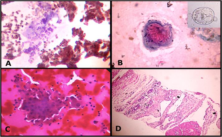
neutrophils and necrotic celldebris. Few mononuclear cells and degenerating hepatocytes were present. In a case of amoebic liver abscess, smears prepared from the centrifuged sediment showed trophozoites of Entamoeba Histolytica in a background of necrotic cellular debris, degenerating hepatocytes and mixed inflammatory cells. Mallikarjuna, et al and Nasit, et al reported similar findings 9,23 . These cases were diagnosed cytologically thus validating our results that FNAC can serve a dual purpose of therapeutic as well as diagnostic procedure where serum anti amoebic antibodies are not available9 . Biopsy of pyogenic abscess was diagnosed as granulomatous hepatitis as sampling error was responsible and amoebic liver abscess showed only necrosis and no malignancy.
Two cases of Fungal Liver Abscesses due to Aspergillosis and Candida were diagnosed cytologically in both immuno-compromised patients. Smears showed slender hyphae and budding spores of Aspergillosis. Smears showed pseudo hyphae and budding yeast forms of Candida. Subsequent microbiologic and cultural studies also confirmed the cytological findings in both the cases. FNAC thus is a rapid, sensitive and important method of diagnosing fungal infections which may be lifesaving in immunocompromised patients. Similar findings were seen in Jha, et al study and in individual case reports done by Vairani, et al and Menachery, et al3,24,25. Chronic hepatitiswas diagnosed in one case with hepatocytes showing predominantly reactive cellular changes. Soudah et al reported similar cytological features on FNAC and opined that its utility lies in pointing out the etiology, but not convincingly in viral diseases of the Liver7
In our study, we concluded that FNAC in cirrhotic nodules are only helpful in detecting fibrosis. Similar findings were seen in the study done by Soudah, et al and Geramizadeh, et al7,8. In our case cytologically diagnosed as cirrhosis, biopsy was
done and concluded as HCC due to presence of a few atypical cells. Mallikarjuna, etalreported a similar case9 Aspiration of a radiologically suspected pyogenic liver abscess case showed scoleces, scattered hooklets and hyaline fragments which was confirmed as hydatid cyst on histopathology. Swamy MC, et al and Mahajan, et al both concluded that definite diagnosis of hydatid disease of liver should involve a combination of imaging, microbiology, and cytology, as reliance on a single diagnostic modality cannot conclusively confirm the presence of hydatid disease9,26
3 cases of large cell dysplasia were reported in our study which were consistent with the findings seen in the studies done by Jha, et al and Sultana, et al3,11 Biopsy in one case of liver cell dysplasia was diagnosed as cavernous hemangioma as repeated aspirations yielded only blood. Similar findings were seen in studies done by Guy, et al and Nasit, et al who concluded that the most frequent spindle-cell lesion of the liver is hemangioma and radiological correlation is necessary27 (Fig 3).
In the present study, a single case of liver cell adenoma (1.78%) was diagnosed cytologically along with radiological correlation in a young female patient with a past history of treatment with oral contraceptives. Proper placement of the needle and sampling within the lesion is suggested by Nasit, et al to get adequate yield and avoid diagnostic confusion with focal nodular hyperplasia23. Similar findings were
Pap - Papanicolau stain; H&E - Hematoxylin and Eosin
Fig 3 — (A) FNAC of Fungal Liver Abscess – Aspergillus hyphae and spores Pap stain (X 400). (B) FNAC of Hydatid Cyst- Scolex of E Granulosus Pap stain (X 400) along with wet preparation showing intact scolex (inset). (C) FNAC of Large cell dysplasia on H&E stain (X 400) and (D) Cavernous hemangioma (Tru-cut biopsy) H&E stain (X 400).
Vol 121, No 9, September 2023Journal of
seen in the study conducted by Jha, et al and Nasit, et al3,23
In the present study (82.14%) lesions were cytologically malignant followed by(12.5%) benign lesions and (5.35%) pre-neoplastic lesion. Most of the studies including Jha K, et al, Sawke, et al and Rastogi, et al reported similar findings3,6,20
In the present study, the key cytological features of HCC were macronucleoli, trabecular pattern of cells, hyperchromatic nuclei, increased nuclear-cytoplasmic ratio and pleomorphism. Wee A, et al, Balani S, Shruthi HY, et al, observed similar cytological features as noted in our study28,15,13 (Table 3).
Cytological features of CC diagnosed in our study was similar to those described by Soudah, et al and Mallikarjuna, et al7,9
Metastatic tumors constituted (25%) and adenocarcinoma was the commonest type (16.07%). Primary sites of adenocarcinoma which could be ascertained were: Colon and Rectum (1), Ovary (1), Breast (1) and Pancreas (1). In the present study, the most common metastatic adenocarcinomas were of unknown primary origin (5 cases). Similar findings were seen in the studies conducted by Ali SR, et al, Giriyan S, et al and Garg, et al3,14,12. Cytological features of metastatic adenocarcinoma noted in our study were similar to those described by Shruthi and Garg, et al13,12. Histopathological correlation was done in 3 patients which confirmed the diagnosis.In the study done by Sahin, et al, majority of metastatic adenocarcinoma cases presented as acinar pattern while the key differentiating features between HCC and metastasis were uniform atypia, increased N/C ratio, hepatocytic appearance and atypical naked nuclei29 The findings of present study were in concordance with these observations. Further workup of the patients to determine the primary sites was not possible as they were referred to higher centres for further management.
Two cases of malignant lymphoma clinically diagnosed as NHL were seen in our study. The cytomorphological features were similar to those described by Collins, et al30
A case of lymphoma included by Jha, et al in their study was diagnosed as Diffuse large B cell lymphoma by flow cytometry3. However, this was not possible in our institute which is a major drawback.
Metastatic malignant melanoma showed similar cytological features when compared to De Las Casas, et al who have reported certain diagnostic criteria for liver metastasis of melanoma31. In the study conducted by Prosser, et al, metastatic neuro-endocrine tumors to the liver were subtyped on the basis of
Table 3 — Comparison of cytological HCC features in the present study with other studies
CytologicalWee A and Balani SShruthi HYPresent features Nilsson B(2013)15 (2021)13 study (2003)26
Macronucleoli75.14 85.7 100.0080.00
Trabecular pattern88.5765100.00 66.7
Hyperchromatic nucleus75.71100 96.8890.00
Increased N : C ratio100100 93.75 100.00
Pleomorphism80.00 71.487.5090.00
Traversing blood vessels82.85 57.171.8766.7
Endothelial rimming35.71 35.765.6260
cytomorphology into round, spindle and polygonal cell types and further emphasized the need for using IHC in distinguishing the cell of origin in such tumors as diagnostic confusion in differentiating them from HCC32 However, in our study we observed a single case of metastatic neuro-endocrine tumor. A case of metastatic poorly differentiated carcinoma was cytologically similar as described by Mallikarjuna, et al who suggested IHC be utilized to differentiate Poorly differentiated HCC from other poorly differentiated carcinomas9 (Fig 4).
The present study observed to have overall values of Specificity (100%), Sensitivity (94%), NPV (75%), PPV (100%) and Diagnostic efficacy (95%). The studies done by Kuo, et al,Ceyhan, et al and Okzara, et al, have mentioned similar values of above parameters for diagnosing SOL’s of liver with FNAC33,18,34. These values were comparable with the observations of the present study. There are reports of Reddy, et al and Franca, et al, who have reported low NPV of 58.8% and 64% respectively which is a contrary observation to the studies reviewed for literature because of low false negative reporting16,35. Kuo, et al, who reported sensitivity of 78.4% which is low as compared to other published studies and present study33. The studies of Ceyhan, et al, Rastogi, et al, and Franca, et al, have reported PPV of 100% due to low false positive cases18,20,35. The present study also made similar observations.
In our study, 20 cases were histopathologically correlated. 3 cases were diagnosed as non-neoplastic lesions on FNAC, which were confirmed histopathologically. One case diagnosed as cirrhosis on FNAC was reported as HCC on biopsy. Sampling error was the main reason for false-negative result in this case. Such limitation for erroneous diagnosis of false negative cases has been quoted in the studies of Mallikarjuna, et al and Ceyhan, et al9,18
16 cases reported as malignant on FNAC were concordant with the biopsy. With an efficacy rate as high as 95% for liver lesions, FNAC is an extremely useful procedure that facilitates rapid diagnosis and
Vol 121, No 9, September 2023Journal of
prevents further surgical intervention in inoperable cases.
Limitation(s) :
The present study utilized FNAC as the primary diagnostic method for various hepatic lesions. A small sample size of patients our study is also not representative of the gamut of hepatic lesions commonly encountered during cytological examination. Further due to the advent of superior imaging techniques such as Fibro scan which measures Fibrosis and Steatosis thereby obviating the need for further invasive procedures. Use of ancillary techniques like IHC and flow cytometry were not possible in our institute.
Fig 4 — (A) FNAC of Well differentiated HCC – Pseudo acinar pattern on H&E stain (X 400) and cell block (inset). (B) FNAC of Moderately differentiated HCC-Dispersed atypical naked nuclei on H&E stain (X 400) along with peripheral endothelial rimming (inset). (C) FNAC of Poorly differentiated HCC on Pap stain (X 400) and cell block (inset) (D) Cholangiocarcinoma (CC) HPE on H&E stain (X 400) and FNAC of Cholangiocarcinoma (CC) on Pap stain (X 400).
Pap - Papanicolau stain; H&E - Hematoxylin and Eosin
fine needle aspiration cytology in cases of hepatic mass lesions. Int J H Biomed Res 2015; 3: 149-55.
CONCLUSION
Image guided FNAC utilizing USG of hepatic masses is a reliable, rapid and cost effective procedure having reasonably high accuracy and good sensitivity and specificity. When used in conjunction with relevant tests such as LFT’s, Serology and Serum tumor markers it can yield valuable information which can be used for early diagnosis and planning of rational therapeutic management of patients. Conflict of interest : Nil.
REFERENCES
1de Boer B — Liver and spleen. In: Orell SR, Sterret GF, et al editors. Fine Needle Aspiration Cytology. 5th ed. Churchill Livingstone, Elsevier, 2012: 271-96.
2Edoute Y, Osamah H, Malberger E, Yerushalmi R, Tibon-Fisher O, Assy N — Diagnostic accuracy of direct FNAC of liver lesions:Aprospective study of 107 patients in peripheral community center with limitedtechnological capabilility. Arch Gastro Enterohepatol 2001; 20(1): 19-24.
3Jha K, Gupta A, Pangarkar M, Kumbhalkar DT, Raut WK — Guided FNAC of nodular lesions in liver. ArchCytolHistopathol Res 2018; 3(4): 166-72.
4Ali SR, Jayabackthan L, Rahim S, Sharel MB, Prasad K— Hegdekatte N-Role of fine needle aspiration cytology in the diagnosis of hepatic lesions. Muller JMed Sci Res 2015; 6: 125-8.
5Mane A, Kanetkar SR, Saini S, Saini N — Role of image guided
6Sawke N, Madhaw N, Sawke GK — Study of image guided fine needle aspiration cytology in cases of hepatic mass lesions. Trop J Path Micro 2018; 4(6): 437-41.
7Soudah B, Schirakowski A, Gebel M, Potthoff A, Braubach P, Schlue J, et al — Overview and evaluation of the value of fine needle aspiration cytology in determining the histogenesis of liver nodules: 14 years of experience at Hannover Medical School. Oncol Rep 2015; 33(1): 81-7.
8Geramizadeh B, Asadi N, Tabei SZ— Cytologic comparison between malignant and regenerative nodules in the background of cirrhosis. Hepat Mon 2012; 12(7): 448-52.
9Swamy MC, Arathi C, Kodandaswamy C — Value of ultrasonography-guided fine needle aspiration cytology in the investigative sequence of hepatic lesions with an emphasis on hepatocellular carcinoma. J Cytol 2011; 28(4): 178-84.
10Manik AH, Ekram R, Mallik PK, Mahzabeen M, Islam MR, Amin MR — FNAC of Hepatic Malignancy and it’s Clinical Correlation. Bangladesh Critical Care Journal 2019; 7(2): 81-5.
11Sultana SS, Dewan RK, Ferdousi F, Sarker R, Jinnah SA, Jeba R, et al — Evaluation of Space Occupying Lesion of Liver by Fine Needle Aspiration Cytology and Cell Block Examination. Journal of Histopathology and Cytopathology 2018; 2(1): 11-8.
12Rachana G, Rao A — Clinicocytological Analysis of Hepatic Neoplastic Lesions with ParticularReference to Morphological Pattern Assessment. J Cytol Histol 2021; 12: 561.
13Shruthi HY, Chavan SS — A Prospective Study of Ultrasound Guided Fine Needle Aspiration Cytology of Focal Lesions in Liver. NJLM 2021; 10(2): 1-4.
Vol 121, No 9, September 2023Journal of
14Giriyan S, Arati S — Ultrasound guided FNAC in diagnosis of space occupyinglesions of liver. IJPRP 2017; 6(2) part 2.
15Balani S, Malik R, Malik R, Kapoor N— Cytomorphological variables of hepatic malignancies in fine needle aspiration smears with special reference to grading of hepatocellular carcinoma. J Cytol 2013; 30(2): 116-20.
16Reddy CV, Goud YG, Poornima R, Deshmane V, Madhusudhana BA, Gayathridevi M — Role of FNAC in Hepatic lesions: Risk of track metastases. South Asian J Cancer 2015; 4(1): 35-7.
17Ji XL — Fine-needle aspiration cytology of liver diseases. World J Gastroenterol 1999; 5(2): 95-7.
18Ceyhan K, Kupana SA, Bektaº M, Coban S, Tuzun A, Cinar K et al — The diagnostic value of on-site cytopathological evaluation and cell block preparation in fine-needle aspiration cytology of liver masses. Cytopathology 2006; 17(5): 26774.
19Khurana U, Handa U, Mohan H, Sachdev A — Evaluation of aspiration cytology of the liver space occupying lesions by simultaneous examination of smears and cell blocks. Diagn Cytopathol 2009; 37(8): 557-63.
20Rastogi N, Bhake A, Agarwal D, Wankhede A — Ultrasound guided fine needle aspiration cytology of space occupying lesions of Liver. Int J Res Med Sci 2019; 7: 192-8.
21Roy SK, Sultana S, Mollah NU, Yasmin T, Sarker A, Jahan MU — Role of ultrasonography in diagnosis of solid space occupying lesion in the liver correlation with FNAC. Bangladesh Med Res Counc Bull 2015; 41(2): 81-8.
22Khanna M, Kaur K, Sharma S, Khanna A, Manjari M, Garg S, et al — Cytological-Radiological Correlation of Image Guided FNAC Of Hepatic Mass Lesions: Our Experience In Tertiary Care Centre. Ann Pathol Lab Med 2016; 577-81.
23Nasit JG, Patel V, Parikh B, Shah M, Davara K — Fineneedle aspiration cytology and biopsy in hepatic masses: A minimally invasive diagnostic approach. Clin Cancer Investig J 2013; 2: 132-42.
24Vairani G, Rebeschini R, Barbazza R — Hepatic and subcutaneous abscesses due to aspergillosis. Initial diagnosis of a case by intraoperative fine needle aspiration cytology. Acta Cytol 1990; 34(6): 891-4.
25Menachery J, Chawla Y, Chakrabarti A, Duseja A, Dhiman R, Kalra N — Fungal liver abscess in an immunocompetent individual. Trop Gastroenterol 2012; 33(3): 232-3.
26Mahajan S, Thapar S, Khillan V, Gupta V, Rastogi A, Gupta E — Comparitive evaluation of Echinococus serology with cytology for the diagnosis of hydatid disease. J Lab Physicians 2020; 12: 98-102.
27Guy CD, Yuan S, Ballo MS — Spindle-cell lesions of the liver: diagnosis by fine-needle aspiration biopsy. Diagn Cytopathol 2001; 25(2): 94-100.
28Wee A, Nilsson B — Highly well differentiated hepatocellular carcinoma and benign hepatocellular lesions. Can they be distinguished on fine needle aspiration biopsy? Acta Cytol 2003; 47(1): 16-26.
29Davutªahin — Cytological Differential Diagnosis Criteria of Liver Masses. Haydarpasa Numune Med J 2021; 61(2): 21722.
30Collins KA, Geisinger KR, Raab SS, Silverman JF— Fine needle aspiration biopsy of hepatic lymphomas: cytomorphology and ancillary studies. Acta Cytol 1996; 40(2): 257-62.
31De Las Casas LE, Gokden M, Baker SJ, Korourian S, Hermonat PL, You H, et al — Malignant melanoma metastatic to the liver. A cytomorphologic comparative study to identify reproducible diagnostic criteria. Acta Cytol 2004; 48(1): 328.
32Prosser JM, Dusenbery D — Histocytologic diagnosis of neuroendocrine tumors in the liver: a retrospective study of 23 cases. Diagn Cytopathol 1997; 16(5): 383-91.
33Kuo FY, Chen WJ, Lu SN, Wang JH, Eng HL — Fine needle aspiration cytodiagnosis of liver tumours. Acta Cytol 2004; 48(2): 142-8.
34Kaçar Özkara S, Ozöver Tuneli I — Fine needle aspiration cytopathology of liver masses: 101 cases with cyto-/ histopathological analysis. Acta Cytol 2013; 57(4): 332-6.
35Franca AV, Valerio HM, Trevision M, Escanhoela C, Sevapereira T — FNAB forimproving the diagnostic accuracy of focal liver lesion. Acta Cytol 2003; 47(3): 332-6.
Ifyouwanttosendyourqueriesandreceivethe responseonanysubjectfromJIMA,pleaseuse the E-mail or Mobile facility.
Website:https://onlinejima.com
For Reception:Mobile : +919477493033
For Editorial:jima1930@rediffmail.com
Mobile : +919477493027
For Circulation:jimacir@gmail.com
Mobile : +919477493037
For Marketing: jimamkt@gmail.com
Mobile : +919477493036
For Accounts: journalaccts@gmail.com
Mobile : +919432211112
For Guideline:https://onlinejima.com
Original Article
Assessment of Mortality of Admitted Patients having Nicotine Dependence in a Tertiary Care Hospital and its Relationship with Other Clinical Variables
Background : Presence of co-morbidities with COVID-19 is a poor prognostic factor for survival. The Nicotine dependence may enhance the risk of death during any immuno-compromised state. This cross sectional study aims to find out the correlation between mortality due to COVID and its relationship with Nicotine dependence.
Materials & Methods : 136 patients admitted in COVID hospital in a Tertiary Care Hospital have been recruited in this study. The Nicotine dependence (FTND Scale), Stress (PSS) and outcome (alive or dead) have been analyzed by appropriate statistical methods.
Results: Demographic variables like residence has emerged as statistically significant (p=0.003) variable. Clinical variables like length of stay in hospital, presence of co-morbidity, vaccination status, outcome (all p value=0.000) and SpO2 during admission (p=0.002) have been found statistically highly significant. The linear regression analysis with cause of death as dependent variable and other predictors like nicotine dependence, vaccination status, co-morbidity, other substance dependence, stress, length of stay at hospital, SpO2 during admission; has found the adjusted R square value 0.678. Paired T test has found all above predictors except perceived stress scale total are significant. Paired samples t test has found all paired variables are significant except vaccination status and cause of death (p=0.362) and vaccination status and PSS total score (p=0.142). However, survival analysis and hazard function of outcome when plotted in Kaplan Meier curve, the length of stay, Nicotine dependence and survival have not found statistically significant (p=0.304).
Conclusion : Nicotine dependence has not been found directly attributed for death due to COVID-19. When compared with parametric tests the causality appears to be directly related. However, when logistic regression, survival analysis and Omnibus Tests of Model Coefficients have been carried out which nullifies the impact of confounding variables; the tobacco dependence has not been found to be directly attributed to mortality in this study.
The outcome of in patients who’re admitted in COVID depends on multiple factors.
The direct causal relationship between nicotine dependence and mortality due to COVID is difficult to ascertain due to confounding variables.
In this present study, on 136 patients admitted in a Tertiary Care Government Medical Colege & Hospital in Eastern India, the direct causal relationship has not been found between mortality due to COVID and nicotine dependence.
Department of Psychiatry, Murshidabad Medical College and Hospital, Berhampore 742101
1MBBS, MD (Psychiatry, Gold Medal), DNB (Psychiatry). MNAMS Associate Professor and Head and Corresponding Author
2MBBS, Deputy Chief Medical Officer of Health II, Department of Public Health, Office of the Chief Medical Officer of Health, Murshidabad, Berhampore 742101
3MSW, PGD (Psychology), Psychologist, Counsellor, Tobacco Cessation Unit, NTCP, Murshidabad Medical College and Hospital, Berhampore 742101
4MBA (Finance), B Com, Data Manager, OSTC, Murshidabad Medical College and Hospital, Berhampore 742101
Received on : 04/11/2022
he presence of Co-morbidities in COVID-19 is a poor prognostic factor. The mortality rate of COVID patients in a COVID hospital where moderate and severe cases are being admitted is expected to be higher if Nicotine dependence is present. There has not been any systematic study to find direct causal association and mortality due to COVID. There could be confirming variables attributed to death due to COVID and this study aims to find out the direct or indirect evidence of nicotine dependence, which could be an important prognostic variable of outcome of the admitted COVID patients. A multicentric study in Malaysia has found that smokers have higher risk of Acute Respiratory Distress Syndrome (ARDS) [OR, Odds ratio] 1.69; renal injury (OR 1.55) and acute liver injury (OR 1.33) than non-smokers in COVID-19 positive patients1. Similar study done in Bangladesh has also suggested that smoking is directly attributed to the COVID risk and course of illness. Smokers are at higher risk on having mild (OR=1.35), severe (OR=1.3) and critical (OR=2.45) cases than non-smokers2. In a group of 622 patients has been found smoking as an
Accepted on : 02/04/2023
121, No 9, September 2023Journal
independent variable was statistical significant while comparing non survivors versus survivors in patients with COVID-19 even after adjustment of other variables like age, gender and underline diseases Hazard Ratio (HR) 1.897, P=0.0073. Another study group from Middle East has found that both mortality and morbidity due to COVID-19 are significantly higher among smokers than non-smokers4
MATERIALS AND METHODS
The hypothesis of the present study is that Nicotine dependence increases the mortality in patients diagnosed of COVID-19. The primary objective of the present study is to find out the association between Nicotine dependency and mortality in COVID-19. The secondary objectives of the present study are to analyze survival function, hazard ratio and length of stay as outcome measures and their association with Nicotine dependence. The study design is a cross sectional in person interview maintaining COVID protocol. The age group selected was 15-60 years of both genders with desired sample size for the study applying the formula is N=4pq where p=prevalence, q= (100-p) and permissible error is 15%. N = 4X 2X98X.15=118 in round figure. The recovered patient and the close family members of patients who died due to COVID have been included in this study. The study has been done by household survey from records available in Chief Medical Officer of Health (CMOH) office in Murshidabad district for 18 weeks as per the list available. From the database of register of COVID patients admitted in COVID hospital, 152 patients or the survivors of individual who died due to COVID have been included in the study on the basis of computerized random number generation. A written informed consent has been taken from the patients or their survivors. The subjects from whom voluntary informed consent couldn’t be taken and those who are physically and mentally unable to respond in this interview have been excluded from the study. In this group, 16 patients or their survivors have refused to participate in the study. So, finally 136 subjects have been recruited in the present study. The following instruments have been used in the present study (i) Clinical data profile sheet for demographic variables, (ii) Fagerstorm Test for Nicotine Dependence (FTND) to assess the level and pattern of dependence5 There are six questions in FTND. Scoring in this instrument has been done as follows (a) less than 4 = Low Dependence, (ii) 4 to 6 = Moderate Dependence. And (iii) Above 6 = High Dependence. Fagerstorm Test for Nicotine dependence is a validated standard instrument which provides
ordinal measures of Nicotine dependence. The validated scale has been used by various researchers in local vernacular as well6-8
Perceived Stress Scale (PSS) to assess the stress of deceased family members and COVID survivors9. There are 10 questions in PSS. Scoring has been done as follows (a) Scores ranging from 0-13 is considered as Low stress, (ii) Scores ranging from 14-26 is considered as Moderate stress and (iii) Scores ranging from 27-40 is considered as High perceive stress. The patients were interviewed by Trained Doctors (Public Health Specialist and Psychiatrist) and Psychologist/ Counselor by the house to house visit. The clinical psychologist National Tuberculosis Control programme (NTCP) has been instructed to collect the data by the communication of the area related Health worker likeAccredited Social Health Activist (ASHA), Auxiliary Nurse Midwife (ANM), Senior Treatment Supervisors (STS), Community Health Officer (CHO), Health Supervisor etc, and related Block Medical Officer of Health (BMOH). In this purpose the incidental cost and mobility support will be spent from the fund of NTCP, Murshidabad. The data has been tabulated and analyzed is Statistical Package of Social Sciences (SPSS) Version 18.0, IBM Corp10.
RESULTS
In this study, majority were male subjects (89.0%) and most of patients are from rural sector (52.2%), most of patients are educated up to primary standard (Class 4), majority of them are self employed or business man (23.5%) most of the study population are Hindu by religion (70.6%) and having lower family income (INR<10,000/month). Among the demographic variables only residence has been found to be statistically significant in one-way ANOVA test (p = 0.003) (Table 1).
Among the clinical variables, the length of stay has been found to be statistically significant. The majority of the study subjects have been admitted in COVID Hospital for more than seven days. (n=79, 58.1%, p = 0.000). The majority of patients stayed in COVID Hospital (n = 79, 58.1%, p = 0.000) for more than 7 days which suggests the chronicity and severity of illness among the study population. The SpO2 during admission has emerged a statistically important clinical variable in the study population. During admission around one fourth of the study population had SpO2 ranging from 81-94% (n = 33, 24.2%, p = 0.002) and around one in tenth admission cases had SpO2 <80% during admission (n = 13, 9.6%, p = 0.002). The majority of admitted patients had COPD,
121, No 9, September 2023Journal
Table 1 — Demographic Variables & Frequencies of Study Population
AgeFrequencyF
Below 18 years14 (10.3) 0.0770.781
18-30 years23 (16.9)
31-45 years48 (35.3)
46-60 years51 (37.5)
Gender :
Male 121 (89.0) 1.6990.154
Female15 (11.0)
Residence :
Urban42 (30.9) 4.222 0.003**
Semi Urban23 (16.9)
Rural71 (52.2)
Education :
Illiterate17 (12.5) 8.1660.347
Primary (up to 4th standard)36 (26.5)
Secondary (5th-10th standard)30 (22.1)
Higher Secondary (11th and 12th standards)17 (12.5)
Graduate31 (22.8)
Postgraduate/Master’s degree5 (3.7)
Occupation :
Unemployed/Students25 (18.4) 3.5520.110
Manual Workers25 (18.4)
Semi Skilled worker5 (3.7)
Skilled worker5 (3.7)
Private Job3 (2.2)
Government Job19 (14.0)
Business32 (23.5)
Retired22 (16.2)
Family Income (per month) :
<10,00061 (44.9) 0.3330.172
10,001 - 20,00035 (25.7)
20,001 - 30,00020 (14.7)
30,001 - 40,0009 (6.6)
40,001 - 50,0005 (3.7)
> 50,0006 (4.4)
Religion :
Hindu96 (70.6) 0.0520.585
Muslim40 (29.4)
Christian0 (0.0)
Others0 (0.0)
Migratory labour :
Yes2 (1.5) 0.3090.289
No134 (98.5)
Tobacco Dependence : Yes77 (56.6) 0.2460.637
No59 (43.4)
*Significance p<0.05;**Significance p<0.01
followed by Hypertension and Type 2 Diabetes Mellitus as co-morbidity, which has been found to be statistically significant (p = 0.000). During the study period both doses of COVID vaccine have been received by around half of the study population (n = 67, 49.3%, p = 0.000) and single dose of COVID vaccine has been received by around one eighth of study population (n = 18, 13.2%, p = 0.000) (Table 2). While analyzing the tabulated data by linear regression statistics with cause of death as dependent variable and other
predictors (Fagerstorm’s test for Nicotine dependence, vaccination status, co-morbidities, any dependence other than tobacco, Perceived Stress Scale (PSS), length of stay at COVID hospital, SpO 2 during admission), it has been found that the cause of death as a significant predictor with the above variables (adjusted R square = 0.678, F = 36.480, p = 0.000)(Table 3). By analyzing Paired 't' test it dependent variables as cause of death the variables like Tobacco dependence (p = 0.022), any dependence other than tobacco (p = 0.005), Length of stay at COVID Hospital (p = 0.000), SpO2 during Admission (p = 0.000), Comorbidity (p = 0.014), Vaccination status (p = 0.002) and Fagerstorm’s Questionnaire (FQ) score total (p = 0.016) have been found to be statistically significant. However, Perceived Stress Scale (PSS) total score has not been found to have co-relation with cause of
Table 2 — Clinical Variables of Study Population
Length of Stay FrequencyF Signifiin COVID Hospital (n%) cance (p)
ModelRR squareAdjusted RStandard error square of the Estimate 10.835a 0.6970.6780.836
a. Dependant Variable: Cause of Death
b. Predictors: (Constant), FQTOT, Whether Vaccinated, CoMorbidity, Any dependence other than Tobacco, PSSTOT, Length of stay at COVID Hospital, SpO2 during admission, Tobacco Dependence (any)
121, No 9, September 2023Journal
death (p = 0.231)(Table 4). The Paired samples statistics like (i) PSS (total score) and FQ (total score), (ii) Cause of death and co-morbidity, (iii) PSS total and Comorbidity, (iv) FQ total and Cause of Death, (v) Tobacco Dependence and Cause of Death, (vi) Tobacco Dependence and Co-morbidity; all have been found to be highly significant statistically (p = 0.000), have been found to be statistically significant. However, (vii) Vaccination Status and Cause of Death (p = 0.362) and (viii) Vaccination status and PSS total (p = 0.142) have not been found to be significant statistically (Table 5). The Kaplan Meier survival table analysis has been done with respect to actual length of staying in hospital (dependant variable) with outcome (alive or dead) and level of Tobacco dependence. The computation has been done with respect to cumulative proportion surviving at the time (estimate and standard error) and number of cumulative events (Table 6). The link between length of stay in Hospital,Tobaco dependence and survival outcome plotted in Survival function (Fig 1) and Hazard function (Fig 2).
The length of stay in the hospital, tobacco dependence and survival (outcome alive and dead) has been analyzed by Kaplan Meier survival analysis (Fig 1). Time frame analyzed was ‘actual number of days spent in COVID Hospital’ and status factor analyzed was survival in binary variable as outcome (Alive & Dead). The factor & strata analyzed was whether there is history of Tobacco dependence or not. The Cox regression model and omnibus test of model coefficient has been analyzed to find out the likelihood based on chi square test whether there is any correlation between mortality due to COVID and Tobacco dependence with respect to length of stay in the hospital which again didn’t show any significance (p=0.350) (Table 7).
Pair
Dependant variable:
DISCUSSION
Some researchers have found that smoking may have negative correlation with the disease progression of COVID-19 and adverse outcome which has been challenged explaining multiple mechanisms like influence of Renin-angiotensin System (RAS), affinity of Angiotensin Converting Enzyme II (ACE-2 ) to small airway epithelia of smokers, expression of nicotinergic acetylcholine receptors ( α7 subunit of nAChR) in bronchial epithelial cells, type II alveolar epithelial cells and interstitial lung fibroblasts12 Significantly, higher CHRNA7 expression has been found in current smokers especially in obese individual affected with COVID13
The majority of study subjects represent male, hindu subjects with rural background, educated upto primary level and monthly income is less than 10,000 per month. As expected all the clinical variables like length of stay in hospital, SpO2 during admission,
Table 4 — Paired 't' Test
*Significance p<0.05; **Significance p<0.01
Table 5 — Paired Samples Test
Pair
Table 6 — The Kaplan Meier survival analysis with length of stay in hospital versus Nicotine dependance and survival Tobacco Meana Chi squaredfSignificance Dependence95%
2.7970.0442.7102.884 *Significance p<0.05; **Significance p<0.01; df =degree of freedom
Table 7 — Cox Regression Model with Length of Stay (dependant variable), survival and Tobacco dependence
Omnibus Tests of Model Coefficients -2
df = degree of freedom, SE = Standard Error, Wald = Wald statistic of logistic regression, B = unstandardized regression weight
presence of Co-morbidities and outcome have been found to be statistically significant and Perceived Stress Scale (PSS) score has also been found to be statistically significant with the clinical variables.11 Linear regression analysis with cause of death as dependant, criterion variable and other variables as predictor variable or regressor has been analyzed. The coefficient of determination (adjusted R square) value shows 0.678 which means 67.8% of the observed variation can be explained by this model. Tobacco dependence can be attributed for higher risk of contamination of COVID for using hand, touching lips, sharing tobacco products etc. Smoking has direct relationship with faster disease progression both in ever and current smokers in meta-analysis14. Patient with any history of smoking has been found to have poor outcome in meta-analysis of 47 studies but many of them have smaller sample size or methodological issues. This study attempts to eliminate those confounding variables, selection and response biases 15 In other Preferred Reporting Items for Systemic Reviews and Meta-analysis (PRISMA) analysis 207 studies with 49 variables have analyzed including Co-morbidities and smoking history but again direct causal ratio couldn’t be extracted16 In UK, using Open SAFELY software which examined the database of 10,926 COVID related death but again direct causal
relationship in between Nicotine dependence and COVID death couldn’t be ascertained17 In fact, in one meta-analysis has found current smokers have reduced risk, former smokers have higher risk of hospitalization and never smokers fall in between18 Even cumulative exposure with duration of use with mortality due to COVID have been compared in one cohort study which showed Odds Ratio (OR) 2.25 for hospitalization and OR 1.89 for death with smokers compared to non-smokers19
CONCLUSION
The cause of the relationship between COVID death and degree with pattern of Nicotine dependence could be an important epidemiological variable to formulate public health strategy as well as early formulation strategy for appropriate COVID management. The direct causal relationship can’t be attributed between Nicotine dependence and mortality in COVID in this present study by regression, survival and hazard analysis. However, Nicotine dependence can be attributed as a significant risk of mortality due to COVID, synergistic adverse prognostic factor with Comorbidity, perceived stress, length of stay, SpO2 during admission and vaccination status of COVID infected admitted indoor patients having Moderate and Severe COVID illness. There is limitation of the present study.
Fig 2 — Hazard functions of outcome
Fig 1 — Survival functions of outcome
Vol 121, No 9, September 2023Journal
A longitudinal study with larger sample across multiple centers could have larger effect size and strong statistical association. Despite the above shortcomings, the present study has explored the direct causal relationship between Nicotine dependence and mortality due to COVID controlling confounding variables. The above findings need to be extrapolated with different Race, Ethnicity, Culture and Socio-economic status across nations and continents to sum up and make a final conclusion.
Source of Funding: Incidental cost and mobility support received from National Tobacco Control Programme (NTCP) fund of Murshidabad district, West Bengal, India.
Conflicts of Interest : None.
Acknowledgement : Prof Amit Dan, Principal, Professor Anadi Roychowdhury, Medical Superintendent Cum Vice Principal, Murshidabad Medical College & Hospital & Dr Sandip Sanyal, Chief Medical Officer of Health, Murshidabad district.
REFERENCES
1Ismail N, Hassan N, Abd Hamid MHN, Yusoff UN, Khamal NR, Omar MA, et al — Association of smoking and severity of COVID 19 infection among 5,889 patients in Malaysia: A multicenter observational study. Int J Infect Dis 2022; 116: 18996. doi: 10.1016/j.ijid.2022.01.011. Epub 2022 Jan 10. PMID: 35021062; PMCID: PMC8743378.
2Mohsin FM, Tonmon TT, Nahrin R, Tithy SA, Ame FA, Ara I, et al — Association Between Smoking and COVID-19 Severity: Evidence from Bangladesh. J Multidiscip Healthc 2021; 14: 1923-33. doi: 10.2147/JMDH.S317603. PMID: 34326643; PMCID: PMC8315768.
3Peng F, Lei S, Zhang Q, Zhong Y, Wu S — Smoking Is Correlated With the Prognosis of Coronavirus Disease 2019 (COVID-19) Patients: An Observational Study. Front Physiol 2021; 12: 634842. doi: 10.3389/fphys.2021.634842. PMID: 33762967; PMCID: PMC7982916.
4Heydari G, Arfaeinia H — COVID-19 and smoking: More severity and death - An experience from Iran. Lung India 2021; 38(Supplement): S27-S30. doi: 10.4103/ lungindia.lungindia_757_20. PMID: 33686976; PMCID: PMC8104333.
5Meneses-Gaya IC, Zuardi AW, Loureiro SR, Crippa JA — Psychometric properties of the Fagerström Test for Nicotine Dependence. J Bras Pneumol 2009; 35(1): 73-82. English, Portuguese. doi: 10.1590/s1806-37132009000100011. PMID: 19219334.
6Bhattacharyya R, Sanyal D, Bhattacharyya S, Chakraborty K, Neogi R, Banerjee BB — Depression, Sexual Dysfunction, and Medical Comorbidities in Young Adults Having Nicotine Dependence. Indian J Community Med 2020; 45(3): 295298. doi: 10.4103/ijcm.IJCM_153_19. Epub 2020 Sep 1. PMID: 33354005; PMCID: PMC7745808.
7Heatherton TF, Kozlowski LT, Frecker RC, Fagerstrom KO — The Fagerstrom Test for Nicotine Dependence: a revision of
the Fagerstrom Tolerance Questionnaire. Br J Addict 1991; 86: 1119-27.
8Pomerleau C S, Majchrezak MI, Pomerleau OF — Nicotine dependence and the Fagerstrom Tolerance Questionnaire: a brief review. J Substance Abuse 1989; 1: 471-7.
9Simon PD — The 10-item Perceived Stress Scale as a valid measure of stress perception. Asia Pac Psychiatry 2021; 13(2): e12420. doi: 10.1111/appy.12420. Epub 2020 Sep 1. PMID: 32869503.
10IBM SPSS Statistics for Windows, Version 24.0. Armonk, NY: IBM Corp. (Released 2016)
11Russo P, Bonassi S, Giacconi R, Malavolta M, Tomino C, Maggi F — COVID-19 and smoking: is nicotine the hidden link? Eur RespirJ2020; 55(6): 2001116. doi: 10.1183/13993003.011162020. PMID: 32341101; PMCID: PMC7236819.
12Leung JM, Yang CX, Sin DD — COVID-19 and nicotine as a mediator of ACE-2. Eur Respir J 2020; 55(6): 2001261. doi: 10.1183/13993003.01261-2020. PMID: 32350104; PMCID: PMC7191112.
13Chatterjee SS, Bhattacharyya R, Bhattacharyya S, Gupta S, Das S, Banerjee BB — Attitude, practice, behavior, and mental health impact of COVID-19 on doctors. Indian J Psychiatry 2020; 62(3): 257-65. doi: 10.4103/psychiatry. Indian J Psychiatry_333_20. Epub 2020 May 15. PMID: 32773868; PMCID: PMC7368446
14Patanavanich R, Glantz SA — Smoking is associated with worse outcomes of COVID-19 particularly among younger adults: a systematic review and meta-analysis. BMC Public Health 2021; 21: 1554.
15Reddy RK, Charles WN, Sklavounos A, Dutt A, Seed PT, Khajuria A — The effect of smoking on COVID-19 severity: A systematic review and meta-analysis. J Med Virol 2021; 93(2): 1045-56. doi: 10.1002/jmv.26389. Epub 2020 Aug 13. PMID: 32749705; PMCID: PMC7436545.
16Izcovich A, Ragusa MA, Tortosa F, Lavena Marzio MA, Agnoletti C, Bengolea A, et al — Prognostic factors for severity and mortality in patients infected with COVID-19: A systematic review. PLoS One 2020; 15(11): e0241955. doi: 10.1371/ journal.pone.0241955. PMID: 33201896; PMCID: PMC7671522.
17Williamson EJ, Walker AJ, Bhaskaran K, Bacon S, Bates C, Morton CE, et al — Factors associated with COVID-19-related death using Open SAFELY. Nature 2020; 584(7821): 430-6. doi: 10.1038/s41586-020-2521-4. Epub 2020 Jul 8. PMID: 32640463; PMCID: PMC7611074.
18Simons D, Shahab L, Brown J, Perski O — The association of smoking status with SARS-CoV-2 infection, hospitalization and mortality from COVID-19: a living rapid evidence review with Bayesian meta-analyses (version 7). Addiction 2021; 116(6): 1319-68. doi: 10.1111/add.15276. Epub 2020 Nov 17. PMID: 33007104; PMCID: PMC7590402.
19Lowe KE, Zein J, Hatipoglu U, Attaway A — Association of Smoking and Cumulative Pack-Year Exposure With COVID19 Outcomes in the Cleveland Clinic COVID-19 Registry. JAMA Intern Med 2021; 181(5): 709-11. doi: 10.1001/ jamainternmed.2020.8360. Erratum in: JAMA Intern Med. 2021 May 1;181(5):727. PMID: 33492361; PMCID: PMC7835916.
Original Article
Utility of Fine Needle Aspiration Cytology on Evaluation of Lymphadenopathy
Fine Needle Aspiration Cytology (FNAC) is one of the first line investigations advised in a patient with Peripheral Lymphadenopathy. The aim of this study is to determine the diagnostic utility of FNAC in a case with lymphadenopathy and the spectrum of cytological diagnoses associated with Lymphadenopathy. This is a one year retrospective study conducted in the Department of Pathology of VIMSAR, Burla. Patients presenting with Peripheral Lymphadenopathy referred to our cytology department for FNAC were included in our study. Previous records were studied and the corresponding stained smears of the aspirations were retrospectively examined and were categorized according to their diagnoses. Available histologic sections of these cases were correlated. Out of 448 FNAC done on cases of lymphadenopathy, 149(34.81%) were reactive hyperplasia of lymph node, 105(23.44%) were Granulomatous Lymphadenitis, 46(10.27%) were Suppurative Lymphadenitis, 5(1.11%) were Hodgkin’s Lymphoma, 46(10.27%) were non-Hodgkin’s Lymphoma, 97(21.65%) showed metastatic deposits. 292 cases were histologically correlated. The overall diagnostic accuracy was 88.36%. FNAC serves well in diagnosing the cause of Lymphadenopathy. It has been found to be highly beneficial in cases of Tuberculosis and Metastasis where further radiological and invasive procedures can be avoided.
[J Indian Med Assoc 2023; 121(9): 39-43]
Key words :Lymphadenopathy, Fine Needle Aspiration Cytology (FNAC), Reactive Lymphadenitis, Metastases, Lymphoma.
Lymphadenopathy is one of the commonest presentations in the outpatient department with the causes ranging from a reactive process to a malignancy. Fine Needle Aspiration Cytology (FNAC) can be called the choicest investigation to determine the cause behind it having given the fact that it is cheap, simple, easy, less invasive or less traumatic and above all early availability1
In a developing country like ours where the costbenefit ratio is highly taken into account, FNAC has a greater advantage of being cost effective in comparison to other surgical and radiological procedures. It almost always gives an accurate diagnosis for Reactive Hyperplasia, Granulomatous Lymphadenitis, Lymphoma and Metastatic Malignancy thus avoiding the need for excisional biopsy in most cases. This also aids the clinician in starting specific therapy without further investigations2
A cytological diagnosis of Granulomatous Lymphadenitis directs the clinician to search for any of the several conditions like mycotic, viral and bacterial
Department of Pathology, Veer Surendra Sai Institute of Medical Sciences and Research, Odisha 768017
1MBBS, Senior Resident and Corresponding Author
2MBBS, MD, Associate Professor
3MBBS, MD, Assistant Professor
Received on : 19/07/2022
Accepted on : 21/01/2023
Editor's Comment :
FNAC is an effective and accurate procedure for diagnosing benign tumors including infections as well as high grade malignancies. However, its utility in diagnosing low grade lymphomas still remain doubtful and needs further research.
infections like Tuberculosis, Leprosy, Syphilis, Sarcoidosis, Toxoplasmosis or any carcinomas or lymphomas3 ZN staining of aspirations can give a definite diagnosis of Tuberculosis or Leprosy4.
More than 90% of the Metastases in the lymph node are diagnosed initially by FNAC5. The diagnosis of metastatic tumor to the lymph node on cytological smear is highly crucial especially in cases of occult carcinoma6. However, in cases where the primary tumor is clinically known, FNAC is used for making out the treatment plan and follow up of these patients. Most of metastatic malignancies can be identified by their typical cytomorphological characteristics. However, in instances where precise diagnosis of the primary tumor becomes difficult, ancillary techniques such as immuno-cytochemistry must be used to support the cytodiagnostic interpretation7,8
MATERIALS AND METHODS
The present study is a retrospective one conducted in the Cytology section of Postgraduate Department of Pathology, VIMSAR, Burla, Odisha over a period of 1 year (2021).
121, No 9, September 2023Journal
The cytology record of the year 2021 was reviewed and the cases with a presentation of Peripheral Lymphadenopathy were noted. The corresponding Diff-Quik and Papanicolaou stained smears of aspirations were reexamined. Ziehl-Neelsen Staining smears wherever available were also reviewed (Table 1).
Table 1 — Distribution of lymph nodes in different regions
Site Number
Cervical231
Axillary90
Supra-clavicular62
Sub-mandibular49
Inguinal36
Sub-mental10
The Hematoxylin & Eosin (H&E) stained sections wherever available were correlated with their corresponding cytologic smears.
RESULTS
Out of the 428 cases with Peripheral Lymphadenopathy, 241 cases were males while 187 were females.The age ranged from 2 years to 84 years. 273 patients had single enlarged lymph node and the rest 155 had multiple to generalized Lymphadenopathy.
All the cases with Lymphadenopathy were cytologically divided into 7 categories (Table 2).
The patients with reactive hyperplasia of lymph node ranged from 2 years to 75 years of age, with a mean of 26.29 years. Out of the 122 cases, 64 were male while 58 were female. In 59 of the patients presented with cervical Lymphadenopathy and 13 had multiple palpable lymph nodes. Five cases with axillary lymph nodes with cytological characteristics of reactive hyperplasia had concomitant breast lumps diagnosed as ductal carcinoma.
Granulomatous Lymphadenitis was found in patients from 7 years to 75 years with a mean age of 32.52 years. 62 were male and 43 were female. Seventy of these patients had cervical lymph nodes with 26 of them having multiple matted lymph nodes.
Suppurative Lymphadenitis was seen in 26 cases out of which 17 were male and 9 were female. The patients age ranged from 4 years to 65 years. The mean age was calculated to be 35.35 years.
HL was comparatively seen in very few (5 cases). The youngest patient was 8 years old male while the
Table 2 — FNAC diagnostic categories and their distribution ReactiveGranulo- Suppura-HLNHL Metahyperplasia matous tive static
Max Age757565407884
Min Age27481618
Mean26.2932.5235.3522.445.5952.72
Median 24.52836.5234555 Mode302815-4560
oldest was 40 years old male. NHL was diagnosed in 46 cases out of which 34 were male and 12 were female. The age of the patients ranged from 16 years to 78 years with a mean of 45.6 years. 23 patients had generalized Lymphadenopathy with cervical, axillary and inguinal lymph nodes. 10 patients had radiologic reports showing mediastinal and retroperitoneal lymph nodes.
Metastatic deposits were seen in 97 cases, where the youngest patient was 18 years. 33 cases with axillary lymph nodes had underlying carcinoma of the breast. 13 cases with supra-clavicular lymph node had radiologically visible lung masses. Other cases with known primary were angiosarcoma (1), squamous cell carcinoma in the oral cavity (9), Squamous cell carcinoma of the penis (1), melanoma in the lower limb (1), carcinoma cervix (1) and seminoma (1). Rest 37 cases had unknown primary.
Non-specific Lymphadenitis was given as a diagnosis in 27 cases where the cytological picture was not attributable to any of the other headings.
The corresponding slides and reports of available 292 histology sections were reviewed. The 292 samples for histopathology were cytologically reported as: 68 cases as Reactive hyperplasia, 66 cases as Granulomatous Lymphadenitis, 5 cases as Suppurative Lymphadenitis, 5 cases as HL, 37 cases as NHL, 90 cases as metastatic deposits and 21 cases as Nonspecific lymphadenitis.
With a diagnostic accuracy of 97.05%; 66 cases were confirmed to be Reactive Follicular Hyperplasia, and 2 cases were NHL (Follicular Lymphoma).
Of the 66 cytological Granulomatous Lymphadenitis cases, 63 were histologically confirmed to be Granulomatous lymphadenitis (95.45% accuracy), with 32 of them proved to be tuberculous in origin by means of culture. 1 case was Hodgkin’s lymphoma- mixed cellularity type and 2 cases had foreign cells (Metastases).
Two of the 5 suppurative lymph nodes were histologically granulomatous and culture confirmed to be Tuberculosis. FNAC was found 100% accurate in HL cases. 4/5 were histologically nodular sclerosis and 1 was mixed cellularity.
Of the 37 cytological NHL cases, histological examinations of the sections confirmed 31 (diagnostic accuracy 83.78%). 15 cases were Diffuse Large BCell Lymphoma (DLBCL), 8 Small Lymphocytic Lymphoma (SLL), 4 Follicular Lymphoma, 3 Anaplastic Lymphoma, and 1 Burkitt’s Lymphoma. 6 cases turned out to be Reactive Follicular Hyperplasia.
All the 90 cases (100%) with a cytological diagnosis
Vol 121, No 9, September 2023Journal of
of metastatic deposits were histologically confirmed. Sections of 28 lymph nodes with unknown primary were diagnosed as 13 Adenocarcinoma and 15 Squamous cell carcinoma.
The sensitivity of FNAC in suppurative Lymphadenitis is 100% while the specificity is 99.31%. The specificity for both HL and metastases into lymph nodes are 100% specific (Table 3).
DISCUSSION
FNAC can provide an accurate diagnosis in reactive Lymphoid hyperplasia, Suppurative conditions, granulomatous lymphadenitis, and metastatic tumor deposits, thus aiding in the initiation of rapid treatment without the need for an excisional biopsy7. When correlated with clinical and radiological findings, FNAC can be a diagnostic method that can prevent unnecessary procedures. It can also distinguish between benign and malignant lesions with significant accuracy 8 . Although FNA is a simple, fast and inexpensive method to diagnose lymph node lesions and is increasingly used in routine practice, histopathological evaluation of the organ (or lymph nodes as in this study) is still considered to be the gold standard for the final diagnosis9,10
In this study cervical lymph nodes constituted majority (53.97%) of the sites of Lymphadenopathy. Haque and Talukder similarly found 102/117 cases (87.18%) had cervical Lymphadenopathy11. Reddy, et al too had a similar report. They found that cervical group of lymph nodes were the ones most commonly involved (82% of cases). They also reported that granulomatous Lymphadenitis was most common in young adults and reactive nonspecific lesions were most common in paediatric group and metastatic carcinomas were most common type after 5th decade which is similar to our study12. Ali, et al similarly found cervical Lymphadenopathy in 69.01% cases and Nesreen, et al in 37.6% of their total cases13,14
Badge, et al conducted a study for 2 years and found 310 cases with lymphadenopathy. They found tubercular Lymphadenitis in 151 (48.70%) cases, granulomatous Lymphadenopathy in 102 (32.90%) cases, reactive Hyperplasia in 25 (8.06%) cases, Metastatic carcinoma in 12 (3.87%) cases, Suppurative Granulomatous in 7 (2.25%) cases, acute suppurative Lymphadenitis in 5 (1.61%) cases, nonHodgkin’s Lymphoma in 2 (0.64%) cases, and Hodgkin’s Lymphoma in 1 (0.32%) case15. In 168 cases of Lymphadenopathy were studied by Dharwadkar, et al out of which 63 cases (33.69%) were reactive hyperplasia, 54 cases (28.88%) were tubercular Lymphadenitis and 33 (17.65%) cases were
Table 3 — Sensitivity and specificity of FNAC Category Diagnostic SensitivitySpecificity Accuracy (In %)(In %)
Reactive97.05%91.6799.09
Granulomatous95.45%96.9298.68
Suppurative-100 99.31
HL100%83.33100
NHL83.78%93.9497.69
Metastases 100%97.83100
metastatic malignancy, 12 cases (6.42%) were acute suppurative Lymphadenitis and 4 cases (2.14%) were non-Hodgkin’s Lymphoma (NHL)4
In this study, metastasis in the lymph nodes constituted 22.66% of the total cases which is similar to studies conducted by Kochar, et al; Ali, et al and Sharma, et al16,13,17. While Mitra, et al, Bhaskaran, et al; Khajuria, et al; Srivastav, et al; Hirachand, et al; and Bhavani, et al in their separate studies found that the incidence of metastatic lesions were13.7%, 5.6%, 3.8%, 11.84%,12.3% and 11.32% respectively which are lower than the present study18-23
In a 10 years study by Ali, et al 39.22% cases were reactive, 33.52% tubercular, 24.60% metastatic lymph nodes and 2.60% cases were hematological malignancies. They found that cervical lymph nodes were the commonest site for metastasis (69.01% cases)13
Contrary to all the above studies, Zhou, et al found that malignancy constituted majority of lymph node cytology cases which was 53.6% while the others like chronic non-specific Lymphadenitis constituted 15.2%; reactive lymph node 7.5%; pyogenic abscess 2.9%; tuberculosis Lymphadenitis 8.7%; Hodgkin lymphoma, 4.8%; and non-Hodgkin lymphoma, 7.16%24
Sharma, et al in their study of 1026 lymph node aspirates, identified 188 cases (18%) as malignancies in lymph nodes. There were 145 males (77%) and 43 females (23%) and the male to female ratio was 3.5:1. Cervical lymph node (140/188; 74%) was the main group of lymph node involved. They found that 158 cases (84%) had metastasis to lymph nodes while 30 cases (16%) had primary lymphomas.They identified Squamous cell carcinoma in 96/158 (60%) cases. 51/ 158 (32%) cases had primary in the lungs and 12% cases had unknown primary17
In this study the overall diagnostic accuracy is 88.36% (258 out of 292 cases). Bhaskaran, et al and Giri, et al19,25 reported similar correlation rates of 88% and 87% respectively. Al-Mulhim, et al; Sumyra, et al and Anuradha, etalreported a higher overall accuracy of Fine Needle Aspiration at 93%, 93% and 94% respectively26-28. Adhikari, et al reported a slightly
121, No 9, September 2023Journal
similar correlation rate of 91% (50/55 cases) 29 . Nesreen, et al reported the lowest correlation rates at 82%14
In the present study FNAC for reactive hyperplasia correlated well (97.06%) with the Histopathological Examination (HPE). The diagnostic accuracy in both the studies by Adhikari, et al and Al-Mulhim, et al was found to be 100%. While Nasar, et al30. Nesreen, et al14 and Sumyra, et al27 reported an accuracy of 89%, 58% and 76.78% respectively. Giri, et al25 reported the lowest correlation rate at 33.33% due to their study on only 6 cases of reactive hyperplasia.
In this study FNAC for Granulomatous Lymphadenopathy has a correlation rate of 95.45% which is similar to that of Nasar, et al (95%)30, Giri, et al (94.11%)25 and Al-Murham, et al (93%)26. Sumyra, et al27, Adhikari, et al29 and Nesreen, et al14 reported lower correlation rates of 91.11%, 87%, 70% respectively.
In this study accuracy of FNAC for suppurative lymphadenitis was only 60% as only 5 out of the total 26 cases were available for HPE. While Nasar, et al30 reported a correlation rate of 86%, Patra, et al31 reported 100%.
Al-Murham, et al reported a diagnostic accuracy of 90% in Hodgkin’s lymphoma, 86% in non-Hodgkin’s lymphoma, and 91% in metastasis lymphadenopathy26
In the present study, sensitivity and specificity for metastatic lesions has been found to be 97.83% and 100% respectively. Gupta, et al reported similar sensitivity and specificity for metastatic as 98.5% and 100% respectively32. The sensitivity and specificity in our study for HL is 83.33%, 100% respectively and for NHL is 93.94%, and 97.69% respectively. Kim, et al reported that sensitivity of metastatic carcinoma was 98.0% and that of malignant lymphoma was 87.9%33
Similar to our study (90/90), Wilkinson et al found 100% diagnostic accuracy with all 50 of the cytologically malignant cases which were histologically confirmed34
CONCLUSION
Fine Needle Aspiration Cytology (FNAC) has become one of the most beneficial diagnostic procedures that provide not only a provisional diagnosis but also gives an idea of treatment approach of the lesion. It is also conclusive in lesions of infectious or metastatic origin.
REFERENCES
1Keith VE, Harsharan SK and Jerald GZ — Fine needle aspiration biopsy of lymph nodes in the modern era: Reactive lymphadenopathies. Pathol Case Rev 2007; 12: 27-35.
2Howlett DC, Harper B, Quante M, Berresford A, Morley M, Grant J — Diagnostic adequacy and accuracy of fine needle aspiration cytology in neck lump assessment: results from a regional cancer network over a one year period. J Laryngol Otol 2007; 121(6): 571-9.
3Metre MS, Jayaram G — Acid-fast bacilli in aspiration smears from tuberculous lymph nodes. An analysis of 255 cases. Acta Cytol 1987; 31(1): 17-9. [PubMed]
4Vimal S, Dharwadkar A, Chandanwale S, Vishwanathan V, Kumar H — Cytomorphological study of lymph node lesions: A study of 187 cases. Med J Dr DY Patil Univ 2016; 9: 43.
5Patel MM, Italiya SL, Dhandha ZB, Dudhat RB, Kaptan KR, Shah MB, et al — Study of metastasis in lymph node by fine needle aspiration cytology: our institutional experience. Int J Res Med Sci 2013; 1: 451-4.
6Kollur SM, Hag IBE — Fine needle aspiration cytology of metastatic nasopharyngeal carcinoma in cervical lymph nodes: comparison with metastatic squamous cell carcinoma and Hodgkin’ and Non Hodgkin’ lymphoma Diagn Cytopathol 2003; 28: 18-22. Scopus Google Scholar
7Howlett DC, Harper B, Quante M, Berresford A, Morley M, Grant J, et al — Diagnostic adequacy and accuracy of fineneedle aspiration cytology in neck lump assessment: results from a regional cancer network over a one year period. J Laryngol Otol 2007; 121: 571-9.
8Paker IO, Kulaçoðlu S, Eruyar T, Ergül G — Fine needle aspiration cytology of head and neck masses: a cytohistopathological correlation study with emphasis on false possives and false negatives. Kulak Burun BogazIhtis Derg 2013; 23: 163-72. [CrossRef ]
9Moatamed NA, Naini BV, Fathizadeh P, Estrella J, Apple SK — A correlation study of diagnostic fine-needle aspiration with histologic diagnosis in cystic neck lesions. Diagn Cytopathol 2009; 37: 720-6.
10Saatian M, Badie BM, Shahriari S, Fattahi F, Rasoolinejad M — FNA diagnostic value in patients with neck masses in two teaching hospitals in Iran. Acta Med Iran 2011; 49: 85-8.
11Haque MA, Talukder SI — Evaluation of fine needle aspiration cytology of lymph node in Mymensingh. Mymensingh Med J 2003; 12(1): 33-5. Scopus Google Scholar
12Reddy S, Vinay Kumar R, Kushtagi A V, Reddey H, Husain K W, Morphological patterns of FNAC of lymph nodes. Indian J Pathol Oncol 2018; 5(2): 269-72.
13Ali MB, Kaushik A, Rizvi G — Morphological spectrum of enlarged peripheral lymph nodes on Fine Needle Aspiration Cytology: A study of 16,985 cases from tertiary care center in Uttarakhand. JMSCR 2020; 8(1):
14Nesreen H. Hafez, Neveen S — Tahoun, Reliability of fine needle aspiration cytology (FNAC) as a diagnostic tool in cases of cervical lymphadenopathy. Journal of the Egyptian National Cancer Institute 2011; 23(2): 105-14. ISSN 11100362, https://doi.org/10.1016/j.jnci.2011.09.009.
15Badge SA, Ovhal AG, Azad K, Meshram AT — Study of fineneedle aspiration cytology of lymph node in rural area of Bastar District, Chattisgarh. Med J DY Patil Univ 2017; 10: 143-8.
16Kochar AK, Duggal G, Singh K, Kochar SK — Spectrum of
Vol 121, No 9, September 2023Journal of the Indian Medical Association
Cytological Findings In Patients with Lymphadenopathy In Rural Population of Southern Haryana, India - Experience in a Tertiary Care Hospital. Internet J Pathol 2012; 13(2)
17Manupriya S, Aruna G, Kaul R — Utility of FNAC in Diagnosis of Lymph Node Malignancies: An Audit from a Rural Medical College. Int J Cancer Cell Biol Res 2017; 2(2): 34-8.
18Mitra S, Ray S, Mitra PK — Analysis of FNAC of cervical lymph nodes: experience over a three-year period. J Indian Med Assoc 2013; 111(9): 599-602. PMID: 24968522.
19Bhaskaran CS, Kumar HG, Sreenivas M, Kamleshwari R, Rao G, Aruna CA — Fine needle aspiration cytology. Review of 1731 cases. Indian J Pathol Microbiol 1990; 33(4): 387-97.
20Khajuria R, Gosswami KC, Singh K, Dubey VK — Pattern of lymphadenopathy on fine needle aspiration cytology in Jammu. 2006; 8(3): 157-9.
21Srivastav N, Shah HA, Agarwal NM, Santwani PM, Srivastava G — Evaluation of peripheral lymphadenopathy by fine needle aspiration cytology: A three year study at tertiary center. J NTR Univ Health Sci 2014; 3: 86-91.
22Hirachand S, Lakhey M, Akhter J, Thapa B — Evaluation of fine needle aspiration cytology of lymph nodes in Kathmandu Medical College, Teaching Hospital. Kathmandu Univ Med J 2009; 7: 139-42.
23Bhavani C, Neeraja M, Varalakshmi KP, Ramana Babu PV, Chaitanya B, Sravani P — Role of FNAC in the diagnosis of cervical lymphadenopathy. Int J Med Res Rev 2014; 2: 599603.
24Zhou J, Li F, Meng L, Hao F, Liu X, Zhao C, et al — Fine needle aspiration cytology for lymph nodes: a three-year study. Br J Biomed Sci 2016; 73(1): 28-31. doi: 10.1080/ 09674845.2016.1144947. Epub 2016 Mar 16. PMID: 27182674.
25Giri S, Singh K — Role of FNAC in evaluation of patients with superficial lymphadenopathy. International Journal of Biological and Medical Research 2012; 3(4): 2475-2479.
26Al-Mulhim AS, Al-Ghamdi AM, Al-Marzooq HM, Mohammad HA, Gharib IA — The role of fine needle aspiration cytology and imprint cytology in cervical lymphadenopathy. Saudi Med J 2004; 25: 862-5. Scopus Google Scholar
27Qadri SK, Nissar H, Praveen S, Iqbal Lone, Khalil B — Profile of Lymphadenopathy in Kashmir Valley: a cytological study. Asian Pacific J Cancer Prev 2012; 13: 3621-5.
28Anuradha S, Parthasarathy V — Usefulness of imprint and FNAC, in diagnosis of lymphadenopathies and other tumours. Indian Journal of Pathol, Microl 1989; 32: 291-6.
29Adhikari P, Sinha BK, Baskota DK — Comparison of fine needle aspiration cytology and histopathology in diagnosing cervical lymphodenopathies. Australasian Medical Journal 2011; 4(2): 97-9.
30Nasar MA, Salma M, Kouser T — Cytological and histopathological correlation of lymph node swellings. Indian Journal of Pathology and Oncology; 3(2): 427-31/ 2394-6792
31Patra AK, Nanda BK, Mahapatra Pandu AK, Nanda BK, Mahapatra, Pandu AK — Diagnosis of lymph adenopathy by fine needle aspiration cytology. Ind Jour of Path Micro 1983; 26: 273-8.
32Gupta ML, Singh K — Correlation of fine needle aspiration cytology lymph node with histopathological diagnosis. International Journal of Research in Medical Sciences 2016; 4(11): 4719-23, dec. 2016. ISSN 2320-6012.
33Kim HS, Kim DS, Oh YL, Ko YH, Ree HJ — Diagnostic Usefulness and Limitation of Fine Needle Aspiration Cytology of Lymph Node: Analysis of 176 Cases Confirmed by Biopsy. J Pathol Transl Med 1999; 10(1): 35-42.
34Wilkinson AR, Mahore SD, Maimoon SA — FNAC in the diagnosis of lymph node malignancies: A simple and sensitive tool. Indian J Med Paediatr Oncol 2012; 33(1): 21-4. doi:10.4103/0971-5851.96964.
Disclaimer
The information and opinions presented in the Journal reflect the views of the authors and not of the Journal or its Editorial Board or the Publisher. Publication does not constitute endorsement by the journal.
JIMA assumes no responsibility for the authenticity or reliability of any product, equipment, gadget or any claim by medical establishments/institutions/manufacturers or any training programme in the form of advertisements appearing in JIMA and also does not endorse or give any guarantee to such products or training programme or promote any such thing or claims made so after. — HonyEditor
Background : Snake-bite is a common medical emergency in developing tropical countries. In India 46,000 people are dying every year from Snake bites. Because of under-reporting, the actual pattern of morbidity and mortality in India is unknown. Aim of the study is to analyse the epidemiology, clinical profile and outcomes of the snake bite patients admitted to a rural tertiary care Hospital of West Bengal.
Materials and Methods : It was a hospital based cross sectional study carried out in the department of general medicine in Midnapore Medical College and Hospital. All the snake bite patient admitted with signs of envenomation from July, 2019 to June, 2020 were included. Data were collected using a pre-tested semi structured questionnaire and analysed using Statistical Package for Social Sciences version 27.0.
Results : Among 201 patients with signs of envenomation, majority of victims were male (66.2%) and maximum patients were in the age group between 31-50 years. Majority of victims were from rural areas (88.6%) and most of the bites occurred at night (56.7%), mainly on the lower limbs (82.1%), during March to October (84%) and during the farming activities (57.7%). Russell Viper was the pre-dominant identified snake (34.8%). Common symptoms were local pain(82.1%) and swelling (81.1%), followed by ptosis (22.4%) and hematuria (18.4%). Mean dose of Anti-snake Venom Serum was 16.91±7.84 vials. Acute Kidney Injury was the predominant complication (13.4%) and the mortality rate was 4.5%. Mean bite to hospital admission time, oliguria, albuminuria, complications were significantly higher in non-survivors.
Conclusion : Snake bite is a neglected and life-threatening medical emergency in India. General awareness, easy availability and appropriate use of Anti-snake Venom Serum, close monitoring, institution appropriate therapy and timely referral may reduce the mortality. There is an urgent need to educate the rural population about the complication of Snake bite and early intervention.
Key words : Clinico-epidemiological profile, Snake bite.
S[J Indian Med Assoc 2023; 121(9): 44-9]
Editor's Comment :
Snake bite is a neglected and life-threatening medical emergency mainly in developing countries and predominantly affects the younger and working population.
The longer the time span between the bite and hospital admission, the higher is the mortality.
General awareness, appropriate preventive measures, easy availability and appropriate use of ASV, institution of renal replacement therapy or ventilator support if required and timely referral may reduce the mortality.
nake bite is a major public health problem throughout the world especially in tropical and subtropical countries. Snake bite is one of the neglected tropical diseases that World Health Organization (WHO) aimed to eradicate. South Asia, South East Asia and Sub-Saharan Africa regions are mainly affected by Snake bite. Because of under reporting, usual disability and mortality is unknown. Globally every year, more than 5 million people are bitten by snakes, resulting in approximately 20,000 to 1,25,000 deaths1. Envenomation in India is estimated to be highest in the world followed by Sri Lanka, Vietnam and Brazil2
Department of General Medicine, Midnapore Medical College and Hospital, Midnapore, West Bengal 721101
1DCH, MD (Medicine), DM (Endocrinology) Assistant Professor and Corresponding author 2MD (Medicine), Senior Resident 3MD(Medicine), Assistant Professor 4MD (Medicine), Associate Professor 5MD (Medicine), Professor & Head
Received on : 04/05/2022
Accepted on : 31/07/2022
India has the highest mortality due to Snake bite in the world with 35,000-50,000 deaths every year4. Because of under reporting, the actual pattern of morbidity and mortality in India is still unknown. Rural people of the lower Socio-economic strata especially those who are engaged in agriculture and forestry bear a high burden of Snake bite. Some Snake bite victims seek traditional treatment that have been found to be ineffective and even fatal.
Vol 121, No 9, September 2023Journal
Out of 216 species of snake in India, only 52 species are poisonous. Mainly four venomous land snakes, spectacled cobra (Najanaja), common krait (Bungarus caeruleus), saw-scaled viper (Echiscarinatus) and Russell’s viper (Daboia russelii) pose public health problems in India and accounts for 99% of venomous bite4. Clinically, Snake envenomation is categorized into hemotoxic, neurotoxic and myotoxic syndromes. Hemotoxic clinical features is caused by snakes of Viperidae family like Russel viper and Saw scale viper, neurotoxic features are caused by Kraits and Cobras and myotoxic features are caused by sea Snake bite.
In spite of the availability of Anti-snake Venom Serum (ASV), the mortality rate in India is high. Illiteracy, faith on traditional methods of treatment, delay in hospitalization, scarcity of health care facilities and lack of trained medical professional contribute the high mortality.
There is increasing trend in both the cases and deaths in West Bengal. Hati AK, et al reported an annual mortality of 16 per 100,000 population in rural areas of West Bengal. Unlike other public health problems, the problem of Snake bite has been neglected by Public Health Professional, Physicians and Policy makers though its social and economic impact is wide-spread. There is limited data on clinicoepidemiological profile of Snake bite from the district of Paschim Medinipur in West Bengal. Hence the study is carried out to know the epidemiology, clinical profile and outcome of Snake bite in a Rural Tertiary Care Hospital in West Bengal and to assess the number of vials of ASV required for the treatment.
MATERIALS AND METHODS
This is a single centred, hospital based cross sectional study conducted in the Department of Medicine in Midnapore Medical College and Hospital, West Bengal over a period of one year between July 2019-June 2020 after taking approval of the institutional Ethics committee. In 201 patients were selected with history of Snake bite with signs of envenomation and willingness to participate the study. We excluded patients less than 12 years of age and patient with history of Snake bite without signs of envenomation. A detailed information of demographic and epidemiological parameters such as Age, Sex, Residence, Occupation, Education, Place and Site of bite, Time and Month of Bite, Type of snake and Time to reach hospital were obtained. Examination details like presenting symptoms and signs, complications and period of survival were noted. 20-minutes Whole Blood Clotting Test (20 WBCT), Complete Blood Count,
Renal Function Test and Urine Routine and microscopic examination were carried out in patients with hematotoxic Snake bite. In neurotoxic bite, muscle weakness was assessed by Single Breath Count Test, inability to maintain upward gaze and head lag. Initial dose of ASV, observation of the response to ASV, and criteria for repeating the initial dose of ASV were followed according to the WHO/SEARO guidelines5 For statistical analysis data were entered into a Microsoft excel spreadsheet and then analysed by SPSS (version 27.0; SPSS Inc, Chicago, IL, USA) and Graph Pad Prism version 5. Data had been summarized as mean and standard deviation for numerical variables and count and percentages for categorical variables. p-value <0.05 was considered for statistically significant.
RESULT
In 201 cases of venomous Snake bite cases were analysed. The mean age of the study population was 39.23 ± 14.23 years.The highest proportion of Snake bite was reported in the age group of 31-50 years (n=89, 44.3%) followed by >50 years. Majority of Snake bite victims were males (133, 66.2%) with a male to female ratio of 1.95:1. 57.7% of the population was engaged in agriculture followed by Housewife (16.9%), Students (14.4%), Labourer(4.5%)and others accounting to 6.5% were unemployed. More than three-fourth (178, 88.6%) of the patients resided in the rural areas.62 patients (30.8%) were illiterate. 84.1% (169) of the bites occurred in outdoor. Most of the bites occurred in the lower limbs (165, 82.1%) between 06.00 PM to 12.00 AM (114, 56.7%). The maximum number of Snake bite was reported during March to October (169, 84%) followed by November to February (32,15.9%). The species was identified by 98 patients and majority of snake bite in the study area was inflicted by Russel’s viper (70, 34.8%), followed by Common krait (22, 10.9%) and Cobra (6, 3%)(Table 1).
The most common symptom was local pain (82.1%) and swelling (81.1%). Hematotoxic symptoms like hematuria (18.4%), gum bleeding (5.5%) and oliguria (13.4%) were observed in patients with Russell viper bite. The neurotoxic symptoms such as Ptosis (22.4%), Dysphagia (16.4%), Head lag (10.4%), Slurred speech (10%) and breathlessness (7.5%) were reported in Common Krait and Cobra bite (Table 2).
In 13.4% patients had Acute Kidney Injury (AKI), 0.5% patients had Disseminated Intra-vascular Coagulation (DIC), 4.0% patients had respiratory failure, and 2.0% patients had Shock (Fig 1). The incidence of complications were more in the sub-group of patients who admitted after a delay of >6 hours.
121, No 9, September 2023Journal
Gender
Table 2 — Clinical features of snake bite victims
2 — Dose of ASV given in different type of Snake bite
urticaria -2 cases and anaphylactic shock in 1 patient).
The mean duration of hospital stay was 5.16 ± 3.56 days. The mean duration of hospital stay in non-survivor was 5.14± 3.54 days.
The mean dose of ASV for the treatment of Snake envenomation was 16.91±7.84 vials. 98(48.8%) patients received 10 vials of ASV, 71(35.3%) patients received 20 vials and other 28(13.9%) and 4(2.0%) patients received 30 vials and 40 vials respectively. The mean dose of ASV for the treatment of Russell viper envenomation was 21.57±8.62 vials, 17.27±5.50 vials in common Krait envenomation and 20±0 vials for Cobra envenomation (Fig 2). Allergic reaction to ASV were observed in 5 cases (chills and rigor -2 cases,
The case fatality rate was 4.5%. The mean bite to hospital admission time was 8.89± 2.26 hours in nonsurvivors and 5.54± 2.69 hours in survivors. The difference of mean of bite to hospital admission time within both the groups was statistically significant (p=0.0003). Oliguria was reported significantly (p=0.0052) more in non-survivor 4(44.4%) than survivor 23(12.0%). Complications were significantly higher in non-survivor group (p-value < 0.0001). Albuminuria was more in non-survivor 4(44.4%) than survivor 16(8.3%) with statistical significance (p=0.0004). The association of other parameters with outcome (survived/death) were not statistically significant (Table 3).
DISCUSSION
Snake bite is one of the medical emergencies, common in developing countries in tropical region. In India, approximately one person is bitten by Snake in every 10 seconds and one person dies every two hours. It is a common occupational hazards in Farmers, Plantation workers and labourers with a significant morbidity and
Fig 1 — Distribution of complications of Snake bite
Table 1 — Demographic profile of snake bite victims
Fig
121, No 9, September 2023Journal
Table 3 — Comparison of parameters between Survivor versus
We found that most of the patients belonged to age group of 31-50 years, which was similar to studies conducted by Hotwani R, et al10 and Anjum A, et al11 This may be due to the higher exposure of the people in this age group to agricultural work and most of the people in this age group contribute to the working population of the society.
The higher incidence of Snake bite in rural areas can be due to large number of people working in the field, and the habit of walking bare foot. In rural areas, majority of houses are built of mud that provide access and shelters to Snakes, as well as the habit of sleeping on the floor and outside the house may be responsible for the increased incidence of snake bites.
Majority of Snake bite was reported during the month of March to October that was in supported by other studies6,12. Whereas some studies reported higher incidence during post-monsoon season13,14 Maximum Snake bite occurs during the monsoon season, because of the flooding habitats of snakes and their prey. This is the life cycle of natural prey of these Snakes that govern their contact with human.
Common krait121 Unknown bite499 Russell viper466
Clinical Features : Local Pain81570.5862 Local Swelling8155 0.5411 Hematuria235 0.7625
Oliguria423 0.0052
Gum bleeding1100.4466
Abdominal pain1400.4792 Ptosis2430.9902
Dysphagia2310.6305
Head lag1200.9469
Slurred speech2180.2082
Mortality that remains largely under-reported.
Male to female ratio in our study was 1.95:1. Similar results had been reported in different parts of India6,7 Whereas the studies from Karnataka reported a lower male to female ratio8,9. Men are the main earning members of the family and the agriculture is the main occupation in India. As the males are engaged in agricultural activities than the females and sleep in the farmyards during harvesting season, so males are
In 52% cases snakes were identified in our study and Cobra, Russel Viper and Common Krait were commonly encountered species. Our observation was in accordance with other studies in India9,14. Mana K, et al15 found that 5% of people could not identify the snakes in Ghatal Subdivision hospital in Paschim Medinipur.
Most of the bites reported to occur between 6 pm to 12 am in our study which was similar to other studies9,12,16. Snakes come out in search of their prey during this period. People may accidentally step on the Snakes causing in bites as a result of poor vision at dusk and night.
Commonest site of bite was lower limbs which was in conformity with other observations6,17,18. Bite on upper limbs and chest was due to the habit of sleeping in the floor in rural areas and working in the field.
Commonest symptoms of Snake bite was local pain and swelling. The symptoms of hematotoxic snake bite were hematuria, gum bleeding and oliguria and symptoms of neurotoxic bite were ptosis, dysphagia, slurred speech, head lag and breathlessness. The findings were close to study conducted by Indrani D, et al19 and Ghosh R, et al20
We observed, most common complication was AKI followed by cellulitis, respiratory failure, shock and DIC.13.4% patients had AKI, among which 70.4% patients developed AKI due to the Russell viper bite and 29.6% patients developed AKI due to unknown
121, No 9, September 2023Journal
bite. Meenakshi B, et al21 reported that cellulitis was the major complication followed by AKI and DIC. We noticed AKI in13.4% patients whereas Saini, et al reported only 4% of AKI22. This difference in different studies was attributed to the subtle differences among venoms of the viperine sub-species in different areas or delay in initiation of treatment.
In our study, the mean dose of ASV required for treatment was 16.91±7.84 vials and majority (48.8%) of patients received 10 vials (range10-40 vials). A study on use of ASV and clinical outcome of Snake envenomation showed a similar result where mean dose ASV was18.21±15.51 vials23. Whereas Sharma, et al reported that average number of ASV used to treat elapid bite was 51 vials and 31 vials for viper bite. Punde, et al found that the dose of ASV for treatment of neurotoxic bite was 4-32 vials and 2-25 vials for viper bite24
Allergic reaction to ASV were observed in 2.5% of cases in our study. Halesha BR, et al8reported reaction to ASV in 12.7% of patients which was higher than our study. The lesser adverse reaction to ASV may be due to prophylactic use of subcutaneous adrenal in prior to administration of ASV.
The case fatality rate in our study was 4.5%. Ghosh R, et al reported 3.5% mortality rate in our institution during the period of 2012-2016. 3-10% mortality rate were reported from different parts of the country6,25 (Table 4).
Mean bite to hospital admission time, oliguria, albuminuria, occurrence of complications were significantly higher in non-survivors than the survivors. The association of age, residence, literacy, occupation, type of Snake bite, diurnal and seasonal variation, dose of ASV and 20 WBCTwith outcome were not statistically significant. Kumar KS, et al reported that thrombocytopenia, prolongation of 20 WBCT, serum creatinine >3 mg/dL and albuminuria were associated with an adverse outcome 26 . Most of the studies observed this association between bite to hospital time and complications or mortality 27,28 . This can be explained by the fact that incidence of complications is directly proportional to the duration of venom in the blood prior to its neutralization by ASV28
Several limitations are acknowledged. This is single
Table 4 — Comparison of mortality rate in different studies Author
DP, et al 2005427 4.7%
Suchitra N, et al 20082003%
SM, et al 201259 5.1%
centred study with only 201 patients. As the study is from a Tertiary Care Hospital, Hospital bias cannot be ruled out.
CONCLUSION
Snake bite is a neglected and life- threatening medical emergency mainly in developing countries. Snake bite is an occupational hazardand predominantly affects working population. General awareness, easy availability and appropriate use of ASV, close monitoring, institution appropriate therapy and timely referral may reduce the mortality. There is an urgent need to educate the rural population about the complication and early intervention of Snake bite. People should be educated to use gumboots to prevent bite, removal of sheltering of rats by closure of crevices and holes or use of mesh to cover the outlets in rural houses.
ACKNOWLEDGEMENT
We express our sincere gratitude to the participants for their cooperation and willingness.
REFERENCES
1Kasturiratne A, Wickremasinghe AR, de SilvaN, Gunawardena NK, Pathmeswaran A, Premaratna R, etal—The global burden of snakebite: A literature analysis and modelling based on regional estimates of envenoming and deaths. PLoS Med 2008; 5: e218.
2World Health Organization (2007) — Rabies and envenomings. A neglected public health issue. Geneva: WHO. http:// www.who.int/bloodproducts/animal_sera/Rabies.p df Accessed on 09.08.2017
3National Snakebite Management Protocol, India. Directorate General of Health and Family Welfare, Ministry of Health and Family Welfare, India; 2009.
4Whitaker R — Common Indian Snakes, A Field nd Guide. 2 edition. 2006. Macmillian, Chennai.
5World Health Organization — Regional Office for South-East Asia.(2010).Guidelines for the management of snake-bites. World Health Organization.http://www.who.int/iris/handle/ 10665/204464
6Inamdar IF, Aswar NR, Ubaidulla M, Dalvi SD — Snakebite: Admissions at a Tertiary Health Care Centre in Maharashtra, India. S Afr Med J 2010; 100: 456-8.
7Logaraj M, Thirumavalavan R, Gopalakrishnan S — Epidemiology of snake bite reported in a Medical College Hospital in Tamil Nadu. Int J Health Allied Sci 2013; 2: 53-5.
8Halesha BR, Harshavardhan L, Lokesh AJ, Channaveerappa PK, Venkatesh KB — A study on the clinico epidemiological profile and the outcome of snake bite victims in a Tertiary Care Centre in Southern India. J Clin Diagn Res 2013; 7: 122-6.
9Saravu K, Somavarapu V, Shastry AB, Kumar R — Clinical profile, species specific severity grading, and outcome determinants of snake envenomation: An Indian Tertiary Care
Vol 121, No 9, September 2023Journal of the Indian Medical Association
Hospital based prospective study. Indian J Crit Care Med 2012; 16: 187-92.
10Hotwani R, Bhavika S, Shetti AN — Clinico-Epidemiological characteristics of snakebite patients admitted in rural tertiary care unit of Maharashtra, 2019.
11Anjum A, Husain M, Hanif SA, Ali SM, Beg M — Epidemiological Profile of Snake Bite at Tertiary Care Hospital, North India. J Forensic Res 2012; 3: 146. doi:10.4172/2157- 7145.1000146.
12Sharma N, Chauhan S, Faruqi S, Bhat P, Varma S — Snake envenomation in a north Indian hospital. Emerg Med J 2005; 22: 118 20.
13Lal P, Dutta S, Rotti SB, Danabalan M, Kumar V — Epidemiological profile of snakebite cases admitted in JIPMER Hospital. Indian J Community Med 2001; 26: 36.
14Buranasin P — Snakebites at Maharat Nakhon Ratchasima Regional Hospital. SoutheastAsian J Trop Med Public Health 1993; 24: 186-92.
15Mana K, Ghosh R, Gantait K, Saha K, Parua P, Chatterjee U, Sarkhel S — Incidence and treatment of snakebites in West Bengal, India. Toxicology Reports 2019; 6: 239-43
16Chew KS, Khor HW, Ahmad R, Rahman NH — A five year retrospective review of snakebite patients admitted to a tertiary university hospital in Malaysia. Int J Emerg Med 2011; 4: 41.
17Monteiro FN, Kanchan T, Bhagavath P, Kumar GP — Epidemiology of Cobra bite in Manipal, Southern India. JIndian Acad Forensic Med 2010; 32: 224-7.
18Logaraj M, Thirumavalavan R, Gopalakrishnan S — Epidemiology of snake bite reported in a Medical College Hospital in Tamil Nadu. Int J Health Allied Sci 2013; 2: 53-5.
19Indrani D, Pragnadyuti M, Rahin M, Srabani G, Balaram G, Rabindranath D — Clinical and Epidemiological Profile of Snake Bite Cases in a Tertiary Care Medical College and Hospital in Eastern India. Saudi J Med Pharm Sci 2017; 3(5): 408-13.
20Ghosha R, Manaa K, Gantaitb K, Sarkhel S — A retrospective study of clinico-epidemiological profile of snakebite related deaths at a Tertiary care hospital in Midnapore, West Bengal, India. Toxicology Reports 2018; 5: 1-5.
21Meenakshi B, Shantaraman K, Indhumathi M, Arumugapandian SM — Clinical profile and outcomes of snake bite poisoning in a tertiary care centre in South Tamilnadu. Int J Basic Clin Pharmacol 2017; 6: 418-21.
23Pore SM, Kumbhar AV, Shah RD, Patil PT, Patankar SM, Herekar RN, et al — Use of anti-snake venom and clinical outcomes in snake envenomation: a prospective observational study. Int J Basic Clin Pharmacol 2017; 6: 1472-6.
24Punde DP — Management of snake-bite in rural Maharashtra: a 10-year experience. Natl Med J India 2005; 18: 71-5.
25Ahmed SM, Nadeem A, Islam MS, Agarwal S, Singh L — A retrospective analysis on the snake victims in northern India, who were admitted to a tertiary level institute. Anaesthesiol Clin Pharmacol 2012; 28(1): 45-50.
26Kumar KS, Narayanan S, Udayabhaskaran V, Thulaseedharan NK — Clinical and epidemiologic profile and predictors of outcome of poisonous snake bites–an analysis of 1,500 cases from a tertiary care center in malabar, north kerala, india. International journal of general medicine. 2018;11:209.
27Kalantri S, Singh A, Joshi R, Malamba S, Ho C, Ezoua J, et al Clinical predictors of inhospital mortality in patients with snakebite: A retrospective study from a rural hospital in central India. Trop Med Int Health 2006; 11: 22-30.
28Narvencar K — Correlation between timing of ASV administration and complications in Snake bites. J Assoc Physicians India 2006; 54: 717-9.
Original Article
Study of TOPS Score as a Predictor of Survival in Transported Neonates in a Tertiary Care Centre
Safe Neonatal Transport act as a bridge between referring and receiving units. Simple, timely and efficient approach is required to reach sustainable development goal-3. Reliable and easily applicable score is required to predict mortality in this sick neonates in resource limited setups. Present study is aimed to evaluate TOPS score as a predictor of survival in this transported neonates also intend to find strength and lacunae of referral systems. A prospective observational study was done in a Tertiary Care SNCU enrolling 430 Transported Neonates. All clinical and demographic data were noted. All transport details were recorded. Clinical physiological parameter of TOPS Score was applied at time of admission and was related with outcome, expired, or survived (temperature by digital Thermometer in axilla, saturation by Pulse Oximeter, capillary refill time in mid sternum region, Blood Sugar by reagent strip). Pre-stabilisation of transported neonates were strongly associated to reduce mortality in referring systems (p=0.001). All parameters of TOPS score were strongly associated with mortality.
[J Indian Med Assoc 2023; 121(9): 50-3]
Key words : TOPS Score, Neonatal Transport, High Risk Neonate, Neonatal Mortality.
Neonatal Mortality is a major concern globally, around four million neonates die every year primarily in underdeveloped countries with limited resources. During the last two decades, India has significant reduction in child death with immense effort from the government and improvement of health care facility. The NMR is not uniform across India. The NMR in rural areas is twice the NMR in urban areas. Though institutional delivery and in-utero transport of new born is safest but unfortunately preterm delivery and perinatal illness cannot always be anticipated resulting in continued need of transfer of these neonates after delivery1. These neonates are often critically ill, and their outcome is dependent on effectiveness of transport system. There is expansion of Special Care Newborn Unit, still sick neonates do not arrive in time and once irreversible patho-physiology has set in before reach to health facility, the probability of mortality is increased. The assessment of illness severity in neonates at admission is central to assessing the risk of fatality. For this purpose various survival score like SNAPPE, CRIB, TRIPS etc. have been devised and are used in developed countries, but most of them are
Department of Pediatrics, Smt NHL Municipal Medical College, Ahmedabad, Gujarat 380006
1MD, Associate Professor and Corresponding Author
2MBBS, MD (Pediatrics), Assistant Professor
3MBBS, MD (Pediatrics), Professor 4MBBS, MD (Pediatrics), Resident
5MBBS, Resident
Received on : 04/11/2022
Accepted on : 10/01/2023
Editor's Comment : Assessment of Transported Neonated at the time of admission by using TOPS Score will help as a Prognostic tool in countries where advanced Diagnostic and Therapeutic resources are limited and it will also help to asses quality improvement in health care facility.
time consuming and required sophisticated equipment also. One of the score devised by Mathur et al2, and was given the acronym TOPS is a simple and rapid score.
Present study was aimed to evaluate TOPS score as survival score in all transferred sick neonates. Being a Tertiary Care Neonatal Institute there are ample of references from home, PHC, CHC, private sector for further management. So, this study was undertaken to know the condition of neonate at the time of presentation by TOPS score and final outcome in form of morbidity and mortality. This study was intended to find strengths and lacunae of referral system and emphasize a clinical score (TOPS) to identify high risk neonates for prompt action (triage) for intact survival of these tiny babies.
MATERIAL AND METHOD
Present study was prospective observational study done in a Tertiary Care Hospital over period of two years over 430 out born neonates, who were transported either from home, PHC, CHC or any other hospital. Extreme low birth weight <1000 gms and neonates having severe congenital malformation were excluded from study.
121, No 9, September 2023Journal
All the Details of birth history including mode of delivery, place of delivery, antenatal maternal history (TT inj, antenatal visits, major/minor medical illness)
Gestational age (PT/FT), Birth weight and Resuscitation detail were documented. Transport detail including referring centre, mode of transport used, accompanying trained person, transportation time, details of pre-transport stabilization were recorded. The referral documents were recorded and categorized as no document or detail document.
Clinical physiological parameter of TOPS score on admission were observed and recorded in data capturing sheet by on duty clinician.
TOPS scoring on admission.
Included 4 variables —
I)Temperature by digital thermometer in axilla,
II)Oxygenation by spo2 monitor (Nelcor Pulse Oximeter)
III) Perfusion by capillary refilling time on mid sternum,
IV)Sugar by reagent strip (one touch glucometer strip)
Abnormality of each parameter defined as —
Hypothermia- <36.5oc
Hypoxia- < 90%
Hypo-Perfusion- > 3 second
Hypoglycemia- < 40 mg/dl
Each parameter was assigned as score 1 if abnormal and 0 if normal (Lowest Score 0 and highest Score is 4). Patient were managed according to standard NICU protocol by on duty clinician. Cases were followed up till outcome. Outcome was observed and recorded in form of discharge, DAMA and expiry. In case of mortality cause of death were recorded. Computerised analysis of data was done with the help of MedCalc Version 19.1. The study variable were analysed for their association with immediate outcome by applying Chi-square test or fisher exact test as applicable. All P value were too tallied and p<0.05 was considered statistically significant. Variable that was found significant on Chi-square test were further analysed using logistic regression analysis for their possible independent association with mortality. Univariate analysis of TOPS parameter was done, after that cumulatively TOPS Score was analysed. ROC curve analysis was done.
RESULTS
Total 430 neonates were enrolled in study out of them 253 (58.8%) were male and 177 (41.1%) were female. Out of 430 neonate Majority of neonate were low birth weight 228 (53%), very low birth weight was 45(10%) and rest 157 (37%) were normal birth weight.
With mean birth weight was 2250 grams (Table 1).
Out of 430 neonate 346(80%) were discharged, 25(6%) were taken DAMA and 59(14%) expired (Table 2).
TOPS score was measured in all referred neonates and outcome according to score and according to all variables of score was observed and analyzed (Table 3).
At admission Hypothermia was present in 217(50.4%) neonates,150(34.8%) were Hypoxic, 86(20%) had poor perfusion and 107(24.9%) were hypoglycaemic. All abnormal parameters were significantly associated with increase mortality. Highest mortality was found in poor perfusion group (53.4%) whereas Hypothermic group had least mortality (18.4%). On univariate statistical analysis of TOPS variable in form of sensitivity, specificity, predictive value, and likelihood ratio was analysed in Table 4.
Table 1 — Demographic Profile
Character Frequency (430)
Postnatal age at time<1 Week 301(70%) of admission1-2 Weeks92 (21%) >2 Weeks37 (8%)
Gestational agePreterm <37 weeks)125 (29%) Full term (>37 weeks)305 (71%)
Accompanied by Trained Person : Yes 19(4.4%) 1(1.6%)(p=0.01) No411(95.6%)58(98.3%)
Availability of Documentation : Yes36 (8%)4(11.11%)
No394(91.6%) 55(13.9%)
121, No 9, September 2023Journal
For prediction of mortality, Hypothermia and Hypoxia were most sensitive (68.2 and 76.6%) with the best negative predictive value (91.08 and 95.02%) Hypoperfusion had highest specificity (86.7%) and maximum positive predictive value (38.64%)(Table 5).
Table 3 — Different variables of TOPS score with outcome in form of mortality
Variables of score Affected survived expiredP valueOdds ratio
Out of 430 neonates, 138(32.1%) babies had normal TOPS score and only 1(0.72%) baby expird, 292 (67.9%) presented with abnormal TOPS score among which 58 (19.8%) expired.. Amongst which higher the score more the mortality was observed (Table 6) (Fig 1).
Table 4 — Accuracy of individual parameter Individual parameter of TOPS score Hypo- Hypoxiao Hypo-Hypothermiat perfusionp glycaemias
A score of =2 had the best discrimination (AUC-0.816) on ROC curve plotted between TOPS score and mortality (P<0.0001).
DISCUSSION
Infant Mortality Rate (IMR) is an indicator of health as well as accessibility of health services by the people in a developing country like India. NMR contributes to about two third of all infant deaths3. India contributes about a quarter of all neonatal death in the world Delivery of sick neonates needing special care still take place at places with extremely limited resources, necessitating need for transport. Percentage of hospital deliveries in our study was 408(94.5%) comparable to recent data of Gujarat as per SRS was 85.7% probably due to JSSK and lakshya progrrame4,5. With the initiative of state governments in developing Special Care New born Units (SNCU) at District Hospitals and introduction of 108 ambulance services the transport and survival of sick neonate to higher centers has revolutionized.
Prematurity125 (29%) was the most common cause of referral followed by Sepsis 111 (25.8%) followed by pathological Jaundice 66(15.3%) followed by respiratory distress 58 (11.3%) followed by birth Asphyxia 32 (7%) followed by convulsion 21 (4.8%) followed by meconium aspiration syndrome 17 (3.9%). In our study higher number of neonates transported by 108 (286) or private ambulance (41) which was comparable to similar study done in Tamil Nadu 2019. There is currently no dedicated neonatal transport service by Government in India. Prior stabilisation, trained attendant, proper documentation was still lacking in most of transported neonates in our study leads to deranged TOPS score of neonates at time of admission and leads to increase mortality. Similar phenomenon was observed in Mathur, et al2.
Table 5 — Tops Score of Neonate and Relation with Mortality TOPS ScoreAffected Neonate (N=430)Mortality (N=59) 0138(32.09%)1 (0.72%) 1109(25.34%)10 (9%) 2117(27.2%)17 (14%) 353(12.32%)19 (35.8%) 413(3.02%)12 (92.3%)
Table 6 — Comparison of Accuracy of TOPS Score in Predicting Mortality
Fig 1 — Sensitivity and Specificity
121, No 9, September 2023Journal
In our study Hypothermia was commonest physiologic derangement at time of presentation followed by Hypoxia, Hypoglycaemia and Hypoperfusion. Which was comparable to Dalal, etal; Mathur, etaland Suresh Kumar, et al2,6,7 odds ratio for fatality was high with hypoperfusion which was comparable to all other studies. this parameters can be easily manged before and during transportation by very simple means including Kangaroo mother care. In our study, out of all parameter Hypothermia, Hypoperfusion was most specific predictive parameters.
Neonatal mortality rate among our neonates was 20.76% which is in line with the findings of a recent study done by Begum, et al, in Telangana (22.8%)8 but is lower than older studies done by Buch, et al, and Mathur, et al, and Narang, et al2,9,10 (32.2% in Gujarat and 34.25% in New-Delhi, 46.3% in New-Delhi respectively).
In the present study among TOPS parameters, odds ratio for mortality was maximum for hypoperfusion, followed by Hypoxia, Hypoglycaemia and Hypothermia, these findings are comparable to Dalal, et al; Verma, et al; Mathur, et al2,6,7
The present study showed increase in mortality with increase in TOPS score, these findings are in concordance with the previous studies (Table/Fig 4). ROC curve analysis showed that the score of =2 has got the maximum discrimination for prediction of mortality as noted in previous studies also (AUC: present study 0.816, Verma, et al 0.83, Mathur NB, et al, 0.89, Begum A, et al, 0.76) (Table/Fig 6)2,6-8
In today’s era intrauterine transport or sick neonate by special neonatal transport vehicle as best transport to get minimum mortality. India had got wide network of peripheral health centre in rural area where most population resides. There is need of special SNCU is needed in this centre to minimised neonatal refer to higher centre. A neonate should be referred always with Prestabilisation, proper documentation, accompany by trained person, and minimum transport time to reduced mortality further at higher centre. The presence study showed that TOPS Score is a reliable predictor of mortality among transported neonate.
Limitations : Limitations of our study include no head to head comparison between out-born and inborn data and lack of long term follow-up.
CONCLUSION
Though, institutional delivery and transport facilities are gradually improving in the country but faster reduction of NMR requires a well-developed transport system for sick neonates, which include Prestabilisation, good documentation, expert medical accompanying staff, well equipped and quickly
available ambulances, road traffic guidelines for faster clear road during transportations, and well equipped SNCU with enough trained staff for neonatal care.Safe neonatal transport act as a bridge between referring and receiving centres. TOPS seems to be score of choice for assessment of neonatal unit, especially in countries where advanced diagnostic and therapeutic resources are limited. TOPS score is practical, since it uses variables that are part of the routine care of new-borns, and they are quickly obtained. TOPS score can be easily reproduced, avoiding interpretation error due to individual subjectivity. The present study revealed that derangements of TOPS variables had good sensitivity, specificity, positive and negative predictive values. TOPS score can be used to assess severity of illness and help to give prognostic information to parents about their baby, and to identify high risk neonates for prompt action (TRIAGE).
REFERENCES
1Community- based interventions for improving perinatal and neonatal health outcomes in developing countries; a review of the evidence. Pediatrics 2005; 115(2 suppl): 519-617.
2Mathur NB, Arora D — Role of TOPS (a simplified assessment of neonatal acutephysiology) in predicting mortality in transported neonates. Acta Paediatrica 2007; 96(2): 172-5.
3India Newborn Action Plan (INAP) Guideline, Government of India, Ministry of Health and Family Welfare, September 2014.
4Janani Suraksha Yojana: Guidelines for Implementation, Ministry of Health and Family Welfare, Government of India, 2005, http://www.mohfw.nic.in/ NRHM/RCH/guidelines/ JSY_guidelines_09_06.pdf,
5Ekta D, Vishal G, Devang S — Study on neonatal transport at tertiary care center. International Journal of Science and Research 2013; 2(12): 289-92.
6Verma SK, Nagura CP, Goyal VK, Raheja KK, Singh A — Status of transported neonate and evaluation of TOPS as a survival score. Indian Journal of Neonatal Medicine and Research 2017; 5(2): PO01-PO05.
7Begam A, Ashwani N, Kumar CS. “TOPS: a reliable and simplified tool for predicting mortality in transported neonate”.
8Buch PM, Makwana AM, Chudasama RK, Doshi SK — Status of newborn transport in periphery and risk factors of neonatal mortality among referred newborns. Journal of Pharmaceutical and Biomedical Sciences 2012; 16(9): 1-6.
9Narang M, Kaushik JS, Sharma AK, Faridi MMA — Predictors of mortality among the neonates transported to referral centre in Delhi, India. Indian Journal of Public Health 2013; 57(2): 100-04.
10Chheda A, Khadse S, Valvi C, Kulkarni R, Hiremath A — Importance of Temperature, Oxygen Saturation, Perfusion, Sugar (TOPS) Parameters and the concept of TOPS score for Neonatal Transport in India - A pilot Project. Pediatr Oncall J 2018; 15: 69-72. doi: 10.7199/ped.oncall.2018.27
11Hapani PT, Berwal A, Dave H — Application of Tops Score in sick neonate received at level 3 Nicu and its ipmpact on the outcome of the neonate.IOSR Journal of Dental and Medical Sciences (IOSR-JDMS) 2019; 18(3): PP 11-5.
12The CRIB (clinical risk index for babies) score: a tool for assessing initial neonatal risk and comparing performance of neonatal intensive care units. The International Neonatal Network. Lancet 1993; 342: 193-8.
13Broughton SJ — The Mortality Index for Neonatal Transportation Score: A New Mortality Prediction Model for Retrieved Neonates. Pediatrics 2004; 114(4):
Original Article
Contemporary Knowledge on the Genetic Basis of Polycystic Ovarian Syndrome
Polycystic Ovarian Syndrome (PCOS) is a reproductive disorder commonly found in adolescent and young women. It is a leading cause of female infertility. So, considerable attention should be given to explore its cause and mechanism. Primarily, scientists noted that several environmental and lifestyle related factors like lack of physical activity and overeating lead to insulin resistance, hyperinsulinemia and finally to PCOS. In addition, some extraovarian factors like different cytokines, biomolecules, heat shock proteins and oxidative stress were found to be involved in the disease process. But all of these are external causes. If we want to go deeper to extend the causal chain, we should set out on a journey to the genetic domain. In the meanwhile, scientists picked up a valuable clue. They observed that PCOS runs in families and is more prevalent in specific ethnic groups. This observation supplied an indication of possible chromosomal or genetic link behind this syndrome. Now, the trip began and after a lot of research they determined that a single chromosomal site or gene cannot be blamed rather multiple genes and their cross-talk is responsible behind this complex process. Further analysis revealed that, in fact, 241 gene variation might be involved in the dynamics. Present article is a faithful representation of this historic journey in a lucid yet scientifically rigorous manner. Authors conclude that further intensive, careful and elaborate research is necessary in this direction to discover the genetic links behind the entire system and process of the globally uprising problem.
[J Indian Med Assoc 2023; 121(9): 54-9]
Key words : Polycystic Ovarian Syndrome (PCOS), Genetics, Epigenetics.
Polycystic Ovarian Syndrome (PCOS) is a multifactorial endocrine reproductive disorder common among a large population of women across the globe. It is also seen to run in families and found to be more prevalent amongst specific ethnic groups. Several studies have already been directed to find specific chromosomal or genetic clues behind causation of this syndrome. Some of them are very optimizing, but none is conclusively indicative of a single chromosomal site or gene. This narrative review is intended to extensively surf the possible factors behind the causation and/or phenotypic expression of this widely prevalent syndrome.
Clinical Manifestations :
PCOS usually presents in adolescent and young adults with variety of manifestations, like Oligo/ Amenorrhea, Obesity, Acne, Hirsutism, Hypertension,
ICARE Institute of Medical Science and Research & Dr B C Roy Hospital, Haldia, West Bengal 721645
1MBBS, DPH, MD, Associate Professor, Department of Medical Physiology
2MD, DNB, FRCOG, Professor Head, Department of Obstetrics and Gynaecology
3MBBS Student, Phase 3, Part II
4MBBS, MD, MNAMS, FICS, FICOG, FICMCH, Professor Emeritus, Department of Obstetrics and Gynaecology and Corresponding
Author
Received on : 20/08/2022
Accepted on : 11/07/2023
Editor's Comment :
Despite extensive genetic research, no specific genes have been identified as related to the aetiopathogenesis of PCOS. Other types of omics studies, such as transcriptomics, epigenomics, proteomics, metabolomics, etc, have also not provided specific insights into the underlying causes of PCOS.
It is crucial to intensify and expand research efforts to understand the origin, development, prevention, and treatment of PCOS, possibly leading to a more appropriate naming of the condition.
Impaired Glucose Tolerance, Type-2 Diabetes Mellitus (T2DM), etc, in various combinations1. There remains no consensus of diagnostic criteria of the syndrome. NIH Criteria (1990) – long standing anovulation, hyperandrogenism; Rotterdam Criteria (2003) – any two of oligo or anovulation, clinical or biochemical hyperandrogenaemia, Ultrasound features of Polycystic ovary; and AE-PCOS Society Criteria (2006) – biochemical and clinical evidence of hyperandrogenism, dysfunction ovaries, polycystic ovary morphology, are the three major criteria for diagnosis of PCOS2 . Diagnostic workup includes evaluation of androgen level, LH, FSH and serum prolactin levels, SHBG level, fasting and postprandial glucose, clinical evaluation of hirsutism by modified Ferriman-Gallwey Score and ultrasonic evaluation of number of antral follicles and ovarian volume; and second-line investigations include estimation of Anti-
Vol 121, No 9, September 2023Journal
mullerian Hormone (AMH), androgen levels and lipid profile (Homburg, et al, 2013; Lam et al, 2006).
Over the time it has been differently termed as Sclerocystic ovaries, Multicystic ovaries, Stein Leventhal syndrome, etc3. But Teede H, et al, 2013 and Khadilkar SS, 2016 raised some controversies regarding the appropriateness of globally-used terminology, Polycystic Ovarian Syndrome (PCOS).
PCOS is related to upto 40% of female infertility4; and is a leading cause of endometrial carcinoma5 Insulin-resistance in PCOS leads to not only glucose intolerance, T 2 DM and Hypertension, but also dyslipidemia, hepatic steatosis and cardiovascular morbidities6. Overall global prevalence among female under 18-44 age group is 5-15% (Teede, et al, 2010; Ricardo, 2016). Following NIH Criteria, prevalence of PCOS in Caucasian and black races were around 4%, (Knochenhauer etal, 1998) but in another study it was found to be 8% in white and 4% in black races7. Among Greek women, it was 6.8%, and so among Spanish Caucasians also (Asunción, et al 2000), Among Chinese women of reproductive age group, it is 5.6%, (Li, et al, 2013) but among similar cohort of Indian women, it is nearly 9.13% (Nidhi, et al, 2011). Using Rotterdam Criteria, a study in Pakistan reported about 50% prevalence rate (Aqram, etal,2015) and in another study it was 40% using the same criteria in Pakistan (Baqai, et al, 2010).
Not ovarian morphology but hyperandrogenism, Body Mass Index (BMI) and menstrual irregularities predict metabolic dysfunction in PCOS8,9. Considering all the common criteria – Ultrasonographic Ovarian Morphology, Oligo/Anovulation and Hyperandrogenism, Khan, etal(2019) classified PCOS into four phenotypes – A, B, C and D, as follows10-12 (Table 1) :
1 — Phenotypic Classification of PCOS (consolidated)
(phenotype C) with normal routine menses, besides having high score Hirsutism, they were having mild degree of insulin resistance, atherogenic dyslipidemia and metabolic syndrome, which might be due to excessive diet and physical inactivity among the high Socio-economic group in which these phenotypes were more prevalent 11,16 . Non-hyperandrogenic PCOS (phenotype D), had regular menses with intermittent irregularities17, showing elevated SHBG with normal androgen level and low T3, T4, FSH and LH levels18, as well as least metabolic dysfunction19,20
But this classification was not agreed by many investigators as they found wide variation of PCOS phenotypes. In Germans, different PCOS phenotypes were found but with no significant variation in BMI, IR and Dyslipidemia21 . In Greeks, similar metabolic alterations were found only in PCOS women with more than 25 kg/m2 BMI22. In Sri Lanka and Brazil also, significant variation in metabolic syndrome were not observed among PCOS women (Wijeyaratne, et al, 2011; Melo, et al, 2011. In Turkish PCOS cohort, elevated LDL levels were found in phenotype C group with respect to phenotype D group as above (Ates, et al, 2013). Some investigators were also doubtful regarding the method of androgen estimation across the globe (Rosner, et al, 2007). The three standard diagnostic criteria, as described before, accepted different phenotypes in their categorization, eg, phenotype A and B by NIH, all four by Rotterdam Consensus and phenotypes A, B and C by AESPCOS23,24
Etiology of PCOS :
PCOS is a multifactorial syndromic disorder involving both genetic and environmental interplay. Franks, et al defined PCOS as ovarian pathology of androgenic overproduction and heterogenous manifestations according to interplay of genetic predisposition –determining the ovarian pathology –with other genetic and environmental factors (Franks, et al, 2006).
In short, phenotype A and B belong to classic PCOS, phenotype C to ovulatory PCOS and phenotype D to non-hyperandrogenic PCOS. Classic PCOS (phenotypes A and B) were related to hyperinsulinemia, insulin-resistance, Metabolic Syndrome, Obesity, Atherogenic Dyslipidemia13, Hepatic Steatosis14 and significantly elevated level of AMH15. Ovulatory PCOS
According to database, 241 gene variation might be involved in its etiology (Joseph, etal, 2015). A defect in the transcriptional activity of a gene leading to PCOS might be due to polymorphism or any nucleotide change. Genes encoding for androgen receptors, LH receptors, FSH receptors and leptin receptors are mostly considered to be responsible for these25. PCOS were considered to be related to polymorphisms of StAR, FSHR, FTO, VDR, IR and IRS, GnRHR26 Progression and severity of PCOS was found to be related to hyperinsulinemia and hyperandrogenemia. Hyperinsulinemia acting over the ovarian theca cells
Vol 121, No 9, September 2023Journal
raise androgen level and reduce hepatic biosynthesis of SHBG and IGFBP-1. Again, Androgen level stimulating Visceral Adipose Tissue (VAT) increases Free Fatty Acid (FFA) leading to IR, thus a vicious cycle actually escalates the etiopathogenesis of PCOS2. 5α-reductase in the theca cell increased 5αandrostane-3,17-dione concentration and inhibited the activity of aromatase in granulosa cells. In PCOS, LH and progesterone acting over the granulosa cells yielded high androgen level and low Estrogen level (Denis, 2006). But the intrauterine Androgen excess was considered to be fetal, not maternal, as the fetus got protected by placental aromatase activity and high maternal SHBG concentration1. Usually quiescent fetal ovary, in subjects genetically pre-disposed to PCOS,could produce excess androgen under influence of maternal HCG1.
Some investigators suggested a cross-generational relationship between the degree of maternal hyperandrogenism and development of PCOS in their daughter during adolescence, particularly when the HPO axis got activated1. Gestational hyperglycemia, vis-a-visfetal hyperinsulinemia, might be accompanied by mid-gestational androgen excess in female fetuses; and excess amount of mid-gestational maternal Testosterone level could predict high AMH levels in their daughter during adolescence (Hart, et al, 2010).
Physiological excess of insulin level and reduction of SHBG level during puberty could escalate the effect of hyperandrogenemia; and physiological hyperinsulinemia directly could stimulate ovarian steroidogenesis, hyperandrogenism and anovulation. Overweight girls pre-disposed to insulin resistance were found to be more susceptible for developing early adrenarche and PCOS in adolescence (Lewy, et al, 2001). Hyperandrogenemia in obese pre-pubertal girls were found to have frequent low amplitude LH surges, thus causing reduced inhibition of GnRH pulse frequency by progesterone and increase in Ovarian androgen production by rapid LH pulses (Rosenfield, et al, 2010; Burt, et al, 2010; Blank, et al, 2009).
Epigenetics of PCOS :
Epigenetic factors were found to be significantly related to the etiopathogenesis of PCOS. Influences of different epigenetic factors were considered to attribute even from the womb among female fetuses genetically pre-disposed to PCOS. Subsequent lifestyle pattern, like paucity of physical activity and overeating in girl child also led to insulin resistance, hyperinsulinemia and its sequelae resulting in development of PCOS. Hyperandrogenic exposure in utero epigenetically reprogrammed fetal reproductive
tissue, which ultimately got translated into PCOS phenotype in later life. Such epigenetic alteration might even persist in germ cell line leading to transgenerational transmission of PCOS11
Adverse in utero hormonal milieu which could lead to SGA (small for gestational age) neonates and subsequent insulin-resistance could epigenetically promotegenetic makeup predisposed to PCOS to manifest as PCOS in adolescence (Moghetti, et al, 1996).
Genetics of PCOS :
Though there are several studies indicating genetic pre-disposition to PCOS, yet the matter is not so clearly discernable. Not only are there difference in genetic basis of PCOS between families but also genetic susceptibility of different genes is found to be different in patient from the same family cohort (Goodarzi, et al, 2011). Thus, genetic screening to search for a candidate gene in PCOS is not that realistic as even linkage analysis in family cohorts also gives negative results. Parental analysis is also not practical, although there remains few known risk factors only large population-based case-control studies and Genome-wide Association Studies (GWAS) could be of some help in seeking for possible association. Multiple gene mutation was reported from different families, but low or no true penetrance of single gene mutation has as yet been reported. Considering such diverse findings, PCOS turns out to be polygenic and multifactorial systemic disorder involving all genes or mutations that could affect ovaries in direct/indirect ways.
Although it was initially thought by some researcher that PCOS is an autosomal dominant disorder involving a single gene allele, subsequently most of the researchers postulated it to be a polygenic disorder considering the candidate gene either related to androgen synthesis or insulin-resistance or both, along with a long list of possible genes involving different systems of the body of PCOS patients. Intensive study involving women with Chinese or European ancestry indicated allelic variance of fibrillin-3 (FBN3), (Chen, et al, 2011; Goodarzi, et al, 2012; Welt, et al, 2012; Xie, et al, 2013) and variance of luteinising hormone receptor (LHR) (Chen, etal, 2011; Capalbo, etal, 2012; Mutharasan, et al, 2013) FBN3 encoding for extracellular matrix protein regulates Transforming Growth Factor (TGF) signaling particularly during early to mid-gestation in the ovary and many other organs and tissues. A8, its allelic variant in PCOS, manifests a metabolically distinct phenotype including insulin resistance (Urbanek, et al, 2007, Hatzirodos, et al;
Vol 121, No 9, September 2023Journal of the Indian Medical Association
2011, Sabatier et al, 2011). During this early and midgestational period, testosterone exposure induces altered DNA methylation of TGF-β – regulating genes, and subsequently results in PCOS-like traits (Xu, et al, 2011). LHR variants might alter LH stimulation of adipogenesis (Dos Santos, et al, 2007), leading to further insulin-resistance and hyperandrogenemia perpetuating the etiopathogenesis of PCOS1
Extensive studies in search of all possible gene and mutations affecting ovary, either directly or indirectly in association to PCOS, proposed series of genes in some functional groups like ovarian and adrenal steroidogenesis, steroid hormone effect, gonadotrophin action and regulation, insulin action and secretion and few others (Table 2).
Table 2 — Proposed Series of Genes in Some Functional Groups Associated with PCOS
Insulin Action and Secretion INS, INSR, IRSP & CPAN 10
Others FTO, PCOS1, SRD5A1 & SRD5A2
Genes related to Steroid Synthesis of Ovary and Adrenal Gland :
Of all the steroidogenic enzymes, aromatase, which converts androgen to estrogen, is of paramount importance in physiologic development of HPO axis and normal female phenotypes. Alteration related to this particular enzyme, right from fetal to pubertal age, might result in various PCOS phenotypes. Aromatase deficiency leads to altered ovarian function and hyperandrogenemia, following low or no alteration of C19 to C18. Any abnormality in cytochrome P450 increases risk in PCOS progression through alteration in either single or multiple genes, like CYP11A1 (chromosome 15q24.1), CYP11B2 (chromosome 8q24.3), CYP17A1 (chromosome 10q24.32), CYP19A1 (chromosome 15q21.2), CYP1A1 (chromosome 15q24.1), CYP21A2 (chromosome 6p21.33), CYP3A7 (chromosome 7q22.1)2.
Genes related to Steroid Hormone Effects : Androgen Receptor (AR) gene and Sex Hormone-
Binding Globulin (SHBG) gene are the principle genes related to PCOS. AR gene allele is on chromosome Xq1, alteration of which, as assessed by GWAS, was reported to be related in causation of PCOS (Urbanek, 2007, Urbanek, 2014) SHBG gene, located on chromosome 17P13-P12, promotes synthesis of SHBG by hepatocytes under control of androgens, insulin and few other metabolic factors (Edmunds, et al; 1990, Nestler, et al, 1991; Plymate, et al, 1998). Variant of SHBG gene and loss of function of this gene due to inhibitory effect of hyperinsulinemia reduce the level of SHBG (Nestler, et al, 1991), Single nucleotide polymorphism in the SHBG gene was found to be significantly associated with PCOS (Wickham, et al, 2011; Chen, et al, 2010.
Genes related to Gonadotrophin Action and Release :
Luteinising Hormone (LH) and its LH Receptor (LHR) gene, Anti-mullerian Hormone (AMH) gene, and FSH Receptor (FSHR) gene are the three major candidates in this group.
(1)Genes for LH and LHR : Elevated LH increases androgen concentration directly as described earlier; and also, indirectly by decreasing FSH level though negative feedback control and thus limiting androgen to oestrogen conversion (Pigny, et al, 2003; Nardo, et al, 2008). A point mutation (Trp8Arg and Ile15Thr) was reported in cases with PCOS in a gene encoding the β-subunit of LH. (Furui, et al, 1994); although similar mutation was reported in 15% of normal population (Nilsson, et al, 1997). Polymorphism of the same gene was also noted in association with PCOS (Roy, et al, 1996).
(2) Gene for AMH : AMH gene (chromosome 19q13.3) encodes AMH that was involved in infertility, and variation of this gene as assessed by whole exon sequencing and GWAS was found to be associated with PCOS (Cate, et al; 1986, Gorsic, et al, 2017).
(3) Gene for FSHR :
Disruption of FSHR, a GPCR, is associated with abnormalities in gonadal development and hormonal imbalance related to PCOS. Polymorphism of FSHR gene, located on the petty arm of chromosome 2, were noted in higher frequency in PCOS patients than healthy individuals in a study in North Iraq (Gromoll, et al, 2005, Baban, et al, 2018).
Genes related to Insulin Action and Secretion : Genes for the Insulin, Insulin Receptor (INSR), Insulin Receptor Substrate (IRS) proteins and Calpain 10 (CPAN 10) gene are the candidate genes considered to be associated with PCOS.
121, No 9, September 2023Journal
(1) The Insulin Gene :
Insulin acts on theca cell to increase androgen synthesis physiologically at pre-pubertal age (Nardo, et al, 2008). This action of insulin is further provoked in PCOS to phospoinositide 3-kinase / protein kinase B pathway (Munir, etal, 2004). INS is a sandwich gene (11p15.5) between tyrosine hydroxylase and insulin growth factor – II (IGF-II) (Akram, et al, 2015). Transcriptional rate of INS and IGF-II is regulated by VNTR polymorphism at the 5’ untranslated region (Paquette, et al, 1998), which is associated with PCOS (Waterworth, et al, 1997).
(2) INSR Gene :
INSR is similar to turmeric protein with a pair of α and β chains, and both are encoded by the same gene. INS gene is located at chromosome 19p13.2, a larger part of which was extensively searched and D19S884 was only reported to have strongest association with PCOS27. Except for this finding, no other association was detected between PCOS and INSR (Sorbara, et al, 1994; Kashima, et al, 2013; Diamanti-Kandarakis, et al, 2006).
(3) IRSP Gene :
High frequency association of Arg972 IRS-1 was reported in women with PCOS by Petermann, et al, and much higher frequency of Gly972Arg in IRS-1 was reported in Turkish women with PCOS in Dilek, et al. On the other hand, El Mkadem, et al reported no such association of significance. Such varied results of association and no association could only be due to environmental and ethnic contribution10
(4) CPAN 10 Gene :
CPAN 10 gene, located on the long arm of chromosome-2, encodes a heterodimeric protein, calcium-dependent cysteine protease, and also associated with T1DM (Sáez, etal, 2008). The protein calpain 10 impedes insulin secretion and metabolism. Mutation in CPAN 10 is related to PCOS.
Other Genes related to PCOS:
(1) Fat Mass Obesity (FTO) Gene :
FTO gene, located on chromosome 6q, encodes α-ketoglutarase and is reported to be associated with obesity and T2DM (Wojciechowski, etal, 2012). Single Nucleotide Polymorphism, SNP, rs9939609 was significantly associated in PCOS as reported in a study on Pakistani women28.
(2) PCOS1 Gene : PCOS1 gene or PCO gene, located on 19p13.2, was initially identified in 2 siblings in 1971, and was ascribed as a susceptible region for PCOS. Urbanck, et al. (2005) replicated this study27
(3) SRD5A2 and SRD5A1 Genes : Jakimiuk, et al (1999) first reported increased SRD5A activity among women with PCOS. Subsequently, in 2006, SRD5A1 variants were found to be related to increased risk of hirsutism and PCOS, but variant of SRD5A2 was found to have protective role against PCOS29
CONCLUSION
PCOS is a multifactorial syndromic disorder involving multiple organ system, endocrine and metabolic pathways. There are a number of factors, like environmental, epigenetic – right from the mother’s womb to the lifestyle of the affected person. Extraovarian factors, different cytokines, several biomolecules, heat shock proteins, oxidative stress are also considered to be related to etiopathogenesis of PCOS. Genetic factors are also considered to be very significant in its causation and manifestation. There were extensive studies on several fields to find out the candidate gene or factor but nothing conclusive has yet been there. More intensive studies in regard to the genetic field are imperative. Further discussions on the other factors are also necessary.
REFERENCES
1De Leo V, Musacchio MC, Cappelli V, Massaro MG, Morgante G, Petraglia F — Genetic, hormonal and metabolic aspects of PCOS: an update. Reproductive biology and endocrinology: RB&E 2016; 14(1): 38. https://doi.org/10.1186/s12958-0160173-x
2Ajmal N, Khan S, Shaikh R — Polycystic ovary syndrome (PCOS) and genetic predisposition: A review article. European journal of obstetrics & gynecology and reproductive biology: X, Contemporary Knowledge on the Genetic Basis of PolycysticOvarianSyndrome 2019; 3: 100060. https://doi.org/ 10.1016/j.eurox.2019.100060
3Legro RS — Stein-Leventhal syndrome. Encyclopedia britannica 2009.
4Krysiak R, Okopie B, Gdula-Dymek A, Herman ZS — Update on the management of polycystic ovary syndrome. Pharmacol Rep 2006; 58: 614
5Hardiman P, Pillay OS, Atiomo W — Polycystic ovary syndrome and endometrial carcinoma. Lancet 2003; 361: 1810-2. doi:10.1016/S0140-6736(03)13409-5
6Liu AL, Xie HJ, Xie HY — Association between fat mass and obesity associated (FTO) gene rs9939609 A/T polymorphism and polycystic ovary syndrome: a systematic review and meta-analysis. BMC Med Genet 2017; 18. doi:10.1186/ s12881- 017-0452-1
7Chan JL, Kar S, Vanky E — Racial and ethnic differences in the prevalence of metabolic syndrome and its components of metabolic syndrome in women with polycystic ovary syndrome: a regional cross-sectional study. Am J Obstet Gynecol 2017; 217: 189.e1–189.e8. doi: 10.1016/ j.ajog.2017.04.007
Vol 121, No 9, September 2023Journal of
8Ehrmann DA, Liljenquist DR, Kasza K — Prevalence and predictors of the metabolic syndrome in women with polycystic ovary syndrome. J Clin Endocrinol Metab 2006; 91: 48-53. doi:10.1210/jc.2005-1329.
9Brower M, Brennan K, Pall M, Azziz R— The severity of menstrual dysfunction as a predictor of insulin resistance in pcos. J Clin Endocrinol Metab 2013; 98: E1967–E1971. doi:10.1210/ jc.2013-2815
10Khan MJ, Ullah A, Basit S — Genetic Basis of Polycystic Ovary Syndrome (PCOS): Current Perspectives. The Application of Clinical Genetics 2019; 12: 249-60. https:// doi.org/10.2147/TACG.S200341
11Guastella E, Longo RA, Carmina E — Clinical and endocrine characteristics of the main polycystic ovary syndrome phenotypes. Fertil Steril 2010; 94: 2197-201. doi: 10.1016/ j.fertnstert.2010. 02.014
12Yilmaz M, Isaoglu U, Delibas IB, Kadanali S — Anthropometric, clinical and laboratory comparison of four phenotypes of polycystic ovary syndrome based on Rotterdam criteria. J Obstet Gynaecol Res 2011; 37: 1020-6. doi:10.1111/ jog.2011.37. issue-8
13Lizneva D, Suturina L, Walker W, Brakta S, Gavrilova-Jordan L, Azziz R — Criteria, prevalence, and phenotypes of polycystic ovary syndrome. Fertil Steril 2016; 106: 6-15. doi:10.1016/j. fertnstert.2016.05.003
14Goverde AJ, Van Koert AJB, Eijkemans MJ — Indicators for metabolic disturbances in anovulatory women with polycystic ovary syndrome diagnosed according to the consensus criteria. Hum Reprod 2009; 24: 710-7.
15Sahmay S, Atakul N, Oncul M, Tuten A, Aydogan B, Seyisoglu H — Serum anti-mullerian hormone levels in the main phenotypes of polycystic ovary syndrome. Eur J Obstet Gynecol Reprod Biol 2013; 170: 157-61. doi: 10.1016/ j.ejogrb.2013. 05.019
16Di Fede G, Mansueto P, Longo RA, Rini G, Carmina E — Influence of sociocultural factors on the ovulatory status of polycystic ovary syndrome. Fertil Steril 2009; 91: 1853-6. doi:10.1016/ j.fertnstert.2008.02.161
17Panidis D, Tziomalos K, Papadakis E — Associations of menstrual cycle irregularities with age, obesity and phenotype in patients with polycystic ovary syndrome. Hormones 2015. doi:10.14310/horm.2002
18Jamil AS, Alalaf SK, Al-Tawil NG, Al-Shawaf T — Comparison of clinical and hormonal characteristics among four phenotypes of polycystic ovary syndrome based on the Rotterdam criteria. Arch Gynecol Obstet 2016; 293: 447-56. doi:10.1007/s00404-015- 3889-5
19Zhang HY, Zhu FF, Xiong J, Shi XB, Fu SX — Characteristics of different phenotypes of polycystic ovary syndrome based on the Rotterdam criteria in a large-scale Chinese population. BJOG an Int J Obstet Gynaecol 2009; 116: 1633-9. doi:10.1111/j.1471- 0528.2009.02347.x
20Dewailly D, Catteau-Jonard S, Reyss A-C, Leroy M, Pigny P — Oligoanovulation with polycystic ovaries but not overt hyperandrogenism. J Clin Endocrinol Metab 2006; 91: 39227. doi:10.1210/jc.2006-1054
21Cupisti S, Haeberle L, Schell C — The different phenotypes of polycystic ovary syndrome: no advantages for identifying women with aggravated insulin resistance or impaired lipids. Exp Clin Endocrinol Diabetes 2011; 119: 502-8. doi:10.1055/ s-0031- 1277136
22Panidis D, Tziomalos K, Misichronis G — Insulin resistance and endocrine characteristics of the different phenotypes of polycystic ovary syndrome: a prospective study. Hum Reprod 2012; 27: 541-9. doi:10.1093/humrep/der418
23Azziz R, Carmina E, Dewailly D — Position statement: criteria for defining polycystic ovary syndrome as a predominantly hyperandrogenic syndrome: an androgen excess society guideline. J Clin Endocrinol Metab 2006; 91: 4237-45. doi:10.1210/jc.2006-0178
24Barik S, Faruque F — Etiological Factors of Irregular Periods in Young Women in a Semi Urban Tertiary Care Hospital of West Bengal, India Introduction 2020; 10: 60-63.
25Xita N, GaAT I— The genetic basis of polycystic ovary syndrome. Eur J Endocrinol 2002; 147: 717-25.
26Fang Shu-ying, Ca Yao — Potential genetic polymorphisms predicting polycystic ovary syndrome. Endocr Connect 2018; 7: 187-95.
27Urbanek M, Woodroffe A, Ewens KG — Candidate gene region for polycystic ovary syndrome on chromosome 19p13.2. J Clin Endocrinol Metab 2005; 90(12): 6623-9. doi:10.1210/jc.2005- 0622
28Rizwan S, Ghazanvi S, Rasheed N, Mi U — Association of FTO common RS9939609 polymorphism with obesity and association of FTO common RS9939609 polymorphism with obesity and polycystic ovarian syndrome in Pakistani women. J Med Res Biol Stud 2018; 1.
29Goodarzi MO, Shah NA, Antoine HJ, Pall M, Guo X, Azziz R — Variants in the 5α-reductase type 1 and type 2 genes are associated with polycystic ovary syndrome and the severity of hirsutism in affected women. J Clin Endocrinol Metab 2006; 91: 4085-91. doi:10.1210/jc.2006-0227
Case Series
Hemoadsorption (CytoSorb®) in the Management of Septic Shock : our Experience from North East India
Tauhidul Alam Choudhury1, Brajendra Lahkar2, Reza A Rasul3
CytoSorb® is a Hemoadsoption device that helps in reduction of cytokines that mediate Sepsis. We present three cases of CytoSorb® therapy in sepsis.
Case 1 : A 43-year-old male presented Septic Shock due to Pneumonia with Acute Respiratory Distress Syndrome, Acute Kidney Injury and was on high dose of vasopressor. His condition progressed to Multiorgan Dysfunction syndrome.
Case 2 : A 69-year-old hypertensive woman presented with sepsis due to complicated urinary tract infection complicated by Acute Kidney Injury, Hepatic Dysfunction and Hyperinflammatory state.
Case 3 : A 70-year-old man diagnosed case of Chronic Kidney Disease and Chronic Airway Diseases presented with Sepsis because of Pneumonia. His condition progressed to Shock requiring high dose of noradrenaline and acute Respiratory Distress Syndrome.
All the three patients required Mechanical ventilation and Dialysis beyond the standard care for Sepsis. CytoSorb® therapy was performed for 1-2 session.
Results : Rapid stabilization of hemodynamics with improvement in sepsis markers were prominent findings. Improvements were observed in SOFA score, conscious level and ventilatory support. Patient of case 1 survived while case 2 & 3 expired.
Key words : Septic Shock, CytoSorb®, Hemoadsorption.
Sepsis is defined as life-threatening organ dysfunction caused by a dysregulated host response to infection and its most severe state, Septic Shock, represents a highly lethal condition that causes substantial morbidity and mortality among critically ill patients1. If not managed properly, Sepsis can result in Septic Shock, systemic hyper inflammation and multiple organ failure 2 . Host response to sepsis activates inflammatory response which leads to dysregulation of inflammatory homeostasis with increased levels of both proinflammatory [interleukin (IL)-1β and Tumor Necrosis Factor (TNF) α] and anti-inflammatory (IL-6, IL-8, IL-10) plasma mediators2. These mediators (cytokines) lead to responses like hypotension and multiple organ failure. Removal of these cytokines through blood purification therapy may attenuate these responses particularly in the early phase of Sepsis3
CytoSorb® is an European CE mark approved and ISO certified hemoadsorption device which helps in reduction of excess inflammatory cytokines in the blood4
In previous studies, CytoSorb® therapy has shown
1DM (Nephrology), Senior Consultant Nephrologist, Department of Nephrology, Dispur Hospital, Guwahati, Assam 781006 and Corresponding Author
2MD (Medicine), Director, Department of Internal Medicine and Critical Care, Health City Hospital, Guwahati, Assam 781022
3DA, IDCCM, Consultant, Department of Critical Care Medicine, Dispur Hospitals, Guwahati, Assam 781006
Received on : 24/06/2022
Accepted on : 31/08/2023
[J Indian Med Assoc 2023; 121(9): 60-2]
Editor's Comment :
CytoSorb® therapy in combination of standard care stabilizes hemodynamics and control of hyperinflammatory response. It may be used as bridge therapy in septic shock.
clinical benefits if used early (<24 hours) in patients with Septic Shock5,6. Here, we present 3 cases of CytoSorb® therapy in severe Sepsis to share our experience.
CASE 1
A 43-year-old man was admitted at Intensive Care Unit (ICU) on 14.7.2019 (Day 0) with Breathlessness, Cough and Fever for 7 days and reduced urine output for 2 days. He had history of hypertension and drinking ethanol. He was diagnosed as Acute Kidney Injury (AKI) and Acute Respiratory Distress Syndrome (ARDS) secondary to Sepsis because of pneumonia.
At admission :
The patient was tachypneic (Respiratory rate24/min), tachycardic and hemodynamically unstable requiring high doses of noradrenaline(0.20mcg/kg/min) to maintain adequate Mean Arterial Pressure (MAP). He was put on mechanical ventilation because of deteriorating sensorium (GCS 6/15) and development of hypoxemia and ARDS. Antibiotic regimen was Meropenem and levofloxacin. Subsequently, he had multiple organ injury (SGOT-298U/L, SGPT-105U/L, Serum Bilirubin 1.2mg/dl, Serum Amylase 723U/L, Lipase 4409U/L, Lactate
Vol 121, No 9, September 2023Journal of
Dehydrogenase 1442U/L, Creatinine 18.7mg/dl, Urea209mg/dl) and highly elevated markers for Sepsis (Total leukocyte count-23000/ μ l with Neutrophils of 95%, CReactive Protein (CRP)-296mg/dl, Procalcitonin-30.5mg/ dl). Sequential Organ Failure Assessment (SOFA) score was 15 and Serum Lactate level was 3.0mml/L.
His condition deteriorated with higher requirement (0.4mcg/kg/min) of Noradrenaline and additional vasopressin on Day1. Sustained Low Efficiency Dialysis (SLED) was done on Day 1 for severe AKI with metabolic acidosis.
CytoSorb® therapy was done on Day 2 and Day 4. Each treatment was performed with a 4008S machine (Fresenius Medical Care) with treatment duration of 8-10 hours, Blood flow rate 100ml/min, Anticoagulation-low dose heparin.
Changes with CytoSorb® therapy (Table 1) :
Hemodynamic stabilization with improvement in lung function, acidosis, sepsis markers and multi-organ function. Vasopressin infusion was stopped on Day 3 and Noradrenaline infusion was reduced by 50% on Day 3 and stopped on Day 5.
Patient Follow-Up :
Extubating was possible Day 6 but reintubated on day 7 due to refractory hypoxemia and hypercapnia because of Influenza (H1N1). The patient was transferred to normal ward on day 11 and discharged on day 13.
CASE 2
A 69-year-old female presented on 1.8.2019 (Day 0) with breathlessness and altered speech for 2 days. She was bedridden at home and had accidental fall twice prior to admission. She had multiple co-morbidities e.g. Hypertension, obstructive sleep apnea, morbid obesity, Coronary Artery Disease (CAD), Chronic Kidney Disease (CKD), lumbar canal stenosis with cauda equina syndrome and chronic bronchial asthma. No history of diabetes. Clinical evaluation and investigations revealed complicated urinary tract infection, acute on chronic kidney disease and sepsis.
At admission :
She had AKI (Serum creatinine 3.99mg/dl, urea 78mg/ dl) and liver dysfunction (total Bilirubin 3.3mg/dl, SGOT64U/L, SGPT 48U/L). She was put on mechanical ventilation because of her deteriorated (GCS 4/15) conscious level. There was sudden drop in Blood Pressure (SBP by 30 mm of Hg). She received intermittent SLED and antibiotic therapy with meropenem which was escalated to tigecycline on day 7. Laboratory parameters revealed severe hyperinflammatory state (CRP 284mg/ l, procalcitonin>200ng/ml, TLC 29900/ μl). SOFA score was 14.
CytoSorb® therapy was done on Day3 and Day5 for 10 hours for each session. Blood flow rate was 100ml/ min with 4008s (Fresenius medical care) machine.
Results of CytoSorb® therapy (Table 1) :
Improvement on hemodynamic parameters, conscious level and Sepsis markers but there was dialysis associated hypotension requiring temporary noradrenaline support.
Follow-up :
Though the patient showed improvement after CytoSorb® therapy but not sustained. Total Leukocyte count was increasing from day 8. Her Blood Pressure was not recordable with maximum dose of noradrenaline and vasopressin on day 10 and finally succumbed to death on day10.
CASE 3
A 70-Year-old man presented on 3.1.2020 (Day 0) with complain of Fever, increased urinary frequency, reduced urine output and hematuria for 6 days and cough with dyspnea for one day. He had multiple co-morbidities eg, Hypertension, CAD, CKD, Chronic obstructive airway disease and smoking. He was provisionally diagnosed as sepsis because of pneumonia with AKI. Antibiotic therapy with Meropenem and clarithromycin was initiated as per hospital protocol.
After 8 hours of admission, he was drowsy and hemodynamically unstable (BP-90/50 mm of Hg with Noradrenaline 0.2mcg/kg/min). He was intubated and put on Mechanical ventilation due to hypoxemia with ARDS. Laboratory parameters suggested severe hyperinflammatory state (TLC-27000/ μ l, CRP-280mg/dl, Procalcitonin 27.7mg/dl).
SOFA Score was 16. SLED was done on Day1 in view of AKI (Creatinine 3.7mg/dl, Urea 204mg/dl) with metabolic acidosis.
CytoSorb® therapy was done on Day3 for 10 hours with blood flow rate of 120ml/min with 4008S machine (Fresenius Medical Care) with low dose heparin.
Results of CytoSorb® therapy (Table 1) :
Hemodynamic stabilization with reduction of noradrenaline dose and improvement of conscious level, ventilatory support and inflammatory markers.
Follow-up :
Patient party counselled for 2nd CytoSorb® therapy but they refused. His condition deteriorated from day 8. Tracheal aspirate culture showed multi-drug resistant klebsiela pneumoniae. His leukocyte counts were rising rapidly. He became hemodynamically unstable and finally succumbed on Day 15.
DISCUSSION
The above three cases showed hemodynamic stabilization with reduction of vasopressor and control of hyperinflammatory state with CytoSorb® therapy in severe Septic Shock. Studies6,7 have observed the hemodynamic stabilization with improvement of MAP with CytoSorb® therapy which is consistent with our cases. Besides, CytoSorb® therapy showed better clinical outcomes in
Vol 121, No 9, September 2023Journal
laboratory and vital parameters, Sepsis scores, cytokine levels and vasopressor needs7. Out of 3 patients, two expired and one survived. Multiple comorbidities were present in two cases that expired. Although there was transient improvement of these two cases after CytoSorb® therapy, but later on condition deteriorated. Infection with multidrug resistant organism was behind the deterioration. In case 3, only one session of CytoSorb® therapy was feasible due to financial constrain. In the first case infection was controlled with appropriate antibiotic and the clinical response with CytoSorb® therapy was persistent which was not seen in rest two cases. These cases highlight that CytoSorb® therapy may provide bridge therapy till definite control of infection.
Note : Post-Cytosorb means 24-hour after last CytoSorb® therapy, VC-Volume Control, CPAP-PS: Continuous Positive Airway Pressure-pressure Support, MAP-mean Arterial Pressure, GCS-Glasgow Coma Scale, Sofa-sequential Organ Failure Assessment, CRP-C-Reactive Protein, TLC-Total Leukocyte Count
REFERENCES
1Singer M, Deutschman CS, Seymour CW, Shankar-Hari M, Annane D, Bauer M, et al — The third international consensus definitions for sepsis and septic shock (Sepsis-3). JAMA 2016; 315: 801-10.
It has been shown5,6 that CytoSorb® therapy if initiated in early Septic Shock (within 24 hours of onset) give better clinical benefits. We performed CytoSorb® at 48 hours for Case 1 & 3 and 72 hours for Case 2. In Case 2, there was drop of Blood pressure more than 30% but was above the target MAP (65 mm of Hg) prior to CytoSorb®. Late initiation of CytoSorb® may be another reason for poor response. Mehta, et al7 developed scoring system for initiation of CytoSorb® therapy in Septic Shock patient that might help in selecting patients.
CONCLUSION
In patients with Sepsis due to infection, CytoSorb® in the combination with standard therapy resulted in rapid stabilization of hemodynamics and control of hyperinflammatory response. CytoSorb® is safe and easy to administer and advantageous extracorporeal therapy for Sepsis with multiorgan dysfunction syndrome in the sense that it provides bridge therapy by controlling the inflammatory response till the definite cure.
2Genga KR, RussellJA. Update of Sepsis in the Intensive Care Unit. J Innate Immun 2017; 9: 441-5. [PMID:28697503 DOI:10.1159/000477419]
3Ronco C, Tetta C, Mariano F — Interpreting the mechanisms of continuous renal replacement therapy in sepsis: the peak concentration hypothesis. Artif Organs 2003; 27(9): 792–80
4Shum HP, Yan WW, Chan TM. Extracorporeal blood purification for sepsis. Hong Kong Med J 2016; 22: 478-85 [PMID: 27538388 DOI: 10.12809/hkmj164876]
5Kogelmann K, Jarczak D, Scheller M, Druner M — Hemoadsorption by CytoSorb in septic patients: a case series. CritCare2017; 21: 74 [PMID: 28343448 DOI: 10.1186/s13054017-1662-9]
6Friesecke S, Stecher SS, Gross S, Felix SB, Nierhaus A — Extracorporeal cytokine elimination as rescue therapy in refractory septic shock: a prospective single-center study. J Artif Organs2017; 20: 252-9. [PMID: 28589286 DOI: 10.1007/ s10047-017-0967-4]
7Mehta Y, Mehta C, Kumar A, George JV, Gupta A, Nanda S, et al — Experience with hemoadsorption (CytoSorb®) in the management of septic shock patients. World J Crit Care Med 2020; 9(1): 1-12. URL: https://www.wjgnet.com/22203141/full/v9/i1/1.htm. DOI: https://dx.doi.org/10.5492/ wjccm.v9.i1.1.
Case Report
Transverse Venous Stenting in Medically Refractory Idiopathic Intracranial Hypertension — A Case Report
Idiopathic Intracranial Hypertension (IIH) is a disease characterized by persistent intracranial hypertension but in the absence of any distinguishable pathology. This disease has been associated with obesity and thus weight reduction constitutes major lifestyle related modification for its management but in patients not responding to it are then planned for pharmacological and surgical treatment options. Medically refractory cases who have trans stenosis venous pressure gradient of 8-10 cm H20 and more are the candidates planned for venous sinus stenting but it is very dilemma to optimize these patients preoperatively as they are on high dose dual antiplatelet drugs. Here we report management and follow up of such case in 39 year old female on dual antiplatelet agents posted for transverse venous sinus stenting.
[J Indian Med Assoc 2023; 121(9): 63-4]
Key words :Idiopathic Intracranial Hypertension, Antiplatelet Drugs, Obesity, Transverse Venous Sinus Stenting, Case Report.
Idiopathic Intracranial Hypertension (IIH), also known as pseudotumor cerebri or benign intracranial hypertension, is characterized by persistent intracranial hypertension without a distinguishable pathology. A 2011 United Kingdom study reported an incidence of 11.9 per 100,000 obese women1. Hereby we report management of a case of medically refractory IIH due to transverse venous sinus stenosis.
CASE REPORT
A 39-year-old female weighing 80 kg presented to neurosurgical outpatient department with complaint of headache since past 6 years. It was insidious in onset, progressive in nature and associated with transient blurring of vision (lasting 5-10 seconds), vomiting and tinnitus. There was no history of trauma/weakness of limbs/ seizures. She was taking tablet acetazolamide 250mg 6 hourly since 2016 for IIH. Her vitals and higher mental functions were normal. All cranial nerves were normal on examination except second nerve which revealed bilaterally normal sized and normally reacting pupils with 6/6 visual acuity in both eyes but bilateral papilledema on fundoscopy. Magnetic Resonance Imaging (MRI) brain revealed intact gray-white differentiation with no areas of abnormal signal. Bilateral optic nerves were torturous with prominent subarachnoid spaces, partial empty sella was also seen. MR arteriography was normal but on MR Venography
Department of Anesthesiology and Critical Care, All India Institute of Medical Sciences, Rishikesh, Uttarakhand 249203
1MBBS, MD, Senior Resident
2MBBS, MD, DM, Senior Resident
3MBBS, Junior Resident
4MBBS, MD, PDCC, Professor and Head and Corresponding
Author
Received on : 03/01/2022
Accepted on : 21/01/2023
Editor's Comment : Venous Sinus Stenting is the treatment of choice in medically refractory cases of Idiopathic Intracranial Hypertension. Antiplatelets and anticoagulants peri-procedurally in these patients must be vigilantly monitored to maintain risk-benefit ratio.
arachnoid granulations were seen along bilateral transverse sinus causing significant stenosis.
The opening CSF pressure was 24cm of H2O. She was planned for diagnostic cerebral angiogram (DSA) with manometry under conscious sedation where right transverse sinus stenosis was confirmed (Fig 1) with pressure gradient of 12cm H20 across the stenosed segment.
She was started on tablet aspirin 150mg once daily and tablet clopidogrel 75 mg once daily for 7 days and scheduled for right transverse sinus stenting under general anesthesia. All the anticoagulants were continued till the day of intervention and injection enoxaparin 0.4 mg subcutaneous was advised to be added on the morning of day of intervention. Her coagulation profile on the morning of intervention was checked and was within normal limits. Left radial artery cannulation was done for beat to beat invasive blood pressure monitoring and intermittent arterial blood gas analysis. The interventional radiologist took access via right femoral vein and left femoral artery. The sheath system was then advanced till the stenosed right transverse sinus and beyond 10mm. Injection heparin was given in the dose of 5000 IU intraarterially at the start of procedure and then 1000 IU intermittently every hour, with total dose of 9000 IU. In view of partial opening of stenosed segment via stent, balloon dilatation was done to ensure complete opening of the stenosed segment (Fig 2).
Injection nitroglycerin 50 microgram was given intraarterially to prevent vasospasm. Throughout the
procedure end tidal Carbon-di-oxide was kept around 32-35 mmHg to prevent any rise in Intra-cranial Pressure (ICP). After successful dilatation, check angiogram was done which was found satisfactory. The patient was then smoothly extubated to prevent any rise blood pressure. Postoperatively, she complained of severe headache, for which she was started on injection fentanyl via peripheral cannula at the rate of 0.05 microgram/kg/ minute with maintained spontaneous ventilation. Enoxaparin was continued for 5 days and her antiplatelets were continued further in the postoperative period to prevent stent thrombosis. On follow-up her headache got relieved and papilledema was also improving.
DISCUSSION
Idiopathic Intracranial Hypertension (IIH) has classic triad which includes chronic headaches, papilledema and visual loss without any neurological signs or ventriculomegaly or intracranial lesions on imaging, except abducent nerve palsy that could be seen in severe cases2 The modified Dandy criteria are the most widely accepted criteria for diagnosing IIH3. There has been various studies suggesting association of obesity and IIH4 Our patient was also overweight with Body Mass Index (BMI) of 30.1kg/cm2
The treatment of choice in medically refractory IIH is nowadays getting more inclination towards transverse Venous Sinus Stenting (VSS). The goal of VSS is to normalize the trans-stenosis pressure gradient. A pressure gradient of 8-10 cm H2O has been the threshold for intervention in most studies5-8. In our case the trans stenosis pressure gradient was 12 cm H2O, thus becoming the ideal candidate for venous stenting. With any vascular stenting, the most feared complication is acute thrombus and stenosis of the stent. To prevent thrombosis of stents, patients are put on high dose of dual antiplatelet drugs 1-2 weeks prior to procedure including aspirin 325 mg once daily and clopidogrel 75 mg once daily. And anticoagulation escalated on the day
Fig 2 — Showing successful stenting with balloon dilatation of stenosed segment
of surgery by addition of heparin.
During the procedure heparin bolus are given intermittently with goal-activated clotting time of 250-300 seconds through the procedure. The dual antiplatelet drugs at the escalated doses are continued post procedure upto 3 months after which repeat check angiograms are performed for follow-up5
The postoperative headache which patient had is a noted postoperative complication that occurs due to dural stretch and it resolved gradually.
CONCLUSION
VSS is now a days being increasingly opted for treatment of medically refractory IIH. The high coagulation in these patients form important consideration.
REFERENCES
1Raoof N, Sharrack B, Pepper IM, Hickman SJ — The incidence and prevalence of idiopathic intracranial hypertension in Sheffield, UK. Eur J Neurol 2011, 18: 1266-8.
2Radhakrishnan K, Ahlskog JE, Garrity JA, Kurland LT — Idiopathic intracranial hypertension. Mayo Clin Proc 1994; 69: 169-80.
4Sugerman HJ, DeMaria EJ, Felton WL 3rd, Nakatsuka M, Sismanis A — Increased intra-abdominal pressure and cardiac filling pressures in obesity-associated pseudotumor cerebri. Neurology 1997; 49: 507-11.
5Liu KC, Starke RM, Durst CR, Wang TR, Ding D, Crowley RW, Newman SA — Venous sinus stenting for reduction of intracranial pressure in IIH: a prospective pilot study. J Neurosurg 2017; 127: 1126-33.
6Fields JD, Javedani PP, Falardeau J, Nesbit GM, Dogan A, Helseth EK, et al — Dural venous sinus angioplasty and stenting for the treatment of idiopathic intracranial hypertension. J Neurointerv Surg 2013; 5(1): 62-8.
7Ahmed RM, Wilkinson M, Parker GD, Thurtell MJ, Macdonald J, McCluskey PJ, et al — Transverse sinus stenting for idiopathic intracranial hypertension: a review of 52 patients and of model predictions. AJNR Am J Neuroradiol 2011; 32(8): 1408-14.
8Raper DMS, Buell TJ, Chen CJ, Ding D, Starke RM, Liu KC — Intracranial venous pressures under conscious sedation and general anesthesia. J Neurointerv Surg 2017; 9: 986-9.
Drug Corner
A Clinical Study to Evaluate the Efficacy and Safety of Lincomycin Tablet in the Treatment of Surgical Site Infection (SSI) and Skin & Soft Tissue Infection (SSTI)
Background : Skin and Soft Tissue Infections (SSTI) and Surgical Site Infection (SSI) have been more common in the general population in recent years, resulting in a greater number of patients being seen and treated in the Emergency Department (ED). Lincomycin is an antibiotic belonging to the lincosamide class. The aim of this study was to evaluate the effectiveness and safety of Lincomycin Tablet in the treatment of SSI and SSTI.
Method : In this open-label, prospective, interventional clinical study, 33 participants undergoing surgical operations were enrolled. During the study, they received 1000 mg of lincomycin once daily orally for 14 days. The signs & symptoms associated with SSI (reduction in incidence, severity, signs of infection at the surgical site, wound healing, post-operative pain) & SSTI (Erythema, Purulence, Crusting, Edema, Redness, Swelling, Warmth and Pain) after receiving lincomycin tablet were evaluated. Patient satisfaction was also evaluated during the study.
Results : A significant reduction in the severity of SSI, signs of infection, wound healing, erythema, oedema, redness, swelling, and warmth was observed (P<0.05). It was also observed that due to its safety profile patients were satisfied with the treatment of oral lincomycin.
Conclusion : Oral lincomycin 1000 mg tablet once daily was found to be safe and effective in relieving signs and symptoms of SSIs and SSTIs.
Skin and Soft Tissue Infections (SSTI) have been more common in the general population in recent years, resulting in a more significant number of patients being seen and treated in the Emergency Department (ED), as well as being admitted to the hospital1-3. More than four million patients are seen in the ED each year for SSTI, an increase of approximately one million from 20074. This trend correlates significantly with the rise in community-associated methicillin-resistant Staphylococcus aureus (CA-MRSA) prevalence5,6 Each year, they put the lives of millions of people in danger and help spread antibiotic resistance. 11% of patients who undergo surgery in low- and middleincome nations develop an infection7
With a surge of SSTI in ED, treatment guidelines for antibiotic choice and hospitalisation remain vague. The most recent guidelines from the Infectious
1MBBS, MS, DHA, FICS, FASI, PGCDFM, Head of Medical Surgery Department, Khan Bahadur Bhabha Hospital, Mumbai 400070
Diseases Society of America (IDSA) attempted to categorise patients into mild, moderate, or severe infections while allowing physicians to interpret systemic symptoms of illness8. However, the primary recommendations for disposition include immunocompromised patients, failure of the current course of treatment, necrotised infection, and inconsistent patient adherence. An emphasis on provider decisionmaking concerning disposition was included in one study that previously assessed patient and infection variables related to in-patient admission9-12. Attempts have been made to identify traits that increase the likelihood of treatment failure, but no strong relationships have been found12. Identifying symptoms indicative of more severe disease is crucial given the morbidity, mortality, and expense linked to the rising occurrence of SSTI in the ED, primarily due to inadequate initial therapy12.
Depending on the clinical manifestation and anatomical location, there are numerous forms of SSTIs, including the following: Impetigo, Folliculitis, Furuncles, Carbuncles, Erysipelas, Cellulitis, Necrotizing Fasciitis (also called Hemolytic Streptococcal Gangrene), Meleney Ulcer, Synergistic Gangrene, Fournier Gangrene (when limited to the scrotum and perineal area), Pyomyositis13. SSTIs can
Vol 121, No 9, September 2023Journal of
be either monomicrobial or polymicrobial and can be occurred by any pathogenic bacteria such as Staphylococcusaureus(the most common pathogen), Streptococcuspyogenes, site-specific infections (eg, gram-negative bacilli in perianal abscesses), immunocompromised hosts and complicated SSTIs (eg, Pseudomonas aeruginosa, beta-hemolytic streptococci, Enterococcus), Polymicrobial necrotising fasciitis (eg, streptococci, staphylococci, or aerobic gram-negative bacilli) and anaerobes (eg, Peptostreptococcus, Bacteroides, or Clostridium); Monomicrobial necrotising fasciitis (Spyogenes)13
Lincomycin is an antibiotic belonging to the lincosamide class first discovered in 1964 from the actinomycete Streptomyces lincolnensis. It works by attaching to the bacterial ribosome’s 50 S subunits and blocking the peptide bond formation during transcription, thereby inhibiting protein synthesis in susceptible bacteria14,15. Although it is regarded as bacteriostatic, it also exerts bactericidal action when administered at high concentrations against sensitive microorganisms. Its range of activity includes anaerobic bacteria like Clostridium (tetani and perfringens) and Propionibacterium as well as Grampositive bacteria including Staphylococcus, Streptococcus(Pyogenes,Viridans,Pneumoniae), and Cdiphtheriae. There are multiple uses of lincomycin, such as treating bacterial infections of the respiratory system, soft tissue, bone, and joints and oro-dental infections. It is particularly effective against strains that produce penicillinase and are erythromycin resistant. Lincomycin has negligible efficacy against the Enterobacteriaceae family, including Neisseria and Hemophilus, and no activity against Enterococcus faecalis. Based on local epidemiology and susceptibility patterns data, lincomycin needs to be utilised in cases proven or strongly believed to be caused by susceptible bacteria. Lincomycin administration with food considerably lowers oral bioavailability, ranging from 25 to 50% in a fasted state. In 2-4 hours, peak plasma concentrations of 2-5 ug/ml are reached and maintained for 6-8 hours. Peak bone concentrations are often reached at a level of 2-2.5ug/ ml in 2-3 hours. Most excretion occurs through bile, with 10-15% occurring through urine. Lincomycin can play an important role in the management of SSTIs as it can act on gram-positive and anaerobic bacteria16,17
MATERIALS AND METHODS
Ethics Statement :
The Suraksha Institutional Ethics Committee approved the study protocol and related materials in
compliance with ICMR (Indian Council of Medical Research), New Drugs and Clinical Trials Rules, 2019, ICH GCP, and the declaration of Helsinki.
Study Design and Participants :
This was an open-label, prospective, interventional clinical study. The goal of the study was to evaluate the efficacy and safety of Lincomycin Tablet (1000 mg) once daily in the treatment of SSI and SSTI. Study sites obtained approval from local independent ethics committees, and each patient signed informed consent before participating in the trial.
The study included patients above 18 years of age & undergoing a surgical operation with a clinical diagnosis of impetigo, folliculitis, or minor soft tissue infection, including secondarily infected eczema presumed to be caused by Staphylococcusaureus
The study excluded pregnant or breastfeeding women and subjects with known sensitivity to the study medication. It also excluded the subject with signs of systemic infection (such as fever), or with evidence of abscess or cellulitis at the site to be treated, known history of hypersensitivity to lincomycin or clindamycin. Furthermore, subjects who has used a topical antibacterial medication to the area being treated within the last 48 hours were excluded from the study.
Study Intervention :
Lincomycin tablet (1000 mg) was administered once daily till the end of the treatment.
Outcome Measures :
The primary outcome measure involves the evaluation of the signs & symptoms associated with SSI (reduction in severity, signs of infection at the surgical site, wound healing, post-operative pain) & SSTI (Erythema, Purulence, Crusting, Oedema, Redness, Swelling, Warmth and Pain) after administering lincomycin tablet.
The secondary outcome involves the measurement of adverse event incidence such as (Allergic contact Dermatitis, Antibiotic resistance, and Anaphylaxis) during the antibacterial treatment & patient satisfaction at the end of therapy.
RESULTS
A total of 33 participants were recruited during the study in which 81% of subjects were male &19% were females. The average age of the subjects were 38 years and average duration of treatment was 5 days. Since observations are on ordinal scale (gradations), we have used Wilcoxon Signed Rank Test to test efficacy.
121, No 9, September 2023Journal
Efficacy :
Treatment with oral lincomycin 1000mg significantly reduced the mean score of post operative pain (1.42 to 1.00), severity of SSI (1.57 to 0.96), signs of infection (1.54 to 0.75) and wound healing (0.81 to 0.3). Similarly, reduction of symptoms scores from baseline to end of the treatment of Erythema (0.81to 0.09), Crusting (0.27 to 0.15), Swelling (0.63 to 0.12), Redness (1.18 to 0.36), Oedema (1.18 to 0.57), Incision and Drainage (0.21 to 0.06) and Warmth (0.63 to 0.12) were observed. (p<0.05) (Fig 1).
Safety :
Adverse effects such as allergic contact dermatitis, and anaphylaxis was not observed in any patients. All the participants completing the study reported no bothersome side effects or antibiotic resistance during the study period.On surveying patients regarding their satisfaction towards the treatment, we observed that 18.18% of patients were satisfied on day 3 after treatment, 33.33% were satisfied on day 4, and 96% were satisfied on day 9 with lincomycin treatment.
DISCUSSION
The study was conducted to investigate the efficacy and safety of lincomycin in SSTI. Study results revealed that Lincomycin (1000 mg) oral dose shows significant reduction in the severity of SSI, Signs of Infection, Wound Healing, Erythema, Oedema, Redness,
Swelling and Warmth, while there was a non-significant reduction in postoperative Pain, Crusting, Incision and Drainage.
In a previous study, 30 patients with SSTIs were evaluated for response to Lincomycin 500mg oral capsules given twice/thrice daily. Complete relief of clinical signs and symptoms by day 14 was overall around 80% as follows: cellulitis 60%, folliculitis 85.7%, furuncles 66.7%, carbuncles 50%, oozing wounds 90.9%, and open wounds/surgical site infections 100% 18 . Another study with 14 patients showed improvements within 24 hours and average healing time in eczematous dermatitis and folliculitis was 3-5 days, for furuncles and carbuncles was 7-13 days & for cellulitis was 3 to 7 days after Lincomycin treatment19
CONCLUSION
The clinical study demonstrated that treatment with oral Lincomycin 1000mg was effective and safe in the treatment of patients with SSI and SSTI.
Declaration : Article is not published / submitted in any other journal.
Acknowledgments : The authors thank all the study investigators, study coordinators, and other study personnel who participated in the study, for their contributions.
Fundings : The study and publication were financially supported by Wallace pharmaceuticals Ltd.
Fig 1 — Reduction in signs and symptoms of SSI and SSTI
Vol 121, No 9, September 2023Journal of the Indian Medical Association
REFERENCES
1Hersh A, Chambers H — JMA of internal, 2008 undefined. National trends in ambulatory visits and antibiotic prescribing for skin and soft-tissue infections. jamanetwork.com
2Simonsen SME, Van Orman ER, Hatch BE, Jones SS, Gr En LH, He Gm A NnA N KT — Cellulitis incidence in a defined population. cambridge.org. doi:10.1017/S095026880500484X
3Edelsberg J, Taneja C, Zervos M — NHE infectious, 2009 undefined. Trends in US hospital admissions for skin and soft tissue infections. ncbi.nlm.nih.gov
4Prusakowski M — Control DKA journal of infection, 2015 undefined. Trends in emergency department management of skin abscesses. Elsevier
5Moran GJ, Krishnadasan A, Gorwitz RJ — MethicillinResistant S. aureus Infections among Patients in the Emergency Department. New England Journal of Medicine 2006; 355(7): 666-74. doi:10.1056/NEJMOA055356
6Pallin DJ, Egan DJ, Pelletier AJ, Espinola JA, Hooper DC, Camargo CA — Increased US emergency department visits for skin and soft tissue infections, and changes in antibiotic choices, during the emergence of community associated. Elsevier 2007; Published online. doi:10.1016/ j.annemergmed.2007.12.004
7WHO — Global Guidelines for the Prevention of Surgical Site Infection, second edition. World Health Organization Published online 2016: 27-37.
8Stevens DL, Bisno AL, Chambers HF — Practice Guidelines for the Diagnosis and Management of Skin and Soft Tissue Infections: 2014 Update by the Infectious Diseases Society of America. Clinical Infectious Diseases 2014; 59(2): e10e52. doi:10.1093/CID/CIU296
9Talan D, Salhi B, Moran G — WMWJ of, 2015 undefined. Factors associated with decision to hospitalize emergency department patients with skin and soft tissue infection. ncbi.nlm.nih.gov
11Sabbaj A, Jensen B, Browning MA, Ma OJ, Newgard CD — Soft tissue infections and emergency department disposition: predicting the need for inpatient admission. Wiley Online Library 2009; 16(12): 1290-7. doi:10.1111/j.15532712.2009.00536.x
11Volz K, Canham L, Kaplan E — LSTAJ of, 2013 undefined. Identifying patients with cellulitis who are likely to require inpatient admission after a stay in an ED observation unit. Elsevier
12Black N, Schrock JW — Evaluation of Skin and Soft Tissue Infection Outcomes and Admission Decisions in Emergency Department Patients. EmergencyMedicineInternational 2018; 2018: 1-7. doi:10.1155/2018/7142825
13Skin and Soft Tissue Infections - Incision, Drainage, and Debridement: Background, Indications, Contraindications. Accessed January 24, 2023. https://emedicine. medscape.com/article/1830144-overview
15Lincomycin: Indication, Dosage, Side Effect, Precaution | CIMS India. Accessed January 24, 2023. https://www.mims.com/ india/drug/info/lincomycin?type=full&mtype=generic
16Clapper We, Meade GH, Stewart DB — The Susceptibility of Certain Bacteria to Lincomycin as Related to Attainable Serum Levels in Human Adults. TheAmerican journal of the medical sciences. 1964; 247: 274-77. doi:10.1097/00000441196403000-00004
17Lincocin (lincomycin) dosing, indications, interactions, adverse effects, and more. Accessed January 24, 2023. https://reference.medscape.com/drug/lincocin-lincomycin342555
18Desai A, Narayanan V — Assoc SAJIM, 2021 undefined. Drug Corner Lincomycin: A review and meta-analysis of its efficacy and tolerance in common infections encountered in clinical practice. researchgate.net. 2021;119(8).
19Kanee B — Lincomycin in Dermatologic Practice. Canadian Medical Association Journal 1965; 93(5): 220.
Vol 121, No 9, September 2023Journal of the Indian Medical Association
Image in Medicine
Bhoomi Angirish1, Bhavin Jankharia2
Quiz 1
A 23-year-old male presented with Backache and Stiffness since long time.
Questions :
(1)What is the Diagnosis ?
(2)What are the other associations with this condition?
Answers :
(1)Ossification of anterior longitudinal ligament (red arrow) and formation of syndesmophytes is seen. Bone marrow edema (short arrow) with reactive sclerosis is seen at corners of vertebral bodies. There is bilateral symmetrical fusion of sacroiliac joints (yellow arrow). These imaging findings are suggestive of Ankylosing spondylitis.
(2)The other systemic associations with ankylosing spondylitis are anterior uveitis, psoriasis, inflammatory bowel disease, interstitial lung disease, aortitis etc.
Quiz 2
A 26-year-old female presented with Flank Pain and Hematuria. She also had history of Seizures.
Questions :
(1)What is the Diagnosis ?
(2)What are the common syndromic associations of this lesion?
Answers :
(1)Innumerable varying sized fat density lesions with enhancing soft tissue density areas are seen in both kidneys suggestive of Angiomyolipomas.
(2) Angiomyolipomas are the most common benign solid renal lesion. The majority of angiomyolipomas are sporadic and are
Department of Radiology, Picture This by Jankharia, Mumbai, Maharashtra 400004 1MD, DNB (Radiology) 2MD, DMRD (Radiology)
typically identified in adults . The remaining are seen in association with phakomatoses, vast majority are seen with tuberous sclerosis. Angiomyolipomas are also found with von-Hippel-Lindau Syndrome and Neurofibromatosis type 1.
Letter to the Editor
[The Editor is not responsible for the views expressed by the correspondents]
Is there any Vaccine available which does have Zero side effect?
SIR, — We recently read of the Indian government’s recent declaration, in answer to an RTI, that all COVID vaccinations had adverse effects.We don’t understand why people are making such a big deal about the adverse consequences of vaccines. We are expressing our viewpoint on the adverse effects of vaccinations1
The word “vaccine” is derived from the Latin word vacca, which means “cow,” and it was originally used by Edward Jenner to describe the cowpox virus vaccinia in beginning of 19th Century. It was set up for illness prevention, however after injecting several humans lost their lives. This was not only the time of vaccine invention, but adverse effects are still there now, a century later2
Researchers developed vaccines to protect the population from the infectious diseases caused by various bacteria and viruses that affect humans.The vaccination prevents polio, smallpox, measles, and many more diseases. We were able to eradicate polio from the world thanks to vaccines 3 . Every vaccine has negative consequences, whether it is administered orally, intravenously, or for prophylaxis or therapeutic. Some live but weakened organisms or antigens used in vaccines can sensitise the human immune system and stimulate it to create antibodies to fight infection. A few vaccines contain antibodies that were synthesized in another animal during lab testing. The human body may create the same immunity against the virus thanks to that antibody.Both types of vaccines contain substances (antigens or antibodies) that the immune system of the human body identifies as foreign antigens.Anytime a foreign antigen enters the body and is recognised by the immune system, the body reacts to it, which can manifest in a variety of signs and/or symptoms.The type of response can be local or systemic, short-term or long-term, self-limiting or requiring therapy4.
The most frequent adverse effects of vaccines are anaphylactic reactions such fever, chills, itching/rash, localised swelling and/or discomfort, and muscle spasms, which can subside on their own or are easily managed. Although occasionally the patient may experience those symptoms for several weeks, months, or longer. Despite the fact that there are very few incidences of this sort of disability, patients experience irreversible impairment that cannot be reversed.We would much rather get immunized against the disease than experience the disease and its side effects.Vaccine benefits outweigh any potential
drawbacks by a wide margin. Even after administration of the vaccine on proper time and appropriate dose still patient can get infections5.
Taking a vaccine for any disease is individuals’ choice. Without the patient’s or the patient’s attendant’s prior agreement, no one is allowed to administer the vaccine to any patients. The majority of vaccination side effects are mild to moderate. It does not mean we have to throw away the vaccines. We are neither in favour of nor against vaccination.We merely want to point you that there isn’t a single vaccine which is offered without any adverse effects. We have our own choice, no one can pressurise you to take vaccine. Vaccine is not a magical wand which can make diseases vanish altogether6.
We conclude that, choose the vaccine yourself and don’t hold others accountable for any adverse effects.Every choice for treatment and prevention, including medications and vaccines, has drawbacks that we must accept if we decide to use it.
REFERENCES
1Vaccines or villains? Government admits multiple side-effects of COVID-19 jabs in RTI reply by IANS Healthworld.com on von 16 th January 2023. Available at https:// health.economictimes.indiatimes.com/news/pharma/ vaccines-or-villains-government-admits-multiple-sideeffects-of-covid-19-jabs-in-rti-reply/97025651 Last accessed on 19th January 2023.
2Turley PK — Vaccine: From vacca, a cow. AJO-DO Clinical Companion. 2021 Jun;1(1):5–6. doi: 10.1016/ j.xaor.2021.02.005. Epub 2021 Mar 29. PMCID: PMC8216846.
3Polio eradication: falling at the final hurdle? The Lancet 2022; 400(10358): P1079. https://doi.org/10.1016/S01406736(22)01875-X
4Pulendran B, Ahmed R — Immunological mechanisms of vaccination. Nat Immunol 2011; 12(6): 509-17. doi: 10.1038/ ni.2039. PMID: 21739679; PMCID: PMC3253344.
5Gupta A, Bhoyar R, Mirza S, Jolly B, Senthivel V, Shamnath A, et al — SARS-CoV-2 vaccine breakthrough infection following a previous infection in a healthcare worker. J Infect 2022; 84: 418-67.
6Mukhida S, Khan S, Das NK, Patil R, Vyawahare C — How long time to learn lessons and move on from pandemic?. Med J DY Patil Vidyapeeth 2022; 15: 960.
Department of Microbiology, Sahjid Mukhida1, Dr D Y Patil Medical College, Hospital and Sameena Khan2, Research Centre, Pune Nikunjakumar Das3, 1MBBS, Postgraduate Resident Sriram Kannuri1 2MD, Assistant Professor, 3MD, Associate Professor