


t is now well known that diabetes mellitus is of different types. The main classification of diabetes is as Type 1 Diabetes, Type 2 Diabetes, gestational diabetes and other types of diabetes. Under ‘other types’ are included various genetic forms of diabetes, secondary diabetes, endocrine forms of diabetes, drug induced diabetes and many other forms. Unfortunately, once such classifications are published, there is a tendency to consider Type 2 Diabetes as a homogenous entity. Based on this, till recently, various guidelines for treatment of diabetes have suggested algorithms whereby metformin is used first for all patients with Type 2 Diabetes and then, subsequently, various choices of antidiabetic drugs were prescribed including sulfonylureas, DPP4 inhibitors, Glitazones (Pioglitazone), SGLT2 inhibitors, GLP1 analogs and insulin. More recently, due to the increasing evidence of benefits for the heart and the kidney, the SGLT2 drugs have been considered as the drug of choice, particularly for those with heart failure or with high risk of cardiovascular disease. The GLP1 receptor analogs have also been suggested for those in whom weight reduction or prevention of heart disease is a priority. While these changing guidelines point to the increasing role of precision medicine in the diagnosis and treatment of diabetes, it still considers Type 2 Diabetes as one single entity.
Vol 120, No 9, September 2022Journal of the Indian Medical Association 9
During the last few years, scientists have been trying to subclassify Type 2 Diabetes in several ways. However, the early attempts to segregate Type 2 Diabetes into different subtypes, did not really take off.
In 2018, Ahlqvist, et al1 published their seminal paper in Lancet Diabetes Endocrinology, classifying Type 2 Diabetes into 5 different subtypes.Severe Autoimmune Diabetes (SAID) Severe Insulin Deficient Diabetes (SIDD), Severe Insulin Resistant Diabetes (SIRD) Mild Obesity Related Diabetes (MOD) and Mild Age-Related Diabetes (MARD). This paper was a turning point for sub dividing Type 2 Diabetes into various clusters. The paper was based on 3 Scandinavian registries and indeed in that population these subtypes seemed to have worked very well. However, the SAID variety is a form of autoimmune diabetes and one could argue that it either represents a variant of Type 1 Diabetes or that it is nothing but what was earlier called as Latent Autoimmune Diabetes of Adults (LADA). The replication of these subtypes soon followed and many countries, including China, Mexico, Portugal and others described clustering of Type 2 Diabetes in their respective populations with some getting exactly the same results as was obtained in Sweden by Ahlqvist, et al1 and others reporting some variations in the clustering.
Type 2 Diabetes : One Disease or of Many ISubtypes?
What about India? For many years we have known that Type 2 Diabetes in Indians (and in South Asians) differs considerably from that seen in Europeans. Some of the characteristics of ‘Asian Indian Phenotype’or ‘South Asian Phenotype’ are that Type 2 Diabetes occurs at least 10-15 years earlier in Indians compared to that seen in Europeans and that a rapid decline in beta cell function in this ethnic group thereby leaving to a faster progression to pre-diabetes to diabetes in South Asians and in Indians2-4. Moreover Indians have a major dyslipidemia characterized by very low HDL (good) cholesterol and high serum triglycerides.
How does one subclassify Type 2 Diabetes as a Clinician?
What is the significance of the clusters of Type 2 Diabetes ?
(1)In terms of the time taken for individuals to reach the HbA1c target of 7%, the MARD variety was easiest group to treat, followed by the IROD variety. The most difficult to control group was the SIDD variety, not unexpectedly, because they have the lowest insulin secretion. The CIRDD variety behaved similar to the SIDD variety, because they also have insulin deficiency.
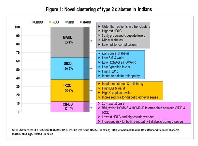
The study was done on 19,084 patients with Type 2 Diabetes using simple clinical parameters which included age at diagnosis, body mass index, waist circumference, glycated hemoglobin, HDL cholesterol, serum triglyceride and fasting and stimulated C-peptide. The clustering was initially performed using data of patients seen at Dr Mohan’s Diabetes Specialities Centres (DMDSC) across the country. Later it was also replicated in a representative sample of the whole of India, through the ICMR-INDIAB study. We found that 4 clusters were present in Indians :Severe Insulin Deficient Diabetes (SIDD), Insulin Resistant Obese Diabetes (IROD), Combined Insulin Resistant and Deficient Diabetes (CIRDD) and Mild Age-Related Diabetes (MARD). The SIDD and the MARD varieties were similar to that described in Scandinavia, although there were some differences here also. The SIDD variety, for example appeared to have more severe insulin deficiency than in Europeons and the MARD variety seemed to develop diabetes at a younger age group than in Europeons. eg, the mean age at the diagnosis of the Scandinavian MARD patients were 67 years compared to 50 years in the Indian population. The characteristics of four subtypes of Type 2 Diabetes in Indians is shown in Fig 15.
Given the differences in phenotype of Type 2 Diabetes, we looked at the clustering of Type 2 Diabetes in Indians, in collaboration with the University of Dundee by taking up the India-Scotland Partnership for Precision Medicine in Diabetes (INSPIRED) project. Specifically, we looked at the type of Type 2 Diabetes clusters in our population5
It was gratifying to note that the clusters were validated in the whole of India through the ICMR-INDIAB population. Subsequently, in another study, the prescribing patterns of treatment at different diabetes centres was looked at and was confirmed in a larger sample size of 32,867 patients that the same four clusters were identified across different clinic populations across India6
(3)A novel finding of our study was that the CIRDD variety was more prone to both retinopathy and nephropathy5
Using the simple clinical characteristics described above, it is possible to make a mental diagnosis of the subtype of diabetes that we are treating, even as the patient walks into our consultation room. For example, if a young, thin individual walks in (and Type 1 and Fibrocalcific Pancreatic Diabetes have been ruled out in them) it is most likely that they have the SIDD variety. If an obese individual walks in, most likely this individual has IROD. If in some of these individuals whom we suspect to have IROD, the HDL cholesterol is very low and the triglyceride levels very high, and the insulin secretion is on the lower side, they have the CIRDD variety. Finally, if an older person, say above 60 years of age, who has just been diagnosed, walks in, most likely this individual has MARD type of diabetes.
What about the Therapeutic Approach to Individuals in these Various Categories ?
Vol 120, No 9, September 2022Journal of the Indian Medical Association 10
The clustering of diabetes has several clinical implications :
Till date we do not have a randomized clinical trial to prove that a particular group of drugs will work better in a particular subtype of Type 2 Diabetes. However, the hypothesis is that the SIDD variety will respond better to insulin secretagogues like sulfonylureas or DPP4 inhibitors, or
More recently, these Indian clusters have also been replicated in South Asians in the UK (Pakistani’s and Bangladeshis) 7. It is notable that the CIRDD variety appears to be unique to South Asians and they also have the lowest HDL cholesterol and highest serum triglycerides among the four subtypes.
(2)With regard to the risk of complications, it was shown that the SIDD variety is more prone to retinopathy and neuropathy, whereas the IROD variety is more prone to nephropathy. These findings were similar to what was reported by the Ahlqvist, et al1.
REFERENCES
Vol 120, No 9, September 2022Journal of the Indian Medical Association 11
We have also recently developed an App called ‘Diabetes Novel subgroup Assessment (DIANA) of we feed in the basic clinical characteristics, the App will tell us which type of Type 2 Diabetes that particular patient is likely to have, ie, SIDD, IROD, CIRDD or MARD. It will also suggest the first line drugs which can be used for that patient. Finally, it will also inform us about the risk of developing the retinopathy or nephropathy within the next five years. The App is just being launched and this could help the clinician to offer individualized or personalized care to diabetes.
5Anjana RM, Baskar V, Nair ATN, Jebarani S, Siddiqui MK, Pradeepa R, et al — Novel subgroups of type 2 diabetes and their association with microvascular outcomes in an Asian Indian population: a data-driven cluster analysis: the INSPIRED study. BMJ Open Diabetes Res Care 2020; 8: e001506.
7Hodgson S, Huang QQ, Sallah N, Griffiths CJ, Newman WG, Trembath RC, et al — Integrating polygenic risk scores in the prediction of type 2 diabetes risk and subtypes in British Pakistanis and Bangladeshis: A population-based cohort study. PLoS Med 2022; 19: e100398.
6Anjana RM, Siddiqui MK, Jebarani S, Vignesh MA, Kamal Raj N, Unnikrishnan R, etal— Prescribing Patterns and Response to Antihyperglycemic Agents Among Novel Clusters of Type 2 Diabetes in Asian Indians.Diabetes Technology & Therapeutics 2022; 24: 190-200.
The era of precision medicine in diabetes has finally dawned. Besides classifying patients into Type 1 or Type 2 Diabetes, or Monogenic forms of diabetes, or other specific forms of diabetes, with the further refinement into subclasses of Type 2 Diabetes, the field is now moving on at a rapid pace.It is hoped that this will set the scene for precision diabetes diagnosis and treatment in India and elsewhere.
1Ahlqvist E, Storm P, Käräjämäki A, Martinell M, Dorkhan M, Carlsson A, et al — Novel subgroups of adult-onset diabetes and their association with outcomes: a data-driven cluster analysis of six variables. Lancet Diabetes Endocrinol 2018; 6: 2Gujral361-9.UP, Pradeepa R, Weber MB, Narayan KM, Mohan V — Type 2 diabetes in South Asians: similarities and differences with white Caucasian and other populations. Ann N Y Acad Sci 2013; 1281: 51-63.
MD, PhD, DSc, Viswanathan Mohan President & Chief of Diabetes Research, Madras Diabetes Research Foundation, ICMR Centre for Advanced Research on Diabetes & Chairman & Chief of Diabetology, Dr Mohan’s Diabetes Specialities Centre, Chennai 600086
3Staimez LR, Weber MB, Ranjani H, Ali MK, Echouffo-Tcheugui JB, Phillips LS, etal—Evidence of Reduced Beta Cell Function in Asian Indians With Mild Dysglycemia. Diabetes Care 2013; 15: 4Sattar315-22.N,Gill JM — Type 2 diabetes in migrant south Asians: mechanisms, mitigation, and management. Lancet Diabetes Endocrinol 2015; 3: 1004-16.
may need insulin early. The IROD variety on the other hand, would respond better to insulin sensitizers, and thus, metformin and SGLT2 drugs would be more suitable. In the CRIDD variety, we can speculate, that they would need an insulin secretagogue as well as a sensitizer. Finally, the MARD variety is the easiest to treat and most likely all that they would need is metformin. This hypothesis is currently being tested at our centre through a randomized clinical trial (CITR No. CTRI/2021/ 11/037753), The control group in each of the subtypes would start with metformin and then go on to one of the other drugs as we conventionally treat now. In the intervention arm in each of the subgroups, the specific drugs based on the pathophysiological defect would be given. This RCT, when completed, should throw more light on whether the classification of Type 2 Diabetes into clusters could translate into better control of diabetes by using the appropriate antidiabetic drugs.
1PhD Student, Department of Bacteriology and Virology, School of Medicine2MD,Faculty Member, Gastroenterohepatology Research Center3MD, Faculty Member, Transplant Research Center
4PhD, Faculty Member, Department of Bacteriology and Virology, School of Medicine and Corresponding Author
Received on : 06/12/2021
The results of the present study indicated the relatively high frequency of amino acid substitutions and deletion, especially in amino acids part of 88-154 from HBx protein in patients with CH and C/HCC.
of 154 Amino acids and manipulates the transcription of cellular and viral genes, signal transduction pathways and Protein degradation; also, it controls the cell cycle and apoptosis6. Protein X is encoded by a gene with 462 nucleotides spanning the overlapping pre-core/core promoter, enhancer II, DR1, and DR2. Therefore, mutation inside the X region not only affects its own functionality, but also might influence other related sequences7. In addition, several critical cis-elements such as microRNA-binding region, EnhII and the core promoter exist in HBx protein. It has been suggested that mutations which occur naturally in various parts of X gene could be associated with different Hepatitis disease statuses8. The reported mutations of X gene could change the function of wild Protein on activation of NF-KB pathway, apoptosis process, P53 interaction and induction of potent responses of T cells9. Moreover, these variations affect viral propagation through affecting
Vol 120, No 9, September 2022Journal of the Indian Medical Association
The relationship between HCC and various mutations of HBx gene has been Suggested.
Shiraz University of Medical Sciences, Shiraz, Iran
Seyed Mohammad Ali Hashemi1, Neda Sanaei1, Mohammad Reza Fattahi2, Seyed Ali Malek- Hosseini3, Seyed Younes Hosseini4, Jamal Sarvari5
12
Editor's Comment :
D
Conclusion : The results of the present study indicated the relatively high frequency of Amino acid substitutions and deletion, especially in part of region 88-154 from HBx Protein in patients with CH and C/HCC. The findings should be considered in a larger population.
Background: Chronic HBV (CH) infection and its consequences including cirrhosis (C) and Hepatocellular Carcinoma (HCC) still represent a major Global health. The relationship between HCC and various mutations of HBx gene has been reported. In the present study, we aimed to determine the sequence variation of HBx gene in patients with Chronic HBV infection or C/HCC.
Materials and Methods : In this cross-sectional study, 15 patients with HBV chronic infection and 13 with C/HCC were included. After viral DNA extraction using commercial kit HBX gene was amplified using an in-house nestedPCR. Then, bi-directional sequencing was performed on the PCR product. The data resulting from sequencing were aligned with reference HBV sequence to identify the mutations.
Original Article
Hepatocarcinogenesis of HBV is mediated by three factors including Chronic inflammation, Integration of viral DNA in the host genome and Oncoproteins encoded by HBV4. Among seven polypeptides encoded by HBV, protein X plays a critical role in Carcinogenesis5. The 16-kD HBx protein is composed
5PhD, Faculty Member, Department of Bacteriology and Virology, School of Medicine and Gastroenterohepatology Research Center, Shiraz University of Medical Sciences, Shiraz, Iran and Corresponding Author
espite availability of an effective Vaccine against Hepatitis B Virus (HBV), about 220 million chronic HBV (CH) infected patients are at risk of the sequel disease including Cirrhosis (C) and Hepatocellular carcinoma (HCC)1. Up to 10 genotypes of HBV have been identified with their own geographic territory; among them, the dominant genotype is D in Iran2,3
Accepted on : 02/03/2022
Investigation of Hepatitis B Virus X Gene Mutations in Patients with and without Cirrhosis/Hepatocellular Carcinoma
Results : The mean age of CH and C/HCC groups was 38.23±12.46 and 50.67±14.22 years old, respectively. We found 43 and 20 Amino acid substitutions inside the region of 88–154 from HBx protein in CH and C/HCC groups, respectively. In addition, K130M+V131I mutation was found in 13.34% (2/15) and 30.7% (4/13) of patients in the CH and C/HCC groups, respectively (P=0.36). Furthermore, 10 deletion mutations were observed in both groups with no significant difference (P=0.8).
Key words :Hepatitis B virus, HBx, Mutations, Cirrhosis, HCC.
[J Indian Med Assoc 2022; 120(9): 12-6]
In this study, 15 out of 23 CH samples and 13 out of 24C/HCC samples had acceptable quality of sequencing data. The mean age of the participants was 38.23±12.46 and 50.67±14.22 years in the CH and C/HCC groups, respectively. Totally, 12 patients (80%) in the CH and 12 (92.3%) in C/HCC groups were male and the rest in each group were female. The level of ALT (P=0.016) and AST (P=0.002) and age (P=0.029) were significantly higher in the C/HCC group than the CH group. Demographic and clinical data for the patients in both groups are shown in Table 1.
In the amplification stage, each PCR mixture at the first round contained 0.5 pmol of each outer primer, 5µL of extracted DNA, 1.5 mM MgCl2, 1U Taq DNA polymerase (CinnagenInc, Tehran, Iran) and 200 mM of each dNTPs (25 µL total volume) and PCR condition: 95°C for 5 min, 28 cycles of 94°C for 35 seconds, 58°C for 45, 72°C for 40 seconds and 72°C for 3 minutes. The second round PCR was performed in a similar amount of PCR mixture and also cycling time parameters with set 2 of primers but the number of cycles was 35 and annealing temperature was 56°C. Sequencing and Multiple Sequences Alignment :
Patient’s Selection :
MATERIALS AND METHODS
Viral DNA Extraction and HBx Gene Amplification:
After purifying the PCR products from the gel electrophoresis by using PCR Product Purification Kit (MN Inc, Germany), bi-directional sequencing was performed using nested internal primers. The results from sequencing were aligned with a group of reference genomic sequences of HBV from data bank by using MEGA7 software to identify the mutations. Every difference between reference sequences and PCR products was considered as mutation.
HBV DNA was extracted from 200 µL of each Serum samples by viral DNA extraction kit (Cinnagen Inc. Tehran, Iran), according to the manufacturer ’s instructions.A nested PCR assay was performed using specific outer and inner primers (Table 2). Primers design was carried out with NCBI homepage primer designing software based on genomic sequences of B, C and D HBV genotypes. Due to overlapping of gene X with precore/core promoter, enhancer II, DR1 and DR2, the primers were designed in such a way that they amplified both X gene and these overlapping regions.
regulatory sequences of the genome which directs the disease toward Cirrhosis and HCC10,11. Moreover, deletions and insertions in C-terminal region of HBx can be related to clinical output and disease severity in patients suffering from Chronic Hepatitis. Therefore, these mutations which might be related to C/HCC can be used as biomarkers to predict disease progression12. The present study was conducted to investigate the sequence variations of HBx gene in chronic HBV infection and C/HCC patients.
13
Vol 120, No 9, September 2022Journal of the Indian Medical Association
In this study, 47 subjects including those with Chronic Hepatitis (CH) as well as C/HCC were enrolled consecutively from Gastroentrohepatology Unit at Mottahari Clinic and Liver Transplant Research Centers at referral Abu-Ali Sina Hospital, affiliated to Shiraz University of Medical Sciences, from 2013 to 2016. All the enrolled patients had Chronic HBV infection and were divided into CH and C/HCC groups by a Liver Specialist according to Biochemical, Virological and clinical records based on EASL guidelines 13 . Demographic and Clinical data of patients were collected using Medical Records. The CH group consisted of chronic patients who were positive for HBsAg and positive/negative for HBeAg. The C/HCC patients were enrolled based on Ultrasound Scanning, histology grading, Abnormal Liver Function Tests, and α-fetoprotein levels. All the patients were negative for HCV, HDV and HIV. Based on the sequencing of preS1, S2 and S region of these samples, all patients were infected with HBV genotype D14,15. Written consent was obtained from each patient before sampling and the study was approved by the Ethics Committee of Shiraz University of Medical Sciences. Five milliliter of venous blood without anti-coagulation was taken from each participant. The sera were separated by centrifuge and stored at -20oC until used.
Epi Info™ software and SPSS 22 were used for statistical analysis. Chi square test was used for analysis of the mutation data and P value <0.05 was considered significant. Age, ALT, AST were expressed as Mean ± SD.
RESULTS
In the case of detected mutations, in total, 53 and 25 substitutions were detected in the X Protein sequence of CH and C/HCC groups, respectively. However, no significant difference was determined. Also, 43 and 20 Amino acid substitutions were found in Amino acids sequence 88–154 of HBx Protein in the CH and C/HCC groups, respectively. Moreover, 13.34% (2/15) of the patients in the CH group and 30.7% (4/13) of those in the C/HCC group had K130M+V131I double mutation (p=0.36). Additionally, we also showed K130N (n=2), K130Y (n=1) and K130C (n=1), V131L (n=3) mutations in the CH group and K130Q (n=1) and V131L
Statistical Analysis :
Amino acids from 52-65 and 88-154 in HBx protein have a key role in the transactivation, transcription and replication of the HBV genome17. In this study, we detected 43 and 20 Amino acid substitutions in 88154 region of HBx Protein in the CH and C/HCC groups, respectively. In addition, R56L(1), S65P(1) and Del4772(3) mutations were found in 52-65 region of HBx in the C/HCC group and L58H(1) and Del47-72(1) Amino acid substitutions were found in the CH group. Mani, et al reported H52Y and S64T from patients suffering from Chronic Liver Disease. In addition, they observed T36A mutation in 4 participants with Chronic LiverMoreover,Disease1813.34% (2/15) of patients
AST*±SD 31.58±21.29130.9±106 0.002
Vol 120, No 9, September 2022Journal of the Indian Medical Association
Gender :
DISCUSSION
Table1 — Demographic and clinical data of the study groups
HCC ranks the sixth among the most prevalent cancers, represents the third cancer-related death across the world and is mainly caused by HBV16 Mutation in HBx, especially in the COOH-terminal region, has been suggested to direct the disease progression toward HCC. Thus, investigation of HBx mutations can lead to confirming the predictors of endstage Liver disease by HBV.
Mean Age±SD 38.23±12.4650.67±14.22 0.029
(n=2) mutations in the C/HCC group. Furthermore, H94Y(3), I127N(n=1)/T(n=2)/F(n=1), F132Y(n=3) mutations in the CH group, H94Y(n=2), I127L/T/S(5) and F132Y(n=1) mutations in the C/HCC group were observed. Interestingly, 10 deletion mutations were determined in each group albeit no statistically significant difference was seen. These mutations included single deletion at position 135 in the CH group and 101 in the C/HCC group. Moreover, there were some partial deletions at the 48-72 and the C-termini of X Protein region; they included Del47-72(1), Del77end (1), Del130-133(3), Del 129-131(1), Del 130-132(1) in the CH group and Del47-72(2), Del77-end (2), Del76end (1), Del47-72(1), Del74-152(1), Del74-149 and Del130-133(2) in the C/HCC group. However, based on the deleted residues and blocks, any special difference was extrapolated when comparing the two groups (Table 3).
ALT*±SD 47.08±43.93119.2±98.73 0.016
ALT: Alanine Aminotransferase;
AST: Aspartate Aminotransferase; CH: Chronic Hepatitis; C/HCC: Cirrhosis/Hepatocellar Carcinoma Table 2 — The sequences of primers used in the nested PCR Sequence Position Product Size HBx Forward 1 5'-CGATCCATACTGCGGAACT-3' 1262-1946 685 bp HBx Reverse 1 5'-GTAACTCCACAGWAGCTCCA-3' HBx Forward 2 5'-GCTTGYTTTGCTCGCAG-3' 1288-1886 599 bp HBx Reverse 25'-CAAGGCACAGCTTGGAG-3' Table 3 — The frequency of HBx variations in the CH and C/ HCC groups CH(n=13) C/HCC(n=15) P value Substitution M1T(1)-mutation 0.48 C6G (1)- 0.48 P11H(1),CH:DelDelDelDel47-72(1),-I283L(1)T152I(1)-N149H(2)-C148W(2)C148W(1)P147L(2)-A146E(1),A144P(2),K140M(1),-H139A(1),C137Y(1),-0.48G135E(1),-V133I(1)-0.48F132Y(3),F132Y(1)0.29V131L(3),K130N(2),R128I(1),-I127N(1),E122D(1),E122D(1),E121G(4)E121G(1)D114Q(1),-F112V(2)F112V(1)0.47G107D(1),-L98I(1),-R96W(1),-H94Y(2),H94Y(2)0.65F88L(1),-Q87P(1)R78S(1),--R72C(1)-S65P(1)L58H(1),--R56L(1),S41Y(2),S41Y(1),S38A(2),S31P(1),-H30Y(1),-0.48D14N(1),-A12T(1),-0.480.480.480.48H30Y(1),S38A(1),0.290.470.550.480.550.550.480.55F88S(1),F88S(2),F88L(1)0.590.480.480.480.480.330.72I127T(2),I127L(1),I127T(3),I127F(1),I127S(1)0.610.48K130Y(1),K130M(5),K130Q(1)K130M(2),K130C(1),0.83V131I(1)V131I(4),V131L(2)0.720.48R138T(1)0.55H139D(1),-0.240.48A144T(2)0.65A146P(1),A146T(1),-0.120.240.470.240.480.55DeletionMutationDel77-end(1),Del47-72(2),Del77-end(2),0.8101(2),Del130-133(3),Del76-end(1),Del47-72(1),129-131(1),Del74-152(1),Del74-149,130-132(1),Del135(1)Del101(1),Del130-133(2)ChronicHepatitis;C/HCC:Cirrhosis/HepatocellarCarcinoma 14
Male 12(80%)12 (92.3%)Female 3(20%)1 (7.7%)
CharacteristicsCH group C/HCC group P value (N=15)(N=13)
In spite of mutations in the D and E domain including H94Y(3), I127N/T/F(n=4) and F132Y(n=3) in the CH group and H94Y(n=2), I127L/T/S(n=5), and F132Y(n=1) in the C/HCC group, there was not any correlation between the groups and mutations. D and E functional domains in HBx Protein are associated with nuclear transactivation, signal transduction as mutations in these domains may be modulating its transactivation property21. It has been reported that H94Y, I127T, K130M, V131I and F132Y/I/R mutations that are located in the D and E domains might be related to modulation of HBx transactivation property 21 . In addition, I127T+K130M+V131I triple mutation was reported with progression of Liver Disease21
This study also demonstrated that 10 small and partial deletions of HBx Protein in both groups separately. There were some deletions in the C-termini of X protein region including Del 47-72(1), Del 77-end (1), Del 130-133(3), Del 129-131(1) and Del 130-132(1) in the CH group and Del 47-72(2), Del 77-end (2), Del 76-end (1), Del 47-72(1), Del 74-152(1), Del 74-149 and Del 130-133(2) in the C/HCC group. Deletion in the COOH-terminal of HBx is a frequent event in Hepatocellular Carcinoma 23 . Some studies have reported X gene containing different deletions in the COOH-terminal region of HCC patients24,25. Al-Anazi et al reported that there was evidence for an effect of deletion mutation in HBx on cell cycle regulators24 They showed that HBx-WT enhanced modulation of p21, p27 and cyclin D1, whereas truncated forms of HBx (61-124) inhibited p53 expression significantly. Similarly, truncated forms including HBx (1-94) and
The present study showed that amino acid substitutions in the HBV-HBx gene, especially in amino acids 88-154, are frequent in patients with CH and C/ HCC. These mutations might be related to HBVassociated liver injury and progression of infection. However, a more detailed study on a larger population of HBV-infected patients is recommended to confirm this claim.
15
Mutations have various biological outcomes, and based on the mutation type, they play different roles in the HBV infection outcome. Moreover, some of these mutations can be used as prognosis and predict the outcome of the disease. Relatively small sample size as well as lack of available data regarding the viral replication parameters could be the limitations of this study.
et al reported that HCC-related mutations mainly resided in the HBx transactivation domain, immune epitopes, viral promoter and protein/ miRNA binding sites29. In B cell epitope region including aa36, aa44 and aa50, we did not detect any mutation. Xie et al. reported that T36P/S/A mutation in the B cell epitope was not significantly higher in HCC than non-HCC patients infected with genotype A/C/D. Muroyama, et al and Cho, et al reported that A44V and G50R were significantly higher in the HCC group than non-HCC (genotype A/D). Also, in aa118 and aa 123 related BH3-like motif, core promoter and EnhII, NRE region we did not detect any mutation; However, in aa127 related BH3-like motif, core promoter and NRE regions we reported 4 and 5 mutations in CH and C/HCC, respectively. Fan, et al reported L123S and a silent mutation in aa118 that were significantly higher in the HCC group than non-HCC ones (genotype D1)
Vol 120, No 9, September 2022Journal of the Indian Medical Association
CONCLUSION
in the CH group and 30.7% (4/13) in the C/HCC group had K130M+V131I double mutation. Additionally, we found K130Y/N/C/L (n=7) mutations in the CH group and K130Q/L(n=3) in the C/HCC group. In the same line, Mani, et al showed that 27% of patients in different stages of HBV infection had (K130M+V131I) double mutation18. Double mutation (K130M+V131I) affects the cell cycle regulation, DNA repairing mechanism, and HBeAg expression19. In this regard, Shi, et al for the first time reported HBx10-144 double mutation that may be involved in progression toward HCC20. 130 and 131 sites in HBx overlap with A1762T and G1764A sites in the basal core promoter, which are common substitutions in HCC21. Liu, et al reported that K130M/V131I mutation promoted transcription activity of hypoxia-inducible factor-1 (HIF-1) which is enhanced in human tumors22. It is believed that the interaction of HIF-1 and HBx is caused by formation of a stronger secondary structure in HBx as a result of this double mutation22
HBx (61-154) suppressed the expression of PARP and Bax efficiently. Fu, etalreported that HBx-d382 deletion mutant (128-145aa) enhanced the cell proliferation26
The author found out that C-terminal truncations and deletion mutations, in contrast, attenuated the HBx ability to promote transcription activity of HIF-122. In the present study, we did not find any difference between the frequencies of deletion mutations in Cterminal of HBx in C/HCC patients in comparison with CH patients that may be related to sample size. Salarnia, et al reported that deletion and insertion mutations in C-terminal of HBx were more frequent in cirrhotic patients compared to chronic HBV patients27. Recently, it was reported that C-terminal truncated HBx by downregulating TXNIP initiated hepatocarcinogenesis28MoreoverLi,
8Kim H, Lee S-A, Kim B-J — X region mutations of hepatitis B virus related to clinical severity. World journal of gastroenterology 2016; 22(24): 5467.
11Buckwold1239-44.
The present study was financially supported by Shiraz University of Medical Sciences (grant no: 903259)
15Taghiabadi M, Hosseini SY, Gorzin AA, Taghavi SA, Monavari SHR, Sarvari J — Comparison of pre-S1/S2 variations of hepatitis B virus between asymptomatic carriers and cirrhotic/ hepatocellular carcinoma-affected individuals. Clinical and experimental hepatology 2019; 5(2): 161.
29Li W, Goto K, Matsubara Y, Ito S, Muroyama R, Li Q, et al — The characteristic changes in hepatitis B virus x region for hepatocellular carcinoma: a comprehensive analysis based on global data. PloS one 2015; 10(5): e0125555.
ACKNOWLEDGMENTS
14Hosseini SY, Sanaei N, Fattahi M-R, Malek-Hosseini SA, Sarvari J — Association of HBsAg mutation patterns with hepatitis B infection outcome: Asymptomatic carriers versus HCC/ cirrhotic patients. Annals of hepatology 2019; 18(4): 640-5.
9Malmassari SL, Deng Q, Fontaine H, Houitte D, Rimlinger F, Thiers V, etal— Impact of hepatitis B virus basic core promoter mutations on T cell response to an immunodominant HBx derived epitope. Hepatology 2007; 45(5): 1199-209.
10Li J, Buckwold VE, Hon M-w, Ou J-h — Mechanism of suppression of hepatitis B virus precore RNA transcription by a frequent double mutation. Journalofvirology 1999; 73(2):
17Tang H, Delgermaa L, Huang F, Oishi N, Liu L, He F, et al — The transcriptional transactivation function of HBx protein is important for its augmentation role in hepatitis B virus replication. Journal of Virology 2005; 79(9): 5548-56.
4Berasain C, Castillo J, Perugorria M, Latasa M, Prieto J, Avila M — Inflammation and liver cancer: new molecular links. Annals of the NewYorkAcademy of Sciences 2009; 1155(1): 5Bréchot206-21.
C, Gozuacik D, Murakami Y, Paterlini-Bréchot P, editors. Molecular bases for the development of hepatitis B virus (HBV)-related hepatocellular carcinoma (HCC). Seminars in cancer biology; 2000: Elsevier.
6Murakami S. Hepatitis B virus X protein: a multifunctional viral regulator. Journal of gastroenterology 2001; 36(10): 651-60.
19Lin X, Xu X, Huang Q-L, Liu Y-Q, Zheng D-L, Chen W-N, et al— Biological impacts of “hot-spot” mutations of hepatitis B virus X proteins are genotype B and C differentiated. World journal of gastroenterology: WJG 2005; 11(30): 4703.
23Liu X-H, Lin J, Zhang S-H, Zhang S-M, Feitelson MA, Gao HJ, et al — COOH-terminal deletion of HBx gene is a frequent event in HBV-associated hepatocellular carcinoma. World journal of gastroenterology: WJG. 2008; 14(9): 1346.
27Salarnia F, Besharat S, Zhand S, Javid N, Khodabakhshi B, Moradi A — Mutations in Hepatitis-B X-Gene Region: Chronic Hepatitis-B versus Cirrhosis. Journal of clinical and diagnostic research: JCDR 2017; 11(3): OC31.
16
20Shi Y, Wang J, Wang Y, Wang A, Guo H, Wei F, et al — A novel mutant 10Ala/Arg together with mutant 144Ser/Arg of hepatitis B virus X protein involved in hepatitis B virus-related hepatocarcinogenesis in HepG2 cell lines. Cancer letters 2016; 371(2): 285-91.
3Pujol FH, Navas M-C, Hainaut P, Chemin I — Worldwide genetic diversity of HBV genotypes and risk of hepatocellular carcinoma. Cancer letters 2009; 286(1): 80-8.
1Chen GF, Wang C, Lau G — Treatment of chronic hepatitis B infection 2017. Liver International 2017; 37(S1): 59-66.
7Rajput MK. Mutations and methods of analysis of mutations in Hepatitis B virus. AIMS microbiology 2020; 6(4): 401.
VE, Xu Z, Chen M, Yen T, Ou J-h — Effects of a naturally occurring mutation in the hepatitis B virus basal core promoter on precore gene expression and viral replication. Journal of virology 1996; 70(9): 5845-51.
24Al-Anazi MR, Nazir N, Colak D, Al-Ahdal MN, Al-Qahtani AA — Deletion and functional analysis of hepatitis B virus X protein: evidence for an effect on cell cycle regulators. Cellular Physiology and Biochemistry 2018; 49(5): 1987-98.
28Zhang Y, Yan Q, Gong L, Xu H, Liu B, Fang X, et al — Cterminal truncated HBx initiates hepatocarcinogenesis by downregulating TXNIP and reprogramming glucose metabolism. Oncogene 2021; 40(6): 1147-61.
REFERENCES
12Zhu Pa, Tan D, Peng Z, Liu F, Song L — Polymorphism analyses of hepatitis B virus X gene in hepatocellular carcinoma patients from southern China. Acta biochimica et biophysica Sinica 2007; 39(4): 265-72.
18Mani M, Vijayaraghavan S, Sarangan G, Barani R, Abraham P, Srikanth P — Hepatitis B virus X protein: The X factor in chronic hepatitis B virus disease progression. Indian journal of Medical Microbiology 2019; 37(3): 387-92.
25Fu X, Tan D, Hou Z, Hu Z, Liu G, Ouyang Y, et al— The effect of miR-338-3p on HBx deletion-mutant (HBx-d382) mediated liver-cell proliferation through CyclinD1 regulation. 2012.
21Barbini L, Tadey L, Fernandez S, Bouzas B, Campos R — Molecular characterization of hepatitis B virus X gene in chronic hepatitis B patients. Virology journal 2012; 9(1): 1-7.
Conflicts of interest : All the authors declared that there is no conflict of interest.
Vol 120, No 9, September 2022Journal of the Indian Medical Association
2Amini-Bavil-Olyaee S, Hosseini SY, Sabahi F, Alavian S-M — Hepatitis B virus (HBV) genotype and YMDD motif mutation profile among patients infected with HBV and untreated with lamivudine. International Journal of Infectious Diseases 2008; 12(1): 83-7.
13Liver EAFTSOT. EASL clinical practice guidelines: management of chronic hepatitis B virus infection. Journal of hepatology 2012; 57(1): 167-85.
16Niu B, Hann H-W — Hepatitis B virus–related hepatocellular carcinoma: carcinogenesis, prevention, and treatment. Updates in Liver Cancer 2017; 13.
26Fu X, Tan D, Hou Z, Hu Z, Liu G, Ouyang Y, et al — The effect of miR-338-3p on HBx deletion-mutant (HBx-d382) mediated liver-cell proliferation through CyclinD1 regulation. PLoS One 2012; 7(8): e43204.
22Liu L, Hu B, Ye C, Ho R, Chen G, Lai P — HBx mutants differentially affect the activation of hypoxia-inducible factor1á in hepatocellular carcinoma. British Journal of Cancer 2014; 110(4): 1066.
B
complications, duration of drain and duration of stay at the hospital. Also the diabetic control pre operatively plays a role in these associations. Also the breast cancer surgery in turn can have a beneficial effect on the control of sugars in the post operatIve period. Objectives of the Study : (1) To compare the rate of postoperative complications in Diabetic and non-diabetic patients undergoing breast cancer surgeries (2) To compare the in hospital outcomes (drain duration, inpatient days, stage at presentation) in Diabetic and non-diabetic patients
Received on : 31/01/2022
Higher morbidity is observed in diabetic patients. However these morbidities are lesser in patients with well controlled diabetes.
[J Indian Med Assoc 2022; 120(9): 17-9]
Methods : Data will be collected from the in patient files and the surgery case completion register in the Medical records department of hospitals attached to Bangalore Medical College and the data will be retrospectively scrutinized for a period of 3 years from 01/09/2018 to 30/08/2021. Patients diagnosed with breast cancer and planned for surgical management of the same were included in the study.
Key words :Breast Cancer, Diabetes Mellitus, Postoperative period.
2MBBS, MS, Head of the Department
Aims And Objectives : (1) To compare the rate of postoperative complications in Diabetic and non-diabetic patients undergoing breast cancer surgeries. (2) To compare the in hospital outcomes in diabetic and non diabetic patients. (3) To study the pre and Postoperative control of sugars in Diabetic patients undergoing breast cancer surgeries.
Editor's Comment :
Results : A total of 100 breast cancer patients were studied. 34 were Diabetic of which 16 had uncontrolled sugars pre operatively. Out of which only 5 patients remained to have uncontrolled sugars Postoperatively. A total of 27 patients developed Postoperative complications. 13 were Diabetic of which 9 had uncontrolled sugars. Of the diabetic patients a total 27 had a drain duration of more than 7 days.
The Effect of Diabetes Mellitus on the Postoperative Period in Breast Cancer Patients — A Three Years Retrospective Study
Background : Breast cancer is the most common type of cancer amongst women. Amongst the many factors which affect the outcome of breast cancer surgeries is Diabetes Mellitus. Diabetes Mellitus is an ongoing problem Worldwide. It is a predictor for Postoperative complications in women who undergo surgical management for breast cancer.
Original Article
Accepted on : 21/02/2022
1MBBS, MS, Associate Professor
Breast cancer is the most common cancer amongst women. Diabetes mellitus being a lifestyle disease has become a burden in the modern world.
When these both unfortunately are present in an individual, the Postoperative period can be different from a non diabetic breast cancer patient.
17
Department of General Surgery, Bangalore Medical College and Research Institute, Karnataka 560002
3MBBS, Postgraduate Trainee and Corresponding Author
Conclusion : Diabetic women who undergo breast cancer surgeries at an increased risk of complications post operatively, more so if they have uncontrolled sugars. In hospital outcomes too are different in patients with Diabetes as in the duration of drain thereby the total in patient days were higher in patients with diabetes. Also patients with uncontrolled sugars saw an improvement in their sugars postoperatively.
Manjunath Byadigere1, Harindranath H R2, Nagashri Suresh Iyer3
Vol 120, No 9, September 2022Journal of the Indian Medical Association
reast cancer is the most common type of cancer amongst women. It is also one of the major cause of death and morbidity Worldwide4,5 Surgical management like Breast Conservative Surgery and Mastectomy, are the mainstay of treatment of breast cancer alongside hormonal, chemotherapy and radiotherapy. These surgeries come along with a variety of complications associated with them. These complications can be infective and non infective thereby affecting the inpatient stay of the patient.Diabetes is a leading cause of morbidity worldwide. Diabetes being a lifestyle disease and also a part of the Syndrome X, when associated with breast cancer can have a varying effects on the outcomes post operatively such as infective and non infective
Vol 120, No 9, September 2022Journal of the Indian Medical Association
The data collected was entered into excel sheet and was analysed using SPSS 27.0 Grad pack. The data regarding the data thus compiled was statistically represented using mean, Standard Deviation, frequency and percentage using chi square test. The variables included in the study were age, comorbidities like Hypertension, Diabetes and Hypothyroidism, complications like seroma, Flap necrosis and Surgical Site Infections (SSI), drain duration, inpatient days, stage at presentation and recurrence. Also the diabetic control was compared pre-operatively and post operatively. A p value of <0.05 was considered to be significant.
Statistical analysis :
RESULTS
A total of 100 breast cancer patients who had underwent Mastectomy in the study period were
1 — Presence of uncontrolled sugars Fig 2 — Drain duration in diabetic patients Fig 3 — Stage at presentation Table 1 — Age AgeFrequencydistribution Percent < or equal to 40 years18 18.0 40-59 years5454.0 60-79 years2727.0 > or equal to Total100100.08011.0Table2—DistributionaccordingtothepresenceofcomorbiditiesDiabetesHypertensionHypothyroidismPresent34262Absent667498Total100100100Table3—DistributionaccordingtotheoccurrenceofcomplicationsComplicationsNonDiabetic14ControlledDm4UncontrolledDm9 18


MATERIALS AND METHODS
Fig
We performed a retrospective observational study by collecting data from the inpatient files and case completion registers in the Medical Records Department of Hospitals attached to Bangalore Medical College and the data was retrospectively scrutinized for a period of 3 years from 01/09/2018 to 30/08/2021
(3) To study the pre and postoperative control of sugars in Diabetic patients undergoing breast cancer surgeries.
Inclusion criteria were set to identify the patients diagnosed with breast cancer who were undergoing surgical management. Then the data were analysed to identify the associated co morbidities, days of stay at hospital, stage at presentation, drain duration, inpatient days, Postoperative complications and pre and post surgical control of sugars in case of Diabetic patients. The results were compiled and analysed and compared with regard to patients with and without DiabetesThestudy was approved by our Institute Ethics Committee and 100 people >18 years of age, who had undergone surgery for breast cancer were included in theAstudyGRBS of >140 was considered as uncontrolled sugars. The inpatient days were divided into less than or equal to 15 days. Drain duration was analysed as less than or equal to 7 days. The Postoperative complications were divided into infectious complications like surgical site infections and non infectious complications like seroma and flap necrosis and compared with the Diabetic and non-diabetic counterparts.

Diabetic women who undergo breast cancer surgeries at an increased risk of complications post operatively, more so if they have uncontrolled sugars. In hospital outcomes too are different in patients with Diabetes as compared to their non-diabetic counterparts.Patients with Diabetes usually have other comorbidities. In our study they were associated with other comorbidities like Hypertension and Hypothyroidism, thus adding on to the morbidity. Similar results were seen in the study conducted by Lopez-de-Andres A, et al1
Of the Diabetic patients 16 patients had uncontrolled sugars pre-operatively whereas only 5 patients remained to have uncontrolled sugars post operativelyAtotal of 27 patients developed Post-operative complications like Seroma, Flap necrosis, Surgical site Infections of which 13 were diabetic. 9 of these 13 patients had uncontrolled sugars pre-operatively. Relationship between Diabetes and Complications was significant (p = 0.020); p <0.05 is significant
39 patients presented at an early stage of which 10 were Diabetic. 5 patients presented with metastasis of which 3 were Diabetic.
Of the Diabetic patients a total 27 had Drain duration of more than 7 days of which 9 patients had uncontrolled pre-operative sugars. Diabetes and Drain duration was significant (p=0.003); p<0.05 is significant
Of the Diabetic patients 17 had a inpatient stay of >15 days and 17 had an in patient stay of <15 days.
With the inclusion criteria being more than 18 years, patients studied belonged to ages between 32 years and 87 years, Mean age being 50 years
DISCUSSION 2,3
Occurring in 1 out of 4 women breast cancer is most common in Indian women6
The Postoperative complications in patients with
The duration of Drain insitu too was significantly higher in pre-operatively uncontrolled Diabetes patients.Butthe relation of stage at presentation, duration of stay at hospital and recurrence with diabetes status of the patient showed no significant relation in our study.Hence the presence of Diabetes acts as an additional factor to the morbidity in post mastectomy patients.
Through this study we conclude that diabetic patients with breast cancer usually have other associated comorbidities and also have an increased risk of Postoperative complications, more so if the diabetes is not well controlled. Thus having other significant in hospital stay outcomes compared to their non-diabetic counterparts.
1Lopez-de-Andres A, Jimenez-Trujillo I, Hernandez-Barrera V, et al — Association of type 2 diabetes with in hospital complications among women undergoing breast cancer surgical procedures. A retrospective study using the Spanish National Hospital Discharge Database, 2013-2014.
2Ferroni2017;7:e017676.doi:10.1136/bmjopen-2017-017676BMJOpenP,RiondinoS,BuonokaO,PalmirottaR,GuadagniF,RoselliM—2015.Type2DiabetesandBreastcancer:TheinterplaybetweenImpairedGlucosemetabolismandOxidantstress.OxidativeMedicineandCellularLongevity2015;pp1-10.3LarssonS,MantzorosC,WolkA—Diabetesmellitusandriskofbreastcancer:Ameta-analysis.InternationaljournalofCancer2007; 121(4): pp.856-62.
4Peairs K, Barone B, Snyder C, Yeh H, Stein K, Derr R, Brancati F, Wolff A — Diabetes mellitus and Breast cancer outcomes: A systematic review and meta-analysis. Journal of clinical oncology 2011; 29(1): pp.40-6.
6Panda S, Chakrabarti S, Chakraborty J, Bhattacharyya R — Correlation between Her2Neu status with molecular classification, Cyclin D1 status and Ki67 expression in intraductal carcinoma of the breast. Journal of Indian Medical Association 2021; 119(4): 29-33.
Vol 120, No 9, September 2022Journal of the Indian Medical Association
SSI01Seroma89Necrosis63
19
5Sainsbury R — The Breast. Bailey and Love’s short practice of surgery 2018; 27: 871-8.
REFERENCES
CONCLUSION
And only 1 patient presented with recurrent Carcinoma breast who was non Diabetic.
Table 4 — Distribution according to various complications Complications Non Diabetic Diabetic Flap
uncontrolled Diabetes were significantly higher than in those with non diabetic patients. Also our study shows no significant increase in the risk of complications in diabetic patients whose sugar levels were under control. Thus signifying the importance of the maintenance of good Diabetic Control. Similar results showing that both infective and non infective complications were higher in diabetic patients was seen in the study by Lopez-de-Andres A et al1
studied and analysed (Tables 1-4 & Figs 1-3).
Of the total patients, 34 patients had Diabetes, 26 were Hypertensive and 2 had Hypothyroidism.
We undertook a prospective observational study between June 2019 to June 2020 at NICU of Silchar Medical College and Hospital.Written consent was taken from the parents of the participating Neonates. Institutional Ethical Clearance was also taken. Neonates were included within the study were term, weight more than 2 kgs, not receiving any pharmacological drug for pain relief. All sick babies including neurologically unstable and on Ventilator support were excluded.
Accepted on : 28/07/2022
Anupama Deka1, Gourav Das2, Sanjib Kumar Debnath3
Vol 120, No 9, September 2022Journal of the Indian Medical Association
20
Result : The duration of cry was found to be higher in the group receiving EBM. The neonates in 25%D groups had lower Neonatal Infant Pain Scale (NIPS) score than EBM group (chi-sqr-10.34 & p-0.0057).
1DCH, MD, Professor, Department of Paediatrics, Gauhati Medical College and Hospital, Guwahati 781006
A Study Comparing the Efficacy of Different Non-pharmacological Methods to Reduce Pain in Neonates Admitted in A Tertiary Care Hospital
2MD, Paediatrician, Life Line Hospital and Research Centre, Karimganj, Assam 788710
Aim : To compare the efficacy of different non-pharmacological methods for reducing pain in Neonates.
[J Indian Med Assoc 2022; 120(9): 20-2]
Received on : 21/07/2022
For each Neonate, a pre-structured proforma was filled out, including the outcome variables used in our study as well as other pertinent information such as the mother’s name, birth weight, sex, gender and the
3MD, Assistant Professor, Department of Pediatrics, Silchar Medical College & Hospital, Silchar 788014 and Corresponding
Procedural Analgesia in newborn is often an ignored and underutilized area.
Author
eonates receiving medical care are subjected to multiple painful procedures as a part of their medical management. Neuroendocrine Systems and neuroanatomic components of the neonate are sufficiently developed to permit transmission of painful stimuli1. As premature and full-term infants experience pain there is growing awareness to the fact that stress and discomfort in hospitalised infants is largely undertreated. As pre-natal pain and stress may alter neurodevelopment and later perceptions of painful stimuli and behavioural responses, prevention and control of pain likely to benefit infant.
Methodology : During the study period of one year from July, 2019 to July, 2020, a total of 70 infants were consecutively recruited and divided into two groups. One group received 2ml of EBM and other group 2ml of 25% D is administered which was given 1 minute before Venepuncture. The outcome variables are the duration of cry after Venepuncture & NIPS score for both group.
Original Article
Editor's Comment :
Key words :Expressed Breast Milk (EBM), 25%D- 25% Dextrose, Neonatal Infant Pain Score (NIPS).
25% Dextrose is a readily available effective Analgesic and should be used before any painful procedure in newborn. Proper analgesia and prevention of pain in neonatal period has better neurodevelopmental outcome in future.
N
Conclusion : In our study we found 25% Dextrose to be a better non-pharmacological Analgesic as compared to EBM during painful procedure in newborn.
As part of their intensive care, infants frequently require investigations and procedures,painful situation are quite common. Heel lance and Venepuncture are examples of several such painful procedures.There are pharmacological therapies available. Pharmacological treatments are often not used during these procedures due to apprehension of side effects and attending physician’s ignorance. Non-pharmacological interventions are valuable alternatives. These include use of non-nutritive sucking2, oral sucrose or Glucose solution 2,3 , Kangaroo Mother Care 4,5 and
breastfeeding6,7. Measurement of pain can be tricky in neonates, however, several tools or pain scales are there to assessment of pain in infants. The Neonatal Infant Pain Scale (NIPS), for example, has been validated as a reliable tool for measuring pain during various procedure in newborns8,9. Our objective is to compare the efficacy of non-pharmacological method in reducing pain during painful procedure.
MATERIALS AND METHODS
Before the study one volunteer was trained in the assessment of NIPS score. We assessed pain by using NIPS during Venepuncture .The NIPS is a scale for assessment of pain during procedures in Neonates and was developed in 1993 by Lawrence, et al10. NIPS includes some behavioural parameters such as (facial expression, crying and movement of arms/legs) and two clinical indicators (state of arousal and breathing pattern). The maximum possible score is 7 and minimum score is 2. The NIPS is usually divided into mild pain (1-2/7), moderate (3-4/7) and severe pain (57/7). The duration of the cry was timed from the instant of Venepuncture till cessation of the cry. If the crying persisted for >180 sec, it had been simply recorded as duration >180 sec. The data was analysed using SPSS. The Fisher’s Exact Test and Chi-square tests were done as test of significance between two groups.
The mean NIPS score in EBM group (3.86±1.7) and in 25% Dextrose group (3.05±1.50). A total of 6 babies in EBM group experienced mild pain, 16 babies had NIPS score of moderate pain and 13 babies had severe NIPS score. 8 babies in 25 Dextrose group NIPS score of mild pain, 25 babies of moderate NIPS score and 2 babies of severe NIPS score. When the NIPS score was compared between two group a highly statistically significant result was found in favour of 25% Dextrose group (Chi sq=10.32, p=0.0057).
Vol 120, No 9, September 2022Journal of the Indian Medical Association
RESULT
DISCUSSION
Table 1 — Distribution of Descriptive Characteristics of EBM25%Chi-Neonates p-value Dextrosesqr SEXMale: 20(57%)18(51%)0.05 P=0.810 Female 15(42%)17(48%) Gestational Age : <36 Weeks 24(68%)27(77%)0.28P= 0.591 36-38 Weeks11(31%)8(22%) Delivery Mode Caesareans18(51.4%): 17(48.6%)0.00 P=0.99 Normal 17(48.6%)18(51.4%) Postnatal age1.29±1.3221.09±1,12Z=0.57 P=0.569 Table 2 — Duration of Cry in Two Study Group DurationEBM25 DextroseTest ValueP Value 0-1 minute1822 1-2 minutes109 2-3 minutes32 >3 Mean±SD1.65±0.92minutes421.31±0.79Z= 1.48 P=0.138 Table 3 — NIPS Score of Two Subgroup NIPSEBM 25% DextroseTest ValueP Value Mild68 Chi-sq=10.32 P=0.0057 Severe132Moderate1625 21
In the EBM group 18 babies cried until 1 min,10
babies ceased to cry within 1-2minutes, 3 babies cried within 2-3 minutes and 4 babies cried after 3 minutes. Mean±SD of cry in EBM group is 1.65±0.92. Likewise in the 25% Dextrose group 22 babies didn’t cry at all or cease to cry within 1 minute, 9 babies ceased cry within 1-2 minutes, 2 babies in 2-3minutes and 2 babies in more than 3 minutes mean±SD of cry in 25 dextrose group is 1.31±0.79. The Mann-whitney U test doesn’t reveal any statistically significant difference between EBM and 25% Dextrose group (p= 0.138) (Table 2).
gestational age. Total 70 eligible babies were randomly assigned to either the EBM or the 25D groups at delivery. 2 ml of Expressed Breast Milk was drawn up in a sterile dropper by a Nurse before the procedure (for the EBM group)and for Infants in the 25D group, 2 ml of commercially available 25D was used.
The study population consist of 70 babies who were equally divided in two groups ie, 35 in each. In the EBM group 20(57.14%) were male and 15(42.8%) babies were female. Term babies were 24(68.3%) and preterm babies were 11(31.3%). In the 25% Dextrose group 18(51.4%) babies are male and 17(48.5%) babies are female, 27(77.1%) term and 8(22.9%) are preterm. The distribution of the data on the sex, gestation week, postnatal age and type of delivery of the newborns in the study and a comparison of these characteristics has been provided in Table 1. Both groups were similar and there was no statistical difference (p>0.05).
Procedural pain has been reduced using a variety of non-pharmacological approaches. Our study result suggests that 25% Dextrose is more Analgesic than EBM.This result is evident from lower pain score obtained in Neonates receiving 25% Dextrose during Venepuncture. A study conducted by Stevene B, et al11 showed that Sucrose is effective for reducing procedural pain from single events like heel lance, venepuncture and intramuscular injection in both preterm and term Neonates. Another study by R Carbajal, et al12 have shown that infant with oral Glucose or Sucrose of around 20% concentration feels less pain during heel puncture indicated by less cry and these effects will be blocked by naltrexone, an opioid antagonist, suggesting a link between the orogustatory effects of a sweet solution orally and endogenous opioid pathways.
Our study was comparing the efficacy of nonpharmacological methods like EBM and 25% Dextrose to reduce procedure pain like Venepuncture in neonates. We found that 25% Dextrose is better in reducing pain perception during Venepuncture. Vigorous cry was seen in more Neonates of the EBM group when compared to that of the 25D group.
9Taksande Amar M, Vilhekar KY, Jain M, Chitre D — Pain response of neonates to venepuncture. Indian J Pediatr 2005; 72: 751-3
A study by Osinaike et al13 showed that breastfeeding during Venepuncture reduces pain in infants. Another study done by Upadhyaya, et al14 observed similar analgesic effects of EBM during painful procedure.Areview by Harrison, et al15 showed sufficient evidence of the effectiveness of sweet-tasting solutions. In another study by Gradin, et al16, comparing the Analgesic effect of oral Sucrose (30%) with breastfeeding, shortly before the invasive procedure, showed that a combination of oral Glucose and breastfeeding have lowest pain score and significantly shorter duration of crying, however in our study duration of cry was not statistically significant. Shann reported that combination of breastfeeding and Sucrose acts better than breastfeeding or Sucrose alone for procedural pain in infants17. However, Brovedani, et al18, using the Premature Infant Pain Profile (PIPP), found no difference in groups where infants were given 20% Glucose along with breastfeeding during Venepuncture and those who were only breastfed. Their inference was that using Glucose on a regular basis would add to the nursing burden. In another study done by Harrison et al18 to compare pain responses in late preterm during heel lances demonstrated that as compared to breast milk, 25% Glucose reduces both pain scores and duration of cry more effectively.
3Stevens B, Yamada J, Ohlsson A — Sucrose for analgesia in newborn infants undergoing painful procedures. Cochrane Database of Systematic Reviews 2001; CD001069.
6Gray L, Miller LW, Philipp BL, Blass EM — Breastfeeding is analgesic in healthy newborns. Pediatrics 2002; 109: 590-3.
CONCLUSION
Limitations of this study are : Consecutively admitted newborns were included in the study rather than randomised Neonates. Preterm babies are not included as appreciation of sweetness would possibly be different in them. The precise time till the cry ended was not clearly defined. Both the outcome variables in our study were subjective.
7Carbajal R, Veerapen S, Couderc S, Jugie M, Ville Y — Analgesic effect of breast feeding in term neonates: randomised controlled trial. Br Med J 2003; 326: 13.
22
5Bellieni CV, Bagnoli F, Perrone S — Effect of multisensory stimulation on analgesia in term neonates: a randomized controlled trial. Pediatr Res 2002; 51: 460-3.
2Carbajal R, Chauvet X, Couderc S, Olivier-Martin M — Randomised trial of analgesic effects of sucrose, glucose, and pacifiers in term neonates. Br Med J 1999; 319: 1393-7.
10Lawrence J, Alcock D, McGrath P, Kay J, MacMurray SB, Dulberg C, et al — The development of a tool to assess neonatal pain. Neonatal Netw 1993; 12: 59-66.
For procedural Analgesia in Neonate, the firstchoice should be 25% Dextrose as we have demonstrated significant reduction of pain score during Venepuncture.
REFERENCES
4Gray L, Watt L, Blass EM — Skin-to-skin contact is analgesic in healthy newborns. Pediatrics 2000; 105:e14.
1Eichenwald EC, Hansen AR, Martin CR, Stark AR. Cloherty and Stark’s Manual of Neonatal care. 8th ed. Philadelphia, PA: Wolters Kluwer; 2017. 1022-3
13Osinaike BB, Oyedeji AO, Adeoye OT, Dairo MD, Aderinto DA — Effect of breastfeeding during venepuncture in neonates. Ann Trop Paediatr 2007; 27: 201-5.
14Upadhyay A, Aggarwal R, Narayan S, Joshi M, Paul VK, Deorari AK, et al — Analgesic effect of expressed breast milk in procedural pain in term neonates: A randomized, placebo-controlled, double-blind trial. Acta Paediatr 2004; 93:
17Shann F — Suckling and sugar reduce pain in babies. Lancet 2007; 369: 721-3.
18Brovedani P, Montico M, Shardlow A, Strajn T, Demarini S — Suckling and sugar for pain reduction in babies. Lancet 2007; 369: 1429-30.
15Harrison518-22. D, Bueno M, Yamada J, Adams-Webber T, Stevens B — Analgesic effects of sweet-tasting solutions for infants: Current state of equipoise. Pediatrics 2010; 126: 894-902.
16Gradin M, Finnström O, Schollin J — Feeding and oral glucose additive effects on pain reduction in newborns. Early Hum Dev 2004; 77: 57-65.
11Stevens B, Yamada J, Lee GY, Ohlsson A — Sucrose for analgesia in newborn infants undergoing painful procedures. Cochrane Database Syst Rev 2013; 1: CD001069.
12Carbajal R, Chauvet X, Couderc S, Olivier-Martin M — Randomised trial of analgesic effects of sucrose, glucose, and pacifiers in term neonates. Br Med J 1999; 319: 1393-7.
Vol 120, No 9, September 2022Journal of the Indian Medical Association
8Pereira AL, Guinsburg, de Almeida MF — Validity of behavioral and physiologic parameters for acute pain assessment of term newborn infants. Sao Paulo Med J 1999; 117: 72-80.
Paediatric Femur Fractures Treated by Ender’s Nail — A Prospective Study of 15 Cases
Objectives : To know prevalence of Femoral Fractures in paediatric age groups, to classify fracture type, mode of injury, course of healing and to evaluate the result of low cost-least invasive Ender’s Nail Fixation in Paediatric patients in developing country.
Materials and Methods : It is prospective study with 15 patients of 6-15 years age group with Femur Diaphyseal Fracture were treated with retrograde Ender’s Nail in our Orthopaedic Department with minimum 6 months follow up. Fracture location was in the upper third of the femur in four cases (26.66%), mid shaft in nine (60%) and (13.34%) lower third in two.
Editor's Comment :
Background : The most common major Paediatric injuries treated by Orthopaedic Surgeons are Femoral Shaft Fractures. Early reduction and hip spica are used to treat young children under five years old, whereas intramedullary interlocking nail is used to treat young teenagers over 15 years old.
Results : All patients had union within an average of twelve weeks (8 to 16 weeks ). Skin irritation caused by a nail was found in one case. Twelve patients achieved excellent results, while three individuals good results, according to Flynn criteria.
hildren’s bone injuries comprise 1.6% of faemoral Diaphyseal Fractures. Transverse fractures result from low-velocity trauma while comminuted or segmental fractures are caused by high-velocity trauma1,2. Due to the operational treatment’s quicker recovery and shorter immobilization period, it has become more appropriate to treat Paediatric Femoral fractures in recent years as opposed to conservative treatment3. Early reduction and hip spica are used to treat young children under five years old, whereas intramedullary interlocking nail is used to treat young teenagers over 15 years old. Traction, hip spica, flexible/elastic stable retrograde intramedullary nail,
Received on : 08/08/2022
C
Vol 120, No 9, September 2022Journal of the Indian Medical Association
Accepted on : 29/08/2022
Key words :Paediatric Femur Fractures, Ender’s nail fixation,
Ender’s nail fixation is a good and satisfactory way of treatment for femoral shaft fractures in children aged 6 to 15 years in developing country.
or external fixators are used to treat children aged 5 to 13 years4. The experience of numerous practitioners and time have proven that children with Diaphyseal Femur Fractures do not usually recover with conservative treatment. Operative approach is the main stand for the management of Femoral Shaft Fractures in children presently. The benefits of flexible intramedullary nails as a fixation device include closed insertion, which preserves the fracture haematoma and lowers the risk of fracture site infection; no reaming is necessary and thus the endosteal blood supply is generally retained5. With the advantages of safe, minimally invasive, economic ,simple learning curve and few complication flexible intramedullary nailing has become well established now6 External fixators and antegrade intramedullary nailing both carry the risk of bone refracture and pin tract infection, respectively, as well as osteonecrosis of the Femoral Head.
[J Indian Med Assoc 2022; 120(9): 23-6]
The use of intramedullary nails to treat femoral shaft fractures in children is becoming more common due to the benefits of early mobilisation, rapid healing, and better alignment control.
1MS (Orthopaedics), Professor and Head, Department of Orthopaedics, Banas Medical College and Research Institute, Palanpur 385001
2MS (Orthopaedics), Assistant Professor, Department of Orthopaedics, Smt. NHL Municipal Medical College, Ahmedabad 3800063MS (Orthopaedics), Assistant Professor, Department of Orthopedics, Smt. NHL Municipal Medical College, Ahmedabad 3800064MS, Associate Professor & Head of Unit, Department of Orthopaedics, Smt. NHL Municipal Medical College, Ahmedabad 380006 and Corresponding Author
5MBBS, Resident Doctor, Department of Orthopaedics, Smt. NHL Municipal Medical College, Ahmedabad 380001
The most common significant paediatric orthopaedic injury necessitating hospitalisation is a femoral shaft fracture.
Anil J Nayak1, Harshal N Damor2, Dhrumil S Dave3, Parag M Tank4, Dhavalkumar V Patel5
Original Article
Conclusion : Enders nailing is recommended for Femoral Diaphyseal Fracture Fixation because it is safe, simple to apply and economical with a lower rate of complications.
23
Therefore, the care of Paediatric Diaphyseal Femoral Shaft Fractures has increasingly relied on retrograde flexible or elastic stable intramedullary nailing. Early good results using flexible (Ender) or elastic stable (Nancy) intramedullary rods have been reported by several European and American Researchers.
Surgical Technique :
During surgery after proper anaesthesia patients is taken on fracture table. Traction is given to achieve reduction under IITV guidance. Over the medial and lateral surfaces of the distal femur, a 2 to 3 cm linear skin incision was made. Nail size is determined by keeping it on femur under IITV. Entery taken with awl just above proximal to distal femur physis. It is not less than 40% of the narrowest diameter of the diaphysis. C-shaped curved is given and the tip of the enders nail is bent slightly more than the curve. We usually used 3 and 3.5 mm Ender's Nail. First proper size nail is inserted medial side with the help of inserter under image intensifier up to fracture site then reduction
OBSERVATION
We had fifteen patients with Shaft Femur Fracture treated with Ender's Nail . There were nine male(60%) and six female (40%) patients. Out of fifteen patients, Nine patients (60%) presented with road traffic accident injury, four patients (26.6%) had sports injury and remaining two patients (13.4%) had fall down history. Closed fracture was seen in eleven patients (73.3%) whereas open fracture was seen in four patients (26.7%). On the Gustilo Classification three fracture were type I(75%), one fracture was type II(25%), none of type III (Table 2). Fracture location was in the upper third of the femur in four cases (26.66%), mid shaft in nine (60%) and (13.34%) lower third in two (Table 3). In eleven patients Femur Fracture was an isolated injury, three had associated head injury and one had Distal Radius Fracture. Nine were left side and six were right side fracture. Mode of injury in Nine patients was road traffic accident, in four patients was fall from height and in two patients was sport injury. Operation of Enders nailing was done on first day in
MATERIAL AND METHODS
Vol 120, No 9, September 2022Journal of the Indian Medical Association
The Exclusion criteria were : (1) Skeletally matured patients (2) Open fracture grade 3 (3) Pathological Fracture.
Data were analyzed for 15 patients 6 female, 9 male (Table 1); mean age 10.7 years (range 6-15). On admission patients were given above knee slab up to groin region, elevation, analgesic and antibiotic in open fracture. Routine Anteroposterior and Lateral Radiographs are performed following temporary fracture immobilisation. The fracture pattern was subdivided into AO, transverse, oblique and spiral types and according to Femur Bone segment. There were no additional complications and everyone underwent surgery in three days.
was achieved and nail is advance up to 1-1.5 cm distal to proximal physis of Femur. Second nail is inserted to achieve and maintain better reduction at fracture site. Long above knee plaster was applied after operation. Both stitches and plaster were removed 15 days after surgery. Then Physiotherapy started and allowed weight-bearing around 4 weeks. The initial follow up was done weekly for 2 months then monthly for 6 months. Measured all the parameters like observed fracture alignment after surgery, any infection, union status and time of union, limb length discrepancy and knee rang of motion with Radiological and Clinical Examination. Removal of implant is mandatory in growing children in view of growing bone. Nail were removed as soon as Clinical and Radiological evidence of solid union was present usually 8-9 months after Surgery.
Table 1 — The distribution of fractures according to Gender GenderNumber of Total15100%Female0640%Male0960%patientsPercentage Table 2 — The distribution of fracture types according to gustilio classification system Type of Fracture accordingNumber ofPercentage to Gustilio classification patients Type I0375% Type II0125% Type Total15100%LowerMid-shaftUpperFemurTotal04100%III000Table3—DistributionaccordingtoFemurBonesegmentBoneSegmentNumberofpatientsPercentage3rdofFemur0426.66%ofFemur0960%3rdofFemur0213.34% 24
(2)Closed fracture (3)Open fractures up to grade 2 (4)Diaphyseal fracture
Between May 2019 to May 2021, 15 consecutive patients with femur diaphyseal fracture were treated with retrograded Ender's Nail in our Orthopaedic Department after Institutional review board approval. Studies were considered acceptable for inclusion in the prospective if they meet the following criteria: (1)Age 6-15 years
Table 4 — Distribution according to Flynn Criteria Criteria RatingNumber of
Flynn
nine patients, between two to three days in six patients. None of them required special postoperative care or blood transfusions. Postoperative above knee plaster was given for 2 weeks. At two weeks, Physiotherapy was begun using quadriceps drills, quadriceps strengthening exercises and hamstring strengthening exercises. Patients were prevented from bearing weight for eight weeks and Partial weight bearing was started from eighth to tenth week. After ten weeks full weight bearing, gait training was advised. Only one patient (6.6%) had skin impingement whereas rest fourteen patients (93.4%) didn’t have any complication. Patients were planned for follow-up assessments one, two, three and six months after surgery. Within one to two months, clinical union signs showed. The earliest Radiological Union was observed at eight weeks, the latest at sixteen weeks with a mean of twelve weeks (Figs 1,2). Patients were advised to schedule visit after nine months of Surgery. Results were evaluated using Flynn, et al scoring criteria for Ender’s Nail. Twelve patients (80%) had excellent and three patients (20%) had good outcome (Table 4).
Children treated conservatively for lower limb fractures experienced longer cast immobility, study abandonment, joint stiffness and psychosocial difficulties with their parents13

Children today are more likely to have Lower Limb Long Bone Fractures from playground injuries or traffic accidents. One of the most frequent fractures in children’s age groups that needs hospitalisation and surgery is a Shaft Femur Fracture. Besides that, numerous articles have been written for various fixation
Vol 120, No 9, September 2022Journal of the Indian Medical Association
DISCUSSION
Fig 2 — Radiograph of 9 year/Male paediatric patient. (A) Pre-operative, (B) Immediate postoperative, (C) Final Follow up.
Since a stainless steel implant has more advantages than a Titanium one, we exclusively chose these. Stainless steel nails are more stable under bending, torsion, and axial forces10. When compared to Titanium nails, enders nails were superior in adolescence and unfavourable canal diameter11. Lascombes, et al recommended using elastic nails with accuracy of entry and complete comprehension of fracture geometry with fixation procedures in the upper and lower limbs. It takes skill and a tough task to cure a Femur Fracture with these elastic nails, which have a precise diameter depending on the child’s age and are inserted successively in the proximal, transverse, distal shaft, comminuted, and long oblique fractures12
Total15100%Poor000Fair000Good0320%Excellent1280%patientsPercentage
Excellent biomechanical loading demonstrated perfect compatibility, strength in cortical connections and stiffness under bending and shearing11 They had a significant advantage over elastic nails when it came to steel nails. In
Fig 1 — Radiograph of 10 year/Male paediatric patient, (A) Pre-operative, (B) Immediate Postoperative, (C) Final Follow up.

25
Kaiser researched composite femur synthetic bone models using two groups, one for elastic nail fixation and the other for steel nail fixation.
methods using Titanium elastic nail systems, newly plated/interlocking nails, although the most of them lean toward TEN system7. Few studies have been done on the effectiveness of Enders’ nailing in treating Paediatric Shaft Femur Fractures. nailing has proven superior to plating, external fixation or interlocking nails and conservative management. Since its inception in early 1900s stainless steel implants were modified gradually from type 302 to 316L for Orthopaedic Surgery which contain 17-19% Chromium and 14% Nickel and later molyblednum. Corrosion resistance was enhanced with a minimal amount of Carbon8,9. Even more Ender’s nail benefits include economical, pre-countered, instant fracture fixation, least soft tissue interruption, lesser infection and low refracture rates, early mobilisation and rapid return to normal day-to-day activity with little to no complication and quick removal with the aid of the enders nail’s “eye.”
Coll (JRMC) 2012; 16: 25-7. Back to cited text no. 2
9Shrivastava S — Medical Device Materials. Proceedings of the Materials & Processes for Medical Devices Conference. ASM International. 2004.
17Talari P, Bachu S — Evaluation of fracture shaft femur fixation with ender’s nailing in children. Int J Orthop Sci 2017; 3(4):
14Marengo L, Nasto LA, Michelis MB, Boero S — Elastic stable intramedullary nailing (ESIN) in paediatric femur and tibia shaft fractures: comparison between titanium and stainless steel nails. Injury 2018; 49: S8-S11.
REFERENCES
Enders nailing is recommended for femoral Diaphyseal Fracture fixation in age group of 6-15 years because it is safe, simple to use and economical, with a lower rate of complications.
13Hunter JB — The principles of elastic stable intramedullary nailing in children. Injury 2005; 36: A20-4.
16Parsch KD — Modern trends in internal fixation of femoral shaft fractures in children. A critical review. Journal of pediatric orthopedics. Part B 1997; 6(2): 117-25.
18Sutphen28-30.
19Yamaji T, Ando K, Nakamura T, Washimi O, Terada N, Yamada, H — Femoral shaft fracture callus formation after intramedullary nailing: a comparison of interlocking and Ender nailing. Journal of orthopaedicscience : Official Journal of the Japanese Orthopaedic Association 2002; 7(4): 472-6.
Vol 120, No 9, September 2022Journal of the Indian Medical Association
10Kaiser MM, Wessel LM, Zachert G, Stratmann C, Eggert R, Gros N, et al — Biomechanical analysis of a synthetic femur spiral fracture model: Influence of different materials on the stiffness in flexible intramedullary nailing. ClinBiomech (Bristol, Avon) 2011; 26(6): 592-7.
SA, Mendoza JD, Mundy AC, Yang JG, Beebe AC, Samora III WP, et al — Pediatric diaphyseal femur fractures: submuscular plating compared with intramedullary nailing. Orthopedics 2016; 39(6): 353-8.
comparison to elastic titanium nails, stainless steel nails had superior healing, stability, tolerance to deforming forces, a lower rate of complications, no metal allergy reaction and were economic14 Proximal entry for Femoral Shaft Fractures causes coxa valga, greater throchanter epiphyseal growth inhibition and varying degrees of Capital Femoral Dysplasia15 In spite of Diaphyseal Long Bone Fractures in either direction and with extended indication in children, Parsch, et al Ender's Nails were used in patients from three to eighteen years of age with good outcomes16 Enders nails were an adequate implant for paediatric femoral diaphyseal fracture stabilization and expense, as well as for the technique’s adaptability once acquired for surgeons of all ages17. Sutphen, et al in his 198 paediatric population femoral fracture group treated with flexible nails, rigid nails and submuscular plates found that 23% malunion rate as compared to 11% owing to long oblique unstable Femoral Diaphyseal Fracture and heavy weight 18 With comparison to locked nails, the callus and bone healing around minimum Telescopy with outstanding elasticity and flexibility were seen in enders nailing19 No cases of infection, growth arrest , femoral epiphysis injury, delayed union, nonunion , refracture after implant removal. Skin impingement Sutphen, et al in his 198 paediatric 17 population femoral Fracture group treated with flexible nails, rigid nails and submuscular plates found that 23% malunion rate as compared to 11% owing to long oblique unstable Femoral Diaphyseal Fracture and heavy weight of enders nail present in two patients were undergone implant removal when Xray revealed full callous after 9 month. Ender's Nail is appropriate for open fractures of grades 1, 2, and 3A.
3Carey TP, Galpin RD — Flexible intramedullary nail fixation of pediatric femoral fractures. Clin Orthop Relat Res 1966; 332: 4Canale110-8.ST— Fracture and dislocation in children ST Canale, JH Beaty (Eds.), Campbell’s Operative Orthopaedics (11th ed.), Mosby, Philadelphia (2007), 1651-61.
1Allison P, Dahan-Oliel N, Jando VT, Yang SS, Hamdy RC — Open fractures of the femur in children: Analysis of various treatment methods. J Child Orthop 2011; 5: 101-8. Back to cited text no. 1
11HerscoviciJr D, Scott DM, Behrens F, Nelson B, Benton J — The use of Ender nails in femoral shaft fractures: what are the remaining indications? Journal of Orthopaedic Trauma 1992; 6(3): 314-7.
12Lascombes P, Haumont T, Journeau P — Use and abuse of flexible intramedullary nailing in children and adolescents. Journal of Pediatric Orthopaedics 2006; 26(6): 827-34.
15Herzog B, Affolter P, Jam L — SpatbefundenachMarknagelung kinder-licherFemurfrakturen. Z Kinderchir l976; l9: 74-80.
26
2Sheikh SI, Ullah M, Khan A, Iqbal J — Ender’s nail for diaphyseal long bone lower limb fractures in children. J Rawalpindi Med
7Damor HN, Dave D, Tank PM, Patel HN, Upadhyay KA, Katara DL, et al — The use of Ender’s Nail in Tibial Diaphyseal Fractures in Pediatric Patients–A Clinical Study. European Journal of Molecular & Clinical Medicine (EJMCM) 2022; 9(02):
5Venkatesh Gupta SK, Sirish Aditya S — Role of Enders Nail in Diaphyseal Fractures of Long Bones in Pediatric Age Group. IOSR-JDMS 2013; 5(2): 28-31.
6Hedin H, Hjorth K, Rehnberg L — External fixation of displaced femoral shaft fractures in children: a consecutive study of 98 fractures. J Orthop Trauma 2003; 17: 250-6.
CONCLUSION
8Davis JR — Handbook of Materials for Medical devices. ASM International, 2003.
Editor's Comment :
Department of Medicine, Bharati Vidyapeeth (Deemed to be University) Medical College, Pune, Maharashtra 411043
It can be concluded that hyperfibrinogenemia may precede the onset of clinical vascular complications.It has been shown that diabetes itself is a procoagulant state.
Additional hypercoagulability as evidenced by increased fibrinogen levels may contribute to this state.
27
Key words :Diabetes Mellitus, Fibrinogen, Microalbuminuria, Cardiovascular complications.
Aditya Girish Borawake1, Sukanya Swapankumar Dasgupta2, Supriya Salil Barsode3, Nihal Shah4, Apurva Maindarkar4, Uday Kumar Vangala4, Yasodeep Ashokrao Girwalkar4
Evaluation of Plasma Fibrinogen Levels and Its Association with Microalbuminuria and Glycemic Control in Type 2 Diabetes Mellitus
Vol 120, No 9, September 2022Journal of the Indian Medical Association
Results : Mean Fibrinogen level in study cases was 507.8 mg/dl with 26% had Fibrinogen levels of more than 500 mg/dl. Micro and Macro-albuminuria were seen in 25% and 9% cases. Mean Fibrinogen level was significantly more in cases with a duration of Diabetes 5 years, poor glycemic control and Microalbuminuria.
[J Indian Med Assoc 2022; 120(9): 27-31]
ype 2 Diabetes Mellitus (T2DM) is a nonautoimmune, complex, heterogeneous and polygenic metabolic disease condition in which the body cannot produce enough insulin and is hence characterized by abnormal Glucose Homeostasis1
Received on : 25/12/2020
This procoagulant state may contribute to atherosclerosis, which is the major cause of cardiovascular related morbidity and mortality.
Materials and Methods : A hospital-based Cross-Sectional Study was conducted at the Department of Medicine, Bharati Hospital and Research Centre. The study aimed to evaluate the levels of plasma fibrinogen and its association with microalbuminuria and glycemic control in patients with T2DM. A total of 100 subjects (males and females) presenting with Diabetes Mellitus to our hospital were included in the study after informed consent. A detailed clinical history and relevant laboratory investigations were done.
The magnitude of the impact of T2DM on the kidney is such that nearly 25-40% of patients develop kidney damage and Chronic Kidney Disease (CKD) 2
Additionally, among these patients, the risk of Cardiovascular Disease (CVD) morbidity, and premature mortality associated with DM and CKD is greatest3Ithas been reported that high Fibrinogen concentration increases the risk of Cardiovascular disease in diabetic patients. There is a significant correlation between Fibrinogen3 level and duration of Diabetes, FBS, PPBS & HbA1C5.
Increased level of Fibrinogen is supposed to be a risk factor for Macrovascular disease. Insulin acutely increases Fibrinogen production in an individual with Type 2 Diabetes. There is a correlation between fibrinogen level and duration of Diabetes.
Chronic complication can be vascular (Microvascular and Macrovascular) and non-vascular (foot ulcer, infections, and dermatological manifestations). Microvascular complications include Diabetic Nephropathy including Microalbuminuria, Diabetic Retinopathy and Diabetic Neuropathy. Studies on Diabetes-related complications are, therefore, vital to assess the burden of Diabetes.
Fibrinogen levels were significantly higher in microalbuminuric diabetic patients and cases with poor glycemic control.
The increase in Urine Albumin excretion rate is also a marker of poor control of Diabetes. Microalbuminuria has been recognized as an important biomarker to
Original Article
Aim : To evaluate the levels of Plasma Fibrinogen and its association with Microalbuminuria and glycemic control in patients of Type 2 Diabetes Mellitus (T2DM).
Conclusion : Microalbuminuric Diabetic patients and poor Glycemic control patients showed higher fibrinogen levels. It can be concluded that Hyperfibrinogenaemia may precede the onset of Clinical Vascular Complications.
T
1MBBS, MD (Medicine), Senior Resident 2MBBS, MD (Medicine), Assistant Professor 3MBBS, MD (Medicine), Professor and Head 4MBBS, Junior Resident
Statistical Analysis : The quantitative data was represented as their Mean±SD. Categorical and nominal data were expressed in percentage. The t-test was used for analysing quantitative data, or else non-parametric data were analysed by Mann Whitney test. All analysis was carried out by using SPSS software version 21.
Accepted on : 01/04/2021
RESULTS
Vol 120, No 9, September 2022Journal of the Indian Medical Association
AIMS
MATERIAL AND METHODS
A hospital-based Cross-sectional Study was conducted at the Department of Medicine, Bharati hospital and Research Centre. The study aimed to evaluate the levels of Plasma Fibrinogen and its association with Microalbuminuria and glycemic control in patients with T2DM. A total of 100 subjects (Males and Females) presenting with Diabetes Mellitus to our hospital were included in the study after informed consent. One standard questionnaire for each subject4 included: Personal data, drug usage, disease history and physical examination. Demographic information was taken along with history and physical examination. Clinical features and Blood Biochemistry investigations were also noted. Laboratory investigations required were: Plasma Fibrinogen Levels, Urine Protein: Creatinine Ratio (UPCR) and HbA1c. Following observations were made during the study :
Statistical Analysis : The quantitative data was represented as their Mean ± SD. Categorical and nominal data was expressed in percentage. The Ttest was used for analysing quantitative data, or else non-parametric data was analysed by Mann Whitney test and categorical data was analysed by using chisquare test. Pearson’s correlation coefficient was used to compute correlation between quantitative variables. The significance threshold of p-value was set at 0.05. All analysis was carried out by using SPSS software version 21.
Table 1 —Distribution of study group as per fibrinogen levels Fibrinogen levels Total100100701-80055601-70066501-6001515401-5002626301-4002121201-3001515100-2001212mg/dlN% 28
OBJECTIVES
Mean age of the study cases was 63.24 years with 50% of the cases being above 60 years of age. Male pre-dominance was seen in the study group with 59% males to 41% females.
To evaluate the levels of Plasma Fibrinogen and its association with Microalbuminuria and glycemic control in patients of T2DM.
Study Duration : 18 months.
Methodology : The study was carried out amongst 100 patients attending Bharati Hospital, after Ethical Clearance from review board and informed consent taken from the patients. One standard questionnaire for each subject included: Personal data, drug usage, disease history and physical examination 5 Demographic information was taken along with history and physical examination. Clinical features and Blood Biochemistry investigations were also noted. Laboratory investigations required were: (1) Plasma Fibrinogen levels. (2) Urinary Protein: Creatinine Ratio (UPCR) 3. HbA1c Normal values of lab investigations: (1) Plasma Fibrinogen levels- 180-360 mg/dl. (2) HbA1c- up to 6.4%. (3) UPCR- 0.2 gm proteins per gram of Creatinine.
Exclusion Criteria : (1) Previous history of myocardial infarction. (2) Previous history of renal disease (Acute kidney injury or Chronic Kidney disease).
The mean fibrinogen level in study cases was 507.8 mg/dl with 26% had Fibrinogen levels of more than 500 mg/dl (Table 1)1. The mean duration of Diabetes was 6.9 years with 49% of cases being diagnosed as Diabetes from over 5 years (Table 2). Micro and Macroalbuminuria were seen in 25% and 9% cases (Fig 1). Poor glycemic control was noted in 39% cases (HbA1c>7%) while in 8% cases glycated haemoglobin was more than 10%. Mean Fibrinogen level was significantly more in cases with a duration of Diabetes >5 years (511.29 versus 303.24 mg/dl), poor glycemic
predict Microvascular and Macrovascular diabetic complications6. Very few studies have been carried out to demonstrate the association of high plasma fibrinogen levels with glycemic control and albuminuria in T2DM. In the present study, we aimed to evaluate the levels of plasma Fibrinogen and its association with Microalbuminuria and glycemic control in patients of Diabetes Mellitus.
(1) To study the Plasma Fibrinogen levels in T2DM. (2) To determine the prevalence of microalbuminuria in T2DM1.(3)Tocorrelate the association of high Fibrinogen levels and Microalbuminuria with poor glycemic control.
Inclusion Criteria : (1) Either gender with age of >18 years and confirmed diagnosis of Diabetes [ADA 2017]. (2) Patients willing to give written informed consent and follow study related procedures. Diabetes Mellitus was diagnosed by fasting Plasma Glucose >126 mg/dl (7 mmol/l), random plasma glucose >200 mg/dl(11.1 mmol/l), along with other symptoms of Diabetes and with HbA1c >6.5%, 2-hour plasma glucose >200 mg/dl(11.1 mmol/l) during OGTT(75 g).
Vol 120, No 9, September 2022Journal of the Indian Medical Association
control (482.8 versus 308.7mg/dl) and Microalbuminuria (520.56 versus 272.73 mg/dl) (Table 3). Out of the 39 cases with poor glycemic control, 59% had albuminuria as compared to 18% cases with good glycemic control (Table 4). On multivariate analysis, micro-albuminuria (OR1.21; 1.01-1.59) and poor glycemic1 control (OR-1.67;
DISCUSSION
Mean age of the study cases was 63.24 years with 50% of the cases being above 60 years of age. Male predominance was seen in the study group with 59% males to 41% females. The mean duration1 of Diabetes was 6.9 years with 49% of cases being diagnosed as Diabetics from over 5 years. Poor glycaemic control (HbA1c>7%) was noted in 39% cases while in 8% cases, glycated haemoglobin was more than 10%. Global report on Diabetics by WHO mentions that type 2 Diabetes rises after the age of 40 and males are affected more than females. Similar demographic details have also been observed by other authors. Venishetty S, et al. We observed that the mean for age in subjects were 59.04 (±13.47) years predominated by age group of 61-70 years (31.67%), followed by 51-60 years (30%). There were 71-80 years (13.33%), 41-50 years (11.67%) 31-40 years was (7.5%) and <30 years (3.33%). There were 69 males (57.5%) and 51 females (42.5%) in this study. They also observed that maximum of their patients had uncontrolled diabetes, they had mean HbA1c levels of 8.15 ± 1.7 majority number of subjects (51.67%) had levels of more than 8.1, some (20%) had controlled sugars with levels of less than 6.5, while others had levels of 7.1-7.5 (11.67%), 7.6 - 8 (9.16%) and 6.5 - 7 (7.50%). Fibrinogen is the major Plasma Protein coagulation factor in the hepatocyte. Its plasma concentration is 1.5-4.5 g/L when measured as cuttable Protein and approximately 8.8mmol/L Total Protein. It is a heterodimeric molecule, with each half containing three different polypeptide chains (Aa, Bb, and g) which are linked by disulphide bridges. The mean Fibrinogen level in the present study cases was 507.8 mg/dl with 26% had Fibrinogen levels of more than 500 mg/dl. Micro and macro-albuminuria were seen in 34% of the cases and the Mean Fibrinogen level was significantly more in cases with a duration of diabetes >5 years (511.29 versus 303.24 mg/dl), poor glycemic control (482.8 versus 308.7 mg/dl), and Microalbuminuria (520.56 versus 272.73 mg%). On multivariate analysis, micro-albuminuria (OR- 1.21; 1.01-1.59) and poor glycaemic control (OR-1.67; 1.17-2.43) was observed to be significantly correlated with higher fibrinogen levels
Similar(p<0.01)results have been reported by Das US, et al8. In their study, 60% were Males & 40% Females. The majority of the patients belonged to the age group of 41 to 50 years both in the case of Males and
Table 5 — Linear regression analysis for prediction of fibrinogen levels. Albuminuria Glycaemic controlTotal Good% Total6110039100100Present1118235934Absent5082164166Poor%P-value<0.01Table3—Meanfibrinogenlevelcomparisonwithduration of Diabetes mellitus,glycaemic control and microalbuminuria VariableNMean fibrinogenSDpvalue Duration of DM : < 5 years51303.24 120.2<0.01 >5 years49511.29 129.4 Glycaemic control (HbA1C) : Good61308.77131.21 <0.01 Poor39 482.8 154.37 Microalbuminuria : Absent66 272.73 FigHbA1CDurationUPCRVariableORLinearTablePresent33520.56199.87<0.0116.74—AssociationofAlbuminuriawithglycaemiccontrolregressionanalysis:Fibrinogenasdependentvariablep-value95%CI1.21<0.051.01-1.591.120.060.68-1.331.67<0.011.17-2.431—DistributionofofstudygroupasperurinealbuminlevelsTable2—DistributionofstudygroupasperdurationofDiabetesMellitusDurationofDMN%<1years18181-5years33336-10years3030>10years1919Total100100 29
1.17-2.43) was observed to be significantly correlated with higher Fibrinogen levels (p<0.01) (Table 5).

Fibrinogen level was associated with haemoglobin A1C value and Albumin excretion rate measured by Microalbuminuria. Clinic-based studies state that Plasma Fibrinogen levels were higher in diabetic patients with Microalbuminuria than in Diabetic patients with normal albuminuria. Because microalbuminuria has been recognized as a powerful predictor of cardiovascular-related illness and death, high Fibrinogen levels may be considered a potential additional risk factor in patients with Diabetes. Based on the present study findings, it can be concluded that Hyperfibrinogenemia is a mechanism of the
Vol 120, No 9, September 2022Journal of the Indian Medical Association
CONCLUSION
In this study, metabolic syndrome was studied as a risk factor for cardiovascular mortality in T2DM. Other studies have also shown higher levels of Fibrinogen in diabetics compared to the non-diabetic population and have stated that Fibrinogen levels showed an increasing trend with the duration of Diabetes.
This study determines Micro-albuminuria (OR1.21;1.01-1.59) to be significantly correlated with higher Fibrinogen levels. Fibrinogen levels were much higher in patients who had Microalbuminuria compared to those with no proteinuria (p<0.05). This is in comparison to several studies listed below. Vestra D, et al15 found higher fibrinogen levels in patients with in Microalbuminuria and overt proteinuria compared to those with no Proteinuria. In this study they studied the association of nephropathy and Glomerular Basement Membrane (GBM) thickening with acute phase markers of inflammation. Similar results were also obtained in other studies, which suggested that the positive association seen between albumin excretion rate and Fibrinogen level could explain the increased cardiovascular-related morbidity and mortality in diabetic patients with Microalbuminuria and Macroalbuminuria.Therelationbetween Fibrinogen and mild to moderate renal dysfunction was evaluated in the Diabetic Control and Complications Trial (DCCT) study by Klein, etal18. Elevated levels of fibrinogen have been associated with progression to overt nephropathy and higher 5-year mortality. Similar results were obtained in studies by Gomes MB, et al16 and Festa, et al17 which suggested that the positive association seen between Albumin excretion rate and Fibrinogen level could explain the increased Cardiovascular-related morbidity and mortality in Diabetic patients with Microalbuminuria and Macroalbuminuria.
30
Association of Fibrinogen with HbA1c (Glycemic Control) :
Ceriello A, et al14 suggested that Hyperfibrinogenemia is one way by which Hyperglycemia activates coagulation. Therefore, both epidemiologic and clinical findings support the hypothesis that poor glycemic control may lead to Thrombophilia, a condition that might be involved in the increased Cardiovascular risk in patients with Diabetes.
Females. The values of Serum Fibrinogen were 352.49±123.31, Urinary Albumin Creatinine ratio was 468.76±613.95. Their study also revealed a statistically significant positive correlation between log ACR and Serum Fibrinogen level with a Pearson correlation value of 0.613 and significance (2-tailed) of 0.000. These results are also corroborative of previous studies. Also observed that increased coagulability may impair endothelial function thus 9 promoting macrovascular and microvascular diseases. Plasma PAI1(Plasminogen Activator Inhibitor-1) concentrations were significantly higher in obese than lean Diabetic patients (P<0.0001). In conclusion, both coagulation and Fibrinolytic Systems are enhanced in lean and obese Type 2 Diabetic patients compared with healthy subjects. The coagulation system activation was found to be similar in both lean and obese diabetic individuals, but the fibrinolytic activity was significantly lower in obese individuals. VM Dalla, et al10 also supported the associations between plasma fibrinogen and diabetic Nephropathy in the form of albumin to Creatinine ratio, reduced glomerular function or increased glomerular basement membrane width in their studies. Fibrinogen may have some association with GBM thickening not only via inflammatory mechanism but also through endothelial damage, coagulant activity, and platelet activation. Casale Monferrato’s study by Bruno G, et al11. demonstrated that Fibrinogen acts as an independent predictor of progression to overt Nephropathy in Type 2 diabetes.
In our study, we also found an association between the duration of DM and Fibrinogen levels. Our results were also by Chikkamath V, et al12 their study In also, there was the association between the two. Patients with more than 5 years of Diabetes history had hyperfibrinogenemia.
HbA1c generally denotes the overall control of BSL’s over the past 3 months. It has been reported that Fibrinopeptide A is positively related to blood Glucose. The present study observed that poor glycemic control (OR-1.67; 1.17-2.43) was significantly correlated with higher Fibrinogen levels (p<0.01). In a study done by Bruno G, et al13, Fibrinogen level was significantly associated with HBA1c value. Another study by
Microalbuminuria and Fibrinogen Levels :
2RemuzziS10. G SA, Ruggenenti P — Nephropathy in patients with type 2 diabetes. N Engl J Med 2002; 346: 1145-51.
G — Metabolic syndrome as a predictor of all-cause and cardiovascular mortality in type 2 diabetes: the Casale Monferrato Study. Diabetes Care 2004; 27(11): 2689-94.
5Pierpaolo DP, Gan MG, Haymond MW— Differential effect of insulin deficiency on albumin and fibrinogen synthesis in humans. J Clin Invest 1991; 88: 833-40.
9Aso Y — Impaired fibrinolytic compensation for hypercoagulability in obese patients with type 2 diabetes: association with increased plasminogen activator inhibitor-1. Metabolism 2002; 51(4): 471-6.
increased Cardiovascular risk faced by patients with T2DM. It has been shown that Diabetes itself is a procoagulant state. Additional hypercoagulability determined by increased Fibrinogen levels may contribute to a procoagulant state. This procoagulant state may contribute to Atherosclerosis. Fibrinogen levels were significantly higher in Microalbuminuric Diabetic patients and cases with poor Glycemic control. It can be concluded that Hyperfibrinogenemia precedes the onset of Clinical Vascular Complications and therefore, it could be a possible mechanism of the increased cardio-vascular risk in patients with T2DM.
7Venishetty S — Serum uric acid levels in Type 2 diabetes mellitus: Is there a linear relationship with severity of carotid atherosclerosis? Indian Journal of Endocrinology and Metabolism 2018; 22(5): 678-82.
8Das US — A Study of Plasma Fibrinogen Level in Type 2 Diabetic Patients with or Without Microalbuminuria and Retinopathy. IOSR Journal of Dental and Medical Sciences (IOSR-JDMS) 2019; 18(4): 40-8.
13Bruno G — Impact of glycaemic control, hypertension and insulin treatment ongeneral and cause-specific mortality: an Italian population-based cohort of type II (non-insulindependent) diabetes mellitus. Diabetologia 1999; 42(3): 297-
3Gaede P L-AH, Parving H — Effect of a multifactorial intervention on mortality in type 2 diabetes. N Engl J Med 2008; 358: 580-91.
1American Diabetes Association. Diagnosis and classification of diabetes mellitus. Diabetes Care 2004; 27(Suppl 1): S5-
12Chakkamath V — Epidemiology of type 2 diabetes: Indian scenario. Indian J Med Res 2007; 125(3): 217-30.
REFERENCES
16Gomes MB, Nogueira VG — Acute-phase proteins and microalbuminuria among patients with type 2 diabetes. Diabetes Res Clin Pract 2004; 66(1): 31-9.
14Ceriello301. A, Giacomello R, Stel G, Motz E, Taboga C, Tonutti L, et al — Hyperglycemia-induced thrombin formation in diabetes. The possible role of oxidative stress. Diabetes 1995; 44(8): 15Dalla924-8. V — Acute-phase markers of inflammation and glomerular in patients with type 2 diabetes. JournalofAmerican Society of Nephrology (JASN) 2005; 16 Suppl1: S78-82.
31 Submit Article in JIMA - Online See website : https: // onlinejima.com Any queries : (033) 2237-8092, +919477493027; +919477493033
17Festa A — Inflammation and microalbuminuria in nondiabetic and type 2 diabetic subjects: The Insulin Resistance Atherosclerosis Study. Kidney Int 2000; 58(4): 1703-10.
6Bruno G, Merletti F, Biggeri A — Fibrinogen and AER are major independent predictors of 11-year cardiovascular mortality in type 2 diabetes: the CasaleMonferrato Study. Diabetologia 2005; 48(3): 427-34.
Vol 120, No 9, September 2022Journal of the Indian Medical Association
10Dalla VM, Mussap M —Type 2 diabetes mellitus with early phase acute inflammatory protein on serum protein electrophoresis. Journal of Pathology of Nepal 2012; 2: 21111Bruno4.
4Ganda OP, Arkin CH — Hyperfibrinogenemia, an important risk factor for vascular complications in diabetes. Diabetes Care 1992; 15: 1245-50.
18Klein RL, Hunter SJ, Jenkins AJ, Zheng D — Fibrinogen is marker for nephropathy and peripheral vascular disease in type 1 DM : Studies of fibrinogen gene polymorphism in DCCT /EDK cohort. Diabetes Care 2003; 26: 1439-48.
The types of exercise are : (a) Anaerobic (Isometric / Isotonic) : Improves Flexibility, Muscular strength & Endurance. (b) Aerobic (Jogging / Brisk walking etc) : Improves Muscular Blood Supply, Weight, Blood Pressure & Cardio-respiratory Fitness.
Received on : 08/11/2021
Physical Activity — Addressing Effectively a Neglected Issue
Editor's Comment :
Key words :Exercise, Physical activity, Sedentary time, Structured & individualized.
Vol 120, No 9, September 2022Journal of the Indian Medical Association
1MD, DRM, FICP, FIACM, FACP, FDI, FICCMD, FRSSDI; Consultant Physician & Cardio-Diabetologist, Virus' Diabetes and Cardiac Care Centre, Jamshedpur, Jharkhand 831011 and Corresponding Author
Sedentary lifestyle amongst Asian Indians is likely to contribute to the high risk of Diabetes & CardioVascular Disease (CVD)3. The prevalence of T2DM and IGT has been shown to be significantly lower in those with higher levels of physical activity ie, 16.8%, 13.2%, and 11% for sedentary, moderately heavy and heavy workers in South India, respectively4. Infact, studies have shown that higher levels of cardiorespiratory fitness is associated with lower mortality rates5. When looking at the percentage that different risk factors contribute to mortality, a low level of Cardiorespiratory fitness exposes an individual to a greater risk of dying than does Smoking, Obesity, Hypertension or High Cholesterol – all risk factors that have traditionally received greater attention from Physicians and the medical field.
The recent ADA guidelines on physical activity and exercise for people with diabetes recommend 3 or more minutes of light activity every 30 minutes during prolonged sedentary Theactivities.focus is to increase the physical activity during the office hours, traveling time and also during the leisure time2
Review Article
Exercise leads to the depletion of the Glucose stored in the muscles, which leads to two important changes occurring in the muscles, in order to refill their supplies: Not only their sensitivity to insulin increases but they start to absorb Glucose independently of any insulin action.
Physical Activity Guidelines for Healthy Indian Adults9
improvements in Obesity and Glucose homeostasis. Irisin leads to changes in mature adipocytes and subcutaneous white adipose tissue by increasing Cellular Thermogenesis and leading to “browning”. On the other hand, it inhibits Adipogenesis and promotes Osteogenesis7Itstimulates the growth of Neurons, improves cognition & slows the ageing process.
Accepted on : 09/04/2022
32
Exercise has been called the “Silver Bullet” in Diabetes treatment. Exercise, as a prescription medicine has become a Global health initiative1
[J Indian Med Assoc 2022; 120(9): 32-3]
At least 30 minutes exercise in daily routine is essential to improve the physical and mental health of an individual. Sedentary time is defined as the time spent sitting during the non-exercising wake hours Associated with obesity, diabetes, CVD and other NCD (Non-Communicable Thediseases).health authorities are trying to promote the awareness to increase the leisure time physical activity as a strategy to prevent the spread of the NCD.
The health benefits of regular exercise are enormous both in adults and children & adolescents. It decreases pro-inflammatory cytokines and secretes a myokine by the contracting skeletal muscles, called Irisin6.
(1)Avoid: Sedentariness as much as possible.
Along with Diet & Medications, Physical activity is one of the pillars of treatment of Diabetes but unfortunately is often a neglected issue. A universal prescription that could not only prevent, but also treat all the life-style diseases would be most desirable and everyone would prescribe it.
It lowers the HbA1c by modest 0.66%8.
It has been shown in mice that even in the absence of any change in movement or food intake, Irisin can increase the energy expenditure, resulting in
Anil Kumar Virmani1
There are four pillars in the treatment of Diabetes : (1) Diet (2) Exercise (3) Drugs (4) Education. Unfortunately, exercise advice to patients in the most proper and scientific way is often neglected. The focus of all guidelines has been to decrease the sedentary time and introduce a structured, individualized exercise or physical activity program in all patients with Diabetes. In fact, this should be a way of life and be introduced from the earliest years in childhood as a routine part of school curriculum. However, no patient should leave the doctors clinic without a formal prescription for regular exercise as per the individuals’ requirement.
(10) Physically intensive Yoga exercises should be encouraged but more research is required in this area.Measures to reduce sedentary behavior10 :
33
Declaration of Conflicting Interests : No potential conflicts of interest with respect to the research, authorship and/or publication of this article.
1Lobelo, Felipe & Stoutenberg, Mark & Hutber, Adrian. (2014). The Exercise is Medicine Global Health Initiative: 2014 update. British journal of sports medicine. 48. 10.1136/bjsports-20132Institute093080.of Medicine (US) and National Research Council (US) Committee on Childhood Obesity Prevention Actions for Local Governments; Parker L, Burns AC, Sanchez E, editors. Local Government Actions to Prevent Childhood Obesity. Washington (DC): National Academies Press (US); 2009. 5, Actions for Increasing Physical Activity. Available from: https:/ 3Misra/www.ncbi.nlm.nih.gov/books/NBK219690/A—Preventionoftype2diabetes:thelong and winding road. Lancet 2009; 374(9702): 1655-6.
8Bouleajpendo.00094.2016.NG—Effects of exercise on glycemic control and body mass in type 2 diabetes mellitus: a meta-analysis of controlled clinical trials. JAMA 2001; 286(10): 1218-27.
9Misra A, Nigam P, Hills AP, Chadha DS, Sharma V, Deepak KK, et al — Physical Activity Consensus Group. Consensus physical activity guidelines for Asian Indians. Diabetes Technol Ther 2012; 14(1): 83-98. doi: 10.1089/dia.2011.0111. Epub 2011 Oct 11. PMID: 21988275.
(2)Those with chronic diseases, particularly CVD, those who are symptomatic and those who are sedentary, should undergo a medical consultation prior to starting any exercise.
7Zhang, Yuan & Xie, Chao & Wang, Hai & Foss, Robin & Clare, Morgan & George, et al — Irisin exerts dual effects on browning and adipogenesis of human white adipocytes. American Journal of Physiology - Endocrinology And Metabolism 2016; 311. ajpendo.00094.2016. 10.1152/
(8)Physical activity should be initiated gradually and in a structured manner in those who have been sedentary for a long time.
Ethical Approval : Not applicable, because this article does not contain any studies with human or animal subjects
(9) The total duration of physical activity could also be accumulated in small 10-15 minutes periods of physical activity 2-3 times a day.
4Viswanathan M — Familial aggregation of type 2 (non-insulindependent) diabetes mellitus in south India; absence of excess maternal transmission. Diabet Med 1996; 13(3): 232-7.
REFERENCES
Vol 120, No 9, September 2022Journal of the Indian Medical Association
(6)Park your car a distance from the shopping venue(7)Daily physical activity
(1)Reduce the screen / TV time to <30 mts. / day
(2)Take stairs instead of Lift / Escalator (3)Walk in the office for atleast 5 mts. Every hour (4)Avoid prolonged sitting
(6)Moderate-intensity aerobic physical activity can be increased to 300 minutes per week, or 150 min of vigorous intensity aerobic physical activity per week, can give additional health benefits.
5Imboden MT, Harber MP, Whaley MH, Finch WH, Bishop DL, Leonard A — Kaminsky,Cardiorespiratory Fitness and Mortality in Healthy Men and Women. JournaloftheAmericanCollege of Cardiology 2018; 72(19): 2018, 2283-92,ISSN
(3) In contrast to the ADA guidelines, Indian adults should ideally have a total of 60 minutes of physical activity daily which includes Aerobic activity, Workrelated activity and Muscle-strengthening activity.
(7) Brisk walking (walking at an intensity wherein an individual finds speaking difficult but not impossible) is the easiest and most common initial mode of exercise, simply because of its simplicity and not requiring any special equipment.
Trial Registration : Not applicable, because this article does not contain any clinical trials
CONCLUSIONS
(1)Every patients’ prescription should have a proper documentation of the current Physical activity levels (2)A structured, individualized prescription for physical activity should be documented on the prescription.
(5)A minimum of about 10 minutes of Aerobic activity should be performed at any one time.
Today, Sedentariness is considered the new smoking!
10Torres OL, Lobo P, Baigún V, De Roia GF — How to Reduce Sedentary Behavior at All Life Domains. In: Marques A, Gouveia ÉR, editors. Sedentary Behaviour - A Contemporary View [Internet]. London: IntechOpen; 2021 [cited 2022 Mar 25]. Available from: https://www.intechopen.com/chapters/ 76205 doi: 10.5772/intechopen.97040.
6Severinsen M, Pedersen BK — Muscle-Organ Crosstalk: The Emerging Roles of Myokines. Endocrine reviews 2020; 41(4): 594-609. https://doi.org/10.1210/endrev/bnaa016
(4) The above timing can be distributed as follows : at least 30 minutes of moderate-intensity aerobic activity (eg, Brisk Walking, Jogging, Hiking, Gardening, Bicycling etc), 15 min of work-related activity (eg, carrying heavy loads, climbing stairs etc) and 15 min of Muscle strengthening exercises at least 3-4 times a week using light weights.
(5)Use cycle for nearby activities
(3)Patients should be encouraged, counselled and motivated to use simple strategies to reduce sedentary time and engage in moderate physical activity at all times.(4)
At times, a referral to qualified physical instructor may be needed for further counselling.
he COVID-19 pandemic has affected most Countries Globally and is showing minimal signs of abating. there have been 216,303,376 confirmed cases of COVID-19, including 4,498,451 deaths reported as of 30 August 2021, Globally. In India there have been 32,737,939 confirmed cases with 4,38,210 deaths1COVID-19 safety measures and use of masks are the backbone of the management protocol to prevent spread of the COVID-19 infection. Various vaccine platform have been created to fight this pandemic. In current situation, COVID-19 vaccines are being
Results : Of 564 Healthcare Workers (HCWs) who consented to participate, only 503 filled in the questionnaire completely. Majority of the HCWs received Covishield vaccine (78.56%). The infection rate postvaccination was 8.28% (with a median of 22 days and IQR of 8-43 days). This infection rate was significantly higher in those who were not vaccinated as compared to the Vaccinated HCWs (OR = 0.10, 95 CI% = 0.05–0.22, p <0.0001). Breakthrough infection rate was 2.42. Although 58.39% of the participants suffered adverse reactions after vaccination, like myalgia, Fever, Headache, these were mild in nature lasting for an average of 3-4 days. The vaccine hesitancy rate at our hospital HCWs was 6.36%, the main issue being the concerns regarding safety and effectiveness of the vaccines against the COVID-19 infection.
Department of General Surgery and Breast Diseases, Topiwala National Medical College & BYL Nair Ch Hospital, Mumbai, Maharashtra 400008
Key words :COVID-19, Healthcare Workers, Infection rate, Breakthrough infection, Vaccination, Vaccine hesitancy, Adverse reactions.
Accepted on : 15/02/2022
Discussion/Conclusion : The infection and Breakthrough infection rates in our study were low and severity of COVID infection post vaccination was mild, not requiring hospitalisation.
considered as the main stay to halt the progression of the Indiapandemic.launched COVID-19 Mass Vaccination campaign from January 2021, in a phased manner beginning with Health Care Workers, essential frontline workers, followed by Geriatric population, people with comorbidities, those aged more than 45 years later the entire adult population2.
34
The Covishield (ChAdOx1-S/nCoV-19) recombinant vaccine that expresses the SARS-CoV-2 spike protein gene5 while Covaxin is a whole-virion inactivated
2MBBS, MD, DNB, Professor and Head, Department of Clinical Pharmacology, Topiwala National Medical College, Maharashtra 4000083MBBS, MS (Genral Surgery), FMAS, FIOS, Assistant Professor 4BAMS, Research Officer, Department of Clinical Pharmacology, Topiwala National Medical College, Maharashtra 400008
Background : India launched COVID-19 mass Vaccination campaign after regulatory approval of Covishield & Covaxin vaccines. However, inspite of Vaccination, cases of COVID-19 infection are being reported. Hence, this study is aimed to assess the COVID-19 infection and/or re-infection rate, including breakthrough infections, following vaccination among Health Care Workers at a Tertiary Care Dedicated COVID Hospital. This questionnaire-based survey was initiated following Institutional Ethics Committee approval. We also looked at reasons for Vaccine hesitancy and occurrence of adverse reactions following vaccination, their management and duration amongst the Healthcare Workers.
Editor's Comment : Vaccination and Booster are a must irrespective of said Vaccinationcomplication.is a must not only for HCW but also community at large.
Vol 120, No 9, September 2022Journal of the Indian Medical Association
T
Two vaccines were approved and deployed in India. AZD1222 - ChAdOx1-S (Covishield), was manufactured in India by Serum Institute of India through license from AstraZeneca-Oxford. BBV152 (Covaxin), indigenous Vaccine developed by Bharat Biotech in collaboration with the Indian Council of Medical Research (ICMR)3,4
Review Article
Received on : 25/10/2021
1 MBBS, MS, FICS, Professor and Head of Unit and Corresponding Author
COVID-19 Infection Rate following COVID-19 Vaccination among Healthcare Professionals at a Tertiary Care Public Hospital in City of JayashriMumbaiPandya1, Renuka Munshi2, Sagar Ramesh Ambre3, Dipti Kumbhar4
[J Indian Med Assoc 2022; 120(9): 34-8]
Vaccine among Healthcare Workers. The secondary outcome was to assess Vaccine hesitancy amongst healthcare workers and reasons for the same, and to assess adverse reactions post vaccination. Healthcare workers who gave consent were included in the study and they were asked to complete a pre-tested google questionnaire. The questionnaire covered issues regarding COVID-19 infection in the past, COVID-19 infection postvaccination, vaccination details, Vaccine hesitancy prior or post 1st dose of Vaccine, adverse reactions if any post vaccination with treatment and duration of these reactions
Statistical Analysis :
There have been reports of COVID-19 infection in vaccinated individuals especially amongst the Healthcare Workers (HCWs)7. Among the total number of Healthcare Workers Vaccinated In USA between December to February 2021, 71% tested positive within the first 2 weeks after the first dose. The number of Health Worker testing positive within 7 days was more compared to after 7 days to 15 days. These findings correspond to a positivity rate of 0.05%.
Vol 120, No 9, September 2022Journal of the Indian Medical Association
METHODOLOGY
Although the reported percentage of individuals getting infected post vaccination was less than 0.5%8, we proposed to enrol all Healthcare Workers at our Institute who consented to participate in the study. The data was collected over a period of 4 months after receiving Institutional Ethics Committee approval.
Statistical analysis : Results are expressed in frequency and proportions for categorical variables and mean and standard deviation for continuous variables. Difference between proportions was assessed using Chi-square test. A p-value <0.05 was considered statistically significant.
Sample size calculation : The sample size was calculated using the reported prevalence of Breakthrough Infections among HCWs in India as 13.3%10. At 95% confidence level & 5% precision, the sample size was 185. Assuming 20% non-response, the sample size was estimated as 250.
In the study cohort of University of California, San Diego (UCSD) and the University of California, Los Angeles (UCLA), the absolute risk of testing positive for SARS-CoV-2 after Vaccination was 1.19% among Health Care Workers at UCSD and 0.97% among those at UCLA8ACDC report dated 29th March 2021 provides strong evidence that mRNA COVID-19 Vaccines are highly effective in preventing SARS-CoV-2 infections in realworld conditions among Healthcare personnel, first responders, and other essential workers. Results showed that following the second dose of Vaccine (the recommended number of doses), the risk of infection was reduced by 90 percent two or more weeks after Vaccination. Following a single dose of either vaccine, the participants’ risk of infection with SARS-CoV-2 was reduced by 80 percent two or more weeks after Vaccination9InIndia, there is incidence of COVID infection amongst Healthcare Worker following Vaccination1013 Hence the aim of this study was to measure the occurrence of COVID-19 infection, re-infection and breakthrough infections following vaccination in the past, also Vaccine hesitancy and adverse reactions post vaccination and its management were also assessed.
Those who had suffered COVID-19 infection in the previous year (2020) before the vaccines were available and then Post Vaccination (first or second dose) were considered as ‘Reinfection’. The Infection was considered as a ‘Breakthrough infection’ when a participant got infected after the 14-day gap following both doses of the vaccine14. Information about vaccine hesitancy with reasons for the same were documented. The questionnaire was completed either physically or through an online Google form by the participants.
In 564 Healthcare Workers (HCWs) consented to participate in the study and filled in the questionnaire. However, a review of the filled questionnaires showed that 503 of the 564 (89.18%) were complete and hence only this data was considered for analysis. The 503 HCWs whose data was considered included 246 doctors, 117 Nurses, 13 Occupational and Physiotherapy Doctors, 54 Ward Boys and 5 Ayahs and 68 Healthcare Workers not directly interacting with
The efficacy of Covishield (ChAdOx1-S) after administration of two doses of the Vaccines irrespective of interval is 62.1%, with possibly higher efficacy with longer intervals is 78%6.
SARS-CoV-2 adjuvanted Algel-IMDG vaccine.
An observational Study was conducted among Healthcare Workers working in a tertiary care hospital in Mumbai, a dedicated COVID Hospital during the pandemic. From January 2021, following Emergency Use Authorisation (EUA) of the two COVID-19 Vaccines, as per the Government of India policy, two doses of either Covaxin or Covishield Vaccine at least 4 weeks apart were available for Administration to all HCWs. The primary outcome of our study was to calculate the incidence of COVID-19 infection/reinfection following vaccin ation with a COVID-19
RESULTS
35
5034713218453370101
•No benefit from the vaccine.
Fig 1 — Details of the adverse reactions experienced by the HCWs post vaccination

Table 1 — Summary of vaccinations and breakthrough infections
Out of 503 participants, 135 were infected with COVID-19. Of these, 96 participants were infected prior to the availability of the COVID-19 vaccines while 39 participants got infected after being vaccinated. Of the 135 participants, 2 participants suffered from COVID-19 Pre- and Post Vaccination and 1 participant who had not taken either vaccine was also infected. The details are summarised in Table 1.
In 224 of the 471 (47.56%) participants who received the COVID-19 vaccine suffered one or more mild adverse reactions after the 1st and/or 2nd dose like myalgia, fever, headache, which lasted for average of 3-4 days. The only medications taken for these adverse reactions was Paracetamol thrice a day till the adverse reaction lasted. The most common adverse reactions reported by the participants is summarised in Fig 1.
TotalVaccinatedNotVaccination dosingType of VaccinationNumber Infected number of participants1vaccinatedst dose2nd doseCovishieldCovaxinPrior to vaccination1st dose2nd dose 962316
•Had got COVID-19 infection previously hence no need for Vaccination
•There is not enough data on safety/efficacy of the vaccine. Also the virus is continuously mutating so there is no evidence that any of the vaccines will actually be effective. COVID-19 precautions are anyway
Regarding vaccine hesitancy, the most common reasons given by the HCWs are listed below:
Of these, 11 HCWs got Breakthrough Infections as they were infected after the 14-day gap following the 2nd dose of the vaccine (range- 18-60 days). All 11 participants had received 2 doses of Covishield Vaccine. All 11 participants however, only had a mild episode of COVID-19 infection and were quarantined for the same. None of them required hospitalization and recovered over a period of 5-7 days.
patients but working in the hospital (Administrative staff).The mean (SD) age of the participants was 34.69 (±11.7) years including 282 (56.06%) men and 221 (43.94%) women. 453 (90.06%) of the enrolled participants had received both doses of the COVID vaccine while 18 had taken only one dose of a COVID19 Vaccine during the study period and 32 had not yet been vaccinated. There were 370 (78.56 %) Covishield recipients and 101 (21.44 %) Covaxin recipients. The interval between the 2 doses of the Vaccine was 41.58 (±16.93) days for the Covishield recipients and 38.05 (±9.03) days for the Covaxin recipients.
Of the 39 participants who had COVID-19 infection post vaccination, 29 had received Covishield while 10 had received Covaxin. Of the 29 who received Covishield, 15 got infected after receiving the 1st dose while 14 got infected after receiving the 2nd dose. In case of Covaxin, 8 got infected after the 1st dose while 2 got infected after the 2 nd dose. This has been summarised in Table 2. Thus, the infection rate was significantly higher in those who were not Vaccinated as compared to the Vaccinated HCWs (OR = 0.10, 95 CI% = 0.05–0.22, p < 0.0001)2
Table 2 — Breakup of COVID-19 infection as per the vaccine received Vaccine AfterAfterTotal Breakthrough Severity of the 1st dose2nd dose Infections COVID-19 n [%]infection
Total23163911Covaxin82100tobefollowedirrespectiveofvaccination
Covishield15142911 [2.42] Mild status.
•I have autoimmune and/or allergic or other medical disorders, so prefer not to take the Vaccine.
•Fear of injections
DISCUSSION
36
Vol 120, No 9, September 2022Journal of the Indian Medical Association
•Not interested in taking the Vaccine
This study mainly aimed to assess the rate of COVID-19 infection, re-infection and Breakthrough infections among vaccinated Health care workers working at a dedicated COVID Tertiary Care Hospital in Mumbai. The data of 503 HCWs who participated in the study was analysed and it was seen that 453 (90%) HCWs were vaccinated with both doses of the COVID Vaccine while 18 had taken only one dose of a COVID19 vaccine at the time of the survey. There were 81.67%
Vaccine hesitancy was another issue that we have addressed in this paper. The hesitancy rate at our hospital HCWs was 6.36%, the main issue being the
Workers do have a high risk of getting infected due to sustained occupational exposure to SARS-CoV-2. Vaccination is one of the main resources to fight against the virus as it decreases the severity of the illness and mortality. It is a known fact the no vaccine provides 100% protection against any disease, and variants will evolve with mechanisms to bypass the Vaccine induced antibody response. However, even if infected with the highly infectious delta variant of the SARS-CoV-2 virus, the severity and mortality has been shown to be less in those vaccinated compared to the unvaccinated22,23
37
A study assessing Vaccine effectiveness among US Veterans who had received 2 doses of either PfizerBNT-162b2 or Moderna-mRNA-1273 vaccine between December 15, 2020 and March 30, 2021, showed that the breakthrough infection rate was 0.1% among vaccinated persons compared to 6.4% among the unvaccinated matched controls16 A retrospective cohort study looking at the association between receipt of the Pfizer-BioNTech BNT162b2 vaccine and the incidence of symptomatic and asymptomatic SARS-CoV-2 infection among Health Care Workers in Tel Aviv, Israel showed that vaccination with the BNT162b2 vaccine was associated with an adjusted incidence rate ratio of 0.03 for symptomatic infection and 0.14 for asymptomatic infection more than 7 days after the second dose17,18.
Thus, the rate of infection with COVID-19 in our HCWs post vaccination at our hospital was 8.28% (with a median of 22 days and IQR of 8-43 days) and the reinfection rate was 0.42%. The infection rates were higher in the Covishield recipients as compared to the Covaxin recipients, however, this is acceptable as the number of Healthcare Workers who received the Covishield Vaccine was 3 times that of Covaxin recipients. However, the difference in the infection rate in the Covishield group as compared to the Covaxin group was not statistically significant. The COVID infection rate after receiving the 1st dose of vaccination was 4.88% (with a median of 18 days and IQR of 8-44 days) and 3.40% (with a median of 27 days and IQR of 8-42 days) after complete vaccination, As per Dr Balram Bhargava, Secretary, Department of Health Research (DHR) and Director-General, ICMR, the infection rate was 0.04% after the first dose and 0.04% after the second dose of Covaxin & 0.02% after the first dose 0.03% after the second dose of Covishield respectively15. The Breakthrough infection rate at our hospital was lower than that reported from 3 studies done on HCWs, 2 in New Delhi - 11.3% at MAMC, New Delhi10 and 13.2% at a Chronic Care Facility in Delhi11 and 1 in Kerala (8.17%)12. It was however, higher than that reported from PGIMER (1.6%)13.
Covishield recipients and 22.29% Covaxin recipients. Out of 503 participants, 135 were infected with COVID19. Of these, 97 (20.59%) participants were infected prior to the availability of the COVID-19 Vaccines while 39 (7.75%) participants got infected after being Vaccinated. Breakthrough infections occurred in 11 participants Post Vaccination.
Vol 120, No 9, September 2022Journal of the Indian Medical Association
Most of our study participants reported mild to moderate adverse reactions post vaccination lasting for 3 to 4 days with the most common symptoms being fever, headache, pain at injection site, soreness, fatigue, tiredness, diarrhea, runny nose & vomiting. These symptoms were similar to those reported in the study done by Jayde et al however, the percentage of participants who reported these symptoms was higher21Healthcare
concerns regarding safety and effectiveness of the vaccines against the COVID-19 infection. Analysis showed that the infection rate was significantly higher in the unvaccinated as compared to vaccinated HCWs. ‘The Covid Symptom Survey’ (CSS) is a survey being conducted by Facebook in partnership with the University of Maryland, in 200 Countries, including India. The survey findings reveal that a significant proportion of the population across states are Vaccinehesitant. The proportion of the population hesitant to COVID vaccines is highest in Tamil Nadu (40%), Punjab (33%), Haryana (30%), Gujarat (29%) and Andhra Pradesh (29%) while it is lowest in Uttarakhand (14%), Assam (15%), Jharkhand (19%), Kerala (19%) & Odisha (19%). The top five reasons for not taking Vaccine included “waiting for others to get it first” (42%), “other people need it more than me” (35%), “fear of any side-effects” (34%), “vaccines won’t work” (21%) and “don’t believe in the vaccine” (11%). Contrary to popular perception, the proportion of people choosing “high price of vaccines” and “religious belief” as reasons to opt-out remains minuscule19 A similar response was noted by Taman El Elimat et al following a survey conducted in Jordan regarding acceptance and attitudes toward COVID-19 vaccines wherein the public acceptability of COVID-19 vaccines was fairly low (37.4%). Many participants believed that there was a conspiracy behind COVID-19 (OR = 0.502, 95CI% = 0.356-0.709, p<0.001) and they did not trust any source of information on COVID-19 vaccines (OR = 0.271, 95CI% = 0.183-0.400, p<0.001)2
17Angel Y, Spitzer A, Henig O — Association Between Vaccination with BNT162b2 and Incidence of Symptomatic and Asymptomatic SARS-CoV-2 Infections Among Health Care Workers. JAMA. 2021;325(24):2457–2465. doi:10.1001/ 18Bergwerkjama.2021.7152M,Gonen T, Lustig Y, Amit S, Lipsitch M, Cohen C, et al — Covid-19 Breakthrough Infections in Vaccinated Health Care Workers. N Engl J Med 2021 Jul 28:NEJMoa2109072. doi: 19India10.1056/NEJMoa2109072.hasavaccinehesitancy challenge.
12Rana K, Mohindra R, Pinnaka L — Vaccine Breakthrough Infections with SARS-CoV-2 Variants. N Engl J Med 2021. doi: 13Niyas10.1056/NEJMc2107808.VKM,ArjunR—Breakthrough COVID-19 infections among health care workers after two doses of ChAdOx1 nCoV-19 vaccine, QJM: An International Journal of Medicine, 2021; hcab167, https://doi.org/10.1093/qjmed/hcab167
8Keehner J, Horton LE, Pfeffer MA, Longhurst CA, Schooley RT, Currier JS, etal— SARS-CoV-2 Infection after Vaccination in Health Care Workers in California. NEnglJMed 2021; 384: 1774-1775 DOI: 10.1056/NEJMc2101927
6Ella5Voysey,4COVID-193Coronavirus:vaccine-launch-indiahttps://www.unicef.org/india/stories/covid-19-Lastaccessedon30August2021IndiaapprovesvaccinesfromBharatBiotechandOxford/AstraZeneca.BBCNews.3January2021.Lastaccessed30August2021vaccine:DCGIgrantsapprovaltoCovishield,Covaxin;Moditermsit‘decisiveturningpoint’datedJanuary03,2021.https://www.firstpost.com/health/covid-19-vaccine-dcgi-grants-approval-to-covishield-covaxin-modi-terms-it-decisive-turning-point-9167341.htmlMerrynAban,Maritesetal.SafetyandefficacyoftheChAdOx1nCoV-19vaccine(AZD1222)againstSARS-CoV-2:aninterimanalysisoffourrandomisedcontrolledtrialsinBrazil,SouthAfrica,andtheUK.TheLancet,Volume397,Issue10269,99-111.R,ReddyS,JogdandH,SarangiV,GanneruB,PrasadS,etal—SafetyandimmunogenicityofaninactivatedSARS-CoV-2vaccine,BBV152:interimresultsfromadouble-blind,randomised,multicentre,phase2trial,and3-monthfollow-upofadouble-blind,randomisedphase1trial.LancetInfectDis2021; 21(7): 950-61.
Vol 120, No 9, September 2022Journal of the Indian Medical Association
7Dagan N, Barda N, Kepten E — BNT162b2 mRNA Covid-19 vaccine in a nationwide mass vaccination setting. N Engl J Med DOI: 10.1056/NEJMoa2101765.
21Jayadevanjournal.pone.0250555R,Sheboygan R, Anita Devi TS — Survey of symptoms following COVID-19 vaccination in India medRxiv February 12,2021. https://doi.org/10.1101/ 22Lopez2021.02.08.21251366BernalJ,Andrews N, Gower C, Gallagher E, Simmons R, Thelwall S, et al — Effectiveness of Covid-19 Vaccines against the B.1.617.2 (Delta) Variant. N Engl J Med 2021; 385(7): 585-94. doi: 10.1056/NEJMoa2108891.
REFERENCES
38
The positive outcome from our study is that although our hospital was a Dedicated COVID Hospital in both the first & second waves of the pandemic and our Health care Workers worked diligently to manage COVID patients, the infection as well as re-infection rate was low indicating that proper precautions were being followed inspite of being overwhelmed by the number of admitted patients.
Thus to summarize, both the infection and Breakthrough infection rates in our study were low and severity of COVID infection post vaccination was mild, not requiring hospitalisation.
9CDC Real-World Study Confirms Protective Benefits of mRNA COVID-19 Vaccines. Press release March 29, 2021. [Available at 10Sharma19-Vaccines.html]https://www.cdc.gov/media/releases/2021/p0329-COVID-P,MishraS,BasuS,TanwarN,KumarR—BreakthroughinfectionwithSARS-CoV-2anditspredictorsamonghealthcareworkersinamedicalcollegeandhospitalcomplexinDelhi,India.medRxiv2021.06.07.21258447;doi:https://doi.org/10.1101/2021.06.07.2125844711TyagiK,GhoshA,NairD,DuttaK,SinghBhandariP,AhmedAnsariI,etal—BreakthroughCOVID19infectionsaftervaccinationsinhealthcareandotherworkersinachroniccaremedicalfacilityinNewDelhi,India.DiabetesMetabSyndr2021; 15(3): 1007-8.
23Thompson MG, Burgess JL, Naleway AL — Prevention and Attenuation of COVID-19 with the BNT162b2 and mRNA1273 Vaccines. N Engl J Med 2021; 385(4): 320-9. doi: 10.1056/NEJMoa2107058.
20El-ElimatSeptemberhesitancy-challenge-7388907/;indianexpress.com/article/opinion/india-has-a-vaccine-https://lastaccessedon122021T,AbuAlSamenMM,AlmomaniBA,Al-SawalhaNA,AlaliFQ—AcceptanceandattitudestowardCOVID-19vaccines:Across-sectionalstudyfromJordan.PLoSONE2021; 16(4): e0250555. https://doi.org/10.1371/
2Boye BA — COVID-19 Vaccine launch in India dated 28 January 2021.
Our study does have its limitations. Firstly, due to paucity of funds, we were not able to assess the Vaccine induced Antibody response. Also, data analysis with respect to the infection rate in HCWs suffering from comorbidities such as Diabetes Mellitus, hypertension and other Cardiac and Respiratory Disorders has not been done. Thirdly, although the sample size in our study was 503, this was a single center study and multicentric studies in HCWs in various Mumbai hospitals would be required to get the bigger picture of the infection rates in our city.
152-4 per 10,000 people infected after getting vaccinated, Covaxin works against mutant viruses: Government. https:/ 16Butt11619022062884.html;infected-after-getting-vaccinated-govt-/www.livemint.com/news/india/24-per-10-000-people-lastaccessedon10September2021AA,YanP,ShaikhOS,MayrFB—OutcomesamongpatientswithbreakthroughSARS-CoV-2infectionaftervaccinationinahigh-risknationalpopulation.EClinicalMedicine2021; 40: 101117. doi:10.1016/j.eclinm.2021.101117
14COVID-19 Vaccine Breakthrough Infections Reported to CDC — United States, January 1–April 30, 2021. MMWR Morb Mortal Wkly Rep 2021;70:792–793. DOI: http://dx.doi.org/ 10.15585/mmwr.mm7021e3external icon
1WHO Coronavirus (COVID-19) Dashboard; https:// covid19.who.int/. Last accessed on 30 August 2021.
Rational prescribing is essential for adequate patient compliance and proper therapeutic outcome. Often medicines are prescribed randomly to take care of the drug induced adverse reactions without changing the culprit drug or modifying it’s dose, rather commonly by adding another drug towards amelioration of the presenting complain, ignoring it’s drug related occurrence. This ‘Prescribing Cascade’ turns out to be a vicious cycle by promoting polypharmacy thus leading to it’s adverse consequences. This can happen to any person at any age but elderly population are more vulnerable because of their age related physiological changes and co-morbidities. There are several ways to curb the vicious cycle down like anticholinergic burden assessment, selecting the right drug for the right person, medication reconciliation etc.
et us analyse a case scenario where a middle aged male with Chronic Renal Failure (CRF) receiving high dose ARB (Temisartan 80 mg twice daily and torsemide- spironolactone combination once daily develops bradycardia, respiratory distress). ECG revealed features of complete heartblock and he was advised pacemaker implantation. Subsequently he was diagnosed as hyperkalemia in a case of advanced CRF and corrected with dialysis. So, failure to recognise the initial adverse drug reaction encouraged to commute another adventure posing potential physical, mental and of course financial harm.
Prescribing Cascade by definition indicates the use of additional drugs or device to treat an iatrogenicinduced condition by a first drug (Adverse Drug Reaction or ADR) in the wrong idea that this is a different medical event (but not the ADR) requiring obligatory treatment and whose outstanding feature is that it could have been prevented if first drug had been properly used or the ADR recognized1.
Editor's Comment :
Prescribing cascade is a relatively new term introduced by Rochon and Gurwitz in 1995 to identify a major Geriatric problem2. It has immense impact in practice of Medicine, yet not achieved due recognition till date.
Prescribing Cascade can occur in anyone receiving more than one drug therapy, regardless of age but it is more common in older adults because they are more likely to have co-morbidities that require a number of drug therapies (Fig 1).
Review Article
Cholinesterase Inhibitors like donepezil, rivastigmine are often used for Dementia in elderly persons. These drugs may cause Diarrhoea and Urinary incontinence because of cholinergic side effects. This may perpetuate a Prescribing Cascade upon prescription of an anticholinergic drug like oxybutinin causing urinary retention, dry mouth, constipation
L
Received on : 31/08/2022
Examples : Elderly subjects are more prone to develop Anticholinergic adverse drug reactions. There are several drugs which have additional anticholinergic activities and when many drugs are prescribed, their individual anticholinergic activities are summed up leading to overall increased anticholinergic burden (Fig 2).
Prescribingetc.Cascade can occur in both elderly and in other age groups. Besides anticholinesterases and
3MD (Medicine), FRCP (UK), WHO Fellow, Professor, Department of Medicine, RG Kar Medical College & Hospital, Kolkata 700004
Prescribing cascade often leads to harmful health consequences particularly in the elderly population. Rational pharmacotherapy and selective deprescribing are the key measures to prevent it’s occurrence.
1DGO, MD (Pharmacology), DM (Clinical Pharmacology), Associate Professor, Department of Clinical & Experimental Pharmacology, Calcutta School of Tropical Medicine, Kolkata 700073 and Corresponding Author
Prescribing Cascade — What, When, How and the Ways to Mitigate Chiranjib Bagchi1, Biswajit Majumder2, Jyotirmoy Pal3
39
It is also to be noted that antipsychotic drugs, when used in combination, often land up with increase in side effects by promoting Prescribing Cascade and finally not with standing their purpose to improve efficacy. The cascade occurs in the following order
Add on specialized Clinical Pharmacology service can improve the patient care and treatment outcome across the health care facilities.
[J Indian Med Assoc 2022; 120(9): 39-43]
Accepted on : 05/09/2022
Key words :Prescribing cascade, Polypharmacy, Rational prescribing, Pharmacovigilance, Anticholinergic burden, Deprescribing
2MD (Medicine), DM(Cardiology), Professor, Department of Cardiology, RG Kar Medical College & Hospital, Kolkata 700004
Vol 120, No 9, September 2022Journal of the Indian Medical Association
HaloperidolParkinsonismProcyclidineDry mouth, Glaucoma, like featuresRetention of urine
40
Anti-histamines Sedation, increased frequency of fall
Anti-Cholinesterase Urinary Incontinence Oxybutynin Urinary Constipationretention,
Vol 120, No 9, September 2022Journal of the Indian Medical Association
Common ADRs in this category are dry mouth, blurred vision, constipation, urinary retention, decreased sweating, dry eyes, heat intolerance, tachycardia, dizziness, confusion, delirium, drowsiness, cognitive impairment and fall. The anticholinergic effect increases if highly potent anticholinergics are used, in high dose or in combination. Older patients more likely to have multiple co-morbidities and are on several medications. As the body ages, it’s drug metabolising activity, renal
NSAIDs Rise in Blood
Initial treatmentAdverse Effect
TreatmentSubsequentSubsequentadverseevent
Thus Physicians should be aware of the potential adverse effects of the medications they prescribe to recognise ADRs early and take appropriate actions by either withdrawing the medications or reducing the dose.
Table1 — Examples of Prescribing Cascade2,3
Thiazide HyperuricaemiaXanthine-oxidaseGI upset, Acute inhibitor (allopurinol,kidney injury, Skin rash febuxostat)
Anti-hypertensives Dizziness (orthostatic Prochlorperazine Parkinsonism like hypotension) features
StatinsMuscle pain Baclofen Sedation
Fig 1 — Prescribing Cascade when many drugs are used in tandem
Tamsulosin Postural hypotensionVestibular
Adverse Drug Reaction (ADR) with anticholinergic properties :
AmitryptilineProlonged QT interval Antiarrhythmics Further arrhythmia and arrhythmia
Anticholinergic Burden :

Anticholinergic drugs are meant to block the neurotransmitter acetylcholine. Anticholinergics have systemic effects on smooth muscle function including the Lungs, Gastrointestinal System and Urinary Tract. These drugs are therefore prescribed in variety of medical conditions including Parkinson’s Disease, Allergies, Chronic Obstructive Pulmonary Disease, Depression and Urinary Incontinence. Some commonly used drugs in elderly like Oxybutynin, propertiesmedicationsprovidestoanticholinergicalternativesinpropertiessubstantialandDiphenhydramine,Benztropine,OlanzapineAmitryptilinehaveanticholinergicandshouldbeusedcautionandsuitablewithlessburdenarebetterbechosen4,5(Table2)acomprehensivelistofwithanticholinergicprescribedinelderly.
Anti-hypertensiveDizziness, Orthostatic Pressure Hypotension

antipsychotics, an ample number of medications can lead to prescribing Cascade (Table 1).
Sedation, Increased and dizziness sedatives frequency of falls
Metoclopramide Parkinsonism like Levodopa Visual and auditory features hallucination
ACE inhibitorsDry cough
Atropine (GI spasms, sialorrhea)#Amitriptyline (depression, neuropathic pain)# Benztropine (movement disorders)#Carisoprodol (muscle spasms)# Darifenacin (overactive bladder) Chlorpheniramine (allergic rhinitis)# Dicyclomine (GI spasms)#Chlorpromazine (agitation, N/V, psychosis) # Hyoscyamine (GI spasms, sialorrhea)# Cyclobenzaprine (muscle spasms)
Table 3 — Alternatives to medications with strong anticholinergic properties Medications With Strong Alternatives Anticholinergic
Vol 120, No 9, September 2022Journal of the Indian Medical Association clearance are diminished and because of age related dementia older patients are more sensitive to the anticholinergic effects of the medications4,5,7. Thus, assessing anticholinergic burden is mandatory in elderly
Trimethobenzamide (N/V)Promethazine (motion sickness, N/V)# Trospium (overactive bladder)Quetiapine (agitation, psychosis)
Muscle relaxants for muscle spasmsNon-pharmacologic interventions (eg, carisoprodol) (eg, massage, physical therapy) and appropriate pain management (eg, acetaminophen, oxycodone)
Anticholinergic PropertiesUnrelated to Their Primary Use
Reducing Anticholinergic Burden :
Medications Used Specifically for theirMedications With Anticholinergic Properties
Trihexyphenidyl (movement disorders)Procainamide (arrhythmias)
#Drugs with strong ACh properties Table adapted from reference 6
Anticholinergic Burden tables were created in 2008 in an attempt to quantify the effects of these medications, and provide a practical tool for optimising prescribing for older patients4. It is worth mentioning that anticholinergic use for long duration and risk of developing cognitive impairment, dementia have been reflected in several research studies5,6 (Table 3).
TCAs for neuropathic painGabapentin, TCAs with weak ACh (eg, amitriptyline)properties (eg, nortriptyline)
Moreover,population.anticholinergic ADRs may be misdiagnosed as clinical presentations due to advancing age or worsening of the existing disease conditions instead of recognising as medication related issues. This can promote prescribing cascade by using medications for the remedy and thus lead to increased medication costs, healthcare expenditures, more bodily harm, lower quality of life of the patients and caregiver burden as well5,7.
Solifenacin (overactive bladder) Methocarbamol (muscle spasms)
The concept of deprescribing is now coming up in a big way. According to Dementia Antipsychotic
Oxybutynin (overactive bladder)# Doxepin (depression, insomnia)
TCAs for depressionSSRI antidepressants (eg, citalopram, (eg, amitriptyline)sertraline), SNRI antidepressants (eg, duloxetine, venlafaxine), TCAs with weak ACh properties (eg, nortriptyline)
TCAs for insomniaNon-pharmacologic interventions (eg, doxepin)(eg, eliminate caffeine, reduce daytime napping), low-dose trazodone, non-benzodiazepine sedative-hypnotic (eg, eszopiclone, zolpidem)
The corrective actions include, to avoid the drugs with strong anticholinergic properties in the elderly and if used at all, should be used in lowest possible doses and duration. The anticholinergic burden can be further reduced by replacing medications having strong anticholinergic properties with low potent alternatives, medications without such activities or with behavioural and physical therapies as appropriate. Deprescribe and Reassess :
SNRI = Serotonin-Norepinephrine Reuptake Inhibitor, SSRI = Selective Serotonin Reuptake Inhibitor, TCAs = Tricyclic Antidepressants. Table adapted from reference 6
Scopolamine (motion sickness, N/V, sialorrhea)Metaxalone (muscle spasms)
41
Ipratropium (bronchospasm)Diphenhydramine (insomnia, pruritus)# Meclizine (motion sickness, N/V, vertigo)#Disopyramide (arrhythmias)
Tiotropium (bronchospasm)Olanzapine (agitation, psychosis)
Assessing Anticholinergic Burden :
First-generationPropertiesantihistamines forSecond-generation antihistamines allergic rhinitis (eg, chlorpheniramine)(eg, cetirizine, loratadine)
Anticholinergic burden can be assessed by anticholinergic rating scales and radioreceptor assays. A detailed review of different methods has been published earlier. Currently the useful tools for assessing anticholinergic burden
First-generation antihistamines for Non-pharmacologic interventions insomnia (eg, diphenhydramine)(eg, eliminate caffeine, reduce daytime napping), low-dose (eg,non-benzodiazepinetrazodone,sedative-hypnoticeszopiclone,zolpidem)
Table 2 — Common medications with anticholinergic properties used in the elderly populations*
in routine clinical practice is the use of anticholinergic rating scales. Many such scales are in use6,8-10Thepurpose of the Anticholinergic Burden Calculator is to aid the clinician in their decision making during a medication review and to offer alternative drugs with a lower anticholinergic burden.
Tolterodine (overactive bladder)Paroxetine (depression, panic disorder)
Bladder antispasmodics for Non-pharmacologic interventions overactive bladder (eg, oxybutynin)(eg, Kegel exercises, scheduled toileting)
GI = Gastrointestinal, N/V = Nausea/Vomiting, *Uses are provided within bracket.
(3) Take the help of several case studies and tools to detect prescribing cascade.
(4) To reverse prescribing cascade conduct a medication review and dose tapering or discontinuation if feasible.(5)To identify clinically important prescribing cascades there is a need to do research on health database and patient data sources. Electronic prescribing and record keeping is highly recommended for this purpose.
Withdrawal Trial (DART-AD), 2009, significantly less number of patients died with deprescribed medications (antipsychotics) at 1to 3 years follow-up than those without deprescribing 11 . In another study some medications were selectively deprescribed from 120 elderly patients following ‘Geriatric palliative deprescribing algorithm’ like nitrates (in case no chest pain), H2 blockers (without gastrointestinal bleed), and antihypertensives (among many prescribed). In comparison with a age, gender and comorbidity matched control group sans deprescribing, the deprescription group had reduced 1-year death rate (21% versus 45%) and substantial reduction in referral to Emergency Medicine Departments over 1 year (12% versus 30%), revealing highly signiûcant differences12
Truly speaking, deprescribing is not a once and final action. The scope for drug discontinuation should be periodically assessed in long term care settings with regular monitoring of patients for adverse effects. Several barriers to deprescribing also exist. Care providers often feel shaky to change the drugs prescribed by other physicians and patients are also often very much attached to the drugs and their regular physicians. This emphasises the need for patienteducation and proper communication by the healthcare professionals. Additionally they need to recognise the evils of prescribing cascade and the benefits of deprescribing. Though a less talked area, deprescribing is now an internationally recognised term and this concept is growing very fast.
Recommendations for better prescribing and research activities to prevent prescribing cascade :15
(1) To prevent prescribing cascade consider starting drug therapies of lower initial doses, selecting drugs with fewer side effects,
An unrecognised adverse drug reaction can be unfolded further by the consequent prescribing cascade. An Australian veterans study revealed increased rate of prochlorperazine prescription in cases of dizziness caused by prescription drugs like antihypertensives. However, prochlorprazine leads to further exacerbation of dizziness by causing postural hypotension. Thus a prescribing cascade may cause
Vol 120, No 9, September 2022Journal of the Indian Medical Association
Pedal edema is a common side effect of amlodipine which is often unnecessarily treated with diuretic like frusemide or thiazide. To counter act the assumed urinary incontinence in this patient, tolterodine is often prescribed promoting dry mouth for which anetholtrithion, a drug to stimulate salivary secretions is prescribed making a case of prescribing cascade13. These practice of over prescribing without recognising the root cause of the problem merely increases the pill burden and consequent fatalities. These ventures should be discouraged and the scope of drug discontinuation and replacement with safer alternatives should be reassessed periodically in the long term care settings. This can be done in consultation with a Clinical Pharmacologist when available.
42
(6) The effect of such prescribing cascades on Healthcare system should be evaluated for designing appropriate policies and using tools to reduce them.
increased morbidity and mortality due to increased rate of fall leading to hip fracture and hospitalisation14 Prescribing Cascade and Pharmacovigilance : Different factors are responsible for Adverse Drug Reactions (ADR) eg, taking multiple or inappropriate medications, altered pharmacokinetics and pharmacodynamics associated with aging, female gender and having genetic factors, sustaining multiple co-existing medical problems and having cognitive issues. ADR are often misunderstood and lead to a huge economic, ethical and legal burden to the society. Thus proper understanding of the concept of pharmacovigilance and developing an insight for prevention, detection, reporting and monitoring of ADR is utmost necessary for the Healthcare Professionals. The patients should also be sensitized and made aware to recognise any bodily abnormality and bring it to the knowledge of the physician at the earliest. In India, Pharmacovigilance Programme of India (PvPI) has been functioning for many years. So every Healthcare Professional and even public should make use of it by timely reporting the suspected ADRs, enriching database,facilitating signal generation for unknown ADRs and propagating the message for wider circulation. If we strengthen our Pharmacovigilance activities it will indirectly decrease the occurrence of prescribing cascade with increased awareness and vigilance about suspected ADRs facilitating corrective actions to be taken at the earliest.
(2) Initiation of a reconciliation process by separate caregivers (Clinical Pharmacology Service), by actively interviewing the patients whether a new drug is being used to treat a side effect from another drug they are taking.
REFERENCES
Prescribing cascade is an universally recognised issue. Being informed from the range of resources this review is intended to percolate this message to prevent, identify, and rectify prescribing cascades and find the ways to implement the same with an ultimate goal to improve drug prescribing. We all know that many commonly used medications by elderly patients results in cumulative high anticholinergic effect often leading to catastrophic health hazards. Firstly, those medications should be avoided at best in elderly subjects but some are used specifically for the anticholinergic activities, rendering the withdrawal decision more challenging. Treating physicians need to be well informed, vigilant and should utilize the anticholinergic rating scales to assess the anticholinergic burden while prescribing to foster better medication safety among elderly patients.
9Carnahan RM, Lund BC, Perry PJ, Culp KR, Pollock BG — The relationship of an anticholinergic rating scale with serum anticholinergic activity in elderly nursing home residents. Psychopharmacol Bull 2002; 36(4): 14-9.
As deprescribing initiatives are on rise, it is assumed that raising awareness about the prescribing cascade concept and methods and modalities to detect, deprescribe and reverse the same will grow and attract attention within medical fraternity and patient population as well.The importance of a specialised Clinical Pharmacology Service by the supervision of a Clinical Pharmacologist is being recognised globally and is the need of the hour even
10Carnahan RM, Lund BC, Perry PJ, Pollock BG, Culp KR — The Anticholinergic Drug Scale as a measure of drug-related anticholinergic burden: Associations with serum anticholinergic activity. J Clin Pharmacol 2006; 46(12): 1481-6.
7Feinberg M — The problems of anticholinergic adverse effects in older patients. Drug Aging 1993; 3(4): 335-48.
12The war against polypharmacy: A new cost-effective geriatric-positive approach for improving drug therapy in disabled elderly people. The Israel Medical Association Journal. IMAJ 2007; 9(6): 430-4.
(7) There is a need to do intervention studies to assess the efficacy of decision making tools and guidelines in clinical practice.
1Ponte ML, Wachs L, Wachs A, Serra HA — Prescribing cascade. A proposed new way to evaluate it. Medicina (B Aires) 2017; 77(1): 13-6.
15Brath H, Mehta N, Savage RD, Gill SS, Wu W, Bronskill SE, et al — What is known about preventing, detecting, and reversing prescribing cascades: a scoping review. J Am Geriatr Soc 2018; 66(11): 2079-85.
13Prescribing cascade in an elderly woman. Patrick Viet-Quoc Nguyen, Caroline Spinelli. Can Pharm J (Ott) 2016; 149(3):
Prescribing is a continuous process which spans from the decision to prescribe to stopping the medication in the context of medical care. In this continuum of care there are every opportunity to review the drug therapy and the prescribers should be aware of the possibility of prescribing cascade, the commonly occurring ones, their clinical presentation and interventions available to reduce the prevalence. Actually prevention of prescribing cascade starts even before the act of prescribing and continues during prescribing while detection can be done during prescribing, reviewing medications or even during population research. Reversal of prescribing cascade is the shared responsibility of the prescriber and the patients even after detection. There are several tools to assist prescribing cascade detection like algorithms, protocols, check lists and even mobile based software applications. Studies revealed their good acceptance and efficacy in changing prescriber’s behaviour by reducing medication burden in individuals. These tools are easy to be implemented in clinical practice to reduce prescribing cascade and polypharmacy.
Vol 120, No 9, September 2022Journal of the Indian Medical Association
Conclusion :
5Mintzer J, Burns A — Anticholinergic side-effects of drugs in elderly people. J R Soc Med 2000; 93(9): 457-62.
43
8Rudd KM, Raehl CL, Bond CA, Abbruscato TJ, Stenhouse AC — Methods for assessing drug-related anticholinergic activity. Pharmacotherapy 2005; 25(11): 1592-601.
2 Rochon PA,Gurwitz, JH — Optimising drug treatment for elderly people: the prescribing cascade. BMJ 1997; 315: 3Arafat1096. SM, Mahmood T — Prescribing Cascades in Elderly. 4Chatterjeemu_81.pdfhttp://www.apiindia.org/pdf/progress_in_medicine_2017/(Accessedon23.09.2019)S,MehtaS,ShererJT,AparasuRR—Prevalenceandpredictorsofanticholinergicmedicationuseinelderlynursinghomeresidentswithdementia:Analysisofdatafromthe2004NationalNursingHomeSurvey.DrugsAging2010; 27(12): 987-97.
14Roughead122-4.
11The dementia antipsychotic withdrawal trial (DART-AD): longterm follow-up of a randomised placebo-controlled trial. Lancet Neurol 2009; 8(2): 151-7.
6Rudolph JL, Salow MJ, Angelini MC, McGlinchey RE — The anticholinergic risk scale and anticholinergic adverse effects in older persons. Arch Intern Med 2008; 168(5): 508-13.
in a country like India. However, in spite of it’s huge potential impact on patients’ health and quality of life, still it is a less traversed area by the clinicians and researchers even today. As highlighted in this article, different physicians including Clinical Pharmacologists should keep their hands together in the identification, monitoring and resolution of prescribing cascades and taking measures to prevent it’s future occurrence in their respective patient care arenas. We also need focused research on prescribing cascade and generate epidemiological data to explore the possible reasons or factors determining this specific problem.
EE, Anderson B, Gilbert AL — Potentially inappropriate prescribing among Australian veterans and war widows/widowers. Intern Med J 2007; 37: 402-5.
Methods : Primary, non-randomized, prospective cohort study, Patients having fracture shaft of femur who was admitted in MGM Medical College & LSK Hospital, Kishanganj was taken for the study, The study period from October 2019 to April 2021. Total 40 cases were enrolled, Open Kuntcher’s Nail-20 cases & Closed Interlocking Nail-20 cases.
Unlocked Kuntscher nails (K-nails) are exclusively appropriate for Winquist type I and type II fractures of the femur. Other types of femoral shaft fractures could not be stabilized with K-nails due to their lack of rotational stability. In industrialized countries, practically all types of femoral shaft fractures are treated using an interlocking intramedullary nail due to its superior rotational stability.
[J Indian Med Assoc 2022; 120(9): 44-8]
Aims : To assess the results of intramedullary nailing of femoral shaft fractures by both open and closed methods.
Conclusion : Except for the period from injury to surgery and operating time, there was no significant difference between the two groups in terms of demographic data, fracture type, and associated co-morbidities and radiological union. When utilised to fix short oblique and transverse fractures near the isthmus of the femur, Kuntscher’s intramedullary nailing can yield a comparable rate of union to interlocking intramedullary nailing.
2MBBS, MS (Ortho), Assistant Professor, Midnapore Medical College & Hospital, Midnapore, West Bengal 721101
Editor's Comment :
The gold standard of care for these fractures is now intramedullary nailing1,2. Due to decreased infection rates, earlier weight-bearing, and a lower risk of nonunion, intramedullary fixation is preferred over plate fixation. For intramedullary internal fixation of femoral shaft fractures, the cloverleaf nail was first introduced
F
Results : Male cases are predominantly high than females among the two groups. Maximum number of fracture (70% in Closed Interlocking Nail group and 80% in Open K-nail group) Radiological union within 15 weeks.Maximum number of the cases found excellent results in both groups. ie, 70% & 65% respectively. In this study, we have not found any poor & fair patients after surgical outcome. Chi-square value 0.1139 & P-value- is 0.735.
Vol 120, No 9, September 2022Journal of the Indian Medical Association
Review Article
Surjangshu Roy1, Sudhanshu Sekhar1, Jaydip Pal2
However, the use of K-nail in Winquist type I and type II fractures is still contested in countries like India with insufficient healthcare facilities. For example, the operation can be done without an image intensifier, and it has a similar functional effect to an Interlocking Nail (ILN) in these types of fractures.
Unlocked Kuntscher nails, are only suitable for femur fractures of the Winquist types I and II. Due to their lack of rotational stability, other types of femoral shaft fractures could not be stabilised by K-nails.
Because it can preserve the soft tissue envelope,
Fractures of the femoral shaft are one of the most common injuries treated by orthopedic surgeons. These fractures are often associated with polytrauma and can be life-threatening.For physiologically stable individuals, Intramedullary Nailing (IMN) is the most prevalent therapy. Early healing and long-term functional recovery are the goals of fixation. Treatment of modern-day femoral shaft fractures results in excellent outcomes.
1 MBBS, MS (Ortho), Junior Resident, Department of Orthopaedics, MGM Medical College & LSK Hospital, Kishanganj, Bihar 855107
Received on : 23/06/2022
44
Key words :Femoral shaft fracture, OKN, CIN, Polytrauma, Union.
by Gerhard Kuntscher in 19404
emoral Shaft Fractures (FSF) have a bimodal distribution, with high-energy trauma in the young population and low-energy trauma in the elderly. FSFs are also linked to other comorbidities, demanding a multidisciplinary examination and Advanced Trauma Life Support (ATLS). For physiologically stable individuals, intramedullary nailing (IMN) is the most prevalent therapy. Early healing and long-term functional recovery are the goals of fixation. Treatment of modern-day femoral shaft fractures results in excellent outcomes.
Accepted on : 23/06/2022
An interlocking intramedullary nail is used to treat nearly all femoral shaft fracture types in developing country due to its greater rotational stability.
Comparative Study of Outcome of Treatment of Fracture Shaft of Femur by Open Intramedullary Kuntscher’s Nail and Closed Intramedullary Interlocking Nail
Operative procedure :
Expose the fracture through a posterolateral incision. Follow the intermuscular septum to the bone and retract the soft tissue anteriorly to minimize damage to the quadriceps muscle. After exposing the fracture; proceed as follows.
Broad-spectrum antibiotic was given one hour prior to surgery after proper skin sensitivity test. Availability of all the equipments and implants were ensured before surgery. Pre-anesthetic check-up was done properly with blood reports,
After Treatment :
When the nail is properly seated, its eye faces posteromedially and its proximal end does not extend more than 2.5 cm proximal to the trochanter. The distal end of the nail should extend to the level of the proximal pole of the patella.
•Pathological fractures
•Duration of fractures maximum 2 weeks
Surgical Management : All the patients of our study were treated operatively by either open kuntscher’s nail or closed interlocking nail.
The relevant blood and other investigations were done and Anesthetics and physician’s opinion was taken for surgical fitness during this period.
Close the wound in a conventional manner over suction drainage tubes.
•Open fracture
First, mobilize the fragments and reduce the fracture with due regard for correct rotary alignment.
The patient can be allowed crutch ambulation as
Vol 120, No 9, September 2022Journal of the Indian Medical Association
•Any associated fracture in the same limb
Initial Management : Traction
ECG, Chest X-ray, cardiologist’s and physician’s reports.
•Pelvic fracture and associated with serious internal organ injury and soft tissue injury.
A series of rigid reamers corresponding to the diameter of the nail are then passed, first proximally, and then distally, to ream the medullary cavity to the exact diameter of the nail to be chosen to be used. The smallest reamer is introduced into the proximal fragment. If it passes too easily the next size is introduced and so on until the medulla is reamed to take the selected nail with a comfortable push-fit.
If all is proceeding well, drive the nail into the distal fragment while the assistant holds firm pressure against the flexed knee to maintain reduction and prevent distraction at the fracture site.
planning is very much essential for any injury, which helps the surgeon to prepare an operative strategy. Good quality radiographs were taken in all the patients. The type of fracture and degree of comminution was assessed.Tetanus
For simpler fractures an optional program of after treatment can be used. Support not immobilization, is provided by a Thomas splint for 5 to 7 days. Quadriceps – and hamstring setting exercises should be practiced faithfully as soon as the reaction after surgery permits.
Since the fracture tends to bow anterolaterally, exert manual pressure to overcome this. As the nail is driven into the distal fragment of the femur, fair resistance is desirable because it indicates a snug fit.
•Fractures involving the narrow part of the femur shaft i.eits proximal and middle third
METHODOLOGY
•Short oblique and transverse fractures
After initial resuscitation, the patient was given upper tibial skeletal traction.
which is very important for fracture healing, Intramedullary (IM) nailing is the first-line treatment and gold standard for femoral shaft fractures7-9. It is important that the fracture site is not directly exposed during the operation, as IM nailing was designed to be a minimally invasive treatment. Because the muscles around the femur are powerful and thick, both closed reduction and maintaining the reduction effectively during IM nail implantation are challenging for orthopaedic surgeons, and these procedures are experience-dependent and require repeated attempts, resulting in a long duration of radiation exposure. The fracture table is widely used in surgery and is effective in restoring the length of the femur. However, the fracture table cannot achieve alignment independently10
Inclusion Criteria :
prophylaxis was given to patients.
Primary, non-randomized, prospective cohort study, Patients having fracture shaft of femur who was admitted in MGM Medical College & LSK Hospital, Kishanganj was taken for the study, The study period from October 2019 to April 2021. Total 40 cases were enrolled, Open Kuntcher’s Nail-20 cases & Closed Interlocking Nail-20 cases.
Pre-operative Planning : Proper pre-operative
45
MATERIALS AND METHODS
•Age between 16 to 60 Exclusion Criteria :
•Patient medically , physically and mentally fit
•Fracture with vascular injury
Open Intramedullary Nailing11 :
•Comminuted fractures
Routine follow up done at OPD with proper rehabilitation protocol with proper clinical and radiological assessment-at 2 weeks, 4 weeks, 6 weeks, 3 months, 6 months, 12 months, 18 months and in between if required. The patients were assessed regarding wound healing, painless motion, time of fracture healing, alignment and complication like infection, nail bending, nail migration, non-union etc. The result were graded as excellent, good, fair and poor as compared with the grading system done by Thoresen Scoring System, that include parameters such as valgus/varus, procurvatum/recurvatum, shortening and rotation (internal and external).
RESULTS
soon as muscular control of the leg is obtained, usually at 7 to 10 days in a young patient. The patient ambulates with crutches and just toe touches on the extremity for the first 4 to 6 weeks. As bridging callus appears, progressive weight-bearing with crutches can be permitted. In young patients with a stable fracture at the optimal level, full weight-bearing without crutches may be allowed as early as the twelfth week if healing appears to be progressing satisfactorily.
Toe touch weight bearing is allowed after overcome of surgical reaction, and hip and knee range of motion is encouraged. Quadriceps-setting and straight leg raising exercises are begun before hospital discharge. Hip abduction exercises are begun after wound healing. Weight-bearing is progressed as callus formation occurs. There is no specific time at which dynamization (removal of either the proximal or the distal locking screw to allow axial loading of the femur) should occur, and it is not always indicated to promote fracture healing.
Interval between the fracture and surgery was noted in each point. We found that maximum number of patient was operated within 5-10 days (Table 5).
Age inClosed Interlocking NailOpen Kuntcher’s Nail
Table 1 — Age distribution of study population among two groups
We found in Table 2. Male cases are predominantly high than females among the two groups. In Closed Interlocking Nail group male is 80% and female is 20%, another Open Kuntcher’s Nail group Male is 75% and Female 25% respectively. There is no statistical inference between the groups, p-value is 0.704 (Tables 3&4).
Maximum number of the cases injured by motor vehicle accident in both group, in Closed Interlocking
Vol 120, No 9, September 2022Journal of the Indian Medical Association
Age distribution among study population we have found 20 cases were treated with open Kuntscher’s nail and 20 were treated with interlocking nail. The majority of the cases belong to 16-30 years of age in both groups, ie, 80 % & 70 % patients respectively. Statistical inference between the group we found chisquare value is 0.8 with no significant p-value is 0.849 (Tables 1&2).
Nail group 90% cases injured by Motor vehicle accident, only 10 % cases fall from height, another Open Kuntcher’s Nail group 85% cases injured by MVA and 15% cases fall from height. In between the group there was no significant difference, p-value is 0.632.
Follow-up :
46
Maximum number of fracture (70% in Closed Interlocking Nail group and 80% in Open K-nail group) Radiological union within 15 weeks (Table 6).
With antibiotics, a superficial wound infection was cleared in one patient in the Closed Interlocking Nail group. There was no evidence of deeper infection or osteomyelitis. Medial leg sensory impairment is reported by one patient in the INL-nail group. In the Open K-nail group, there were two cases of implant
MeanStatisticalTotal20100.020100.051-60015.0015.041-50015.0015.031-400216-3016Year(n=20)(n=20)NO%No%80.01470.010.00420.0Inference:Chi-square-0.8,Pvalue-0.849&SD28.40±8.0428.35±9.03Table2—SexdistributionamongtwogroupsSexClosedInterlockingNailOpenKuntcher’sNail(n=20)NO%No%Male1680.01575.0Female0420.00525.0Total20100.020100.0StatisticalInference:Chi-square-0.1433,P-value-0.704Table3—ModeofInjuryamongtwogroupsModeofInjuryClosedInterlockingOpenKuntcher’sNailNail(n=20)NO%No%Motorvehicleaccident1890.01785.0Fall0210.00315.0Total20100.020100.0StatisticalInference:Chi-square-0.2285,P-value-0.632Table4—DurationofSurgeryamongtwogroupsDurationofClosedInterlockingOpenKuntcher’sSurgery(Day)NailNail(n=20)NO%No%1-4days0420.00315.05-10days1470.01575..011-15days0210.00210.0Total20100.020100.0StatisticalInference:Chi-square-0.1773,P-value-0.915
migration. In both groups, there were no implant failures. The p-value of 0.248 indicates that it was not statistically significant (Table 7).
In this study Age distribution among study population we have found 20 cases were treated with open Kuntscher’s nail and 20 were treated with interlocking nail. The majority of the cases belong to 16-30 years of age in both groups, ie, 80 % & 70 % patients respectively. Mean age among interlocking nail group was 28.40±8.04. & 28.35±9.03was open Kuntscher’s nail group. With no significant p-value is 0.849. Male cases are predominantly high than females among the two groups.
So far, this study has found no evidence of deeper infection or osteomyelitis in any of those in the Closed Interlocking Nail group that had superficial wound
There was an equal rate of union between the two therapy groups, and the time it took to reach union was the same for both, according to a case series published by Harper. He also discovered that closed intramedullary nailing had a greater rate of malunion. Our findings echo those of Harper, who found that almost exactly the same number of patients in both groups suffered from malunion. Unionization took about the same amount of time. There was, however, one case of malunion recorded after open intramedullary nailing by Tahririan, et al in a research comparing the two techniques. However, it took 3.5 months to get married. Our findings are at odds with these results. Revision surgery was required by 8.6 percent of closed group patients, compared to 16.2 percent of open group patients. Statistically, there was no difference between these two groups15
Vol 120, No 9, September 2022Journal of the Indian Medical Association
Table 5 — Radiological Union Time among two groups Radiological Union Closed InterlockingOpen Kuntcher’s Time (Week) NailNail NO%No%(n=20) Up to 15 weeks14 70.01680.0 >15 StatisticalTotal210.0210.0SensoryImplantDeepSuperficialComplicationsClosedStatisticalTotal20100.020100.0weeks0630.00420.0Inference:Chi-square-0.7843,P-value-0.375Table6—ComplicationsaftersurgeryamongtwogroupsInterlockingOpenKuntcher’sNailNail(n=20)NO%No%skininfection015.0210.0infection00.000.0failure00.000.0deficit15.000.0Inference:Chi-square-1.333,P-value-0.248 Table 7 — Final Outcome Final OutcomeClosed InterlockingOpen StatisticalTotal2010.0210.0Fair00.000.0Poor00.000.0Good6Excellent14NO%No%NailNailKuntcher’s(n=20)70.01365.030.0735.0Inference:Chi-square-0.1139,P-value-0.735 47
Discoveredstudy. Only 10% of the patients in the Closed Interlocking Nail group fell from a height, while 90% of the cases in the Open Kuntcher’s Nail group fell from a height. There was no significant difference between the groups; the p-value is 0.632.
DISCUSSION
Maximum number of the cases found excellent results in both groups. ie, 70% & 65% respectively. In this study, we have not found any poor & fair patients after surgical outcome. Chi-square value 0.1139 & P-value- is 0.735.
A similar study of Halil Burç, et al13 found Twentyeight (63.6 %) patients were male and 16 (36.4 %)
The most common causes of injury in the two treatment groups were car accidents and slips and falls. Patients in open reduction group who had union did so in a mean of 15.53 weeks vs a mean of 15.71 in the closed nailing group (P=0.495). found in Tahir, et al14
Femoral shaft fractures are observed across all age groups and are attributable to a variety of mechanisms12. There tends to be an age and genderrelated bimodal distribution of fractures with injuries occurring most frequently in young males after highenergy trauma and in elderly females after falls from standing.Themechanisms in young patients tend to be motor vehicle crashes, motorcycle crashes, pedestrians struck by vehicles, or falls from height. The relative distribution of these fractures depends on multiple factors including the geographic location (urban versus rural) and country of study.
were female. The average age of patients was 44 (1770 years old).
For the treatment of femoral shaft fractures, there have been numerous studies comparing open and closed intramedullary nailing. The results of this investigation seem to match the conclusions presented by Telgheder15. A total of 91.6 percent of femoral fractures were successfully repaired, although there was no significant difference between the open and closed procedures. A little longer than in our study, Telgedher found that the elapsed time before union was 5.6 months (3.7 months)15.
CONCLUSION
16King7. KF, Rush J — Closed intramedullary nailing of femoral shaft fractures. A review of one hundred and twelve cases treated by the Küntscher technique. J Bone Joint Surg Am 1981; 63: 1319-23.
3Babalola OM, Ibraheem GH, Ahmed BA, Olawepo A, Agaja SB, Adeniyi A — Open intramedullary nailing for segmental long bone fractures: an effective alternative in a resourcerestricted environment. Niger J Surg 2016; 22: 90-5.
17Clinical outcomes after open locked intramedullary nailing of closed femoral shaft fractures for adult patients in a Nigerian Hospital. Salawu ON, Ibraheem GH, Babalola OM, et al. Niger J Clin Pract. 2017; 20: 1316-21.
48
infection after receiving antibiotics. Medial leg sensory impairment is reported by one patient in the INL-nail group. In the Open K-nail group, there were two cases of implant migration. In both groups, there were no implant failures. The p-value of 0.248 indicates that it was not statistically significant.
1Denisiuk M, Afsari A — Treasure Island, FL: StatPearls Publishing; Femoral Shaft Fractures. 2020.
6Yu CK, Wong HY, Vivek AS, Se To BC — Unlocked nailing vs. interlocking nailing for winquist type I and II femoral isthmus fractures. Is there a difference? Malays Orthop J 2008; 2:
5Ling HT, Ng WM, Kwan MK, Aizuddeen LKF, Tay PCM — Use of unlocked intramedullary nailing in winquist type I and II femoral isthmus fracture. Malays Orthop J 2008; 2: 17-22.
8Clawson DK, Smith RF, Hansen ST — Closed intramedullary nailing of the femur. J Bone Joint Surg Am 1971; 53: 681-92.
10Wolinsky P, Johnson K — Length of operative procedures: reamed femoral intramedullary nailing performed with and without a fracture table. J Orthop Trauma 1998; 12: 495.
REFERENCES
11Campbell’s Operative Orthopaedics, S — Terry Canale, Kay Daugherty and Linda Jones,(10th edition), 2003, Mosby,An affiliate of Elsevier Science,V:3, shaft of femur, 2835-283.
Vol 120, No 9, September 2022Journal of the Indian Medical Association
13Burç H, Atay T, Demirci D, Baykal YB, Kirdemir V, Yorgancigil H — The Intramedullary Nailing of Adult Femoral Shaft Fracture by the Way of Open Reduction is a Disadvantage or Not? Indian J Surg 2015 Dec; 77(Suppl 2): 583-8.
7Bohler23-7. J — Closed intramedullary nailing of the femur. Clin Orthop Relat Res 1968; 60: 51-67.
18Harper MC — Fractures of the femur treated by open and closed intramedullary nail-ing using the fluted rod. J Bone Joint Surg Am 1985; 67: 699-708.
2Testa G, Vescio A, Aloj DC — Definitive treatment of femoral shaft fractures: comparison between anterograde intramedullary nailing and monoaxial external fixation. J Clin Med 2019; 8: 1119.
Except for the period from injury to surgery and operating time, there was no significant difference between the two groups in terms of demographic data, fracture type, and associated co-morbidities and radiological union. When utilised to fix short oblique and transverse fractures near the isthmus of the femur, Kuntscher’s intramedullary nailing can yield a comparable rate of union to interlocking intramedullary nailing. Kuntscher’s intramedullary nailing is still a viable option for selected femoral fractures in many hospitals, especially those with limited financial resources or technical expertise, when considering the cost and surgical components of this treatment approach.
9Rollo G, Falzarano G, Ronga M — Challenges in the management of floating knee injuries: results of treatment and outcomes of 224 consecutive cases in 10 years. Injury 2019; 50(Suppl 4): S30-8.
After open intramedullary nailing, Salawu, et al17 investigated the clinical results of closed femoral shaft fractures. Two patients suffered malunion, damaged nails (4.7 percent), infection, loosening of the distal screw, and limb length disparity after radiological fracture union of 14.0 1.2 weeks (2.3 percentManyeach).examples in this study showed outstanding results for both groups. in other words, 70% and 65%, respectively. In this investigation, we discovered no individuals with poor or fair outcomes following surgery. P-value of 0.735 and chi-square value of 0.1139.
4Kuntscher G — Die marknagelung von knochenbruchen. Arch Klin Chir. 1940; 200: 44355.
Harper found that the prevalence of postoperative problems was equal in both open and closed groups, except for rational malunion, which was more common in the closed nailing group. The other findings were comparable between the two groups18
If a fracture can’t be reduced using closed procedures, open nailing can be a useful alternative. Co-morbidities and multiple severe injuries are among the conditions that these patients have to contend with. However, because the treatment takes longer, it leads to more problems and more radiation exposure. In a study of 112 patients who had closed nailing, King et al. discovered that four experienced infections and that 7 percent had limb shortening of 1 to 2 cm16
14Tahir M, Ahmed N, Faraz A, Shafiq H, Noah Khan M — Comparison of Open and Closed Nailing for Femoral Shaft Fractures: A Retrospective Analysis”Cureus 2021 Jun; 13(6):
15Telghedere16030. ZL, Albanese MA, Bloom DS, Kurra S, Sullivan MP — A comparison of complications and union rates in intramedullary nailing of femoral shaft fractures treated with open versus closed reduction. Orthopedics 2020; 43: 103-
12Arneson TJ, Melton LJ 3rd, Lewallen DG — Epidemiology of diaphyseal and distal femoral fractures in Rochester, Minnesota, 1965-1984. Clin Orthop 1988: 188-94.
1MBBS, Junior Resident and Corresponding Author
2MD, Senior Resident
Editor's Comment :
• Haemoglobin: 8.9, TLC: 3100, Platelet:1,80,000/ cubic mm;
A 52-year-old married female presented with a history of generalized weakness for last 4 months aggravating over last two weeks. On clinical examination, we found Hyperpigmentation on her face and proximal muscle weakness in all four limbs, Alopecia; in routine investigations we found Anaemia, Thrombocytopenia, Raised Creatine Kinase. In imaging we found consolidation in left lower lobe, Nerve Conduction Velocity test showed brachial plexopathy and Sural Sensory Neuropathy, Electromyography showed fibrillation potentials and increased insertional activity in Gastrocnemius medial head, Tibialis anterior, Biceps brachii. We also performed muscle biopsy which showed findings suggestive of Dermatomyositis. Summarizing all the findings, we thought the cause to be an underlying Connective Tissue Disorder, hence we send samples for ANA, ENA profile which showed ANA 4+ homogenous, Ro52+++, RNP++, SS-A++; which led us to the final diagnosis of Systemic Lupus Erythematosus.
Vasculitis in Lupus may mimic myositis like changes in muscle biopsy.
• Direct Coombs Test: Positive
Soutrik Ghosh1, Sumesh Putthenveetil Mony2, Subhra Shankar Sen3, Umakanta Mahapatra4
• HBsAg, Anti HCV, ICTC: NON-REACTIVE
• Urine: trace proteins; 24 hours urine protein: 650 mg
• Chest x-ray: consolidation in left lower lobe; HRCT Thorax: Consolidation in right upper, middle segment of left lower lobe; CORADS-3; CECT Thorax:
Received on : 09/03/2022
upus is commonly seen in females in the child bearing age group; most of the patients present at a young age; but this patient presented with Lupus in her fifth decade that too with proximal muscle weakness as her presenting feature. As the presentation and the age; both are rare so we decided to write this case as a “food for thought” for all the clinicians that an elderly female with chronic muscle weakness can be a manifestation of Lupus.
Accepted on : 30/03/2022
INVESTIGATIONS
3MBBS, Junior Resident
Case Report
Key words :Lupus, Myositis and Autoimmunity, Systemic Manifestations of Lupus, Proximal Myopathy in Elderly.
Vol 120, No 9, September 2022Journal of the Indian Medical Association
Our patient presented with a history of proximal muscle weakness for last four months, aggravated over last two weeks. On examination we found that she had Alopecia, Hyperpigmentation over her face and was unable to stand without aid and unable to comb her hair; muscle wasting in both lower limbs; deep tendon jerks were absent in all four limbs; she could not move her lower limbs against gravity, while she could move her upper limbs against gravity, she could not do so against resistance. In routine investigations, we detected Anaemia, Leukopenia, 650 mg proteins in 24 hours urine sample, raised Creatine Kinase. In High Resolution CT scan of thorax, we found consolidation right upper, middle segment of lower lobe. As this was a case of proximal muscle weakness in an elderly female and we had already found raised Creatine Kinase so we were thinking it to be either a case of Myositis caused by an underlying malignancy or a Connective Tissue Disorder. However, she lacked the skin manifestations of
Department of General Medicine, Midnapore Medical College and Hospital, Midnapore 721101
49
Myositis can be a presenting feature of Lupus.
L
Lupus patients having myositis usually present at a young age, but in case they present at an older age they have worse prognosis than primary myositis.
4DM, Assistant Professor
Elderly Lupus Patient Presenting with Myositis — An Uncommon Presentation of a Common Disease
CASE REPORT
Dermatomyositis such as heliotrope rash, gottron papule making Dermatomyositis an unlikely diagnosis. As her condition was deteriorating so we started her on pulse Methylprednisolone Therapy for 3 days followed by oral steroids and was also started on Immunomodulator therapy. To look for malignancy we needed to perform imaging investigations; Contrast Enhanced CT scan of thorax showed consolidation in left lower lobe, Contrast Enhanced CT scan of abdomen showed no abnormality. Nerve conduction velocity test showed brachial plexopathy and sural sensory neuropathy; while Needle Electromyography showed increased insertional activities and fibrillation potentials in Gastrocnemius medial head, Tibialis anterior, Biceps brachii. At this point we sent samples for ANA, ENA profile; did muscle biopsy from the Gastrocnemius medial head; the results showed ANA 4+, nuclear homogenous;( Ro-52 +++, SS-A ++, RNP ++); muscle biopsy showed findings suggestive of Dermatomyositis. Hence, we concluded this to be a case of Lupus presenting as Myositis.
[J Indian Med Assoc 2022; 120(9): 49-50]
• Serum Creatine Kinase: 8469 U/L (normal: 26192 U/L)
• Physiotherapy as advised for the proximal muscle weakness
• Tab Ramipril 5 mg once daily after breakfast
Fig 2 — Muscle biopsy showing perivascular inflammation with lymphocytes and plasma cells
• COVID RTPCR: NEGATIVE, Sputum for AFB, CBNAAT: NEGATIVE

• Injection Cyclophosphamide 500 mg in 1000 ml normal saline over 3 hours
Our patient presented with proximal muscle weakness; we started to progress with the case in the lines of Myositis. As we progressed the investigations revealed evidence of Myositis such as raised Creatine Kinase, Electromyography showing fibrillation potentials, as well as peripheral neuropathy suggested by Brachial plexopathy and sural sensory neuropathy on Nerve conduction velocity test. Now in this case we are dealing with an elderly patient so at this stage we were thinking that this could be a case of myositis itself or any underlying malignancy with Myositis as a paraneoplastic feature; or a Connective Tissue Disorder presenting as Myositis. Finally, we diagnosed this as a case of Systemic Lupus Erythematosus; with a rare presentation such as Myositis.
3Yan Ling — Associated variables of myositis in Systemic Lupus Erythematosus. Nov 2016.
• Nerve Conduction Velocity: Brachial plexopathy (C5, C6) and sural sensory neuropathy
DISCUSSION
• Sunscreen lotion to be applied 30 minutes before going outside
although it is thought to be milder than primary Myositis but it has been found that Lupus myositis is often as severe as primary Myositis hence should be treated with equal vigour1. SLE patients of black race with childhood onset SLE, who possess myositis specific antibodies or Myositis associated antibodies should be regularly screened for Myositis2. The point prevalence of myositis was 2.6% in SLE patients. The significant association of Alopecia, Leukopenia and active disease with myositis suggests that organ damage, haematological abnormality, and high disease activity promote the progression of Myositis in Lupus patients3. We looked for Lupus cases with Myositis and in all such cases the age of the patient was below 30; hence we decided to write this case report to emphasis on the fact that an elderly patient can also present with Myositis as a feature of Lupus in her fifth decade!
TREATMENT
• Muscle biopsy showed severe myofiber degeneration with moderate endomysial fibrosis perifascicular atrophy seen, hypertrophy seen in 20% fibers, moderate perivascular inflammation seen with Lymphocytes and Plasma cells; suggestive of Dermatomyositis (Fig 2).
We discharged the patient with the aforementioned medications and was asked to come at our Out-patient Department (OPD) after 4 weeks; however, we got the news of her demise 2 weeks after being discharged from our hospital.This showed us that in case of Lupus, age at the time of presentation determines the prognosis to a great extent; and it is poor prognosis in case of elderly Lupus patient.
1Garton M, Isenberg D — Clinical features of lupus myositis versus idiopathic myositis: a review of 30 cases. Published 1 October 1997, DOI:10.1093/RHEUMATOLOGY/ 36.10.1067Corpus ID: 176770.
2Nicole B, Solow EB, Wright T, Bermas BL — Inflammatory myositis in systemic lupus erythematosus. First Published April 11, 2020. Vol 29, Issue 7, 2020. Brief Report Find in PubMed : https://doi.org/10.1177/0961203320918021.
50
REFERENCES
• ANA 4+, nuclear homogenous, Ro-52+++, SSA++, RNP++.
• Electromyography: Increased insertional activities and fibrillation potentials in Gastrocnemius medial head, Tibialis anterior, Biceps brachii (Fig 1).
• Tab Hydroxychloroquine sulphate 200 mg at bedtime
Fig 1 — NCV AND EMG showing brachial plexopathy, and increased insertional activities in Gastrocn
of
OUTCOME AND FOLLOW- UP
Myositis is a rare but significant complication of Lupus,
Vol 120, No 9, September 2022Journal the Indian Medical Association
DIFFERENTIAL DIAGNOSIS

• Tab Azithromycin 500 mg once daily for 5 days· Tab Rabeprazole 20 once daily in empty stomach
• Injection Methylprednisolone 1000 mg IV once daily for 3 days; followed by Tab Prednisolone 40 mg once daily
consolidation in left lower lobe
51
1MD (Paediatrics), DAA (CMC Vellore), Fellowship in Neonatology (London, UK), Consultant and Corresponding Author
USG revealed mild hepatic fibrosis with normal liver function test. Eye examination revealed mild retinal degenerative changes. Patient was discharged and was asked to follow-up in the Neonatology OPD with gene analysis report.
4MD, Fellowship in Neonatology (IAP), Consultant
uene syndrome, asphyxiating thoracic dystrophy is an uncommon autosomal recessive condition with a frequency of 1 in every 100000-130000 live births1. It has got variable seriousness and different musculoskeletal indications. It is an uncommon short rib skeletal dysplasia described by short limbed dwarfism, small narrow bell-shaped thorax,varying degrees of rhizomelic brachymelia, micromelia, polydactyly of hands and feet, pelvic peculiarities, renal abnormalities with an extensive neonatal mortality because of respiratory distress. Renal, Hepatic, Pancreatic and Visual complications may happen in later life. Radiological confirmation is needed. Prognosis of the disease depends on the seriousness of the chest deformities and the greater part of the patients are lost in the principal year in view of respiratory issues. We present a newborn, born in our NICU with milder form of Juene Syndrome (IFT52 gene mutation, exon6, pArg142Ter variant) and report this case because of its rarity.
CASE REPORT
Juene Syndrome otherwise called asphyxiating thoracic dystrophy is an uncommon autosomal disorder. Different studies have demonstrated that the locus on12p,15q13 chromosome is responsible for this syndrome 4 .The molecular basis of this condition has been elucidated showing inclusion of the IFT80(3q25.33), DYNC2H1(11q22.3), WDR19(4p14) and TTC21B (2q24.3) genes each encoding an intraflagellar transport protein which affirms that Juene disorder has a place with ciliopathies group. Mutations in different genes may also be implicated in the disorder and still remains unidentified.Exome sequencing recognizes DYNC2H1 transformations as a typical reason for Asphyxiating Thoracic Dystrophy (Juene Syndrome) without major polydactyly renal or retinal involvement. It is because of
Case Report
AC Mammen1, Binukuttan PV2, Reni G Varghese3, Salini Sasidharan4
p.ARG142Ter Variant Causing IFT52 Gene Mutation Resulting in Asphyxiating Thoracic Dystrophy (Juene Syndrome)
horizontal clavicles. The newborn required CPAP support for 5 days due to respiratory distress.
DISCUSSION
Key words :Juene syndrome, pArg142Ter, Asphyxiating thoraccic dystrophy, Skeletal anomalies.
Department of Paediatrics and Neonatology, Sanjivani Multispeciality Hospital, Chenganur, Kerala 689121
Received on : 12/04/2021
2MD (Paediatrics), Consultant
Accepted on : 27/04/2021
Editor's Comment :
After 5 days, when the respiratory distress came down patient was shifted to headbox. Septic screen was negative. Feeds were gradually increased as per tolerance and keeping a check on the respiratory rate. Once full feeds were achieved patient was shifted to mother side. Baby was not able to feed directly from the breast because of respiratory distress, so baby was fed by paladay feeds. Blood was collected and sent for gene analysis (Fig 1&2).
Asphyxiating thoracic dystrophy is a rare cause of congenital dwarfism. Index of suspicion should be there in case of babies born with bell shaped thorax, small limbs and polydactyly.
J
— A Rare Case Report
Asphyxiating Thoracic Dystrophy or Juene syndrome is a rare condition of congenital dwarfism. Incidence is 1 per 100000-130000 live births 1 . A term neonate born in our hospital had respiratory distress soon after birth. On examination, the baby had polydactyly on all four limbs and small bell-shaped thorax. X-ray revealed small metacarpals, short ribs, short iliac bones acetabular spurs. Mutation analysis revealed mutation in the gene coding IFT52 in Exon 6 with pArg142Ter as variant. Since this variant is reported less in literature, we report this case.
A first order full term male baby born by LSCS to a primigravida non-consanguineous married couple, developed respiratory distress soon after birth.The baby had skeletal abnormalities. Weight of the baby was 2.6 kg, length being 43cm, head circumference being 36cm with chest circumference of 33cm. The neonate had typical bell-shaped thorax, bilateral short upper limb and polydactyly. X-ray revealed typical finding of horizontal short ribs, enlarged costochondral junction, small and
[J Indian Med Assoc 2022; 120(9): 51-2]
3DCH), Consultant
Vol 120, No 9, September 2022Journal of the Indian Medical Association
3Morgan NV, Bacchelli C, Gissen P, Morton J, Ferrero GB, Silengo M, et al —A locus for asphyxiating thoracic dystrophy, ATD, maps to chromosome 15q13. J Med Genet 2003; 40:
Vol 120, No 9, September 2022Journal of the Indian Medical Association
7Das Bibhuti B, Nagaraj A, Fayemi A, Rajegowda Benemanahalli K, Giampietro Philip F — Foetal thoracic measurements in prenatal ultrasonography of Jeune syndrome. Indian J Pediatr 2002; 69: 101-3.
— HonyEditor
Fig 2 — PolydactylyFig 1 — Bell shaped thorax with short upper limbIfyouwanttosendyourqueriesandreceivetheresponseonanysubjectfromJIMA,pleaseusetheE-mailorMobilefacility.KnowYourJIMAWebsite:https://onlinejima.comForReception:Mobile:+919477493033ForEditorial:jima1930@rediffmail.comMobile:+919477493027ForCirculation:jimacir@gmail.comMobile:+919477493037ForMarketing:jimamkt@gmail.comMobile:+919477493036ForAccounts:journalaccts@gmail.comMobile:+919432211112ForGuideline:https://onlinejima.com52
Disclaimer
REFERENCES
Juene syndrome although a rarity should be kept in mind on seeing a baby with polydactyly, narrow chest wall, mesomelia etc. We report this case because the gene analysis revealed IFT52 gene mutation at Exon 6 with pArg142Ter as variant which is less reported in the literature.
5Casteels I, Demandt E, Leguis E — Visual loss as the presenting sign of Jeune syndrome. Europ J Pediatr Neurol 2000; 4: 243-7.

The information and opinions presented in the Journal reflect the views of the authors and not of the Journal or its Editorial Board or the Publisher. Publication does not constitute endorsement by the journal.
JIMA assumes no responsibility for the authenticity or reliability of any product, equipment, gadget or any claim by medical establishments/institutions/manufacturers or any training programme in the form of advertisements appearing in JIMA and also does not endorse or give any guarantee to such products or training programme or promote any such thing or claims made so after.
4Beales431-5. PL, Bland E, Tobin JL, Bacchelli C, Tüysüz B, Hill J, et al — IFT80, which encodes a conserved intraflagellar transport protein, is mutated in Jeune asphyxiating thoracic dystrophy. Nat Genet 2007; 39: 727-9.
9Chen CP, Lin SP, Liu FF, Jan SW, Lin SY, Lan CC — Prenatal diagnosis of asphyxiating thoracic dysplasia (Jeune syndrome). Am J Perinatol 1996; 13: 495-8.

the dysfunction of interflagellar transport or primary ciliary dysfunction. It is named one of the 6 Short Rib Polydactyly Condition (SRPS) disorders 5 . The principle clinical features are dwarfism with short ribs, short appendages, polydactyly of hands and feet and classical radiographic changes in the ribs (small bell-shaped thorax) and pelvis (short iliac bones with acetabular spurs)1with retinal degeneration. Lung hypoplasia probably because of limited thoraccic cage causes alveolar hypoventilation and around 60-70% of the patients of Juene condition die from respiratory failure in early life. Chronic renal failure can happen in survivors. Other skeletal dysplasia which are close differentials are achondrogenesis, achondroplasia, osteogenesis imperfecta, thanatrophic dwarfism, hypophosphatasia. Pre-birth Ultrasonography can help in conclusion.
CONCLUSION
8Callen PW — Prenatal ultrasonography. In: Callen PW, eds. Ultrasonography in Obstetrics and Gynaecology. 4th ed. Philadelphia: WB Saunders; 2000.
6Twining P, McHugo JM, Pilling DW — Textbook of foetal abnormalities. In: Twining P, McHugo JM, Pilling DW, eds. A Book. 1st ed. Edinburgh: Churchill Livingstone; 2000: 254-55.
1Verma A, Gurudatta HS — Jeune syndrome. Indian Pediatr 2004; 41: 2Murotsuki,954-5.Nishizawa H, Udagawa Y — Ultrasonic diagnosis of fetal bone and small parts. Donald School J Ultrasound Obstet Gynecol 2011; 5(1): 45-55.
echo was normal & Foetal Umbilical Artery Doppler measured was normal Pl – 1.43 (Fig 2).
Received on : 25/10/2021
53
Accepted on : 08/12/2022
Case Report
Percutaneous Ostium Secondum Atrial Septal Defect Device Closure in a Pregnant Patient with Transeosophageal Echocardiography Guidance under Total Intravenous Anaesthesia

[J Indian Med Assoc 2022; 120(9): 53-5]
Fig 1 — Pre-procedure Atrial Septal Defect as on TEE
Atrial Septal defects are among the most common congenital heart lesions occurring in 1.64 per 1000 live births 2 . Although many are diagnosed and treated in childhood, a significant number of ASDs are diagnosed in adulthood, accounting for 25-30% of newly diagnosed congenital heart lesions Worldwide with incidences ranging between 17-20% in Indian population3,4
Vol 120, No 9, September 2022Journal of the Indian Medical Association
Jeril George Kurien1
a 21 year old lady (G2P0A1) with 22 weeks of pregnancy with a diagnosis of large Atrial Septal Defect with features suggestive of congestive heart failure. Physical examination revealed tachycardia, tachypnoea and hepatomegaly, an Ejection systolic murmur. Preprocedure blood investigations were normal (CBC, RFT, LFT, PT/PTT/INR, Triple H markers) except microcytic anaemia (Hb 8 gm/dl) for which Packed RBCs were reserved. Chest X-ray revealed cardiomegaly and increased pulmonary blood flow. 2D Echocardiography revealed large 16 mm Ostium Secondum atrial septal defect with a PASP of 50 mm Hg and it was decided to take her up for ASD device closure.
Congenital heart disease has become the most prevalent chronic maternal heart disease in pregnancy, accounting for 66% to 80% of cases with ASDs being the mostWecommon.presenthere
In a developing country like India, where maternal and fetal mortality are high and where cause of perinatal mortality is often unexplained and not thoroughly investigated, early diagnosis and management of reversible maternal cardiac disease can be extremely beneficial and revolutionary, more so among the urban poor and rural population.
She was kept nil by mouth for 4 hours pre-procedure & maintenance intravenous fluid started & premedicated with ranitidine & planned for ASD device closure under
Atrial Septal Defects (ASDs) are one of the most common acyanotic congenital heart lesions. We present a case report of a pregnant lady who successfully underwent Percutaneous Ostium Secondum ASD device closure employing total intravenous anaesthesia guided by Transoesophageal Echo (TEE). She was followed up after discharge every 2 weeks with echocardiogram to confirm the ASD device in position and no vegetations and shunt across the device and underwent a normal delivery at 33 weeks.
In our country with lack of awareness and inadequate infrastructure, adequately timed cardiac interventions with minimal to no harm to the mother and foetus can not only decrease overall incidences of perinatal mortality but can also decrease the risk of cardiovascular disease and metabolic syndrome in later life.
CASE REPORT
Editor's Comment :
In a registry of the European Society of Cardiology, congenital heart disease was the most prevalent form of structural heart disease (66 percent) affecting pregnancy outcomes Worldwide 1.
Total Intravenous Anaesthesia. After oral lignocaine gargle & intravenous Midazolam 1mg & Fentanyl 25 ug, Transoesophageal Echo (TEE) probe was inserted which showed a centrally located 16 mm 14 mm os Secondum ASD (shunting left to right) (FigFoetal1).
Key words :Atrial Septal Defects, Pregnant, Device closure, TIVA.
1MBBS, DA, FCPS, DNB (Anaesthesia), PDCC, Consultant, Department of Paediatric Cardiac Sciences, Sir H N Reliance Foundation Hospital and Research Centre, Mumbai, Maharashtra 400004 and Corresponding Author
Pre-procedure assessment should include a detailed clinical evaluation (Airway, Heart, Lungs, Foetal USG).
She was shifted to the ICU & continued on Nasal prongs @ 2 L/min for 2 hours, (R) leg mobilised after 6 hrs & Aspirin started & shifted to ward the following day.
Although many women with ASDs tolerate the haemodynamic changes of pregnancy, Maternal Cardiovascular Complications occur in 5-25% of such pregnancies. The most common clinical presentation of these patients include palpitations (68.38%), breathlessness (23.59%), leg oedema (8.45%) and chest pain (8.1%). The common complications are Congestive Heart Failure, Thromboembolism & Arrhythmias6
The anaesthetic management of pregnant patient with an ASD can present unique challenges. Besides the changes in airway anatomy, maternal – foetal physiology, shunt across the defect, specific risks & potential crises all have to be considered during assessment of such patients.
The assisting Cardiologist regularly monitored the Foetal Echocardiography during the procedure.

The right femoral vein was cannulated using a 6 Fr short sheath by modified Seldinger technique. The ASD was crossed using a 5 Fr JR Catheter which was parked in the left atrium under TEE guidance. A J tip regular Teflon wire was then placed in the LA. The sheath was then placed in the LA and through the sheath a 20 mm St. Jude device was then passed and deployed across the defect so that LA disc was in the LA and the RA disc was in the RA (Fig 3). TEE showed proper device position and stability and thereby released using the plastic vise. The patient tolerated the procedure well. There were no adverse events during the procedure. The sheath was removed and haemostasis achieved.
Pregnancy is associated with severe haemodynamic changes like increasing blood volume (35-50%), increase in Cardiac output (by up to 30-50%), redistribution of regional blood flow and increased oxygen consumption which are well tolerated by normal women but can be challenging for women with heart disease5 Our young lady had all the features of decompensated Cardiac Status with her symptoms and signs.
DISCUSSION
Potential concerns during the procedure are risks resulting from physiological changes of pregnancy, conditions compelling surgery during pregnancy, placental transfer of drugs, teratogenicity, preterm labour, radiation dose exposure & maternal factors leading to foetal compromise7,8
Fig 3 — Deployed device across Atrial Septal defect
54
Post procedure maternal haemodynamics were Heart Rate 76 /min, BP 106/76 mmHg and saturations 98% on O2 nasal prongs 2 l/min and normal Foetal echo and Foetal Umbilical Artery Doppler measured Pl-1.43.
Fig 2 — Intraprocedure foetal monitoring
Follow up after 1 year was uneventful and during her last follow up she was two months pregnant with no residual shunt on Echocardiography.
Vol 120, No 9, September 2022Journal of the Indian Medical Association
Discharge Echocardiography revealed ASD Device in situ and no vegetation or shunt across device and advised aspirin 75 mg after food for 6 months .She was followed up every 2 weeks after discharge and had a normal delivery at 33 weeks.
The presence of associated severe pulmonary hypertension carries a very high Maternal & Foetal Mortality and our patient had a PA systolic pressure of 50 mm Hg suggestive of moderate Pulmonary Hypertension but given the patient’s past pregnancy history and the present condition it was decided to go ahead with procedure after explaining the risks preprocedure.
Intraprocedure maternal haemodynamics were monitored regularly with Heart Rate 86-94 /min, BP 104110/ 66-78 mmHg and saturations 97-99% on O2 nasal prongs 2 l/min & fentanyl 25 ug IV.
6Manh TN, Bui Van N, Le Thi H, Hoang LV — Pregnancy with heart disease: Maternal outcomes and risk factors for foetal growth retardation. Int J Environ Res Public Health 2019: 16:
Surgical Closure of ASD with Cardiopulmonary bypass carries mortality risk similar to non-pregnant women, there is significant morbidity including late Neurological impairment in 3-6% of children and high foetal mortality. The surgical closure should be the last option when the device closure is not feasible
Nonetheless7 we were able to perform a percutaneous closure under Total Intravenous Anaesthesia with Transoesophageal echocardiography, thus avoiding the use of inhalational agents and radiation and consequently foetalWerisk.continuously monitored haemodynamics and Foetal Cardiac Function by Foetal Echo to prevent Neonatal depression.
1Roos Hesselink JW, Ruys TP, Stein JL — Outcome of
5Yadav V, Sharma JB, Mishra S, Bhatta N, Kachhawa G — Maternal and fetal outcomes in operated vs non operated cases of congenital. Heart disease cases in pregnancy. Indian Heart Journal 2018; 70: 82-6.


8Wagdi P, Ritter M — Patient radiation dose during percutaneous intervention closure of interatrial communication. J Cardiol 2009; 53: 368-73.
3Saxena A — Congenital heart disease in India: A status report. Indian J Paediatr 2005; 72: 595-8.


2Van der Linde D, Konings EM, Slager MA — Birth prevalence of congenital heart disease worldwide: a systematic review and meta- analysis. J Am Coll Cardiol 2011: 58(21): 2241-7.
10Drenthen W, Pieper PG, Roos JW, Hesselink — Outcome of pregnancy in women with congenital heart disease – A literature review. J Am Coll Cardiol 2007; 49: 2303-11.
Timing of the percutaneous ASD device closure (after 18 weeks gestation), 22 weeks in our case is also of relevance to minimise the risk of teratogenicity due to placental transfer of anaesthetic drugs.
REFERENCES
9Yap S-C, Drenthen W, Meijboom FJ, Moons P, Mulder BJM — Comparison of pregnant outcomes in women with repaired versus unrepaired. ASD BJOG 2009; 116: 1593-601.
55
4Khalil A, Aggarwal R, Thirupuam S, Arora R — Incidence of congenital heart disease among hospital live births in India. Indian Pediatr 1994; 31: 519-27.

Here we limited the anaesthetic effects on Foetomaternal circulation by avoiding inhalation agents and minimising the dose of Intravenous Anaesthetic Agents. Also women with an unrepaired ASDs are at increased risk of neonatal events in comparison with women with repairedAlthoughASDs6,9,10Cardiac
Vol 120, No 9, September 2022Journal of the Indian Medical Association


7Arnoni2075.RT, Arnoni AS, Bonini RC — Risk factors associated with cardiac surgery during pregnancy. Ann Thorac Surg 2003; 76: 1605-8.
pregnancy in patients with structural or ischaemic heart disease: Results of a registry of the European Society of Cardiology. Eur Heart J 2013; 34: 657.
fact that the FDA has approved only two treatmentsFinasteride and Minoxidil for hair loss. Our awareness of molecules and pathways regulating hair follicle formation and hair growth has significantly advanced recently. One of the markets for beauty products with the fastest growth is nutricosmetics, an emerging product category. Among the most popular nutricosmetics are nutritional alternatives and supplements to treat hair loss. Many individuals who encounter hair loss turn to nutraceuticals since healthy hair demands more than just balanced nutrition. Natural supplements appeal to patients looking for safe and effective treatments for hair loss5,6. This article highlights some of the most significant nutraceuticals having pharmacologic effects that can prevent hair loss and promote hair growth. While there is no magic bullet or single natural ingredient to address all of the mechanisms at play in the multiple forms of clinical hair loss, using the combination of bioactive nutraceuticals offers a promising approach for hair loss. Physiology of Hair Growth :
Editor's Comment : Various factors, including illness, poor nutrition, hormonal imbalances, and stress, can contribute to hair loss, which is associated with lowered self-esteem and decreased Nutraceuticalsconfidence.
with bioactive properties are essential in promoting modest hair growth in men and women with different hair conditions and are safe and effective for long-term use.
56
Hair growth is cyclic, with phases of growth
Drug Corner
1MBBS, MD, Senior Dermatology, Vivekanand Hospital, Lucknow 226007
Received on : 09/09/2022
The demand for treatments for hair loss fuels a multibillion-dollar industry. Despite this, most currently marketed products are ineffective, as evidenced by the
[J Indian Med Assoc 2022; 120(9): 56-61]
Hair loss is among the most frequent complaints among all patients who visit a dermatologist, which profoundly impacts social interactions and patients’ psychological well-being. It can be temporary or longlasting. Alopecia areata, which causes bald patches, has a visible hair loss pattern, whereas Telogen effluvium, which causes diffuse hair loss, has a more subtle pattern. As with most conditions, the physician begins the evaluation with a detailed history and physical examination. The diagnosis is a comprehensive clinical history, physical examination, clinical diagnostic tests, laboratory analysis, and, in some cases, a scalp biopsy. Infectious, nutritional, congenital, autoimmune, or environmental factors may all have a role in the pathophysiology of such disorders1,2. Androgenic alopecia, telogen effluvium, and alopecia areata are the most common types of non-scarring alopecia. A cross-sectional study conducted on 393 men aged between 18-50 years in India showed that the total prevalence of hair fall was 60.4%, whereas female pattern hair loss accounts for 15.3 % of hair loss in women in India3,4.
2MD, FCP, PGDHEP, Clinical Pharmacologist & Nutraceutical, Physician, Founder & CEO, IntelliMed Healthcare Solutions
Vol 120, No 9, September 2022Journal of the Indian Medical Association
Accepted on : 10/09/2022
Key words :Nutraceuticals, Hair loss, Supplements, Hair strength, Hair care.
Exploring Multi-dimensional Approach for Treating and Preventing Hair Loss with Nutraceuticals
Kanchan Porwal1, Anish Desai2
Although hair disorders are not life-threatening, there is no denying that they significantly influence social interactions and patients’ psychological well-being. A sufficient and well-balanced nutritional intake is responsible for normal skin and hair function integrity. Dietary imbalance can disturb this equilibrium, whether it takes the form of an overall deficiency, a more specific shortage, or an excess of one component over another. Human skin and hair can be affected by nutritional factors, resulting in excessive hair shedding and hair loss. It is essential to separate those nutritional factors that directly affect the hair cycle and promote hair growth. One of the most emerging areas in dermatology is the role of nutraceuticals in hair loss without any side effects. However, with increasing awareness among patients, there has been a tremendous demand for natural hair care and treatment products. An effective combination of bioactive ingredients derived from natural sources is essential in hair growth stimulation and provides a therapeutic benefit in hair conditioning. When it comes to hair health, dietary supplements and nutraceuticals can be part of a plan to address a visible problem that impacts self-esteem and confidence in men and women.
HormonalAn imbalance in the levels of Estrogen and progesterone slows down the hair growth, and hair becomes thinner. imbalanceA decrease in these hormones also triggers an increase in the production of androgens that shrink hair follicles, resulting in hair loss15
Minoxidil promotes hair growth when it has been affected by various conditions, including androgenetic alopecia. Topical Minoxidil is now the most widely recommended and is available in 2% and 5% solutions for hair loss. However, Minoxidil can induce allergic contact dermatitis, and the most common complaint among users is scalp pruritus and scaling. In addition to irritant and allergic contact dermatitis, these
Pollutants Nano-size suspended air particle matter causes oxidative stress apart from the biological interaction with the hair cells. Polycyclic aromatic hydrocarbons in the environment damage the hair due to skin penetration, transepidermally or through the hair follicles16
57
Stressors
Minoxidil :
EmotionalStress can exert profound hair growth-inhibitory catagen-inducing and hair-damaging pro-inflammatory effects12
Table 1 — Factors affecting the hair quality
(anagen), involution (catagen), and rest (telogen). The cycles of active growth and rest are regulated by complex messages between the epithelium and the dermis. In a normal scalp, most follicles are growing (90 to 95 percent), a few are undergoing involution (less than 1 percent), and the remainder are resting (5 to 10 percent). At the end of telogen, hair is released and shed, and the next cycle is initiated (Fig 1). Each day, up to 100 hairs in telogen are shed from the head, and about the same number of follicles enter anagen7. The duration of anagen determines the length of hair, and the volume of the hair bulb determines the diameter. Hair follicles form during embryogenesis in humans, and no new hair follicles develop after birth. However, the character of individual follicles can change drastically over time. Thicker and darker hairs replace fine, lightly pigmented hairs in the beard at puberty. Conversely, thick scalp hairs convert into fine small hairs later in life. The hair shaft’s size and length correspond to the hair follicle’s size and the duration of the anagen8,9.

Hair loss is often distressing and can have a significant effect on the patient’s quality of life Different treatment options are available in the treatment of hair loss such as use of drugs such as minoxidil & finasteride. However, these drugs displays side effects such as irregular heartbeat, blurred vision, loss of libido etc.
UVExcessive sun exposure is the most frequent cause of hair shaft structural impairment and is responsible for hair radiationprotein loss and color changes. Absorption of radiation in hair produces free radicals, which affect keratin.
With successive hair cycles, the duration of anagen shortens, and the follicles become smaller, producing shorter, more delicate hairs that cover the scalp poorly. Androgenetic alopecia is distinguished by these miniaturized hairs of varying lengths and diameters. Dihydrotestosterone (DHT) is formed by the peripheral conversion of testosterone by 5 α-reductase. Higher levels of 5-reductase, more androgen receptors, and lower levels of cytochrome P-450 aromatase are found in young men and women with androgenetic alopecia. Cytochrome P-450 aromatase converts testosterone to estradiol in hair follicles in the frontal region of the scalp than in the occipital region, which develops into androgenetic alopecia 11 . Among these hair loss causes, other factors also affect the hair (Table 1). Limitations of the Current Treatment :
Drugs Anticoagulants, retinol, interferon, and antihyperlipidemic drugs have two different effects on anagen follicles: either they cause the follicles to enter an early stage of rest (telogen effluvium), or they cause the rapidly dividing hair matrix cells to abruptly stop mitosis18
Fig 1 — Physiology of hair structure and its growth cycle10
Effect on hair
Nutritional Hair loss in premenopausal women is due to nutritional deficiency of iron, Severe protein, caloric restriction, deficiencyVitamin D deficiency, zinc deficiency, and chronic starvation, which can induce diffuse telogen hair loss17
Factors Affecting the Health of Hair :
Dermal papilla undergoes progressive cell loss and eventual miniaturization that contributes to impairment in hair follicles13 Infections Fungal infections like Tinea capitus cause hair loss that generally is associated with pruritis; Bacterial infections like Staphylococcus bacteria can produce scarring alopecia14
Vol 120, No 9, September 2022Journal of the Indian Medical Association
Agingstress
Finasteride :
Wheat Germ Oil
58
Wheat germ is a by-product of wheat milling from which wheat germ oil (WGO) can be obtained. The WGO is rich in Phosphorus, Vitamins B and E, containing mainly phytosterol and tocopherol. The higher amount of vitamin E stabilizes cell membranes by protecting unsaturated fatty acids from peroxidase cleavage and reducing the incidence of reaction with peroxide radicals, which helps prevent hair loss29. Plant pigments such as carotenoids are efficient antioxidants capable of eradicating singlet molecular oxygen and peroxyl radicals. They can play an essential role in the antioxidant defense system in the human body as they establish synergistic relations with other antioxidants30. Ceramides in WGO are associated with skin aging and can protect and moisten skin, with moisturizing and soothing effects on scalp skin. Ceramide protects the extracellular matrix against leukocyte proteinases which cause degradation by inhibiting this elastase, making it an excellent antiinflammatory agent. Moreover, WGO stimulates the scalp’s microcirculation and cures dystrophic cells in the hair bulb. It plays a vital role in hair loss prevention. It has also been shown to improve the texture of damaged hair31,32
and proliferation and promoted the process of hair growth26. Ex vivo studies show that treating scalp fragments with miliacin alone significantly increased the number of proliferative cells in the hair bulb (P<0.01) and in the outer root sheath. Moreover, there was an increase of 140% in the number of proliferative cells and keratinocytes mitotic index in the hair bulb. Another study with miliacin demonstrated a significant (p<0.01) increase (20.8%) of collagen thickness in the connective tissue sheath of the hair in comparison with control scalps. Miliacin acts on dermal papilla where it stimulates growth factor production like IGF1 and increases the thickness of the extracellular matrix of the connective tissue sheaths. Moreover, it stimulates the renewal of keratinocytes in the hair bulb. In a placebo-controlled, randomized, double blind trial conducted on women with telogen effluvium, it was demonstrated that, in comparison with a placebo treatment, a 12-week supplementation of miliacin decreased telogen density (P<0.001) and increased anagen density (P<0.001) in subjects. In addition, scalp dryness was significantly reduced, and there was improved hair brightness and beauty for the miliacin group after 6 and 12 weeks of supplementation (P<0.001). These studies confirm the role of millet extract in preventing hair loss and limiting telogen effluvium in women27,28
Vol 120, No 9, September 2022Journal of the Indian Medical Association
symptoms may be due to an exacerbation of seborrheic dermatitis. Topical Minoxidil can also induce hypertrichosis in different areas out of the scalp. It affects temples, forehead, and cheeks, which is the consequence of contamination of these areas. The fair or white-haired can become yellow or green after applying this drug. Postural hypotension/dizziness and increased heart rate by 6.5% were associated with oral minoxidil19,20.
In dermatological research, millet ( Panicum miliaceum) and its main component, miliacin, are of great interest, particularly for their capacity to promote tissue repair and wound healing. Miliacin, also called Panicol or Prosol, belongs to the class of organic compounds known as triterpenoids. Cellular studies using thymocyte and splenocyte cultures have revealed a protective effect of miliacin from DNA fragmentation and apoptosis25. More recent studies focusing on miliacin have shown that it improved cellular renewal
Finasteride is a 5 α reductase inhibitor that decreases the conversion of testosterone to the more potent androgen DHT used for treating male pattern hair loss. Many reports describe adverse effects in men during treatment, such as sexual dysfunction and mood alteration. This condition, termed post-finasteride syndrome, is characterized by sexual side effects (ie, low libido, erectile dysfunction, decreased arousal, and difficulty in achieving orgasm), Depression, Anxiety, and cognitive complaints that are still present despite drug withdrawal. Animal studies show external genital abnormalities in male fetuses exposed to Finasteride in utero. The drug is classified in FDA pregnancy category X and is contraindicated in women who are or may become pregnant21.22
Nutraceuticals for Hair Growth and Maintaining Healthy Hair :
Millet Extract :
Nutraceuticals include vitamins and minerals, herbs/botanicals, and probiotics, all of which are globally marketed as dietary supplements and do not require Food and Drug Administration (FDA) approval. According to the 2012 National Health Interview Survey, natural products are the most popular complementary and Alternative Medicine (CAM) approach for dermatologic conditions, used by 17.7% of Americans. Different clinical papers are available on the use of amino acids, caffeine, capsaicin, millet extract, wheat gum, soya protein isolate, vitamin B7 (biotin), vitamin D, vitamin E, and zinc to treat hair loss23,24
L-cysteine is an amino acid that is classified as semi-essential in humans. L-cysteine primarily contributes to the observed protection against endogenous oxidative stress. It must be considered a candidate when considering which nutrients could be used for improving hair growth. Treatment of telogen effluvium includes L-cysteine-containing oral combinations available on the market. L-cysteine plays an essential role in the protection against oxidative stress, along with a positive effect on proliferation. Studies on the impact of dietary supplements containing L-cysteine have shown improvements in the trichogram and hair tensile strength. Moreover, its effect has significant hair growth in healthy women with telogen effluvium42,43. A study by Lengg N et al. demonstrated that L-cysteine in combination with pantothenic acid significantly improves mean anagen hair rate within six months of treatment (p = 0.003)44 Additionally, L-cysteine promotes the repair of structural lesions, slows down hair loss experienced by patients affected by certain disorders (diffuse alopecia), and provides strength and rigidity to keratin45.
Pantothenic Acid :
for its claimed benefits on healthy hair and nail growth39. Of the reported cases in the literature, all patients receiving biotin supplementation had some underlying pathology for either poor hair or nail growth. Moreover, all cases showed evidence of clinical improvement after receiving biotin. Cases reported with alopecia subsequently resolved after varying months of biotin supplementation. Additionally, there were reported cases of uncombable hair syndrome that all showed improvement in hair quality after a few months of treatment. In another case of low serum and urine levels of biotin, hair regrowth in the patient occurred after two months of biotin supplementation. The administration of the cofactor, biotin, positively changed the character of hair since the root strength increased, scaling disappeared, the growth rate accelerated, and the hair became pliant and more combable. Biotin’s function in protein synthesis and, more specifically, in keratin production explains its contribution to healthy hair growth. Moreover, biotin improves strength by altering the matrix proteins rather than the keratin itself40,41
One of the studies evaluated the effects of a formula composed of millet extract, wheat germ oil, calcium pantothenate (Vitamin B5), and L-cysteine in women
Soya protein isolate found in soybeans contains large amounts of protein and amino acids, indicating that it has various pharmacological activities, including antioxidants, is essential for hair growth, and is also effective in preventing hair loss33. Moreover, soya protein is rich in high-quality minerals (eg, Potassium, Calcium and Iron), Vitamin B groups, and Vitamin E, which can support smooth blood circulation, thus preventing hair loss and keeping hair healthy. Based on preclinical studies, soya protein is thought to effectively prevent hair loss by nourishing the hair roots through improved scalp blood circulation34. It has long been used as a health food for hair loss prevention or as a raw material for herbal medicine prescriptions. One of the studies conducted by Sung HY, etal showed that after applying soya protein extract once a day for 12 weeks, there were significant changes in the number of lost hair, the diameter of the hair, hair condition, and satisfaction score of subjects. The application of soya protein extract to the scalp led to hair growth promotion, which is thought to result from activated breast papillary cells, expanded capillaries, and improved blood flow. Considering the excellent hair loss prevention and hair growth-promoting effects, soya protein isolate may be used for hair loss treatment35
Biotin (also known as Vitamin B7 or Vitamin H) is a water-soluble vitamin that is an essential cofactor for carboxylase enzymes in multiple metabolic pathways. Biotin has gained commercial popularity
Biotin :
Soya Protein Isolate :
Pantothenic acid, a water-soluble Vitamin, also known as vitamin B5, is required for synthesizing CoA and for the metabolism of carbohydrates, proteins, and fats. The deficiency of Pantothenic acid affects the synthesis of CoA and results in alopecia and loss of hair pigment36. Pantothenic acid dietary supplements are commonly used as over-the-counter products for hair loss treatment. It is used in cosmetics due to its anti-static and hair conditioning properties. Studies have reported that exogenous Calcium D -pantothenate could alter gene expressions to increase the synthesis of secreted proteins in human dermal fibroblasts (which regulate hair growth) and promote the proliferation and migration of human dermal fibroblasts. In vitro studies show that Pantothenic acid treatment promoted the proliferation and migration of dermal papilla cells and accelerated hair growth37. In addition, it regulates the proliferation of several cells, including keratinocytes and fibroblasts38
L-cysteine :
59
Combination of Millet Extract, Wheat Germ Oil, Calcium Pantothenate and L-cysteine :
Vol 120, No 9, September 2022Journal of the Indian Medical Association
12Hadshiew IM, Foitzik K, Arck PC — Burden of Hair Loss: Stress and the Underestimated Psychosocial Impact of Telogen Effluvium and Androgenetic Alopecia. J Invest Dermatol 2004; 123: 455-7.
10Hair Growth Cycle - The 3 Stages Explained | Grow Gorgeous, 11Esendoes-hair-grow/https://www.growgorgeous.co.uk/blog/the-hair-lab/how-(accessed3September2022).SalmanK,KucukunalNA,KivancAltunayI—Frequency,severityandrelatedfactorsofandrogeneticalopeciaindermatologyoutpatientclinic:hospital-basedcross-sectionalstudyinTurkey.AnBrasDermatol2017; 92: 35.
23Fuhrmann T, Smith N, Tausk F — Use of complementary and alternative medicine among adults with skin disease: updated results from a national survey. J Am Acad Dermatol 2010; 63: 24Statistics1000-5.From the National Health Interview Survey, 25Panfilovahealth-interview-surveywww.nccih.nih.gov/health/statistics-from-the-national-https://(accessed3September2022).TV,Shtil’AA,PolosukhinaER—Effectofthetriterpenoidmiliacinonthesensitivityoflymphocytesinthe
15Grymowicz M, Rudnicka E, Podfigurna A — Hormonal Effects on Hair Follicles. Int J Mol Sci 2020; 21: 1-13.
Conclusion :
3Varman PM, Paul CMP, Rajan P — Study on hair fall with hair related problems among males of age 18-50 years: Study on Chennai based population. J Clin Diagnostic Res 2018; 12:
Vol 120, No 9, September 2022Journal of the Indian Medical Association
17Paus R, Botchkarev VA, Botchkareva NV — The Skin POMC System (SPS): Leads and Lessons from the Hair Follicle. Ann N Y Acad Sci 1999; 885: 350-63.
Oral nutraceuticals have demonstrated efficacy in promoting modest hair growth in men and women with different hair conditions. As the popularity of nutraceuticals grows, dermatologists and physicians need to know the potential benefits of nutraceuticals and appropriately counsel patients seeking treatment for hair loss. Further randomized, controlled trials are required to investigate the efficacy of supplements to maintain healthy hair and those which promote hair growth. Moreover, nutraceuticals are coming up with natural constituents with bioactive properties which are safe and effective for long-term use.
5Almohanna HM, Ahmed AA, Tsatalis JP — The Role of Vitamins and Minerals in Hair Loss: A Review. DermatolTher(Heidelb) 2019; 9: 51.
4RavikiranLC09-LC12. SP, Syrti C, T S — An epidemiological study of female pattern hair loss at a referral centre in South India. IP Indian J Clin Exp Dermatology 2021; 2: 106-10.
with diffuse alopecia. The patients were given a capsule containing 140 mg millet extract, 271 mg wheat germ oil, 2 mg L-cysteine, and 10 mg calcium pantothenate three times a day for three months. There was a significant increase (P<0.05) in anagen ratio and a parallel decrease in telogen ratio post-treatment in the frontal and occipital regions. In addition, A statistically significant difference was found between the pretreatment hair pulling test and the post-treatment hair pulling test, and a decrease in the number of hair shedding per day after the treatment. After three months of treatment, 43.4% of the subjects had a positive response in the frontal region and 49.1% in the occipital region, which show that formulation consisting of L-cysteine, calcium pantothenate, wheat germ oil, and millet extract is effective in the treatment of telogen effluvium46.
REFERENCES
19Rossi A, Cantisani C, Melis L — Minoxidil Use in Dermatology, Side Effects and Recent Patents. Recent Pat Inflamm Allergy Drug Discov 2012; 6: 130-6.
2França K, Rodrigues TS, Ledon J — Comprehensive Overview and Treatment Update on Hair Loss. J Cosmet Dermatological Sci Appl 2013; 3: 1-8.
16Rajput R — Understanding Hair Loss due to Air Pollution and the Approach to Management. Hair Ther Transpl 2015; 5: 1.
20Randolph M, Tosti A — Oral minoxidil treatment for hair loss: A review of efficacy and safety. J Am Acad Dermatol 2021; 84: 21Diviccaro737-46.S, Melcangi RC, Giatti S — Post-finasteride syndrome: An emerging clinical problem. Neurobiol Stress 2020; 12: 100209.
ACKNOWLEDGMENT
14Novak MA, Meyer JS — Alopecia: Possible Causes and Treatments, Particularly in Captive Nonhuman Primates. Comp Med 2009; 59: 18.
6Cotsarelis G, Millar SE — Towards a molecular understanding of hair loss and its treatment. Trends Mol Med 2001; 7: 2937Malkud301. S — Telogen Effluvium: A Review. J Clin Diagn Res 2015; 9: WE01.
18Tosti A, Misciali C, Piraccini BM — Drug-Induced Hair Loss and Hair Growth. Drug Saf 1994 104 2012; 10: 310-7.
22Sallout BI, Al Wadi KA — Aphalangia possibly linked to unintended use of finasteride during early pregnancy. Ann Saudi Med 2009; 29: 155.
1Trüeb RM, Henry JP, Davis MG — Scalp Condition Impacts Hair Growth and Retention via Oxidative Stress. Int J Trichology 2018; 10: 262.
8Hirt PA, Paus R — Healthy Hair (Anatomy, Biology, Morphogenesis, Cycling, and Function). Alopecia 2019; 19Erdoðan22. B — Anatomy and Physiology of Hair. Hair Scalp Disord. Epub ahead of print 3 May 2017. DOI: 10.5772/67269.
13Shin W, Rosin NL, Sparks H — Dysfunction of Hair Follicle Mesenchymal Progenitors Contributes to Age-Associated Hair Loss. Dev Cell 2020; 53: 185-198.e7.
60
We want to acknowledge Hemen Ved from IntelliMed Healthcare solutions for providing all the support for drafting the manuscript.
40Patel DP, Swink SM, Castelo-Soccio L — A Review of the Use of Biotin for Hair Loss. Ski Appendage Disord 2017; 3: 41Shelley166-9. WB, Shelley ED — Uncombable hair syndrome: Observations on response to biotin and occurrence in siblings with ectodermal dysplasia. J Am Acad Dermatol 1985; 13: 42Riegel97-102.K, Hengl T, Krischok S — L-Cystine-Containing HairGrowth Formulation Supports Protection, Viability, and Proliferation of Keratinocytes. Clin Cosmet Investig Dermatol 2020; 13: 499.
thymus and spleen to dexamethasone-induced apoptosis. Bull Exp Biol Med 2003; 136: 336-9.
35Sung H, Kim KH — Hair Growth Promotion with Black Soybean Extracts: case series. J Pharmacopuncture 2022; 25: 63-7.
36Tahiliani AG, Beinlich CJ — Pantothenic acid in health and disease. Vitam Horm 1991; 46: 165-228.
46Çetinkünar D, Önder M — Evaluation of the Effects of “Millet Extract, Wheat Germ Oil, Calcium Pantothenate and L-cystine Combination” Therapy in Women with Diffuse Alopecia by Using “Digital Phototrichogram”. Turkish Arch Dermatology Venereol 2012; 46: 78-83.
61
29Megahad OA, El Kinawy OS — udies on the extraction of wheat germ oil by commercial hexane. Grasas y Aceites 2002; 53: 414-8.
inhibitor of DNA Binding 3/Notch signaling pathway. Life Sci; 252. Epub ahead of print 1 July 2020. DOI: 10.1016/
27S B, MC B, E G — Miliacin Associated with Polar Lipids: Effect on Growth Factors Excretion and Extracellular Matrix of the Dermal Papilla Hair Follicle Model Maintained in Survival Conditions. Hair Ther Transplant 2016; 6: 6-10.
39Goldberg LJ, Lenzy Y — Nutrition and hair. Clin Dermatol 2010; 28: 412-9.
31Sytar O, Cai Z, Brestic M — Nutritional Compotition, Ekstraction and Utilization of Wheat Germ Oil. October 2013; 1-22.
44Lengg N, Heidecker B, Seifert B — Dietary supplement increases anagen hair rate in women with telogen effluvium: results of a double-blind, placebo-controlled trial. Therapy 2007; 4:
45Clemente59–65.Plaza N, Reig García-Galbis M, Martínez-Espinosa RM — Effects of the Usage of l-Cysteine (l-Cys) on Human Health. Mol AJSynthChemNatProdChem; 23. Epub ahead of print 3 March 2018. DOI: 10.3390/MOLECULES23030575.
Vol 120, No 9, September 2022Journal of the Indian Medical Association
37Wang Z, Nan W, Si H — Pantothenic acid promotes dermal papilla cell proliferation in hair follicles of American minks via
28Keophiphath M, Courbière C, Manzato L — Miliacin encapsulated by polar lipids stimulates cell proliferation in hair bulb and improves telogen effluvium in women. J Cosmet Dermatol 2020; 19: 485-93.
43Rizer RL, Stephens TJ, Herndon JH — A Marine Proteinbased Dietary Supplement for Subclinical Hair Thinning/Loss: Results of a Multisite, Double-blind, Placebo-controlled Clinical Trial. Int J Trichology 2015; 7: 156.
30Stahl W, Sies H — Antioxidant activity of carotenoids. Mol Aspects Med 2003; 24: 345-51.
38KobayashiJ.LFS.2020.117667.D,Kusama M, Onda M — The Effect of Pantothenic Acid Deficiency on Keratinocyte Proliferation and the Synthesis of Keratinocyte Growth Factor and Collagen in Fibroblasts. J Pharmacol Sci 2011; 115: 230-4.
32Kim J-S — Effect of Wheat Germ Oil on Hair Texture Improvement. Asian J Beauty Cosmetol 2020; 18: 609-18.
33Kovalenko I V., Rippke GR, Hurburgh CR — Determination of amino acid composition of soybeans (Glycine max) by nearinfrared spectroscopy. J Agric Food Chem 2006; 54: 3485-
26Hoeller Obrigkeit D, Oepen T, Jugert FK — Xenobiotics in vitro: the influence of L-cystine, pantothenat, and miliacin on metabolic and proliferative capacity of keratinocytes. Cutan Ocul Toxicol 2006; 25: 13-22.
34Kim91. K, Lim KM, Kim CW — Black soybean extract can attenuate thrombosis through inhibition of collagen-induced platelet activation. J Nutr Biochem 2011; 22: 964-70.
Grade II : major contusion or laceration without duct injury
(1)What is the Diagnosis?
(2)The classification of pancreatic injury as per AAST is as follows:
Answers
Vol 120, No 9, September 2022Journal of the Indian Medical Association
2MD, DMRD (Radiology)
In a clinical case of Bilateral Leucocoria in a 8 months child, MRI of the Orbit is Questionsdone. :


(2)Persistent(PHPV).
(1)Full: thickness tear of the pancreatic parenchyma is seen in its tail (arrow) with associated loculated peripancreatic collection. Mild ascites is also seen. These findings are suggestive of pancreatic transection.
Grade III : distal transection or deep parenchymal injury with duct injury
Quiz 1
Grade I : hematoma with minor contusion or superficial laceration without duct injury
Quiz 2
(2)Mention the grading of Pancreatic Inury according to American Association for the Surgery of Trauma (AAST) classification.
Grade IV : proximal transection or deep parenchymal injury involving the ampulla (and/or intrapancreatic common bile duct)
Grade V: massive disruption of the pancreatic head / shattered pancreas
hyperplastic primary vitreous (PHPV) is a rare congenital developmental malformation of the eye.It arises due to a failure of normal regression of the embryonic hyaloid vascular system. Persistent fetal vasculature in PHPV can lead to fibrosis, resulting in elongation of the ciliary processes, retinal detachment and spontaneous cataracts.
A 23-year-old female presented with Blunt Trauma to Abdomen in an Motor Vehicle QuestionsAccident.:


62
1MD, DNB (Radiology)
Image in Medicine Bhoomi Angirish1, Bhavin Jankharia2
(2)What is the Pathology of this Answerscondition?:
Department of Radiology, Picture This by Jankharia, Mumbai, Maharashtra 400004
(1)What is the Diagnosis?
(1)A trianglular shaped retrolental tissue is seen extending from the head of the optic nerve to the posterior surface of the lenswhich appears low T2 signal against the normal high T2 signal of the globe, characteristic of Persistent Hyperplastic Primary Vitreous
This is the ECG of 20-year-old female who is asymptomatic and stable

Student's Corner
The patient is asymptomatic with congenital complete heart block with stable junctional escape rhythm. Most often this patient may not require any intervention but however risk stratification of this complete heart block should be done with Holter monitoring and Echocardiography as well as serial follow up.


consultant cardiologist,
M Chenniappan1
“When: it is Half, The Diagnosis is Most Often Off”
(2)Why is this clue?
Vol 120, No 9, September 2022Journal of the Indian Medical Association
(3)What are practical implications?
(1) Describe all ECG changes
Answers :
ECG shows basic bradycardia with narrow QRS rhythm. The sinus rate is about 108/min and ventricular rate is 54/min. Alternate P waves are followed by QRS and the P wave which is not followed by QRS which is falling on the descending limb of T wave (Relative Refractory Period). There are no significant ST T changes. These findings suggest 2:1 Atrio Ventricular Block (AVB) but careful and close examination of the PR intervals of the conducted beats in L II rhythm strip show they are not constant and slightly varying (Expanded Figs a & Thisb).
63
Fig a — PR in first beat of LII rhythm strip Fig b — pr in the last beat of LII rhythm strip
(2) CLUE :
It is easy to diagnose 2:1 AVB in ECG with superficial examination but whenever the ECG is showing 2:1 AVB like picture one should carefully look for the constant PR interval in the conducted beats. If the PR interval is varying it is likely to be complete AV Block with fortuous but not constant relationship between P and QRS complexes due to atrial rate being exactly twice of the ventricular rate. If in doubt, one can do manoeuvres like coughing or mild exercise to change atrial rate to bring out classical A
V dissociation. So, in this ECG when you diagnose atrio ventricular conduction as half (2:1 AVB) your diagnosis of complete heart block is off. So, that is why the clue of “ when it is half the diagnosis is most often off” is given.
Become a Sherlock Holmes in ECG
1Adjunct Professor, Dr MGR Medical University, Tamilnadu; Senior Tamilnadu; Ramakrishna Medical Centre, Apollo Trichy
Series 9
variation in PR interval cannot happen in 2:1 AVB where it is fixed. In complete AVB when atrial rate is almost twice of the ventricular rate there is apparent near constant P wave, QRS relationship which will mimic 2:1 AVB. So, this ECG is probably congenital complete heart block (in view of Age), supra HIS (Junctional escape rhythm) with apparent but not true constant P , QRS relationship. So, this is “Pseudo 2:1 AVB” .The second blocked P is sinus P because P-P intervals are constant and they have similar configurations.
(1) ECG CHANGES :
(3) PRACTICAL IMPLICATIONS:
Speciality Hospital,
Questions :
blood. I cannot be deceived in that colour. That drop of blood is my death warrant. I must die.”
ohn Keats, one of the foremost romantic poets of English literature, was originally trained as a surgeon. In those days, the path to the medical profession was quite different from the present university education. Keats was apprenticed to their family doctor when he was just 14 years old. In 1810, he moved in with the doctor and became a helper and observer in his clinic. He continued there for 5 years and in 1815, he was admitted to Guy’s Hospital as a student. There, he had extensive experience in different surgeries, the dissection of corpses and wound dressing. He also passed the final examination and obtained his license in 1816.
64
Later art historians have discovered that Keats’ poetry in general, and his choice of words or imagery in particular, were widely influenced by his profession as a surgeon. But it was this profession which drew a lot of flak from contemporary literary critics. The “ Blackwoods’ Edinburgh Review ” was one magazine which was highly critical of Keats and often used his profession to denigrate his poems.
— The sculpture of John Keats at his alma mater, Guy’s Hospital

Medical History
Keats’ memories of his surgery residency days in hospital often came back as imagery in his poems. For example, in his “Ode to a Nightingale” he writes:“Where palsy shakes a few, sad, last grey hairs, Where youth grows pale, and spectre-thin, and dies;”In one of his last untitled unfinished poems, Keats wrote, “a poet is a sage, / A humanist, a physician to all men.” Thus, this talented physician decided to leave the crude world of nineteenth century medicine and pursue his dreams of a better world beyond all humansuffering.Fig1
Vol 120, No 9, September 2022Journal of the Indian Medical Association
1Consultant Physician, Kolkata
John Keats : The Doctor and the Poet Rudrajit Paul1
J
“I have the choice as it were of two Poisons (yet I ought not to call this a Poison) the one is voyaging to and from India for a few years; the other is leading a feverouslifealonewithPoetry—Thislatterwillsuitmebest—”Thus,theyoungdoctorfinallydecidedtodevotehislifetopoetry.Buthismedicaltrainingdefinitelyinfluencedhisliterarycreations.Unfortunately,hiscareerwasshort-lived.Aswasquitecommonatthattime,doctorswhoattendedpatientswithnopersonalprotectiveequipment,oftengotinfectedwithvariousgerms.Keatscontractedtuberculosisfromhisdaysatmedicalschool(andalsofromnursinghisbrotherathome)andpassedawayin1821.Butinspiteofbeingadoctor,Keats’finaldayswerespentinintenseagony.MedicalsciencewasinitswombinthosedaysandKeats,apatientoftuberculosis,wastreatedwithbloodletting,averylowcaloriedietandhewasdeniedanyopiatestoeasethepain.Theall-pervadinganguishpresentinKeats’poemsarenotonlytheresultofdepressionbutalsothislackofanypalliativetreatmentfromhisdoctor.Butasadoctor,Keatsknewwhenhisdiseasewastakingaturnfortheworse.Forexample,when,inhisfinaldays,hehadaboutofhemoptysis,heexclaimed,“Iknowthecolourofthatblood!Itisarterial
However, Keats decided to pursue a career in poetry and never really practiced as a surgeon. It was common practice for young newbie British physicians at that time to travel to the new colony of India to start a medical practice. Keats, for a short time, considered this career too. He wrote in a letter,
[The Editor is
Mariam Ansar1
65
Medical College and Hospital, Arkaketan Chatterjee2 Kolkata 700073
Vol 120, No 9, September 2022Journal of the Indian Medical Association
Organisation (WHO) released a public health advise for homosexual, bisexual and other men who have sex with men and emphasised the need to stopstigmatising people as any human could get this disease and pass it onto another regardless of their sexuality. Transmission can occur from animal to human, human to human and from contaminated environments to humans. It is found to spread via direct contact with someone infected with a rash or scab, or contact with objects including clothing, beddings, or surfaces used by them. It can spread via respiratory droplets, bodily fluids including pus, oral, anal, and vaginal secretions4
3MD, Senior Resident, Department of Neuroanaesthesiology and Critical Care, All India Institute of Medical Sciences, Delhi
Monkeypox (MPX) is a zoonotic orthopoxvirus that was first isolated from a patient with suspected smallpox infection in the Democratic Republic of the Congo (DRC) in 19702. According to latest WHO reports this year, there have been 3413 laboratory confirmed cases and one death from 50 countries/territories in five WHO
WorldRegions3Health
Growing Homophobia due to Monkeypox Outbreak in 2022
1MBBS, Junior resident and Corresponding Author
expressed
Department6American5World4Centers3World2Mooremonkeypox-spread.may/23/un-denounces-homophobic-and-racist-reporting-on-https://www.theguardian.com/world/2022/Accessed27June2022.M,ZahraF.Monkeypox.2022May22.In:StatPearls[Internet].TreasureIsland(FL):StatPearlsPublishing;2022Jan.HealthOrganization.https://www.who.int/emergencies/disease-outbreak-news/item/2022-DON396.Accessed27June2022.forDiseaseControlandPrevention.https://www.cdc.gov/poxvirus/monkeypox/about.html.Accessed27June2022.HealthOrganization.https://cdn.who.int/media/docs/default-source/hq-hiv-hepatitis-and-stis-library/public-health-advice-for-msm-on-monkeypox-22-may-2022.pdf?sfvrsn=7648499_29&download=true.Accessed27June2022.PsychologicalAssociation.https://www.apa.org/pi/aids/resources/exchange/2012/04/discrimination-homophobia.Accessed27June2022.ofMedicine,
1The Guardian.
2MBBS, Junior resident
Letters to the Editor not the views by the correspondents]
responsible for
REFERENCES
Sumit Roy Chowdhury3
SIR, — The emerging multi-country monkeypox outbreak in 2022 has exacerbated the stigma revolving around homophobia and diseases associated with them.United Nations’ Aids agency denounced the homophobic and racist reports on monkeypox spread in May1. Themodes of monkeypox virus transmission needs emphasis so that we can battle this growing fear in people due to misinformation and adequate response to the current outbreak can be made in an effective manner.
Monkeypox is not a ‘gay disease’, although it is more frequently being diagnosed in this community in recent times because of several reasons. Around 40% of the homeless youth belong to the Lesbian, Gay, Bisexual, Transgender (LGBT) community due to strong rejection from their families. These people are more likely to have depression, use illegal drugs and haveunsafe sex. Racism and economic burden for this community is another important factor why they are susceptible to the spread of infections. Monkey pox rashes resemble skin lesions in sexually transmitted diseases including herpes, syphilis, and the generalised lymphadenopathy resembles diseases like Acquired Immunodeficiency Syndrome (AIDS). Because of the positive health seeking behaviour in homosexuals due to their preexisting high risk of sexually transmitted diseases, monkeypox cases have been found to be diagnosed in them more frequently at sexual health clinics5. Studies have shown that the discrimination of homosexuals in 1980’s fuelled the AIDS pandemic as well6
Due to the growing number of monkeypox cases, the authors feel the urgent need for good quality education among the masses for disease burden reduction. Multiple social factors impact the health behaviour of homosexuals in our community. It is our responsibility to make them feel safe to seek health facilities whenever required. Schools and workplace education and behavioural modification should be ensured to prevent criminalising them.
Isolation of confirmed patients and local confinement of suspected cases, regular soap/alcohol based hand wash, disinfection of clothes and surfaces, wearing protective personal equipment (including gloves, masks, gowns, googles),keeping active lesions covered with clothing and most importantly proper knowledge has shown to decrease spread of this disease and is the best way to curb the worldwide outbreak of monkeypox.
MCH (pg ) 25-33
As of 13th August 2021, patient is absolutely pain free but the left forefoot still is slightly hyper pigmented, with very slight wasting of the small muscles of the forefoot and moderate loss of superficial veins in the (L) forefoot area (Fig 1B). Could the whole episode be the result of localized thrombosis involving the veins of the left forefoot? Or was it local oedema causing pressure on the neuro vascular supply.
RBC Count
An Unusual Untoward Reaction to COVID-19 Vaccine-AZD 1222 (ChaAdOx1)
MCV (f1) 77-95
Haemoglobin Estimation (gm/dl) 13-17 13.513. 7 Leukocyte Count (Cumm) 4000-1000080508070 61.464.1 26.825.9 42.341.3 (1012/L)4.5-5.54.454.38 95.194.3 30.331.2
Laboratory Data : Variables NormalOn Rangeadmission45AfterDays(Adult)(23.2.2021)(08.4.2021)
foot elevation and anti-inflammatory drugs. NSAID treatment was not followed regularly due to P/H of DU. Pain and swelling of (L) foot persisted making the patient bedridden for nearly 10 days, after which the swelling and pain gradually started decreasing (Fig 1A). Nearly two months after the 2nd vaccine dose, 5% pain and swelling was still present. Patient personally felt that it could be some sort of a non-infective teno-synovitis involving the extensor halluces tendon.There was also numbness of the (L) 1st interdigital space with desquamation of epidermis.
Total-
Note the absence of superficial veins on the left foot, hyperpigmentation, and mild wasting of interosseous muscles.
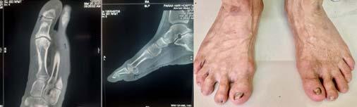
Degenerative arthritic changes seen in 1st metatarso-phalangeal joint in form of articular surface Sclerosis, marginal osteophytes and subchondral cysts of great toe. No fracture of phalanges / metatarsal bones seen. Mild soft tissue swelling seen around 1 st metatarsophalangeal joint. Sesamoid bone seen near 1st metatarsophalangeal joint’ Irregularity of articular surface involving 1st metatarsophalangeal joint with mild sclerosis, osteophytes and subchondral cyst.s/o Arthritic Changes
SIR, — A male 79 years is a practicing general surgeon in good health – non diabetic, non-hypertensive on no regular medication. Relevant past history of NSAID induced acute DU perforation 46 years back, operated, Robotic prostate surgery 16 years ago and Laparoscopic repair of right Inguinal hernia 8 years ago. He was given the first dose of COVID-19 Vaccine - AZD 1222 (ChaAdOx1) on 16th January 2021 at Paras HMRI Hospital, Raja Bazar, Patna. This was well tolerated except for mild low back ache and pain in right (non-injected arm), near the insertion of the deltoid, which lasted in diminishing order for a week to 10 days. Second dose of COVID-19 Vaccine - AZD 1222 (ChaAdOx1) was given on 15th February 2021 at the same institution followed by mild to moderate myalgia particularly in both the pectorals and right arm outer aspect. Developed bowel upset with semi watery stools and mild lower abdominal gripping, marked anorexia, no nausea or vomiting, no fever. Simultaneously gradual swelling, pain and redness started around the base of (L) big toe with increasing episodes of sharp pain involving the (L) forefoot. A detailed laboratory test was performed and the results are shown in Table 1.Swelling, erythema and pain kept on increasing, gradually extending up to the ankle. Nature of pain was sharp shooting initially on mild movement of the big toe but later even without movement and severe enough to cause marked loss of sleep. X-ray and CT with 3-D reconstruction done on 20.03.2021 (Fig 1A). It showed no fracture. He was examined by a senior Orthopedic Surgeon, advised Crepe bandage application,
Vol 120, No 9, September 2022Journal of the Indian Medical Association 66
Neutrophils (%) 40-80
Fig 1 — (A) Representation of CT joints of the left foot with 3D reconstruction. (B) A clinical photograph of the feet after five months of the incident.
Table 2 — Clinical findings Variables Observation on 23.02.2021 Observation Remark on 08.04.2021 AntibodyWITTI3DCT-JOINTStoReactive (3.83)Not seenReactive Hepatitis C virus >1.00 HCV RG RQ_PCR 34709 - IU/ml or 4.54 - log of HCV RNA was detected in the specimen.Not seen Detection Limit: Assay for Hepatitis
MCHC (gm/dL) 31-37 31.933.1 RDW ASOUricCreatenineGlucose–RandomPlatelet(cv%)11.6-14.11413.1Count(Lakhs/Cumm)1.5-41.882.42(mg/dL)70-14010496(mg/dL)0.8-1.51.21.2Acid(mg/dL)3.5-8.57.47.8Titer/AntiStreptolysinOTiter(1U/mL)0<200NA<200
Patient had already planned for cataract surgery in his right eye. In the Pre-operation preparations routine tests were carried out on 23.02.2021.All the reports were within normal range except the Hepatitis C antibody report gave the result as Reactive (Table 2). This was very surprising as reports done on previous occasions were always negative for Viral Markers. The pathologist concerned (Dr S K) repeated the test and confirmed positivity and also did the viral load. (Report attached). The cataract surgery was carried out on 25.02.2021. There were no ophthalmic complains and as far as the eye was concerned patient was back to normal in three days. The pain in the left foot continued in a slowly diminishing pattern. The patient took no medication for the Hepatitis C report as he was certain of not taking any IV or IM Injection during the last one year. The only procedure during this period was one dental extraction done on 26.09.2020. On 08.04.2021 to reassess the situation HCV Viral Load Assay was repeated and came out to be NegativeTarget not seen. For reconfirmation one sample was sent to another laboratory Oncquest and result was again negative.
Lymphocytes (%) 20-40
Eosinophils (%)1-62.42.1 Monocytes (%) 2-106.65.9 Basophils (%)0-10.70.6 LUC (Large unstained cells)NA2.11.4 ESR (Erythrocyte Sed. Rate)0<152650 PCV –Haematocrit (%) 40-50
1MS, FRCS, FICS, Director, Ahmad Abdul Hai1 Department of General Surgery, Paras HMRI Hospital, Patna 800014 C 65 IU/mL or virus RNA 1.81 log
Discussion — keeping in view the above history, it appears that the entire episode was an untoward reaction to the COVID-19 Vaccine - AZD 1222 (ChaAdOx1).

