


Effective research involves a series of systematic procedures that researchers must undertake in order to contribute to the existing evidence base. It includes identification of research problem, formulation of research questions, objectives and hypothesis, selection of appropriate study design, data collection and analysis, and publication1,2 Publication of scientific paper is essential for evolution of science as well as for career development. It helps in dissemination of study findings and gives visibility to the research. Research work that is not published or documented is considered not done 3 . It is mandatory to follow proper ethical guidelines in every step of the research process. Organizations involved with publication ethics
There are some organizations who have developed guidelines for facilitating ethics in publication. Authors, editors and reviewers must adhere to these guidelines in order to produce accurate and unbiased scientific papers. Organizations involved with publication ethics are:
• Committee on Publication Ethics (COPE): It is a forum created by editors of peerreviewed journals for discussing issues related to publication ethics. COPE published “Guidelines on Good Publication Practice” to assist authors, editors, editorial board members, and reviewers in the publication process4.
• Council of Science Editors (CSE):It promotes effective communication of scientific information through identification of research misconduct and guidelines for action5
• International Committee of Medical Journal Editors (ICMJE) : It is a group of medical journal editors who developed guidelines primarily for authors who want to publish in ICMJE member journals. Roles and responsibilities of those involved in publishing are clearly outlined. Most journals follow ICMJE recommendations for manuscript submission 6
• World Association of Medical Editors (WAME): It is a non-profit voluntary association of editors of peer-reviewed journals. WAME facilitates cooperation and communication among medical journal editors in order to improve editorial standards7
• Consolidated Standards of Reporting Trials (CONSORT): It aims to address issues arising from inadequate reporting of randomized controlled trials (RCTs). CONSORT provides guidelines for reporting of RCTs8
• Sense about science: It is an independent charitable organization that promotes public understanding and respect for scientific evidence9
Ethics approval and informed consent
It is mandatory for researchers to get their research proposal approved by InstitutionalEthics Committee (IEC) or Institutional Animal Ethics Committee (IAEC). All clinical trials must be registered with Clinical Trials Registry of India (CTRI) and authors must provide CTRI registration number in their publications. In addition to this, obtaining informed consent from all study participants is vital for conducting effective research3
Scientific misconduct
Scientific misconduct refers to violation of standard codes of scholarly conduct and
ethical behaviour in publication of research. Misconduct may occur at any stage of the research process and puts a question mark on integrity of the research. Research fraud including fabrication or falsification of data are serious forms of scientific misconduct because they distort observed truth. Fabrication refers to publishing results and conclusions from data that were not generated by experiments or observations. Falsification refers to manipulating research materials, images, data, equipment, or processes in order to alter observed results of a study. Most publishers have strict rules against research fraud and may take disciplinary action as per COPE recommendations. Scientific misconduct also includes plagiarism, violation of authorship rules, simultaneous submissions, duplicate submissions, salami slicing, and non-declaration of conflict of interest 10,11 . A study conducted among medical professionals in India indicated that 65.1% offered gift authorship, 56.7% knew of an individual who fabricated data, and 53.5% observed plagiarism 12 . Scientific misconduct, however, does not include ordinary errors, differences in interpretation of data, andscholarly or political disagreements.
Authorship
An individual who has made significant contribution to the research study in question is considered to be an author. Contributions may include conceptualizing and designing the study; data acquisition, analysis and interpretation; writing draft or editing the manuscript; approving the final draft; and being accountable for the accuracy or credibility of the research11. ICMJE guidelines state that all authors should fulfil criteria for authorship and those who fulfil criteria should be named in publication6
Authorship issues should be resolved before conducting the study to avoid conflicts in future. Order of authorship should be a joint decision of co-authors based on extent of contribution of individual authors13. It is to be noted that individuals who have provided technical support, advice, research space, departmental support or financial support do not qualify for authorship. They may be listed as contributors or acknowledged individuals14.
Ghost authorship, gifted authorship, and guest authorship are the most commonly encountered authorship misconduct. Ghost authors are those individuals who have made substantial contribution to research but are not listed in publication. Guest authors are those individuals who are named in publication despite making no significant contribution to the study. Guest authorship is usually provided to improve chances of acceptance of manuscript. Gift authors are usually people in academic or administrative positions who have made no significant contribution to study but are listed as co-authors in publication due to their tenuous affiliation with the study11
Gifted or guest authorship is unethical and unacceptable in publication. Authorship in research publication is a responsibility and authors are accountable for the study findings and results which are published.
Conflict of interest
Conflict of interest (COI) occurs when researchers’ financial or personal interest affects their judgement and influences conclusions of the study. This deteriorates quality and integrity of research. Employment, grants, patents, honorarium from funding agencies, or multiple affiliations are often reasons for potential competing interests. Most journals ask for disclosure of potential competing interests during submission of manuscript for publication6
Plagiarism
Plagiarism refers to use of another author’s work in the manuscript without taking consent, or giving credit or acknowledgement. This is the commonest type of scientific misconduct and research fraud.There are several forms of plagiarism, some of which are listed below:
• Direct plagiarism or plagiarism of text: This is when a researcher reproduces another researcher’s previously published work entirely or parts of it without giving credit or acknowledgement to the original work. Direct plagiarism must be avoided by citing the original source in references or putting the copied part within quotation marks.
• Plagiarism of ideas: This refers to copying and reproducing someone else’s ideas without giving proper credit to the original researcher. This form of plagiarism is difficult to detect and often goes unnoticed. This is a serious offence and must be avoided.
• Self-plagiarism: When an author uses sections of their own previous work in following publications having similar topic, it is called self-plagiarism. This can be avoided by proper citations or putting copied text within quotation marks 13
Simultaneous submission
Simultaneous submission occurs when researchers submit the same manuscript to more than one journal simultaneously. This is a serious scientific misconduct and can lead to the same paper being published in two journals. During manuscript submission, most journals require declaration from the researcher that the manuscript has not been submitted elsewhere simultaneously. It is important for researchers to abide by this rule and wait for decision before submitting to another journal3
Salami slicing
Researchers often publish multiple papers using same dataset, same study population, same methodology, or same hypothesis. Slicing up a large study into multiple segments and publishing them
separately is called salami slicing. This is an unethical practice and must be avoided.
However, large clinical trials or epidemiological studies having multiple endpoints may be published separately. Each publication should have a clearly defined hypothesis and outcome of interest15 Self-citation
It is unethical for authors to cite their previously published work in subsequent publications which are out of context. Self-citation is often done to increase metrics such as G-Index or H-Index. This is scientific misconduct and undermines reputation of the researcher in the scientific community. However, it is acceptable when researchers use self-citation for their subsequent publications which are continuations of their previous work or are within the same context3
Conclusion
Authors must adhere to recommendations by COPE or ICMJE for their publications. The issue of unethical publications must be addressed through appropriate training and guidance. Scientific misconduct harms the reputation of the researcher as well as the journals publishing such work. Serious offences may lead to blacklisting of the researcher and retraction of publication from the journal.
1Gelling L — Stages in the research process. Nurs Stand. 2015; 29(27): 44-9. doi: 10.7748/ns.29.27.44.e8745. PMID: 25736674.
2Gemayel R — How to write a scientific paper. FEBS J 2016 Nov; 283(21): 3882-5. doi: 10.1111/febs.13918. PMID: 27870269.
3Sengupta S, Honavar SG — Publication ethics. Indian J Ophthalmol2017; 65(6): 429-32. doi: 10.4103/ijo.IJO_483_17. PMID: 28643704; PMCID: PMC5508450.
4Wager E — The Committee on Publication Ethics (COPE): Objectives and achievements 1997-2012. Presse Med. 2012 Sep;41(9 Pt 1):861-6. doi: 10.1016/j.lpm.2012.02.049. Epub 2012 Jun 22. PMID: 22727914.
5Editorial Policy Committee, Council of Science Editors. CSE’s White Paper on Promoting Integrity in Scientific Journal Publications. New York, NY: 2021.
6ICMJE — Recommendations for the conduct, reporting, editing and publication of scholarly work in medical journals. Updated May2022.https://www.icmje.org/icmje-recommendations.pdf. Last accessed on November 02, 2022.
7WAME — Recommendations on Publication Ethics Policies for Medical Journals. Prepared by the WAME Publication Ethics Committee. https://wame.org/recommendations-onpublication-ethics-policies-for-medical-journals. Last accessed on November 02, 2022.
8Schulz KF, Altman DG, Moher D, for the CONSORT Group. CONSORT 2010 Statement: updated guidelines for reporting parallel group randomised trials.
9Sense about science- because evidence matters. https:// senseaboutscience.org/. Last accessed on November 02, 2022.
10Sabir H, Kumbhare S, Parate A, Kumar R, Das S. Scientific misconduct: a perspective from India. Med Health Care Philos 2015; 18(2): 177-84. doi: 10.1007/s11019-014-9603-8. PMID: 25316327.
11Singhal S, Kalra BS — Publication ethics: Role and responsibility of authors. Indian J Gastroenterol 2021; 40(1): 65-71. doi: 10.1007/s12664-020-01129-5. Epub 2021 Jan 22. PMID: 33481172; PMCID: PMC7821455.
12Dhingra D, Mishra D — Publication misconduct among medical professionals in India. Indian J Med Ethics 2014; 11(2):1047. doi: 10.20529/IJME.2014.026. PMID: 24727622.
13Wallace MB, Siersema PD — Ethics in publication. Gastrointest Endosc 2015; 82(3): 439-42. doi: 10.1016/j.gie.2015.05.019. Epub 2015 Jun 22. PMID: 26112677.
14Hodge A, Haapala I, Yngve A, McNeill G, Tseng M — A refresher in research publication ethics. Public Health Nutr 2012; 15(3): 377-8. doi: 10.1017/S136898001200002X. PMID: 22280824.
15Luther F — Publication ethics and scientific misconduct: the role of authors. J Orthod 2008; 35(1): 1-4. doi: 10.1179/ 146531207225022347. PMID: 18287388.
1PhD, Scholar, Masuma Yasmin1, Department of Endocrinology Sujoy Ghosh2 and Metabolism, Institute of Post-Graduate Medical Education and Research, Kolkata 700020
2Professor, Department of Endocrinology and Metabolism, Institute of Post-Graduate Medical Education and Research, Kolkata 700020
The Standard of Hand Written Operative Notes; A Long Campaign towards Refinement — A Complete Audit Loop Study in a Teaching Hospital
Background : A comprehensive, thorough, accurate, legible and professional operative note allows seamless and proper transfer of patient care from the operating table to the postoperative care room and beyond. Further, incomplete and illegible handwritten operative notes in medico legal cases may be an Achilles heel in the surgeon’s defense.
Aims : This study audited the quality of operative note keeping of general surgical procedures against the standards set by the Royal College of Surgeons of England (RCSE) guidelines. The aim of the study was to assess the compliance while also improving record keeping, documentation, the quality of operative notes, and educating surgery residents.
Materials and Methods : The information from operative notes of every patient undergoing general surgical procedures was collected over a period of seven months. The data was formulated and analyzed using the SPSS version 20.
Results : Total of 560 operative notes were recruited and audited. All the notes were hand-written, with the majority being written by postgraduate residents (86%).The postoperative care advice, fluid and antibiotic instructions were documented in the finest manner (100%). However, only 1% of the notes mentioned the Patients’ name, Gender and Age. All operative notes (>99 percent) included the names of the operative surgeon and assistants. The consultant in charge was documented in only 12 percent emergency notes and 100 percent elective procedures. The name of runners (Nursing Orderly) was missing from all the notes. Notably, no details of closure techniques were mentioned in any of the operative notes. Almost all of the operative notes were not signed properly to include the resident’s name and code.
Conclusion : The quality of the operation notes that were entered into the patient’s case sheets was poor and insufficient, and it needed to be greatly improved. The findings of the study underline the necessity for residents to receive mandatory training on data collection and how to produce operative notes according to institutional rules.
[J Indian Med Assoc 2022; 120(11): 16-9]
Key words :Audit, Operation notes, General surgery, Medical records.
Clinical auditing is a component of clinical governance that determines whether the healthcare being provided is in accordance with accepted standards, thereby aiding in the improvement of service quality and, if necessary, identifying areas for improvement1. Good medical and operative records are essential for proper medical practice in order to ensure effective patient care. Operative notes not only serve as a record of patient care and evidence for medico-legal issues but also provide critical information for research and auditing the performance of hospitals and the working staff2-4. The Royal College of Surgeons of England published Good Surgical Practice Guidelines for legible, comprehensive and all-inclusive medical record keeping5. These guidelines capture the details of patient, surgical procedure, intra-operative
Department of General and Minimal Access Surgery, SKIMS Medical College & Hospital, Srinagar, Jammu and Kashmir 190018
1MBBS, MS, Senior Resident and Corresponding Author
2MBBS, MS, FRCS, FICS, Professor and Head
Received on : 23/03/2022
Accepted on : 10/04/2022
Editor's Comment :
A comprehensive and precise operative documentation is indispensable not only for superior postoperative care but also for academic and research purposes. Inadequate postoperative notes is an Achilles Heel in a surgeon’s defence and therefore may prove to be a medico-legal liability.
findings, complications and postoperative care instructions. The compliance to Good Surgical Practice Guidelines for operative notes varies from hospital to hospital. The ongoing auditing and assessment of handwritten operative notes is now an important aspect of Departmental clinical governance, as it determines whether the changes listed in the action plan following the baseline audit are being implemented for overall patient care improvement. This study audited the quality of operative note keeping for elective and emergency general surgical procedures against the standards set by the Royal College of Surgeons of England (RCSE) guidelines. The aim of the study was to assess the compliance while also improving record keeping, documentation, the quality of operative notes, and educating surgery residents.
This descriptive hospital-based audit loop study was carried out at the SKIMS, Medical College and Hospital, in a department of General and Minimal Access Surgery over a period of seven months. The audit was carried out with the approval of the Departmental Academic and Research Committee (Order No. SKIMS/MCH/GS/ 2021-571) and under the supervision of the Head of Department. A total of 560 patients who underwent various surgeries in elective and emergency settings were included in the study and operation data were collected. The information from the operative notes of all the patients was collected by authors and formulated for data collection. RCS England, Good Surgical Practice 2014 guidelines on operation note keeping was followed, for completing the components on a checklist. The operative notes were compared against the set data points recommended in the RCS GSP guidelines for various General Surgery Procedures (Table 1). At the conclusion of audit study, the findings were presented in the departmental meeting, where the deficiencies and inadequacies were highlighted to the surgeons in the Department (Fig 1).
Data was entered and analyzed in Microsoft Excel 2016 software, and each item was checked as present or absent. The statistical analysis was carried out with the Statistical Package for Social Science (SPSS) version 20 software, and the results were provided as number or percentage of patients.
A total of 560 operative notes were recruited and audited with 296 (52.86 percent) elective general
Parameters
Patient Name, Gender and Age
Date and Time
Elective/Emergency procedure
Consultant in charge
Names of the operating surgeon and assistant
Scrub assistant
Type of Anaesthesia
Operative procedure carried out
Indication of procedure
Type of Incision
Pre- and postoperative diagnosis
Operative findings
Any problems/complications
Any additional operation performed and the reason why it was performed
Details of tissue removed, added or altered
Any foreign material or prosthesis used
Details of closure technique
Pot operative antibiotics and fluids
Detailed postoperative care advice
Signature

surgical procedures and 264 (47.14 percent) emergency surgeries. As the ongoing practice of the institute, all the operative notes were hand-written. The majority of the notes did not adhere to the RCS England guidelines for General Surgical Practice. The majority of the handwritten notes lacked one or more important items mentioned in the standard guidelines. The notes were mostly written by postgraduate residents (86%), followed by senior residents (13.5%) and junior residents (0.5%). None of the notes was written by the Consultants. The postoperative care advice, fluid and antibiotic instructions were documented in the finest manner (100%).
The Name, Gender and Age of patient were mentioned in only 1% of notes. The date-of-surgery was documented in 95% elective operation notes and 83% in emergency notes. However, time was recorded only in 4% emergency notes and not documented in any of the elective procedures. The duration-of-surgery was documented in 1% emergency operative notes and in none of the elective procedures. The name of the operating surgeon and assistant was documented in all operative notes; 99.34% elective notes and 99.44% in emergency notes. The name of anaesthesiologist was captured in 70% elective cases and only 52% of emergency notes. The name of the scrub assistants was documented in 92% elective and 50% emergency notes. The consultant in-charge was documented in 100% elective procedures and 12% emergency notes. The name of runners (Nursing Orderly) was missing from 100 % notes. The type of anaesthesia was mentioned in 45% of notes. The operative position and type of incision was noted in 1% elective surgical notes and none of the emergency documents. The operative diagnosis and intra-operative finding were mentioned in 99.45% operative notes. The occurrence of any intraoperative complication and any additional operation performed and its reason was inconsistently documented in 12% operative notes. The details of tissue removed, altered or added was mentioned in 8%
120, No 11, November 2022Journal
notes and only 20% operative notes documented the foreign body or prosthesis used. Notably none of the operative notes mentioned the details of closure techniques; however, 100% notes documented the postoperative care advice, antibiotic and fluids. Almost none of the operative notes in both the emergency and elective records were correctly signed with the resident’s name and institutional code.
The accuracy of operative notes is critical to providing effective patient care in the postoperative care room, general wards and beyond in follow-up. The accurate and comprehensive documentation of the operation is essential for providing safe postoperative patient care and forms an important part of the legal documentation in cases of medico-legal importance. Furthermore, insufficient and unreadable handwritten operative notes may lead to misunderstandings and be the surgeon’s Achilles heel in medico-legal issues. An audit done by Lefter, etal demonstrated the medicolegal impact of poor operative notes, with 44.73 percent of 190 operative notes judged to be non-defensible after review by a medico-legal counsel2. The guidelines already exist for preparation of operative notes updated by RCS England in 2014 as Guidelines for Good Surgical Practice5; nonetheless, compliance has been observed to vary amongst institutes and specialties. The overall standard of reporting and documentation in medicine is low, with many reports omitting important and relevant data6. There is insufficient time and effort spent on critically and objectively evaluating the outcomes of clinical audits and the audit loop is frequently not completed7. In our conflict zone of Kashmir Valley, the rates of litigation in trauma and emergency surgical patients are quite high and rising, making legible and precise documentation even more important. To date, a negligible amount of data has been published and no audit loop study concerning the quality of operative notes from the Kashmir valley has been reported. The audit study was conducted to assess compliance and to identify methods of improving and maintaining the quality of operative notes solely for general surgery procedures, as well as to educate the surgery residents at our tertiary care teaching institute.
The patient identification (name, age and sex) is an essential part of operative notes; however its significance was consistently under estimated in the study and reported in 1% notes. This was found to be remarkably less as compared to other studies reported in literature8-11. The patient identification was seen in 28-33% of the operative notes in the study done at a
teaching Hospital, in Sudan8. As a crucial parameter, the personal identification should be documented in each patient’s operation notes. In the event of a lawsuit, operative team members are typically paraded throughout the hearing of proceedings to provide evidence on the events that occurred during the surgery for medical legal clarity. As a result, it is critical to include all of this information in the operative note.
The date-of-surgery was well documented (95 percent in elective operation notes and 83 percent in emergency notes), whereas, time was only recorded in 4 percent of emergency notes and none of the elective procedures. In a study conducted in Sudan 8 the time was documented in 81% of the notes; however, other researchers in Nigeria and Pakistan discovered that the time of surgery was frequently omitted in operative notes12,13. Some data points, such as the name of the operating surgeon and the assistant, the operative diagnosis and intra-operative findings, postoperative care advice, antibiotics and fluids, were completed with a high level of accuracy (>95 percent). This may be secondary to the printed parameters and data points included in the current proforma of operative notes at our centre (Fig 1). The advantages of using a proforma have been highlighted in the literature and it has resulted in better completion of detailed notes14-16. The use of template operational notes can both reduce the complexity of the task at hand and ensure that no vital details are neglected. The findings of our study were comparable to those of a review study of operation notes from nine UK Hospitals and other published literature17,18. The study in UK hospitals revealed a high level of completion (95%) for data points such as the name of the operating surgeon (99.3%), legibility, date, and operation title (99.1 percent)17
In this study, the name of the anaesthesiologist was recorded in 52 percent to 70 percent of the notes, the scrub assistant (50-92 percent), the consultant in charge (12-100 percent) and the type of anaesthesia was recorded in 45 percent of the notes. The runners’ names (Nursing Orderly) were omitted from 100% of the notes and the operative position and type of incision were poorly documented (1 percent in elective surgical notes only). Other studies 9,19,20 found that the Anaesthesiologist’s name, Scrub assistant, Type of anaesthesia, Operative position and Type of incision were all visible in more than 90% of cases. The results of a clinical audit study of operation notes conducted in two different tertiary hospitals in Ethiopia revealed mixed results19. The names of anaesthetists, scrub nurses and runners were documented in almost all operation notes from one hospital but were inconsistently documented at another19.
120, No 11, November 2022Journal
Despite the fact that there is no formal training for surgical postgraduate residents in our setting for operative note writing, it was discovered that the majority of operative notes were written by Postgraduate Residents (86%), Senior Residents (13.5%), and Junior Residents (0.5 percent). This is quite concerning, as it has been discovered that trainees frequently struggle to produce high-quality notes in the absence of proper guidance21. The findings of the study underline the importance of residents receiving mandatory data collecting training as well as training on how to write operative notes in compliance with institutional norms. Additionally, writing operative notes should be included in the early stages of residency training and senior surgeons should invest time in trainees to ensure successful writing of standard operative notes. Also it is recommended that the legibility of signatures should be improved by the use of name-stamps for residents as reported in literature22
The quality of the operation notes entered into the patient’s case sheets was poor and needed to be much improved. To increase the quality of operative notes, regular auditing and the use of templated operative notes in accordance with RCS England criteria are essential. The findings of the study underline the necessity for residents to receive mandatory training on data collection and how to produce operative notes according to institutional guidelines.
Ethical Issue : None
Financial and competitive interest : None
The authors declare that no financial support was received from any organization for the submitted work.
Other Relationships : The authors declare that there are no other relationships or activities that could appear to have influenced the submitted work.
Conflict of interest : On behalf of all authors, the corresponding author states that there is no conflict of interest.
The authors have no other disclosure.
1Williams N, O’Connell PR, McCaskie A — Human factors, patient safety, and quality improvement. In: Bailey & Love’s Short Practice of Surgery, 27th Edition: The Collector’s Edition. Ch. 13. Boca Raton, FL: CRC press; 2018. p. 1301-19. https:/ / doi.org/10.1201/9781315111087.
2Lefter LP, Walker SR, Dewhurst F, Turner RW — An audit of operative notes: facts and ways to improve. ANZ J Surg 2008; 78(9): 800-2. doi:10.1111/j.1445-2197.2008.04654.x [View Article] [PubMed]
3Bateman ND, Carney AS, Gibbin KP — An audit of the quality of operation notes in an otolaryngology unit. J R Coll Surg Edinb 1999; 44(2): 94-5. [PubMed]
4Wauben LS, Goossens RH, Lange JF— Evaluation of
operative notes concerning laparoscopic cholecystectomy: are standards being met?. World J Surg 2010; 34(5): 903-9. doi:10.1007/s00268-010-0422-7 [View Article] [PubMed]
5Good surgical practice: a guide to good practice. Royal College of Surgeons of England. 2014. https://www.rcseng.ac.uk/ standards-and-research/gsp/
6Rogers A, Bunting M, Atherstone A — The quality of operative notes at a general surgery unit. S Afr J 2008; 98: 726-8.
7Kulkarni R, Sudheer K, Worell J, Strenks GJ — Completing the audit loop: A New Objective and Standardized Method. Qual Assur J 2001; 55(1): 33-6.
8Hamza A, Abdalrahim HM, Idris SA, Ahmed OM — Evaluating the Operative Notes of Patients Undergoing Surgery at Omdurman Teaching Hospital , Sudan. Sch J App Med Sci 2014; 1(6): 668-72.
9Jawaid M, Askari R, Qureshi MA, Manzar S— Quality of operative notes. Journal of Postgraduate Medical Institute (Peshawar Pakistan) 2011; 22(4): 174-6.
10Shah S, Dangol B, Kumari S, Guragain RP — An audit of operative notes at TUTH. Nepalese Journal of ENT Head & Neck Surgery 2011; 2(2): 25-6.
11Elfaki EAA, Elhilu AH — Facts about compliance of surgeons. An audit study of the quality of operation notes in the department of general surgery, King Faisal Hospital, Makkah, Saudi Arabia. Sudan JMS 2009; 4(1): 12-6.
12Khan MUR, Ahmed S, Shamim MS, Azhar M, Ur Rehman S — Operative notes at surgical units of a tertiary care hospital. J Surg Pak Int 2010; 10(1): 57-9.
13Babalola RN, Olasehinde O, Sowande OA — An audit of the quality of surgical operation notes in a Nigerian teaching hospital. EastCentAfrJSurg 2016; 21(2): 76-80. doi:10.4314/ ecajs.v21i2.12 [View Article]
14Al Hussainy H, Ali F, Jones S, McGregor-Riley JC, Sukumar S — Improving the standard of operation notes in orthopaedic and trauma surgery: the value of a proforma. Injury 2004; 35(11): 1102-6.
15Chan BKY, Exarchou K, Corbett HJ, Turnock RR — The impact of an operative note proforma at a paediatric surgical centre. Journal of Evaluation in Clinical Practice 2015; 21(1): 74-8.
16Payne K, Jones K, Dickenson A — Improving the standard of operative notes within an oral and maxillofacial surgery department, using an operative note proforma. Journal of Maxillofacial and Oral Surgery 2011; 10(3): 203-8.
17Severn Audit and Research Collaborative in Orthopaedics (SARCO). Assessing the quality of operation notes: a review of 1092 operation notes in 9 UK hospitals [published correction appears in Patient Saf Surg. 2016;10:8]. Patient Saf Surg. 2016;10:5. Published 2016 Feb 6. doi:10.1186/s13037-0160093-x
18Huda F, Kumar N, Verma S — Assessing the Quality of Operation Notes in a Teaching Hospital. Gastroenterol Hepatol Int J 2019; 4(1): 000150.
19Mesale S, Seyoum N, Bekele A, Carter S — Clinical audit of operation notes at the Department of Surgery, Addis Ababa University. East and CentralAfrican Journal of Surgery 2020; 25: 13-7.
20Wauben LSGL, Goossens RH, Lange JF — Evaluation of Operative Notes Concerning Laparoscopic Cholecystectomy: Are Standards Being Met? World J Surg 2010; 34(5): 903-9.
21Nzenza TC, Manning T, Ngweso S, Perera M, Sengupta S, Bolton D, Lawrentschuk N — Quality of handwritten surgical operative notes from surgical trainees: a noteworthy issue. ANZ J Surg 2019; 89(3): 176-9.
22Glisson JK, Morton ME, Bond AH, Griswold M — Does an education intervention improve physician signature legibility? Pilot study of a prospective chart review. Perspect Health Inf Manag 2011; 8: 1e.
Nehad Jaser Ahmed1
Background : Methotrexate is widely used in the treatment of neoplasms, psoriasis and rheumatoid arthritis but it can cause several adverse events. The gastrointestinal system is where methotrexate side effects occur most frequently. The aim of the study was to describe the reported gastrointestinal adverse events of methotrexate. Material and Methods : This was a retrospective, descriptive analysis that was conducted to analyze gastrointestinal adverse events of methotrexate that were reported to the Food and Drug Administration or to the World Health Organization (WHO).
Results : Methotrexate use has been linked to gastrointestinal side effects such as Nausea, Vomiting, Diarrhea, Stomach Pain, Stomatitis and Mouth Ulcers.
Conclusion : Methotrexate can be effective and safe when used and monitored properly, therefore it’s critical to periodically check on patients and inform them of the side effects of methotrexate use and how to prevent or manage them.
[J Indian Med Assoc 2022; 120(11): 20-3]
Key words :Adverse events, FAERS, Methotrexate, Reporting, VigiBase.
Methotrexate is widely used in the treatment of neoplasms, psoriasis and rheumatoid arthritis1 Whether taken alone or in conjunction with other medications, methotrexate has been used successfully to treat a variety of malignancies and autoimmune illnesses. Unfortunately, intestinal toxicity is the primary dose-limiting factor for the administration of methotrexate and patients are significantly burdened by methotrexate-induced intestinal mucositis2. Despite its effectiveness, methotrexate occasionally only receives restricted use because of its side effects, which include kidney or liver damage, bone marrow toxicity and gastrointestinal mucosal injury1.
Although fatal mucosal necrosis caused by methotrexate is relatively rare, it is known that the drug can cause intestinal mucositis, hemorrhage, and peptic ulcers1. The gastrointestinal system is where methotrexate side effects occur most frequently3. Any amount of methotrexate can cause toxicity, although these effects become more frequent and severe with higher doses or more frequent dosing4.
The public can now search for information about human adverse events reported to the Food and Drug Administration by the pharmaceutical industry, healthcare providers, and consumers using the highly interactive Food and Drug Administration Adverse Event
1MSc, Lecturer, Department of Clinical Pharmacy, College of Pharmacy, Prince Sattam Bin Abdulaziz University, Al-Kharj, Saudi Arabia and Corresponding Author
Received on : 08/09/2022
Accepted on : 12/09/2022
Editor's Comment :
Methotrexate is widely used in the treatment of neoplasms, psoriasis, and rheumatoid arthritis but it can cause several adverse events.
The most reported gastrointestinal adverse events of methotrexate were Nausea, Vomiting, Diarrhea, Abdominal Discomfort and Stomatitis.
It is critical to periodically check on patients and inform them of the side effects of methotrexate use and how to prevent or manage them.
Reporting System (FAERS) tool5. They also can use VigiBase which is a distinctive global database of reported possible drug side effects that is maintained by the World Health Organization6. With over 30 million suspected adverse drug reaction reports reported since 1968, it is the world’s largest database of its sort. It is constantly updated as new reports arrive6
The present study aimed to describe the gastrointestinal adverse events of methotrexate using the Food and Drug Administration Adverse Event Reporting System (FAERS) and the World Health Organization database (VigiBase).
MATERIAL AND METHODS
This was a retrospective, descriptive analysis that was conducted to analyse gastrointestinal adverse events of methotrexate that were reported to the Food and Drug Administration (FDA) or to the World Health Organization. The study included all of the reports
120, No 11, November 2022Journal
that were submitted to the US FDA Adverse Event Reporting System (FAERS) before the 1st of July, 2022 and the reports that were submitted to the World Health Organization database (VigiBase) before 22 August, 2022.
The collected data included the total number of reports that were submitted to VigiBase, the geographical distribution of the reports, the gender and age of the patients who had adverse events, the number of gastrointestinal adverse events and the most reported gastrointestinal adverse events. The collected data also included the total number of reports that were submitted to FAERS, the gender of the patients who had adverse events, the age of the patients who had adverse events, the specialty of the reporters, and the most common gastrointestinal adverse events. The data were analysed descriptively and represented as numbers and percentages.
No IRB approval or informed consent was required because the research include only data about the adverse events reported and the data already available for the public and the researchers.
Methotrexate adverse events that were submitted to VigiBase :
Table 1 shows the gender and age of the patients that had an adverse event. The reports that didn’t specify the gender were excluded. More than 68% of the patients were females. The reports that didn’t specify the age were excluded. The age of 39.43% of the patients was between 45 and 64 years and the age of 19.71% of the patients was between 65 and 74 years.
Among these reports, 32,882 reports were reports of Gastrointestinal disorders (19.03%). The most reported gastrointestinal adverse events were Nausea (34.20% of the gastrointestinal events), Vomiting (17.66%), Diarrhea (13.37%), Abdominal Discomfort (7.81%), Stomatitis (7.15%), Mouth Ulceration (6.85%),
Abdominal Pain (6.56%), Gastrointestinal Disorder (4.78%), Upper Abdominal Pain (3.52%) and Dyspepsia (2.72%) (Table 2).
Methotrexate adverse events that were submitted to FAERS : Till 30 June 2022, 1,52,823 reports were submitted to FAERS. The age of 57.51% of the patients was between 18 and 64 years and the age of 25.87% of them was between 65 and 85 years. More than 66% of the patients who had adverse events were females. About 81.90%of the reporters who submitted the adverse events were healthcare professionals (Table 3).
Table 4 shows the most reported gastrointestinal adverse events that were submitted to FAERS. The most reported gastrointestinal adverse events were Nausea (7.00%), Diarrhea (4.78%), Abdominal Discomfort (4.55%), Vomiting (4.27%), Abdominal Pain (3.59%), Stomatitis (2.45%), Glossodynia (2.39%), Gastrointestinal Disorder (2.15) and Mouth Ulceration (1.05%).
Table 4 — The most reported gastrointestinal adverse events were submitted to FAERS
*The percentages represent the percentage of each adverse event among the total adverse events and not among the gastrointestinal events.
The present study showed that methotrexate frequently causes gastrointestinal adverse events such as Nausea, Vomiting, Diarrhea, Abdominal Discomfort, Stomatitis, Mouth Ulceration and Abdominal Pain. According to Maestá, et al the most common side effects of methotrexate were gastrointestinal disorders and abnormal laboratory findings. The most common gastrointestinal side effects were Oral mucositis, Nausea, Abdominal pain, Diarrhea and Vomiting7 According to Bulatovi-Alasan, et al Nausea (32.0%), Stomach Pain (11.3%) and Vomiting (6.5%) were the most common gastrointestinal symptoms following methotrexate administration and 42.3% of arthritis patients who received methotrexate experienced at least one gastrointestinal side event8 . Asai, et al reported that using methotrexate can Result in Reflux, Stomach Discomfort, Indigestion, Diarrhea, or constipation. In their study, the high-dose methotrexate group showed a higher prevalence of Reflux (32% versus 24%) and Abdominal Pain (28% versus 18%) compared to the low-dose methotrexate group. They discovered that the prevalence of dyspepsia, diarrhea, and constipation did not differ significantly across groups9. Sherbini, et al reported that Gastrointestinal (42.0%), Neurological (28.6%), Mucocutaneous (26.0%), Pulmonary (20.9%), increased Alanine Transaminase (18.0%) and HAematological events (5.6%) were the most frequently reported adverse events in individuals with early rheumatoid arthritis10 Moreover, previous studies demonstrated that methotrexate’s gastrointestinal side effects, such as Nausea, Stomach discomfort and Vomiting, frequently occur11-17
Despite its effectiveness, methotrexate sometimes only receives restricted use because side effects include kidney or liver damage, bone marrow toxicity, and gastrointestinal mucosal irritation, according to Tsukada, et al Additionally, they noted that intestinal mucositis, hemorrhage, and peptic ulcers are wellknown methotrexate side effects of the gastrointestinal system, despite the fact that fatal mucosal necrosis instances are relatively uncommon1. Furthermore, according to Higuchi, et al methotrexate is stopped due to gastrointestinal side effects that are frequently seen during the treatment of rheumatoid arthritis18 Anticipatory and associative gastrointestinal symptoms may impair methotrexate use and negatively affect patients’ quality of life. However, these symptoms are not very obvious clinically 13,19 According to Zhou, et al methotrexate-induced intestinal mucositis places a heavy load on patients and is the main dose-limiting factor for methotrexate therapy2
Methotrexate use has been linked to gastrointestinal side effects such as Nausea, Vomiting, Diarrhea, Stomach pain, Stomatitis and mouth Ulcers. Methotrexate can be effective and safe when used and monitored properly, therefore it’s critical to periodically check on patients and inform them of the side effects of methotrexate use and how to prevent or manage them.
This Publication was supported by the Deanship of Scientific Research at Prince Sattam Bin Abdulaziz University.
1Tsukada T, Nakano T, Miyata T, Sasaki S — Life-Threatening Gastrointestinal Mucosal Necrosis during Methotrexate Treatment for Rheumatoid Arthritis. Case Rep Gastroenterol 2013; 7(3): 470-5.
2Zhou B, Xia X, Wang P, Chen S, Yu C, Huang R, et al — Induction and Amelioration of Methotrexate-Induced Gastrointestinal Toxicity are Related to Immune Response and Gut Microbiota. E Bio Medicine 2018; 33: 122-33.
3BPAC — Methotrexate prescribing errors can be fatal. 2011. (https://bpac.org.nz/bpj/2011/february/methotrexate.aspx).
4Pfizer — Methoblastin.2022.(www.medsafe.govt.nz).
5FDA — FDA Adverse Event Reporting System (FAERS) Public Dashboard. 2022. (https://www.fda.gov/drugs/questionsand-answers-fdas-adverse-event-reporting-system-faers/ fda-adverse-event-reporting-system-faers-publicdashboard).
6WHO — About VigiBase. 2022. (https://who-umc.org/ vigibase).
Vol 120, No 11, November 2022Journal
7Maestá I, Nitecki R, Horowitz NS, Goldstein DP, Moreira MDFS, Elias KM, et al — Effectiveness and toxicity of first-line methotrexate chemotherapy in low-risk postmolar gestational trophoblastic neoplasia: The New England Trophoblastic Disease Center experience. GynecolOncol 2018; 148(1) : 161-7.
8Bulatoviæ Æalasan M, van den Bosch OF, Creemers MC, Custers M, Heurkens AH, van Woerkom JM, etal— Prevalence of methotrexate intolerance in rheumatoid arthritis and psoriatic arthritis. Arthritis Res Ther 2013; 15: 1-5.
9Asai S, Nagai K, Takahashi N, Watanabe T, Matsumoto T, Asai N, et al — Influence of methotrexate on gastrointestinal symptoms in patients with rheumatoid arthritis. Int J Rheum Dis. 2019; 22(2): 207-13.
10Sherbini AA, Gwinnutt JM, Hyrich KL, Adebajo A, Ahmed K, Al-Ansari A, et al — Rates and predictors of methotrexaterelated adverse events in patients with early rheumatoid arthritis: results from a nationwide UK study. Rheumatology 2022; 2022: keab917.
11Murray KJ, Lovell DJ — Advanced therapy for juvenile arthritis. Best Pract Res Clin Rheumatol 2002; 16(3): 361-78.
12van der Meer A, Wulffraat NM, Prakken BJ, Gijsbers B, Rademaker CM, Sinnema G — Psychological side effects of MTX treatment in juvenile idiopathic arthritis: a pilot study. Clin Exp Rheumatol 2007; 25(3): 480-5.
13BrunnerHI, Johnson AL, Barron AC, Passo MH, Griffin TA, Graham TB, et al — Gastrointestinal symptoms and their association with health-related quality of life of children with
juvenile rheumatoid arthritis: validation of a gastrointestinal symptom questionnaire. J Clin Rheumatol 2005; 11: 194204.
14Hashkes PJ, Laxer RM — Medical treatment of juvenile idiopathic arthritis. JAMA 2005; 294(13): 1671-84.
15Ruperto N, Murray KJ, Gerloni V, Wulffraat N, de Oliveira SK, Falcini F, et al — A randomized trial of parenteral methotrexate comparing an intermediate dose with a higher dose in children with juvenile idiopathic arthritis who failed to respond to standard doses of methotrexate. Arthritis Rheum 2004; 50(7): 2191-201.
16Ramanan AV, Whitworth P, Baildam, EM — Use of methotrexate in juvenile idiopathic arthritis. Arch DisChild 2003; 88(3): 197200.
17Alsufyani K, Ortiz-Alvarez O, Cabral DA, Tucker LB, Petty RE, Malleson PN — The role of subcutaneous administration of methotrexate in children with juvenile idiopathic arthritis who have failed oral methotrexate. J Rheumatol 2004; 31(1): 179-82.
18Higuchi T, Yoshimura M, Oka S, Kawasaki A, Warabi E, Mizuno S, et al — Dietary Factors Modulate Gastrointestinal Adverse Effects of Methotrexate. JPS 2018; 2018: PO3-13.
19Bulatovic M, Heijstek MW, Verkaaik M, van Dijkhuizen EH, Armbrust W, Hoppenreijs EP, et al — High prevalence of methotrexate intolerance in juvenile idiopathic arthritis: development and validation of a methotrexate intolerance severity score. Arthritis Rheum 2011; 63(7): 2007-13.
Sai Sameera N1, Sangeetha Lakshmi B2, Aishwarya Lakshmi P3, R Ram4, Siva Kumar V5
Introduction : Physicians in India are frequently the first point of contact for patients with chronic kidney disease. Hence, awareness of clinical practice guidelines for Chronic Kidney Disease (CKD) among the Postgraduates/ residents of MD Medicine is of paramount importance. The aim of this study is to assess MD Medicine Postgraduates’ and Residents’ awareness and knowledge of clinical practice guidelines for Chronic Kidney Disease (CKD).
Materials and methods : The present study is a cross-sectional observation study. A questionnaire of clinical vignettes on CKD with multiple choices was prepared. A one-time survey of MD Medicine postgraduates and residents from various medical colleges in three states of southern India has been collected.
Results : A total of 228 Postgraduates or Residents of MD Medicine participated in the study. The awareness of clinical practice guidelines for CKD was low across all postgraduate years (PGYs) of MD Medicine. We measured the CKD awareness in postgraduates and residents of MD Medicine based on the Postgraduate year, Medical college and institute, type of institution (either Government funded or capitation fee), the presence of a nephrology fellowship in the institution, and the consultation given to the Nephrology patients in the Medicine OPD.
Conclusions : The modest awareness of clinical practice guidelines for CKD across all PGYs has suggested that incorporation of these guidelines into the medicine postgraduate training curriculum is not robust at present. We also discussed the means to improve the understanding of nephrology by the postgraduates/residents of MD Medicine.
[J Indian Med Assoc 2022; 120(11): 24-8]
Key words :Chronic Kidney Disease, Clinical practice guidelines, Medicine postgraduates, Residents.
Chronic Kidney Disease (CKD) is defined as abnormalities of kidney structure or function, present for more than three months, with implications for health. (1) Albuminuria (AER>30 mg/24 hours; ACR>30 mg/g [>3 mg/mmol]), urine sediment abnormalities, electrolyte and other abnormalities due to tubular disorders; abnormalities detected by histology; structural abnormalities detected by imaging; and a history of kidney transplantation are all markers of kidney damage. In addition, the Glomerular Filtration Rate (GFR) should be<60 ml/ min/1.73 m2 1
Globally, the incidence of CKD increased by 89% to 21,328,972 (uncertainty interval 19,100,07923,599,380), prevalence increased by 87% to 275,929,799 (uncertainty interval 252,442,316300,414,224), death due to CKD increased by 98% to 1,186,561 (uncertainty interval 1,150,743-1,236,564), and disability-adjusted-life years (DALYs) increased
Department of Nephrology, Sri Venkateswara Institute of Medical Sciences, Andhra Pradesh 517507
1MD, DM, Senior Resident
2MD, DM, Assistant Professor
3MD, Senior Resident
4MD, DM, FISN, FRCP, Professor & Head and Corresponding
Author
5MD, DM, FISN, FRCP, FASN, Professor
Received on : 17/08/2022
Accepted on : 23/09/2022
Editor's Comment :
With the information that the knowledge of CKD awareness is narrow, the means to improve the understanding of Nephrology by the Postgraduates and Residents of MD Medicine should be adapted. Such steps are: Nephrologists should highlight that the management of some conditions could be improved only after proper training in nephrology, eg, hyponatremia, anti-tuberculous therapy in renal failure, systemic lupus erythematosus, and renal artery stenosis. Nephrologists should also ensure that medical postgraduates or residents learn central vein catheter placement, which is at present expected as a basic skill for all Postgraduates or Residents.
by 62% to 35,032,384 (uncertainty interval 32,622,07337,954,350)2. In the worldwide statistics, India is one of the countries with the highest rates of prevalence of CKD2. The prevalence of chronic kidney disease in India is reported to be between 0.78% and 17.2%3. The progressive nature of asymptomatic chronic kidney disease leads to an enormous social and economic burden for the community at large, in terms of burgeoning dialysis and transplant costs, which will only see an exponential rise in the next decade and will not be sustainable unless we reduce chronic kidney disease incidence and prevalence through screening and prevention. India can ill afford to manage all patients with End Stage Renal Disease (ESRD)4. The prevention of CKD is evolving as a crucial issue for the medical fraternity. Diabetes Mellitus and Hypertension
Vol 120, No 11, November 2022Journal
constitute approximately 60% of all the causes of CKD. Both these diseases are also easy to diagnose, follow, and treat. The prevention of the complication of these diseases, CKD, entails involvement at every step, ie, physician, policymakers, government and patient. In the undergraduate medical curriculum, there is no significant mention of CKD as one of the topics to be read and understood. Physicians in India are frequently the first point of contact for patients with chronic kidney disease. Hence, awareness of clinical practice guidelines for chronic kidney disease among the postgraduates/residents of MD Medicine is of paramount importance.
The aim is to study the awareness and knowledge of MD Medicine Postgraduates and Residents on clinical practice guidelines for chronic kidney disease.
We received the approval of the institutional ethics committee. The present study is a cross-sectional observation study. The questionnaire of clinical vignettes on CKD with multiple choices was adapted from the reference5 with the permission of the authors.
A one-time survey of MD Medicine Postgraduates/ residents from various Medical Colleges in three states of southern India has been collected. The questionnaire had been sent by post or by messenger, and also circulated at an Andhra Pradesh Chapter of API conference at Kakinada. The participants are advised not to refer to a book or the internet during their response. We included the Postgraduates/ Residents of MD Medicine of Sri Venkateswara Medical College, Tirupati, Andhra Medical College, Vishakapatnam,Rangaraya Medical College, Kakinada, Government Medical Colleges of Kurnool and Guntur, Government, Naryana Medical College, Nellore, Stanley Medical College, Madras Medical College, Osmania Medical College, Gandhi Medical College, Hyderabad; SRM Institute and SRMC Institute, Madras, Nizam’s Institute of Medical Sciences and Sri Venkateswara Institute of Medical Sciences, Tirupati. In this list, three Medical Colleges are capitation fee colleges; two are institutes that have residency programs; and the remaining nine are Government Medical Colleges.
As this is an exploratory study on knowledge and awareness, the sample size is taken as high as possible, covering 14 institutions.
The official KDIGO guidelines formed the basis for the preparation of the questionnaire. The KDIGO guidelines were reviewed and domains pertinent to a physician offering pre-end-stage renal disease care were identified. The questionnaire consisted of the following 10 questions: 1. Core knowledge: 4 questions, 2. Risk
factors: 1 question, 3. Laboratory evaluation: 1 question, 4. Management of CKD: 1 question, 5. Medications: 1 question, 6. Complications of CKD: 1 question, 7. Referral to nephrology: 1 question.
The questionnaire was validated by three senior nephrologists. A pilot study was performed on medicine residents attending to nephrology postings at Nizam’s Institute of Medical Sciences (n=37), who were excluded from the subsequent analysis. Based on the feedback obtained, we added a question about complications of CKD, which was not in the original list, and improved the clarity of the questions.
The four questions included in core knowledge are on the guidelines of CKD, the definition of CKD and the classification of CKD and hypertension goal. A total of 10 risk factors were given and responses were divided into percentiles. Similarly, A total of eight tests of laboratory evaluation for CKD, 7 aspects of management of CKD, five medications given and 9 complications are divided as percentiles.
Statistics :
Proportions were calculated from the total number of respondents for each question. Each correct option was given a score of 1. The performance score in each section is calculated as the number of correct responses expressed in percentage. The KruskalWallis test was used to compare performance scores among three-year PGs and the Mann-Whitney U test was used to compare categorical variables.
Stepwise multi regression analysis was performed to determine whether overall knowledge is influenced by factors like postgraduate year, type of programme (government funded versus capitation fee), presence of nephrology fellowship, Medical College versus Institute and nephrology patients in OPD clinic. P<0.05 is regarded as significant.
A total of 228 postgraduates or residents of MD Medicine participated in the study. Incomplete responses have been excluded. From institutes, there were thirteen participants, and the rest (215) were from medical colleges. From the Government Medical Colleges, there were 149 (65.3%), and the rest was from 79 (34.6%) capitation fee colleges. The number of Postgraduates/residents of medicine according to the year of study was: Postgraduate year 1 (PGY1): 95 (41.6%), postgraduate year 2 (PGY2): 75 (32.8%); and postgraduate year 3 (PGY3): 58 (25.4%). The number of Postgraduates/residents with nephrology fellowships available at their institutes/medical colleges was 51 (22.3%) and not available in 177 (77.6%). The number of Postgraduates/residents who regularly gave consultation to nephrology patients in the Outpatient 25
120, No 11, November 2022Journal
Department was 184(80.7%) and those who did not were 44(19.2%).
Table 1 displayed the mean CKD awareness score for all 228 MD Medicine postgraduates or residents who took part in the study.
Table 2 showed that the nephrology patients in the Medicine OPD clinic had the highest impact on CKD awareness in Postgraduates/Residents of MD Medicine. The other factors in the descending order of influence were, type of programme: government versus capitation fee, year of postgraduation, medical college versus institute, and the presence of Nephrology fellowship.
Table 3 presents the overview of the responses of all Postgraduates/Residents of MD Medicine. Most postgraduates/residents of MD Medicine identified that the traditional CKD risk factors like diabetes and hypertension had chosen eGFR to assess kidney damage, were aware that the angiotensin converting enzyme inhibitor and angiotensin receptor blockers delay the progression of the CKD, more than 50% were aware that protein restriction, lipid control, glycemic control, and weight loss as preventive strategies and also identified when to refer to Nephrology.
The awareness of clinical practice guidelines for CKD was low across all PGYs, (mean grand score: 52.75±15.25, Table 1). This modest understanding has suggested that incorporation of these guidelines into the medicine Postgraduate training curriculum is not robust at present.
We found postgraduates/ residents in the 2 nd postgraduate year of MD medicine to have significantly more awareness of guidelines in terms of core knowledge, risk factors, medications, management, laboratory tests, and complications when compared to postgraduates/ residents in the 3 rd and 1 st year. However, there is no significant difference in referral to Nephrology. This attests to the fact that the Postgraduates/ residents in the 2nd year are exposed to Nephrology in subspecialty rotations.
Postgraduates/Residents of the 3rd year have lower scores compared to the 2nd year because they have a tendency to concentrate on other subspecialties key for the final practical examinations.
Medical College versus Institute : We discovered that residents in institutes had significantly higher awareness of laboratory evaluation and medication, but there was no significant difference in referral to nephrology or CKD management.
Type of Institution versus CKD awareness : Except for a referral to Nephrology, Postgraduates/ Residents in Government-funded Medical Colleges/ institutions had higher awareness in all domains than those in capitation fee Medical Colleges.
Presence of Nephrology fellowship in the institution versus CKD awareness : Postgraduates or Residents in Medical Colleges or institutes with a
Table 2 — Step wise multiregression analysis Coefficientsa
120, No 11, November 2022Journal
Table 3 — CKD knowledge in all medicine postgraduates and residents of MD Medicine
Question PGY1PGY2PGY3All PGY (n= 95) (n=75) (n=58) (n=228)
(Q1) He has CKD
Proteinuria tested twice423722 101(44.29%)
Needs eGFR<60ml/min/1.73m2 28172065 (28.50%)
He needs to have his urine tested again for proteinuria because 3 abnormal urine tests suggest CKD161915 50(21.92%)
Do not know8020111(4.82%)
(Q2) What are risk factors for CKD
Age > 60 years646145 70(30.70%)
African374234113(49.56%)
American/Hispanic454624115(50.43%)
Male947556 225(98.68%)
Diabetes Mellitus917455 220(96.49%)
Hypertension3755 37129(56.57%)
Obesity394940 128(56.57%)
Systemic lupus erythematosus274328 98(56.14%)
Coronary artery disease807353 206(90.35%)
Daily NSAID use Family history of CKD746844 186(81.2%)
(Q3) What tests would you request to assess kidney damage
Creatinine alone184123 (10.08%)
eGFR716854 193 (84.64%)
Urine analysis474928124 (54.38%)
Urine dipstick for protein/albumin24251564 (28.07%)
Random urine albumin/protein1012729 (12.71%)
Random urine albumin/protein creatinine ratio4351 40134 (58.77%)
24 hour urine creatinine clearance39312797 (42.54%)
24 hour urine protein384534117 (51.31%)
(Q4) How do you manage her CKD
ACEi/ARB646349 176 (77.19%)
Protein restriction4750 36133 (58.33%)
Salt restriction6967 49185 (81.14%)
Lipid control485942149 (65.35 %)
Glycemic control5967 50176 (77.19%)
Weight loss675734158 (69.2%)
Smoking cessation70 5741168 (73.68%)
(Q5) Which medications reduce proteinuria independent of BP
ACEi/ARB916851210 (92.1%)
Diuretics22408 (3.5%)
DHP CCB817843 (18.85%)
Non DHP CCB891027 (11.84%)
Beta blockers59519 (8.33%)
(Q6) What are potential complications when eGFR<60ml/min/m 2
Anemia917454 219 (96.05%)
Bone disease5353 50156 (68.42%)
Coronary Artery Disease264332 101(44.29%)
Stroke20372784 (36.84%)
Malnutrition24332683 (36.40%)
Dementia9191947 (20.61%)
Diabetic complications354233110 (48.24%)
Medication complication394432115 (50.43%)
(Q7) Referral to nephrologist GFR<30 1463432112 (49.12%) 2463933118 (51.75%)
3575339149 (65.35%)
42125 (2.19%)
NSAID: non steroidal anti-inflammatory drugs, ARB: angiotensin receptor blockers, ACEi: angiotensin converting enzyme inhibitor, GFR: glomerular filtration rate, DHP: dihydropyridine, CCB: calcium channel blocker
nephrology fellowship were better aware of laboratory tests and management. This is understandable considering the availability of the laboratory tests and possible interaction and exchange of knowledge with Nephrology postgraduates and residents. More importantly, there was no significant gain in domains where it is desired, like core knowledge, risk factors, and referral to nephrology.
Nephrology patients in OPD versus CKD awareness : We found the examination of CKD patients in Medicine OPD, led to a gain in knowledge of laboratory tests and complications. The knowledge of how to refer to Nephrology, however, remained a lacuna.
Knowledge of Nephrology referral remained an Achilles heel in all categories of MD Medicine Postgraduates and Residents.
Why is CKD awareness low in Postgraduates/Residents of MD Medicine ? The reasons are hardtofathom whyCKDawareness is low. The conceivable reasons could be that the Guidelines for Hypertension and Diabetes mellitus are older, whereas the KDIGO guidelines are relatively new (2012) and some are still being developed and updated. The KDIGO guidelines are very extensive (163 pages) and not concisely presented, like other guidelines. CKD may be asymptomatic until later stages when uraemia sets in and causes nonspecific symptoms. During MD Medicine Postgraduate or Resident training, Postgraduates or Residents work with Nephrologists mainly in inpatient wards. Most patients in the inpatient wards have ESRD; the majority of these inpatients have complications of ESRD. Thus, the opportunity to learn CKD management is in OPDs and not appropriately given. While Davidson’s Principles and Practice of Medicine, 23rd edition6 and Kumar and Clark’s Clinical Medicine, 9 th edition 7 elaborate the pointers for referral of CKD patients to nephrology, several other standard textbooks of medicine referred to by the MD Medicine Postgraduates/ residents understate it.
What can be done to raise CKD awareness among Postgraduates/ residents of MD Medicine? The awareness of the CKD is a surrogate of
120, No 11, November 2022Journal
the awareness of the nephrology, as the CKD forms the principal part of the nephrology. Nephrologists should simplify the teaching of the subject for MD Medicine Postgraduates and residents to be enthused. By projecting the positive outcomes of these morbid events, we should wean off the general impression that Nephrology bristles with emergencies, complications, and effects of toxins. The strengths of Nephrology as a field that offers a diverse range of Medicine, including management of both acutely unwell individuals as well as long-term holistic patient care, should be highlighted. We should emphasise that nephrology is a branch of medicine in which the nephrologist has a regular (often thrice-weekly) engagement with patients and their families, to whom the nephrologist is an absolute specialist. Within renal medicine, there are many subspecialist areas, but a Nephrologist is also a generalist, often serving as the patients’ GP (General Physician) once they have undergone transplant or are on dialysis. Nephrology encompasses every single part of a medical career, and most specialties do not. Nephrologists should supervise and ensure that medical Postgraduates or Residents learn central vein catheter placement, which is at present expected as a basic skill for all Postgraduates or Residents. The training programme for Postgraduates/Residents of MD Medicine must include a combination of a variety of in-patient, outpatient, and procedural experiences. Nephrologists should highlight that the management of some conditions could be improved only after proper training in nephrology, eg, hyponatremia, antituberculous therapy in renal failure, systemic lupus erythematosus, and renal artery stenosis.
The other sources of the attraction to the Postgraduates/Residents of MD Medicine should be Nephrology entails a variety of kidney patient Pathology, there are very few specialties that offer the breadth of conditions seen in Nephrology and the concept that as a Nephrologist one “never gets bored”.
The present study had a few limitations. Of over 750 MD Medicine Postgraduate and Residents in our country, we surveyed only 228 students. We did not include DNB Medicine students, who manage patients in a different realm. In some institutions, a few students have chosen not to respond. We do not have data for non-responders, and we could not account for this bias. Respondents were self-selected to answer the questionnaire.This could have added another bias. The respondents might have chosen answers differently from an interviewer-administered questionnaire.We framed only a few questions testing each domain of CKD management, and this might less accurately test the level of knowledge. We did this to ensure acquiescent participation by students. We made the
questionnaire as practical/non-theoretical as possible. However we had the understanding that the merely asking questions did not assess the practical knowledge in nephrology entirely and why MD medicine Postgraduate and Residents were apathetic and were discouraged in Nephrology. But students in real patient management may perform better through prudence and teamwork.
What is already known on this topic ?
India can ill afford to manage all patients with end stage renal disease unless we reduce chronic kidney disease incidence and prevalence through screening and prevention.
The present understanding is that the knowledge of a MD Medicine Postgraduate or a Resident is limited. As a result, the purpose of this study was to determine MD Medicine postgraduates’ and residents’ knowledge of clinical practise guidelines for chronic kidney disease. What this study adds ?
The study identified that MD Medicine Postgraduates and Residents have only a modest awareness of clinical practice guidelines for CKD. How this study might affect research, practice or policy?
With the information that the knowledge of CKD awareness is narrow, the means to improve the understanding of Nephrology by the Postgraduates and Residents of MD Medicine should be adapted. This study proposes such measures also.
Funding : None of the authors have source of funding.
Conflict of interest : No conflicts of interest for any author disclosed.
1Kidney Disease : Improving Global Outcomes (KDIGO) CKD Work Group. KDIGO 2012 clinical practice guideline for the evaluation and management of chronic kidney disease. Kidney Int Suppl 2013; 3: 1-150
2Xie Y, Bowe B, Mokdad AH, Xian H, Yan Y, Li T, et al — Analysis of the global burden of disease study highlights the global, regional, and national trends of chronic kidney disease epidemiology from 1990 to 2016. KidneyInt 2018; 94(3): 567-81.
3Varma PP — Prevalence of chronic kidney disease in IndiaWhere are we heading? Indian J Nephrol 2015; 25(3): 133-5.
4Ravikumar TS, Abraham G — We need a leap in healthcare spending. The Hindu 2019; 9.
5Agrawal V, Ghosh AK, Barnes MA, McCullough PA — Awareness and knowledge of clinical practice guidelines for CKD among internal medicine residents: a national online survey. Am J Kidney Dis 2008; 52(6): 1061-9.
6Conway B, Phelan PJ, Stewart GD — Nephrology and urology. In: Ralston S, Penman I, Strachan M, Hobson editors. Davidson’s Principles and Practice of Medicine. 23rd ed. Edinburgh: Elsevier 2018; 381-440.
7Yaqueeb MM, Ashman N — Kidney and urinary tract disease.In: Kumar P, Clark M. editors. Kumar and Clark’s Clinical Medicine 9th ed. Edinburgh Elsevier 2018; 721-93.
Guna Bharathi1, Kranti Bhavana2, Vishnudas T V1, Vijay Kumar3, Bhartendu Bharti4, Arun Srinivaasan1, Sheelia Ouseph1, Suraj Pillai1, Kaushik Sadhukhan5, Vijay Kumar6, Surya Kant7, Ankita Mandal8
Mucormycosis has emerged as an important fungal infection with high associated mortality rates. Mucormycosis causes devastating angio-invasive fungal infections, primarily in patients with underlying risk factors. The prevalence of mortality associated with invasive Mucormycosis is high (>30-50%), with 90% mortality contributed by disseminated disease. Sudden rise in Mucormycosis cases during the COVID-19 pandemic came as a surprise to all. Lowered immunity due to COVID and associated conditions like diabetes, made the population susceptible to this dreaded disease. This disease led to both increase in morbidity and mortality among the general population.
Aim of the Study : To interpret in detail the causes of mortality of patients presenting with COVID Associated Mucormycosis (CAM-19) at AIIMS Patna between May-November, 2021.
Materials and Methods : An observational study of all patients who were treated for mucormycosis during the period of May 2021-Nov 2021 in ENT Department, AIIMS, Patna. During the period of study, 219 patients of RhinoOrbital-Cerebral Mucormycosis (ROCM) were admitted for treatment. Five patients had gone on Leave Against Medical Advice (LAMA). So, 214 patients were included in the study.
Results : Among the 214 patients, 165 patients were treated surgically through both endoscopic and open approaches along with antifungal therapy management. 41 patients died during the hospital course of the treatment. The mortality rate of ROCM stood at 19.15% in our series. Pulmonary Mucormycosis had high mortality (100%). Diabetes is the most common risk factor. Multiple co-morbidities and extensive intracranial involvement had a strong association with mortality.
Conclusion : The advanced stage of ROCM was associated with more deaths. Our series mortality rate of 19.15% is lower than most of the other documented mortality rates. Our results support that early aggressive surgical approach, antifungal therapy and multidisciplinary approach has reduced the mortality.
[J Indian Med Assoc 2022; 120(11): 29-36]
Key words :Mucormycosis, Mortality, COVID Associated Mucormycosis (CAM)
Mucormycosis has emerged as an important fungal infection in recent times with a sudden increase of prevalence by approximately 50 times since the beginning of Severe Acute Respiratory Syndrome Coronavirus 2 (SARS-CoV-2) pandemic (pre-pandemic prevalence- 14 cases per 100000, during pandemic –
Department of ENT, All India Institute of Medical Sciences, Patna 801507
1MBBS, Junior Resident
2MS, DNB, MAMS, Additional Professor and Corresponding Author
3MS, Senior Resident
4MS, DLO, Associate Professor
5MS, Associate Professor, Department of Ophthalmology, All India Institute of Medical Sciences, Patna 801507
6MD, Associate Professor, Department of Medicine, All India Institute of Medical Sciences, Patna 801507
7MD, Professor, Department of Respiratory Medicine, King George’s Medical University, Lucknow 226003
8MBBS, Junior Resident, Department of Respiratory Medicine, King George’s Medical University, Lucknow 226003
Received on : 17/08/2022
Accepted on : 01/11/2022
Editor's Comment :
COVID Associated Mucormycosis (CAM) was an unprecedented epidemic in India, complicated by COVID, hyperglycaemia and indiscriminate use of steroids. Mortality in CAM was more in immune compromised patients more so in patients with uncontrolled diabetes. Advanced stage of Rhino-Orbital-Cerberal Mucormycosis was associated with higher mortality rate
700 /100000). It has emerged as a difficult infection to treat owing to the need of multimodality tertiary care management, crucial decision making and associated high mortality rates (30-90%).
Mucormycosis causes devastating angio-invasive fungal infections, primarily in patients with underlying risk factors. It may be acquired through different routes including respiratory tract, injured skin, contaminated needles or catheters or ingestion of contaminated food. Depending on the anatomic localization, mucormycosis can be categorised as one of the six forms- Rhino-Orbito-Cerebral (ROC), pulmonary,
120, No 11, November 2022Journal
cutaneous, gastrointestinal, disseminated and Mucormycosis of uncommon site1. The principal risk factor implicated in Mucormycosis includes uncontrolled Diabetes Mellitus (DM) and Diabetic Ketoacidosis (DKA), prolonged steroid therapy, persistent neutropenia, desferrioxamine therapy, haematological malignancies, and autoimmune disorders. Uncontrolled diabetes with ketoacidosis increases the risk of Mucormycosis exponentially. Ketoacidosis interferes with the normal lymphocyte activity preventing local control of infection while uncontrolled diabetes causes immune dysfunction by downregulating wound healing and thereby exponentially increasing the risk of Mucor. Mucormycosis exhibits a proclivity towards invasion of blood vessels culminating in vessel thrombosis, necrosis and infarction of tissue. The prevalence of mortality associated with invasive Mucormycosis is high (>30-50%) with disseminated disease accounting for 90% mortality2
The annual incidence of Mucormycosis is estimated to range from 1.7 cases per 100,000 inhabitants in the United States of America to 14 cases per 100,000 in India. An increasing trend of Mucormycosis was projected by Chakrabarti, et alfrom a single Centre at successive periods, with annual incidence being 12.9 cases per year spanning 199019993. An annual incidence of 18.4 cases per year during 2005-2015 was put forth by a 10-year old study from southern India (Tamil Nadu)4. Sindhu, etalbrought out a unicentric prevalence of mucormycosis to be 12% amongst the ICU patients from north India5 According to their data, India’s estimated prevalence of mucormycosis is about 70 times higher than global estimates, which amounts to 0.02 to 9.5 cases per 100000 people3. The newly reported and cumulative COVID-19 cases were found to be 256,480,022 and deaths clocked upto 5,145,002 as per WHO regimen as of 23rd November 2021. As of November 23rd 2021, India’s total cases summed upto 34,526,4806. As per Ministry of Health and Family Welfare, Government of India , the total number of COVID-19 Associated Mucormycosis cases in India till 30th November, 2021 were 51775 with the highest number of cases reported from Maharashtra (10366) and 842 cases were reported from Bihar7. An estimated prevalence of 14 cases per 100000 individuals in India was made by a computational model-based study. COVID-19 served as an unprecedented crisis to the medical fraternity. It has spread across the globe at a very rapid pace and hence had been declared a pandemic by the WHO in the year 2020.It has burdened the health infrastructure
across the globe. The case fatality ratio is quite high and a prudent cure for the infection is still elusive. A number of candidate drugs are in the offing vying for efficacy against COVID-19 comprising of Remdesivir, Lopinavir/ritonavir, Chloroquine (CQ) and Hydroxychloroquine (HCQS), Umifenovir (Arbidol), tocilizumab and plasma therapy. Some studies have revealed that HCQ can stall virus entry, transmission and replication. It also regulates pro inflammatory cytokines eg, Tumor Necrosis Factors (TNF), interleukin (IL)-1 & 6 and harbours antioxidant properties 8. Ivermectin has been shown to be a promising drug in reducing the symptoms as well as the viral load9,10. It is a cost-effective alternative. The Clinical Medical Research Centre of the National Infectious Diseases in collaboration with Third People’s Hospital of Shenzhen performed a clinical trial on 14th February 2020. The preliminary results revealed that the antiviral efficacy of Favipiravir is more than that of Lopinavir-ritonavir mixture11,12. The Adaptive COVID19 Treatment Trial study found that the time to recovery and the mortality rate were less for remdesivir as compared to placebo13. It is also pertinent to assess the knowledge, attitude and practice amongst the health care professionals as regards pre-exposure prophylaxis14
The rapid surge of Rhino-Orbital-Cerebral Mucormycosis (ROCM) cases in association with the second wave of COVID-19 infection in India and elsewhere was an unprecedented event for the entire medical fraternity. COVID-19 associated mucormycosis (CAM) suddenly overwhelmed our health care system and put the limits of already stressed-out medical facilities to test. Aggressive surgical debridement of necrotic tissue with appropriate antifungal treatment has been recommended universally as a pivotal part of a multidisciplinary treatment approach and described extensively in various guidelines.
In this article, we plan to analyze the mortality data of our patients which not only gives us an insight into the severity and aggressiveness of COVID-19 associated Mucormycosis but also helps us understand the optimal management of this disease. The mortality rate in our set of patients was remarkably lower in comparison to other studies done before.
MATERIAL AND METHODS
The study was conducted in the Department of Otorhinolaryngology (ENT) at the All India Institute of Medical Sciences, Patna.
Aims and Objectives :
To review in detail the causes of mortality of patients presenting with COVID-19 associated mucormycosis at AIIMS-Patna between May- Nov 2021. Patients who were diagnosed (clinically, radiologically and on the basis of positive KOH mount) and treated for Mucormycosis but died during hospital course were included in our observational study. Method :
All the patients presented to “Mucor clinic” which was a multidisciplinary clinic initiated by ENT department comprising of Departments of ENT, Ophthalmology, Neurosurgery, Oral and Maxillofacial Surgery and Internal Medicine. A 90 bedded Mucor ward along with 30 ventilators bedded Mucor ICU were ear marked in the hospital.
A detailed history of the onset and duration of signs & symptoms such as nasal crusting, blackish nasal discharge, malar paraesthesia, headache, visual disturbance, facial swelling, loosening of teeth, palatal discoloration, blackening of skin, ophthalmoplegia was collected. Additional history regarding history of COVID-19, diabetes mellitus, immunocompromised state, steroid use, and oxygen therapy use was obtained. Systemic mucormycosis (pulmonary / gastric/ cutaneous) other than ROCM was primarily managed by the respective departments. In detail diabetic profile evaluation and its control were initiated in ‘the same sitting.
Tc-99 Gadolinium-enhanced Contrast-enhanced MRI of the Nose, Paranasal Sinuses, Orbit, Skull Base and Brain along with MR angiography were done to look for the complete extent of the pathology. T1 weighted, T1 weighted contrast enhanced, T2 weighted, T2 weighted fat suppression and Diffusion restriction images were performed. They were followed the next day with their KOH/HPE report. Patients who were clinically and radiologically diagnosed with mucor were staged based upon the radiological extent, based on the “Hanover Staging for ROCM”15 (Table 1). They were commenced on empirical antifungal therapy after their baseline investigations which included a complete hemogram, blood sugar levels and Hb1Ac levels, Kidney and Liver Function Tests, Serum Electrolyte Levels along with RTPCR for COVID 19 along with ESR, CRP, Serum Ferritin and IL-6 levels. Empirical antifungal treatment with intravenous Amphotericin –B was begun. The dose was calculated to be 5mg/kg/ day for Liposomal Amphotericin-B and 0.5-1mg/kg/day for Amphotericin deoxycholate 16. In patients with intracranial extension, a dose of 10mg/kg/day
Intravenous liposomal Amphotericin B was administered.
Patients who were fit for surgery underwent surgical debridement of the disease. The approach followed was endoscopic as well as open approach was performed depending on the stage of the disease presentation. Intra-op samples were sent for KOH, fungal culture and sensitivity and histopathological examination to look for tissue fungal hyphae invasion. Postoperatively, once Acute invasive fungal rhinosinusitis was confirmed based on HPE, a complete course of 21 days of Liposomal Amphotericin B at 5mg/kg/day was administered following which a minimum of 3-6 months of tablet Posaconazole (300mg once daily) therapy was advised. As a part of postoperative care, all patients underwent repeat nasal endoscopy and suction cleaning of the operated nasal cavity every 3rd day during the course of hospital stay. After discharge they were followed up on weekly basis in ENT OPD with serial ESR and CRP monitoring until healthy stable cavity was achieved.
In spite of the best possible treatment to these patients admitted at our centre, mortalities were encountered. Out of the 214 patients who formed the study group for this mortality analysis, 41 patients died during the course of their hospital stay. The mortality thus was to the tune of 19.15%.
Surgical Protocol :
Criteria for surgical intervention :
ROCM stage less than 4b with or without vision loss, ptosis and ophthalmoplegia
•Patient fit to undergo surgical intervention under general anaesthesia.
•Patients who gave consent for the procedure.
Criteria for palliative care :
(1) Multifocal or Diffuse CNS involvement.
(2) Bilateral loss of vision.
(3) Extensive skull base involvement with major vessels’ infarction.
(4) Hemodynamically unstable patients.
(5) Pulmonary / Gastric Mucormycosis.
(6) Patients not giving consent for procedure
Special points of mention of COVID-19 associated Mucormycosis epidemic :
Acute shortage of systemic antifungal Amphotericin posed an undue challenge in the management of COVID-19 associated Mucormycosis. Our institute could get adequate supply of Amphotericin in time as our state government had some surplus Amphotericin supply for the management of Kala-azar which is endemic in this part of our country. Timely supply of this life saving drug played an important role in lower
Vol 120, No 11, November 2022Journal
Table 1 — HONAVAR Staging of Rhino-Orbito-Cerebral Mucormycosis (ROCM)
Staging of Rhino-Orbito-Cerebral Symptoms
Signs Primary Confirmation Mucormycosis Assessmentof Diagnosis
Stage 1: Involvement of the nasal mucosa
1a: Limited to the middle turbinate
1b: Involvement of the inferior turbinate or ostium of the nasolacrimal duct
1c: Involvement of the nasal septum 1d:Bilateral nasal mucosal Involvement
Stage 2: Involvement of paranasal sinuses
2a: One sinus
2b: Two ipsilateral sinuses
2c: > Two ipsilateral sinuses and/ or palate/oral cavity
2d: Bilateral paranasal sinus involvement or involvement of the zygoma or mandible
Stage 3: Involvement of the orbit
3a: Nasolacrimal duct, medial orbit, vision unaffected
3b: Diffuse orbital involvement (>1 quadrant or >2 structures), vision unaffected
3c: Central retinal artery or ophthalmic artery occlusion or superior ophthalmic vein thrombosis; involvement of the superior orbital fissure, inferior orbital fissure, orbital apex, loss of vision
3d: Bilateral orbital involvement
Stage 4: Involvement of the CNS
4a: Focal or partial cavernous sinus involvement and/or involvement of the cribriform plate
4b: Diffuse cavernous sinus involvement and/or cavernous sinus thrombosis
4c: Involvement beyond the cavernous sinus, involvementof the skull base, internal carotid artery occlusion, brain infarction
4d: Multifocal or diffuse CNS disease
Nasal stuffiness, nasal discharge, foul smell, epistaxis
Foul-smelling sticky mucoid or black-tinged, or granular or hemorrhagic nasal discharge, nasal mucosal inflammation, erythema, violaceous or blue discoloration, pale ulcer, anaesthesia, ischemia, eschar
Diagnostic nasal endoscopy, Contrastenhanced MRI (preferred) or CT- scan
Symptoms in Stage 1 + facial pain, facial edema, dental pain, systemic symptoms (malaise, fever)
Symptoms in Stage 1 and 2 + pain in the eye, proptosis, ptosis, diplopia, loss of vision, infraorbital and facial V1 V2 nerve anesthesia
Signs in Stage 1 + unilateral or bilateral, localized or diffuse facial edema, edema localized over the sinuses, localized sinus tenderness
Diagnostic nasal endoscopy, Contrastenhanced MRI (preferred) or CTscan
Deep nasal swab or endoscopy- guided nasal swab or nasal mucosal biopsy for direct microscopy, culture and molecular diagnostics; nasal mucosal biopsy for rapid histopathology with special stains
Same as Stage 1 + sinus biopsy for direct microscopy, culture and molecular diagnostics and rapid histopathology
Symptoms in Stage 1 to 3 + bilateral proptosis, paralysis, altered consciousness, focal seizures
Signs in Stage 1 and 2 + conjunctiva! chemoses, isolated ocular motility restriction, ptosis, proptosis, infraorbital nerve anesthesia, central retinalartery occlusion, features of ophthalmic artery occlusion and superior ophthalmic vein thrombosis. V1 and V2 nerve anesthesia, and features of Ill, IV and VI nerve palsy indicating orbital apex/ superior orbital fissure involvement.
Signs in Stage 1-3 (some features overlap with Stage3) + V1 and V2 nerve anesthesia, ptosis, and features of Ill, IV and VI nerve palsy indicate cavernous sinus involvement. Bilaterality of these signs with contralateral orbital edema with no clinico-radiological evidence of paranasal sinus or orbital involvement on the contralateral side indicate cavernous sinus thrombosis. Hemiparesis, altered consciousness and focal seizures indicate brain invasion and infarction.
mortality in our series of patients. In all our patients who could tolerate Amphotericin, we were able to complete at least three weeks of dosage. Many patients who presented to the Mucor Clinic were already COVID positive which made operating them challenging for the surgical personnel due the risk of
Diagnostic nasal endoscopy, Contrastenhanced MRI (preferred) or CT- scan
Diagnostic endoscopy, Contrastenhanced CT Scan,MRI (preferred)
Same as Stage 2 + orbital biopsy if indicated and if feasible (if the disease is predominantly orbital) for direct microscopy, culture and molecular diagnostics and rapid histopathology
Same as Stage 3
spreading the deadly infection. The sudden surge of patients requiring surgical debridement increased exponentially. Due to COVID positivity in many of these patients, we also modified our surgical planning and approach aiming to reduce overall surgical time in order to reduce viral exposure to health care professionals.
2 — Patient HBA1C Values
Case NoAgeSexHbA1C 154F06.9 266M07.2 344M14 469M06.5 526M07.3 665M08.7 764M07.6 873M08.8 960F13.8 1073M08.3 1170F07.9 1229F10.2 1357F07.0 1440M08.6 1562M10.1 1657M10.5 1758M09.8 1870M09.60 1962M09.20 2052M12.4 2160M10.6 2252M06.90 2365F08.60 2449F08.30 2560F14.40 2656M11.60 2746M11.10 2872M07.20 2937M06.10
3053M06.8 3145F12.701 3255F10.60
3350F07.5 3446F08.20 3524M05.90
3672M07.20 3756M06.90
3835M07.0 3969M10.40 4040M07.50 4159M09.90
In 219 patients were admitted for treatment. Five patients Left Against Medical Advice (LAMA). 214 patients were included in the study. Amongst the 214 patients, 41 patients died during the course of hospital stay (19.15%). The following observations were made from the deceased Mucormycosis patients regarding demographic profile. Among the 41 patients, 28 were males and 13 were females with mean age of 55 years (Range 2473). Around 36.5 % of people were from urban areas and 63.5% of people were from rural areas.
All the deceased patients (41/41-100%) had uncontrolled Type-2 Diabetes Mellitus (with mean HbA1c of 9.20), 20(48%) patients had both diabetes mellitus and Hypertension, 6(14.63%) patients had both diabetes and Chronic Kidney Disease, 1 (2.43%) patient had both diabetes and coronary artery disease (Table 2).
The diagnostic details showed that out of the 41 deceased patients, 30(73.17%) patients had been diagnosed by positive KOH mount, 7(17.07%) patients were diagnosed by positive histopathology of the necrosed nasal mucosa, 4 (9.75%) patients were diagnosed by Broncho Alveolar Lavage (Table 3).
Stage of disease at the time of death in these 41 cases were - Stage 2b-1 ,Stage 2c- 3, Stage 2d- 2 , Stage 3a-2, Stage 3b-1 , Stage 3c-4, Stage 3d-1, Stage 4a-4 Stage 4b- 7, Stage 4c-2, Stage 4d-10 and Pulmonary Mucor- 4 (Table 4)
Table 3 — Diagnostic Investigations
Consequences which led to death were Sepsis14(34.14%), Stroke-10(24.39%), Respiratory failure9(21.95%), CKD-4(9.75%), Myocardial infarction2(4.87%), Electrolyte Imbalance-2(4.87%) (Table 5).
20 patients (52.5%) out of the total 41 deceased patients had an active COVID-19 infection during the onset of Mucormycosis symptoms. Out of 41 patients, 29 (73.2%) were only on medical management as their disease was far advanced and did not qualify for surgical intervention protocols. These patients were directly admitted to our Mucor ICU with unstable general condition. 11(26.8%) patients were operated but could not survive due to poor general condition postoperatively as a result of electrolyte imbalances, uncontrolled Diabetes and poor response to adjuvant medical treatment. Amongst the ROCM patients with intracranial extension, four patients underwent intracranial clearance via bifrontal craniotomy approach, of which only one patient survived.
ROCM is an acute, angio-invasive fungal infection that disseminates to the paranasal sinuses and invades the orbit via the ethmoid and maxillary sinuses or via the nasolacrimal duct 17 . An intracerebral extension may occur from the orbit through orbital apex, orbital vessels, or cribriform plate. It is deemed as an opportunistic infection. Innate immunity composed of macrophages and neutrophils is thought to be critical in the host defence against fungal infection, mediating hyphal damage through
120, No 11, November 2022Journal
phagocytosis, oxidative and non-oxidative mechanism.
The fungus invades along blood vessels particularly arteries and proliferates within the internal elastic lamina, dissecting it from media. So it can cause an extensive tissue necrosis18.
The most common causative organism are of the Rhizopus, Mucor and Absidia species19. Diagnosis is confirmed through biopsy and HPE of affected tissues. On microscopy, Broad based ribbon like non septate hyphae with irregular right-angled branching are a key diagnostic microscopic feature20. In a prospective study of mucormycosis in North India by Bala, et al KOH was positive in 84%, histopathology in 58% and culture in 61%21. In our study, 30(73.17%) were found to be positive by KOH mount, 7(17%) patients were diagnosed by positive histopathology of the necrosed nasal mucosa, 4 (9.7%) patients were diagnosed by Broncho Alveolar Lavage. Diabetes predisposes to this infection, as is seen in the majority of instances of ROCM (60-81%) in different series. The predisposition of patients with diabetes to acquire the disease may be particularly related to hyperglycaemia and the presence of ketoacidosis is presumed to induce a neutrophil defect, resulting in reduced phagocytosis and chemotaxis. Yohai, et al reviewed 145 cases of ROCM, 60% of them had diabetes and analysed their ophthalmic and non-ophthalmic symptoms22. Ferry and Abedi reported 16 cases of ROCM; 13(81%) of them had diabetes23.
In our series all the deceased patients (100%) had uncontrolled Type-2 Diabetes Mellitus [with mean HbA1c of 9.20(Table 2)], 20 patients (48%) had both diabetes mellitus and hypertension, 6 patients (14.63%) had Diabetes Mellitus and chronic kidney disease whereas 1 patient (2.43%) had both diabetes mellitus and coronary artery disease. The mortality was thus high in immunocompromised patients with co-morbidities and uncontrolled diabetes mellitus was a poor prognostic factor. Mortality in Mucormycosis is common in immunocompromised hosts. Advanced stage lesions contribute to mortality and overall mortality being higher in patients with advanced disease along with comorbidities. Infarction, haemorrhage, cavernous sinus thrombophlebitis, systemic mucormycosis involvement and other medical complications like Myocardial infarctions and septicaemia are all common causes of mortality in Mucormycosis. In our series, maximum number of diseased patients belonged to advanced 4d stage as per the Hanover Classification. Roden, et al revealed that mortality from rhinocerebral Mucormycosis was high, ranging from
25% to 60%17. Marcio, et al showed mortality was high (48.3%) but was within the range (40-70%)24 Mortality among patients with malignancy was relatively high (57.91%) and a mortality rate of 100% was observed in three allogeneic HSCT recipients. In the study by Jeong, et al overall mortality was found to be 46%, out of which the highest was reported in disseminated mucormycosis (68%) and least in cutaneous mucormycosis(31%)25. Mortality in ROCM (43%) was in the range of 34% to 75% with sinocerebral having the worst mortality (75%). Van Burick, et al reported an overall mortality in the range of 2560% with the best prognosis in patients with infection restricted to sinuses. Our mortality was in the confines of 19.15% which was lower in comparison to other studies quoted above. The prognosis in our study was poor for patients with brain, cavernous sinus, or carotid involvement which was similar to all the studies. On analysing our data, we were flabbergasted to know that more than half (52.5%) of the 41 deceased patients were admitted in our centre with an active COVID-19 infection. Jyotsna, et al in their study on COVID-19 associated mucormycosis, came to a conclusion that phagocytic dysfunction, GRP78 overexpression, hyperglycaemia and ferritin derived iron are the major pathogenic mechanisms behind the surge of mucormycosis in COVID-19 patients26. The first line medical management comprises of intravenous amphotericin-based antifungals with course of at least 6 weeks. Intravenous Posaconazole can be employed as a salvage therapy for patients who do not respond to or tolerate amphotericin B. Patients who received surgical treatment in addition to Amphotericin B showed better outcomes than those who received AmphotericinB alone27-29. We routinely treated all our patients with a minimum of three weeks of systemic Amphotericin (5mg/kg/day - for Rhino-orbital and 10mg/kg/day- For intracranial involvement) which was followed by tablet Posaconazole for a minimum of three months. All the deceased patients were on Amphotericin injections and none of them could finish u their their regime as we lost them before the minimum three weeks cut off period of Amphotericin administration as per our drug protocol.
Out of 41 patients, 23 (56.2%) patients had advanced disease (Stage 4) and were on 10mg/kg/ day Inj Liposomal Amphotericin-B and 11 patients were operated.
Our mortality rate in this series of COVID-19 associated Mucor mycosis was low in comparison with other series reported in world literature. This could be due to multiple reasons. Mortality was more in the
120, No 11, November 2022Journal
earlier part of the epidemic when many patients came to us with advanced disease. As awareness grew among people, patients started presenting earlier with initial symptoms and could be managed by both surgical intervention and optimal medical management. The role of both print and digital media along with health awareness programs regarding the importance of early detection and management of Mucor mycosis in the general public, was also one reason for patients seeking medical advice in time.
The role of teamwork at our institute where all the stakeholders from different departments of ENT, Eye, Medicine, Neurosurgery, Oral and maxillofacial Surgery, Radiology, Microbiology, Pathology and other allied branches of General Surgery and Surgical Oncology (who assisted in surgical work) came together and worked in unison in designated Mucor Wards and ICU, made a significant difference in our results. The timely availability of life-saving drugs Amphotericin and Posaconazole along with meticulous follow up of all patients who underwent local debridement and cleaning of their operated cavity at every three days intervals, contributed significantly towards lowering of our mortality rates.
COVID associated Mucormycosis took our whole nation by surprise and assumed epidemic proportions in the ongoing pandemic. Mortality was high in immunocompromised hosts more so with those suffering from diabetes mellitus. The advanced stage of Rhino-Orbito-Cerebral Mucormycosis was associated with more deaths. Our series mortality rate of 19.15% is lower than most of the other documented mortality rates which could be due to early presentation of patients in the later part of the epidemic, timely availability of multipronged treatment arms (timely surgical and medical interventions) by a dedicated team, meticulous local debridement and regular follow up, availability of life-saving drugs-Amphotericin and Posaconazole and a protocol based overall management of this disease.
1Prakash H, Chakrabarti A — Epidemiology of Mucormycosis in India. Microorganisms [Internet] 2021 Mar 4 [cited 2021 Nov 10]; 9(3): 523. Available from: https:// www.ncbi.nlm.nih.gov/pmc/articles/PMC8000977/
2Reid G, Lynch JP, Fishbein MC, Clark NM — Mucormycosis. Semin Respir Crit Care Med 2020; 41(1): 99-114.
3Prakash H, Chakrabarti A — Global Epidemiology of Mucormycosis. J Fungi (Basel) [Internet] 2019 Mar 21 [cited 2021 Nov 8]; 5(1): 26. Available from: https:// www.ncbi.nlm.nih.gov/pmc/articles/PMC6462913/
4Manesh A, Rupali P, Sullivan MO, Mohanraj P, Rupa V, George B, et al — Mucormycosis—A clinicoepidemiological review of cases over 10 years. Mycoses [Internet]. 2019 [cited 2022 Mar 8]; 62(4): 391-8. Available from: https:// onlinelibrary.wiley.com/doi/abs/10.1111/myc.12897
5Sindhu D, Jorwal P, Gupta N, Xess I, Singh G, Soneja M, et al
Clinical spectrum and outcome of hospitalized patients with invasive fungal infections: a prospective study from a medical ward/intensive care unit of a teaching hospital in North India. Infez Med 2019; 27(4): 398-402.
6India Situation Report [Internet]. [cited 2022 Apr 4]. Available from: https://www.who.int/india/emergencies/coronavirusdisease-(covid-19)/india-situation-report
7Sabha L, Patel SKD — Government of India Ministry of Health And Family Welfare Department of Health and Family Welfare. 5.
8Bajpai J, Pradhan A, Singh A, Kant S — Hydroxychloroquine and COVID-19 – A narrative review. Indian Journal of Tuberculosis [Internet]. 2020 Dec 1 [cited 2022 Jul 5]; 67(4, Supplement): S147–54. Available from: https:// www.sciencedirect.com/science/article/pii/ S0019570720300706
9Vora A, Arora VK, Behera D, Tripathy SK — White paper on Ivermectin as a potential therapy for COVID-19. Indian J Tuberc [Internet]. 2020 Jul [cited 2022 Jul 5]; 67(3): 448–51. Available from: http s://www.ncbi.nlm.nih.gov/pmc/articles/ PMC7434458/
10Kant S, Rastogi H, Bajpai J, Aggarwal KK — Ivermectin-A potent weapon in the anti-COVID-19 armamentarium. Indian Journal of Clinical Practice 2020; 31(5): 413-21.
11Thanesekaraan V, Samaria JK, Kant S, Koul P, Mishra N, Tampi PS, et al — Consensus statement on: Favipiravir as an empirical therapy for influenza-like illness during COVID-19 pandemic. Journal of the Indian Medical Association [Internet]. 2020 [cited 2022 Jul 5]; 70-6. Available from: https:// search.bvsalud.org/global-literature-on-novel-coronavirus2019-ncov/resource/en/covidwho-875422
12Bolarin JA, Oluwatoyosi MA, Orege JI, Ayeni EA, Ibrahim YA, Adeyemi SB, et al — Therapeutic drugs for SARS-CoV-2 treatment: Current state and perspective. Int Immunopharmacol [Internet]. 2021 Jan [cited 2022 Jul 5]; 90: 107228. Available from: http s://www.ncbi.nlm.nih.gov/pmc/articles/ PMC7691844/
13Bajpai J, Kant S, Pradhan A, Verma AK — Remdesivir - Current evidence & perspective in management of COVID-19 infection. J Family Med Prim Care 2021; 10(5): 1808-13.
14Verma AK, Pandey AK, Singh A, Bajpai J, Kant S, Bajaj DK — Assessing Knowledge, Attitude, and Practices towards Ivermectin Pre-exposure Prophylaxis for COVID-19 among Health Care Workers. Indian Journal of Community Health [Internet]2021 Sep 30 [cited 2022 Jul 5]; 33(3): 430-4. Available from: https://www.iapsmupuk.org/journal/index.php/IJCH/ article/view/2221
15Honavar SG — Code Mucor: Guidelines for the Diagnosis, Staging and Management of Rhino-Orbito-Cerebral Mucormycosis in the Setting of COVID-19. Indian Journal of Ophthalmology[Internet].2021 Jun [cited 2021 Nov 12]; 69(6):
Vol 120, No 11, November 2022Journal of the Indian Medical Association
1361-5. Available from: https://journals.lww.com/ijo/fulltext/ 2021/06000/code_mucor__guidelines_for_the_diagnosis, _staging.1.aspx
16Cornely OA, Alastruey-Izquierdo A, Arenz D, Chen SCA, Dannaoui E, Hochhegger B, et al — Global guideline for the diagnosis and management of mucormycosis: an initiative of the European Confederation of Medical Mycology in cooperation with the Mycoses Study Group Education and Research Consortium. Lancet Infect Dis 2019; 19(12): e405–21.
17Roden MM, Zaoutis TE, Buchanan WL, Knudsen TA, Sarkisova TA, Schaufele RL, et al — Epidemiology and outcome of zygomycosis: a review of 929 reported cases. Clin Infect Dis 2005; 41(5): 634-53.
18Petrikkos G, Skiada A, Lortholary O, Roilides E, Walsh TJ, Kontoyiannis DP — Epidemiology and clinical manifestations of mucormycosis. Clin Infect Dis 2012; 54 Suppl 1: S23-34.
19Kontoyiannis DP, Lewis RE — Agents of Mucormycosis and Entomophthoramycosis. In: Mandell, Douglas, and Bennett’s Principles and Practice of Infectious Diseases [Internet]. Elsevier Inc.; 2014 [cited 2022 Mar 8]. p. 2909-19. Available from: h ttp:// www.scopus.com/inward/record.url?scp= 84925062249&partnerID=8YFLogxK
20Walsh TJ, Gamaletsou MN, McGinnis MR, Hayden RT, Kontoyiannis DP — Early Clinical and Laboratory Diagnosis of Invasive Pulmonary, Extrapulmonary, and Disseminated Mucormycosis (Zygomycosis). Clinical Infectious Diseases [Internet]. 2012 Feb 1 [cited 2022 Mar 8]; 54(suppl_1): S55–60. Available from: https://doi.org/10.1093/cid/cir868
21Bala K, Chander J, Handa U, Punia RS, Attri AK — A prospective study of mucormycosis in north India: Experience from a tertiary care hospital. Medical Mycology [Internet]. 2015 Apr 1 [cited 2022 May 11]; 53(3): 248–57. Available from: https:// doi.org/10.1093/mmy/myu086
22Yohai RA, Bullock JD, Aziz AA, Markert RJ — Survival factors in rhino-orbital-cerebral mucormycosis. Surv Ophthalmol 1994; 39(1): 3-22.
23Ferry AP, Abedi S — Diagnosis and management of rhinoorbitocerebral mucormycosis (phycomycosis). A report of 16 personally observed cases. Ophthalmology 1983; 90(9): 1096-104.
24Nucci M, Engelhardt M, Hamed K — Mucormycosis in South America: A review of 143 reported cases. Mycoses [Internet]. 2019 Sep [cited 2022 Mar 8]; 62(9): 730-8. Available from: https://www.ncbi.nlm.nih.gov/pmc/articles/PMC6852100/
25Jeong W, Keighley C, Wolfe R, Lee WL, Slavin MA, Kong DCM, et al — The epidemiology and clinical manifestations of mucormycosis: a systematic review and meta-analysis of case reports. Clinical Microbiology and Infection [Internet]. 2019 Jan 1 [cited 2022 Mar 8]; 25(1): 26-34. Available from: https://www .sciencedirect.com/science/article/pii/ S1198743X18305299
26Gumashta J, Gumashta R — COVID19 associated mucormycosis: Is GRP78 a possible link? J Infect Public Health [Internet]. 2021 Oct [cited 2022 Mar 8]; 14(10): 13517. Available from: https://www.ncbi.nlm.nih.gov/pmc/articles/ PMC8431836/
27Ochi JW, Harris JP, Feldman JI, Press GA — Rhinocerebral mucormycosis: results of aggressive surgical debridement and amphotericin B. Laryngoscope 1988; 98(12): 1339-42.
28Smith JL, Stevens DA — Survival in cerebro-rhino-orbital zygomycosis and cavernous sinus thrombosis with combined therapy. South Med J [Internet]. 1986 Apr 1 [cited 2022 Mar 8]; B 501-4. Available from: https://doi.org/10.1097/00007611198604000-00028
29Humphry RC, Wright G, Rich WJ, Simpson R — Acute Proptosis and Blindness in a Patient with Orbital Phycomycosis. J R Soc Med [Internet]. 1989 May 1 [cited 2022 Mar 8]; 82(5): 304-5. Available from: https://doi.org/10.1177/ 014107688908200523.
Ifyouwanttosendyourqueriesandreceivethe responseonanysubjectfromJIMA,pleaseuse the E-mail or Mobile facility.
Website:https://onlinejima.com
For Reception:Mobile : +919477493033
For Editorial:jima1930@rediffmail.com Mobile : +919477493027
For Circulation:jimacir@gmail.com Mobile : +919477493037
For Marketing: jimamkt@gmail.com Mobile : +919477493036
For Accounts: journalaccts@gmail.com Mobile : +919432211112
For Guideline:https://onlinejima.com
Aparna Devi C1, Ashwini Nargund2, Geeta V Patil Okaly3, Usha Amirtham4, Akkamahadevi S Patil4, Champaka G4, Suma M N5, Arun Kumar AR6
Background : Histiocytic disorders are uncommon entities which arise from the cells of the mononuclear phagocyte system. Most common amongst these are Langerhans Cell Histiocytosis (LCH) and Rosai Dorfman Disease (RDD).
Objective : To study the spectrum and the clinicopathological profile of histiocytic disorders in children at a tertiary cancer institute in south India.
Methods : This retrospective descriptive study included children aged <16 years diagnosed with a histiocytic disorder according to the recent World Health Organisation classification of Haematolymphoid and Paediatric tumours during a two- and half-year period from January, 2020 to July, 2022.
Results : This study included 23 children with a mean age of 6.7 years, comprising of 14 Boys and 9 Girls. We encountered 12 cases of LCH, six cases of RDD, single case each of Histiocytic sarcoma, Erdheim Chester disease (ECD) and Juvenile Xanthogranuloma (JXG) and two cases of unclassified histiocytic neoplasms. Four cases showed recurrence, with three of them showing ambiguous histomorphology on recurrence. Our cohort had two children who were first degree relatives with different histiocytic disorder/neoplasms.
Discussion : LCH was encountered mostly in bone with all our cases expressing CD1a and S100 as in other studies. Histiocytic sarcoma is an aggressive neoplasm which showed recurrence in our study. In concordance with the literature, the case of ECD presented with bilateral bone lesions.
Conclusion : Histiocytic disorders/ neoplasms can rarely have familial predisposition and present with ambiguous histomorphology when they recur. These recurrent tumours carry poor prognosis.
[J Indian Med Assoc 2022; 120(11): 37-41]
Key words :Histiocytic disorders/ neoplasms, Langerhans cell histiocytosis, Rosai Dorfman Disease, Histiocytic sarcoma, Erdheim Chester disease.
Histiocytic and dendritic cell neoplasms include clonal inflammatory disorders and overtmalignancies which are known to show differentiation towards monocytes, macrophages, or dendritic cells. They account for <1% of all lymph node and soft tissue tumours1. The World Health Organisation (WHO) Paediatric Tumour classification online blue book incorporates in its histiocytic disorders classification, LCH, Juvenile Xanthogranuloma (JXG), Erdheim Chester Disease (ECD), Rosai Dorfman Disease (RDD) and histiocytoses of uncertain malignant potential2 whereas, the 5th edition of WHO classification of Haematolymphoid tumours also includes Langerhans cell sarcoma, Interdigitating
Department of Pathology, Kidwai Memorial Institute of Oncology, Karnataka 560029
1MD, Senior Resident
2DCP, DNB, Associate Professor and Corresponding Author
3MD, Associate Professor 4MD, Professor 5MD, DNB, Associate Professor 6MD, Associate Professor, Department of Paediatrics
Received on : 11/10/2022
Accepted on : 20/10/2022
Editor's Comment :
Histiocytic disorders neoplasms are rarely encountered clonal proliferative disorders/ neoplasms. They can be confused with a variety of benign and inflammatory conditions. Infrequently, they can recur and behave aggressively. Familial predisposition is also documented in a minority of cases.
dendritic cell sarcoma (IDCS), ALK – positive histiocytosis and Histiocytic Sarcoma (HS) in addition to the above mentioned entities. Follicular Dendritic Cell Sarcoma (FDCS) and Fibroblastic reticular cell tumour are no longer considered as histiocytic neoplasms1. Histiocytic disorders/ neoplasms mimic a variety of reactive processes and a deep insight is vital to avoid misdiagnosis.
Extensive literature is available on individual histiocytic disorders, but a collective study encompassing all entities are limited3. We aimed to study the spectrum of histiocytic disorders and analyse their clinicopathological and immunohistochemical characteristics in children aged <16 years. In addition, we intend to discuss the diagnostic challenges faced by the pathologists.
120, No 11, November 2022Journal
This retrospective descriptive study was carried out in the Department of Pathology at Kidwai Memorial Institute of Oncology, Bengaluru, Karnataka, India. As this was a retrospective study and did not involve any intervention, an exemption from the Institutional Ethics Committee was taken and a broad consent was taken for all procedures. All cases aged <16 years diagnosed with a histiocytic disorder/ neoplasm from January 2020 to July 2022 were included in the study.Cases diagnosed as FDCS or plasmacytoid dendritic cell neoplasms were excluded from the study. Case details were collected from the Medical Records Department. Haematoxylin and Eosin (H&E) and immunohistochemical slides were retrieved from the departmental archives and reviewed. ImmunoHistochemistry (IHC) panel done wereS100 (4C49, TBS), CD68 (CD68/G2, BGX), CD1a (010, BGX), BRAF(v600e, Roche). Demographic details, sites involved, Bone Marrow (BM) involvement, IHC profile and additional clinical findings were collected. Qualitative and quantitative variables are expressed as frequency with percentage and mean± standard deviation or median with range respectively.
Our study included 23 Indo Asian children comprising of 14 (60.9%) Boys and nine (39.1%) Girls. LCH accounted for majority of the cases (52.17%) followed by RDD (26.09%). Two cases were diagnosed as unclassified histiocytic disorder, as the immunomorphology did not allow definitive categorization. Spectrum of histiocytic disorders in this study is listed in Table 1.
Table 1 — Spectrum of histiocytic disorders in children <16 years in this study
DiagnosisNumber (%)
Langerhans cell histiocytosis 12 (52.17)
Rosai Dorfman disease6 (26.09)
Erdheim Chester Disease1 (4.35)
Histiocytic sarcoma1 (4.35)
Juvenile Xanthogranuloma1 (4.35)
Histiocytic disorder – unclassified2 (8.69)
The age of the children ranged from 5 months to 16 years with a median of 3 years. The mean age of the children with LCH and RDD were 59.3±78.2 and 82±51.2 months respectively. Male to female ratio was 2:1 in LCH and RDD. HS was encountered in six-yearold child while children with ECD and JXG were 3 years old. Both the cases of unclassified histiocytic disorder were 16 years old.
Out of the 12 LCH cases, 5 were Single system LCH(SS - LCH) and 7 were multisystem LCH (MSLCH). All the cases of SS – LCH showed unifocal bone involvement with frontotemporal bone being the most common site. MS – LCH involving skin, bone, lymph nodes, Central Nervous System (CNS) in various combinations were encountered in four cases. Three cases of MS – LCH involving liver, spleen, or BM were stratified as MS -LCH high risk. One of the LCH cases was a known case of Immune Thrombocytopenic Purpura (ITP). The child with ECD presented with bilateral symmetrical bone lesions.Isolated orbital soft tissue involvement was seen in the child with JXG.The sites of involvement are summarized in Table 2.
BM performed in all the cases of LCH, ECD and JXG were not involved by the tumour. However, the BM of a 5-month-old baby with LCH showed myeloid hyperplasia with histiocytic aggregates and giant cells, raising suspicion of involvement. However, it was rendered uninvolved as CD1a was negative.
Our small cohort had two siblings born of first-degree consanguineous marriage. The elder girl was diagnosed with HS and the younger boy was diagnosed with LCH.
Histologically, all LCH cases showed inflammation with histiocytes having grooved nuclei (Fig 1a). RDD cases showed emperipolesis in almost all cases (Fig 1b). Biopsy of the femur lesion in ECD showed foamy histiocytes and inflammation (Fig 2a). Morphologically, HS was characterized by sheets of plump epithelioid cells (Fig 2b). JXG was composed of sheets of spindled and foamy macrophages with dense inflammation and giant cells (Fig 3a). Among unclassified histiocytic disorders, one case hadan epithelioid foamy appearance and other had a spindly morphology (Fig 3b).
Table 2 — Sites of involvement of histiocytic disorders in this study
Langerhans cell histiocytosis (N = twelve)Rosai Dorfman Disease (N = six)
Single system (unifocal)- Craniofacial bonesFive cases (41.67%)Cervical Lymph node onlyThree cases (50%)
Multisystem- Skin, bone, lymph node, CNSFour cases (33.33%)Central nervous systemTwo cases (33.3%) involvement (dural) only
Multisystem High Risk-with liver and spleenThree cases (25%) Intracranial location and lymph nodesOne case (16.7%)
Erdheim Chester Disease- Bone: bilateral femur, iliac blades
Histiocytic sarcoma- Bone, skin, lymph nodes
Juvenile Xanthogranuloma- Periorbital region
Histiocytic disorder-unclassified- Thoracic vertebra and cervical lymph node respectively (single system in both cases)
N = total number of cases
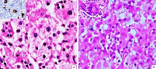
Fig 1 — a. Langerhans cell histiocytosis – histiocytes with grooved nuclei, H&Ex200. Inset: CD1a x200. b. Rosai Dorfman Disease–sheets of histiocytes with emperipolesis H&E x200. Inset: multinucleated giant cell exhibiting emperipolesis

Fig 2 — a.Erdheim Chester Disease– histiocytes with xanthomatous change, H&E x50. Inset: Magnetic resonance imaging showing bilateral symmetrical altered intensities in femur. b. Histiocytic sarcoma–sheets of plump histiocytes, H&E x50. Inset: CD68 x50.
Two cases with atypical morphology, one with an epithelioid appearance and other with a spindly morphology were categorized as unclassified histiocytic disorders as they were positive only for CD68 and negative for S100,CD1a andBRAF.
All LCH cases and HS were treated with chemotherapy. Mean follow up period was 14.7 months with a range of 1-30 months. During this period, four out of 23 cases recurred, which included two RDD cases (at two and six months of diagnosis), one case of LCH with ITP (after 27 months) and HS (after two months). Disease recurrence was observed in both siblings with RDD and HS.Ki67was high (60%) upon recurrence in HS. The immunomorphology of the both the recurrent RDD cases were different from the primary immuno-morphology wherein, one case showed focal positivity for CD1a, and the other case revealed atypical histiocytic proliferation. An infant with MS-LCH high risk succumbed to the illness within three weeks of diagnosis.

Fig 3 — a. Juvenile Xanthogranuloma – sheets of histiocytes dispersed amidst inflammatory cells. indicates multinucleated giant cell, H&E x50. b. Histiocytic disorder – unclassified: sheets and fascicles of histiocytes with spindled morphology, H&E x50.
Diagnoses of LCH was made based on morphology and immunoreactivity for CD1a, (Fig 1a inset) S100 and/ or CD68. Ki67 ranged from 3-30%. The immunoprofile of these cases are summarized in Table 3. The histiocytes in RDD cases were at least focally positive for S100and CD68. JXG was positive for CD68 and negative for CD1a and ALK.
ECD was diagnosed based on the classic clinical presentation, histomorphology and immunoreactivity for CD68. A diagnosis of HS was rendered after ruling out the possible morphologic differentials by using a panel of IHC markers. The neoplastic cells were positive for S100, CD68 (Fig 2b inset) and negative for CD23, SMA, CD34 etc, with Ki67 index of 20%.
Histiocytic neoplasms are commonly encountered in children and rarely in adults. The classification of histiocytic disorders has evolved over the years, from comprising of three categories in 2004 – L group (LCH, Indeterminate cell tumour, ECD, mixed LCH and ECD), C group (JXG, Adult xanthogranuloma, cutaneous RDD) and the M group (primary and secondary Malignant histiocytosis) to including two other categories namely the R group (RDD and its types) and the H group (primary and secondary Haemophagocytic Lymphohistiocytosis) in 2016 by the Histiocyte Soceity 4 to the current 5 th edition WHO classification of Haematolymphoid tumours. LCH is the most common disorder among histiocytic disorders accounting for approximately 5 cases/ million population per year with a male predominance 5 . In par with the literature, LCH constituted the bulk of the cases in this study (52.17%) showing a male predominance. LCH presents either as SS- LCH or MS- LCH and each account for 50% of cases 6 . In contrast, MS-LCH was slightly more common in this study (58.3%). According to a systematic review, skull bones (42.2%), chest wall (23.4%) are the frequently affected sites followed by the spine (9.4%) and pelvis (9.4%)7. Similarly, in this study, craniofacial bones were commonly involved with a rare case involving iliac bone. Other sites involved included skin, hematopoietic system, lung, and CNS6 In this study, BM of one LCH case was highly
Table 3 — Immunoprofile of Langerhans cell histiocytosis cases
Case CD68 S100 CD1a
Case 1Focal Positive Focal Positive Positive
Case 2 Positive Focal Positive Positive
Case 3Focal PositiveNon-Contributory Positive
Case 4Not DonePositivePositive
Case 5Negative PositivePositive
Case 6Not DonePositivePositive
Case 7Focal PositiveFocal PositiveFocal Positive
Case 8Not DonePositivePositive
Case 9Not DonePositivePositive
Case 10 PositivePositivePositive
Case 11Focal Positive PositivePositive
Case 12Not DoneFocal Positive Positive
suspicious of involvement but was rendered uninvolved as it was negative for CD1a. However, a study by Galluzzo ML, et al showed histiocytic clusters in 40.9% LCH cases, but only 14% cases showed CD1a immunoreactivity8. If these findings are taken, then this case may be considered to be involved. More studies are needed to determine the importance of histiocytic aggregates without CD1a expression in BM of LCH cases. A rare case of a child with ITP presenting with MS - LCHat adulthood has been reported, which was also the scenario in one of the cases9. Although, no theories related to this association are available at present, future studies might elicit relation between these two entities.
The diagnosis of LCH is supported byfocal or strong expression of CD1a, S100 and CD68 which was the case in our study as well2. Despite being known as a specific marker for LCH, CD1a can also be expressed in JXG, HS and RDD10. Ki67 performed in few cases in our study, was as high as 30%. Ki67upto 40% has been reported in literature and high Ki67 correlates with poor prognosis10,11. BRAF immunoreactivity, an indicator of MAPK pathway mutation is seen in >85% LCH cases10. Despite chemotherapy, the girl with ITP and MS-LCH involving the CNS presented with recurrent disease at a different site after two years. An infant diagnosed with MS-LCH high risk expired within few weeks of initiating treatment.The dismal prognosis of recurrent MS-LCH cases is well studied12
The global incidence of RDD (Sinus Histiocytosis with massive lymphadenopathy) is largely unknown1,5 Although, rare studies demonstrated a slight female preponderance, RDD is more common amongst males and this study consisted of four males and two females13,14. Clinically RDD can be nodal or extra nodal and they were seen in equal number of cases in this study as in literature. Extra nodal manifestations include cutaneous, intracranial, spinal, head and neck and intrathoracic sites15. CNS RDD accounts for <5%
of involved cases in literature and we had two cases of CNSRDD15. Although rare, relapses do occur in RDD with soft tissue and CNS involvement13. In this study, recurrence was observed in two multisite disease cases and one among them had a familial background as well. Classic morphology include slymphocytic emperipolesis, increased histiocytes with mild atypia and mitoses16. Atypical histiocytic proliferation was observed in one of the recurrent cases. The other recurrent case expressed only CD1a upon recurrence and this raised suspicion as to whether an overlap disease (RDD – LCH) was present initially or just the mere infrequent expression of CD1a in a RDD case10,14.
HS, a diagnosis of exclusion can present in variable age groups and the exact incidence is presently unknown due to changes in classification5. This can present either denovo or in association with other lymphoid neoplasms and may have an aggressive clinical course17. Available literature shows a slight male predominance whereas in this study, HS was diagnosed in a young girl1. Lymph nodes are usually affected in HS, but any extra nodal site like gastrointestinal tract, spleen, soft tissue, skin, CNS or orbit may be affected1. The child in this study had multisystem disease involving skin, bone and lymph nodes. The diagnosis was rendered after excluding various morphologic differentials like FDCS, IDCS, Langerhans cell sarcoma, inflammatory pseudotumor, Anaplastic Large cell lymphoma, melanoma etc,18Despite chemotherapy, this child presented with recurrent disease within two months17
Hereditary predisposition in RDD is due to germline mutations in SCL29A3 gene or TNFRSF6 gene2,19. No known hereditary factor has been implicated in HS as of now upon extensive literature search. A possibility of germline mutations in MAPK pathway may be considered due to its versatile role in histiocytic disorders1. As the siblings in this study had different histiocytic disorders, namely RDD and HS,further studies are required to adequately study the familial background in these disorders.
Fewer than 1000 cases of ECD have been reported in literature 5 . ECD often presents with a male preponderance between 46-56 years20. Paediatric cases infrequently show the classic adult clinical presentation of bilateral bone lesions2. The diagnosis of ECD is aided by the constellation of clinical (bone pain), radiographic (bilateral cortical sclerosis) (Fig 2a inset) and histological (foamy histiocytes which are negative for CD1a) findings, which was observed in the child in this study21. BRAF p.V600E mutation is seen in >50% of these tumours2. However, this case was negative for BRAFIHC21.
120, No 11, November 2022Journal
JXG is a paediatric histiocytic disorder having excellent prognosis, seen confined to skin of head and neck usually2. It accounts for 1 case/ million children and can undergo spontaneous resolution22. We had one case of JXG with a solitary lesion in the orbit expressing CD68. ECD is a mimicker of JXG and hence, the diagnosis should be made only in the appropriate radiographic and clinical context.
Rarely, histiocytic lesions can have ambiguous morphology and express only CD68. Tissue fixation and IHC pitfalls are key areas to be pondered in these unclassified cases. Overlap diseases like LCH – ECD do exist and awareness of these are vital for arriving at a correct diagnosis23
Histiocytic neoplasms are rare entities which can involve multiple systems. They can have overlapping histomorphology with various benign and reactive conditions thereby posing diagnostic challenges to the Pathologist. Familial RDD is well known, but further studies are required to identify familial relationship between these distinct entities. Although considered indolent, these entities can have a poor prognosis. Although there are many case reports and series, large scale Indian studies are still lacking and our study is one such initiative to collectively look upon the clinicopathological and IHC profile of these histiocytic neoplasms.
1Chan J, Alaggio R, Coupland SE — Histiocytic/Dendritic cell neoplasms: Langerhans cell and other dendritic cell neoplasms. In: WHO Classification of Tumours Editorial Board. Haematolymphoid Tumours [Internet; beta version (in progress)]. Lyon (France): International Agency for Research on Cancer; 2022.
2Picarsic J, Jaques TS — Histiocytic and dendritic cell neoplasms: Langerhans cell histiocytosis and related disorders. In: WHO Classification of Tumours Editorial Board. Paediatric tumours [Internet; beta version ahead of print]. Lyon (France): International Agency for Research on Cancer; 2022. (WHO classification of tumour series, 5th ed.; vol.7)
3Pileri SA, Grogan TM, Harris NL — Tumours of histiocytes and accessory dendritic cells: an immunohistochemical approach to classification from the International Lymphoma Study Group based on 61 cases. Histopathology 2002; 41(1): 1-29. doi:10.1046/j.1365-2559.2002.01418.x
4Emile JF, Abla O, Fraitag S — Revised classification of histiocytoses and neoplasms of the macrophage-dendritic cell lineages. Blood 2016; 127(22): 2672-81. doi:10.1182/ blood-2016-01-690636
5Cassidy DP, Rosenberg AE, Poveda J, Velez-Torres J, Chapman J — Nonlymphoid Hematopoietic Diseases Presenting in Bone, Soft Tissue, and Other Extranodal Sites. Arch Pathol Lab Med 2022; 146(9): 1144-59. doi:10.5858/ arpa.2021-0229-RA
6Rodriguez-Galindo C, Allen CE — Langerhans cell histiocytosis. Blood 2020; 135(16): 1319-31. doi: 10.1182/ blood.2019000934. :13.
7Reisi N, Raeissi P, Harati Khalilabad T, Moafi A — Unusual
sites of bone involvement in Langerhans cell histiocytosis: a systematic review of the literature. Orphanet J Rare Dis 2021; 16(1): 1. doi:10.1186/s13023-020-01625-z
8Galluzzo ML, Braier J, Rosenzweig SD, Garcia de Dávila MT, Rosso D — Bone marrow findings at diagnosis in patients with multisystem langerhans cell histiocytosis. Pediatr Dev Pathol Off J Soc Pediatr Pathol Paediatr Pathol Soc 2010; 13(2): 101-6. doi:10.2350/09-05-0651-OA.1
9Kuan YC, Nurain MN — Idiopathic thrombocytopenic purpura in childhood, Langerhans cell histiocytosis in adulthood: More than a chance association? Med J Malaysia 2017; 72(1): 50-2.
10Bedran NR, Carlos R, de Andrade BAB, Bueno APS, Romañach MJ, Milito CB — Clinicopathological and Immunohistochemical Study of Head and Neck Langerhans Cell Histiocytosis from Latin America. Head Neck Pathol 2018; 12(4): 431-9. doi:10.1007/s12105-017-0867-1
11Bartnick A, Friedrich RE, Roeser K, Schmelzle R — Oral Langerhans cell histiocytosis: antigen-Ki-67 as a indicator for local tumor behaviour. Anticancer Res 2003; 23(2A): 921-6.
12Al Abdulsalam A, Kapila K, Alherz M, Alsayegh M. Recurrence of a Langerhans Cell Histiocytosis bone lesion in a different site: A case report. Ann Med Surg 2022; 75: 103401. doi:10.1016/j.amsu.2022.103401
13Goyal G, Ravindran A, Young JR — Clinicopathological features, treatment approaches, and outcomes in RosaiDorfman disease. Haematologica 2020; 105(2): 348-57. doi:10.3324/haematol.2019.219626
14Bruce-Brand C, Schneider JW, Schubert P — Rosai-Dorfman disease: an overview. J Clin Pathol 2020; 73(11): 697-705. doi:10.1136/jclinpath-2020-206733
15Abla O, Jacobsen E, Picarsic J — Consensus recommendations for the diagnosis and clinical management of Rosai-Dorfman-Destombes disease. Blood 2018; 131(26): 2877-90. doi:10.1182/blood-2018-03-839753
16AlKuwaity KW, Alosaimi MH, Alsahlawi KT — Unusual Presentation of Rosai-Dorfman Disease: Report of a Rare Case. Am J Case Rep 2019; 20: 91-6. doi:10.12659/ AJCR.912423
17Shangpliang D, Baishya P, Raphael V, Dey B, Jagtap VK — Pediatric multifocal histiocytic sarcoma- a fatal diagnosis not to miss! Autopsy Case Rep 2020; 10(2) . doi:10.4322/ acr.2020.170
18Machado ES, de Miranda AC, Escopelli T, Caron R, Escopelli AC — Histiocytic sarcoma. Rev Bras Hematol E Hemoter 2011; 33(2): 155-7. doi:10.5581/1516-8484.20110038
19Chouk H, Ben Rejeb M, Boussofara L — Phenotypic intrafamilial variability including H syndrome and Rosai–Dorfman disease associated with the same c.1088G > A mutation in the SLC29A3 gene. Hum Genomics 2021; 15(1): 63. doi:10.1186/s40246-021-00362-z
20Goyal G, Heaney ML, Collin M — Erdheim-Chester disease: consensus recommendations for evaluation, diagnosis, and treatment in the molecular era. Blood 2020; 135(22): 192945. doi:10.1182/blood.2019003507
21Mazor RD, Manevich-Mazor M, Shoenfeld Y — ErdheimChester Disease: a comprehensive review of the literature. OrphanetJRareDis2013; 8(1): 137. doi:10.1186/1750-11728-137
22Haroche J, Abla O — Uncommon histiocytic disorders: Rosai–Dorfman, juvenile xanthogranuloma, and Erdheim–Chester disease.Hematology Am Soc Hematol Educ Program 2015; 2015: 571-8.doi: 10.1182/asheducation-2015.1.571.
23Ocak S, Bayramoglu Z, Tugcu D, Karaman S, Unuvar A, Karakas Z — Mixed Langerhans Cell Histiocytosis and Erdheim-Chester Disease in a Girl: A Rare and Puzzling Diagnosis. J Pediatr Hematol Oncol 2021; 43(3): e375-9. doi:10.1097/MPH.0000000000001770
Anupam Mandal1, Supratick Chakraborty2, Biswajit Ghosh3, Nandini Chatterjee4
Background : There has been a steady rise in the geriatric population in India and increasing number of elderly patients are being admitted in Critical Care Unit (CCU). They need mechanical ventilation during their hospital stay. Hence, there is continued need for evaluation and research to develop a validating scoring systems used to predict the outcome of CCU patients supported by mechanical ventilation.
Objective : Analysis to predict the outcome (survival or mortality) of mechanically ventilated elderly patients in different age groups at the CCU.
Material and Method : A Prospective observational study was done in CCU for a period of one year. A group of 40 elderly ventilated patients greater than 60 years of age (Group 1-elderly case group) and another group of 40 ventilated patients less than 60 years of age (Group-2- control group) were included in the study. A clinical database was collected which included age, sex, Acute Physiology and Chronic health Evaluation II (APACHE II) score and an Sequential Organ Failure Assessment (SOFA) scores were calculated in the first 24 hours of ventilation,indication of mechanical ventilation, co-morbidity, according to the Charlson Comorbidity Index (CCI), functional capacity according to the Barthel Index (BI). Patients outcome (survival or mortality) were analyzed. All the patients in two groups were on ventilation support.
Result : In case group (n=40), mortality was 55%. In control group (n=40), mortality was 52.5%. On comparison of outcome between two groups (case with control group) the difference was not statistically significant (p= 0.8225). In case group, association of outcome to different age groups (60-65 years, 66-75years, more than75years) (p=0.3357) andto gender (p=0.3854) was not statistically significant.
Multivariate logistic regression analysis of the study variables showed APACHE II score to be statistically significant for outcome (p=0.0229).
Conclusion : Mortality of elderly patients supported by mechanical ventilation at CCU were slightly higher(55%) than in mechanically ventilated younger populations (52.5%) though the difference was not statistically significant between two groups (p=0.82). APACHE II, score measured within 24 hours of ventilation was a significant predictor of mortality in the patients on mechanical ventilation.
[J Indian Med Assoc 2022; 120(11): 42-5]
Key words :Mechanical ventilation, Elderly, Critical care unit outcome, APACHE II Score.
There has been a steady rise in the population of elderly persons in India with 8.6% of population above the age of 60 years as per 2011 census which is projected to go over 10% by 20201
In this context, the hospital admission rate and demand for critical patient beds are expected to increase exponentially in the coming decade. Older age is characterized by emergence of several complex health states and there is a tendency to restrict their
Department of General Medicine, IPGME&R and SSKM Hospital, Kolkata 700020
1 MD (General Medicine), Associate Professor and Corresponding Author
2MD (General Medicine), Associate Professor, Department of General Medicine, Murshidabad Medical College and Hospital, Berhampore 742101
3MD (General Medicine), Ex-postgraduate Student
4MD (General Medicine), Professor
Received on : 31/08/2022
Accepted on : 21/10/2022
Editor's Comment :
Judicious use of mechanical ventilators in the elderly patients to be considered in the context of individualised risk-benefit ratio in a tertiary care centre.
admission to the Critical Care Unit (CCU). Present study compared the mortality outcome of elderly patients with younger individuals in a cohort of critical patients subjected to mechanical ventilation. This study prospectively compared the several variables on the patients requiring mechanical ventilation in CCU, to predict outcome.
Study period : A prospective observational study was done over a period of one year (October 2015 to September, 2016) in the Critical Care Unit of a tertiary care hospital.
Study population : The case group comprised 40 ventilated patients of age 60 years and above and control of 40 ventilated adult patients less than 60 years
120, No 11, November 2022Journal
of age, were included in the study.
Sample size calculation : Sample size was calculated by using the formula n=z(1- α/2)2pq/d2
z (1- α /2)
=1.96=value of the standard normal distribution corresponding to a significance level of á (1.96 for a 2-sided test at the 0.05 level). p= expected proportion of mortality from literature 50% or (0.5). q=1-p. d= absolute precision= 0.2(result to be with in 20% of true value).
Taking into account confounding factors for each variable, the sample size was increased by 10% of calculated value and we have taken 40 patients in each groups.
Study variables, inclusion criteria, exclusion criteria, informed consent and ethics committee: Acute Physiology and Chronic Health Evaluation II (APACHE II)2 score and an Sequential Organ Failure Assessment (SOFA)3 scores were measured in the first 24h of ventilation, age, gender, indication of mechanical ventilation, co-morbidity according to the Charlson Index (CCI)4, functional capacity according to the Barthel index (BI)5 were documented.
Those patients who died within 24 hours of ventilation were excluded from the study.
Informed consent from relative of patient was taken to include the patient in the study. The study was approved by the Clinical Research Ethics Committee of the Institution. The indications of mechanical ventilation were recorded based on the criteria of the Mechanical Ventilation International Study Group6 Table 1).
Detailed history and clinical examination of admitted patients were done. The criterion for CCU admission was decided by the admitting primary physician, based on the clinical condition of the patients. Patients from all specialties were admitted. No patients were refused admission in CCU based on the age. No treatment options were restricted to a
specific group of patients during CCU stay. Indication for which ventilation was initiated was noted. In this study, among case (n=40) and control group (n=40), consecutive patients were evaluated when they were ventilated. The records of parameters were taken within 24 hours of ventilation and subsequently daily for one week or until discharge or death, in all patients. Data was recorded which included age, sex, admitting diagnosis, APACHE II (acute physiology and chronic health evaluation) Score, Barthel Index (BI) Score, Charlson Comorbidity Index (CCI) Score, and Sequential Organ Failure Assessment (SOFA) Score, ABG (Arterial Blood Gas), Pulse rate, Blood Pressure, Temperature in Fahrenheit, Fractional inspiratory O2 concentration, Liver function test, Serum creatinine, Urea or BUN, urine output, Ventilatory rate. Complete Blood Count, and Serum Na, K.
No patients were re-intubated in this study among case and control group patients. The final outcome either survival or death was analyzed.
Demographic Details : In case group, the mean age (Mean ± SD) of patients were 71.1250±8.0166 years with range 60.00 - 92.00 years. Number of patients in case group, in 60 years to 65 years was 37.5%, in 66-75 years was 35.0%, and more than 75 years was 27.5%. In control group, the mean age (Mean±SD) of patients were 41.6750±12.0711 years with range 20.00 - 57.00 years (Fig 1).
Among 40 cases (n=40), 23 patients were male and 17 patients were female. Among 40 control patients, 22 patients were male and 18 patients were female (Fig 2).
Study variable findings
In case group,Barthel Index (BI) score mean ± SD was 12.0000± 13.0973 and in control group mean ± SD was 10.8750 ±12.5006 respectively. Difference of mean BI between two groups was not statistically significant (p=0.6954).
In case group,Sequential organ failure assessment (SOFA) score mean ± SD was 8.9250 ± 3.0834 and in control group,mean ± SD was 9.4250 ± 3.3350 respectively. Difference of mean SOFA between two groups was not statistically significant. (p=0.4884).
In case group,Charlson Co-morbidity Index Score (CCI) mean ± SD was 4.9750 ± 1.8326 with range of 1.0000- 9.0000 and in control group it was 1.9250 ± 3.4744 with a range of 0.0000-21.0000 respectively. The difference of mean CCI between two groups were statistically significant ( p<0.0001)

Fig 1 — Number of patients in control group(<60yrs) and case
In case group, APACHE II score mean ±SD was 23.7500 ± 6.3559 with range of 10.0000 - 41.0000 and In control group, it was Mean ± SD = 23.6250± 6.8713 with rang of 11.0000- 42.0000. Difference of mean between two groups was not statistically significant (p=0.9329).
Outcome in case group (n=40), mortality was 55%. In 60-65 years, mortality was 27.3%. In 66-75years, mortality was 40.9%. In >75years, mortality was 31.8%. Association of sub-groups of age with outcome in case group was not statistically significant (p=0.3357).
Association of gender (male mortality was 60.9%. female mortality was 47.1%) with outcome in case group was not statistically significant (p=0.3854).
The prediction of prognosis in respect to multiple variables like BI, SOFA, CCI, and APACHEII were calculated by regression analysis (Tables 2 & 3).
We found (Table 2) no variable had significant association with outcome in case group i.e in elderly patients. So in elderly patients, no single variable was a significant predictor of mortality.
We found (Table 3) APACHEII score was significant predictor of mortality in control group of patients. So in relatively younger patients, APACHEII score was

significant independent predictor of mortality.
DISCUSSION
Table 2 — Multivariate logistic regression analysis for case (n = 40)
CI = Confidence Interval; SE = Standard Error
Table 3 — Multivariate logistic regression analysis for control patients. (n =40) TermOdds ratio95%CI
CI = Confidence Interval; SE = Standard Error
We were prompted to do this study because of increasing number of elderly patients being admitted in CCU and their need for mechanical ventilation during hospital stay There is also reluctance among physicians to put the elderly patients in mechanical ventilation apprehending development of complications subsequently. So we wanted to see if there is any relationship between the mortality outcome and age of the patients. There are several studies that have described the poor results in elderly patients who were subjected to mechanical ventilation with ages over 65 years,7 70 years,8 80 years,9 85 years,10 Rosenthal, et al in a multihospital study concluded that the adjusted odds of death increased with each 5-years age increment11 Boumendil A, et al in their study concluded that after adjustment for disease severity, ICU mortality rates were higher in elderly patients than in younger populations and age itself explained only a small part of hospital mortality, suggesting that specific information such as functional, cognitive, and nutritional status as well as comorbidities, should be collected to predict mortality in elderly ICU patients12. Vosylius et al. had similar observation with 39% mortality in >75 years age group when compared with 18% in those <65 years (p<0.001)13 Stein, et al in a study concluded that age >76.9 years was an independent determinant of mortality (p<0.001)14. De Rooij, et al in a meta- analysis concluded that it is not age per se but factors such as severity of illness and pre-morbid functional status that are responsible for poor prognosis15
120, No 11, November 2022Journal
Anon JM, et al in a study in Spain, showed that mortality in the ICU was higher in the elderly patients (33.6%) than in the younger subjects (25.9%) (p=0.002)6
In India, a study by Sodhi, et al16 showed that no statistical difference was observed between the control and geriatrics age group in overall ICU mortality (p>0.05). However, mortality rates increased in the geriatric population requiring mechanical ventilation and use of inotropes during ICU stay.
In our study, mortality in elderly case group patients were 55% and mortality of control group patients were 52.5%. The difference was not statistically significant in comparison (p=0.82).
Here we found highest mortality was seen in 66-75 years of age (40.9%) which was higher than older age group( >75 years - 31.8%). But difference of mortality among sub- groups (60-65 years, 66-75 years,>75years) were not statistically significant (p=0.3357).
In a study by Sudarsanam TD, et al at CMCH, Vellore, India, concluded that APACHEII Score measured at admission was one of the independent predictor of mortality17 in the patients on mechanical ventilator. In a study by Nevins and Epstein 18 also showed that the APACHE II associated comorbidities predicted a poorer outcome for COPD patients requiring mechanical ventilation.
In our study, there is no significant difference in outcome in relation to age and in relation to gender. There is no significant difference in outcome in different subgroup of age of elderly people.
Mortality of elderly patients supported by mechanical ventilation at CCU were slightly higher (55%) than in mechanically ventilated younger populations (52.5%) though the difference was not statistically significant between two groups (p=0.82). There should not be any reluctance for ventilation initiation for elderly patients for fear of poor outcome. In CCU, APACHE II, score which has a comparatively high sensitivity in predicting mortality, will be useful to guide to the physician on probable outcome and management decision.Co-morbidity should not restrict the decision for ventilation initiation..
Limitations : Morbidity profile could not be studied here as it needed a long term follow up. Duration of ventilation is not studied here which could be another determinant factor for outcome. Single Centre, short time, small number of study and control population has the limitation of decision making. To draw a inference multi-centre long term study with large number of population will be needed. It is a score based
study, individual organ function related study has not been done.
1Malik C, Khanna S, Jain Y, Jain R — Geriatric population in India: Demography, vulnerabilities and health care challenges. J Family Med Prim Care 2021; 10(1): 72-6.
2Kress PJ, Hall BJ — Approach to the patient with critical illness. In :kasper , Fauci, Hauser, Longo, Jamseson , Loscalzo(eds) Harrisons Principle of internal medicine. 19 th ed. Vol-2. McGraw Hill Education. 2015. pp-1729.
3Lambdens, Laterre PF, Levy MM, Francois B — The SOFA score—development, utility and challenges of accurate assessment in clinical trials. Crit Care 2019; 23(374): 1-9
4Charlson ME, Pompei P, Ales KL, MacKenzie CR — A new method of classifying prognostic comorbidity in longitudinal studies: development and validation. J Chronic Dis 1987; 40(5): 373-83.
5Mahoney FI, Barthel DW — Functional evalution: the Barthal index. Md State Med J 1965; 14: 61-5.
6Anon JM, Tello-Gomez V, Higueras-Gonzalez E, Corcoles V, Quintana M, Lorenzo AGD, et al — Prognosis of elderly patients subjected to mechanical ventilation in the ICU. Med Intensiva 2013; 37(3): 149-55.
7Lieberman D, Nachshon L, Miloslavsky 0, Dvorkin V, Shimoni H A, Zelinger J, etal— Elderly patients undergoing mechanical ventilation in and out of intensive care units: a comparative, prospective study of 579 ventilations. Crit Care 2010; 14: R48
8Esteban A, Anzueto A, Frutos-Vivar F, Alia I, Ely EW, Brochard L, et al — Outcome of older patients receiving mechanical ventilation. Intensive Care Med 2004; 30: 639-46.
9Somme D, Maillet JM, Gisselbrecht M, Novara A, Ract C, Fagon JY — Critically ill old and the oldest-old patients in intensive care: short- and long -term outcome. Intensive Care Med 2003; 29: 2137-43.
10Ip SP, Leung YF, Ip CY, Mak WP — Outcomes of critically ill elderly patients: is high-dependency care for geriatric patients worth while? Crit Care Med 1999; 27: 2351-7.
11Rosenthal GE, Kaboli PJ, Barnett MJ, Sirio CA — Age and the risk of in hospital death: insights from a multi hospital study of intensive care patients. J Am Geriatr Soc 2002; 50: 1205-12.
12Boumendil A, Somme D, Garrouste-Orgeas M, Guidet B — Should elderly patients be admitted to the intensive care unit? Intensive Care Med 2007; 33: 1252-62.
13Vosylius S, Sipylaite J, Ivaskevicius J — Determinants of outcome in elderly patients admitted to the intensive care unit. Age Ageing 2005; 34: 157-62
14Stein FD, Barros RK, Feitosa FS, Toledo DO, Silva Junior JM, Isola AM, etal— Prognostic factors in elderly patients admitted in the intensive care unit. Rev Bras TerIntensiva 2009; 21: 255-61.
15de Rooij SE, Abu-Hanna A, Levi M, de Jonge E — Factors that predict outcome of intensive care treatment in very elderly patients: A review. Crit Care 2005; 9: R307-14.
16Sodhi K, Singla MK, Shrivastava A, Bansal N — Do intensive care unit treatment modalities predicts mortality in geriatrics patients :An observational study from an Indian intensive care unit. Ind J of Crit Care Med 2014; 18(12): 789-95.
17Sudarsanam TD, Jeyaseelan L, Thomas K, John G — Predictor of mortality in mechanically ventilated patients. PostgradMed J 2005; 81: 780-3.
18Nevins ML, Epstein SK — Predictors of outcome for patients with COPD requiring invasive mechanical ventilation. Chest 2001; 119: 1840-9.
Prashanth
Mada1, R Shyamala2, Pragnesh Parmar3, Yadukul S4, Divya Reddy Pannala5
The changing disease burden, living conditions and anticipations of end-users in health care have resulted in the decision of regulatory bodies in the Indian medical education system to shift the MBBS curriculum from Traditional to Competency-Based Medical Education (CBME).The efforts taken by the National Medical Commission (NMC) erstwhile Medical Council of India (MCI) to successful implementation of the herculean task of shifting curriculum are praiseworthy. MCI initiated a National Faculty Development Programme (FDP) in 2009 in all medical colleges under its ambit.MCI started with five regional centres, which now expanded to 22 centres, out of which 12 are regional centres, and 10 are advanced nodal centres. Nearly 44932 faculties were trained till December 2018. Despite all the advantages of CBME & the efforts taken up by governing bodies, there are many challenges. Some are common for all subjects, but few are unique or specific to a subject. This article views the possible challenges and the way forward for the successful implementation of CBME in Forensic Medicine and Toxicology.
[J Indian Med Assoc 2022; 120(11): 46-9]
Key words :Medical Education, CBME, Forensic Medicine & Toxicology, National Medical Commission, Indian Medical Graduate.
The changing disease burden, living conditions & anticipations of end-users in health care have resulted in the decision of regulatory bodies in The Indian medical education system to shift the MBBS curriculum from Traditional to Competency-Based Medical Education (CBME). Frank and colleagues state that CBME is “an approach to preparing physicians for practice that is fundamentally oriented to graduate outcome abilities and the organization around competencies derived from an analysis of societal and patient needs1.”
There is no doubt that the CBME is superior, valuable and essential in the present scenario for making a competent Indian Medical Graduate (IMG)2. The advantages of CBME are innumerable and a few to mention are that it focuses on outcomes, which ensures that the IMG is competent enough to practice. It deemphasises time-bound learning, which addresses the struggles of slow learners. Promoting student-centric training, which synergistically acts with the deemphasizes of time-bound learning2.
Department of Forensic Medicine & Toxicology, AIIMS, Bibinagar, Telangana 508126
1MBBS, MD (Forensic Medicine), ACME, Assistant Professor and Corresponding Author
2MD (Microbiology), ACME, FAIMER, Associate Professor, Department of Microbiology
3MBBS, MD(FM), FAIMER, PGDHL, Additional Professor
4MBBS, MD, PGDMLE, Associate Professor
5MBBS, MD (Forensic Medicine), Senior Resident
Received on : 23/06/2021
Accepted on : 10/10/2022
Editor's Comment :
In the making of a globally competent Indian Medical Graduate to serve the community effectively, Competency Based Medical Education (CBME) introduced by National Medical Commission will play a significant role. Challenges will be there in the implementation of CBME But collaborative effort of all stake holders involved will bring out the desired outcome.
The traditional curriculum, which focuses on the cognitive domain, was replaced by CBME which gives balanced learning of all the three domains viz Cognitive, Psychomotor & Affective. More stress on learning skills and improving affective domain than on retention of mere knowledge. The efforts taken by the National Medical Commission (NMC) erstwhile Medical Council of India (MCI) to successful implementation of the herculean task of shifting curriculum are praiseworthy. MCI initiated a National Faculty Development Programme (FDP) in 2009 in all medical colleges under its ambit. MCI started with five regional centres, which now expanded to 22 centres, out of which 12 are regional centres, and 10 are advanced nodal centres. Nearly 44932 faculties were trained till December, 20183
The long-awaited shift of Forensic Medicine and Toxicology subject to third MBBS was possible with introducing a new curriculum. An increase in the University examination marks and an increase in the teaching hours from 100 to 125 hours are all welcome changes.
Despite all the advantages of CBME & the efforts taken up by Governing bodies, there are many
120, No 11, November 2022Journal
challenges. Some are common for all subjects, but few are unique or specific to a subject. This article views the possible challenges and the way forward for the successful implementation of CBME in Forensic Medicine & Toxicology.
Challenges :
The challenges can be discussed under two groups viz, stakeholders & methodology.
Stakeholders : Policymakers, regulatory bodies, management/administration of colleges, faculty & students.
Methodology : The guidance provided by the regulatory body in the form of training, documentation of competencies, integration, assessment, AETCOM, etc.
Policy Makers :
Acquisition of competency is a critical component of CBME. In Forensic Medicine & Toxicology, conducting and preparing post-mortem examination reports of varied aetiologies in a supervised environment is the desired competency. To acquire these competencies, one should observe real cases under supervision in a mortuary. This particular competency cannot be acquired in a simulated environment.
Mere changing of curriculum & defining competencies without making specific guidelines mandatory will not change the ability of IMG to perform better. So, to make the best use of the CBME, specific guidelines like compulsory post-mortem work in all medical colleges, irrespective of government or private, should be made mandatory. There are approximately 250 government and 300 private medical colleges in India. Out of the 300 private medical colleges, less than 20% do post-mortem work. So, all the IMG’s graduating from the rest of the private colleges will be incompetent.
MBBS doctors are doing the majority of post-mortem work in the country4. Data regarding the number of autopsies done in India is not available. Total suicidal deaths in 2015 were 1,33,623, accidental deaths were 413457, including traffic accidents. Considering these numbers, at least 6-7 lakh autopsies in India might have been done in 2015. Out of these, the majority of autopsies have a little value or at times opposite effect due to them being done by incompetent doctors4. Many students of private colleges also do not have exposure to the examination of victims/accused of sexual offence cases.
Regulatory bodies (NMC / MCI) :
Why was 2019 selected for the shift from traditional to competency-based?
For students who aspire to pursue a medical career in foreign countries, one of the essential criteria is to
graduate from an accredited medical school by (World Federation for Medical Education (WFME). The WFME has announced that from 2024 CBME curriculum will be taken into consideration for accreditation of medical institutes for ECFMG (Educational Commission for Foreign Medical Graduates)5 For this requirement and the welfare of the students, the regulatory body had to shift to a new curriculum in 2019.
Administrative (Managements) :
Not all private medical colleges are encouraging Medical Education Units & do not conduct faculty development programs. The regulatory body has recently amended the teacher eligibility qualifications in medical institutions and mandated completion of the Basic course in Medical Education Technology from Institution(s) designated by MCI to encourage private medical colleges for conducting faculty development programmes.Still, the majority of private medical colleges do not provide financial support to the faculty for registration, travel and accommodation6 Recruitment of additional faculty beyond minimum standard requirements to handle small group teaching may not be encouraged by some of the private institutions. Even if the Government grants permission to conduct medico-legal autopsies, most private medical college managements in Telangana may not be interested in taking up medico-legal autopsies as they are not profitable.
Faculty :
As per the National Medical Council website, the total number of faculty is 96649, out of which nearly 44,932 were trained in CBME. Hence nearly 50% are yet to be trained, a hurdle for successful, smooth implementation of the new curriculum. So active measures are required to increase the number of faculty trained in CBME. A reasonable number of faculty trained in the new curriculum are also not confident enough, as they were trained for or studied traditional curriculum. For them, the shift is a little cumbersome. Still, some are reluctant to adapt to the new shift.However, faculty should change their mindset and adapt to unlearn and relearn.
Students :
The new curriculum has advantages and lacunae. It is better structured and incorporates more critical skills, like basic life support, communication skills, and ethical issues, which benefits students. There is no clarity regarding the exit exam. To write a proper reflection, training the students is crucial. In CBME, not only does the teacher gives feedback to the student, but the student also gives feedback to the teacher in the form of reflective writing. Reflective writing is sharing
120, No 11, November 2022Journal
a student’s experience of the session, which will improve in teaching in the future.
Competencies :
Competency is the ability to do something successfully or proficiently.7
The efforts to create and consolidate Undergraduate Curriculum, Volume I,II & III must be lauded, and many changes suggested are progressive and welcome, though there are some minor errors.However, it is unclear why the MCI has stopped providing only competencies and why not Specific Learning Objectives’? Another challenge was that a few of the essential documents related to the implementation of CBME were not provided before the onset of the process, which created confusion.
Specific Learning Objectives (SLOs) :
An SLO describes what the learner must do upon completion of educational activity. It should be specific, measurable, attainable, relevant and time-bound8 Many feel challenged to devise their own SLO’s; most of them are prepared by a few and copied by the rest.
Teaching Learning Methods :
According to the new curriculum, there is more focus on small group teaching. Small group learning is defined as a process of learning that takes place when students work together in groups of 8-10. According to this definition, if 100 students are there in an institute, we may at least require 5-7 faculty to conduct a session of effective small group teaching9 Instead of increasing the faculty strength, the regulatory body has decreased the faculty strength in forensic medicine & toxicology. At the same time, to conduct small group teaching, we require proper infrastructure and teaching aids, which are not adequate in most government and private institutes.9 The class can be divided into small batches and can be rotated among the second or third MBBS subjects, but faculty has to repeat, and there will be a class almost every day. The best possible solution to address this issue can be increasing the faculty strength in Forensic Medicine and Toxicology on par with other subjects.
Integration & Alignment :
Alignment implies teaching subject material that occurs under a particular organ system/disease concept from the same phase in the same time frame, ie, temporally.
Integration implies that concepts in a topic/organ system that are similar, overlapping, or redundant are merged into a single teaching session where subjectbased demarcations do not exist.10
The main aim of integration & alignment is to prevent
repetition and make the best use of time. However, the NMC has given no clear guidelines as to who has to take the topics of integration, leading to the defeat of the main objective of integration.
Faculty coordination has changed a lot, but it requires changing more to implement newer methodologies like integration & alignment successfully.
Assessment :
A robust assessment is key to the successful implementation of CBME. The focus from summative assessment has shifted to formative assessment with the change in curriculum. Feedback is an important integral part of formative assessment.
Many medical teachers are getting trained from the faculty development programmes but still, a reasonable number of them are not trained. The trained faculty are aware of the terminology of various activities in CBME, but to master them, it needs practice and time. For example, the art of giving feedback to students as it should be timely, practical, sufficient, and by using the sandwich technique.
Logbook versus Practical record :
The Logbook is a verified record of the progression of the learner documenting the acquisition of the requisite knowledge, skills, attitude and competencies11
It is still unclear whether the logbook is a replacement or an addition to the practical record and what entries need to be recorded in the logbook? Who will record the details in the logbook? If it is the responsibility of the faculty, then the faculty should first assess the student for an activity, enter those details into the logbook. If the student does not attain the desired level, then feedback should be given, suggesting remedial measures, reassessing the student till the desired level is achieved. However, with the available number of faculty in the Department, it will be a herculean task11
Skills Training :
The following are certifiable skills mentioned in Forensic Medicine & Toxicology specialty :
1.Documentation and certification of trauma (I)
2.Diagnosis and certification of death (D)
3.Legal documentation related to emergency cases (D)
4.Certification of medical-legal cases, e.g., Age estimation, sexual assault etc. (D)
5.Establishing communication in medico-legal cases with police, public health authorities, other concerned departments, etc12
There is no mention of medico-legal autopsies in the certifiable skills. Furthermore, as per data, more than
120, No 11, November 2022Journal
80% of Medico-legal autopsies in the country are done by MBBS qualified Doctors. There is a need to revisit the competencies and certifiable skills in the subject.
AETCOM :
In the old curriculum, the Forensic Medicine Department only dealt with medical ethics. To an extent, it is not tricky for forensic medicine faculty to teach medical ethics and law. However, due to the introduction of Attitude, Ethics & Communication (AETCOM) into the new curriculum, it is not clear whether MEU or any specific department is responsible for AETCOM. Are all faculty well trained to teach the different components of the affective domain is a big question? More clarity should come regarding what tools are used to assess the students in this domain?
Electives :
An elective is a learning experience created in the curriculum to allow the learner to explore, discover and experience areas or streams of her/his interest. Two choices of electives are offered to medical students before the commencement of III MBBS part 2, called Block 1 and Block 2. Each block is of 4 weeks duration and is allowed before the III MBBS, part 2. In the module released by NMC, there is mention of examples of block 1 & 2 learning experiences; however, Forensic Medicine & Toxicology topics have found no place in electives. It needs to be included in electives as well13
COVID-19 :
The pandemic has affected all walks of life, including medical education. No one anticipated or was prepared for such a scenario, but despite all the hurdles, everyone got adjusted to the virtual platform rapidly for teaching, assessment, CME’s & treating patients. One of the learnings from the pandemic is a backup plan in the curriculum to deal with such situations. In addition to all the challenges described above, it was not possible to follow CBME or timelines since last year. Due to virtual sessions, many compromises have been made in teaching, learning, and assessment. There is a need for guidance on how these issues will be tackled in the ongoing pandemic time.
Way forward :
Despite all the challenges, collective proactive participation from faculty of all the Departments, students & administration will help in the successful implementation of the new CBME curriculum. The barriers between the departments should be erased, and each faculty, irrespective of the cadre, should be highly motivated to participate & identify their new role as facilitator.
NMC should take measures for successful implementation of CBME. Increasing faculty strength
can be one of the strategies.
The administration should encourage Medical Education Units in their respective colleges and implement regular faculty development programmes. Teaching institutions must incentivize high-quality teaching and leadership that contribute to the professional development of trainees and reward engaged faculty, for example, promotion and monetary benefits.
Differences between Government & Private Institutions should disappear in the student’s best interest. Uniformity across institutes among forensic departments should be attained.
There should also be training programs for the students in various new aspects like reflective writing, how to take feedback properly given by mentor, etc.;
Proper evaluation of the programme should be done at regular intervals to identify the lacunae and appropriate measures should be undertaken to fill the lacunae. CBME can raise the standards of medical education in India, but it can only achieve this goal if intensive planning and support from all stakeholders. Implementing any new programme will have hurdles, but those hurdles can become stepping-stones with collective effort.
1Frank JR, Mungroo R, Ahmad Y, Wang M, De Rossi S, Horsley T — Toward a definition of competency-based education in medicine: a systematic review of published definitions. Med Teach 2010; 32(8): 631-7.
2Frank JR, Linda S, Ten Cate O, Eric SH, Carol S, Susan H — Competency-based medical education: Theory to practice. Medical Teacher 2010; 32(8): 638-45.
3Medical Council of India. Faculty Development initiatives for the Undergraduate Medical Education Program, ten years’ experience –status report, 2020: pp 1-24.
4Sengupta R — The autopsy report. Mint. 2018 Sep 07. [Cited 2021 Feb 25]. Available from: https://www.livemint.com/ Politics/Lofa7q6gweBypR05IoERrO/The-autopsy-report.html
5Ahn D — Current trend of accreditation within medical education. J Educ Eval Health Prof 2020; 17: 30.
6Board of Governors in supersession of Medical Council of India. Minimum Qualifications for Teachers in Medical Institutions (Amendment) Regulations, 2019. No. MCI-12(2)/ 2019-Med.Misc./189334 (12th February, 2020)
7Ten Cate O — Competency-Based Postgraduate Medical Education: Past, Present and Future. GMS J Med Educ 2017; 34(5): Doc69.
8Chatterjee D, Corral J — How to Write Well-Defined Learning Objectives. J Educ Perioper Med 2017; 19(4): E610.
9Meo SA — Basic steps in establishing effective small group teaching sessions in medical schools. Pak J Med Sci 2013; 29(4): 1071-6.
10Medical Council of India — Alignment and Integration Module for Undergraduate Medical Education Program, 2019: 1-34.
11Medical Council of India — Guidelines for preparing Logbook for Undergraduate Medical Education Program, 2020: 1-23
12Medical Council of India — Skills Training Module for Undergraduate Medical Education program, 2019: 1-49.
13Medical Council of India — Electives for the Undergraduate Medical Education Training Program, 2020: 1-30.
Arnab Bhattacharyya1
Due to increase in longevity, the number of people in geriatric age group is increasing day by day. Due to multiple physical and socio-economic problems they have to depend upon care-givers and sometimes circumstances compel them to stay in “old age homes”. Addressing their problems, care-givers or support-staffs of these institutions should be trained in user-friendly as well as patient-friendly screening procedures.
Key words :Geriatrics Screening, Support Staff.
Though medical research often defines person as elderly, when he is of 65 years or above; defining elderly age by chronology alone has its limitations. Although WHO defines persons as elderly, if they are 65 years or older, for monitoring of demographic and socio-economic profile in Africa, a person is defined as elderly, if they are 50 years or older (United Nations 2012)17th September 2015. Again as per OECD (Organization for economic co-operation and development-an organization of 38 countries) data, the elderly population is defined as people aged 65 years and above1
As per 2011 census, there are 104 million older people (60+years) in India, constituting 8.6% of total population and among the elderly, females outnumber males2
Problems :
Ageing is a natural phenomenon, being governed by theories like defects in mitochondrial DNA repair, Telomere attrition etc and manifested by multiple problems viz, Sarcopenia, Cancer, Dementia, Cataracts, Heart Disease, Arthritis, Osteoporosis, Immuno-senescence, frailty (thus coining terms viz, multimorbidity) and thereby increases the mortality and morbidities of this vulnerable population. This condition of multimorbidities invites another problem ie, polypharmacy, as the effects as well as side-effects of many drugs are potentiated in this old age group.
Though older people are a valuable resource for any society, increase in longevity and decline of joint family and breakdown in social fabric pushes seniors into loneliness and neglect. Here comes the role of “Care-givers” and the concept of “old age homes”,
1MD (General Medicine), FICP, Assistant Professor, Department of Medicine, Barasat Government Medical College, Kolkata 700124 and and Corresponding Author
Received on : 06/09/2022
Accepted on : 01/11/2022
[J Indian Med Assoc 2022; 120(11): 50-3]
Editor's Comment :
Support staff play an important role in addressing the multimorbidities of geriatric age group.In fact they being the missing links between the treating physician and elderly persons living in institutions like "Old age home" or in remote places, they should not be overburdened. And as such a simple,user-friendly, ready to use screening tool must be there to address the different problems old age, so that the treating doctor can intervene at the ealiest, according to gravity of the situation.
where these old people can live safely in a community, with availability of nursing home level care, as and when required.
Proposals :
So to maintain a comprehensive care of this vulnerable population, there must be a guideline for the support-staffs of these institutions, like one exists as” 5M’s of geriatrics” (mentation, medication, mobility, multicomplexity and matters)3, meant to optimize utilization of existing resources during hospitalization of older adults,by focusing on key geriatric issues. So, there must be a screening procedure to address the problems of this vulnerable population, which would be comfortable not only to treating doctors, but also to support-staffs of these institutions.
Hence, a quick reminder for Care providers or support staff of elderly is being proposed-its in the form a mnemonic,"PAID-IVF".
P — Pain (chronic pain due to any cause viz, Cancer, Arthritis, Osteoporosis etc); Paralysis
A — Anxiety; Altered bowel habits
I — Insomnia
D — Depression; Drugs; Dentures; Difficulties in breathing
I — Incontinence; Impaired hearing
V —Voluntary movements; Vaccination; Visual impairment
F — Falls (due to cardiovascular or neurological or orthopedic causes); Forgetfulness
120, No 11, November 2022Journal
Pain & Paralysis :
Degenerative spine and arthritis related disorders of musculoskeletal systems are the most common cause of chronic pain in the elderly. Ischemic pain, neuropathic pain and cancer related pain are other important contributors. Prevalence of vertebral compression fractures is high in geriatric women resulting pain and discomfort. Identification of pain, assessment of intensity using different subjective tools, impact on quality of life, non-pharmacological therapy including physiotherapy, cognitive behavioural therapy and introducing pharmacotherapy following the concept of ‘analgesic ladder’’ after adequate informed counselling by the prescriber. Analgesic ladder concept includes assessment of intensity followed by specific drugs. For (a) mild pain - the first choice is paracetamol, (b) mild to moderate pain or pain uncontrolled with paracetamol - NSAIDs, (c) Pain refractory to NSAIDs - weaker opioid agonist like tramadol (d) For pain refractory to the previous plan, or severe pain - pure opioid agonist like morphine, pethidine, buprenorphine, trapentadol like adjuvant medication may be added for synergism with the existing medications4.
Paralysis of limbs needs urgent attention for care givers. Paresis or complete paralysis; flaccidity or spasticity; location of paralysis are different issues which need to be understood by care givers and communicated promptly to physicians for early interventions.
Anxiety & Altered bowel habits :
Anxiety is common in elderly, 10% of adults over the age of 65 estimated to have an anxiety disorder. It includes generalized anxiety disorder, panic disorder and Post-Traumatic Stress Disorder (PTSD). As stressful life events are very common in elderly like experiencing recent losses in the family or suffering from a chronic illness, development of an anxiety disorder is common. Caregivers should be adequately trained to identify these conditions to address promptly to doctors5.
Altered bowel habits in elderly need vigilant approach. Many cases these are associated with irritable bowel syndrome which requires proper assurance and education. In a few cases they should be counselled for warning signs like frequent alteration of bowel habits, bleeding per rectum, weight loss which requires urgent follow up by physicians. Chronic constipation is a common problem in this age group,which should be initially dealt with life style modification,such as scheduled toileting after meals, increased fluid intake,increased fibre intake; and if not
relieved,then with the judicious use of drugs along with necessary investigations (if required).
Insomnia :
Prevalence of insomnia symptoms is very common in geriatric population ranging between 30% to 48%. Complain of difficulty falling or maintaining sleep, or non-restorative sleep, producing significant daytime symptoms including difficulty concentrating and mood disturbances are concerning complications in elderly which affect their quality of life. Clinical diagnosis of insomnia is extremely crucial considering factors like demographic, psychosocial, biologic and behavioural issues. Late-life insomnia results in increasing risk of different medical and psychiatric illnesses. Caregivers should be adequately educated to identify insomnia and for providing non-pharmacological management6
Depression, Drugs, Dentures & Difficulties in breathing :
4 ‘D’s including “Depression”,”Drugs”, “Dentures” and”Difficulties in breathing” need special attention while caring elderly population.
Depression in elderly population is a common psychiatric disorder affecting quality of life significantly. Study conducted in USA had suggested that 2% of adults aged 55 years or older are suffering from major depression. With increasing age prevalence rises. Clinically significant depressive symptoms were found in 10% to 15% of older adults. Undetected or inadequately treated depression is common in geriatric population. Antidepressants, psychotherapy, exercise therapy, and in limited cases electroconvulsive therapy have some role. Patients suffering from mild to moderate severity depression require psychotherapy. Same doses of antidepressant medications are required in elderly population like younger adults. It is important to educate care givers to raise awareness regarding adverse effects of antidepressants, interactions with other medical comorbidities and drug-drug interactions. Use of drug therapy for depression in patients with dementia is not beneficial as per high quality evidences7
Drugs and related issues like PK-PD changes with ageing, polypharmacy, drug-drug interactions are important concerns in elderly. Medication history and using medicine card (brown bag concept) need to be taken care and treating physician should be explained by care givers about ongoing therapeutics in great details. They should be aware regarding common adverse effects associated with drugs, so that they can presume them early and seek medical care attentions. Adherence is another issue need to be
120, No 11, November 2022Journal
emphasized while discussing drugs. Screening Tool of Older People’s Prescriptions (STOPP) and Screening Tool to Alert to Right Treatment (START) criteria are important tools to prevent inappropriate prescribing in elderly, detailed medication history provided by care givers including history of OTC drug intake, AYUSH drugs are essential to ensure appropriate prescribing 8 . Elderly persons are susceptible for prescribing cascade related atrocities. Early diagnosis and timely deprescribing can prevent this9. Caregivers should be adequately trained for providing detailed medication history.
Eating, social interaction and communication difficulties along with compromised quality of life are commonly associated problems with dentures in geriatric population. Information about oral health in geriatric population needs to be generated. To raise the confidence and self-esteem of geriatric population with poor oral health need to establish access of proper therapy, affordable quality artificial dentures and proper counselling is needed. Care givers should be adequately educated to take care of elderly patients with dentures10
Difficulties in breathing should be assessed by care givers using pulse oxymeter, respiratory rate counting, blood pressure and pulse measurement. They should be adequately trained to check any bluish discoloration of tongue or any specific part of body. In case of provision of telemedicine, video consultation they should be adequately trained on JVP examination posture and location to focus by camera. In case of increasing pedal swelling they should be counselled to monitor sacral oedema and how to examine it.
Incontinence & Impaired hearing :
Urinary incontinence(accidental or involuntary loss of urine from the bladder) is another important issue in this age group,as urine incontinence makes an individual feel isolated,rejected from the society and make them anxious and dependant on others.Nearly 5 to 15 percent of men with the age of 60yrs, and above suffer from urinary incontinence 11 . Urine incontinence may be broadly classified into stress incontinence,urge incontinence,overflow incontinence etc, and may be due to various reasons, which need special attention by an expert in this field,such as Urologist. Women generally suffer from stress or urge incontinence. There may be embarrassing situations like “Nighttime incontinence” in the elderly, which may be due to diabetes, urinary tract infections, side effects of medications, neurological disorders, anatomical abnormalities, overactive bladder, enlargement of prostate and many other causes (or rarely due to
anxiety or emotional problems); which are to be addressed accordingly.
Impaired hearing is another problem,which may cause depression,anxiety,social withdrawal,frustration or decline in cognitive skills in older adults.Hearing loss may be accompanied by tinnitus, another irritating problem. Presbycusis or age related hearing loss may run in families and a person,suffering from this ,cannot tolerate loud sounds or may not understand clearly,what others are saying12
Voluntary movements, Vaccination & Visual impairment :
There may be problems with voluntary movements due to neurological, cardiovascular, locomotor (arthritis) or many other causes, which are to be addressed sympathetically both by the treating physician and the support staffs or care givers
Vaccination is a big issue as the vaccines like influenza and pneumococcal vaccines are safe and effective.Decreased immunity along with physiological changes, poor health and multiple co-morbidities make this population vulnerable to various infections. Infections like pnemococcal, influenza, tetanus, zoster and COVID-19 are major causes of morbidity and mortality of this age group, causing large number of deaths and hospitalisations, thus further strengthening the need for a comprehensive immunisation programme of this age group.
As per the LASI (Longitudinal Aging Study In India, Wave 1) data prevalence of blindness is higher among the elderly (60 years and above) residing in rural areas (4.3%) than among those in urban areas (2.7%)13. The most common causes of vision loss among the elderly are age related macular degeneration, glaucoma, cataract and diabetic retinopathy12. In a further study regarding “Falls and visual impairment among elderly residents in ‘homes for the aged ‘ in India Srinivas Maramula, et al showed that the prevalence of falls was higher among those with visual impairment due to uncorrected refractive errors and this was more so in elderly individuals living in ‘ homes for the aged ‘ in Hyderabad, India14. So addressing impairment of vision may result in fewer falls and thus contribute to healthy ageing in addition to other benefits of correction of vision for this age group.
Falls & Forgetfulness :
Falls in the elderly are a common and serious health problem with devastating consequences, for which various risk factors have been attributed and thus falls can be prevented through several evidence based interventions. So identifying at-risk patients is the most important part of management, as applying various
120, No 11, November 2022Journal
preventive measures in this vulnerable population can have a profound effect on public health. So treating physicians and accompanying care-givers must be very much vigilant about screening of older patients, who are at risk of falls, so as to apply preventive measures as and when required.
Regarding forgetfulness, this can be stated that age related memory loss and dementia are two different conditions though they may share some overlap in symptoms. Though normal forgetfulness, often caused by lack of focus, never progresses into serious territory, whereas dementia will generally get worse over time15 Memory and other thinking problems may be due to depression, infection, injury or side-effects of medication and sometimes cognition improves if the problem can be identified and treated successful. But the course of the problem like a serious brain disorder, such as Alzheimer’s disease will go more or less downhill. So a treating physician as well as a caregiver should be vigilant about the fact that, whether the loss of memory is accompanied by warning signs like impairment in reasoning skills, judgement, language or there is personality disorder or not .
However the mnemonic “PAID IVF” is, so as to say, user-friendly, almost self-explanatory, and easy to remember the geriatric problems—which are to be dealt with, according to the importance and gravity of the situation. Obviously, enquiries by the support staffs of “old age homes” or “Health care facilities” (specially designed for elderly people) are to be made in local dialects/vernaculars, so as to make it patient-friendly.
1OECD (2022), Elderly population (indicator). doi: 10.1787/ 8d805ea1-en (Accessed on 05 September 2022)
2Lekha S, Bansod DW — Demographics of Population Ageing in India: Trends and Differentials, BKPAI Working Paper No. 1, United Nations Population Fund (UNFPA), New Delhi 2021.
3Molnar F, Frank CC — Optimizing geriatric care with the GERIATRIC 5Ms. CanFamPhysician 2019; 65(1): 39. PMID: 30674512; PMCID: PMC6347324.
4Ali A, Arif AW, Bhan C, Kumar D, Malik MB, Sayyed Z, Akhtar KH, etal— Managing Chronic Pain in the Elderly: An Overview
of the Recent Therapeutic Advancements. Cureus 2018; 10(9): e3293. doi: 10.7759/cureus.3293. PMID: 30443463; PMCID: PMC6235641.
5Patel D, Steinberg J, Patel P — Insomnia in the Elderly: A Review. J Clin Sleep Med 2018; 14(6): 1017-1024. doi: 10.5664/jcsm.7172. PMID: 29852897; PMCID: PMC5991956.
6Mohlman J, Bryant C, Lenze EJ, et al — Improving recognition of late life anxiety disorders in Diagnostic and Statistical Manual of Mental Disorders, Fifth Edition: observations and recommendations of the Advisory Committee to the Lifespan Disorders Work Group. Int J Geriatr Psychiatry 2012; 27(6): 549-56. doi:10.1002/gps.2752
7Kok RM, Reynolds CF — Management of Depression in Older Adults: A Review. JAMA 2017; 317(20) : 2114-22. doi:10.1001/ jama.2017.5706
8Gallagher P, Ryan C, Byrne S, Kennedy J, O’Mahony D — STOPP (Screening Tool of Older Person’s Prescriptions) and START (Screening Tool to Alert doctors to Right Treatment). Consensus validation. Int J Clin Pharmacol Ther 2008; 46: 72-83.
9Samajdar SS, Mukherjee S, Tripathi SK, Pal J, Joshi S — Deprescribing for Better Patient Outcomes in Chronic Long Term Care and Role of Clinical Pharmacological Review. J Assoc Physicians India 2021; 69(11): 11-2. PMID: 34781620.
10Bekiroglu N, Çiftçi A, Bayraktar K, Yavuz A, Kargül B. Oral Complaints of Denture-Wearing Elderly People Living in Two Nursing Homes in Istanbul, Turkey. OHDM 2012; 11(3): 10715. [PubMed] [Google Scholar]
11Shamliyan TA, Wyman JF, Ping R, Wilt TJ, Kane RL — Male urinary incontinence: prevalence, risk factors, and preventive interventions. Rev Urol 2009 Summer; 11(3): 145-65. PMID: 19918340; PMCID: PMC2777062.
12Ciorba A, Bianchini C, Pelucchi S, Pastore A — The impact of hearing loss on the quality of life of elderly adults. ClinInterv Aging 2012; 7: 159-63. doi: 10.2147/CIA.S26059. Epub 2012 Jun 15. PMID: 22791988; PMCID: PMC3393360.
13International Institute for Population Sciences (IIPS), NPHCE, MoHFW, Harvard T. H. Chan School of Public Health (HSPH) and the University of Southern California (USC) 2020. Longitudinal Ageing Study in India (LASI) Wave 1, 2017-18, Executive Summary, International Institute for Population Sciences, Mumbai
14Marmamula S, Barrenkala NR, Challa R, Kumbham TR, Modepalli SB, Yellapragada R, et al — Falls and visual impairment among elderly residents in ‘homes for the aged’ in India. Sci Rep 2020; 10(1): 13389. doi: 10.1038/s41598-02070066-2. PMID: 32770042; PMCID: PMC7414840.
15Deak F, Kapoor N, Prodan C, Hershey LA — Memory loss: Five new things. Neurol Clin Pract 2016; 6(6): 523-9. doi: 10.1212/CPJ.0000000000000314. PMID: 28058207; PMCID: PMC5200851.
Sribatsa Kumar Mohapatra1, Satya Prakash Dhal2
Varicose veins of lower limbs is very common in clinical practice with its associated complications like venous ulcer, edema and pigmentation. Sapheno-femoral in competence and perforator incompetence are the principal cause of varicose veins. Trendlenburg operation of sapheno-femoral ligation and perforator ligation is standard procedure for its treatment. But for a beginner surgeon the procedure has some potential risks like injury to saphenous vein, injury to femoral vein and tributaries during dissection and ligation producing complications like bleeding, haematoma and long operative time. We have modified this Trendlenburg operation by a simple technique of canulating the saphenous vein from mid thigh end to easily identify the T-junction and tributaries. This Modified Trendlenburg Operation reduces the intra-operative complications and decreases the operation time.
Key words :Trendlenburg operation, Beginners, Infant feeding tube.
Varicose vein is a Chronic Venous Disorder (CVD) characterised by dilated, tortuous, and elongated veins of the lower limbs and located in the subcutaneous plane, 3 mm or more in size, measured in the erect posture and with demonstrable reflux1,2 Varicose veins may be of three types based onthe basic etiology – primary, secondary, and congenitaltype3 Primary varicose veins result from valvularreflux in superficial venous system commonly in the Sapheno-Femoral Junction (SFJ) / SaphenoPopliteal Junctions (SPJ).
Open surgery is even now the “gold standard” inthe care of a patient with varicose veins. However, its role is being increasingly threatened with advent ofthe endovenous treatment. With the advent of endovenous interventions, the indications for open surgery is restricted to those patients with sapheno-femoral junction incompetence and saphenous vein with marked local blowouts and saphena varix4,5
Sapheno-femoral T junction ligation with ligation of tributaries is known as Trendlenberg operation. Although Trendelenburg operation is a safe procedure, but for beginners it is frequently associated with increase in operative time, injury to great saphenous vein, injury to femoral vein, injury to tributaries of great saphenous vein at close to its termination and sometimes injury to femoral artery. Modified Trendelenburg procedure is a safe procedure to avoid these difficulties.
Department of General Surgery, Veer Surendra Sai Institute of Medical Sciences and Research, Burla, Orissa 768017
1MS, DNB, FRCS, Professor and Corresponding Author
2MS, Senior Resident
Received on : 18/03/2021
Accepted on : 25/01/2022
[J Indian Med Assoc 2022; 120(11): 54-6]
Editor's Comment :
During Trendlenburg operation for varicose veins sometimes it becomes difficult to indentify distal end of GSV.
It’s May lead to various complications.
This difficulties can be overcome by palpating the cannula introduced in GSV in Modified Trendlenburg procedure.
Procedure :
History taking clinical examination and necessary investigations like Duplex Scan are done to confirm the presence of varicose veins, sapheno-femoral junction incompetence and perforator incompetence. In standing position superficial tortuous veins with the blowout perforators are mapped using skin marker. On the day of operation Patient is placed in Trendelenburg position. Limb is elevated to empty the blood vessels. Under anaesthesia thorough painting from toe to mid abdomen done and followed by draping. A transverse incision is given at level of thigh perforator where great saphenous vein is easily approachable.Great saphenous vein venesection is done and a 6 FG feeding tube is introduced into great saphenous vein until it reaches the sapheno-femoral junction which is confirmed by palpation. Then incision is given at 3.5cm below and lateral to pubic tubercle and saphenous vein is identified by palpating through the feeding tube very easily at groin level.Tributaries to great saphenous vein like superficial pudendal vein, superficial epigastric vein, superficial circumflex iliac vein, medial and lateral superficial femoral vein are dissected, ligated and divided.After identifying the T-junction,the feeding tube is withdrawn downwards, so as to make the terminal 2 cms of Great saphenous vein free of feeding tube. Great saphenous vein is now dissected, ligated and divided at T-junction. The feeding tube is then removed.
Other perforator incompetence sites below knee are dissected ligated and excised. Local blowout at various level are dissected and avulsed. Postoperative sterile dressing is done and compression bandage given. All stitches were removed on 14 th postoperative day (Figs 1-4).
Varicose veins and leg ulcers have plagued humanity ever since man assumed erect posture. It used to be commented that the patients with varicose veins were relegated to the end of the operation list and delegated to the junior most member of the team. Rational principles for surgical treatment of great saphenous vein varicosity were introduced by Friedrich Trendelenburg in 18906. He conceived the idea of ligating the saphenous vein as part of treatment for varicose veins. Unfortunately he did notper form the flush tie; instead he ligated the vein a tmid-thigh level. Flush ligation was first performed by Perthes, a student of Trendelenburg. Moore, an Australian surgeon in1896, also is reported to have performed flush tie7. Even today, high ligation is gold standard procedure in the treatment of varicose veins8. In the era of endovenous procedures open surgery is reserved for largely distended truncal varices with marked local blowouts and saphena varix. The drawbacks of open surgery are longer hospital stay andmore severe postoperative pain in comparison to endovenous procedures. The pre-operative measures include Duplex Ultrasound scan to locate relux at sapheno-femoral junction, trunk of great saphenous vein, perforators and also deep veins. In standing position mapping of sapheno-femoral junction, great saphenous vein, localvarices and blowouts done. Great saphenous vein venesection at mid- thigh and feeding tube insertion intogreat saphenous vein until it reaches at saphenofemoral junction helps in easy identification of saphenous vein, its tributaries and Tjunction during operation. The great saphenous vein now a days is preserved to facilitate venous drainage into deep veins and for future venous graft purpose. The drainage function of superficial veins is maintained by a reverse flow9. The potential complications


occuring during groin exploration are laceration or division of femoral vein which can lead to profuse bleeding and accidental arterial injury may lead to loss of limb or very serious morbidities. Anatomical knowledge and awareness of possibilities of vascular injury though preventive, sometimes it happens with in experienced hands. Groin exploration and identification of sapheno-femoral T junction is often a time consuming event for beginners. This Modified Trendlenburg Operation is considered as a solution for all these preventable complications which also


Vol 120, No 11, November 2022Journal of the Indian Medical Association
decreases the operation time by easy identification of veins. Routine checking of arterial circulation during surgery and in postoperative period is very important also10
Varicose veins are very common problem in clinical practice. In past many clinicians consider it as a minor problem not deserving serious attention. However it is now realized that Varicose veins and its complications can impose a significant social as well as financial burden and needs early interventions. In the era of endovenous procedures like Endovenous Laser Ablation (EVLA) , Radiofrequency Ablation (RFA), Ultrasound guided Foam Sclerotherapy (USFS) open surgery has a significant contribution towards treatment of Varicose Veins and performed routinely in various centers for reflux varicosities. The difficulties during Trendlenburg Operation and the potential complications are restricting its popularities among surgeons. Modified Trenlenburg operation can be considered as a very safe and time saving procedure for budding surgeons to learn and understand the procedure.
1Eklof B, Rutherford RR, Berghan J, Carpentier PH, Gloviczki P, Kistner RL, Meisssner MH, et al — Revision of the CEAP classification for chronic venous disorders: consensus statement. J Vasc Surg 2004; 40: 1248-52.
2McCollum P, Chetter I — Venous Problems. In: Williams NS, Bulstrode JK, O’Donnel R, editors. Bailey & Love’s short practice of surgery. 26th ed. Boca Raton: CRC Press; 2013. p. 901-22.
3Kistner RL, Eklof B — Classification and etiology of chronic venous disease. In: Gloviczki P, editor. Handbook of venous disorders; Guidelines of theAmerican Venous Forum. 3rd ed. London: Hodder Arnold; 2009. 37-46.
4Gloviczki P, Comerota AJ, Dalsing MC, Eklof BG, Gillespie DL, Gloviczki ML, et al — The care of patient with varicose veins and associated chronic venous diseases: clinical practice guidelines of the society for vascular surgery and the American Venous Forum. J Vasc Surg 2011; 53(5 Suppl): 2s-48.
5Darwood RJ, Gough MJ — Endovenous laser treatment for uncomplicated varicose veins. Phlebology 2009; 24Suppl 1: 50-61.
6Royle J, Somjen GM — Varicose veins: Hippocrates to Jerry Moore. ANZ J Surg 2007; 77(12): 1120-7.
7Perrin M — History of venous surgery part 1. Phlebolymphology 2011; 18(3): 123-9.
8Vaidyanathan S,Menon RR, Jacob P, John B —Chronic Venous Disorders of the Lower Limbs, A Surgical Approach, Page 6.
9Gloviczki P, Comerota AJ, Dalsing MC, Eklof BG, Gillespie DL, Gloviczki ML, et al — The care of patient with varicose veins and associated chronic venous diseases: clinical practice guidelines of the society for vascular surgery and the American Venous Forum. J Vasc Surg 2011; 53(5 Suppl): 2s-48
10Rudstrom H, Bjorek M, Bergqvist D — Iatrogenic vascular injuries in varicose vein surgery: a systematic review. World J Surg 2007; 31: 228-33.
Suvendu Maji1, Dipankar
Epithelial tumours are the most common ovarian neoplasm in postmenopausal age group. Yolk sac tumour is a type of germ cell tumour which is common in younger age group. Occurrence of such tumour in postmenopausal women is extremely rare and not more than 55 cases have been reported so far in English literature. Often in such cases an association with benign or malignant epithelial tumours have been described. We report a strange case of Yolk sac tumour in a 57 year old postmenopausal lady who presented to us with chief complaints of abdominal distension and early satiety. She underwent exploratory laparotomy with presumed diagnosis of carcinoma ovary. Upon exploration she had extensive disease with peritoneal nodules and metastatic deposits over small bowel mesentery. Debulking surgery in form of total abdominal hysterectomy with bilateral oophorectomy, infracolic omentectomy and stripping of anterior peritoneum was done. Postoperative histopathological examination showed presence of high grade adeno carcinoma of left ovary with omental metastasis. On immunohistochemistry cells were immunoreactive for cytokeratin, Glypican 3, SALL 4 and negative for EMA,CK-7,CK-20,PAX8 and OCT 4.The clinical picture was consistent for Yolk Sac tumour. Postoperatively she was planned for chemotherapy with BEP. Her pulmonary function testing was suggestive of severe restrictive disease. She received one cycle of EP. One month later she developed ascites and melena and succumbed to her disease.
[J Indian Med Assoc 2022; 120(11): 57-8]
Key words :Yolk Sac tumour, Immunohistochemistry, Germ cell tumour, Ovarian Cancer.
Majority of the malignant ovarian tumours are epithelial in nature (85-90%).They are primarily seen in women older than 50 years of age. Contrary to this malignant germ cell tumours which account for 1-2% of all ovarian malignancies are pre-dominantly found in younger population (15-19)1. Yolk sac tumours are the second most common type of germ cell tumour after dysgerminoma. Their presence in postmenopausal women is extremely rare2. Since their clinical presentation is similar to that of classical epithelial ovarian cancers diagnosis is often a surprise as was seen in our case3. Although the treatment modality remains almost identical consisting of surgery followed by chemotherapy, the prognosis remains guarded when compared to those found in younger age group. Morphologically Yolk Sac Tumours are extremely diverse and can differentiate as well as dedifferentiate into variety of histopathological patterns making histopathological diagnosis difficult4. As such immunohistochemistry is often required to establish tumour lineage and dissect between different tumour types5.
A 58-year-old lady was admitted under our care with complaints of progressively increasing abdominal swelling and discomfort for last 2 months along with generalised fatigue (Fig 1). She was diabetic and was a chronic betel nut chewer. She had no personal or family history of cancer. An ultrasonography done outside
1MS (General Surgery), DNB (Surgical Oncology), RMO, Department of Surgical Oncology, Medical College, Kolkata 700073 and Corresponding Author
2MBBS, DGO, MS (Surgery), Consultant Surgeon, Department of Surgery, Arambagh Superspeciality Hospital, Arambag 712601
Received on : 17/08/2021
Accepted on : 29/01/2022
Editor's Comment :
Although rare it should be kept in differential diagnosis of pelvic mass in postmenopausal women presenting with atypical symptoms.
Acute abdominal pain and palpable mass is the commonest symptom.
AFP is a common biomarker of this tumour.
Judicious use of immunohistochemistry markers.
revealed a complex pelvic mass of size 20 x13 x17 cm mass, extending up to epigastrium, probably of ovarian origin. A computed tomography scan showed a welldefined heterogeneously enhancing left sided ovarian mass of size 18 x19.7 cm with perilesional fat stranding (Fig 2). Preoperative CA 125 was 631.92 U/ml. The case was discussed in Multidisciplinary Tumour Board and the patient was taken up for exploratory laparotomy. She underwent total abdominal hysterectomy with bilateral salpingo-oophorectomy and infracolic omentectomy and excision of nodules over parietal abdominal wall. Microscopical examinations of the specimen showed presence of high grade adenocarcinoma of left ovary with omental metastasis. The right ovary showed presence of serous cystadenoma. Rest of the specimen was unremarkable. On immunohistochemistry the morphology was surprisingly consistent with yolk sac tumour. Patient was discharged after a week. Postoperative AFP levels were normal. She was planned for multiagent chemotherapy with BEP regimen. However due to presence of poor pulmonary status she was started with etoposide and cisplatin.She tolerated the chemotherapy well but later developed ascites and
120, No 11, November 2022Journal
melena before the next cycle of chemotherapy and succumbed to the disease.
Yolk Sac Tumours are the second most common germ cell tumour in adolescent age group, the median age of presentation being 19 years. Although they can occur at any age group and at any site 6 , they rarely occur in postmenopausal women. So far 56 cases have been reported in the last 3 decades, one of them only from India7. Majority of them were discovered postoperatively as yolk sac tumours are often not considered in the list of differential diagnosis while treating cases of adnexal massespresenting in postmenopausal age group. They commonly present with acute abdominal pain and palpable abdominal mass 8 They are often large in size ranging between 10 and 30 cm. They are frequently unilateral and AFP levels are often elevated. Schiller Duval bodies when present are often hall mark of yolk sac tumours. In true sense yolk sac tumours do not represent a discrete histopathological entity9. Rather it is an amalgamation of heterogeneous group of neoplasm capable of differentiating into variety of endodermal tissues. With greater understanding of pathobiology 10 , Oosterhuis and Looijenga broadly classified germ cell tumours into seven type from Type 0,Type I to Type VII. While GCTs occurring in younger age group correspond to type I, those in sexually mature women belong to type VI category. They can be either pure or mixed with other germ cell (usually dysgerminoma) or epithelial tumours (usually endometroid carcinoma). The microscopic pattern can present a wide array of architectural patterns ranging from the common microcystic (reticular) pattern to rare polyvesicular, sarcomatoidor hepatoid type. Case reports show, glandular pattern of YST may mimic endometroid adenocarcinoma11. This extreme morphological diversity often makes it difficult to diagnose and highlights the importance of immunohistochemistry in such cases. Due to broad immunophenotype of YST, a number of markers are essential to clinch diagnosis. While SALL4 is a marker of stemnessit is useful for differentiating YST arising from somatic tumours. Glypican 3 is another totipotent marker capable of differentiating YSTs from close counterparts like ovarian clear cell carcinoma. Among other markers CK 712 & EMA negative staining is useful in differentiating YSTs from clear cell and endometroid ovarian carcinoma. Theirtreatment mirrors that followed for germ cell tumours in younger age group although the prognosis is often worse as was seen in our case.


As such newer cases should be reported as it leads to better understanding of this disease. Our case is second case report from Indian subcontinent and highlights the difficulties in diagnosis and mangement of Yolk Sac Tumour when it occurs in atypical age group.
1Roma Andres A, Przybycin, Christopher G — Yolk Sac Tumor in Postmenopausal Patients. International Journal of Gynecological Pathology 2014; 33(5): 477-82. doi: 10.1097/ PGP.0000000000000078
2Wang Y, Yang J, Yu M, Cao D, Zhang Y, Zong X, et al — Ovarian yolk sac tumor in postmenopausal females. Medicine 2018; 97(33): e11838. doi: 10.1097/MD.0000000000011838
3Nogales FF, Bergeron C, Carvia RE — Ovarian endometrioidtumors with yolk sac tumor component, an unusual form of ovarian neoplasm. Analysis of six cases. Am J Surg Pathol 1996; 20: 1056-66.
4Oh C, Kendler A, Hernandez E — Ovarian endodermal sinus tumor in a postmenopausal woman. Gynecol Oncol 2001; 82: 392-4.
5Parker VL, Sanderson P, Naik V — Post-menopausal presentation of yolk sac germ cell tumour. Gynecol Oncol Rep 2015; 11: 16-9.
6Sukumaran R, Somanathan T, Mathews A, Kattoor J — Primary extragonadal pure yolk sac tumor in a post-menopausal female. South Asian J Cancer 2013; 2(3): 178. doi:10.4103/2278330X.114151
7Lakshmanan A, Kurian A, Gubbala K — Postmenopausal Woman with Ovarian Yolk Sac Tumor and Associated Mucinous Carcinoma: A Case Report and Review of Literature. Indian J Gynecol Oncolog 2019; 17(25): https://doi.org/ 10.1007/s40944-019-0271-8
8Cicin I — Yolk sac tumours of the ovary: Evaluation of clinic pathological features and prognostic factors. European Journal of Obstetrics & Gynecology and Reproductive Biology 2009; 146(2): 210-4.
9Carine N — Ovarian Yolk Sac Tumour in a 47 Years Old Patient: A Case Report. GynaecologyandPerinatology 2017; 1(2): 115-9.
10Lobo J, Gillis AJM, Jerónimo C, Henrique R, Looijenga LHJ — Human Germ Cell Tumors are Developmental Cancers: Impact of Epigenetics on Pathobiology and Clinic. Int J Mol Sci 2019; 20(2): 258. Published 2019 Jan 10. doi:10.3390/ijms20020258
11Ashihara T — Ovarian yolk sac tumor in a postmenopausal woman/ : case report and review of the literature”. International Cancer Conference Journal 2012; 1(2): 96-102.
Yolk sac tumour is extremely rare in postmenopausal age group. With only 55 cases reported worldwide ,the experience with its management and diagnosis is limited.
12Ramalingam P, Malpica A, Silva EG — The use of cytokeratin 7 and EMA in differentiating ovarian yolk sac tumors from endometrioid and clear cell carcinomas. The American Journal of Surgical Pathology 2004; 28(11): 1499-505. DOI: 10.1097/01.pas.0000138179.87957.32.
Mohamad Safwan1, Manoj Haridas2, Shafy Ali Khan SL3
Although Compartment Syndrome is a common surgical emergency with a plethora of aetiologies, Intravenous extravasation of Computed Tomography (CT) contrast medium causing acute compartment syndrome has been reported very rarely. We present a 61-year-old female who underwent abdominal CT with Intravenous contrast for irreducible, recurrent incisional hernia, presented with persistent excruciating pain and progressive multiple blister formation over the left forearm and hand following intravenous contrast material injection via the left dorsum of the hand. Clinical diagnosis of compartment syndrome was made, X-ray left forearm and hand confirmed soft tissue contrast extravasation. She was taken for emergency decompression fasciotomy of the left forearm and hand compartments, and later partial wound closure and split skin graft into remaining areas were carried out. Clinicians and radiologist should aware of this potential complication for its early recognition, management and prevention.
[J Indian Med Assoc 2022; 120(11): 59-62]
Key words :Contrast Extravasation injury, Compartment syndrome, contrast medium, Fasciotomy.
Acute Compartment Syndrome (ACS) is defined as increased intra compartmental pressure (within 30 mmhg of Diastolic Pressure) causes compression of intra compartment structures which compromises the microcirculation and function of the tissues within that space 1,2
It occurs most frequently soon after significant trauma involving long bone fractures of the extremity, penetrating trauma, crush injury, severe thermal burns, electric burns or animal envenomation.Non-traumatic causes of ACS occur less frequently from ischemia-reperfusion injury, coagulopathy,spontaneous haemorrhage or hematoma, soft tissue infection or prolonged circumferential limb compression by constrictive bandages, splints, or casts 1,2
Nevertheless, compartment syndrome caused by intravenous extravasation injury from an injected highpressure contrast mediumfor a CT scan is seldom reported. To the best of our knowledge, only 15 such cases2,5,7-18 have been reported in world literature till now. This is the third case of ACS involving both forearm and hand due to contrast extravasation injury to be reported.
The aim of reporting this case is to contribute in part to the better understanding and awareness of this rare limb-threatening complication of contrast extravasation injury for its early recognition, management and prevention.
Kerala Institute of Medical Science, Trivandrum, Kerala 695011
1MBBS, Postgraduate Resident, Department of General Surgery and Corresponding Author
2MS (Ortho), D (Ortho), PDCC (Hand and Micro Vascular Surgery), Senior Consultant, Department of Hand and Micro Vascular Surgery
3MBBS, DNB, MRCS, MNAMS, Senior Consultant, Department of General Surgery
Received on : 07/10/2021
Accepted on : 31/10/2022
Editor's Comment :
It is necessary to have a high clinical suspicion of acute compartment syndrome following intravenous contrast injection.
Prompt decompression fasciotomy may salvage the limb before permanent damage occurs.
Proper selection of high calibre vein and catheter, supervised contrast injection, early identification of contrast extravasation and prompt termination may prevent the extremity from major contrast extravasation injury.
A 61-year-old woman underwent abdominal CT with contrast material injected intravenously in her left hand. Her medical history was significant for right breast carcinoma treated with breast conservation surgery followed by chemotherapy and radiotherapy 1 year ago, and her surgical history was significant with total abdominal hysterectomy 21 years ago for multiple fibroid uterus and open incisional hernia repair 11 years ago. She was taken up for a contrast-enhanced CT abdominal scan for recurrent irreducible incisional hernia in another institution on an outpatient basis. She revealed that there were multiple attempts of vein puncture during intravenous cannulation and the contrast medium was injected intravenously through a rapid infusion pump in her dorsum of the hand. During infusion, she developed severe pain in her left dorsum of the hand. Hence, further infusion and CT were abandoned. Analgesics and local ice application were advised and she was sent home.
About 9 hours following the procedure, she presented to our Emergency Department with persistent excruciating pain and rapidly progressing swelling over her left hand and forearm. Physical examination of the forearm and hand revealed a tender, tense, diffuse oedema with multiple skin blisters on the dorsum of the
hand and forearm (Fig 1).
Passive stretching of the fingers were painful. Gross blunted sensation along the median nerve distribution was noted. The radial pulse was palpable. Active movements of fingers were painful and capillary refilling time was normal. Oxygen saturation was 99% at room air.
Significant contrast extravasation into the subcutaneous and sub-fascial compartment was evident with the X-ray left forearm and hand (Fig 2).
Based on clinical findings, compartment syndrome was diagnosed. Hand surgeon opinion was sought and decided to proceed with emergency decompression fasciotomy.

Under anaesthesia, decompression fasciotomy of forearm and hand with carpal tunnel release was performed, interstitial fluid admixed with partially clotted blood was released.
At 72 hours, the wound was inspected and found to be healthy (Fig 3), next day the wound was dealt with by partial closure and the remaining areas being resurfaced with split-thickness skin graft harvested from the ipsilateral ulnar border of the forearm. The patient was discharged on the 4 th postoperative day with an immobilizer above-elbow slab. At 4 weeks, she recovered well without any complications and undergoing physiotherapy (Fig 4).

Extravasation of an intravenous contrast medium is defined as the accidental delivery of a variable amount of contrast solution, from the intravascular compartment into the adjacent muscular,sub-fascial or subcutaneous compartment 3
Although extravasation is one of the well-known complications of intravenous contrast medium injection, its incidence is very low, accounting for approximately 0.1 to 0.9% 3,6. It commonly occurs with an automated mechanical contrast injector2,5,7-18
Risk factors associated with extravasation injury are injection in distal and or small calibre veins, multipunctured veins,use of a metallic catheter, hyperosmolar contrast, high flow rate and lack of supervision during injection, and patient dependent factors such as extreme ages,unconscious patient, obesity, or vascular fragility due to chemotherapy drugs3,4
In our case, a high flow Infusion pump, chemotherapy treatment for breast carcinoma and injection at the dorsum of hands are recognized risk factors for extravasation injury.
The location of the intravenous catheter tip may influence the probability of extravasation of the contrast and compartment syndrome2-4
A-List of documented cases of acute compartment syndrome of an extremity due to extravasation of


Computed Tomography contrast material, risk factors and management has discussed in Table 1.
Diagnosis of ACS is mainly based on history and examination findings.
Rapid progression of symptoms like persistent intense pain, numbness and paresthesia, and signs like blistering, redness, oedema, pain on passive stretching of flexors muscles (early sign), tense compartment with firm “wood-like” feeling, decreased sensation in the form of prolonged two point discrimination, pallor, pulselessness, muscles weakness and paralysis (late sign) over a few hours following intravenous contrast injection are consistent with the diagnosis of ACS3-5.
The great majority of patients suffering minor extravasation injury with small volumes of contrastmedium resolved spontaneously within 24 to 48 hours 3-5 . However,early decompression fasciotomy of involved extremity within six hours of symptom onset is the only recognized treatment for major extravasation injury-causing ACS to relieve neurovascular compromise and to salvage the limb2,6
Complication due to extravasation injury can be prevented by proper choice of puncture site, adequate calibre catheter, use of plastic venous lines, and intravenous aspiration before contrast injection, low
Table 1 — A List of documented cases of Acute Compartment Syndrome of an extremity due to extravasation of computed tomography contrast material in literature
CaseAgeRisk factor
Injection InvolvedSite of Management
Reference no.site compart- extravasated ment contrast
1N/AAutomated injection
Dorsal hand Forearm SubfascialN/A benson
248yAutomated injection N//A Forearm Subcutaneous Fasciotomy grand and subfascial
372yMal positioned vein catheterDorsal handHand Subcutaneous Fasciotomy strav and subfascial
463ymechanical bolus injector Dorsal handHand Subcutaneous Fasciotomy vinod and subfascial
560-y mechanical bolus injectorDorsal handHandN/A Fasciotomy yudakul
650ymechanical bolus injector
Dorsal handHand
Subcutaneous Fasciotomybelzungui
781ymechanical bolus injector Dorsal handHand Subcutaneous Fasciotomy D’ASERO 848yAutomated pump Dorsal handHand Subcutaneous Fasciotomy Wang
970yAutomated pump,Chemotherapy Dorsal handHandN/A Fasciotomy Selek 1051yAutomated pump,Chemotherapy Dorsal handHand Subcutaneous Fasciotomy stein 1166ymechanical bolus injector AntecubitalBiceps Subfascial Fasciotomy chew fossa brachi
1243yinfusion pump ForearmForearm Subfacial Fasciotomyvan Veelen 1342yautomated injectorDorsal handHand and Subcutaneous Fasciotomy Jae-Won forearm and subfascialJung, 1480yinjector pump,altered level of Dorsal handHandN/A Conservative managementJH Kwon1 consciousness with immediate suction by a squeezing manoeuvre, ice pack local application, limb elevation, regular dressing and split-thickness skin graft after 4 weeks
1523Infusion pump, HandHand andN/A Fasciotomy Egemen days infancy forearm
1661yInfusion pump, chemotherapyHand Hand and Subcutaneous FasciotomyIndex case forearm and subfascial
Vol 120, No 11, November 2022Journal of the Indian Medical Association
osmolar contrast and supervised intravenous injection3,4.
Despite being one of the rare complications of contrast-enhanced imaging studies, it is necessary to have a high clinical suspicion of acute compartment syndrome following intravenous contrast injection. X-ray of the involved limb may show contrast extravasated compartment. Prompt decompression fasciotomy may salvage the limb before permanent damage occurs. Proper selection of high calibre vein and catheter, supervised contrast injection, early identification of contrast extravasation and prompt terminationmay prevent the extremity from major contrast extravasation injury.
Source(s) of Support : Nil
Conflicting Interest : Nil
1Matsen FA 3rd, Krugmire RB Jr — Compartmental syndromes. Surg Gynecol Obstet 1978; 147(6): 943-9.
2Van Veelen NM, Link BC, Donner G, Babst R, Beeres FJ — Compartment syndrome of the forearm caused by contrast medium extravasation: A case report and review of the literature. Clinical Imaging 2020; 61: 58-61.
3Cristián Varela U, Paulina Sepúlveda P, J Prieto R, Sebastián Pavanati C — Extravasation of intravenous contrast media: What every radiologist should know. Rev Chil Radiol 2015; 21(4): 151-7.
4Ding S, Meystre NR, Campeanu C, Gullo G — Contrast media extravasations in patients undergoing computerized tomography scanning: a systematic review and meta-analysis of risk factors and interventions. JBI database of systematic reviews and implementation reports 2018; 16(1): 87.
5Kwon JH, Kim JS, Eo SR — Extensive Soft Tissue Necrosis Due to Extravasation of Computed Tomography Contrast Medium. Hong Kong Journal of Radiology 2015; 18(3): 2402.
6Wang CL, Cohan RH, Ellis JH, Adusumilli S, Dunnick NR — Frequency, management, and outcome of extravasation of nonionic iodinated contrast medium in 69 657 intravenous injections. Radiology 2007; 243(1): 80-7.
7Benson LS, Sathy MJ, Port RB — Forearm compartment syndrome due to automated injection of computed tomography contrast material. J Orthop Trauma 1996; 10(6): 433-6.
8Grand A, Yeager B, Wollstein R — Compartment syndrome presenting as ischemia following extravasation of contrast material. Canadian Journal of Plastic Surgery 2008; 16(3): 173-4.
9Stavrakakis IM, Daskalakis II, Detsis EP, Karagianni CA, Papantonaki SN, Katsafarou MS — Hand compartment syndrome as a result of intravenous contrast extravasation. Oxford Medical Case Reports 2018; 2018(12): omy098.
10Vinod KV, Shravan R, Shrivarthan R, Radhakrishna P, Dutta TK — Acute compartment syndrome of hand resulting from radiographic contrast iohexol extravasation. Journal of Pharmacology & Pharmacotherapeutics 2016; 7(1): 44.
11Yurdakul E, Salt Ö, Durukan P, Duygulu F — Compartment syndrome due to extravasation of contrast material: a case report. The American Journal of Emergency Medicine 2014; 32(9): 1155-e3.
12Belzunegui T, Louis CJ, Torrededia L, Oteiza J — Extravasation of radiographic contrast material and compartment syndrome in the hand: a case report. Scandinavian Journal of Trauma, Resuscitation and Emergency Medicine 2011; 19(1): 1-4.
13D’Asero G, Tati E, Petrocelli M, Brinci L, Palla L, CerulliP, etal— Compartment syndrome of the hand with acute bullous eruption due to extravasation of computed tomography contrast material. Eur Rev Med Pharmacol Sci 2010; 14(7): 643-6.
14Selek H, Özer H, Aygencel G, Turanlý S — Compartment syndrome in the hand due to extravasation of contrast material. Archives of Orthopaedic and Trauma Surgery 2007; 127(6): 425-7.
15Stein DA, Lee S, Raskin KB — Compartment syndrome of the hand caused by computed tomography contrast infiltration. Orthopedics 2003; 26(3): 333-4.
16Chew FS, Boles CA, Mattern CT — Biceps brachii compartment contrast media extravasation with surgical correlation. Radiology Case Reports 2006; 1(1): 4-6.
17Jung JW, Lim YJ, Kim BS, Cho CH — Compartment Syndrome of the Upper Extremity Induced by Extravasation of Contrast Media after Computed Tomography: A Case Report. Clinics in Shoulder and Elbow 2017; 20(4): 240-3.
18Altan E, Tutar O, Senaran H, Aydýn K, Acar MA, Yalçýn L — Forearm compartment syndrome of a newborn associated with extravasation of contrast agent. Case Rep Orthop 2013; 2013:638159. doi: 10.1155/2013/638159.
Vilas Magarkar1, Akshay Bafna2, Ankur Gupta3, Nikhil Motiramani4, Shweta Sharma5, Kumar Gaurav6
Dual anti-platelet therapy (DAPT) and statins are recommended by guidelines for the management of cardiovascular diseases (CVDs), even though the duration of treatment is guided by ischemic and bleeding risk. Clopidogrel and aspirin are the most commonly used DAPT in CVDs. Adding a statin to DAPT is helpful in reducing the thrombosis risk. Fixed-dose combination (FDC) therapy in CVD can help to address the factors of convenience, compliance, control, cost, and complication better than free drug combinations. Therefore, the FDC of rosuvastatin (10 mg or 20 mg) + clopidogrel (75 mg) + aspirin (75 mg) is likely to improve compliance in CVD patients, thereby reducing adverse cardiovascular outcomes and cost of treatment. There is lack of awareness on long term benefits of this FDC in Indian patients.
[J Indian Med Assoc 2022; 120(11): 63-8]
Key words :Fixed Drug Combination, Coronary Artery Disease, Acute Coronary Syndrome, Peripheral Vascular Disease, Percutaneous Coronary Intervention, Coronary Artery Bypass Graft, DAPT, Dual Antiplatelet Therapy, Statin
Cardiovascular diseases (CVD) are the leading cause of mortality in India accounting for 30%42% of all deaths1. More than 80% of CVD deaths are due to acute coronary syndrome (ACS), coronary artery disease (CAD) and stroke1. Peripheral artery disease (PAD) is another CVD of high prevalence in India2
Dual Anti-platelet Therapy (DAPT) and statins are usually prescribed for long periods in the management of CVD,although the duration of treatment is guided by ischemic and bleeding risk. Continued treatment with DAPT and statins in high risk patients is important to prevent further adverse cardiovascular events, however, adherence to these medications decreases over time3,4. This results in further complications and
1MBBS, MD (Medicine), DM (Cardiology), Senior Consultant & Head of Department of Cardiology, Seth Nandlal Dhoot Hospital, Aurangabad, Maharashtra 431210
2MBBS, MD (Medicine), DM (Cardiology), DGM (Geriatric Medicine), Head of Department of Cardiology, CPRH Kolhapur, Maharashtra 416002
3MBBS, MD (Medicine), DM (Cardiology), Consultant & Interventional Cardiologist, Meditrina Heart Centre, Panchkula, Haryana
4MBBS (Gold Medalist), MD (Medicine), DM (Cardiology), Consultant & Interventional Cardiologist, Shri Medishine Hospital, Raipur, Chattisgarh 492015
5MBBS, MD, Medical Affairs, Dr Reddy’s Laboratories Pvt Ltd, Hyderabad 500049 and Corresponding Author
6MBBS, MD, Medical Affairs, Dr Reddy’s Laboratories Pvt Ltd, Hyderabad 500049
Received on : 06/10/2022
Accepted on : 09/11/2022
re-admissions, thereby increasing cost of treatment.4Therefore,the FDC of rosuvastatin (10 mg or 20 mg) + clopidogrel (75 mg) + aspirin (75 mg) is likely toaddress these concerns.
This position paper was formed through a series of virtual advisory board meetings held in 2021. The main objective of the advisory boards was to understand the positioning of the FDC of Rosuvastatin (10 and 20 mg) + clopidogrel (75 mg) + aspirin (75 mg) in post ACS/CAD and PAD patients. The advisory boards were attended by eminent cardiologists and cardio-thoracic surgeons across India. The panels deliberated extensively on available literature, guideline recommendations and their own clinical experience to formulate this position paper. Points endorsed by majority of panelists were considered for the positioning.
(1) Rationale of the FDC use in post ACS/CAD/ PAD:
1A.Panel discussion based on available literature
i) Rationale based on efficacy, safety and mechanism of action
The FDC contains a DAPT (clopidogrel and aspirin) combined with a statin (rosuvastatin). The concomitant use of clopidogrel and aspirin is universally accepted as an effective and safe DAPT therapy in post ACS/ CAD/PAD setting 5. Rosuvastatin is a reversible competitive inhibitor of 3-hydroxy-3-methyl-glutarylcoenzyme A (HMG-CoA) reductaseand plays a strong
120, No 11, November 2022Journal
role in plaque regression6,7. Rosuvastatin is highly potent in decreasing low density lipoprotein cholesterol (LDL-C), triglycerides (TG) and increasing high density lipoprotein cholesterol (HDL-C); thereby decreasing the cardiovascular risk6,8. The metabolism of clopidogrel plus rosuvastatin show synergism. Clopidogrel is a prodrug and requires CYP3A4 to form its active metabolite9 . Rosuvastatin is not metabolized by CYP3A4, and therefore does not compete with clopidogrel for CYP3A4.
All the three drugs have proven clinical efficacy in primary and secondary prevention of thrombosis and thromboembolic events5,10-12. The R-GOLD study from India on the use of the FDC of clopidogrel, aspirin and rosuvastatin in stable ACS patients (N=13,410), suggested that this FDC is non-inferior to the free drug combinations of the three drugs13. However, further evidences are needed on long term benefits of this FDC in Indian patients even though individually these drugs are recommended in high risk ACS/CAD patients.
ii) Rationale based on compliance and adherence Patients with ACS/CAD/PAD usually experience high pill burden due to long-termtherapies and comorbidities.3,14Adherence to medications in CVD is highest at one month, declines by 12 months, and further reduces to about 3% at 5-years post CVD4,15 Non-adherence leads to adverse outcomes, hospitalreadmissions and increase risk of mortality.14,15 FDC reduces pill burden, increases compliance and is also cost-effective3,4,15
1B.Positioning of the FDC based on the rationale of use
•FDCs reduce pill burden, cost of treatment and thus, improve patient adherence, satisfaction, and outcomes.
•Factors associated with non-adherence include bleeding, poor education status, and lack of information regarding the importance of continued adherence to DAPT. Patient should be clearly explained about the consequences of missing a FDC dose which increases ischemic risk.
•It is preferable to initiate with free drug combinations especially in patients with high bleeding risk (HBR) as dose titration becomes easy. However, in HBR patients with poor compliance to free-drug combinations, FDCs can be used with strict monitoring of bleeding risk. The FDC can be safely given in patients with high ischemic risk and low bleeding risk (LBR).
(2)Role of the FDC post ACS/CAD :
2A.Panel discussion
Points to be considered while deciding the ideal
patient profile and duration of FDC – Is it ACS or stable CAD? Secondly, is the patient being managed conservatively or through intervention (PCI or CABG)? Lastly, how much is the bleeding risk, high or low?
i)FDC in conservative management of ACS and chronic stable CAD
The ACC/AHA and the ESC guidelines state that clopidogrel and ticagrelor is preferred (class I recommendation) for DAPT with aspirin in patients with ACS managed with conservative therapy. DAPT can be effectively and safely combined with statins to reduce thrombosis risk.13 The FDC is not indicated in chronic stable CAD as DAPT is not indicated in this situation.
ii)Role of FDC post PCI
Most patients with ACS/CAD are prescribed DAPT, for secondary prevention of athero-thrombotic complications, and this usually includes aspirin and a platelet P2Y12 inhibitor.5
a)Stable CAD undergoing PCI : All three clopidogrel, ticagrelor and prasugrel have class I recommendation in these patients.5,13,16The FDC is not indicated in PCI for chronic stable CAD as DAPT is not indicated in this situation.
b)Patients with ACS undergoing PCI : In patients with non-ST elevation (NSTE) and STEMI undergoing PCI, all three drugs have a class I recommendation5,16. Though DAPT with clopidogrel 75 mg and aspirin 75 mg is effective and safe in post ACS/CAD/PAD cases5,13, some patients, especially those who underwent complex PCIs, have high ischemic risk and LBR, require a stronger antiplatelet like ticagrelor or prasugrel5. Patients who are intolerant to ticagrelor can be switched to clopidogrel without affecting the clinical outcomes17. Alternatively, patients with resistance to clopidogrel can be effectively and safely switched to ticagrelor17. The PCI-CURE study in NSTE ACSpatients showed that pre-treatment and long term post PCI treatment with clopidogrel in patients receiving aspirin significantly reduced cardiovascular outcomes18.
c)Choice of drugs based on bleeding risk and ischemic (thrombosis) risk :
Post-ACS when risk of thrombosis is high, DAPT is given with prasugrel or ticagrelor, and thereaftercan be de-escalated to clopidogrel + aspirin if HBR persists. Non-ACS patients with HBR and are usually started with clopidogrel with aspirin.
Affordability alsomajorly affects the choice of drug; clopidogrel + aspirin is preferred in non-affording patients. However, use of prasugrel or ticagrelor is associated with better cardiovascular outcomes and lower readmission rates than clopidogrel19. This cost
120, No 11, November 2022Journal
benefit needs to be explained to the patients who need a stronger antiplatelet due to high ischemic risk.
In HBR patients, DAPT can be effectively and safely combined with statins to reduce thrombosis risk13. In high ASCVD risk, guidelines recommend high intensity statin to reduce LDL cholesterol by >50% 20 Rosuvastatin is effective and can be used in patients with Chronic Kidney Disease (CKD), elderly, patients with risk of calcification and patients at HBR20. High risk patients need to be continued on high intensity statins.
d)Treatment in patients who are already bleeding:
Treatment depends on the type(major/minor) and area of bleed. In patients with minor bleeds, ticagrelor or prasugrel are stopped as bleeding risk is high with these drugs. DAPT with clopidogrel is associated with lower bleeding risk. DAPT may be initiated after the minor bleed is appropriately controlled21. No change is made in the P2g12 inhibitor. In case of a major bleed, all antiplatelets, anticoagulants and antithrombotics need to be stopped21
e)Duration of DAPT: Depends on risk of thrombosis, risk of bleeding and on whether the disease is acute or chronic stable.21 Risk of thrombosis further depends on the complexity of procedure (type, length, diameter of stent, presence of dissection), previous history of CAD, diffuse disease etc. Bleeding is a risk factor for thrombosis as it activates whole thrombotic system.
In ACS patients with LBR undergoing PCI, DAPT is recommended for a year whereas in HBR itis recommendedfor 6 months followed by antiplatelet monotherapy. 5 In patients with chronic coronary syndrome undergoing PCI, DAPT is recommended for 6 months followed by antiplatelet monotherapy.5 In HBR patients it is recommended for 3 months.
DAPT is recommended for one month after bare metal stent (BMS) followed by antiplatelet monotherapy5. The latest generation ultra-thin drug eluting stent (DES) are at lower risk of thrombosis.22 The benefit of short-DAPT is also seen in Asian population.23 The STOPDAPT-2 trial showed that ultrashort DAPT (1 month) with clopidogrel + aspirin followed by clopidogrel monotherapy (11 months) significantly reduced major bleeding and cardiovascular events, especially in HBR patients.24
iii)Role of FDC post CABG
The guidelines state that P2g12 inhibitor should be resumed early (class I recommendation)5. However, time to resume DAPT depends on post-surgical bleeding and patient’s bleeding risk. Ticagrelor is
usually preferred in LBR and clopidogrel in HBR patients. The DAPT combination with rosuvastatin will be effective in these patients.Patients undergoing CABG for chronic stable CAD require only aspirin.
Usually, aspirin is started within the first 24 hours. DAPT is started after 24 hours to one week. Some surgeons prefer to start DAPT after surgical drain is removed.
The recommended duration of DAPT post CABG is one year followed by monotherapy with aspirin usually 5,16 . In case of multiple risk factors, like diabetes, PAD, cerebrovascular disease, high intensity statins like Rosuvastatin is started along with DAPT for minimum one year and maybe even more if there are no contraindications. The FDC can be given for a year in these patients.
2B.Positioning of the FDC in post ACS/CAD:
•Clopidogrel (75 mg) is a universal antiplatelet with low bleeding risk that can be safely given with aspirin (75 mg) and rosuvastatin in post ACS/CAD patients.
•In patients being managed conservatively: In ACS patients: The FDC with the 20 mg rosuvastatin dose can be given or an additional 20 mg rosuvastatin pill can be given along with the FDC for aggressive LDL and CV risk reduction
In chronic stable CAD patients: The FDC is not indicated as DAPT is not indicated in this situation.
•The FDC may be indicated for the duration DAPT is given. The duration depends on the bleeding and ischemic risk. Post ACS: risk of ischemia is high in first 3 months.
Standard guidelines should be followed: DAPT to be given for at least a year. The duration can be extended up to 30 months.
Clinicians can use short DAPT (<6 months) and ultra-short DAPT (1 month) based on patient’s bleeding and ischemic risk. So individualization of DAPT is important.
• Positioning of FDC in patients requiring DAPT + statin for a year:
Most clinicians initially prefer to give aspirin, clopidogrel and rosuvastatin, as free drug combinations for 3-6 months. After 3-6 months, FDC can be started in patients with HBR and low ischemic risk who need to be continued on DAPT + statin for a year. The FDC serves the purpose of improving compliance, which is a major issue in these patients. The FDC may not be indicated in patients with LBR and high ischemic risk as usually ticagrelor or prasugrel is preferred in these patients.
120, No 11, November 2022Journal
• Positioning of FDC in patients requiring DAPT + statin for longer duration (>24 months):
Patients with previous ACS/MI, chronic smokers, with renal dysfunction, diabetes, left ventricular ejection fraction <30%, stent diameter <3 mm, vein graft PCI, complex procedures, high CAD burden, and diffuse thrombosis (CAD+PAD) benefit from extended duration DAPT (>1 year).22
The FDC can be given to patients with high ischemic risk but LBR who require DAPT up to 6-12 months. The following set of patients are considered to high ischemic burden and increased risk for in-stent thrombosis/restenosis: Patients in whom multiple and long stents are deployed (total length is >60 mm), angioplasty is performed in left main PCA, bifurcation PCA, venous graft PCA, single remaining vessel, >3 vessels or chronic total occlusions (CTOs); angioplasty attempted in patients with in-stent thrombosis, CKD or diabetes. 25 In such patients this FDC can be continued for as long as 30-36 months especially in patients who are on clopidogrel + aspirin because of HBR.25
•In young patients (< 40 years) with MI: High intensity rosuvastatin (40 mg) with DAPT or FDC with additional 20 mg of rosuvastatin can be given up to 1 year followed by single antiplatelet therapy with statins.
•In patients who are bleeding:
FDC may be indicated if DAPT is initiated after the minor bleed is appropriately controlled. However, usually these patients are switched to antiplatelet monotherapy.
FDC is not indicated in patients with major bleeds as all antiplatelets, anticoagulants and antithrombotic drugs are stopped
•The FDC is not indicated in chronic stable CAD (either post PCI or CABG) as DAPT is not indicated in this situation.
•Post ACS/CAD patients are at high risk of thrombosis. Hence, high dose rosuvastatin is required.Most panelists recommended initial one-year use of this FDC that contains 20mg of rosuvastatin. If LDL goals not achieved in 4-6 weeks, additional 20 mg can be added as a separate pill to achieve LDL goal. FDC with lower dose rosuvastatin should be used in patients who are intolerant to higher statin dose.
(3)Role of FDC in patients with non-coronary artery disease :
3A.Panel discussion
The ESC guidelines on PAD26recommend a narrow window of one month of DAPT (clopidogrel + aspirinclass I recommendation) in patients undergoing carotid artery stenting. In patients with lower extremity artery
disease undergoing percutaneous revascularization, DAPT (clopidogrel+aspirin) is again recommendedfor one month but recommendation is class IIA.26
However, in patients with CAD with PAD/or polyvascular disease who have LBR, dual inhibition pathways (anticoagulant + antiplatelet) are preferred over DAPT. 27 In patients with PAD with HBR or thosebeing managed medically, monotherapy with either anticoagulant or antiplatelet agent can be used.28
On the other hand, in patients with HBR undergoing stenting for ACS and/or PAD, the standard DAPT guidelines for ACS with clopidogrel + aspirin5can be followed easily.
3B.Positioning of the FDC in non-coronary artery disease
•The FDC can be used in patients with noncoronary artery disease (Carotid/PAD), especially in LBR patients
•In patients with both PAD/CAD undergoing PCI, the recommendation for ACS/chronic coronary syndrome should be followed.
(4)Role of platelet reactivity in positioning the FDC in post ACS/CAD/PAD :
4A.Panel discussion
Platelet reactivity is highest in ACS patients with diabetes, and hence these patients have high ischemic risk compared to ACS patients without diabetes29 However, platelet reactivity is found to be not clinically useful in assessing thrombosis risk.The ACCELSTATIN study showed that ACS patients treated with clopidogrel and a CYP3A4 metabolized statin (atorvastatin) after PCI demonstrated high platelet reactivity (HPR) butwhen switched to non- CYP3A4 metabolized statin (rosuvastatin) resulted in significant decrease in the platelet reactivity and HPR prevalence was reduced by 24%30. There is no recommendation in guidelines to choose an antiplatelet drug based on platelet reactivity
4B.Role of platelet reactivity in positioning the FDC
•Routine assessment of platelet reactivity is not required while choosing the antiplatelet therapy and statin.
•Platelet reactivity is of importance in a patient with diabetes; patient who has developed stent thrombosis; patient who has a single surviving vessel which has been stented; patient with left main bifurcation stenting. It may be assessed in these patients to avoid adverse outcomes.
•Clinical trials are required to assess the role of HPR in assessing the choice of drugs and duration of the FDC.
120, No 11, November 2022Journal
(5)Bleeding risk calculator for monitoring FDC post ACS/CAD/ PAD :
5A.Panel discussion
Many tools are available to calculate the bleeding risk 31 However, most of these risk scores have been validated in western population.
5B.Role of bleeding risk calculators in monitoring the FDC
•In clinical practice, clinicians can rely on their experience and patient profile to assess bleeding risk of the patient.Patients with HBR include elderly, thin and frail, low weight, patients with ESRD/renal failure, patient who had previous bleeding, patients with moderate or severe liver diseases, patients already on anticoagulants.
Table 1 — Key Summary points in positioning of FDC IndicationsContraindications
FDC can be universally used in all patients post ACS/CAD as clopidogrel is safe to use irrespective of bleeding profile. The FDC may be indicated for the duration DAPT is given.
The FDC can be the combination of choice in HBR patients, both after PCI and after CABG: Can be given safely in patients with high bleeding risk, very elderly patients, frail patients, patients with ESRD or renal replacement therapy or when the patient is intolerant to prasugrel or ticagrelor.
FDC can also be given in ACS/CAD patients with low bleeding and low ischemic risk.
The FDC may be continued in adequately treated patients with minor bleeds but with strict monitoring. Usually a monotherapy is preferred in a patient who has developed a bleed.
The FDC can be used in patients with noncoronary artery disease (Carotid/PAD), especially in LBR patients
•No bleeding risk calculator has been validated in Indian population. Hence, clinicians may use a bleeding risk calculator they are comfortable with and have the experience to assess the bleeding risk profile of the patient.
Way Forward
The FDC is not indicated in patients with complex PCIs or high ischemic risk as they will require a stronger antiplatelet like ticagrelor or prasugrel and higher statin dose than available in the FDC.
The FDC is unlikely to be used in LBR patients with high ischemic risk as ticagrelor or prasugrel are preferred in these patients.
The FDC is not indicated in chronic stable CAD (either post PCI or CABG) as DAPT is not indicated in this situation.
The FDC needs to be stopped immediately in patients who develop major bleed
Abbreviations: CABG, coronary artery bypass graft;ESRD, end stage renal disease; PCI, percutaneous coronary intervention
The position paper is attempted to help clinicians understand the positioning of the FDC of rosuvstatin (10 or 20 mg) + clopidogrel (75 mg) + aspirin (75 mg) in post ACS/CAD and PAD patients. There is a felt need to disseminate this knowledge among clinicians to help them better manage their patients. There is also a need for prospective observation real-world study to observe how doctors are currently practicing use of DAPT + statin (FDC/free drug combinations) to understand the long-term outcomes. Also, it is important to assess which bleeding score is best for use in Indian population. Hopefully, the information in the position paper and the knowledge gaps identified in patient management will set the path forward for better management of these patients.
Ethics Compliance : This is a position statement based on consensus through a series of advisory board meetings. Hence, there are no issues of ethical compliance.
Conflicts of Interests : This study represents original work and has not been published elsewhere. All named authors meet the International Committee of Medical Journal Editors (ICMJE) criteria for
authorship for this manuscript, take responsibility for the integrity of the work, and have given final approval for the version to be published. Dr Shweta Sharma and Dr Kumar Gaurav declare that they work in the Medical Affairs Department in Dr. Reddy’s Laboratories Ltd, Hyderabad, India.
Funding : The study was funded by Dr. Reddy’s Laboratories
1Prabhakaran D, Jeemon P, Roy A — Cardiovascular Diseases in India Circulation 2016; 133(16): 1605-20. doi:10.1161/ CIRCULATIONAHA.114.008729
2Tummala R, Banerjee K, Mahajan K, Gupta A — Abstract 12714: Prevalence and Risk Factors of Peripheral Vascular Disease in Rural India: A cross Sectional Study. Circulation 2016; 134(suppl_1): A12714-A12714. doi:10.1161/ circ.134.suppl_1.12714
3Baroletti S, Dell’Orfano H — Medication adherence in cardiovascular disease. Circulation 2010; 121(12): 1455-8. doi:10.1161/CIRCULATIONAHA.109.904003
4Nansseu JR, Tankeu AT, Kamtchum-Tatuene J, Noubiap JJ — Fixed-dose combination therapy to reduce the growing burden of cardiovascular disease in low- and middle-income countries: feasibility and challenges. J Clin Hypertens (Greenwich) 2018; 20(1): 168-73. doi:10.1111/jch.13162
3Capodanno D, Alfonso F, Levine GN, Valgimigli M, Angiolillo DJ — ACC/AHA Versus ESC Guidelines on Dual Antiplatelet Therapy: JACC Guideline Comparison. Journal of the American College of Cardiology 2018; 72(23, Part A): 29152931. doi:10.1016/j.jacc.2018.09.057
Vol 120, No 11, November 2022Journal of
6Luvai A, Mbagaya W, Hall AS, Barth JH — Rosuvastatin: A Review of the Pharmacology and Clinical Effectiveness in Cardiovascular Disease. Clin Med Insights Cardiol 2012; 6: 17-33. doi:10.4137/CMC.S4324
7Takayama T, Hiro T, Yamagishi M — Effect of rosuvastatin on coronary atheroma in stable coronary artery disease: multicenter coronary atherosclerosis study measuring effects of rosuvastatin using intravascular ultrasound in Japanese subjects (COSMOS). CircJ2009; 73(11): 2110-7. doi:10.1253/ circj.cj-09-0358
8Jones PH, Hunninghake DB, Ferdinand KC — Effects of rosuvastatin versus atorvastatin, simvastatin, and pravastatin on non-high-density lipoprotein cholesterol, apolipoproteins, and lipid ratios in patients with hypercholesterolemia: additional results from the STELLAR trial. Clin Ther 2004; 26(9): 1388-99. doi:10.1016/j.clinthera.2004.09.006
9Jiang XL, Samant S, Lesko LJ, Schmidt S — Clinical Pharmacokinetics and Pharmacodynamics of Clopidogrel. Clin Pharmacokinet 2015; 54(2): 147-66. doi:10.1007/s40262014-0230-6
10Jadhav KP, Jariwala PV — Evolving Concepts in Post-PCI Antiplatelets Therapy. Indian Journal of Clinical Cardiology 2020; 1(3-4): 186-194. doi:10.1177/2632463620953677
11Shah CP, Shah BP, Dani SI — Efficacy and safety of the intensive dose of rosuvastatin 40mg/day in patients with acute coronary syndrome and at high risk of cardiovascular disease-ROSUVEES-2. Indian Heart J 2016; 68(6): 766-71. doi:10.1016/j.ihj.2016.09.002
12Kim JS, Kim JH, Shin DH — Effect of High-Dose Statin Therapy on Drug-Eluting Stent Strut Coverage. Arteriosclerosis, Thrombosis, and Vascular Biology 2015; 35(11): 2460-7. doi:10.1161/ATVBAHA.115.306037
13Pillai K, Kavathiya K, Jha P, Korukonda K — Cross-sectional, Case Cohort, Observational Study to Assess Drug Utilization of Rosuvastatin, Clopidogrel, and Aspirin Fixed-dose Combination in Indian Patients with Stable Acute Coronary Syndrome -R-GOLD Study. Published online November 1, 2020
14Leslie KH, McCowan C, Pell JP — Adherence to cardiovascular medication: a review of systematic reviews. Journal of Public Health 2019; 41(1): e84-e94. doi:10.1093/pubmed/fdy088
15Kolandaivelu K, Leiden BB, O’Gara PT, Bhatt DL — Nonadherence to cardiovascular medications. European Heart Journal 2014; 35(46): 3267-76. doi:10.1093/eurheartj/ehu364
16Mach F, Baigent C, Catapano AL — 2019 ESC/EAS Guidelines for the management of dyslipidaemias: lipid modification to reduce cardiovascular risk. Eur Heart J 2020; 41(1): 111188. doi:10.1093/eurheartj/ehz455
17Senguttuvan NB, Sankaran R, Rajeev Y — Effect of discontinuation of ticagrelor and switching-over to other P2Y12 agents in patients with acute coronary syndrome: a single-center real-world experience from India. The Egyptian Heart Journal 2021; 73(1): 7. doi:10.1186/s43044-020-001283
18Mehta SR, Yusuf S, Peters RJ — Effects of pretreatment with clopidogrel and aspirin followed by long-term therapy in patients undergoing percutaneous coronary intervention: the PCI-CURE study. Lancet 2001; 358(9281): 527-33. doi:10.1016/s0140-6736(01)05701-4
19Kim K, Lee TA, Touchette DR, DiDomenico RJ, Ardati AK, Walton SM — Comparison of 6-Month Costs Between Oral Antiplatelet Agents Following Acute Coronary Syndrome. J Manag Care Spec Pharm 2018; 24(8): 800-12. doi:10.18553/ jmcp.2018.24.8.800
20American Diabetes Association. 10. Cardiovascular Disease and Risk Management: Standards of Medical Care in Diabetes—2021. Diabetes Care 2021; 44(Supplement 1): S125-S150. doi:10.2337/dc21-S010
21Gimbel ME, Minderhoud SCS, ten Berg JM — A practical guide on how to handle patients with bleeding events while on oral antithrombotic treatment. Neth Heart J 2018; 26(6): 341-51. doi:10.1007/s12471-018-1117-1
22Howard CE, Nambi V, Jneid H, Khalid U — Extended Duration of Dual Antiplatelet Therapy After Percutaneous Coronary Intervention: How Long Is Too Long? Journal of the American Heart Association 2019; 8(20): e012639. doi:10.1161/ JAHA.119.012639
23Ki YJ, Kang J, Park J — Efficacy and Safety of Long-Term and Short-Term Dual Antiplatelet Therapy: A Meta-Analysis of Comparison between Asians and Non-Asians. J Clin Med 2020; 9(3). doi:10.3390/jcm9030652
24Watanabe H, Domei T, Morimoto T — Effect of 1-Month Dual Antiplatelet Therapy Followed by Clopidogrel vs 12-Month Dual Antiplatelet Therapy on Cardiovascular and Bleeding Events in Patients Receiving PCI. JAMA 2019; 321(24): 24142427. doi:10.1001/jama.2019.8145
25Costa F, Van Klaveren D, Feres F — Dual Antiplatelet Therapy Duration Based on Ischemic and Bleeding Risks After Coronary Stenting. Journal of the American College of Cardiology 2019; 73(7): 741-54. doi:10.1016/ j.jacc.2018.11.048
26Aboyans V, Ricco JB, Bartelink MLEL — 2017 ESC Guidelines on the Diagnosis and Treatment of Peripheral Arterial Diseases, in collaboration with the European Society for Vascular Surgery (ESVS): Document covering atherosclerotic disease of extracranial carotid and vertebral, mesenteric, renal, upper and lower extremity arteriesEndorsed by: the European Stroke Organization (ESO)The Task Force for the Diagnosis and Treatment of Peripheral ArterialDiseases of the European Society of Cardiology (ESC) and of the European Society for Vascular Surgery (ESVS). Eur Heart J 2018; 39(9): 763-816. doi:10.1093/eurheartj/ehx095
27Mackman N, Spronk HMH, Stouffer GA, ten Cate H — Dual anticoagulant and antiplatelet therapy for CAD and PAD patients. Arterioscler Thromb Vasc Biol 2018; 38(4): 726732. doi:10.1161/ATVBAHA.117.310048
28Bauersachs R, Wu O, Briere JB — Antithrombotic Treatments in Patients with Chronic Coronary Artery Disease or Peripheral Artery Disease: A Systematic Review of Randomised Controlled Trials. Cardiovascular Therapeutics 2020; 2020: e3057168. doi:10.1155/2020/3057168
29Maiocchi S, Alwis I, Wu MCL, Yuan Y, Jackson SP — Thromboinflammatory Functions of Platelets in Ischemia–Reperfusion Injury and Its Dysregulation in Diabetes. Semin Thromb Hemost 2018; 44(02): 102-13. doi:10.1055/s-00371613694
30Park Y, Jeong YH, Tantry US — Accelerated platelet inhibition by switching from atorvastatin to a non-CYP3A4-metabolized statin in patients with high platelet reactivity (ACCEL-STATIN) study. Eur Heart J 2012; 33(17): 2151-62. doi:10.1093/ eurheartj/ehs083
31Lane DA, Lip GYH — Use of the CHA2DS2-VASc and HASBLED Scores to Aid Decision Making for Thromboprophylaxis in Nonvalvular Atrial Fibrillation. Circulation 2012; 126(7): 860865. doi:10.1161/CIRCULATIONAHA.111.060061
K
Background: Esomeprazole, an S-isomer of omeprazole, is a much more potent acid inhibitor than most other currently available PPIs and gives excellent results. Therefore, it is a first-line drug for acid-related diseases like NonErosive Reflux Disease (NERD). Yet, patients demand faster onset and response.
Aim : To establish the role of esomeprazole and the importance of anti-reflux agents like a combination of two antacids (calcium carbonate and sodium bicarbonate) and alginate in treating NERD.
Conclusion: Esomeprazole therapy shows potential efficacy in the continuous maintenance treatment of the NERD. However, it is suggested that to improve the efficiency of esomeprazole for the treatment of NERD; we can supplement the drug with antacids (sodium bicarbonate and calcium carbonate) and alginates.
[J Indian Med Assoc 2022; 120(11): 69-71]
Key words :Esomeprazole, PPI, Antacids, Non-erosive reflux disease, Alginates
Non-erosive Reflux Disease (NERD) is defined as a disease with gastroesophageal reflux disease (GERD)-like symptoms without any signs of endoscopic oesophageal mucosal injury. About 3050% of NERD patients show oesophageal acid exposure within the physiological range1 . GERD prevalence is anticipated to be within the range of 10 to 20% in Western countries and is about 5% in Asian countries2. On performing endoscopy, about 60% of the patients that have been observed are suffering from NERD3. In a multi-centre study conducted across Asia, on screening of a total of 690 patients in terms of racial distribution, 75.4% were Chinese, 9.8% were Malays and14.8 % were others including Indians. The majority of patients (64.8%) with the typical reflux symptoms of heartburn or acid regurgitation had NERD4
A range of Proton Pump Inhibitors (PPIs) are available today and are considered the first line of treatment for NERD and other acid-related illnesses. However, it is suggested that to improve the efficacy for the treatment of NERD, supplementation with antacids might be helpful. Some of the commonly used
1MD, DM (Gastro), Head of the Department of Medical Gastroenterology, Medical College, Kolkata 700073
2MD, DM (Gastro) PDT, Medical College, Kolkata 700073
Received on : 23/08/2022
Accepted on : 09/11/2022
antacids are sodium bicarbonate (NaHCO3), calcium carbonate (CaCO3), magnesium oxide (MgO), and magnesium hydroxide gel (Al2O3),simethicone and alginate5,6
Role of Esomeprazole in NERD :
Esomeprazole is the S-isomer of omeprazole; in comparison with omeprazole, esomeprazole exhibits a better pharmacokinetic profile. It has further been established that, esomeprazole is a more potent acid inhibitor over presently available PPIs. Esomeprazole daily dose of 20 or 40 mg treated chronic heartburn effectively in patients with NERD. This symptom of heartburn resolved completely after treatment for 4 weeks in 33-70% of patients with esomeprazole. Esomeprazole demonstrated good tolerance in clinical trials conducted over 4 weeks to 12 months, including more than 17 000 patients5,7
In a comparative study for symptom relief in patients with gastroesophageal acid reflux, several PPIs were administered. Esomeprazole administration was found to be most effective for symptom relief within 2 days compared with lansoprazole, pantoprazole, and omeprazole administration. In patients with NERD, esomeprazole demonstrated clinical and pharmacological benefits higher than those seen with the other PPIs8.
120, No 11, November 2022Journal
Response of Esomeprazole for NERD management gives excellent results. Still, there is a gap in patient satisfaction as the treatment may need incremental dosage or inapt switching to alternate medications. Moreover, the demands for immediate response and faster onset of effects against acid secretion have raised the requirement to develop treatment using a PPI combined with an antacid salt9.
Role of Antacids in NERD :
Antacids help relieve NERD symptoms only temporarily. They are generally used by patients who fail to respond to standard PPI medication6
SodiumBicarbonate :
Sodium bicarbonate is a common antacid that increases the gastric pH. It forms a chemical umbrella that guards esomeprazole and ensures its safe passage through the duodenum, enabling efficient absorption. In a study the combination of Immediate Release (IR) esomeprazole 20 mg and sodium bicarbonate 800 mg was used to alleviate the delayed action of Esomeprazole (ESO) and achieve an immediate effect. Sodium bicarbonate neutralizes gastric acid quickly, and may lead to quicker symptomatic relief independent of accelerated esomeprazole effect. In another study, a combination of sodium bicarbonate (NaHCO3) with other PPIs exhibited quicker absorption and better inception of the antisecretory effect compared to the enteric-coated PPIs and was approved by FDA in 20042,9
Calcium Carbonate :
Calcium carbonate demonstrates good acidneutralizing capacity in comparison to sodium bicarbonate. Therefore, it is anticipated that CaCO3 might be effective even if lesser amount of the antacid salt was used. It would be able to exhibit similar pharmacokinetic and pharmacodynamic features to other fixed-dose combinations of PPIs and NaHCO3. The combination enables rapid absorption and faster onset of inhibitory action and may substitute the conventional esomeprazole for NERD patients9
Alginates :
Alginates have been used along with antacids as over-the-counter medications. Recent studies have
found alginates better in conditions where NERD is non-responsive to inhibiting therapies. Alginic acid derivatives or alginates are extracted from seaweed. They relieve symptoms of NERD by displacing the postprandial gastric acid pocket (PGAP), preventing exposure to acid in the oesophagus6,10
During postprandial reflux, acid secreted forms a layer on top of the meal taken. Furthermore, this acid extends close to the squamocolumnar junction (SCJ) in the postprandial period. This effect is more evident in NERD.11Alginates form an acid barrier and prevent postprandial acid reflux in the gastrointestinal tract. When they come into proximity of acid in the stomach, they form a gel and a raft that concentrates around the acid pocket10
Alginates can be used in combination with other antacids to guard the oesophagus against effects of acid aggression. This combination is known as alginate-antacid derivative; it is entirely absorbed by the body6. The combination reduced episodes of acid reflux significantly (3.5; range, 0-6.5; P=0.03) in comparison to those patients who received only antacid (15; range, 5-20), and the time to next acid reflux episode increased significantly in patients who receive alginate-antacid combination (63 minutes; range, 2392) compared to those receiving only antacid (14 minutes; range, 9-23; P= .01)12
The acid reflux episodes were dependent on the acid pocket location. If the acid pocket location is above the diaphragm the occurrence of reflux episodes is 82% or if it is at the level of diaphragm the occurrence of acid reflux episode is 60%12.
Globally, PPIs that reduce the development of acid pocket, with combinations of alginate or antacid combinations that work on the acid pocket by displacing it, demonstrate the potential for selectively targeting the acid pocket in acid reflux-related diseases12 Thus, for the treatment of NERD that usually employs PPI therapy, treatment efficacy may be enhanced with antacids and alginates in NERD2.
Conclusion :
Esomeprazole is the first-line drug to treat acidrelated diseases like GERD, NERD, H. pylori infection,
Vol 120, No 11, November 2022Journal of the Indian Medical Association
peptic ulcers, Zollinger–Ellison syndrome, etc. While considering the rising need for a more rapid treatment response, there is a need for faster onset of the antisecretory effect.
Acid-suppressive therapy using antacids like alginates, sodium bicarbonate, and calcium carbonate combined with PPIs might be worth exploring to ensure safer and faster absorption coupled with rapid action.
1Martinez SD, Malagon IB, Garewal HS, Cui H, Fass R— Nonerosive reflux disease (NERD) - acid reflux and symptom patterns. Aliment Pharmacol Ther 2003; 17: 537-45.
2Bae S, Kwon J, Lee SB, Jang IJ, Yu KS, Lee S — Comparative Pharmacokinetics/Pharmacodynamics of Fixed-Dose Combination of Esomeprazole and Calcium Carbonate (AD206) to the Conventional Esomeprazole. Drug design, development and therapy. 2021; 15:5099.
3Hershcovici T, Fass R. Nonerosive Reflux Disease (NERD)An Update. J Neurogastroenterol Motil 2010; 16(1): 8-21. doi: 10.5056/jnm.2010.16.1.8
4Ang TL, Fock KM, Ng TM, Teo EK, Chua TS, Tan J — A comparison of the clinical, demographic and psychiatric profiles among patients with erosive and non-erosive reflux disease in a multi-ethnic Asian country. World journal of gastroenterology: WJG 2005; 11(23): 3558.
5Sugimoto M, Furuta T — Efficacy of esomeprazole in treating acid-related diseases in Japanese populations. Clinical and Experimental Gastroenterology 2012; 5: 49.
6Ebeshi BU, Bunu SJ, Kpun HF, Ezebube CO — Analysis of gastrointestinal acid-neutralizing potency of some commercial antacid tablet formulations. GSC Biological and Pharmaceutical Sciences 2022; 19(2): 008-13.
7McKeage K, Blick SK, Croxtall JD, Lyseng-Williamson KA, Keating GM — Esomeprazole. Drugs 2008; 68(11): 1571607.
8Zheng RN — Comparative study of omeprazole, lansoprazole, pantoprazole and esomeprazole for symptom relief in patients with reflux esophagitis. World Journal of Gastroenterology: WJG 2009; 15(8): 990.
9Kim D, Park MS, Yoo BW, Hong T, Park SJ, Kim CO — The safety, pharmacodynamics, and pharmacokinetics of immediate-release formulation containing esomeprazole 20 mg/sodium bicarbonate 800 mg in healthy adult male. Drug Design, Development and Therapy 2019; 13: 3151.
10Leiman DA, Riff BP, Morgan S, Metz DC, Falk GW, French B, et al — Alginate therapy is effective treatment for GERD symptoms: a systematic review and meta-analysis. Diseases of the Esophagus 2017; 30(5): 1.
11Kwiatek MA, Roman S, Fareeduddin A, Pandolfino JE, Kahrilas PJ — An alginate antacid formulation (Gaviscon Double Action Liquid) can eliminate or displace the postprandial ‘acid pocket’in symptomatic GERD patients. Alimentary pharmacology & Therapeutics 2011; 34(1): 59-66.
12Rohof WO, Bennink RJ, Smout AJ, Thomas E, Boeckxstaens GE — An alginate-antacid formulation localizes to the acid pocket to reduce acid reflux in patients with gastroesophageal reflux disease. Clinical Gastroenterology and Hepatology 2013; 11(12): 1585-91.
Moazzam Jah1, Anish Desai2
Objective : To determine the efficacy and safety of topical glucosamine and chondroitin sulfate in the treatment of knee Osteoarthritis (OA).
Materials and Methods : Thirty-three patients diagnosed with knee OA were included in the study. Subjects received topical application of glucosamine and chondroitin sulfate on the affected knee two times a day for four weeks. Pain, joint stiffness, and physical functions were evaluated by the Western Ontario and McMaster Osteoarthritis Index (WOMAC). A Visual Analog Scale (VAS) was used to evaluate the severity of the initial pain. The patients were assessed before the treatment and four weeks after the initiation of the treatment.
Results : The WOMAC scores for pain, stiffness, and function, as well as the VAS score, were significantly improved (P<0.01) in subjects at week four compared to the baseline. There was a 44.02% improvement in the total WOMAC scores and a 51.11% improvement in the VAS scores with glucosamine and chondroitin sulfate topical gel after four weeks.
Conclusion : Topical glucosamine and chondroitin sulfate are safe and effective in improving knee pain, stiffness, and physical function in knee OA.
[J Indian Med Assoc 2022; 120(11): 72-6]
Key words :Glucosamine, Chondroitin, Topical, Osteoarthritis.
Osteoarthritis (OA) is a common joint condition characterized by the degradation of joint cartilage. Moreover, OA is one of the causes of morbidity, which significantly impairs the individual’s daily activities and social performance. According to global estimates, 9.6% of men and 18% of women over 60 have symptomatic OA. Oral treatments for patients with mild to moderate OA pain include paracetamol, diclofenac, and other NSAIDs.NSAIDs have been shown to be effective in OA patients. Nonetheless, they are linked to dose–duration- and age-related risks of gastrointestinal, cardiovascular, renal, hematological, and hepatic side effects and other drug interactions1 To avoid these complications, the National Institute of Health and Clinical Excellence (NICE) guidelines for managing OA and the American Geriatrics Society guidelines for managing chronic pain in the elderly advocate avoiding oral NSAIDs as much as possible.There has been increased interest in localized treatments for OA, ie, therapies administered to the
1MS (Ortho), Consultant Orthopaedic surgeon, Arthroscopy and sports injury clinic, Nishat Hospital, Lucknow
2MD, FCP, PGDHEP, Clinical Pharmacologist & Nutraceutical, Physician, Founder & CEO, IntelliMed Healthcare Solutions, Mumbai
Received on : 07/11/2022
Accepted on : 10/11/2022
joint or in the region of the joint such as topical treatment.NSAID exposure can be reduced by topical NSAIDs, or NSAIDs can be entirely avoided by topical salicylates or capsaicin. However, clinical trial data suggest that topical NSAIDs act locally at the application site rather than through systemic absorption and distribution.Analgesic effects of diclofenac gel in the knee are probably a product of enhanced concentrations in skin and subcutaneous tissues at the application site. Therapeutic concentrations in synovial tissue and fluid of the knee are not achieved with diclofenac gel or patch2,3. Preclinical data suggest that topical NSAIDs, including diclofenac and piroxicam, penetrate to a depth of 3-4 cm, which might not be adequate to reach the synovial structures of the knee. In the absence of significant tissue penetration or a significant anti-inflammatory effect, capsaicin has shown less efficacy than either topical NSAIDs or salicylates and is associated with application site pain and nerve damage4
It has been demonstrated that glucosamine and chondroitin sulfate reduces pain and stiffness associated with OA. Glucosamine and chondroitin sulfate, the structural components of joint cartilage, have an essential role in the continuity and repair of
120, No 11, November 2022Journal
the cartilage. The glucosamine and chondroitin sulfate combination suppress Interleukin-1 (IL-1) induced gene expression of nitric oxide synthase(NOS), cyclooxygenase-2 (COX- 2), and Nuclear factor-κB (NF-κB) in cartilage explants. This leads to reduced production of NO and Prostaglandin-E2 (PGE2), two mediators that are responsible for the cell death of chondrocytes and inflammatory reactions. Inhibition of the IL-1 beta-induced NF- κ B pathway by glucosamine sulfate results in reduced synthesis of the COX-2 enzyme (Fig 1). Chondroitin sulfate diminishes the nuclear translocation of NF-κB, which reduces the formation of proinflammatory cytokines IL- 1beta and TNF-alpha and proinflammatory enzymes such as COX-2and NOS-25 .Glucosamine sulfate possesses antioxidant properties, thereby suppressing high concentrations of Reactive Oxygen Species (ROS), which cause the breakdown of cartilage due to the presence of free radicals and pro-oxidants. Increased synovial fluid IL-1ß, IL-6, and TNF-α levels are essential findings of inflammation in the development of osteoarthritis. Glucosaminechondroitin combination significantly decreases the synovial fluid IL-1β and IL-6 levels. The decrease in the levels of these mediators in the synovial fluids
indicates the anti-inflammatory effect of glucosaminechondroitin sulfate6,7
In accordance with the recommendations of the European League of Associations for Rheumatology and The Osteoarthritis Research Society International on the treatment of knee OA, glucosamine and chondroitin can be used as symptomatic slow-acting drugs for OA. Only a small percentage is available to the joint when administered orally, glucosamine and chondroitin sulfate daily. With the topical route, rapid onset of action is achieved due to the absorption of glucosamine and chondroitin sulfate into the bloodstream and direct uptake into localjoint tissue. Moreover, a high and sustainable level of glucosamine in the blood is achieved through skin delivery and can provide means for cartilage regeneration in OA8-10 Studies have shown an adequate amount of glucosamine in the subject’s synovial fluid by skin absorption in one to three hours after application. Furthermore, themean glucosamine concentration was100.56ng/ml in the synovial fluid11. Fig 2 shows the penetration of glucosamine and chondroitin sulfate in the knee’s synovial fluid with OA.The study aimed to determine the efficacy and safety of topical emulgel containing glucosamine and chondroitin sulfate in the treatment of knee OA.
MATERIALS AND METHODS

1 — Schematic representation of the cellular mechanisms of action of chondroitin sulfate (CS). ERK, extracellular signal-regulated kinases; JNK, c-Jun N-terminal kinases; IKK, IκB kinase; NFκB, nuclear factor kappa-light-chain-enhancer of activated B cell; ROS, reactive oxygen species; MMP, matrix metalloproteases; IL, interleukin; PGE2, prostaglandin E2; NO, nitric oxide12
Study Design :
The Suraksha Institutional Ethics Committee approved the study protocol and related materials in compliance with ICMR (Indian Council of Medical Research), New Drugs and Clinical Trials Rules, 2019, ICH GCP, and the declaration of Helsinki. The present study was designed as a pilot observational study involving volunteers with OA of the knee. Thirty-three patients diagnosed with knee OA as per the American College of Rheumatology (ACR) criteria were included in the study. Participants recruited were
Vol 120, No 11, November 2022Journal
men and women from 19 to 75 years of age who had been diagnosed with knee OA and had a history of mild to moderate pain in the knee during the painful knee movement during the last month. Pregnant or lactating women or subjects with uncontrolled diabetes and hypertension; any severe cardiac, renal, and hepatic disease or end-organ damage were not included in the study. Exclusion criteria included a history of allergy to herbal products or NSAIDs and acute knee joint trauma. Written informed consent was obtained from all participants before the commencement of the study.
Study Intervention

Outcome measures
An aqueous gel containing Glucosamine Sulfate 0.05 mg, Chondroitin sulfate 2mg, Curcumin (Curcuma longa) 0.02 mg, Guggul (Boswellia serrata) 1.5 mg, Ginger (Zingiber officinale) 0.1 mg, Oil of wintergreen 50 mg, and Menthol 10 mg which was developed and supplied by Stabicon Life Sciences Pvt Ltd and marketed by Universal NutriScience.The emulgel was developed using VesiFuzeEmulgel TM* technology which consists of an aqueous gel containing tiny lipid vesicles in which drugs are embedded and is designed to pass through the skin into the joint with a permeability enhancer.Subjects were asked to use the topical application (2 g single dose treatment) on the affected knee two times a day for four weeks.
Pain, joint stiffness, and physical functions were evaluated by the Western Ontario and McMaster Osteoarthritis Index (WOMAC). A Visual Analog Scale (VAS) was used to evaluate the severity of the initial pain. The patients were assessed before the treatment and four weeks after the initiation of the treatment.
Statistical Analysis
A primary database was created in validated Microsoft Excel spreadsheets while processing registration forms received from the study sites. The data were analyzed using statistical software SAS® version 9.1 Inc, CARY, USA. Outcomes were assessed using an unpaired Student t-test. Two-tailed P-values <0.01 were considered statistically significant. All deviations from the final version of the

120, No 11, November 2022Journal
statistical analysis plan were described and substantiated in the final manuscript. Descriptive statistics were presented for all continuous efficacy and safety indicators obtained during the study, and frequency distribution was presented for all categorical variables available in the data.
The subjects comprised 45.45% females and 54.54% males, with a mean age of 57±8.59 years old. The clinical assessment proved that all the included patients suffered knee pain.There was a significant improvement (P< 0.01) in the WOMAC scores for pain, stiffness, and physical function and VAS scores at four weeks when compared to baseline (day 0) with Glucosamine sulfate and Chondroitin sulfate. At baseline,WOMAC for pain averaged 11.96 ± 0.73, whereas post-4-week treatment averaged 6.03 ± 0.57. The percent improvement in the WOMAC score for pain was 49.58% (P< 0.01). The average WOMAC score for stiffness at baseline was 5.57 ± 0.36, and at four weeks was 3.03 + 0.29, which shows a 45.60% (P< 0.01) change in the scores.The average WOMAC score for physical function at baseline was 43.30 + 2.46, whereas at four weeks was 22.78 + 2.08, which shows a 47.39% (P< 0.01)improvement in the scores. The total WOMAC scores at baseline were observed to be 60.46 + 2.95, and at four weeks,33.84 + 2.76, which shows a 44.02% (P< 0.01) improvement in the total scores. At baseline,the VAS score was found to be 6.71± 0.34, and at four weeks was 3.28 ± 0.35, which is a 51.11% (P< 0.01)improvement in the scores. WOMAC scores and the VAS scores at the start and end of the treatment at four weeks of the topical gel are presented in Fig 3. The subjects reported no allergic or adverse reactions during or after the topical application.
OA of the knee joint is a disorder characterized by multiple symptoms. This study evaluated the efficacy and safety of topical glucosamine and chondroitin sulfate in treating knee OA.The study focused on the outcome following the administration of the topical formulation by VAS score and WOMAC scores. Post four weeks after the treatment, there was a significant improvement of 49.58% in the WOMAC for pain, 45.60%improvement in the WOMAC for stiffness, 47.39% improvement in the WOMAC for physical
function, and overall 44.02% improvement in the total WOMAC scores. In contrast, the VAS score improved by 51.11%. The percentage of improvement in the VAS and WOMAC scores is comparable with other studies. A study conducted by Tandon et al. showed similar results, with a significant decrease in the VAS score for pain at the end of treatment13
Similarly, a study by Erhan, Belgin, et al showed significant improvement in the WOMAC scores at four weeks with the group receiving topical glucosamine sulfate14. Hammad YH, et al showed that overall response and joint stiffness were better in the locally treated group compared to the orally treated group, which suggests that topical application of glucosamine and chondroitin sulfate is more effective than the oral route of administration in improving stiffness and function of the joint15. Moreover, topical administration’s benefits include maximizing the drug’s bioavailability, optimizing therapeutic efficacy, and minimizing side effects.Furthermore, a combination of glucosamine and chondroitin provides efficient pain relief at a similar level as tramadol, which is an analgesic.
The WOMAC subscale scores improved 42.9% for pain, 40.5% for stiffness, 39.3% for physical function, after four weeks of treatment for knee OA with topical diclofenac16. Moreover, one placebo-controlled trial investigating 2 weeks of treatment of knee OA with a topical diclofenac gel was conducted with the use of the WOMAC and found reductions in pain, stiffness and physical function of 37%, 17% and 26% respectively 17 . The percentage improvement in WOMAC subscale scores observed with topical diclofenac therapy (39.3% to 44.4%) are similar to those reported for oral diclofenac treatment (35% to 40%), whereas other trials of topical diclofenac showed 42% to 45% reductions in VAS pain intensity in OA18. Subsequently, the percentage improvement in WOMAC and VAS scores was found to be better in our study at week 4 with topical glucosamine and chondroitin sulfate treatment. However, long-term studies on larger samples should be conducted to investigate the efficacy of topical glucosamine and chondroitin treatment in OA.
In conclusion, this study showed topical glucosamine and chondroitin sulfate effectively improves pain, stiffness, and physical function in knee
OA. Moreover, this topical application can substantially benefit the management of OA symptomsin the knee. This formulation can benefit patients who cannot tolerate analgesics or NSAIDs to manage their symptoms. This study adds to the body of evidence that topical chondroitin sulfate and glucosamine sulfate should be positively considered for the symptomatic management of OA.
Funding : This study was funded by Universal NutriScience Pvt Ltd.
Conflict of interest : The authors declare no conflict of interest.
We acknowledge Dr. Parshuram and Mr. Hemen Ved from IntelliMed Healthcare Solutions for their assistance and contribution to the data analysis of the study and drafting of the manuscript.
1Sinusas K — Osteoarthritis: Diagnosis and Treatment. Am Fam Physician 2012; 85: 49-56.
2Altman RD, Barthel HR — Topical therapies for osteoarthritis. Drugs 2011; 71: 1259-79.
3Karrar S, Mackworth-Young C, Karrar S — Local Therapies for Osteoarthritis — An Update and a Review of the Literature. Osteoarthr - Prog Basic Res Treat. Epub ahead of print 1 July 2015. DOI: 10.5772/60557.
4Fendrick AM, Greenberg BP — A review of the benefits and risks of nonsteroidal anti-inflammatory drugs in the management of mild-to-moderate osteoarthritis. Osteopath Med Prim Care 2009; 3: 1.
5Jerosch J. Effects of Glucosamine and Chondroitin Sulfate on Cartilage Metabolism in OA: Outlook on Other Nutrient Partners Especially Omega-3 Fatty Acids. Int J Rheumatol 2011; 2011: 17.
6Damlar Ý, Esen E, Tatli U — Effects of glucosamine-chondroitin combination on synovial fluid IL-1β, IL-6, TNF-α and PGE2 levels in internal derangements of temporomandibular joint. Med Oral Patol Oral Cir Bucal 2015; 20: e278.
7Teslim OA, Daniel AO, Nesto T — Comparative Effects of a Single Treatment Session Using Glucosamine Sulphate and Methyl Salicylate on Pain and Hamstring Flexibility of Patients with Knee Osteoarthritis. http://www.sciencepublishing group.com 2014; 2: 40.
8Reginster JY, Neuprez A, Lecart MP — Role of glucosamine in the treatment for osteoarthritis. Rheumatol Int 2012; 32: 2959.
9Roman-Blas JA, Castañeda S, Sánchez-Pernaute O — Combined Treatment With Chondroitin Sulfate and Glucosamine Sulfate Shows No Superiority Over Placebo for Reduction of Joint Pain and Functional Impairment in Patients With Knee Osteoarthritis: A Six-Month Multicenter, Randomized, Double-Blind, Placebo-Controlled Clinical Trial. Arthritis Rheumatol 2017; 69: 77-85.
10Theodosakis J, Grove ML, Cohen M — A Randomized, Double Blind, Placebo Controlled Trial of a Topical Cream Containing Glucosamine Sulfate, Chondroitin Sulfate, and Camphor for Osteoarthritis of the Knee [5] (multiple letters). J Rheumatol 2004; 31: 826-7.
11Kong M, Hashim KB, Lin P — Penetration of Topical Glucosamine Sulfate into the Synovial Fluid of Patients with Knee Osteoarthritis: A Nonrandomized, Open-Label, Single Dose, Bioavailability Study. J Biosci Med 2019; 07: 76-90.
12Henrotin Y, Lambert C — Chondroitin and Glucosamine in the Management of Osteoarthritis: An Update. Curr Rheumatol Reports 2013 1510 2013; 15: 1-9.
13Tandon N, Paul R, Kwatra G — A comparison of oral versus topical combination of glucosamine sulphate and diacerein in patients of grade 2 osteoarthritis. Int J Res Orthop 2021; 7: 1104.
14Medicine IP, Hospital RT, Medicine P — Sulfate in Knee Osteoarthritis Treated With Physical Placebo-Controlled Study. 2012; 194-9.
15Hammad YH, Magid HR, Sobhy MM. Clinical and biochemical study of the comparative efficacy of topical versus oral glucosamine/chondroitin sulfate on osteoarthritis of the knee. Egypt Rheumatol 2015; 37: 85-91.
16Bookman AAM, Williams KSA, Shainhouse JZ — Effect of a topical diclofenac solution for relieving symptoms of primary osteoarthritis of the knee: a randomized controlled trial. CMAJ 2004; 171: 333-8.
17Grace D, Rogers J, Skeith K — Topical diclofenac versus placebo: A double blind, randomized clinical trial in patients with osteoarthritis of the knee. J Rheumatol 1999; 26: 265963.
18Altman RD, Barkin RL — Topical therapy for osteoarthritis: Clinical and pharmacologic perspectives. PostgradMed 2009; 121: 139-47.
Rudrajit Paul1
A 50-year-old man was suffering from chronic back pain for 10 years. He underwent an MRI of the spine which revealed some changes (Fig 1, Red arrows).
Questions :
(1)What are these radiological lesions?
(2)What are its clinical associations?
(3)How does this lesion evolve?
(4)Is any other vertebral radiological sign associated with this group of diseases?
Answers :
(1)These vertebral end plate lesions are known as “Romanus” lesions.
(2)Romanus lesions are fairly specific for axial spondyloarthropathy (SpA). These radiological lesions represent inflammatory changes at the site of insertion of annulus of intervertebral disc to vertebral endplate. Thus, it is a modified form of enthesitis. These are very early signs of SpA. It is usually found anteriorly on the vertebral body, but may also be present posteriorly. In the figure 1, the lesions are present posteriorly on D10-11. Very rarely, similar lesions may be found in spinal gout (usually limited to lumbar spine).
(3)These lesions represent inflammation. As the inflammation progresses, the end plate corners undergo sclerosis. Then, the appearance is called shiny corner sign. This sclerosis is visible in X ray, CT or MRI scans. Also, as sclerosis occurs, the vertebral body progressively undergoes squaring.
(4)Another vertebral change is Andersson sign. This represents spondylodiscitis in SpA. In this figure, an evolving Andersson lesion may be seen in D6-7 disc.
1Consultant Physician, Kolkata

The information and opinions presented in the Journal reflect the views of the authors and not of the Journal or its Editorial Board or the Publisher. Publication does not constitute endorsement by the journal.
JIMA assumes no responsibility for the authenticity or reliability of any product, equipment, gadget or any claim by medical establishments/institutions/manufacturers or any training programme in the form of advertisements appearing in JIMA and also does not endorse or give any guarantee to such products or training programme or promote any such thing or claims made so after.
— HonyEditor
Vol 120, No 11, November 2022Journal of
Bhoomi Angirish1, Bhavin Jankharia2 Quiz
1
CT scan images of 27-year-old asymptomatic male , who was diagnosed with a space occupying lesion in the liver on a health check up ultrasound.
Questions :
(1)What is the Diagnosis?
(2)What are the differential diagnosis?
Answers :
(1)A well defined hypodense lesion is seen in the right lobe of liver. The lesion shows nodular peripheral enhancement on arterial phase followed by progressive centripetal filling in the portal venous phase and further filling in the delayed phase, appearing isoattenuating to the liver parenchyma. These findings are suggestive of hepatic hemangioma.
(2)The common differential diagnosis are :
(A)Focal hepatic steatosis – geographic lesion without mass effect or distortion of vessels.
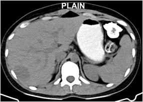

(B)Hepatic cyst- non-enhancing hypodense lesion.
(C)Hepatic abscess – lesion shows peripheral capsular enhancement.
Quiz 2
MRI of the spine of 16-year-old boy who had a fall from tree. This injury was followed by inability to move lower limbs.
Questions :
(1)What is the diagnosis ?
(2)What is the role of MRI in assessment of spinal cord injury ?
Answers :
(1)Fracture - dislocation is seen at D5D6 level with fracture of the anterior part of D6 vertebra. There is resultant retropulsion of the D6 vertebra with rupture posterior longitudinal ligament causing compression over anterior thecal sac - spinal cord and resulting in focal defect in spinal cord at this level (red arrow). Cord edema is seen extending superiorly and inferiorly. There is also rupture of the posterior ligamentous complex (yellow arrow).

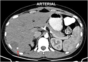


These findings are suggestive of spinal cord transection
Department of Radiology, Picture This by Jankharia, Mumbai, Maharashtra 400004
1MD, DNB (Radiology)
2MD, DMRD (Radiology)
(2)MRI is useful in patients with spine injuries as it helps in the assessment of cord edema, spinal cord contusions, cord compression and cord transection, which decides the treatment plan and hence prognosis of such patients.
M Chenniappan1
Series 10 : “Unexpected Absence and
72 y Palpitations ; Known COPD.
Questions :
(1) Describe ECG findings
(2)Why this clue?
(3)What are practical implications?
Answers :
(1) ECG CHANGES :
This is the ECG of 72 years old man with COPD which shows normal sinus rhythm with frequent VPDs of RVOT origin without compensatory pause- interpolated VPDs. The basic sinus rate is around 60/minute – Basic Bradycardia. So, the PR interval following the VPD is prolonged due to concealed retrograde conduction of VPD. The change in the P wave configuration of the sinus beat following VPD is due to P wave falling on T wave. In addition there is frequent atrial premature beats probably arising from the Right atrium with normal intra ventricular conduction followed by full compensated pause. (The P-P interval which includes the Atrial Premature Beats is exactly twice of the basic sinus cycle). The atrial and ventricular premature beats are alternating with sinus beats. Basic sinus beats do not show significant abnormality. There is no L 1 sign of COPD.
(2) CLUE :

Unexpected absence – absence of complete compensatory pause in VPD
Unexpected presence – presence of complete compensatory pause in APD (Usually APD has incomplete compensatory pause)
That is why the clue of "Unexpected absence and presence" is given.
The ECG shows following interesting findings
-RVOT VPDs
-Interpolated
-Concealed retrograde conduction
-VPD, APD alternating with sinus beat
-Right Atrial premature depolarisation
-APD is having full compensatory pause
-No significant changes in the Basic ECG
1Adjunct Professor, Dr MGR Medical University, Tamilnadu; Senior consultant cardiologist, Tamilnadu; Ramakrishna Medical Centre, Apollo Speciality Hospital, Trichy
APDs and VPDs indicate increased irritability of atrium and ventricles. Interpolated VPDs are expected with basic bradycardia. Because of basic sinus bradycardia and COPD betablockers cannot be given as anti-arrhythmic drugs. Because of the VPD configuration and RVOT origin it is likely to be benign. APDs and VPDs are expected in COPD because of the hypoxia and treatment with sympathomimetic agents. As this patient may be using sympathomimetic agents / inhalers, hypokalemia as the cause of Premature beats has to be excluded. After stopping stimulants like coffee, tobacco, alcohol and sympathomimetic agents, if still these arrhythmias are present, Holter may be done to decide about further management.
[The Editor is not responsible for the views expressed by the correspondents]
Monkeypox as a global health emergency — A threat after COVID-19 pandemic
SIR, — Monkeypox is a rare zoonotic disease caused by the monkeypox virus that belongs to the Poxviridae family1. While the source of infection is primarily zoonotic, and the disease condition is usually seen in Central and West Africa since the 1970s. Recently, there is a rapid spread of Monkeypox all over the world due to climatic change, widespread global travel, and waning herd immunity due to the cessation of smallpox vaccination2. Reemergence of monkeypox across nations had made World Health Organization declare it a public health emergency of international concern (PHEIC) in July 20223. As the disease is mild and not fatal, there are debates on declaring it as a PHEIC as it creates panic among the public but considering the reservoir of infection,pandemic potential and susceptible population declaring monkeypox as PHEIC is the need of the hour. India has reported nine confirmed cases of Monkeypox, including one death (4th August 2022)4. In India, the recent COVID-19 pandemic has equipped us to battle any outbreaks in the future. As we expect more emerging and re-emerging infections in the future, strengthening molecular laboratories will help in the early detection of the disease and containment. Currently, around 70% of the human population issusceptible to Monkeypox infection2. During the COVID-19 pandemic, a significant gamechanger in controlling the outbreak was a quick roll-out of mass vaccination campaigns.As per the CDC recommendations, two FDA-approved vaccines namely JYNNEOS (Imvamune or Imvanex) and ACAM2000 may be used for the prevention of Monkeypox infection5. But the data regarding the effectiveness of these two vaccines in the current outbreak is not available. Hence it is imperative that budget allocation for conducting vaccination effectiveness studies should be implemented in endemic countries where we have an increased incidence of Monkeypox infection.
1Cho CT, Wenner HA. Monkeypox virus. Bacteriol Rev 1973; 37(1): 1-18.
2Simpson K, Heymann D, Brown CS, Edmunds WJ, Elsgaard J, Fine P, et al — Human monkeypox – After 40 years, an unintended consequence of smallpox eradication. Vaccine 2020; 38(33): 5077-81.
3Monkeypox outbreak 2022 [Internet]. [cited 2022 Aug 4]. Available from: https://www.who.int/emergencies/situations/ monkeypox-oubreak-2022
4India Monkeypox Cases: Govt to hold experts’ meeting after India reports 9 monkeypox cases | India News - Times of India [Internet]. [cited 2022 Aug 4]. Available from: https:// timesofindia.indiatimes.com/india/monkeypox-govt-to-holdmeeting-of-experts-today/articleshow/93333192.cms
5CDC — Monkeypox in the U.S. [Internet]. Centers for Disease Control and Prevention. 2022 [cited 2022 Aug 4]. Available from: http s://www.cdc.gov/poxvirus/monkeypox/ considerations-for-monkeypox-vaccination.html
1MD, DTMH, Associate Consultant, Malathi Murugesan1 Department of Infectious Diseases, Srinath Ramamurthy2 Meenakshi Mission Hospital and Research Centre, Tamil Nadu and Corresponding Author 2MD, Consultant, CMR, National Institute of Epidemiology, Tamil Nadu
Laparoscopic repair of a symptomatic direct inguinal hernia in an apparently healthy boy
SIR, — Inguinal hernia is acommon paediatric surgical problem and over 99% of them in children are indirect. The direct inguinal hernias are secondary to other diseases and it’sextremely rare in apparently healthy children1. The incidence in full-term babies is estimated at 1-5%, it is six times more common in boys, therightsided hernias is more than three times that of left-sided hernias while the bilateral hernias are more common in premature infants2 A direct hernia involves herniation of intra-abdominal content through a weakness in the posterior wall of the canal, known as Hesselbach’s triangle. A direct hernia is found medial to the inferior epigastric vessels, while an indirect hernia is found lateral to these vessels3
A 5-year-old boy has noticed a swelling in the right groinwith a change in size with coughing or straining and itgot painful at times especially during defecation and urination as he has chronic constipationin the background. On examination, the patient had reducible, non-tender, non-transilluminated,positive cough impulse, to get above the lump was not possible, was medial to the internal inguinal ring in the Hesselbach’s triangle and diagnosed as right direct inguinal hernia. Right testis was fully descended, of normal size, site, lie and texture. The silk glove sign was negative. At laparoscopy, the direct inguinal hernia defect could be seen in the Hesselbach’s triangle (Fig 1). Posterior wall repair and ligation of the hernial sac was performed. The patient was discharged home same evening and at follow up is well.

Fig 1 — Findings during laparoscopy of right direct inguinal hernia. (A) The defect in the centre of Hesselbach’s triangle. (B) note that this image depicts a right direct inguinal hernia as the inferior epigastric vessels are lateral to it (to the right of the image in this view). (C) Closed deep inguinal ring on the right internal inguinal area
The advantages of the laparoscopic approach may include a lower risk of cord damage, less pain, better cosmetic results and less of postoperative complications. Our patient hadcongenital colorectal motility disorder and bowel dysfunctionwhich is a known risk factor for the development of a hernia4
1Hutson JM, O’Brien M, Beasley SW, Teague WJ, King SK — Jones’ clinical paediatric surgery. 7th edn. Chichester: John Wiley & Sons, 2015. p. 332
2Weaver KL, Poola AS, Gould JL, Sharp SW, St Peter SD, Holcomb GW 3rd — The risk of developing a symptomatic inguinal hernia in children with an asymptomatic patent processus vaginalis. J Pediatr Surg 2017; 52(1): 60-4. doi:10.1016/j.jpedsurg.2016.10.018
3Yeap E, Nataraja RM, Pacilli M — Inguinal hernias in children, AJGP 2020; 49(1-2): 38-42.
4Patel RV, Anthony FM, Govani ND, Govani DR, Panchasara N, Patel RR, Corracia R — Abdominal Wall Defects and Herniaslessons learnt from observations rom the experimental fetal surgery gastroschisis model in rabbits and their clinical extrapolation. Medp Pediatr Child Health Care 2022; 1(1): mppchc-202201001, 2022, 1: 8-15 and Anthony FM, Govani D, Patel RR, Patel RV: Spontaneous regression of clinical inguinal hernias in preterm female infants-role of congenital secreto-motility disorders. Ped Surg Intl (in press, PESI-D-2100637) .
1MB, MD (New York), MS, MCh, Ramnik V Patel1 LLM, MNAMS, DNBS, DNBPS, Rafael Cavalcante Correia2 FRCSEd, FRCSPed Surg, FEBPS, FACS, FAAP, DCH, DRCO, Consultant, Department of Pediatric Surgery, PGICHR, Gujarat and Corresponding Author 2MD, Consultant, Department of Pediatric Surgery, Childrens Hospital of the Sao Jose do Rio Preto Medical School, Santa Casa de Tatui General Hospital, Maneco Pereira Road, Tatui, Sao Paulo, Brazil
Cardio Respiratory faliure cited as — The cause of death, is it a misnomer ?
SIR, — Two types : (1) Heart and Lungs are in intimacy a SINGLE ORGAN for sypply of oxygenated blood to tissues and organs. They may have pre-existing diseases singly or severally like Valvular defects, Septal defects,IHD, Cardiomyopathy, Endocarditis and then Bronchitis, Bronchiectasis, Pneumonia, Pleural effusion, Pneumothorax and so on. Death may ensue primarily from these diseases.
(2) Heart and Lungs are healthy.
Separate disease conditions bring the patient to terminal stage leading to death.
The core crisis created behind death is metabolic derrangement of Acid Base Balance with dysfunction of the K pump regulating expulsion of Na from the cell to extracellular space and transfer of K into the cell.
Thus metabolic acidosis is created. Liver Kidneys try to control the situation but when these vital organs are in compromised condition acidosis becomes irreversible. Cardic and Cerebral cortex are vulnerable to such irregularities. They succumb.
MBBS (Cal), MS (Cal), Sailendra Kumar Chowdhury Consultant Surgeon, Indralok Superspiality Hospital, Birbhum 731101
SIR, — The original article entitled “Perception of undergraduate medical students about the current medical curriculum in India” Volule 119, No 9, September 2021, Page No 27. This study is a need of the hour for new Competency Based Medical Education (CBME) which was implemented in 2019 by a National Medical Council (NMC). Such study we need to encourage to as whole in a different part of India.
The author has mentioned all three professional year students and interns as study participants. But this new curriculum was started 2019 batch. So interns were not being part of this CBME study. The study was conducted in 2021 (Feb-March). Similarly in 3rdprofessional CBME students have not at entered. The Study would be better if only focused on the first professional year. And compare with traditional (Previous curriculum) method of curriculum and CBME curriculum of first-year students. The Second professional year is also not completed when data was collected but they were taught some of the competencies of final year subjects.
The Author also has mentioned the involvement of Government and Private colleges in the f student ratio, but no comparison was mentioned between them. Because many of the colleges as mentioned by author infrastructure facilities like skill lab not at established. Thereforethe implementation of the CBME curriculum is challenging for medical faculty unless until all the colleges are following implementation as per NMC. Till one batch of students will come out with this curriculum. It is very difficult to give an opinion regarding this CBME curriculum. The Author has not mentioned which parts of India, students were enrolled so that in the future remaining part of the students can be covered. It is very important to take the perception of students and faculty regarding the CBME curriculum.
Professor, Department of Community Medicine Rekha Udgiri Member of Medical Education Unit BLDE(DU) Shri BM Patil Medical College Vijayapura 586103, Karantaka
Beard Folliculitis due to Klebsiella pneumoniae
SIR, — Topical steroid damaged/dependent face (TSDF) can be described astemporary or persistentfacial skin damagebrought about by the unreasonable, non-selective, unsupervised, or protracted use of Topical Corticosteroids (TC) causing numerous skin pathologies and psychological dependence on TC1. TC has anti-inflammatory and immunomodulatory effects. TCs have formidable antipruritic, atrophogenic (dermal and/or epidermal), hypopigmentary effects on the corium and can lead to remarkableundesirable effects if used immethodically2. Another facet of TCs squandering is its beautifying application specifically in amalgamation with bleaching creams to make the skin light coloured. Absurd use of TCs inducesnumerous skin changes chaperoned by psychological dependence3. Since the facial skin is comparatively thinnerand there is an sebaceous glands as well, it leads toan escalated percutaneous absorption of drugs . As face is the most uncovered part of the human body it is most blameworthy


to consequences of UV rays, pollution, friction caused by cleaning and rubbing and use of different medications and cosmetics4. On pulling out TCs there is loss of vasoconstriction, resulting in a fixed vasodilatation, which is accountable for the flare which can be seen morphologically after withdrawal of the drug5
Here, we present a 24 years male who presented to us with multiple follicular pustules around the beard area. This patient has history of application of topical mometasone furoate cream since last 3 to 4 years everyday for 1 to 2 times. The patient applied mometasone furoateon suggestion by some friend due for acne spots and scars. The clinical image was consistent with folliculitis (Fig 1). Pus forgram staining and culture and sensitivity testing was sent. Antibiotic susceptibility test was done using disk-diffusion method. After gram staining, gram negative rods were observed under microscope (Fig 2). On blood agar mucoid colonies were seen (Fig 3). On sensitivity testing, the organism was found to be sensitive to few oral antibiotics like Minocycline, Fluoroquinolones, cotrimoxazole and other intravenous antibiotics. The causative organism was Klebsiella pneumoniae, a rare respiratory pathogen. Based on the sensitivity reports, the patient was started on oral Minocycline 100 mg once daily for 20 days. The patient responded well and the patient was followed up after 20 days, he responded well with resolution of all the lesion. The lesions started to resolve. Astonishingly, patient again reported to us with appearance of new lesions once the antibiotic was stopped. This type of recurrent beard folliculitis with a respiratory pathogen is not only rare but peculiar too. This is a rare case of recurrent beard folliculitis caused by Klebsiella pneumoniae due to prolonged use of mometasone from last 3 to 4 years in this COVID era. The most common organism causing beard folliculitis is Staphylococcus aureus6. Acquiring this pathogen might be related to loss of cutaneous immune surveillance to specific respiratory organism. Those organism could be layering on the skin surface due to close vicinity to oral and nasal aperture; and not involving the forehead. K Pneumoniae is rare cutaneous pathogen causing folliculitis in and around mouth; especially notable post topical corticosteroid (mometasone furoate) abuse for long term.
1Lahiri K, Coondoo A. Topical steroid damaged/dependent face (TSDF): An entity of cutaneous pharmacodependence. Indian

J Dermatol 2016; 61(3): 265.
2Brazzini B, Pimpinelli N — New and established topical corticosteroids in dermatology: clinical pharmacology and therapeutic use. Am J Clin Dermatol 2002; 3(1): 47-58.
3Chohan SN, Suhail M, Salman S, Bajwa UM, Saeed M, Kausar S, etal— Facial abuse of topical steroids and fairness creams: a clinical study of 200 patients. J Pak Assoc Dermatol 2016; 24(3): 204-11.
4Sendrasoa FA, Ranaivo IM, Andrianarison M, Raharolahy O, Razanakoto NH, Ramarozatovo LS, et al — Misuse of Topical Corticosteroids for Cosmetic Purpose in Antananarivo, Madagascar. BioMed Res Int [Internet]. 2017 [cited 2021 May 26]; 2017. Available from: https://www.ncbi.nlm.nih.gov/pmc/ articles/PMC5585578/
5Coondoo A, Phiske M, Verma S, Lahiri K – Side-effects of topical steroids: A long overdue revisit. Indian Dermatol Online J 2014; 5(4): 416-25.
6Winters RD, Mitchell M — Folliculitis. In: StatPearls [Internet]. Treasure Island (FL): StatPearls Publishing; 2021 [cited 2021 May 26]. Available from: http://www.ncbi.nlm.nih.gov/books/ NBK547754/
1MBBS, MD, Consultant Dermatologist, Kinnor Das1 Apollo Clinic, Silchar, Assam Surajit Gorai2 and Corresponding Author Piyali Banerjee3
2MBBS, MD, DNB, MNAMS, Consultant Dermatologist, Apollo Multispeciality Hospital, Kolkata 3MBBS, DVDL, Consultant Dermatologist, St. Barnabas Hospital, Jharkhand
1. The World Medical Association (WMA) has developed the International Code of Medical Ethics as a canon of ethical principles for the members of the medical profession worldwide. In concordance with the WMA Declaration of Geneva: The Physician’s Pledge and the WMA’s entire body of policies, it defines and elucidates the professional duties of physicians towards their patients, other physicians and health professionals, themselves, and society as a whole.
The physician must be aware of applicable national ethical, legal, and regulatory norms and standards, as well as relevant international norms and standards.
Such norms and standards must not reduce the physician’s commitment to the ethical principles set forth in this Code.
The International Code of Medical Ethics should be read as a whole and each of its constituent paragraphs should be applied with consideration of all other relevant paragraphs. Consistent with the mandate of the WMA, the Code is addressed to physicians. The WMA encourages others who are involved in healthcare to adopt these ethical principles.
General principles
2. The primary duty of the physician is to promote the health and well-being of individual patients by providing competent, timely, and compassionate care in accordance with good medical practice and professionalism.
The physician also has a responsibility to contribute to the health and well-being of the populations the physician serves and society as a whole, including future generations.
The physician must provide care with the utmost respect for human life and dignity, and for the autonomy and rights of the patient.
3. The physician must practise medicine fairly and justly and provide care based on the patient’s health needs without bias or engaging in discriminatory conduct on the basis of age, disease or disability, creed, ethnic origin, gender, nationality, political affiliation, race, culture, sexual orientation, social standing, or any other factor.
4. The physician must strive to use health care resources in a way that optimally benefits the patient, in keeping with fair, just, and prudent stewardship of the shared resources with which the physician is entrusted.
5. The physician must practise with conscience, honesty, integrity, and accountability, while always exercising independent professional judgement and maintaining the highest standards of professional conduct.
6. Physicians must not allow their individual professional judgement to be influenced by the possibility of benefit to themselves or their institution. The physician must recognise and avoid real or potential conflicts of interest. Where such conflicts are unavoidable, they must be declared in advance and properly managed.
7. Physicians must take responsibility for their individual medical decisions and must not alter their sound professional medical judgements on the basis of instructions contrary to medical considerations.
8. When medically appropriate, the physician must collaborate with other physicians and health professionals who are involved in the care of the patient
or who are qualified to assess or recommend care options. This communication must respect patient confidentiality and be confined to necessary information.
9. When providing professional certification, the physician must only certify what the physician has personally verified.
10. The physician should provide help in medical emergencies, while considering the physician’s own safety and competence, and the availability of other viable options for care.
11. The physician must never participate in or facilitate acts of torture, or other cruel, inhuman, or degrading practices and punishments.
12. The physician must engage in continuous learning throughout professional life in order to maintain and develop professional knowledge and skills.
13. The physician should strive to practise medicine in ways that are environmentally sustainable with a view to minimising environmental health risks to current and future generations.
14. In providing medical care, the physician must respect the dignity, autonomy, and rights of the patient.
The physician must respect the patient’s right to freely accept or refuse care in keeping with the patient’s values and preferences.
15. The physician must commit to the primacy of patient health and well-being and must offer care in the patient’s best interests. In doing so, the physician must strive to prevent or minimise harm for the patient and seek a positive balance between the intended benefit to the patient and any potential harm.
16. The physician must respect the patient’s right to be informed in every phase of the care process. The physician must obtain the patient’s voluntary informed consent prior to any medical care provided, ensuring that the patient receives and understands the information needed to make an independent, informed decision about the proposed care. The physician must respect the patient’s decision to withhold or withdraw consent at any time and for any reason.
17. When a patient has substantially limited, underdeveloped, impaired, or fluctuating decisionmaking capacity, the physician must involve the patient as much as possible in medical decisions. In addition, the physician must work with the patient’s trusted representative, if available, to make decisions in keeping with the patient’s preferences, when those are known or can reasonably be inferred. When the patient’s preferences cannot be determined, the physician must make decisions in the patient’s best interests. All decisions must be made in keeping with the principles set forth in this Code.
18. In emergencies, where the patient is not able to participate in decision making and no representative is readily available, the physician may initiate an intervention without prior informed consent in the best interests of the patient and with respect for the patient’s preferences, where known.
19. If the patient regains decision-making capacity, the physician must obtain informed consent for further intervention.
120, No 11, November 2022Journal
20. The physician should be considerate of and communicate with others, where available, who are close to the patient, in keeping with the patient’s preferences and best interests and with due regard for patient confidentiality.
21. If any aspect of caring for the patient is beyond the capacity of a physician, the physician must consult with or refer the patient to another appropriately qualified physician or health professional who has the necessary capacity.
22. The physician must ensure accurate and timely medical documentation.
23. The physician must respect the patient’s privacy and confidentiality, even after the patient has died. A physician may disclose confidential information if the patient provides voluntary informed consent or, in exceptional cases, when disclosure is necessary to safeguard a significant and overriding ethical obligation to which all other possible solutions have been exhausted, even when the patient does not or cannot consent to it. This disclosure must be limited to the minimal necessary information, recipients, and duration.
24. If a physician is acting on behalf of or reporting to any third parties with respect to the care of a patient, the physician must inform the patient accordingly at the outset and, where appropriate, during the course of any interactions. The physician must disclose to the patient the nature and extent of those commitments and must obtain consent for the interaction.
25. The physician must refrain from intrusive or otherwise inappropriate advertising and marketing and ensure that all information used by the physician in advertising and marketing is factual and not misleading.
26. The physician must not allow commercial, financial, or other conflicting interests to affect the physician’s professional judgement.
27. When providing medical care remotely, the physician must ensure that this form of communication is medically justifiable and that the necessary medical care is provided. The physician must also inform the patient about the benefits and limitations of receiving medical care remotely, obtain the patient’s consent, and ensure that patient confidentiality is upheld. Wherever medically appropriate, the physician must aim to provide care to the patient through direct, personal contact.
28. The physician must maintain appropriate professional boundaries. The physician must never engage in abusive, exploitative, or other inappropriate relationships or behaviour with a patient and must not engage in a sexual relationship with a current patient.
29. In order to provide care of the highest standards, physicians must attend to their own health, well-being, and abilities. This includes seeking appropriate care to ensure that they are able to practise safely.
30. This Code represents the physician’s ethical duties. However, on some issues there are profound moral dilemmas concerning which physicians and patients may hold deeply considered but conflicting conscientious beliefs.
The physician has an ethical obligation to minimise disruption to patient care. Physician conscientious objection to provision of any lawful medical interventions may only be exercised if the individual patient is not harmed or discriminated against and if the patient’s health is not endangered.
The physician must immediately and respectfully inform the patient of this objection and of the patient’s
right to consult another qualified physician and provide sufficient information to enable the patient to initiate such a consultation in a timely manner.
Duties to other physicians, health professionals, students, and other personnel
31. The physician must engage with other physicians, health professionals and other personnel in a respectful and collaborative manner without bias, harassment, or discriminatory conduct. The physician must also ensure that ethical principles are upheld when working in teams.
32. The physician should respect colleagues’ patientphysician relationships and not intervene unless requested by either party or needed to protect the patient from harm. This should not prevent the physician from recommending alternative courses of action considered to be in the patient’s best interests.
33. The physician should report to the appropriate authorities conditions or circumstances which impede the physician or other health professionals from providing care of the highest standards or from upholding the principles of this Code. This includes any form of abuse or violence against physicians and other health personnel, inappropriate working conditions, or other circumstances that produce excessive and sustained levels of stress.
34. The physician must accord due respect to teachers and students.
Duties to society
35. The physician must support fair and equitable provision of health care. This includes addressing inequities in health and care, the determinants of those inequities, as well as violations of the rights of both patients and health professionals.
36. Physicians play an important role in matters relating to health, health education, and health literacy. In fulfilling this responsibility, physicians must be prudent in discussing new discoveries, technologies, or treatments in non-professional, public settings, including social media, and should ensure that their own statements are scientifically accurate and understandable.
Physicians must indicate if their own opinions are contrary to evidence-based scientific information.
37. The physician must support sound medical scientific research in keeping with the WMA Declaration of Helsinki and the WMA Declaration of Taipei.
38. The physician should avoid acting in such a way as to weaken public trust in the medical profession. To maintain that trust, individual physicians must hold themselves and fellow physicians to the highest standards of professional conduct and be prepared to report behaviour that conflicts with the principles of this Code to the appropriate authorities.
39. The physician should share medical knowledge and expertise for the benefit of patients and the advancement of health care, as well as public and global health.
40. The physician should follow, protect, and promote the ethical principles of this Code. The physician should help prevent national or international ethical, legal, organisational, or regulatory requirements that undermine any of the duties set forth in this Code.
41. The physician should support fellow physicians in upholding the responsibilities set out in this Code and take measures to protect them from undue influence, abuse, exploitation, violence, or oppression.
Document no :SMAC221/Violence in the Health Sector REV/Oct2022
Original :English
Title :Proposed revision of WMA Statement on Violence in the Health Sector by Patients and Those Close to Them
Destination :WMA General Assembly, Berlin 2022,
The Ritz-Carlton Hotel, Berlin, Germany, 5-8 October 2022
Action(s) required :For consideration
1. Violence in the health sector has increased substantially in the new millennium, especially in time of COVID-19 pandemic. All persons have the right to work in a safe environment without the threat of violence. Workplace violence includes both physical and nonphysical, such as (psychological) violence, intimidation and cyber harassment, among others.
2. Cyber and social media harassment particularly includes online threats and intimidation towards physicians who take part in a public debate in order to give adequate information and fight disinformation. These physicians are increasingly confronted with, amongst others, malicious messages on social media, death threats and intimidating home visits.
Note : The 220th Council session (April 2022) decided that the WMA Statement on Violence in the Health Sector by Patients and Those Close to Them should undergo a major revision as part of the annual policy review process. Dr Ravindra Wankhedkar of the Indian Medical Association volunteered to undertake this task. and other drugs. Additionally, individuals may threaten or perpetrate violence against health personnel because they oppose a specific area of medical practice, based on their social, political or religious beliefs. Cases of violence from the bystanders are reported as well. Coworker violence, such as bullying, including initiation ceremonies and practical jokes, or harassment, constitutes another important pattern of workplace violence in the health sector.
3. For the purposes of this document, the broad WHO definition of workplace violence will be used : “The intentional use of power, threatened or actual, against another person or against a group, in work-related circumstances, that either results in or has a high degree of likelihood of resulting in injury, death, psychological harm, mal-development, or deprivation”.
4. In addition to the numerous consequences on victims’ health, violence against health personnel has potentially destructive social effects. It affects the entire healthcare system and undermines the quality of the working environment, ultimately impacting the quality of patient care. Furthermore, violence can affect the availability of health care, particularly in impoverished areas.
5. While workplace violence is indisputably a global issue, various cultural differences among countries must be taken into consideration in order to accurately understand the concept of violence on a universal level. Significant differences exist in terms of what defines various levels of violence and what specific forms of workplace violence are most likely to occur. This may create tolerance for some levels of violence in those places. However, threats and other forms of psychological violence are widely recognized to be more prevalent than physical violence.
6. Causes of violence in the healthcare setting are extremely complex. Several studies have identified common triggers for acts of violence by patients and relatives to be delays in receiving treatment, dissatisfaction with the treatment provided, aggressive patient behavior caused by the patient’s medical condition, the medication they take or the use of alcohol
7. Collaboration among various stakeholders (including governments, medical associations, hospitals, general health services, management, insurance companies, trainers, preceptors, researchers, media, police and legal authorities) together with a multi-faceted approach encompassing the areas of legislation, security, data collection, training/education, environmental factors, public awareness and financial incentives is required in order to successfully address this issue. As the representatives of physicians, medical associations should take a proactive role in combating violence in the health sector and also encourage other key stakeholders to act, thus further protecting the quality of the working environment for health personnel and the quality of patient care.
8. The WMA condemns in the strongest terms any forms of violence against healthcare personnel and facilities, which may include coworker violence, aggressive behavior exhibited by patients or family members, as well as acts of malicious intent from individuals in the general public, and calls on its constituent members, the health authorities and other relevant stakeholders to act through a collaborative, coordinated and effective strategy approach :
Policy-making
9. The state has obligations to ensure the safety and security of patients, physicians, and other health personnel. This includes providing an appropriate physical environment.
10. Governments should provide the necessary framework so that the prevention and elimination of workplace violence in the health sector be an essential part of national/regional/local policies on occupational health and safety, human rights protection, healthcarefacility management standards and gender equality.
Financial
11. Governments should allocate appropriate and
sustainable funds in order to effectively tackle violence in the health sector.
Protocols for situation of violence in healthcare facilities
12. Healthcare facilities should adopt a zero-tolerance policy towards workplace violence eliminating its “normalization” through the development and implementation of adequate protocols including the following:
- A predetermined plan for maintaining security in the workplace; including recognition of nonphysical abuse as a risk factor for physical abuse.
- A designated plan of action for health personnel when violence takes place.
- A strengthened internal communication strategy, involving the staff in decisions concerning their security.
- A system for reporting and recording acts of violence, which may include reporting to legal and/or police authorities.
- A means to ensure that employees who report violence do not face reprisals.
13. In order for these protocols to be effective, the management and administration of healthcare facilities should communicate and take the necessary steps to ensure that all staff are aware of the protocols. Managers should be urged to verbalize a no-tolerance policy towards violence in healthcare settings.
14. Patients with acute, chronic or illness-induced mental health disturbances or other underlying medical conditions may act violently toward health personnel; those taking care of these patients must be adequately protected. Except in emergency cases, physicians might have the right to refuse to treat and, in such situations, they must ensure that adequate alternative arrangements are made by the relevant authorities in order to safeguard the patient’s health and treatment.
Training/Education
15. A well-trained and vigilant staff supported by management can be a key deterrent of violent acts. Constituent members should work with undergraduate and postgraduate education providers to ensure that health personnel are trained in the following areas: communication skills, empathy as well as recognising and handling potentially violent persons and high-risk situations in order to prevent incidents of violence.
16. Continuous education should include ethical principles of healthcare and the cultivation of the patientphysician relationships based on respect and mutual trust. This not only improves the quality of patient care but also fosters feelings of security resulting in a reduced risk of violence.
Communication and Social Awareness
17. Medical associations, health authorities and other stakeholders should work together to increase awareness of violence in the health sector, creating networks of information and expertise in this area. When appropriate, health personnel and the public should be informed of acts of violence.
18. Broadcasting agencies, newspapers, and other news outlets are encouraged to thoroughly verify their
sources in order to keep the information shared to the highest standard of professional reporting. Social media companies and associated stakeholders should also take active steps to create a cyber-violence-free environment for its users. This includes strengthening policies to protect user data, making reporting and flagging such violence easy and accessible, and engaging law enforcement for proper legal action when warranted.
Security
19. Appropriate security measures should be in place in all healthcare facilities and acts of violence should be given a high priority by law-enforcement authorities. A routine violence risk audit, including a risk assessment, should be implemented in order to identify which jobs and locations are at highest risk for violence, especially in places where violence has already occurred, and to determine weaknesses in facilities’ security. Examples of high-risk areas include general practice premises, mental health treatment facilities and high traffic areas of hospitals including the emergency department.
20. The risk of violence may be ameliorated by a variety of means which include placing security personnel in highrisk areas and at the entrance of buildings, the installation of security cameras and alarm devices for use by health personnel, the use distinguishable items to identify the staff and by maintaining sufficient lighting in work areas, contributing to an environment conducive to vigilance and safety. The implementation of a system to screen patients and visitors for weapons upon entering certain areas, especially the high-risk ones, should be considered.
Support to victims
21. Adequate medical, psychological and legal support should be provided to victims of violence. Such support should be free of access for all the health personnel.
Investigation
22. In all cases of violence there should be investigation to better understand the causes and to aid in prevention of future violence. The investigation may lead to prosecution of perpetrators under civil or criminal codes. The procedure should be led by relevant officials in law enforcement and should not expose the victim to further physical or psychological harm.
Data Collection
23. Appropriate reporting systems should be established to enable health personnel to report anonymously and without reprisal, any threats or incidents of violence. Such a system should assess in terms of number, type and severity, incidents of violence within an institution and resulting injuries. The system should be used to analyse the effectiveness of preventative strategies. Aggregated data and analyses should be made available to health professional organizations and other relevant stakeholders.
WMA related policies :
- WMA Statement on Violence and Health
- WMA Statement on the Protection and Integrity of Medical Personnel in Armed Conflicts and Other Situations of Violence