


Revival of the Case Series in Publication Circuitry
— Nandini Chatterjee MD, FRCP (Glasgow), FICP Professor, Department of Medicine, IPGME&R and SSKM Hospital, Kolkata 700020 and Hony Editor, JIMA
The current era of evidence based medicine has paved the way for reliance of the medical fraternity on meta analysis, systematic reviews and randomized control trials for therapeutic decision making. The hierarchy triangle of scientific evidence places these at the top while the Case Series and Reports occupy lower rungs. However does this mean that case depictions either singly or in a series have lost their relevance ?
A Case Series is a conglomeration of four to ten cases which have a common thread or bond in terms of pathophysiology, clinical spectrum, diagnosis and have received similar therapy.
The National Medical Council has recognized the Case Series as a valid publication accepted for career progression. But the value of a Case Series extends beyond the boundaries of personal gains and adds to the domain of scientific wisdom.
A Case Series is a descriptive narrative that highlights novel, unusual disease processes or reports a typical presentations of common ailments identified in patients in daily practice. They are empirical enquiries or analysis of clinical problems in a group of patients in a natural, real-world scenario.
Such treatises have immense educational value for the medical graduates to encourage problem based learning and analytical skills for diagnosis and management1
Therefore, it is desirable for medical education programs as well as Journals to uphold the fast disappearing tradition of narrative publication. Medical teachers should encourage students to go through case studies for developing their faculty of critical learning and thinking skills.
Vol 121, No 3, March 2023Journal of the Indian Medical Association 13
Writing case studies is also an efficient way of learning to pen down scientific manuscripts for beginners. Novice writers can benefit from the physician - patient interaction, sharpen their writing and communication skills, and learn about the procedural formalities of manuscript submission in journals.
A Case Series is greatly helpful in pattern recognition that is the central essence of medical diagnosis. Often it also puts forward new research questions and informs us about various aspects of patient management, adverse drug reactions, novel therapies or even ethical dilemmas2
We are publishing a couple of Case Series in this issue – one is on six interesting cases of ocular tuberculosis. The clear message from the series is that in case of chronic inflammatory conditions of eye, a high index of suspicion should be nurtured for ocular tuberculosis in a endemic country such as ours even if direct microbiological confirmation of the bacillus is not possible.
Either hematogenous spread or a hypersensitivity is the common pathophysiological basis of these cases. This observation, as well as the fact that uveal involvement and positive tuberculin test are being noted as clinical pointers,are of an educational value to clinicians that will help in their daily practice.
Another Case Series is of a different flavour –autoimmune hemolyticanemia with IgG and IgA antibodies both found in the patients, which is a rarity. Awareness about autoimmune hemolyticanemia is important as a cause of new onset pallor and jaundice specially in the background of sepsis or autoimmune diseases, as the diagnostic test is very simple ie, DAT and therapy leads to rapid improvement. Thus it is important to recognize the clinical presentation and suspect early.
To summarise, despite many divergent points of view regarding their strength of evidence, Case Reports and Series portray the value of narrative publication that can not be ignored or underplayed.
Real life medical dilemmas along with the depiction of the differential analysis as well as management goes a long way in enriching clinical acumen and experience.
REFERENCES
1Murad MH, Sultan S, Haffar S, Bazerbachi F — Methodological quality and synthesis of case series and case reports. BMJ EvidBasedMed 2018; 23(2): 60-3. doi: 10.1136/bmjebm-2017110853.
2Gagnier JJ , Kienle G , Altman DG — The CARE guidelines: consensus-based clinical case reporting guideline development. J Med Case Rep 2013; 7: 223.doi:10.1186/ 1752-1947-7-223.
Vol 121, No 3, March 2023Journal of the Indian Medical Association 14
Original Article
Acute Kidney Injury among Post Cardiac Surgery Patients : A Retrospective Study
Sunil C V S Mutyala1, Jagadeesh N Vaggar2, Shirish Borker3, Sushmit Kamat4, Shreya S Lotliker5, Jagadish Cacodcar6, Jagannath P Kolwalkar7, Wilroy Anthony Gonsalves8
Background : Acute Kidney Injury (AKI) is a common complication Post Cardiac Surgery with reported incidence of 20-70%. Various studies have been conducted worldwide on risk factors contributing to the etiology of AKI in Cardiac surgery patients. We undertook similar study to understand the etiology and risk factors associated with AKI at Goa Medical College hence we undertook this study.
Methodology : A retrospective record based observational study was conducted at Goa Medical College; wherein records of 419 patients who underwent Cardiac Surgery during the study period were analyzed for pre-operative, intra-operative and postoperative variables. Kidney Disease Improving Global Outcomes criteria were used to study the incidence of AKI. The Data was entered in Microsoft Excel and analysed using SPSS version 22.0. Chi-square test and Student t test were used as a test of significance.
Results : Out of 419 patient records reviewed; 40.3% patients developed AKI after Cardiac Surgery. Age, Sex, h/o previous Cardiac Surgery, CPB duration, Aortic Cross Clamp Time, addition of vasopressor etc. were some of the significant risk factors associated. AKI associated with Cardiac Surgery was associated with a mortality of 8.3%. Mean duration of ventilation 38.48±62.27 hrs. and ICU stay 6.12±3.15 days was comparatively longer than patients without AKI (P<0.001).
Conclusion : We concur that AKI is a serious complication in patients undergoing Cardiac Surgery and has significant impact on the outcome of the patients in terms of duration of ICU stay, duration of ventilation and mortality. There is need to identify modifiable risk factors at the earliest and develop approaches to improve the outcome and decrease the AKI associated morbidity and mortality.
[J Indian Med Assoc 2023; 121(3): 15-20]
Key words :Acute Kidney Injury (AKI), Cardiac Surgery, Cardiopulmonary Bypass.
Acute Kidney Injury (AKI) is defined as an abrupt decrease in kidney function, which encompasses both injury (structural damage) and impairment (loss of function)1. Classification of AKI includes pre-renal
Department of Cardiac Anaesthesiology, Goa Medical College, Bambolim, Goa 403202
1MBBS, MD (Anaesthesiology), PDCC (Cardiac Anesthesia), Professor
2MBBS, MD (Anaesthesiology), DM (Cardiac Anaesthesiology), Associate Professor
3MBBS, MS (General Surgery), MCh (Cardiovascular and Thoracic Surgery), DNB (Cardiovascular and Thoracic Surgery), Professor and Head, Department of Cardiovascular and Thoracic Surgery
4DA, DNB, DM (Cardiac Anaesthesiology), Assistant Professor, and Corresponding Author
5MBBS, DPH, Assistant Lecturer, Department of Community Medicine
6MBBS, MD, PSM, Professor and Head, Department of Community Medicine
7MBBS, MS (General Surgery), MCh (Cardiovascular and Thoracic Surgery), Associate Professor, Department of Cardiovascular and Thoracic Surgery
8MBBS, Assistant Lecturer, Department of Cardiovascular and Thoracic Surgery
Received on : 13/10/2021
Accepted on : 22/04/2022
Editor's Comment : Acute Kidney Injury has significant impact on the outcome of the patients undergoing cardiac surgery
Prevention of modifiable risk factors and early diagnosis is the key to decrease mortality and morbidity associated with Acute Kidney Injury.
There is need to find out biomarker/s to diagnose Acute Kidney Injury at an early stage to initiate Renal Replacement Therapy as early as possible in potential patients to decrease mortality.
AKI, Postrenal obstructive nephropathy and intrinsic Acute Kidney Disease. As the pre-renal and Postrenal causes persist they progress to renal cellular damage1 AKI manifest from mild to serious Renal Derangement /Failure if preventive and reparative measures are not taken in time.
AKI is a common complication Post Cardiac Surgery which carries prolonged morbidity and increased mortality. The reported incidence of AKI varies from 20-70 %2. The mortality rate is around 4070% among patients undergoing Cardiac Surgery who needed Renal Replacement Therapy (RRT)2. Also, the
Vol 121, No 3, March 2023Journal of
the Indian Medical Association
15
increased ICU stay, increased duration of ventilation and the need for RRT leads to a huge or substantial impact on both monetary as well as human resources.
Pathophysiology of Cardiac Surgery associated AKI is multifactorial and complex. Some of the risk factors for AKI that have been well documented by various studies which includes Pre-operative factors (Age, Sex, Hypertension, Diabetes, Type of Cardiac Surgery etc,) Intra-operative factors [Cardio Pulmonary Bypass (CPB) time, Aortic cross clamp time, use of blood products etc,] and Postoperative factors (use vasopressors, hypovolemia etc). To estimate the incidence of AKI among the Post Cardiac Surgery patients; to identify the risk factors associated and also to study the impact of AKI on duration of ventilation, duration of ICU, duration of hospital stay and mortality; we planned to conduct this study using Kidney Disease Improving Global Outcomes (KDIGO) guidelines in our institute and thus get some insights for the prevention of AKI and thereby decreasing the morbidity and mortality.
As per KDIGO guidelines AKI is defined as any of the following2
Increase in Serum Creatinine (sCr) >0.3 mg/dl (>26.5 µmol/l) within 48 hours; or
•Increase in sCr to >1.5 times baseline, which is known or presumed to have occurred within the prior 7 days; or
•Urine volume of 0.5 ml/kg/h for 6 hours
MATERIALS AND METHODS
The present retrospective record based observational study was conducted at Goa Medical College and Hospital Bambolim. Approval from the Institutional Ethical Committee was duly obtained prior to the conduct of the study. The data of 419 patients admitted in the ICU and CVTS wards, who underwent cardiac surgery from 1 st January, 2019, till 31 st December, 2019 was obtained from MRD Department.
The inclusion and exclusion criteria were as follows:
Inclusion criteria : Included all the adult patients >15 years who underwent Cardiac Surgery on elective/ emergency/ urgent basis in the Department of CVTS.
Exclusion Criteria : Included patients who were previously diagnosed with CKD, patients who died within 24 hours of surgery and the patients whose data was incomplete.
Depending on presence or absence of AKI, patients were divided into two groups. In our study we used KIDIGO criteria to define AKI ie, sCr increase of 0.3mg/ dl within 48 hours or an increase >50% within the previous 7 days or urine output < 0.5ml/kg/hour for 6 hours.
The following variables were studied for the present study:
•In the Pre-operative period variables analyzed included physical characteristics (Gender, Age), Biochemical parameters (baseline Serum Creatinine), and Comorbidities (Diabetes, Hypertension, COPD, Stroke), recent MI (<21 days), h/o previous cardiac Surgery, presence of Cardiac dysfunction (LVEF <40%), type of surgery performed (CABG, Valve Replacement Surgery, or the combination of the two, congenital corrections), use of Intra-aortic Balloon Pump (IABP) insertion.
•In the intra-operative period, we evaluated the duration of CPB, Aortic Cross Clamp Time, MAP on CPB, and the use of vasopressor drugs and Use of IABP.
•The variables studied in the postoperative period included MAP (first 3 post op days), duration of ventilation in hours, the duration of ICU stay in days, duration of hospital stay, use of vasopressor drugs, the initiation of RRT and the use of IABP. The Serum Creatinine values were monitored till the 7 th postoperative day. Finally, the patient outcome including mortality if any was noted.
As per our institution protocol Epinephrine was used as primary vasopressor and secondary vasopressors used included Dobutamine or Milrinone or Levosimendan.
The Data was entered in Microsoft Excel and analyzed using the Statistical software namely SPSS 22.0, and R environment ver 3.2.2. Descriptive and inferential statistical analysis was carried out. Continuous variables were expressed as the Means ± Standard Deviations (SDs) and were compared with Student’s t test. Categorical variables were described as frequencies and proportions and were compared with Fisher’s exact tests or chi square tests. Significance is assessed at 5 % level of significance.
RESULTS
Out of 419 patients’ records reviewed it was observed that the mean age was 58.11 ±11.02 years and majority were males (69.7%). It was observed that 169 (40.3 %) out of 419 patients who underwent Cardiac Surgeries during the study period developed AKI.
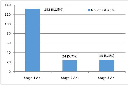
Fig 1 shows 132 (31.5%) presented with stage 1 AKI whereas 24 (5.7%) and 13 (3.1%) presented with stage 2 and stage 3 respectively. The mean Serum Creatinine levels were 0.92±0.20 mg/l. Amongst the comorbid conditions studied majority had h/o Diabetes Mellitus followed by Hypertension, COPD and Stroke as shown in Table 1. Only 13(3.1%) had history of
Vol 121, No 3, March 2023Journal of
the Indian Medical Association
16
11.5–1.9
mg/dl (>353.6 µmol/l)Anuria for >12 hours
recent Myocardial Infarction (MI) <21 days and 8 (1.9%) had h/o previous Cardiac Surgery. The most common procedure performed was CABG followed by Valve Surgery and combination of CABG + Valve surgery.
Table 2-4 shows the association of Preoperative, intra-operative and postoperative variables with the incidence of AKI in the study subjects.
•Chi square test and Student 't' test was used as to study statistical difference between two proportion and means respectively.
Under the Pre-operative variables studied Age, sex, h/o previous Cardiac Surgery had statistically significant association with the occurrence of AKI in patients undergoing cardiac surgery. The incidence of AKI was significantly low in patients with Diabetes Mellitus (P<0.046).
Although other Surgeries like the Bentall procedures are high risk Surgeries and prone for AKI, we could not comment on the statistical association with AKI because number of cases low.
•Student 't' test was used to study significant difference between two means
Amongst the intra-operative variables it was observed that CPB duration, Aortic cross clamp time was found statistically significant. There was no
significant difference between MAP in both the groups. The use of vasopressor during intra-operative period showed increased incidence of AKI, however transfusion of blood products did not show any statistical association with AKI incidence (P=0.243).
Difference in mean and proportion was calculated using student t test and chi square respectively.
As per the institution protocol Diuretics were administered for patients during postoperative period if urine output was found to be <0.5ml /kg for more than 3 hours and hence we could not assess the association between urine output and development of AKI in patients undergoing Cardiac Surgery.
In the Postoperative period MAP was monitored till the patient was in ICU. MAP was analysed for first three post op days and it was observed to have significant association with occurrence of AKI till the second postoperative day as seen in Table 4. The transfusion of blood products namely Packed cells, FFP, Platelets and the mean duration of ventilation, duration of ICU stay also had significant association (P<0.001).It was also observed that incidence of AKI significantly increased in Postoperative period with addition of vasopressors (P<0.001).
The overall mortality was 4.3% in our study with the mortality in AKI group accounting for 8.3% (Stage 1, 2, 3 accounting for 2.3%, 8.3%, 69% respectively) which was significantly higher as compared to non-AKI group ie, 1.6% as seen in Table 4.
Out of 169 patients developing AKI postsurgery 13 patients had stage 3 AKI; among which RRT was initiated in 11 patients, whereas in the non-AKI group we initiated RRT in 1 patient who underwent VSR repair and was on inotropic support. As the hemodynamic were unstable and decreasing trend of urine output in the Postoperative period we considered to initiate RRT early.
It was observed that there was no significant difference between the baseline sCr levels in both groups (P=0.868). However, there was steady rise in levels of sCr from 2nd postoperative day and levels gradually returned to baseline by day 7 as seen in Fig 2.
Usage of IABP had significant association (P<0.002) with occurrence of AKI as seen in Table 2.
DISCUSSION
In the present study, the mean age of the patients undergoing Cardiac Surgery was 58.11 ±11.02 years which was similar to studies by TF Silva, et al3 , Elghoneimy, et al4. We found male predominance in
Vol 121, No 3, March 2023Journal of the Indian Medical Association
of
Stage Serum creatinineUrine output
Fig 1 — Stages of AKI in patients studied Table 1 — KDIGO staging of AKI
Staging
AKI
times baseline<
OR
0.5 ml/kg/h
>0.3 mg/dl (>26.5 µmol/l) increasefor 6–12 hours
22.0–2.9 times baseline<0.5 ml/kg/h for >12 hours
ORfor
Increase
>4.0
OR Initiation
OR,
eGFR
17
33.0 times baseline< 0.3 ml/kg/h
>24 hours
in Serum Creatinine toOR
of renal replacement therapy
In patients <18 years, decrease in
to <35 ml/min per 1.73 m2
our study which was comparable to the study conducted by Machado MN, et al5 ie, 52 %; whereas Wittlinger, et al6 reported significantly more females developed AKI.
The incidence of AKI after Cardiac Surgery was 40.3% which was similar to the findings by Ramos KA, et al7 ie, 43.66%, however studies conducted by Gangadharan, et al8and TF Silva3 it was reported as
9.25% and 83.8% respectively. The variation in incidence is probably due to different criteria used to define AKI.
We found Diabetes was the most common comorbid condition followed by Hypertension, COPD and the predominant procedure performed was CABG followed by Valve Surgery and combination of both which was similar to various other studies (Machado MN, et al,TF Silva, et al)5,3
Redo Surgeries also carried a significant association with AKI, this might be due to the high requirement of blood products and vasopressors.
In our Study Age, Sex, h/o previous Cardiac Surgery had statistically significant association with the occurrence of AKI in patients undergoing Cardiac Surgery (P<0.005), which was contrasting to the findings by Freeland K, et al9 where Age, Gender, Comorbid condition were not predictors of AKI (p<0.005). A study by Xiangcheng Xie, et al10 showed significant association between Coronary Artery Disease and Hypertension (P<0.001).
We did not find any significant association between MI<21 days(p <0.65) and AKI, however S Hanuora, et al11 found those with h/o of MI<30 days were more liable for development of AKI.
The intra-operative period MAP on CPB in mmHg did not show any difference between two groups in our study which was similar to the finding by Freeland K, et al9. However CPB duration, Aortic Cross Clamp Time was significantly associated with the incidence of AKI which was similar to S. Hanoura, et al11and Xiangcheng Xie, et al10.Results demonstrated by Xiangcheng Xie, et al10 showed CPB time longer than 110 minutes was an independent risk factor for AKI.
During the Postoperative period, the AKI group had significantly lower MAP on day 1 and 2 and explains the use ofmore vasopressors compared to non-AKI patients which shows that the higher MAP is required to prevent AKI during the early Postoperative period.
Insertion of IABP had significant association with AKI irrespective of the timing of insertion which was
Vol 121, No 3, March 2023Journal of the Indian Medical Association
Value NoYes
CHARACTERISTICS Age in years : <4019(4.5%)7(1.7%)26(6.2%)0.043 40-60134(32%) 77(18.4)211(50.4%) >6097(23.1%)85(20.3%)182(43.4%) Sex : Female 87(20.8%)40(9.5%)127(30.3%)0.015 Male 163(38.9) 129(30.8%)292(69.7%)
CONDITIONS Diabetes Mellitus : No91(21.7%)78(18.6%)169(40.3%)0.046 Yes159(38%)91(21.7%)250(59.7%) Hypertension : No109(26%)71(17%)180(43%)0.747 Yes141(33.6%)98(23.4%)239(57%) COPD : No240(57.2) 162(38.7%)402(95.9%)0.942 Yes10(2.4%)7(1.7%)17(4.1%) Stroke : No244(58.2%)162(38.7%)406(96.9%)0.313 Yes6(1.4%)7(1.7%)13(3.1%) OTHER VARIABLES Recent MI < 21 days : No240(57.3%)166(39.6%)406(96.9%)0.198 Yes10(2.4%)3(0.7%)13(3.1%) Previous Cardiac Surgeries : No249(59.4%)162(38.7%)411(98.1%)0.006 Yes1(0.2%)7(1.7%)8(1.9%) Cardiac Dysfunction (LVEF) : < 40%22 (5.2%)25 (6%)47 (11.2%) 0.056 >40%228 (54.4%)144 (34.4%)372 (88.8%) Type of Surgery Done : CABG 200(47.7%)122(29.1%)322(76.8%)0.063 Valve Surgery 30(7.2%)32(7.6%)62(14.8%)0.050 CABG+Valve 6(1.4%)6(1.4%)12(2.8%)0.489 ICR9(2.1%) 0(0%) 9(2.1%)0.013 Bentall procedure 0(0%) 3(0.7%)3(0.7%)0.034 VSR Repairs 2(0.5%)1(0.2%)3(0.7%)0.804 Myxoma excision 2(0.5%) 0(0%) 2(0.5%)0.244 Redo surgery 1(0.2%)5(1.2%)6(1.4%)0.031 Total250(59.6%)169(40.3%)419(99.9%) CLINICAL VARIABLES Baseline Serum Creatinine (mg/l)0.92±0.20 0.92±0.210.92±0.20 0.868 IABP insertion : Not inserted 243(58%)150(35.8%)393(93.8%)0.002 Preoperative stage 3(0.7%)2(0.5%)5(1.2%) Intraoperative stage 2(0.5%)9(2.1%)11(2.6%) Postoperative stage 2(0.5%)8(1.9%)10(2.4%) Total250(59.7%)169(40.3%)419(100%)
Table 2 — Association of preoperative variables with incidence of AKI in patients studied
VariablesAKITotalP
BASELINE
COMORBID
18
similar to finding by Wittilinger, et al who found that IABP is an independent risk factor for development of AKI.
We found that duration of ventilation and ICU stay (P<0.001) also showed significant association, which was similar to findings by Yanli liu, et al12. Study by Xiangcheng, et al10showed mechanical ventilation duration greater than 9 hours was an independent risk factor in the development of AKI. However some studies showed null association presumably due to limited sample size and heterogeneity of the patients (Boyles, et al13).
In our study the mortality in AKI group was 8.3% compared to non AKI group (1.6%), however in a study conducted by HY Fu, et al14AKI mortality was reported as 30.4 % mortality in comparison to non AKI ie, 8% (P<0.001).
High mortality was seen in patients initiated on RRT, which may be related to the timing of the initiation of RRT. The patient who was initiated early had a good outcome. This gives an impetus for us to search for those biomarkers which determines AKI early and probably also indicate the timing of RRT initiation. In a study by Yanli liu, et al12 stage 3 AKI patients needed RRT.
CONCLUSION
From our study findings we conclude that AKI in patients undergoing Cardiac Surgery has significant impact on the outcome of the patients in terms of mean duration of ventilation, duration of ICU stay and also mortality; hence prevention of modifiable risk factors and early diagnosis is the key to decrease the mortality and morbidity associated with AKI. Duration of CPB and aortic cross clamp time was found to be a significant risk factor for AKI which needs to be investigated further to reduce incidence of AKI. The other modifiable risk factor includes the usage of blood products can be reduced by using methods like hemofiltration and cell savers and also achieving a proper Haemostasis. Also, there is need to find out biomarker/s to diagnose AKI at an early stage to initiate RRT as early as possible in potential patients to decrease mortality. We look forward to strategies to identify those at risk of AKI Post Cardiac Surgery and develop approaches to improve the outcome.

REFERENCES
1Makris K, Spanou L — Acute Kidney Injury: Definition, Pathophysiology and Clinical Phenotypes. Clin Biochem Rev 2016; 37(2): 85-98.
2Khwaja A — KDIGO clinical practice guidelines for acute kidney injury. Nephron Clin Pract 2012; 120(4): c179-84.
3TF Silva TF, Silva KR, Nepomuceno CM — Incidence of acute kidney injury post cardiac surgery/ : a comparison of the
Vol 121, No 3, March 2023Journal of the Indian Medical Association
VariablesAKITotalP Value NoYes Mean Arterial Pressure (MAP) : MAP Day 1 (mmHg) 72.62±9.8868.62±10.1371.01±10.16 <0.001 MAP Day 2 (mmHg) 84.15±10.0880.59±11.9282.72±10.99 <0.001 MAP Day 3 (mmHg) 87.17±9.7286.30±12.3186.82±10.84 0.421 Transfusion of Blood Products : Packed cells1.19±1.401.82±2.151.44±1.77 <0.001 FFP0.18±0.590.54±1.050.33±0.83 <0.001 Platelets 0.54±1.881.50±3.470.93±2.68 <0.001 Vasopressors Added : Nil222(88.8%)120(71%)342(81.6%) <0.001 One3(1.2%)6(3.6%)8(1.9%) 2 or more 25(10%)43(25.4%)68(16.2%) Other Variables : Duration of Ventilation (hrs.)23.87±23.09 38.48±62.2729.74±43.85 <0.001 Duration of ICU Stay (days)5.02±1.85 6.12±3.155.46±2.51 <0.001 Duration of Hospital stay (days)8.90±7.859.95±4.829.32±6.81 0.123 Outcome : Mortality 4(1.6%)14(8.3%)18(4.3%) <0.001 Discharged 246(98.4%)155(91.7%)401(95.7%)
Table 4 — Association of Postoperative variables with incidence AKI in patients studied
VariablesAKITotalP Value NoYes CPB duration (mins) 138.79±39.59150.63±54.64143.56±46.56 0.010 Aortic Cross Clamp Time (mins)92.99±28.88 101.02±40.4596.23±34.21 0.018 MAP on CPB (mmHg) 56.62±4.5656.81±6.2956.69±5.32 0.720 Vasopressors1.34±1.392.15±1.991.66±1.70<0.001 Transfusion Of Blood Products0.31±0.520.25±0.450.28±0.49 0.243
Table 3 — Association of Intra-operative variables with incidence of AKI in patients studied
19
Fig 2 — Association of Serum Creatinine with incidence of AKI in patients studied
Vol 121, No 3, March 2023Journal of the Indian Medical Association
AKIN and KDIGO criteria. BrazilianJournalofAnesthesiology 2021; 71(5): 511-6.
4Elghoneimy YA, Al Qahtani A, Almontasheri SA — Renal Impairment After Cardiac Surgery: Risk Factors, Outcome and Cost Effectiveness. Cureus 2020; 12(11): e11694.
5Machado MN, Nakazone MA, Maia LN — Acute kidney injury based on KDIGO (Kidney Disease Improving Global Outcomes) criteria in patients with elevated baseline serum creatinine undergoing cardiac surgery. Rev Bras Cir Cardiovasc 2014; 29(3): 299-307.
6Wittlinger T, Maus M, Kutschka I — Risk assessment of acute kidney injury following cardiopulmonary bypass. J Cardiothorac Surg 2021; 16(4): 1-7 .
7Ramos KA, Dias CB — Acute Kidney Injury after Cardiac Surgery in Patients Without Chronic Kidney Disease. Braz J Cardiovasc Surg 2018; 33(5): 454-61.
8Gangadharan S, Sundaram KR, Vasudevan S, Ananthakrishnan B, Balachandran R, Cherian A, et al— Predictors of acute kidney injury in patients undergoing adult cardiac surgery. Ann Card Anaesth 2018; 21(4): 448-54.
9Freeland K, Hamidian Jahromi A, Duvall LM, Mancini MC — Postoperative blood transfusion is an independent predictor of acute kidney injury in cardiac surgery patients. J Nephropathol 2015; 4(4): 121-6.
10Xie X, Wan X, Ji X —. Reassessment of Acute Kidney Injury after Cardiac Surgery: A Retrospective Study. Intern Med 2017; 56(3): 275-282.
11Hanoura S, Omar AS, Osman H, Sudarsanan S, Eissa M, Maksoud M, Khulaifi AA — Prevalence and predictors of acute kidney injury after cardiac surgery: a single-centre retrospective study in Qatar. Netherlands Journal of Critical Care 2018; 26(1): 14-9.
12Liu Y, Shang Y, Long D, Yu L — Intraoperative blood transfusion volume is an independent risk factor for postoperative acute kidney injury in type A acute aortic dissection. BMC Cardiovasc Disord 2020; 20(1): 446.
13Boyle JM, Moualla S, Arrigain S, Worley S, Bakri MH, Starling RC, etal—Risks and outcomes of acute kidney injury requiring dialysis after cardiac transplantation. Am J Kidney Dis 2006; 48(5): 787-96.
14Fu HY, Chou NK, Chen YS, Yu HY — Risk factor for acute kidney injury in patients with chronic kidney disease receiving valve surgery with cardiopulmonary bypass. Asian J Surg 2021; 44(1): 229-34.
Ifyouwanttosendyourqueriesandreceivethe responseonanysubjectfromJIMA,pleaseuse the E-mail or Mobile facility.
Know Your JIMA
Website:https://onlinejima.com
For Reception:Mobile : +919477493033
For Editorial:jima1930@rediffmail.com
Mobile : +919477493027
For Circulation:jimacir@gmail.com
Mobile : +919477493037
For Marketing: jimamkt@gmail.com
Mobile : +919477493036
For Accounts: journalaccts@gmail.com
Mobile : +919432211112
For Guideline:https://onlinejima.com
20
Original Article
Socio-economic and Psychological Correlates of Postpartum Depression at Six Months
Kajal Jitendrakumar Tanna1, Krupa M Unadkat2
Background : A woman undergoes multiple changes physically and emotionally after childbirth. Mothers also experience emotional changes with a new or additional baby related to breastfeeding demands, problems pertaining to maternal dissonance, childcare stress and difficult infant temperament.
Materials and Methods : Overall, 100 women out of 178 women who attended obstetrics and Gynaecology department postpartum in our hospital were selected. Socio-economic factors, psychiatric and maternity characteristics were collected using a standard questionnaire. The main outcome of this study was PPD assessed by Edinburgh postpartum depression scale was used to assess the chief outcome of the study, ie, Postpartum Depression. EPDRS scale consisted of 10 questions that has 4 response scored from 0 to 3, so the highest value shows depressed moods.
Results : Final results are of 100 postpartum females with age ranging between 18 and 30 years with a mean value 26.5 years ± 4.05, 21.3% dwelling in Urban areas and 15.4% having high education. About 2.1% of study participants had postpartum only Depression, 15.3% had only anxiety alone and 23.2% study participant had both. When we look at severity, 8.8%, 10.6%, 2.9%, and 0.4% suffered from Mild, Moderate, Severe and extremely severe Postpartum Depression, respectively. 14.2%, 9.2%, 6.9% and 3.9% suffered mild, moderate, severe, and extremely severe Postpartum anxiety, respectively.
Conclusion : Around 23% female patients in our hospital suffer from Postpartum Depression and/or anxiety. Very low Socio-economic levels, past history of Depression and Anxiety, mothers’ education and occupation levels, family support during pregnancy, mothers’ stress levels are important predictors.
[J Indian Med Assoc 2023; 121(3): 21-4]
Key words :Postpartum depression, Socio-economic correlates, Psychological factors.
Awoman undergoes multiple changes physically and emotionally after childbirth 1 . During pregnancy, common physical changes experienced by mothers are weight gain, stretch marks, and hair growth, while in the postpartum period, the most common changes are weight loss, sagging breasts, and hair loss1. Mothers also experience emotional changes with a new or additional baby related to breastfeeding demands, problems pertaining to maternal dissonance, childcare stress and difficult infant temperament2. Additionally, social demands may contribute to general depressive symptoms and stress, such as financial strain related to low Socio-economic status, compliance to traditional postpartum care practices, and social and sexual relationships with the partner or caretaker of the child2,3
Socio-economic and cultural factors are closely related with the prevalence of Postpartum Depression,
1MBBS MD (Psychiatry), Associate Professor, Department of Psychiatry, GMERS Medical College, Junagadh, Gujarat 362001 and Corresponding Author
2MBBS, Resident Doctor, Department of Psychiatry, Government Medical College, Bhavnagar, Gujarat 364002
Received on : 21/01/2021
Accepted on : 23/02/2022
Editor's Comment :
There is relatively high incidence of Postpartum depression and anxiety. So, Clinicians, especially Gynaecologists should keep an active watch and proactively ask for any symptoms of depression and anxiety in Postpartum patients.
and for different countries, ethnicities and races it varies widely4. As a risk factors of Postpartum Depression, many psychosocial and obstetric parameters have been suggested5. A personal history of depression in non-pregnant state and also in earlier pregnancy is a major risk factor of Postpartum Depression6. History of psychiatric illness in family7, living without spouse13, unemployment of women, spouse or head of family10, lack of monetary and emotional support from spouse11, lack of ‘perceived’ social support from family and friends8,9, unwanted/unplanned pregnancy14, marriage related conflict12, any stressful life events within 12 months 13, no breastfeeding practice 15, childcarerelated stresses15, sick leave while pregnancy because of frequent visits to the ante-natal clinic psychiatric illnesses,uterine irritability 16, and an infant with congenital malformation17 are some other predictors of risk of Postpartum Depression.
Vol 121, No 3, March 2023Journal of the Indian Medical Association
21
As the prevalence of Postpartum Depression has increased in India, all Health Care Workers including Doctors, Staff nurses, EMTs, should be able to identify and treat Postpartum Psychological Disorders. So, current study was done with aim to determine Socioeconomic and psychological correlates of postpartum Depression at six months postpartum period.
MATERIALS AND METHODS
This was a cross-sectional study done in year 2020, conducted at a Tertiary Level Hospital in Gujarat, India. Overall, 178 women who attended obstetrics and gynaecology clinics postpartum in our hospital were selected, of which 100 women were recruited. Inclusion criteria included all women gave informed consent. A questionnaire containing demographic details, Socioeconomic factors, psychiatric and maternity characteristics was completed. Basic instrument to collect data was Face-to-face interview. Interviews took place at 6 months after delivery from 1st June, 2020 to 1 December 2020.
Postpartum Depression and its severity as measured by Edinburgh Postpartum Depression Scale was the main outcome of this study. The scale consisted of 10 questions with 4 response categories scored from 0 to 3, whereby the highest value represents depressed moods. Mothers with a total score of 13 or greater on Edinburgh Postpartum Depression Scale were diagnosed to Postpartum Depression. Score of 0-9 shows no risk of having symptoms of Postpartum Depression, a score of 10-12 shows some risk of having symptoms of Postpartum Depression; and a score of 13 or greater indicates a major risk of having symptoms of Postpartum Depression.
Various Socio-economic parameters, likemother’s educational level (Illiterate, Primary, Diploma, Graduate), per month household income, occupation status of spouse (employed/unemployed), work status at time of pregnancy (housekeeper, employed)were examined. Maternal characteristics like parity, delivery type ie, normal Delivery versus Caesarean, weight gained in pregnancy (inadequate, recommended, excessive), practices of family planning, and psychiatric parameters like past history of depression/ took any antidepressants, satisfaction from husband (Very high, Moderate and very poor), level of stress of mother in pregnancy (Very, Somewhat, No), were studied. All the data were collected by direct interview of mothers. Mother’s reported stress level during a year prior to the child birth was compared with mother’s stress level in pregnancy and postpartum.
RESULTS
Basic Characteristics :
This study included 100 postpartum females and their age ranged between 18 and 30 years with a mean value 26.5 years ± 4.05 and 21.3% were from Urban areas and 15.4% achieved high education.60.3% female had more than 5 family members, 85.5% were belonging to lower Socio-economic class, while 9.8% were of middle Socio-economic class. Average gestational age of infants born was 34.1 ± 3.32, with an average of 32 and 38 weeks, out of them 60.8% were normal vaginal deliveries; out of all babies born 51.3% were female children. The order of the new-born ranged between 0 and 5 with median 3. Most of women (85.4%) were given iron supplementation during pregnancy as 80.2% of them were having anaemia symptoms during pregnancy. 15.7% study participants had history of Postpartum Depression, Anxiety or both. Depression and Anxiety scores ranged between 0 to 37 with mean values of 6.01 ± 27.12 and 5.34 ± 29.1, respectively.
Prevalence and Severity of Postpartum

Depression and Anxiety :
In current study, 2.1% of the studied females suffered Postpartum Depression alone, 15.3% suffered from anxiety alone, and 23.2% suffered from both (Figs 1&2). Considering severity, 8.8%, 10.6%, 2.9%, and 0.4% suffered from Mild, Moderate, Severe and extremely severe postpartum depression, respectively. 14.2%, 9.2%, 6.9%, and 3.9% suffered mild, moderate, severe, and extremely severe postpartum Anxiety, respectively (Tables 1 & 2).
DISCUSSION
In current study, period prevalence of Postpartum Depression was 2.1% and comorbid Postpartum Depression and anxiety were 23.2% at six months Postpartum assessed by Edinburgh Postpartum Depression scale upon females aged 18-30 years. A
Vol 121, No 3, March 2023Journal of the
Indian Medical Association
22
Fig 1 — Pie chart showing the prevalence of postpartum depression and anxiety among the studied postpartum females
study conducted by Taherifard P, et al18 showed that prevalence of Postpartum Depression was 34.8%. In study conducted by Wassif OM, et al19. prevalence of Postpartum Depression was 1.6%. Our figures are less than a study on 325 Australian mothers in Melbourne, with DASS-21done by Miller, et al20 where 19% and 13% of females had Depression and Anxiety, respectively. One possible cause of such difference between figures could be that those females most of the times had combined Mental disorders or stress in addition to depression and anxiety. Fairbrother, et al21 in their study on 115 Canadian mothers showed the prevalence of anxiety was 17%in the early postpartum period, while the prevalence of depression was 4.8%.
In their study, Peñacoba-Puente, et al22 showed significant correlation with each-other between postpartum symptoms of anxiety and depression. Our findings are also similar in this regard, 23% females suffering had comorbid depression and anxiety. Whereas our results are higher than a study of 522 mothers in British-Columbia done by Falah-Hassani et al23,where found that comorbid depression and

anxiety was seen in 13% females.
In current study 8.8% cases had mild, 10.6% moderate, 2.9% severe and 0.4% extremely severe Postpartum Depression and in anxiety 14.2% cases had mild, 9.2% moderate, 6.9% severe and 3.9% extremely severe anxiety. Which are less thana study in Athens on 480 postpartum womenby Deltsidou, et al24 . where anxiety grades for mild, moderate, severe,extremely severe were 31.9%, 21.9%, 19.4%, 2.5%, respectively; while depression levels in their study were 13.1%, 19.3%, 10%, 21.3% for Mild, Moderate, Severe and extremely severe, respectively. The reason for this variation could be Socio-economic characters of the populations ofIndia and other countries. In addition to that, Deltsidou et al. used the DASS-21 scale while we usedby Edinburgh Postpartum Depression Scale (EPDS).
In current study, mean age of females who suffered from comorbid anxiety and depression was higher than the group which had no symptoms. This was in contradiction to Yelland, et al25 who conducted a study in Victoria and south Australian Postpartum women. The reason for this could be that mothers of higher agehave high levels of ability to cope up with emotions associated with child birth and motherhood than the younger mothers.
In current study, we noted that women with past history of similar conditions i.e., anxiety or depression in non-pregnant state had higher prevalence of Postpartum Depressionand anxiety. Which is consistent with systemic review Biaggi, etal26 where they did metaanalysis of 97 studies and reported thatfemales who were having a previous history had recurrence of anxiety or Postpartum Depression in majority.
In current study, we have evaluated the possible predictors of Postpartum Depression and/or Anxiety. We have noted that low Socio-economic status is one of the predictors of Postpartum Depression. Which is similar with the study done on 433 White and African American women in his study by Dolbier, et al27. Also
Vol 121, No 3, March 2023Journal of the Indian Medical Association
FactorsResponses (%) Receiving family support during pregnancy : Yes/always 43.7 No/occasionally 56.7 Mother’s stress level during pregnancy : Very20.2 Somewhat59.8 No20.0 History of depression during pregnancy : Mild 45.5 Moderate/severe4.5 No/never 50.0 Satisfaction from living with husband : Very high 49.2 Moderate41
Table 2 — Psychiatric risk factors of Postpartum Depression.
FactorsResponses (%) Socio-economic status : Low85.5 Moderate9.8 High4.7 Mothers’ educational level : Illiterate1.7 Primary 86.3 Diploma11 University graduate1 Mothers’ occupation : Housekeeper 90.3 Employed9.7 Partners’ occupation : Unemployed 45.5 Employed 54.5
Table 1 — Socio-demographic characteristic risk factors of Postpartum Depression
23
Fig 2 — Bar chart showing the severity of postpartum depression and anxiety among the studied postpartum females
March 2023Journal of the Indian Medical Association
associated with more and severe Postpartum Depression and Anxiety was poor level of education. Which is in contrast to a study by Stewart, et al28 in 583 women in Malawi showed that women with more years of education were more likely to feel the symptoms of anxiety. The reason in highly educated women could be due to various cofactors of anxiety such as job conditions or problems in getting paid leaves or a worry about career in future.
CONCLUSION
Postpartum Depression and/or Anxiety affects around 23% of females in our hospital. Very low Socioeconomic levels, past history of similar conditions, mothers’ education and occupation levels, family support during pregnancy, mothers’ stress levels are the predictors.
REFERENCES
1Zaheri F, Nasab LH, Ranaei F — The relationship between quality of life after childbirth and the childbirth method in nulliparous women referred to healthcare centres in Sanandaj, Iran. Electron Physician 2017; 9:598590.doi:10.19082/ 5985pmid: http://www.ncbi.nlm.nih.gov/pubmed/29560151
2Rai S, Pathak A, Sharma I — Postpartum psychiatric disorders: early diagnosis and management. Indian J Psychiatry 2015;57: S216–21.doi:10.4103/00195545.161481pmid:http:// www.ncbi.nlm.nih.gov/pubmed/26330638
3Norhayati MN, Hazlina NHN, Asrenee AR — Magnitude and risk factors for postpartum symptoms: a literature review. J Affect Disord 2015; 175: 34-52.doi: 10.1016/ j.jad.2014.12.041pmid: http://www.ncbi.nlm.nih.gov/pubmed/ 25590764
4O’Hara MW — Postpartum depression: what we know. Journal of Clinical Psychology 2009; 65(12): 1258-69.
5Bloch M, Rotenberg N, Koren D, Klein E — Risk factors associated with the development of postpartum mood disorders, Journal of Affective Disorders 2005; 88(1): 9-18.
6Dagher RK, McGovern PM, Alexander BH, Dowd BE, Ukestad KL, McCaffrey DJ — The psychosocial work environment and maternal postpartum depression. International Journal of Behavioural Medicine 2009; 16(4): 339-46.
7Beck CT — Predictors of postpartum depression: an update. Nursing Research 2001; 50(5): 275-85.
8Dearing E, Taylor BA, McCartney K — Implications of family income dynamics for women’s depressive symptoms during the first 3 years after childbirth. American Journal of Public Health 2004; 94(8): 1372-7.
9Goyal D, Gay C, Lee KA — How much does low socioeconomic status increase the risk of prenatal and postpartum depressive symptoms in first-time mothers? Women’s Health Issues 2010; 20(2): 96-104.
10Faragher EB, Cass M, Cooper CL — The relationship between job satisfaction and health: a meta-analysis. Occupational and Environmental Medicine 2005; 62(2): 105-12.
11Mayberry LJ, Horowitz JA, Declercq E — Depression symptom prevalence and demographic risk factors among U.S. women during the first 2 years postpartum. Journal of Obstetric, Gynaecologic, & Neonatal Nursing 2007; 36(6): 542-9.
12Dolatian M, Hesami K, Shams J, Majd HA — Relationship between violence during pregnancy and postpartum depression. Iranian Red Crescent Medical Journal 2010;
12(4): 377-83.
13Yonkers KA, Ramin SM, Rush AJ — Onset and persistence of postpartum depression in an inner-city maternal health clinic system. American Journal of Psychiatry, 2001; 158(11): 1856-63.
14Warner R, Appleby L, Whitton A, Faragher B — Demographic and obstetric risk factors for postnatal psychiatric morbidity. British Journal of Psychiatry 1996; 168: 607-11.
15Josefsson A, Angelsio¨o L, Berg G — Obstetric, somatic, ¨ and demographic risk factors for postpartum depressive symptoms. Obstetrics and Gynecology 2002; 99(2): 223-8.
16Rubertsson C, Wickberg B, Gustavsson P, Radestad I — Depressive symptoms in early pregnancy, two months and one year postpartum-prevalence and psychosocial risk factors in a national Swedish sample. Archives of Women’s Mental Health 2005; 8(2): 97-104.
17Rona RJ, Smeeton NC, Beech R, Barnett A, Sharland G — Anxiety and depression in mothers related to severe malformation of the heart of the child and foetus. Acta Paediatrica 1998; 87(2): 201-5.
18Taherifard P, Delpisheh A, Shirali R, Afkhamzadeh A, Veisani Y — Socioeconomic, Psychiatric and Materiality Determinants and Risk of Postpartum Depression in Border City of Ilam, Western Iran. Depression Research and Treatment 2013; 2013: 1–7. https://doi.org/10.1155/2013/653471.
19Wassif OM, Abdo AS, Elawady MA, Abd Elmaksoud AE, Eldesouky RSh — Assessment of Postpartum Depression and Anxiety among Females Attending Primary Health Care Facilities in Qaliubeya Governorate, Egypt. Journal of Environmental and Public Health 2019; 2019:1–9. https:// doi.org/10.1155/2019/3691752.
20Miller RL, Pallant JF, Negri LM — Anxiety and stress in the postpartum: is there more to postnatal distress than depression? Bio Medical Central Psychiatry 2006; 6(1): 12-16.
21Fairbrother N, Janssen P, Antony MM, Tucker E, Young AH — Perinatal anxiety disorder prevalence and incidence. Journal of Affective Disorders 2016; 200: 148-55.
22Peñacoba-Puente C, Marin-Morales D, Carmona-Monge FJ, Furlong LV — Post-partum depression, personality, and cognitive-emotional factors: a longitudinal study on Spanish pregnant women. Health Care for Women International 2016; 37(1): 1-21.
23Falah-Hassani K, Shiri R, C.-L. Dennis — The prevalence of antenatal and postnatal co-morbid anxiety and depression: a meta-analysis. Psychological Medicine 2017; 47(12): 2041-53.
24Deltsidou A, Pappa E, Sarantaki A, Bouroutzoglou M, Kallia T, Nanou C — Postpartum stress in relation with depression and anxiety in a sample of Greek postpartum women. International Journal of Caring Sciences 2018; 11(1): 12-5.
25Yelland J, Sutherland G, Brown SJ — Postpartum anxiety, depression and social health: findings from a populationbased survey of Australian women. BMC Public Health 2010; 10(1): 7-71.
26Biaggi A, Conroy S, Pawlby S, Pariante CM — Identifying the women at risk of antenatal anxiety and depression: a systematic review. Journal of Affective Disorders 2016; 191: 62-77.
27Dolbier CL, Rush TE, Sahadeo LS, Shaffer ML, Thorp J — Relationships of race and socioeconomic status to postpartum depressive symptoms in rural African American and non-hispanic white women. Maternal Child Health Journal 2013; 17(7): 1277-87.
28Stewart RC, Umar E, Tomenson B, Creed F — A crosssectional study of antenatal depression and associated factors in Malawi. Archives of Women’s Mental Health 2014; 17(2): 145-54.
121, No
Vol
3,
24
Original Article
Personal Protective Equipment Associated Symptoms amongst Frontline Health Care Workers in COVID-19 Pandemic — A Cross Sectional Study
Juma Rashid Bin Firos1, Shruthi S2, Balachandra Bhat2, Seema Patil3
Context : During COVID-19 Pandemic, frontline Health Care Worker (HCW) in hospitals were mandated to Personal Protective Equipment (PPE), while caring for suspected or confirmed COVID-19 patients, which involved the donning of close-fitting N95 Face Masks, Protective Eyewear, Gowns, Surgical Gloves and the use of Powered AirPurifying Respirators (PAPR).
Aims : This study is to know the challenges faced during use of PPE among frontline HCW.
Methods and Material : This is a cross-sectional study among HCW at our Tertiary Institution who were working in high-risk hospital areas during COVID-19. All respondents completed a self-administered questionnaire
Statistical analysis used : Data were entered in Microsoft Excel and analyzed using SPSS version 23. Baseline characteristics were described using frequency and percentages. Association between predictors of PPE associated symptoms were assessed using Chi-square test with p-value of <0.05 considered as significant.
Results : Total of 190 Health Care Workers participated in the study. Doctors- contributed most [143/189 (75.2%)]. Majority of the respondents reported usage of Masks, Eyewear, Shield and Gown [126/189 (66.7%)], in which most of them donned N-95 mask [152/189(80.5%)], and Goggles [110/189 (58.2%)] average for 6.32 (2.40) hours a day and 18.15(8.65) days in a month. 83 respondents reported a new onset headache associated with usage of PPE. Majority of the respondents localized Headaches as frontal (69.9%) which was statistically significant. Other symptoms were Tiredness (73.5%), Excess Sweating (45.4%) and Giddiness (20.6%).
Conclusions : Prevalence and characteristics of PPE- associated symptoms in HCW working in high-risk areas in Tertiary Care Centers necessitates better measures and strategies for designing PPE and reducing the exposure time in HCW and also the impact on their work performance. [J Indian Med Assoc 2023; 121(3): 25-9]
Key words :Powered Air-Purifying Respirators (PAPR), Personal Protective Equipment (PPE), Health Care Worker (HCW).
Novel Coronavirus, SARS-CoV-2, named by World Health Organization (WHO) as COVID-19 is a highly transmissible virus causing unprecedented panic across the world1 . Health Care Workers (HCWs) providing care to patients need to ensure Infection Prevention and Control (IPC) measures as it is transmitted through respiratory droplets expelled during talking, coughing, sneezing, etc. Transmission is also likely to occur indirectly through surfaces, objects and fomites. The penetration is through mucous membranes of Upper Respiratory Tract, but also through Eyes and Mouth. WHO recommends the use of contact, droplet and air-borne transmission precautions by HCWs caring for patients with COVID19 to prevent infection in Healthcare settings and the
Department of General Medicine, Yenepoya Medical College, Mangalore, Karnataka 575018
1MBBS, Intern and Corresponding Author
2MD (Gen Medicine), Associate Professor
3MSc (Biostatistics), Department of Biostatistics, Yenepoya (Deemed to be) University, Mangalore 575018
Received on : 10/02/2022
Accepted on : 14/02/2022
Editor's Comment :
Hence, to ensure workplace safety and productivity as well as improve overall occupational health, we recommend through better engineering, the next generation of PPE to have a better design to ensure tolerability and comfort, which can also ensure job satisfaction among the frontlines.
use of Personal Protective Equipment (PPE). The pandemic has forced the HCWs to wear PPE while caring for suspected or confirmed COVID-19 patients, which involves the donning of close-fitting N95 face Masks, protective Eyewear (mainly Goggles/Shields), Gowns, Surgical Gloves and at times, the use of powered Air-Purifying Respirators (PAPR)6. Use of Personal Protective Equipment (PPE) can markedly reduce the infection risk associated with caring for COVID-19 patients7,8. SARS-CoV-2 infections among HCWs can occur due to lack of PPE improper use of PPE, or infection in the community 7. There was increased risk of infection noted among HCW in all Healthcare settings as compared with the general community, with a higher risk in HCW working in
Vol 121, No 3, March 2023Journal of the Indian Medical Association
25
Inpatient and ICU settings. Face Masks were shown to be protective, and having worn one at all times decreased the risk of infection. Hence, PPE is critical for protection of front-line Health Car e Workers. Unfortunately, PPE can also lead to considerable physical and mental distress to the users leading to Headaches, Skin changes and sub-optimal overall performance. Mental impact includes Somnolence, Anxiety and Depression10. In real world practice, donning of the PPE is often felt cumbersome and uncomfortable by the HCWs especially when used for a prolonged period. The objective of the present study is to understand the discomfort experienced by the HCWs with the use of PPE.
MATERIALS AND METHODS
This was a cross sectional study conducted at Yenepoya Medical College Hospital, a Tertiary Teaching Hospital in South India, Karnataka from October 2020 to March, 2021. Study settings included Isolation wards (designated as “COVID wards and ICU"), High Dependency Oxygen Units, and the Medical Intensive Care Unit (MICU), OPD, Fever Clinic, Operation Theatre, Emergency Care Rooms.
We included all Doctors (Postgraduates, House Surgeons) and Nurses working in these areas through random sampling. All participants gave a written and informed consent after understanding the study procedure and they completed a self-administered questionnaire in English. The questionnaire comprised of nine main sections with information on demography, any medical history, place of work, PPE use pattern in terms of duration and type. We also recorded information on any pre-existing Headache and Skin problem, any change in pattern noted by them and any other PPE associated symptoms. Finally, information of location ofHeadache was collected from participants using visual options (Fig 1) by selecting thediagrambelowwherepain,pressureorcompression from wearing the respective PPE equipment is felt.
At our Institution, two types of National Institute for Occupational Safety and Health (NIOSH) certified 3MR N95 face Masks are widely used, with the specification to filter out 95% of particles with a size greater than 0.3 microns. Protective Goggles that provide splash
protection against biological materials are also widely available and are used commonly by the HCWs apart from Face-shields/Visors, while working in high-risk areas. Headcap and Gown are used with N95 Mask and Goggles in COVID ward and ICU, Fever Clinic. Other areas N95 Mask, Scrub, Shields and Headcap were used.
The study was approved by the Institutional Ethical Committee (YEC-1/2020/055).
Statistical Analysis :
Considering the prevalence of PPE associated symptoms as 60% with 7% margin of error and 5% significance, the required sample size was 185.
Data were entered in Microsoft Excel and analyzed using SPSS version 23. Baseline characteristics were described using frequency and percentages. Association between predictors of PPE associated symptoms was assessed using Chi-square test with p-value of <0.05 considered as significant.
RESULTS
To find association between PPE usage and other variables we used Chi-square test. From Table 1 we got significant association between Location of Headache and PPE usage (p=0.008), Likelihood of Headache associated with PPE usage (p = 0.004) and PPE usage due to Facial Mask (p= 0.046).
A total of 190 Health Care Workers were approached to participate in the study, with around 189 consenting to participate giving an overall response rate of 99.5%. Majority of study participants were male [91/189 (51.1%) aged 21-40 years [168/189 (88.4%)]. Doctors- contributed most [ 143/ 189 (75.2%)] followed by Interns [25/189 (13.2%)] then Nurses [17/189 (9%)]. Some respondents also reported concomitant nonheadache comorbidities [50/189 (26.8%)].
Variables Chi-square Value P-value
Pre-existing Headache Disorder6.5770.087
Frequency of headache attack7.2180.843

Symptoms due to Face mask and Eye wear alone12.3220.420
Change due to protective eye wear12.2180.201
Change due to Facial mask and protective Eye wear14.5180.269
Change in acute medication7.2180.843
Other possible factors2.6440.450
Location of Headache22.3420.008
Quality of Headache7.1150.850
Likelihood of Headache due to PPE usage28.5450.004
Any pre-existing Skin Disorder3.2620.353
Likelihood of Skin disorder5.1590.820
Skin Disorder Due to Facial mask76.4720.046
New Skin Disorder6.2390.716
Vol 121, No 3, March 2023Journal
of the Indian Medical Association
Fig 1 — Location of Headache
Table 1 — Association between PPE usage and factors affecting
26
Out of 189 respondents, 40 respondents reported to be diagnosed with Pre-existing Headache Disorder. PPE usage patterns: All health workers reported on increased frequency of usage of PPE due to pandemic. The Respondents donned PPE on average for 6.32 (2.40) hours a day and 18.15(8.65) days in a month. Majority of the Respondents reported usage of Masks, Eyewear, Shield and Gown [126/189 (66.7%)], in which most of them donned N-95 Mask [152/ 189(80.5%)], and Goggles [110/189 (58.2%)] Most of the respondents reported that their primary location of PPE usage as COVID ward (114/189 (60.3%)] followed by COVID ICU [109/189 (57.7%)] and In Patient Ward [82/189 (43.3%)].
New Onset Headaches : Out of 189 Respondents, 83 Respondents reported a new onset Headache associated with usage of PPE. Headaches were described as bilateral (77.1%) by most of the Respondents. Majority of the Respondents localized Headaches as frontal (69.9%), the location of the Headache corresponds to the area of contact of face Mask or Goggles and their corresponding head straps. Majority described the Headache as pressure heaviness [48/83(57.8%)] and some also described it as throbbing [19/83(22.9%)] with moderate intensity (50.6%).
PPE- associated Headache attack lasted for an average of 5-9 days (38.9%) in a month and on average resolved after 45 minutes after removal of PPE (mask, protective Eye wear) in majority of the respondents. Most of the Respondents did not experience any associated symptoms during each attack (38.6%), while some reported to have neck discomfort (33.7%) and nausea/vomiting (21.7%). During a Headache attack majority of the Respondents used Paracetamol/ NSAIDS (56.6%) as Acute Analgesic Treatment while the remaining population did not require any acute treatment. Headaches deemed as “likely” by 37 respondents due to PPE- usage. The majority [45(54.2%)] opined a “slight decrease” in work performance due to PPE-associated Headaches.
Course of Pre-existing Headaches during COVID-19 : Out of 189 Respondents, 40 reported to be diagnosed with a Pre-existing Headache Disorder, out of which most of them were diagnosed with Migraines [30 (75%)], Unilateral [24(60%], throbbing (57.5%), Moderate Intensity (65%). Majority of the Respondents ‘’agree” [19(47.5%)]an increase in average duration of Headache following usage of PPE. Factors that might’ve aggravated Pre-existing Headaches include irregular meal times (25%), sleep deprivation(15%), insufficient hydration(15%). Most of the respondents opined “maybe” [19(47.5%)] there was a change in usage of acute treatment following usage of PPE.
Results are shown in Table 1.
PPE-associated New skin reactions : 54 out of 189(28.6%) Respondents reported a new skin reaction following usage of PPE.
Due to Facial Mask : Majority of the Respondents [30/54(55.6%)] reported new onset acne following usage of PPE, followed by scar at nose bridge [23 (42.6%)] Due to Gloves: Most of the Respondents reported no skin reactions while others reported Dry skin [21(38.9%)]. Due to Gowns: Majority reported no skin reactions due to gowns.
Majority of the respondents “strongly agree” (51.9%) the new skin reaction was due to the usage of PPE.
Course of Pre-existing Skin Disorder: 13 out of 189 Respondents reported to have a Pre-existing Skin Disorder, out of which majority were diagnosed with Acne (12.5%), Eczema (12.5%), Contact Dermatitis (12.5%). Majority of Respondents “agree” (38.5%) that the increased usage of PPE has affected the control of the Pre-existing Skin Disorder.
Other associated symptoms: Apart from the abovementioned symptoms most of the Respondents also experienced Tiredness [139/189 (73.5%)], Excess sweating [86/189(45.4%)], and Giddiness [39/ 189(20.6%)].
The most experienced symptom is Tiredness (73.5%), whereas half of the population experienced excess sweating as well (45.4%) (Table 2).
DISCUSSION
It is indeed very important that we highlight the origin of Personal Protective Equipment so we can deliberate on the reason why was it first donned or worn The first “vulcanized” rubber Gloves was patented in 1840s by Charles Goodyear following which Surgical Masks made from cotton gauze to prevent contamination of surgical wounds in 1900s. The use of Goggles evolved from using polished tortoise shells in the early 15th century to the Goggles we use now, considering the dire need of protection and risk of infection through spread of body fluids.The use of PPE was mainly to protect the Health Care Workers, emphasizing on the occupational health as well as protecting the patients pertaining to the infection control protocols.
Our study elucidates the PPE-associated
Table 2 — Other PPE associated symptoms as reported by study participants
Breathlessness 10/189(0.05%)
Excess sweating86/189(45.5%)
Palpitation 23/189(12.2%)
Giddiness 39/189(20.6%)
Tiredness 139/189(73.5%)
Vol 121, No 3, March 2023Journal of the Indian Medical Association
27
symptoms among frontline Health Workers at a Tertiary Care Hospital in South India state during the current COVID outbreak. About 43.9% of the cohort reported new-onset Headaches, 28.6% reported new onset skin disorders and other symptoms. The combined usage of N-95 Mask, Goggle, Gowns for more than 4 hours per day, and in Respondents with Pre-existing Headache and Skin Disorders had more chances of developing such symptoms due to increased PPE usage.
The findings of our study are in agreement with the report by Jonathan, et al6, which was for PPE associated Headaches only, which reported 82% of the study population developed new-onset Headaches compared to 43.9% reported in this study. Most of the Health Workers who developed symptoms had their primary work location in COVID ward. While more than half of our study participants didn’t require analgesics suggesting use of PPE was not associated with severe Headaches
Nearly half of the study population (54.4%) did not require acute analgesic treatment for Headaches probably due to Moderate intensity and reduced frequency of Headache attacks. PPE associated symptoms also has an impact on occupational health due to “slight decrease in work performance” as reported by the Respondents. The results of this study lead us to postulate that the overall Tiredness, Excessive Sweating caused by PPE could lead to decrease in work performance of Health Care Workers especially if the pandemic prolongs. Hence, reduced work shifts which results in shorter duration of PPE usage can help down the adverse events.
Our results are in agreement with the study by Hoernke K, et al20 which delineated the persistence of HCWs in taking care of the patients despite the challenges faced being shortage of PPE, inadequate training and guidance regarding its usage also considering the prevalence of adverse events amongst PPE workers was very high (78%) as per the study by Galanis P, et al21.
The pathogenesis of new onset Headaches can be due to multiple etiologies which include hypoxia, hypercarbia, mechanical stress and other factors. Forces of tractions or applied pressure due to tight fitting straps may cause local tissue damage and exert effect on the underlying superficial sensory Nerves (trigeminal or occipital nerve branches) innervating the Face, Head and Cervical region. It is important to acknowledge that previous studies also reported Headache due external compression of peri cranial tissues due to tight fitting straps while wearing Helmets, swimming gear or frontal lux devices9-15.
However, the scientific literature on PPE-associated Headaches and the combined usage of N-95 Mask and Goggles including their effect on work performance is scarce. A previous study among health care providers wearing the N95 Face Mask during the 2003 Severe Acute Respiratory Distress Syndrome (SARS) epidemic in Singapore reported new onset face maskassociated headaches with a prevalence rate of 37.3%.
Another study among Nurses Working in a Medical Intensive Care unit reported Headache as one of the main factors accounting for sub-optimal N95 Face Mask compliance. Previous reports highlighted that pain or discomfort (headache, facial pain, and/or ear lobe discomfort) arising from tight-fitting Face Masks as well as elastic head straps resulted in limited tolerability when the N95 Face Mask was used for a prolonged period. The peripheral sensitization may activate the trigeminocervical complex through nociceptive information transmitted via different branches of the trigeminal nerve through the trigeminal ganglia and brainstem to the higher cortical areas thereby triggering the Headache attacks. The etiological factors may be responsible for the development of new onset Headaches as well as exacerbation of pre-existing Headaches.
Majority of Respondents reported acne as the common skin reaction due to Masks, this can be due to the reasons reported in the article by Foo CC, et al19 which explains the acne is due to the hot and humid climate microclimate created in certain regions of face which causes acne flare up and also may do to blockage of pilosebaceous ducts due to local pressure. Skin reactions, like dry skin, itch, rash maybe due Type 1 hypersensitivity reactions to rubber latex, which is one plausible explanation or this could even be due to increased frequency of hand washing and exposure to harsh antimicrobial chemicals and soaps. Unfortunately, the pandemic has brought about or mandated increased use of PPE much more than prior PPE usage patterns under infection control protocols. Considering the additional symptoms like Tiredness, Giddiness as reported by the respondents, it is evident that Health Care Workers especially the front lines have to endure varying degrees of pain despite the discomfort.
We also need to consider that the PPE available does not take into account regarding the overall fit and level of tolerability and comfort when worn, these factors also contribute to the development of the symptoms. Hence, to ensure workplace safety and productivity as well as improve overall occupational health, we recommend through better engineering, the next generation of PPE to have a better design to ensure
Vol 121, No 3, March 2023Journal
of the Indian Medical Association
28
tolerability and comfort, which can also ensure job satisfaction among the frontlines.
We also do acknowledge, certain limitations of our study. First, since the study was conducted through a self-administered online questionnaire, the participants did not respond to all the mentioned questions which might have affected the statistical analysis through recall bias. Second, the initial sample size was considered small which may have been due to the infection control protocols and restrictions imposed due to COVID-19 outbreak. Third, other factors such as anthropometric variables, psychological and sleep patterns and ambient climate and humid environmental condition as the study was set up in a coastal region weren’t taken into consideration in contributing towards the development of the symptoms.
CONCLUSION
Based on discussed results we conclude the prevalence and characteristics of PPE- associated symptoms in HCW working in high-risk areas in Tertiary Care Centers. The impact of increased usage of PPE is clinically significant and might worsen the consequences if the pandemic lasts for a longer time. Better measures and strategies required for designing PPE and reducing the exposure time in HCW and also the impact on their work performance.
ACKNOWLEDGEMENT
We would like to thank Dr Sydney Dsouza and Dr Ibrahim Masoodi for their immense help in in reviewing literature and manuscript. We acknowledge all the study participants for participating in the study.
Presentation at a meeting : Nil
Organisation : Nil
Conflicting Interest (If present, give more details): No conflicts of interest
REFERENCES
1Song P, Karako T — COVID-19 Real-time dissemination of scientific information to fight a public health emergency of international concern. Biosci Trends 2020; 14: 1-2.
2World Health Organization — WHO; 2020. Coronavirus disease (COVID-19) (situation report No. 115)
3The Role of Face Protection for Respiratory Viral Infections: A Historical Perspective.J Pediatric Infect Dis Soc 2020; 24: piaa082
4Suzuki T, Hayakawa K — Effectiveness of personal protective equipment in preventing severe acute respiratory syndrome coronavirus 2 infection among healthcare workers. J Infect Chemother 2021; 27(1): 120-2.
5Gamage B, Moore D, Copes R, Yassi A, Bryce E — The BC Interdisciplinary Respiratory Protection Study Group. Protecting health care workers from SARS and other respiratory pathogens: a review of the infection control literature. Am J Infect Control 2005; 33: 114-21
6Jonathan JY, Bharatendu C — Headaches Associated With Personal Protective Equipment – A Cross-Sectional Study Among Frontline Healthcare Workers During COVID-19. Headache 2020; 60: 864-77.
7Young BE, Ong SWX, Kalimuddin S — Epidemiologic features and clinical course of patients infected with SARS-CoV-2 in Singapore. JAMA 2020. doi:10.1001/jama.2020.3204
8Rebmann TAS, Cain T, Citarella B — APIC Position Paper: Extending the Use and/or Reusing Respiratory Protection in Health Care Settings During Disasters; 2009. Available at: http://www.api c.org/Resource_/TinyMceFileMana ger/ Advocacy-PDFs/ APIC_Position_Ext_the_Use_and_or_ Reus_Resp_Prot_in_Hlthcare_Setti ngs12 09l.pdf. Accessed March5, 2020.
9Gholami M, Fawad I, Shadan S — COVID-19 and healthcare workers: A systematic review and meta-analysis, Diseases Volume, March 2021, Pages 335-46.
10Swaminathan R, Mukundadura BP — Impact of enhanced personal protective equipment on the physical and mental well-being of healthcare workers during COVID-19. Postgraduate Medical Journal Published Online First: 03 December 2020.
11Rahmani Z, Kochanek A, Astrup JJ — Helmet induced headache among Danish military personnel. Scand J Public Health 2017; 45: 818-23.
12Jacobson RI — More “goggle headache”: Supraorbital neuralgia. N Engl J Med 1983; 308: 1363
13Pestronk A, Pestronk S — Goggle migraine. N Engl J Med 1983; 308: 226-7.
14O’Brien JC Jr — Swimmer’s headache, or supraorbital neuralgia. Proc (BaylUniv Med Cent 2004; 17: 418-9.
15Krymchantowski A, Barbosa JS, Cvaigman M — Helmetrelated, external compression headache among police officers in Rio de Janeiro. Med Gen Med 2004; 6: 45.
16Khoo KL, Leng PH, Ibrahim IB — The changing face of healthcare worker perceptions on powered air-purifying respirators during the SARS outbreak. Respirology 2005; 10: 107-10.
17Lim EC, Seet RC, Lee KH — Headaches and the N95 facemask amongst healthcare providers. Acta Neurol Scand2006; 113: 199-202.
18Rebmann T, Carrico R, Wang J — Physiologic and other effects and compliance with long-term respirator useamong medical intensive care unit nurses. Am J Infect Control 2013; 41: 1218-23.
19Foo CC, Goon AT, Leow YH, Goh CL — Adverse skin reactions to personal protective equipment against severe acute respiratory syndrome—a descriptive study in Singapore. Contact Dermatitis 2006; 55(5): 291-4. doi: 1111/ j.1600-0536.2006.00953.x. PMID: 17026695; PMCID: PMC7162267.
20Hoernke K, Djellouli N, Andrews L, Lewis-Jackson S, Manby L — Frontline healthcare workers’ experiences with personal protective equipment during the COVID-19 pandemic in the UK: a rapid qualitative appraisal. BMJ Open 2021; 11(1): e046199. doi: 10.1136/bmjopen-2020-046199.
21Galanis P, Vraka I, Fragkou D, Bilali A — Impact of personal protective equipment use on health care workers’ physical health during the COVID-19 pandemic: a systematic review and meta-analysis. American Journal of Infection Control 2021; S0196-6553(21)00296-0. Advance online publication.
22Jagger J, Powers RD, Day JS, Detmer DE, Blackwell B — Epidemiology and prevention of blood and body fluid exposures among emergency department staff. J Emerg Med 1994; 12(6): 753-65.
Vol 121, No 3, March 2023Journal
of the Indian Medical Association
29
Original Article
Testicular Volume of Boys Aged 5-17 Years in Relation to Sexual Maturity Rating and Clinical Onset of Puberty in an Urban Setting in Gujarat, India
Archana Shah1, Sheena Sivanandan2, Avishek Agrawal3, Rajal B Prajapati4, Nikhil A Gupta5, Rucha J Mehta6, Dipesh M Patel7
Background : Assessment of Sexual Maturity Rating and Testicular Volume are indispensable in the routine assessment of puberty in boys. There is paucity of data in Indian population for Testicular Volume particularly in early adolescence.
Aims : The aims of the study were to collect data for testicular volume,correlate testicular volume with Sexual Maturity Rating (SMR) and the clinical onset of puberty; and to identify Testicular abnormalities in boys aged 5 to 17 years in an Urban setting in Gujarat, India.
Materials and Methods : A prospective observational study was undertaken in boys aged5 to 17years of age from Gujarat from April, 2019 to August, 2019. Mean Testicular Volume was measured with a Prader’s orchidometer. Parameters like Age, Weight and Height were also measured and Body Mass Index (BMI) was calculated. Pubertal stage was categorized using Tanner staging. Data was statistically analyzed using Microsoft Excel and SPSS software.
Results : 977 boys were included in the study. Mean age at SMR stage 2 was 11.22 years. SMR stage 2 was earliest seen at 6 years and latest at 15 years of age. 15% of boys in pre-adolescence, 60% in early adolescence and 94% in middle adolescence showed changes of Puberty. Precocious puberty was detected in 33 boys (3.38%). Delayed Puberty was detected in 4 boys (0.4%) and Undescended Testes in 4 boys (0.4%). Testicular Volume showed positive correlation with Weight, Height and BMI.
[J Indian Med Assoc 2023; 121(3): 30-4]
Key words :Testicular Volume, Puberty, Sexual Maturity Rating, Congenital Anomalies.
The assessment of Testicular Volume has been extensively studied in recent years. In adult males, Testicular Volume is measured in relation to spermatogenic activity, whereas in Paediatric population, it is mainly of importance in assessing pubertal development and to evaluate Testicular abnormalities. The Orchidometer is widely used for this purpose in clinical practice1
1MD (Paediatrics), Professor, Department of Paediatrics, AMC MET Medical College, Ahmedabad, Gujarat 380006
2MD (Paediatrics), Assistant Professor, Department of Paediatrics, GCS Medical College Hospital and Research Centre, Ahmedabad, Gujarat 380025
3MD (Paediatrics), Junior Resident, Department of Paediatrics, Smt NHL Municipal Medical College, Ahmedabad, Gujarat 380006 and Corresponding Author
4MD (Paediatrics), Professor, Department of Paediatrics, Smt NHL Municipal Medical College, Ahmedabad, Gujarat 380006
5MD (Paediatrics), Senior Resident, Department of Paediatrics, Dr Ram Manohar Lohia Institute of Medical Sciences, Lucknow, Uttar Pradesh 226010
6MD, FACP, Senior Consultant, Endocrinologist, Diabetes, Metabolism and Obesity, Apollo Hospitals & EDMO Clinic, Ahmedabad, Gujarat 382428
73rd Year Biology Major Student, Penn State University, Park State College, Pennsylvania, USA
Received on : 18/07/2022
Accepted on : 10/02/2023
Editor's Comment :
It is necessary to generate a baseline data of age appropriate Testicular Volume in every population to assess puberty and Sexual Developmental Disorders.
Due to paucity of data in Indian population for Testicular Volume particularly in early adolescence, local norms of testicular development are necessary to confirm normality and to identify any abnormality.
Early assessment is particularly important when normal pubertal staging may be changing due to environmental or other unknown factors.
Assessment of sexual maturity rating and testicular volume are essential components of routine assessment of puberty in boys, which is unfortunately, often missed during routine office visits. Moreover, there is paucity of data for Testicular Parameters particularly in pre-school and early adolescence. Hence, local norms of Testicular development are necessary to confirm normality and to identify any abnormality.
AIMS AND OBJECTS
The present study was conducted in school going boys from Gujarat to (1) collect data for Testicular Volume, (2) correlate Testicular Volume with Sexual
Vol 121, No 3, March 2023Journal of
the Indian Medical Association
30
Maturity Rating and the clinicalonset of Puberty and (3) Identify testicular abnormalities.
MATERIALS AND METHODS
This prospective observational study was conducted in schools in an urban setting in Gujarat between April, 2019 and August, 2019. The study was approved by the Institutional Review Board of the Hospital. After obtaining informed consent from parents and the concerned authorities of the schools, boys between 5 and 17 years of age were screened.The sample size was calculated from OpenEpi.com (version 3) (confidence level 99%). The boys having Congenital Anomalies, Cerebral Palsy, Epilepsy, Inguinal Hernia Surgery and those not willing to give consent for the study were excluded . Boys with Congenital Anomalies of the Genitalia (such as cryptorchidism, epispadias, and hypospadias) were noted for further follow up but the data was not included for statistical analysis.
The following preliminary parameters were recorded: Age (in completed years) from school records, Weight (in kilograms) and Height (in centimetres). The Weight was determined with a mechanical analogue weighing scale (GVC analogue)and the Height was measured with a Stadiometer (Aussin). BMI was derived from weight and Height (kg/m2).
IAP growth charts were used to chart the height, Weight and Body Mass Index (BMI) in centiles.
The SexualMaturityRating(SMR) of all the enrolled boys was done as per Tanner’s staging criteria (Annexure 1)5
Testicular Examination was done in the undressed child by stretching the scrotal skin over the Testis in a warm room. Examination of the Left Testis was carried out first, followed by the Right Testis with the boy in standing position. Testicular examination was done by the same doctor to minimise inter observer variation. A Prader’s Orchidometer (Genentech Inc) was used to assess Testicular Volume (in cubic centimetre / ml), which consists of 12 ellipsoid models ranging in volume from 1 to 25 cm3 (1 to 6, 8, 10, 12, 15, 20 and 25 cm 3 ).The ellipsoid, which best matched the Testicular size was taken and the Testicular Volumes were recorded.In the present study, Testicular Volume of 4 ml was taken as corresponding to SMR -2 (to assess clinical onset of Puberty)6
Microsoft Excel software was employed to compute the Mean Age, Weight, Height, Testicular Volumes and BMI. SPSS version 20 software was used to determine the Weight centiles and the statistical significance between various parameters.
RESULTS
A total of 1001 students was considered for inclusion in the study, out of which 12 did not give consent and 12 were excluded due to the following reasons. There were 4 boys with Undescended Testes (Incidence 0.4%), 2 boys with Congenital Anomalies (Pectus Carinatum and Left Microtia) and 6 boys with other conditions (Epilepsy, Autism Spectrum Disorder with Epilepsy, Cerebral Palsy and Inguinal Hernia Surgery). Hence 977 boys were included in the present study. All of them belonged to a lower Socio-economic status. The age wise distribution of the sample has been given in Table 1.
All boys at 5 years were Pre-pubertal with SMR 1. Five out of 79 boys aged 6 years, 5 out of 83 boys aged 7 years and 23 out of 94 boys aged 8 years had SMR 2 or more. By definition, these 33 boys could be categorised under Pre-cocious Puberty. Similarly, there were 4 boys aged 14 years who had SMR 1 and no signs of onset of puberty, thus qualifying by definition for delayed Puberty7. All boys in 15, 16 and 17 year category had attained at least SMR 2. In Preadolescent years, 85% boys were Pre-pubertal and had SMR 1. Only 15 % showed onset of Puberty with SMR 2 and 3. In early adolescence, 40% boys were pre-pubertal while rest 60% showed SMR 2, 3 and even 4. In middle adolescence, only 6.3% boys were pre-pubertal whilst the rest 93.7% showed SMR 2, 3, 4 and 5.
In pre-adolescent boys, SMR 2 or more was seen in only 15 % cases, in early adolescence 61% and in middle adolescence 93.5 % . The right testis was larger than the left in 46.3% (n=452) of the children; 25% (n=244) had their left testis larger than the right and
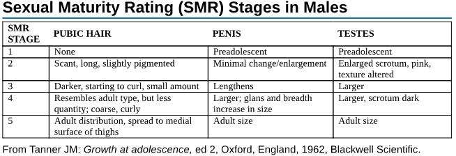
Vol 121, No 3, March 2023Journal of the Indian Medical Association
Table 1
Pre-adolescence Early adolescenceMiddle adolescence (n= 431) (n=484)(n=62) Age(in years)567891011121314151617 N4979839412612513014386381581 31
Annexure 1 : Tanner Stages (Sexual Maturity Rating scales) for males
— Distribution according to Age
both were equal among the rest (28.8%)(n=281). This had statistical significance (p<0.001). According to IAP growth charts, only 1 boy was classified as overweight and 1 boy as obese. We found a significant positive correlation between SMR and Height, Weight and BMI (Tables 2-5).
DISCUSSION
Puberty is associated with accelerated growth velocities. Weight, Height, Testicular Volume and SMR increase proportionally with age. A consistent rise of BMI from 13.7 kg/m2 at 5 years to 21.64 kg/m2 at 17 years was noted indicating accelerated growth velocities and growth spurt.
For the onset of Puberty, most Clinicians use an Orchidometer cut-off of 4 mL8,9, although some studies showed that a volume of 3 mL can already be considered a sign of puberty10. The MTV in present study at the age of 8 years was 3.4 cc, which was higher, compared to studies by Lall11, Goede12 and Boben, et al13. During prepubertal years, we found minimal growth of testicular volume till 9 years of age and rapid increase was observed from 11 years of age; these results are similar to previously reported studies by Lawal14, Tomova15 and Joustra et al8. The MTV at the age of 11 years was 4.68 cc, which was similar to the study by Matsuo16 et al and higher compared to studies done by Lall11, Boben13, Goede12 and Marshall, et al17 But at 12 years, the MTV of 6.05 cc was similar to that of Goede12, lower than Chin 18 and Goede12; and higher than Lall 11 and Boben, et al13
At the age of 16, the MTV in children enrolled in our study was 13.44 cc which was lower than the results of Lall 11 , Goede12 and Boben, et al13. There was a rapid increase in Testicular Volume after the age of 11 years in our study, which was similar in studies done by Chin 18 , Jaiswal 2 , Beres 19 , Joustra 8 , Tomova15 and Lawal, et al14
The mean age attained at SMR Stage-2 in our study was 11.22 years, which was similar to the results of studies done by Hafez20, Singhi21, Wong22 and Papadimitriou, et al23. There was only one boy with SMR stage 5 in our study, so it was not possible to compare with other studies.
Pubertal changes
Vol 121, No 3, March 2023Journal
of the Indian Medical Association
AgenMean WeightMean Height Mean BMI Mean TVSMR (kg)(cm)(kg/m2)(ml)12345 54915.35 ± 1.9105.7 ± 3.8013.7 ± 1.392.88 ± 0.74490000 67916.40 ± 2.19110.13 ± 6.4213.50 ± 1.373 ± 1.00745000 78318.29 ± 3.32115.08 ± 6.9513.83 ± 2.493.34 ± 1.11784100 89419.52 ± 3.22119.40 ± 6.4713.64 ± 1.583.4 ± 0.897122100 912621.45 ± 3.30124.24 ± 5.5613.86 ± 1.563.7 ± 1.009431100 1012524.71 ± 4.89131.01 ± 7.1614.3 ± 1.964.28 ± 1.727646300 1113027.45 ± 6.98135 ± 6.3214.87 ± 2.804.68 ± 1.667063700 1214329.07 ± 5.88139.42 ± 7.3314.82 ± 2.166.05 ± 2.8937951010 138633.6 ± 7.4144.95 ± 8.3915.86 ± 2.57.46 ± 3.3412501950 143835.49 ± 5.65149. 58 ± 9.0715.89 ± 2.128 ± 3.294191041 151538.47 ± 9.49155.7 ± 9.0815.72 ± 3.1710.44 ± 2.32041010 16848.87 ± 6.81160.25 ± 7.7818.2 ± 1.9413.44 ± 3.6403230 1715015221.64700100
Table 2 — Anthropometric parameters, MTV and SMR according to age
SMRnMean ageMean weightMean HeightBMIMTV (Years)(Kg) (cm)(kg/m2)(cc) 15628.53 ± 2.1721.59 ± 6.48122.19 ± 11.7714.18 ± 2.203.5 ± 1.15 233411.22 ± 1.8727.60 ± 7.31136.30 ± 11.6414.59 ± 2.115.88 ± 2.61 36612.88 ± 1.8633.45 ± 8.96145.32 ± 13.2215.59 ± 2.578.83 ± 3.94 41414 ± 1.3041.57 ± 6.63154.07 ± 7.9817.59 ± 2.2612.03 ± 2.42 51143916314.6817.5
Table 5 — Anthropometric parameters and MTV according to stages of SMR
Pre-adolescence Early adolescenceMiddle adolescence (n=431)(n=484)(n=62) SMR >265 (15 %)299 (61%)58 (93.5%) Mean BMI (kg/m2)13.7014.9617.86 MTV (cc)3.26 ± 1.015.61 ± 2.699.72 ± 3.43 Table 3
SMR StagingAge Pre-adolescence Early adolescenceMiddle adolescence (<10 years) n=431(10-13 years) n=484(14-17 years) n=62 SMR 1 (562)(57.5%)3661933 SMR 2 (334)(34.2%)6224527 SMR 3 (66)(6.8%)34023 SMR 4 (14)(1.4%)068 SMR 5 (1)(0.1%)001 32
Table 4 — Growth parameters according to stages of adolescence
—
Stages
of adolescence and SMR
were seen to start in some boys at 9-10 years, the mean age being 11.22 years. This is similar to studies done by Lall11, Tomova15, Joustra8 and Beres19 but earlier compared to Wacharasindhu24 and Largo, et al25. MTV at 15 and 16 years of age in present study were lower than the studies of Lall11 , Goede12, Boben13 and Beres, et al19
The Right Testes was found to be larger in studies by Boben13 and Beres et al19. According to Beres, et al the Left Testis which forms an entity and moves together with the epididymis, gubernaculum, afferent vascular plexus and vas deferens, descends sooner to and settles lower in, the scrotum; whereas the right testis, owing to more abundant blood supply across a shorter plexus, grows to larger size 19 Another explanation is that the pampiniform plexus of veins are more prominent in the left due to sluggish drainage of the Left Testicular Vein into the Left Renal Vein which may lead to increased temperature on the left and hence decreased Sertoli cell proliferation26
A spurt in BMI was noted in boys at 10 years of age. The mean age at onset of Puberty and SMR 2 was 11.22 years in the study. There were 33 boys less than 9 years showing SMR 2 or more who could be categorised in Pre-cocious puberty group. The reasonably large number of boys with “Precocious Puberty” as per existing definition points to an advancement of age of normal onset of Puberty in present times due to improved nutrition. But none of them was overweight or Obese. There were 4 boys with delayed Puberty, ie, SMR 1 at >14 years and none of them were underweight. Since there was a positive correlation of TV with Weight, Height and BMI, it indicates that nutrition might play a role in Puberty progression. But no apparent impact of nutrition on the onset of Puberty could be noted.
Limitations of the study :
The number of boys greater than 15 years of age was limited (0.02 % of study population). Axillary hair staging and confirmation of Testicular Volume by Ultrasonography could not documented.Concomitant LH/FSH/testosterone values could not be assessed for the biochemical confirmation of onset of Puberty.
CONCLUSION
In the present study, 977 school going boys between ages 5 and 17 years in an Urban setting were studied and data regarding Weight, Height, Testicular Volume and SMR were recorded. No role of nutritional status on clinical onset of puberty was noted. Precocious puberty was observed in 3.38% of boys (less than 9 years). Hence, possibility of an earlier
onset of Puberty can be entertained. It is necessary to generate a baseline data of age appropriate Testicular Volume in every population to assess Puberty and Sexual Developmental Disorders.
Source(s) of Support : Nil
Presentation at a Meeting : Nil
Conflicting Interest : Nil
Acknowledgement : Nil
REFERENCES
1Kuijper EAM, Kooten J van, Verbeke JIML, Rooijen M van and Lambalk CB — Ultrasonographically measured testicular volumes in 0- to 6-year-old boys. Human Reproduction 2008; 23(4): 7926. .doi:10.1093/humrep/den021
2Jaiswal VK, Khadilkar V, Khadilkar A, Lohiya N — Stretched Penile Length and Testicular Size from Birth to 18 Years in Boys from Western Maharashtra. Indian Journal of Endocrinology and Metabolism 2019; 23: 3-8.
3Kaplan SA — Clinical pediatric& adolescent endocrinology. W.B. Saunders. Philadelphia, 1982; 307-8.
4Soliman A, De Sanctis V, Elalaily R — Nutrition and pubertal development. Indian Journal of Endocrinology and Metabolism 2014; 18 (Suppl 1 ) : S39-47. doi: 10.4103/ 2230-8210.145073.
5Kliegman, Robert — Chapter 132, Adolescent physical and social development. Nelson Textbook of Pediatrics. Edition 21. Philadelphia, PA: Elsevier, 2020,p 1015
6Emmanuel M, Bokor BR — Tanner Stages. [Updated 2020 Aug 22]. In: StatPearls [Internet]. Treasure Island (FL): StatPearls Publishing; 2020 Jan-. Available from: https:// www.ncbi.nlm.nih.gov/books/NBK470280/
7Dattani MT, Tziaferi V, Hindmarsh PC — Evaluation of Disordered Puberty. Brook’s clinical pediatric endocrinology. 7th ed. Brooks CGD, Clayton PE, Brown RS, editors. 256-65.
8Joustra SD, van der PlasEM, Goede J, Oostdijk W — New reference charts for testicular volume in Dutch children and adolescents allow the calculation of standard deviation scores. Acta Paediatr 2015; 104: e271-8.
9Wu FC, Brown DC, Butler GE, Stirling HF, Kelnar CJ — Early morning plasma testosterone is an accurate predictor of imminent pubertal development in prepubertal boys. J Clin Endocrinol Metab1993; 76: 26-31.
10Biro FM, Lucky AW, Huster GA, Morrison JA — Pubertal Staging in Boys. J Pediatr 1995; 127: 100-2.
11Lall KB, Singh S, Gurani M, Chowdhary B, Garg OP — Normal testicular volume in school children. Indian J Paediatr 1980; 47(5): 389-93.
12Goede J, Hack WW, Sijstermans K — Normative values for testicular volume measured by ultrasonography in a normal population from infancy to adolescence. Horm Res Paediatr 2011; 76(1): 56-64. doi:10.1159/000326057
13Boben GE, Umapathy P, Ravichandran L, Godfrey DA, Ramani G, Srinivasan V — Evaluation of testicular volume in children aged 8-17 years in south India. Indian J Child Health 2016; 3(3): 208-11.
Vol 121, No 3, March 2023Journal
of the Indian Medical Association
33
14Lawal S, Idris HW, Ibinaiye P, Hamidu AU, Tabari MA, Usman B, et al — Normative ultrasonographic values for testicular volumes in Nigerian boys aged 0-15 years. Sub-Saharan Afr J Med 2016; 3: 71-8.
15Tomova A, Deepinder F, Robeva R, Lalabonova H, Kumanov P, Agarwal A — Growth and development of male external genitalia: A cross-sectional study of 6200 males aged 0 to 19 years. Arch Pediatr Adolesc Med 2010; 164: 1152-7.
16Matsuo N, Anzo M, Sato S, Ogata T, Kamimaki T — Testicular volume in Japanese boys up to the age of 15 years. Eur J Pediatr 2000; 159: 843-5.
17Marshall WA, Tanner JM — Variations in the Pattern of Pubertal Changes in Boys. Archives of Disease in Childhood 1970; 45: 13.
18Chin T, Liu C, Wei C — Testicular volume in Taiwanese boys. Zhonghua Yi Xue Za Zhi (Taipei) 1998; 61(1): 29-33.
19Beìres J, Papp G, PazonyiI, Czeize lE — Testicular volume variations from 0 to 28 years of age. Int Urol Nephrol 1989; 21: 159-67.
20Hafez AS, Salem SI, Cole TJ, Galal OM, Massoud A — Sexual maturation and growth pattern in Egyptian boys. Ann Hum Biol 1981; 8(5): 461-7.
21Singhi S, Lall KB, Gurnanl M, Garg OP — Age of appearance of secondary sex characters in Ajmer school children. Indian I Pediat 1982; 49: 547-52.
22Wong GW, Leung SS, Law WY, Yeung VT, Lau JT, Yeung WK — Secular trend in the sexual maturation of southern Chinese boys. Acta Paediatr 1996; 85(5): 620-1.
23Papadimitriou A, Stephanou N, Papantzimas K, Glynos G, Philippidis P — Sexual maturation of Greek boys. Annals of Human Biology 2002; 29(1): 105-8.
24Wacharasindhu S, Pri-Ngam P, Kongchonrak T — Selfassessment of sexual maturation in Thai children by Tanner photographs. Journal of the Medical Association of Thailand 2002; 85: 308-19.
25Largo RH, Prader A — Pubertal development in Swiss boys. Helv Paediatr Acta 1983; 38: 211-28.
26Hadijat Oluseyi Kolade-Yunusa, Ukamaka D Itanyi, Chiedozie J Achonwa — Determination of a normogram for testicular volume measured by ultrasonography in a normal population boys in Abuja. Orient Journal of Medicine 2017; 29[1-2]:
27Karlberg J — Secular trends in pubertal development. Horm Res2002; 57 Suppl 2: 19-30. doi: 10.1159/000058096. PMID: 12065922.
28Surana V, Dabas A, Khadgawat R, Marwaha RK, Sreenivas V, Ganie MA, et al — Pubertal onset in apparently healthy Indian boys and impact of obesity. Indian J Endocr Metab 2017; 21: 434-8.
29Campbell BC, Gillett-Netting R, Meloy M — Timing of reproductive maturation in rural versus urban Tonga boys, Zambia. Ann Hum Biol 2004; 31(2): 213-27.
30Daniel WA Jr, Feinstein RA, Howard-Peebles P, Baxley WD — Testicular volumes of adolescents. J Pediatr 1982; 101(6): 1010-2.
Disclaimer
The information and opinions presented in the Journal reflect the views of the authors and not of the Journal or its Editorial Board or the Publisher. Publication does not constitute endorsement by the journal.
JIMA assumes no responsibility for the authenticity or reliability of any product, equipment, gadget or any claim by medical establishments/institutions/manufacturers or any training programme in the form of advertisements appearing in JIMA and also does not endorse or give any guarantee to such products or training programme or promote any such thing or claims made so after. —
HonyEditor
Vol 121, No 3,
March 2023Journal of the Indian Medical Association
34
Original Article
Evaluation of Risk Factors of Postoperative Urinary Retention in Male Patients Undergoing Surgery Under Spinal Anaesthesia : A Prospective Study
Tusharindra Lal1, Vigneshwar Kumbakonam Sivaraman2, Prabhath Jagath Singh3, Saravanan Sanniyasi4
Background : Postoperative Urinary Retention (POUR) is common after regional anaesthesia with a reported incidence between 5% and 70%. POUR can lead to significant morbidity with additional surprise and mental trauma to the patient when unwarned. This study aimed to assess the occurrence of POUR in male patients undergoing Surgery under Spinal Anaesthesia and to study the risk factors related to it.
Methods : 692 male patients were analysed prospectively for the need for catheterisation which was defined as “the inability to void in the immediate Postoperative period with accompanying discomfort and a palpable Bladder.” All such patients were catheterised as an emergency. A record was made about the mean age, surgical condition, comorbidities, duration of Surgery, use of intra-operative sedatives, intra-operative fluid infused and International Prostate System Score (IPSS).
Results : The overall mean age of patients with POUR was 46 years. The incidence of POUR was highest among Perianal Surgeries ie, 52/70 (13%) followed by Hernia Surgeries, 18/70 (6.3%). An appreciable reduction was observed in urinary retention after administration of intra-operative sedatives (p=0.022) and lower IPSS (p=0.001). Factors such as age, intra-operative fluid administration, duration of Surgery and previous history of Diabetes did not reach statistical significance as being predictive of urinary retention.
Conclusion : An IPSS greater than 7 increases the risk of Postoperative Urinary Retention while the use of intraoperative Sedative in combination with Spinal Anaesthesia decreases the risk. In high-risk patients undergoing perianal procedures, pre-operative patient counselling about the possibility of Postprocedure retention is recommended.
[J Indian Med Assoc 2023; 121(3): 35-8]
Key words :Postoperative Urinary Retention, Spinal Anaesthesia, Catheterisation, IPSS, Intra-operative Sedatives.
Postoperative Urinary Retention (POUR) refers to the patients’ inability to void urine in spite of a full bladder in the postoperative period following surgical intervention1. The reported incidence varies between a wide range of 5% and 70% 2,3 . This extensive variability may be due to the presence of multiple definitions of POUR, types of surgery and anaesthesia used, differences in patient profiles and the perioperative fluid therapy utilized4. Untreated cases
Department of General Surgery, Sri Ramachandra Institute of Higher Education and Research, Chennai 600116
1MBBS Final year
2MBBS, MS, MCh (Neurosurgery) Final year Resident, Institute of Neurosurgery, Madras Medical College and RGGGH, Chennai 600003
3MBBS, MS, Assistant Professor, Department of General Surgery, Sree Uthradom Thirunal Academy of Medical Sciences, Vencode, Vattappara, Kerala 695028 and Corresponding Author
4MS, FRCS Glasgow, MRCS Edinburgh, DNB General Surgery, FACS, Professor
Received on : 03/02/2022
Accepted on : 04/03/2022
Editor's Comment : Perianal procedures have a higher incidence of Postoperative Urinary Retention (POUR) as compared to Hernia repair. An International Prostate System Score (IPSS), score of more than 7 is a risk factor for retention while the administration of intra-operative sedatives in combination with regional anaesthesia reduced the incidence of POUR. Variables such as intra-operative fluid infused, diabetes mellitus, age of the patient and duration of surgical procedure did not significantly contribute to urinary retention in our study.
of POUR can lead to significant morbidity such as prolongation of in-hospital stay, Urinary Tract Infections, Detrusor Muscle Dysfunction, Delirium, Cardiac Arrhythmias, etc5. These have thus led to an increasing focus on early detection of POUR. The current study was conducted to investigate the occurrence of Postoperative Urinary Retention in male patients undergoing surgery under Spinal anaesthesia and to study the contributing risk factors.
Vol 121, No 3, March 2023Journal of the Indian Medical Association
35
MATERIALS AND METHODS
All male surgical patients admitted to our Tertiary Care Centre from June, 2017 to November, 2019 were included in this prospective study. Within this cohort, a total of 692 patients who underwent surgery under Spinal anaesthesia were analysed. POUR was defined as “the inability to void in the immediate postoperative period with accompanying discomfort and a palpable Bladder.” Patients above the age of 18 years were included while those who had been electively catheterized during surgery and those with postTransurethral Resection of the Prostate (TURP) were excluded from the study.
The operative procedures were broadly divided into Hernia, Perianal Surgeries and others. Operative procedures for Hernia included those for Inguinal, Incisional and Umbilical hernias while Perianal procedures were performed for Perianal Fissures, Fistula-in-ano, Hemorrhoids and perianal abscesses. ‘Other’ procedures included surgery for Hydrocele, Varicose Veins and Paraphimosis.
Three methods were used for the diagnosis of POUR namely history and clinical examination, Bladder catheterization and Ultrasonographic assessment2 The primary outcome of this study was to study the contribution of mean age, type of surgical procedure, duration of Surgery (in minutes), intra-operative Sedative use, intra-operative fluid volume and IPSS score to Postoperative Urinary Retention. A p-value of <0.05 was considered to indicate statistical significance.
RESULTS
The mean age of the patients in this study was 46 years (from 18 to 87 years). There was no difference in age between those who voided and those who had retention after Hernia or Perianal surgery (p=0.405) (Table 1). Among the 692 patients subjected to various surgeries, the occurrence of POUR was seen in a total of 70 patients (10.1%). This was found to be maximum in perianal surgeries, 52 (13%) followed by hernia surgeries 18 (6.3%). When comparing individual surgeries, POUR was found to be maximum in bilateral indirect inguinal hernia (20%)
followed by Perianal abscess (17.81%), Hemorrhoids (17.2%) and Fissure-in-ano (12.1%). The difference of proportion among the different types of surgeries: hernia, perianal and ‘others’ with the development of POUR was found to be statistically significant (p= 0.013).
A significant reduction in urinary retention was reported following administration of Intravenous (IV) sedative, midazolam (1 mg) intra-operatively along with spinal anaesthesia (p=0.022). Additionally, a higher IPSS (greater than 7) was associated with POUR (p<0.001)(Table 1).
For purpose of analysing the effect of intra-operative fluids, patients were divided into two groups, those who received up to 1000 ml of intra-operative fluids and those greater than 1000 ml. The study failed to show an association of POUR to the amount of intraoperative fluid administered (p= 0.691).
Urinary retention was found to be more in diabetic patients as opposed to non-diabetics and in cases where the average duration of Surgery was longer (more than 60 minutes), however, a significant p-value was not obtained for both these parameters, p=0.611 and 0.401 respectively.
DISCUSSION
Postoperative Urinary Retention is common among surgical patients and may prolong the in-hospital stay and cause significant pain and morbidity 6 . It is important to identify patients with perioperative risk factors to avoid potential complications in the postoperative period4. Three methods have been described to diagnose urinary retention which includes physical examination, Bladder catheterization and
Vol 121, No 3, March
2023Journal of the Indian Medical Association
VariableVoided POUR Incidence P-value of POUR (%) Mean Age0.405 Up to 50 years38647 10.9 More than 50 years236238.9 Intra-operative sedative0.022 Administered8733.3 Not administered5356711.1 Intra-operative fluid administered0.691 Up to 1000 ml432479.8 More than 1000 ml19023 10.8 International Prostate Symptom Score (IPSS)0.001 Up to 7589599.1 More than 7331125 Duration of surgery (in minutes)0.401 Up to 60 minutes46055 10.7 More than 60 minutes162158.5 Diabetes Mellitus 0.611 Yes841111.6 No538599.9 36
Table 1 — Factors affecting Postoperative Urinary Retention (POUR)
Ultrasonographic assessment 2 . In our study, 70 (10.1%) patients developed urinary retention.
Age has been known to increase the risk of POUR in patients over the age of 50 years2,7,8. Alex, et al also demonstrated that older individuals were at significant risk (67.7 versus 62.0 years, p<0.0001)9 However, in our study, there was no difference in the age groups (p=0.405).
An association between the type of surgical procedure and the incidence of POUR has been reported in the literature. The incidence of POUR has been reported to be higher in patients undergoing Anorectal Surgeries and varies with a wide range between 1% and 52%7. This may be attributed to the risk of injury to Pelvic Nerves and a pain-evoked reflex increase in the tone of the internal sphincter7. The risk of POUR in two studies was found to be higher in haemorrhoidectomy Surgery with the incidence of 21% and 34% respectively7,10. Similarly, POUR was found to be higher among perianal surgeries, 52/70 patients (13%) in our study. However, on comparing individual surgeries, POUR was found to be maximum in bilateral indirect Inguinal Hernia (20%) followed by Perianal Abscess (17.81%), hemorrhoids and Fissure-in-ano. Blair, et al studied the incidence of POUR among Hernia Surgeries and found that the occurrence of POUR in bilateral Inguinal Hernias was 16.7%, as compared to 17.39% in the present study11
Administration of intravenous sedative along with Spinal anaesthesia resulted in the reduction of postoperative pain and subsequent reduction of the incidence of POUR and revealed a highly significant correlation (p= 0.022). Similar studies showed that the analgesic effect of Spinal Anaesthesia was potentiated by the administration of IV midazolam, resulting in a significant reduction of Postoperative Urinary Retention (p <0.01, 0.028 and <0.05 respectively)12-14.
The International Prostate Symptom Score (IPSS) has been used to assess the lower urinary tract in patients suffering from Bladder outlet obstruction as a result of prostatic enlargement15. It was observed from this study that when the IPSS was more than 7, there was a significant increase in the occurrence of POUR (p<0.001). Other reports also demonstrated that patients with increased IPSS (>8) had a higher chance of developing POUR16
It is expected for urinary retention to be higher with excessive administration of intravenous fluids as a result of overdistension of the bladder. Studies have demonstrated a significant correlation between intraoperative fluid administration and POUR (p<0.0001)3,17 An increased intra-operative fluid support (>1000 ml)
failed to increase the risk of POUR in the current study, as was reported in other studies6,7
There are contradictory reports in the literature regarding the duration of surgery and the risk of POUR. Many studies determined that the increase in mean surgery time was associated with an increased risk of urinary retention in the postoperative period3,8,18. A direct correlation was observed in our study between the mean operating time (more than 60 minutes) with the development of POUR. However, this difference was not significant.
Diabetes Mellitus was found to be significantly associated with the development of POUR in many studies (p=0.039, 0.004, <0.01)4,19,20. Another study consisting of 20,77,045 patients described the association of Diabetes to POUR (p<0.0001)9. In our study, the contribution of Diabetes to POUR was not established (p=0.611). Similarly, another study failed to demonstrate a significant correlation between diabetes and urinary retention (p=0.56)11
CONCLUSION
Perianal procedures have a higher incidence of POUR as compared to Hernia repair. An IPSS score of more than 7 is a risk factor for retention while the administration of intra-operative sedatives in combination with regional anaesthesia reduced the incidence of POUR. Variables such as intra-operative fluid infused, Diabetes Mellitus, age of the patient and duration of surgical procedure did not significantly contribute to urinary retention in our study. Patient counselling for risk of retention requiring Postoperative catheterization is recommended for patients undergoing Perianal Surgeries with IPSS scores greater than 7.
REFERENCES
1Rosenstein D, McAninch JW — Urologic emergencies. Medical Clinics 2004; 88(2): 495-518.
2Baldini G, Bagry H, Aprikian A, Carli F, Warner DS, Warner MA — Postoperative urinary retention: anesthetic and perioperative considerations. The Journal of the American Society of Anesthesiologists 2009; 110(5): 1139-57.
3Keita H, Diouf E, Tubach F, Brouwer T, Dahmani S, Mantz J, et al — Predictive factors of early postoperative urinary retention in the postanesthesia care unit. Anesthesia&Analgesia 2005; 101(2): 592-6.
4Çakmak M, Yýldýz M, Akarken Ý, Karaman Y, Çakmak Ö — Risk factors for postoperative urinary retention in surgical population: A prospective cohort study. Journal of Urological Surgery 2020; 7(2): 144-8.
5Agrawal K, Majhi S, Garg R — Post-operative urinary retention: Review of literature. World Journal of Anesthesiology 2019; 8(1): 1-2.
Vol 121, No 3, March 2023Journal of
the Indian Medical Association
37
6Altschul D, Kobets A, Nakhla J, Jada A, Nasser R, Kinon MD, et al — Postoperative urinary retention in patients undergoing elective spinal surgery. Journal of Neurosurgery: Spine 2017; 26(2): 229-34.
7Toyonaga T, Matsushima M, Sogawa N, Jiang SF, Matsumura N, Shimojima Y, et al — Postoperative urinary retention after surgery for benign anorectal disease: potential risk factors and strategy for prevention. International Journal of Colorectal Disease 2006; 21(7): 676-82.
8Lamonerie L, Marret E, Deleuze A, Lembert N, Dupont M, Bonnet F — Prevalence of postoperative bladder distension and urinary retention detected by ultrasound measurement. British Journal of Anaesthesia 2004; 92(4): 544-6.
9Wu AK, Auerbach AD, Aaronson DS— National incidence and outcomes of postoperative urinary retention in the Surgical Care Improvement Project. The American Journal of Surgery 2012; 204(2): 167-71.
10Zaheer S, Reilly WT, Pemberton JH, Ilstrup D — Urinary retention after operations for benign anorectal diseases. Diseases of the Colon & Rectum 1998; 41(6): 696-704.
11Blair AB, Dwarakanath A, Mehta A, Liang H, Hui X, Wyman C, et al — Postoperative urinary retention after inguinal hernia repair: a single institution experience. Hernia 2017; 21(6): 895-900.
12Kim MH, Lee YM — Intrathecal midazolam increases the analgesic effects of spinal blockade with bupivacaine in patients undergoing haemorrhoidectomy. British Journal of Anaesthesia 2001; 86(1): 77-9.
13Hadimioglu N, Ertugrul F, Ertug Z, Yegin A, Karaguzel G, Erman M — The comparative effect of single dose mivacurium during sevoflurane or propofol anesthesia in children. Pediatric Anesthesia 2005; 15(10): 852-7.
14Gottesman L, Milson JW, Mazier WP — The use of anxiolytic and parasympathomimetic agents in the treatment of postoperative urinary retention following anorectal surgery. Diseases of the Colon & Rectum 1989; 32(10): 867-70.
15Barry MJ, Fowler Jr FJ, O’Leary MP, Bruskewitz RC, Holtgrewe HL, Mebust WK, et al — Measurement Committee of the American Urological Association. The American Urological Association symptom index for benign prostatic hyperplasia. The Journal of Urology 1992; 148(5): 1549-57.
16Homma Y, Yamaguchi T, Kondo Y, Horie S, Takahashi S, Kitamura T — Significance of nocturia in the International Prostate Symptom Score for benign prostatic hyperplasia. The Journal of Urology 2002; 167(1): 172-6.
17Pavlin JD, Pavlin EG, Fitzgibbon DR, Koerschgen ME, Plitt TM — Management of bladder function after outpatient surgery. The Journal of the American Society of Anesthesiologists 1999; 91(1): 42-50.
18Hansen BS, Søreide E, Warland AM, Nilsen OB — Risk factors of post operative urinary retention in hospitalised patients. Acta Anaesthesiologica Scandinavica 2011; 55(5): 545-8.
19Dreijer B, Møller MH, Bartholdy J — Post-operative urinary retention in a general surgical population. European Journal of Anaesthesiology (EJA) 2011; 28(3): 190-4.
20Jung HJ, Park JB, Kong CG, Kim YY, Park J, Kim JB — Postoperative urinary retention following anterior cervical spine surgery for degenerative cervical disc diseases. Clinics in Orthopedic Surgery 2013; 5(2): 134-7.
Vol 121, No 3,
March 2023Journal of the Indian Medical Association
38 Submit Article in JIMA - Online See website : https: // onlinejima.com Any queries : (033) 2237-8092, +919477493027; +919477493033
Original Article
Validation of Glasgow-Blatchford Score in Predicting Management of Upper Gastrointestinal Bleeding
Shantanu Shirish Navgale1, Akshay Shirish Deshpande2, Sunny Agarwal3, Satish Balkrishna Dharap4, Rajesh Patil5
Background : Upper Gastrointestinal Bleeding is a common emergency with varying degrees of severity. Haemorrhage is managed by Therapeutic Endoscopy, Radiological Intervention or Surgery and Blood Transfusion which are available only in Tertiary Care Centre. So, when patient presents in primary healthcare setting, it is important to recognize the patients who need this treatment. Glasgow-Blatchford Score is a score which is used for this purpose. The purpose of this research was to validate its reliability in identifying such high-risk patients.
Materials and Methods : This study was prospective and observational, conducted in Medical College and Hospital, from December, 2017 to May, 2019. All adult patients presenting to Emergency Department with sudden onset Upper Gastrointestinal Bleeding were included. Glasgow-Blatchford Score was computed. Patients were followed up till their discharge (or death) from the hospital. The therapeutic management needed and its relationship with the score and treatment modalities were noted. Area under Receiver Operating Characteristic (ROC) Curve was calculated.
Results : Total 100 patients were included in study. 85% were male and 15% were female. Glasgow-Blatchford Score was found as a good predictor in discriminating patients. Patients with score 14. Interventional radiology or surgery was never used. The area under ROC Curve was 0.738 suggesting fair reliability.
Conclusion : Glasgow-Blatchford Score is good predicting tool in cases of Upper Gastrointestinal bleeding and patients with score >7 should be transferred to speciality centres.
[J Indian Med Assoc 2023; 121(3): 39-42]
Key words :Upper Gastrointestinal Bleeding, Glasgow-Blatchford Score.
Any bleeding from a site proximal to the ligament of Treitz in Gastrointestinal Tract is defined as Upper Gastrointestinal Bleeding (UGIB). Hematemesis, Malena, Syncope, Epigastric Pain, Dysphagia, Dyspepsia, Weight Loss, Diffuse Abdominal Pain and Jaundice are the signs and symptoms of acute UGIB. Ulcers in the Gastrointestinal Tract such as Erosion or bleeding Ulcers may lead to Upper Gastrointestinal Bleeding. Rupture of the blood vessels like a Variceal Rupture in the esophagus, fundus and gastric cardia or Mallory-Weiss tear in distal Esophagus are the other causes for Upper Gastrointestinal Bleeding. Carcinoma of the Esophagus, Stomach and Duodenum can also cause UGIB.
Among 100,000 population per year approximately 100 cases present with UGIB1 UGIB is approximately 4 times as common as bleeding from the Lower GI
Department of General Surgery, Topiwala National Medical College, Mumbai 400008
1MBBS, MS, Assistant Professor and Corresponding Author
2MBBS, MS, Assistant Professor
3MBBS, MS, FIAGES, Assistant Professor
4MBBS, MS, DNB, FMAS, FIAGES, Professor and Head
5MBBS, MS, FMAS, Associate Professor
Received on : 11/01/2022
Accepted on : 19/03/2022
Editor's Comment :
Patients presenting with Upper GI bleeding having Glasgow Blatchford Score of 7 or more than 7, should be transferred to higher center for further superspeciality care. This will reduce morbidity and mortality related to upper GI bleeding cases.
Tract and causes significant morbidity and mortality. Mortality rate is around 6-10% for UGIB2 In 98.3% of mortalities in UGIB patients, one or more comorbid illnesses were noted3 In 72.3% of patients, primary cause of death was comorbid illnesses.
Glasgow Blatchford Score helps to assess whether a patient with acute UGIB will need medical intervention such as Endoscopic Intervention or Blood Transfusion. This score is also useful to recognize patients who don‘t need admission to the hospital after UGIB. The validity of this score in present settings will be assessed.
Patients with Haematemesis are resuscitated and stabilized immediately after admission to the hospital. Blood and blood components are transfused if necessary and proton pump inhibitors, vasopressin or its analogues and somatostatin or its analogues are given to start initial treatment.
Vol 121, No 3, March 2023Journal of the Indian Medical Association
39
First-line diagnostic modality and treatment option for UGIB is Endoscopy. It is an important early intervention that is used to recognize the source of bleeding. Endoscopy can also be used for Therapeutic Interventions4. However, findings can be non-diagnostic in about 10% of cases. For example, in a case of massive UGIB, Endoscopy may not be helpful because intraluminal blood cannot be adequately cleared.
Early Upper GI Endoscopy is recommended within 24 hours of presentation in majority patients. This is useful to confirm the diagnosis and it also allows for targeted Endoscopic management like epinephrine injection, thermo-coagulation, clips application and variceal banding4
Endoscopic therapy is useful to bring down morbidity rate, risk of recurrent bleeding and hospital stay. It also reduces the need for surgery to tackle with active Upper Gastrointestinal Bleeding. Inspite of successful endoscopic therapy, 10 to 20 percent of patients can present with rebleeding 5 ; For such patients a second setting of endoscopic procedure is required.
Transjugular Intrahepatic Portosystemic Shunt (TIPS) which is one of the interventional radiological procedure can be indicated in some patients of Upper GI Bleeding. If there is persistent and severe bleeding then Arteriography with embolization or surgery may be needed. Surgery is useful for uncontrolled bleeding from Peptic Ulcer.
However, at Primary Health Care Centre, this advcanced modalities are not available. So, GBS is a screening tool to assess whether a patient with acute UGIB will need medical intervention such as endoscopy or blood transfusion. Advantages of the GBS over the Rockall score, which is useful to assess mortality risk in patients with UGIB, includes lack of need for Oesophago Gastro Duodenoscopy (OGD) and the lack of subjective variables like the severity of systemic diseases to complete the score, which is a feature unique to the GBS.
MATERIALS AND METHODS
This study was done at Medical College and Hospital, after taking approval of the Institutional Ethics Committee during a period of 1 year and 6 months (December, 2017 to May, 2019). This was prospective, observational, descriptive study including 100 patients with age more than 18 years who were presenting in emergency services with chief complaints of having Acute Hematemesis or Malena. Repeated Hematemesis cases that were diagnosed earlier and managed were also involved in the study. All the
patients were included in study after taking informed consent. Patients who were presented electively for management of Upper GI bleeding with past history of Hematemesis or Malena were excluded from study.
A thorough history and detailed clinical examination was carried out. All patients were subjected to the biochemical investigations. The patients were simultaneously resuscitated, stabilized and if required blood and blood products transfusion was done. Once stable, Upper Gastrointestinal Endoscopy was carried out for diagnosis and for therapeutic purpose. Endoscopic therapeutic intervention was performed in same setting if indicated. Interventional radiology and surgery were other options available if needed.
Patients were followed till discharge or death in hospital. Glasgow Blatchford Score (GBS) was computed for every patient (Table 1). Outcome was measured in the form of need for blood transfusion, need for Therapeutic Endoscopy, surgical procedure and any other procedure by interventional radiology and in hospital mortality.
Data was tabulated in Microsoft excel spreadsheet (Version office 8).
Admission Risk MarkerScore Component Value
Other
Presentation with syncope2
Pulse > 100 (per min)1
Presentation with Malena1
The score is equal to “0” if all of the following are present:
•Hemoglobin level >12.9 g/dL (men) or >11.9 g/dL (women)
•Blood urea nitrogen level <6.5 mg/dL
•Pulse <100/minute
•Systolic BP >109 mm Hg
•No Melena or syncope
•No past or present Liver Disease or Heart Failure
•Interpretation of Score:
<0: Minimal risk of needing an intervention
>0: Higher risk of needing an intervention
6 or >6: More than 50% risk of needing an intervention
Vol 121, No 3, March 2023Journal of the Indian Medical Association
Blood Urea (mmol/L) : >256 10.0-254 8.0-10.03 6.5-8.02 Haemoglobin(g/L) for Men : <10.06 10.0-11.93 12.0-12.91 Haemoglobin(g/L) for Women : <10.06 10.0-11.91
Blood Pressure (mm Hg) : <903 90-992 100-1091
Table 1 — Showing Glasgow Blatchford Score (GBS)
Systolic
Cardiac
markers : Hepatic Disease2
Failure2
40
Statistical Analysis : Statistical Package for Social Sciences for Windows, version 16 (SPSS Inc, Chicago, IL, USA) was used for data analysis. To study association between categorical variables Chi-square test was used. Chi-square test for trend was used for ordinal data. ANOVA was used to compare means. Numerical data was shown as mean ± standard Deviation. T-test was used to compare numerical data. Receiver Operating Characteristic (ROC) Curve was plotted. After that Area Under Curve (AUROC)was computed. AUROC between 0.7 to 0.8 was considered fairly reliable for validity of GBS score. P value <0.05 was considered significant.
RESULTS
In our study, total 100 patients were included. The age group in this study varied from 19 years to 95 years with the mean age of 45.9 years with male preponderance as 85% patients were male. 49% patients presented with hematemesis and Malena and 51% patients presented with Hematemesis alone. 31% patients had recurrent episode of Hematemesis. 70% patients were Alcoholic and 9% patients were having addiction of Smoking. 15% patients had tachycardia with pulse more than 100/min. Mean pulse value was 92.9 ± 11.84 per minute. 16% patients presented in shock with systolic BP less than 90 mmHg. Mean Systolic BP was 108.32 ±14.80 mmHg. 67% of patients required blood transfusion. Out of 100 patients, 95% patients underwent Endoscopy and 5% patients could never be Stabilised and Endoscopy could not be performed. On Endoscopy, 68% patients showed Variceal bleeding, 14% patients had Mallory Weiss tear and 5% patients had Peptic Ulcer Disease. 2% patients showed normal study on Endoscopy. 68% of the patients who were having Variceal bleeding, underwent Endoscopic band ligation for oesophageal varices. Remaining 32% of patients were managed conservatively with medications. No surgery was done and neither interventional radiological procedure was done for management of UGIB. Mortality rate in our study was 5%. GBS score ranged from 3 to 18 with mean value of 12.03 ± 2.58.
Patients are divided into 4 groups (Table 2) with respect to GBS Score values as <7, 7-10, 11-14 and >14 and the percentage of patients needing a Blood Transfusion and percentage of patients needing Therapeutic Endoscopy was calculated and the graph was plotted. Both the graphs also show that as the GBS score increases, the need for Blood Transfusion (Fig 1) and Therapeutic Endoscopy (Fig 2) also increases.

Table 2 — GBS Score versus Blood Transfusion & Therapeutic Endoscopy

No ofNo of patientsNo of patients
ROC Curve for GBS Score :
Area under the curve for GBS Score found to be 0.738 with standard error of 0.058. With 95% confidence interval, lower bound value was 0.624 and upper bound value was 0.852.
DISCUSSION
The results indicate that GBS is a fairly good predictor in identifying patients needing referral to specialised centres. Patients with score <7 did not need transfusion or Therapeutic Intervention, when the score was between 7 and 10, 45% needed blood transfusion and 27% needed Therapeutic Endoscopy which further increased to 71% and 75% respectively when score was between 11-14 and 100% when more than 14. 2 patients with score 13 and 3 patients with score 14 succumbed to death. Interventional radiology
Vol 121, No 3, March 2023Journal of the Indian Medical Association
Patients having Blood undergone Transfusion Therapeutic Endoscopy <760 (0%)0 (0%) 7 -10115 (45.45%)3 (27.27%) 11-147352 (71.23%)55 (75.34%) >141010 (100%)100 %
Fig 2 — GBS Score versus Therapeutic Endoscopy
41
Fig 1 — GBS Score versus Blood Transfusion
or surgery was not used in any patient. The area under ROC Curve was 0.738 suggesting fair reliability.
Our findings are similar to the study done by Stevenson, et al6, in which patient with GBS Score less than 6 do not require blood product transfusion. Emergency Endoscopy was done in 86% of patients with GBS score of >6.
In study done by Blatchford O, etal7, the Area Under Curve for ROC Curve of GBS Score was 0.92 which indicates that GBS Score has very good reliability.
In study done by Chandra, et al8, in case of overall prognostic accuracy, GBS performed better than preendoscopy Rockall Score on ROC Curves analysis (AUC=0.79 versus 0.62; P=0.0001; absolute difference, 0.17). The prognostic accuracy of Postendoscopy Rockall Score and GBS was similarly high (AUC, 0.79 versus 0.72; P = 0.26; absolute difference, 0.07).
Study done by Robertson M, et al9, indicates that in predicting need for ICU admission, AIMS65 Score was superior amongst all other scores. Full-Rockall Scores, GBS and AIMS65 were equivalent (AUROCs 0.63 versus 0.62 versus 0.63) and for predicting the composite endpoint, all socres were superior to Preendoscopy Rockall score (AUROC 0.55). For predicting Blood Transfusion, GBS was superior to all other scores.
Limitations of our study was a small sample size. The place of study being a Tertiary Care Centre, only more complicated patients were referred and only 6% of patients had GBS score <7.
CONCLUSION
Glasgow Blatchford Score was found to be a fairly good predictor for need of Blood Transfusion and/or Therapeutic Endoscopic Intervention in cases of Upper Gastrointestinal Bleeding. It’s a simple scoring system that helps a Surgeon or a Physician in any setup to assess requirement for blood transfusion and
therapeutic endoscopy and consider timely transfer to specialised centres if facilities are not available locally.
REFRENCES
1Chason RD, Reisch JS, Rockey DC — More favorable outcomes with peptic ulcer bleeding due to Helicobacter pylori. Am J Med 2013; 3(7).
2Kaplan RC, Heckbert SR, Koepsell TD, Furberg CD, Polak JF, et al — Risk factors for hospitalized gastrointestinal bleeding among older persons. Cardiovascular Health Study Investigators. J Am Geriatr Soc 2001; 49: 126-33. Pmid :11207865 Fujishiro M, Iguchi M, Kakushima N, et al. Guidelines for endoscopic managements of non-variceal upper gastrointestinal bleeding. Dig Endosc. 2016 Feb 22.
3Kelly P, Katerna M, Amadi B, Zimba L, Aparico S, Mudenda K, et al — Gastrointestinal pathology in the University Teaching Hospital, Lusaka, Zambia: review of endoscopic and pathology records. Trans Royal Soc Trop Hyg 2008; 102: 194-9.
4Iwasaki H, Shimura T, Yamada T, Aoki M, Nomura S, Kusakabe A, et al — Novel Nasogastric Tube-Related Criteria for Urgent Endoscopy in Nonvariceal Upper Gastrointestinal Bleeding. Dig Dis Sci 2013; 22(5)
5Giday SA, Kim Y, Krishnamurty DM — Long-term randomized controlled trial of a novel nanopowder hemostatic agent (TC325) for control of severe arterial upper gastrointestinal bleeding in a porcine model. Endoscopy 2011; 43(4): 296-9.
6Stevenson J, Bowling K, Littlewood J, Stewart D— Validating The Glasgow-Blatchford Upper GI Bleeding Scoring System. Gut 2013; 62(Suppl 2): A21.1-A22.
7Blatchford O, Davidson LA, Murray WR, Blatchford M, Pell J — Acute upper gastrointestinal haemorrhage in west of Scotland: case ascertainment study. BMJ 1997; 315: 510-4.
8Chandra S, Hess E, Agarwal D, Nestler D, Montori V, Song L, et al — External validation of the Glasgow-Blatchford Bleeding Score and the Rockall Score in the US setting. The American Journal of Emergency Medicine 2012; 30(5): 673-9.
9Robertson M, Majumdar A, Boyapati R, Chung W, Worland T, Terbah R, et al — Risk stratification in acute upper GI bleeding: comparison of the AIMS65 score to the Glasgow-Blatchford and Rockall scoring systems, Gastrointestinal Endoscopy 2015; doi: 10.1016/j.gie.2015.10.021.
Vol 121, No 3, March 2023Journal
of the Indian Medical Association
42
Original Article
The Prevalence of Isolated Systolic Hypertension at a Tertiary Care Hospital in Eastern India : An Observational Study
Ram Krishna Brahmachari1, Supratik Chakraborty2, Shiladitya Nandi3
Background and Objectives : Hypertension is a significant public health issue. Isolated Systolic Hypertension (ISH) was once considered to be a benign aspect among the aging population, but an association with an increased risk of Cardiovascular Disease is now known. ISH shows an increasing prevalence with increase in age. This study was undertaken to determine the incidence of ISH among adults in Eastern India.
Methods : This study evaluated the adult population (aged 18-70 years) attending the NRS Medical College and Hospital, a Tertiary Care Center in East India. The clinical characteristics and echocardiographic findings were also evaluated.
Results : A total of 800 patients met the inclusion criteria, of whom 75 (9.37%) had ISH. Blood Pressure increased with age. The most common echocardiographic change observed in ISH patients was increased Left Ventricular Mass Index (LVMI), while concentric Left Ventricular Hypertrophy (LVH) was more common in women than men with isolated Systolic Hypertension. The incidence of LVMI increased as the severity of ISH increased. Furthermore, patients with stage 3 ISH were nearly 4 times more likely to develop Proteinuria.
Conclusion : The findings of this study are in line with previous studies evaluating the presence of ISH in the adult Indian population. There is need for effective population screening along with effective treatment for Blood Pressure to reduce morbidity and mortality. Primary prevention strategies may be the need of the hour in the Indian population which is at risk of cardiovascular Disease associated with Hypertension.
[J Indian Med Assoc 2023; 121(3): 43-7]
Key words :Blood Pressure, Cardiovascular Disease, Hypertension, Left Ventricular Hypertrophy, Proteinuria, Systolic Hypertension.
Hypertension is a major public health issue across the globe, with predictions indicating that 1.5 billion adults would be hypertensive in the year 2025. The increased prevalence of this silent killer is attributed to a changing lifestyle. Factors that predispose to Hypertension often vary from region to region and even across Urban and Rural populations. The incidence of Hypertension in India is estimated to be 29.8%, with an evident Urban-rural difference1.
There are various types of Hypertension. Isolated Systolic Hypertension (ISH) is defined as Systolic Blood Pressure (SBP) of >140 mmHg with a Diastolic Blood Pressure (DBP) of <90 mmHg. It is the most common form of Hypertension with 80% of hypertensive patients aged over 60 years having ISH 2 . The Framingham Study reported that 57.4% men and 65% women above 65 years suffered from ISH3. However,
Department of General Medicine, Murshidabad Medical College and Hospital, Berhampore 742101
1MD, Assistant Professor and Corresponding Author
2MD, Associate Professor
3MD, Consultant Physician and Diabetologist, Department of General Medicine, Sir Aurobindo Seva Kendra (EEDF) Hospital, Kolkata 700068
Received on : 15/10/2022
Accepted on : 27/02/2023
Editor's Comment :
Isolated Systolic Hypertension (ISH) is associated with an increased risk of Cardiovascular disease.
ISH was noted in nearly 10% of outpatients at a Tertiary Care Hospital in Eastern India.
Patients with ISH commonly had increased Left Ventricular Mass Index (LVMI).
Patients with stage 3 ISH were nearly 4 times more likely to develop Proteinuria.
Primary prevention strategies may be the need of the hour in the Indian population
ISH the most difficult to treat. In fact, the most common form of Hypertension that is untreated in persons aged over 50 years is ISH4. ISH was once considered to be a benign facet of the process of aging2,4, but it has been demonstrated over the years that ISH is in fact associated with an increased risk of Cardiovascular Disease (CVD)2. SBP tends to increase with age and conversely, DBP tends to decrease with age. Consequently, the prevalence of ISH increases with age. This is reflected in guidelines recommendations that give more importance to SBP, especially in the elderly3
ISH in older adults stems from a stiffening of the Central Arteries 2. This is a result of hyperkinetic
Vol 121, No 3, March 2023Journal of
the Indian Medical Association
43
circulation which leads to elevated cardiac output, Stroke volume and Heart rate. ISH is also associated with Obesity, Smoking, Insulin resistance and Metabolic Syndrome. It is reported that ISH increases the risk of CVD mortality by 28% and that of Coronary Heart Disease (CHD) mortality by 23% in men, and a twofold higher risk of CVD mortality and 55% higher risk of CHD mortality in women5
The aim of this study was to estimate the prevalence of Isolated Systolic Hypertension in the adult population of the catchment area of Nilratan Sircar Medical College and Hospital. Additionally, the clinical characteristics and echocardiographic findings of patients with Isolated Systolic Hypertension were elucidated.
MATERIALS AND METHODS
Study Design and Setting :
This was a prospective, observational study of inhospital BP for all adult patients who met the eligibility criteria described below. Patients attending the Outpatient Department (OPD) and those admitted to the Nilratan Sircar (NRS) Medical College and Hospital, which had an Urban and Rural catchment area, were evaluated.
Participants :
We examined patients aged 18-70 years and over who attended the OPD or were admitted to the NRS Medical College and Hospital between and January, 2013 to December, 2014. All patients attending Medicine OPD or admitted in Indoor of Medicine have their history taken followed by thorough general and systemic physical examination. Blood Pressure was specifically recorded using a standard mercury Sphygmomanometer. Before the measurement, the individual was seated quietly in a chair with feet on the floor for 5 minutes. Care was taken that arm muscles were relaxed and the arm was supported at Heart level. Two measurements were recorded and the Mean Value was considered. Apart from Blood Pressure measurements, all patients underwent Complete Hemogram, Fasting Plasma Glucose and 75 gm glucose load test, glycosylated hemoglobin, Liver Function Test, Urea, Creatinine, Routine and Microscopic Examination of Urine, Proteinuria, Serum Electrolytes, Lipid Profiles, Thyroid Function Tests, Ultrasonography of the Abdomen, Chest X-ray, Electrocardiography, 2-D guided M-mode Echocardiography. Furthermore, Smoking status, Alcohol consumption and Anthropometric measures were recorded. The following exclusion criteria were applied: DBP more than 90 mmHg, Infection,
Malignancy, patients on anti-hypertensive medication, Liver Failure, Chronic Kidney Disease, Thyroid Disorder, Drug-induced Hypertension, Diabetes Mellitus, Heart Diseases and Lipid Disorders.
Outcomes :
The incidence of Isolated Systolic Hypertension (ISH) was determined. ISH was defined as a Systolic Blood Pressure (SBP) of more than 140 mmHg with a Diastolic Blood Pressure (DBP) of less than 90 mmHg. This definition is applicable when person is aged more than 18 years; in absence of illness; and is not on any anti-hypertensives.
Statistical Analysis :
The SPSS version 17 windows was used for statistical analysis of data and Microsoft Word and Microsoft Excel were used for generation of graphs. Chi square and Fisher Exact Test were used to find the significance of proportions between increased Left Ventricular Mass Index (LVMI; males: >131 g/m2, females >100 g/m2) and normal LVMI. Odds Ratio has been used to find the strength of relationship of proportion of risk factors between categories of increased LVMI and normal LVMI and change of stage of BP. The Student ‘t’ test has been used to find the significance of mean values of anthropometry between male and female patients.
Ethical Consideration :
Ethical approval was obtained from the Institutional Ethics Committee of the NRS Medical College & Hospital. Prior written consent was taken from the subjects who volunteered to participate in the study. Participants with ISH were referred to clinicians for appropriate treatment. All procedures were performed in accordance with the ethical standards of our institute as well as the 1964 Helsinki Declaration and its later amendments.
OBSERVATIONS
A total of 800 participants were evaluated for the presence of ISH. Of these, 75 participants had ISH, thus revealing a prevalence of ISH of 9.37% among the total study population. The mean age of these 75 patients was 64.9±15.3 years. Of the 75 participants, 66.7% were males; stage 1 ISH was noted in 34.6%, stage 2 ISH in 42.6% and stage 3 in 42.6% of participants. As age increases, level of BP also increases; no patient aged <35 years had stage 3 ISH (Fig 1). Mean BMI was significantly higher among females than males (27.8±5.6 kg/m2 versus 23.9±4.7 kg/m2, p=0.0022). Clinical characteristics of patients with ISH are depicted in Table 1. Among the
Vol 121, No 3, March 2023Journal
of the Indian Medical Association
44
symptomatic patients, the most common symptom observed was Shortness of breath on exertion (Table 1). Most of the symptomatic patients had Blood Pressure in stage 3 ISH. Risk factors for ISH such as BMI >25 kg/m2, Diabetes Mellitus, Dyslipidemia and Smoking were noted in >40% of participants with ISH, while Alcohol consumption was noted in 26.4%.

Electrocardiography and Cardiac Imaging : Electrocardiographic findings were analysed using two criteria. The Sokolow-Lyons criteria identified 26/ 75 participants (34.4%) as having Left Ventricular Hypertrophy (LVH), while the Romhilt-Estes score identified 18/75 participants (24%) as having LVH. The most common echocardiographic change observed in ISH patients was increased LVMI (n=31; 41.3%) followed by ejection fraction <56% (n=24; 32%), regional wall motion abnormality (n=20; 26.4%) and left ventricular volume >90 mL/ m2 (n=13; 17.3%). Concentric LVH was more common among females than males (56% versus 44%; χ 2=0.96; Odds Ratio [OR] 1.62. p=0.039). Female participants were more likely to have
increased LVMI compared with male participants (60% versus 32%; χ 2=5.39; OR 3.19. p=0.02).
As severity of ISH increased, the incidence of LVMI also increased. Patients with stage 3 ISH were 7.22 times more likely to develop increased LVMI (Fig 2; p<0.001). Patients with Cardiovascular Disease risk factors like Diabetes (p=0.02), BMI (p=0.002) and Smoking (p=0.017) are more likely to develop increased LVMI (Table 2). As severity of ISH increased, the incidence of Proteinuria also increased. Patients with stage 3 ISH were 3.97 times more likely to develop Proteinuria (Fig 3; p=0.036).

DISCUSSION
Identification of ISH has important Cardiovascular implications, due to the significant Cardiovascular risk associated with this condition. The present study identified 9.37% of adults reporting to a Tertiary Care

Vol 121, No 3, March 2023Journal of
the Indian Medical Association
SymptomNumber Percent Asymptomatic4661.3 Shortness of breath29 37.4 Pedal edema19 25.3 Chest pain16 21.3 Headache8 10.4 Giddiness68 Blurred vision68 Palpitation8 10.4 S356.4 S468 Raised Jugular Venous Pressure912 Cardiomegaly68 Pulse912 Hepatomegaly8 10.4 Oliguria56.4 Pallor912 Motor weakness45.3
Table 1 — Clinical characteristics of Isolated Systolic Hypertension
Fig 1 — Age distribution of patients with Isolated Systolic Hypertension (ISH)
VariableIncreased LVMI,
LVMI,
n (%) n (%)ratio BMI >25 kg/m2 8 (22.9)27 (77.1) 0.0020.22 Diabetes Mellitus15 (48.4)16 (51.6) 0.021.64 Smoking9 (26.5)25 (73.5) 0.0170.31 Dyslipidemia11 (36.7)19 (72.3) 0.5020.72 Alcohol consumption10 (50)10 (50) 0.3591.62 BMI : Body Mass Index; LVMI : Left Ventricular Mass Index
Fig 2 — Patients with stage 3 Isolated Systolic Hypertension (ISH) have a greater risk of developing Left Ventricular Mass Index (LVMI) Table 2 — Correlation of associated risk factors with increased LVMI
Normal
p-value Odds
45
Fig 3 — Increased severity of Isolated Systolic Hypertension (ISH) is associated with increased risk of proteinuria
Center as having ISH with a male preponderance. Interestingly, a majority of the patients were asymptomatic. This study revealed increasing severity of ISH with increasing age.
A number of studies have been carried out in India to determine the prevalence of ISH. A study by Midha, et al in Urban and Rural North India demonstrated a higher prevalence in men than women (5.1% versus 3.6%; overall 4.3%)6. The cross-sectional STEPS survey identified ISH in 6.2% of adults, with a slightly higher proportion among males than females, while the older age group (45-69 years) had a higher prevalence than the group aged <45 years (11.5% versus. 8.6%)7 The overall prevalence in these studies was lower than that in our study, but similarities were noted in terms of increasing prevalence with increase in age6.
In another cross-sectional study, the prevalence of ISH was 13.3%, with a higher proportion of males having ISH than females (14.3% versus 12.4%), though this was not statistically significant. Furthermore, ISH was significantly more common among older individuals. Besides age, independent risk factors included lack of physical activity, lower consumption of fruits, high BMI and high salt intake1. Among elderly individuals, a cross-sectional study has reported a higher prevalence of ISH (25%), with risk predictors such as family history of Hypertension, salt intake >5 g/day, Lower consumption of fruits, Smoking, BMI and Waist-hip ratio8
A common thread across all these studies, which is also noted in our study, is the increasing prevalence of ISH with increasing age and a male preponderance. Prior studies have also shown that severity of ISH increases with increasing age and a male preponderance9. However, it is necessary to highlight that nearly 22% of subjects aged <35 years and 45% of subjects aged 35-49 years were detected with ISH, which could indicate that ISH may not be truly agerelated, as suggested by Midha, et al6
This study identified BMI >25 kg/m2, Diabetes Mellitus, Dyslipidemia and Smoking as risk factors for ISH. These are also known risk factors for Hypertension and have been demonstrated as risk factors for ISH6,7,9,10. Therefore, tackling modifiable risk factors such as BMI and Smoking may have a positive impact on the prevalence and prognosis of ISH6
Towards this, primary care practice would have a significant role to play in terms of educating the population, especially the younger age group so as to reap early benefits. Such lifestyle modification would have a positive impact on Cardiovascular and Metabolic
Health and could aid in reducing the burden of Hypertension.
Left ventricular remodelling is known to occur with age, driven by various modifiable and non-modifiable risk factors, the most important being Systemic Hypertension. Systemic Hypertension induces Myocyte Hypertrophy and Interstitial Fibrosis. This leads to altered LV contractility and relaxation. LVH ensues and is considered an important biomarker for Cardiac Disease11. LVH is associated with an 8-fold increase in CV mortality, based on findings from the Framingham study. LVH can lead to ischemia, arrhythmias and ventricular dysfunction, each of which has significant CV consequences12. Analysis of LVMI reveals that risk of CV events and all-cause mortality increase from the lowest to the highest quintile. In this study, LVH was evident in nearly a quarter of patients with ISH. In a study among elderly Indian subjects with ISH, 60% had LVH. The older age of patients could have been the reason for this high incidence13. In contrast, another study among elderly Indian subjects with ISH reported 35% subjects with ISH SokolowLyon criteria and 26.7% as per Romhilt-Estes criteria9 Early identification of LVH followed by aggressive control of BP while also addressing other metabolic issues are important to prevent irreversible LVH14. Thus, identifying ISH could have potential benefits in preventing mortality.
Proteinuria is a known marker of CVD and Renal Disease in hypertensive patients. Hypertension is the second most common cause of end-stage Renal Disease15. Microalbuminuria also correlates with LVH independently of age and BP16, and correlates with higher LVMI, higher carotid femoral Pulse Wave Velocity (PWV) and Carotid Intima Media Thickness (CIMT)17,18. The present study demonstrated increasing prevalence of Proteinuria with increasing severity of ISH, and reduction of Proteinuria is a key aspect of improving renal as well as CV outcomes. Conversely, as Renal function deteriorates, BP increases and BP control becomes difficult to achieve15.
The CV risks of ISH are well-known, with a 6-fold higher risk of CV death, 5.1-fold higher risk of Myocardial Infarction and 6.7-fold higher risk of Stroke reported in the LIFE study. Patients with ISH have stiffer large arteries and poorer prognosis compared to nonISH patients with Hypertension. Thus, one aim of treatment should be normalization of ECG in all patients with ECG-LVH, as this would improve prognosis. Treatments that reduce LVH would be beneficial, as lower in-treatment echo-LVH has a reduced risk of CV events, irrespective of baseline Framingham Risk Score and BP lowering itself19.
Vol 121, No 3, March 2023Journal of the Indian Medical Association
46
CONCLUSION
In conclusion, the findings of the present study reveal that ~9% of patients reporting to a single Tertiary Care Center in Northeast India have ISH, which is in consonance with prior studies across India. Important findings include the incidence of LVH and LVMI in this population, as well as Proteinuria, which are indicators of increased CV risk in this subset of patients. An effective population screening strategy combined with timely initiation of treatment to achieve not just BP goals but also to improve Cardiac and Renal Function are required to reduce morbidity and mortality from this silent killer. This study also adds useful data indicating the need for primary prevention strategies calling for better patient education at early stages and addressing modifiable risk factors, to reduce the burden of CVD and CV mortality in a population which is known to be predisposed to CVD.
REFERENCES
1Anurupa MS, Rashmi R, Shubha DB. To know the prevalence and risk factors of isolated systolic hypertension among adults aged 30 years and above in Davangere Taluk. Natl J Community Med 2019; 10(5): 262-7.
2Wallace SML, Yasmin, McEniery CM, Maki-Petaja KM, Booth AD, Cockcroft JR, et al — Isolated systolic hypertension is characterized by increased aortic stiffness and endothelial dysfunction. Hypertension 2007; 50: 228-33.
3Samal A, Dhadwad JS, Tiwari V, Sairam N, Chaware N, Borle A, et al — Clinical profile of isolated systolic hypertension in elderly. Panacea J Med Sci 2021; 11(2): 245-52.
4Unni TG — Isolated Systolic Hypertension. HypertensJ 2018; 4(4): 200-3.
5Seryan A, Martin M, Hamimatunnsa J, Annete P, Margit H, Karl-Heinz L — Cardiovascular mortality risk in young adults with isolated systolic hypertension: Findings from populationbased MONICA/KORA cohort study. JHumanHypertens2021. Online ahead of print.
6Midha T, Idris MZ, Saran RK, Srivastava AK, Singh SK Isolated systolic hypertension and its determinants - A crosssectional study in the adult population of Lucknow district in North India. Indian J Community Med 2010; 35(1): 89-93.
7Tripathy JP, Thakur JS, Jeet G, Chawla S, Jain S — Alarmingly high prevalence of hypertension and pre-hypertension in North India-results from a large cross-sectional STEPS survey. PLoS ONE 2017; 12(12): e0188619.
8Revanna R, Davalgi SD, Swamy NSS — Prevalence and risk
factors of isolated systolic hypertension among the elderly population in Davangere, Karnataka. Int J Med Sci Public Health 2018; 7(3): 209-15.
9Pandey A, Anand S, Mustafa M, Sharma A — Cardiac changes in isolated systolic hypertension in elderly with special reference to electrocardiography and echocardiography study. Int J Health Clin Res 2021; 4(1): 158-65.
10Xie K, Gao X, Bao L, Shan Y, Shi H, Li Y — The different risk factors for isolated diastolic hypertension and isolated systolic hypertension: A national survey. BMC Public Health 2021; 21: 1672.
11Cuspidi C, Facchetti R, Bombelli M, Tadic M, Sala C, Grassi G, et al — High normal blood pressure and left ventricular hypertrophy echocardiographic findings from the PAMELA population. Hypertension 2019; 73(3): 612-9.
12Artham SM, Lavie CJ, Milani RV, Patel DA, Verma A, Ventura HO — Clinical Impact of Left Ventricular Hypertrophy and Implications for Regression. Progress Cardiovasc Dis 2009; 52: 153-67.
13Kanitkar SA, Kalyan M, Gaikwad AN, Singh N, Bhate AS, Midhun BM. Echocardiographic assessment of hypertensive changes in elderly patients with isolated systolic hypertension and its correlation with pulse pressure. Med J DY Patil Univ 2013; 6: 75-8.
14Lonnebakken MT, Izzo R, Mancusi C, Gerdts E, Losi MA, Canciello G, et al — Left ventricular hypertrophy regression during antihypertensive treatment in an outpatient clinic (the Campania Salute Network). J Am Heart Assoc 2017; 6: e004152.
15Maione A, Annemans L, Strippoli G — Proteinuria and clinical outcomes in hypertensive patients. Am J Hypertens 2009; 22: 1137-47.
16Wachtell K, Olsen MH, Dahlöf B, Devereux RB, Kjeldsen SE, Nieminen MS, etal— Microalbuminuria in hypertensive patients with electrocardiographic left ventricular hypertrophy: The LIFE study. J Hypertens 2002; 20: 405-12.
17Mulè G, Cottone S, Vadalà A, Volpe V, Mezzatesta G, Mongiovi R, et al — Relationship between albumin excretion rate and aortic stiffness in untreated essential hypertensive patients. J Intern Med 2004; 256: 22-9.
18Leoncini G, Sacchi G, Ravera M, Viazzi F, Ratto E, Vettoretti S, et al — Microalbuminuria is an integrated marker of subclinical organ damage in primary hypertension. J Hum Hypertens 2002; 16: 399-404.
19Larstop ACK, Okin PM, Devereux RB, Olsen MH, Ibsen H, Dahlof B, et al — Changes in electrocardiographic left ventricular hypertrophy and risk of major cardiovascular events in isolated systolic hypertension: The LIFE study. J Human Hypertens 2011; 25: 178-85.
Vol 121, No 3, March 2023Journal
of the Indian Medical Association
47
Original Article
Variation in COVID Mortality with Different Demographic Factors in Districts of India
Ashish Goel1, Rhea Wason2, Raghav Gera2
Background : Descriptive data suggests significant disparity in the COVID-19 related deaths across different demographic zones. Several studies have examined these factors at the intra-country or intrastate level. Our study analyzed the data at a District level.
Methods : This cross-sectional study analyzed the association between Socio-demographic factors and COVID19 Mortality at a District level using Univariate and Multivariate linear regression models. Data for sixty randomly selected Districts was collected and compiled from free sources available in the public domain. Linear regression models were built and factors that were found to be significant were used in the model.
Results : Univariate analysis revealed that COVID Mortality has a positive correlation with the literacy rate and a negative correlation with the percentage Rural population of the District. No significant relation was found with primary Health Center accessibility, Sex Ratio and the percentage of chronic illness. On Multivariate analysis, it was negatively correlated to only the percentage of the Rural population.
Conclusion and Relevance : Our study concluded that as the rural population increased in a district, COVID 19 mortality decreased. There was no significant association with other sociodemographic variables.
[J Indian Med Assoc 2023; 121(3): 48-51]
Key words :Demographic Factors, COVID-19, Mortality, India.
More than a year has passed by since COVID-19 was declared to be a pandemic by the World Health Organization and the Severe Acute Respiratory Syndrome Coronavirus 2 (SARS-CoV-2) virus continues to disrupt public life1 . Many countries, despite implementing strict measures such as lockdown, social distancing and travel restrictions struggled to control the spread and contain the death toll2.
The number of deaths and the Case Mortality Rate (CMR) varied enormously across the Globe during the pandemic across different geographical regions3. This can be due to numerous reasons, including the measures taken to control the spread and access to Healthcare facilities. Underlying demographic variations, prevalence of comorbidities and socioeconomic development also played a major role4
Most of the studies studying the Demographic Factors affecting the transmission and Mortality of this disease were carried out at an International, National or state level. These studies are limited in their strength
1MD (Medicine), MPH, Professor and Head, Department of Medicine, Dr B R Ambedkar State Institute of Medical Sciences, SAS Nagar, AIMS Mohali, Punjab 160055 and Corresponding Author
2MBBS, Third Professional Part II Student, Maulana Azad Medical College, Delhi 110002
Received on : 07/02/2022
Accepted on : 28/03/2022
Editor's Comment : Our study helps in understanding the Demographic factors that affected the Mortality of COVID-19 at the District level in India. COVID mortality was negatively correlated to the percentage of Rural population in the District.
and design to account for the variation between geographical areas. A District level study done in England indicates that healthcare accessibility is negatively related to COVID Mortality whereas joblessness has a positive correlation5. An Indian study identified that at the district level population density was positively correlated with higher chances of COVID-19 infection and the ratio of elderly to the total population signified a greater mortality risk6
In this study, we explored the relationship of various socioeconomic factors that operate at the micro-level with the COVID mortality rate in a district to understand the huge disparity that exists across India. Identifying such factors allows us to not only understand the spread of the virus but will also help us take appropriate policy decisions for subsequent waves or emerging infections (Fig 1).
MATERIAL AND METHOD
Study Design :
We conducted a cross sectional analysis wherein linear regression models were developed to study the
Vol 121, No 3, March 2023Journal of the Indian Medical Association
48
variation in COVID Mortality rate in a District with Sociodemographic factors.
Research Data :
We gathered the data for our study from the datasets available for free in the public domain. Data for deaths due to COVID-19 deaths was available on the website of Integrated Disease Surveillance Program (IDSP) under the National Centre for Disease Control (NCDC)7 and was taken on 27th March 2021. The number of total Corona case for each district was available on the district wise corona tracker for India on 27th March, 20218.
The District wise population, Sex ratio, Literacy rate, and percentage Rural population was available for 2011 from the data of the Census 20119. The prevalence of Chronic illnesses and percentages villages in the district with a PHC within 10 Km from the District Level Health Survey-4 (DLHS-4) (2012-2013) on the website of NITI Aayog 10. The number of functioning Primary Health Centers was taken from the Rural Health Statistics (RHS) 2018-1911
Selection of Districts :
There are 718 Districts in India as of 202112 However, for our study we considered the 640 Districts that were present in 2011 and hence their data of our interest was available from the Census 20119

In order to ensure a representation of Districts from all over India, stratified random sampling of the Districts was done. The states were divided into 6 groups based on the Zonal Councils of India. 10 Districts were then randomly selected out of each of these 6 groups of states using Excel function.
Variables of Research :
In order to study the Mortality associated with COVID-19 infection, we chose Mortality per million; defined as the number of deaths due to COVID-19 per million population in the District as the dependent variable. The Independent variables were selected under the following themes:
•Socio-economic Status : The percentage of Rural population, Sex ratio and Literacy rate were considered under this.
• Health Facility Accessibility : We devised a Novel indicator - Primary Healthcare Center (PHC) accessibility defined as the number of functioning PHCs per unit recommended population in India (25000). This was calculated by dividing the number of functioning PHCs in the District by its population and multiplied by the recommended population.
•Morbidity Profile : The prevalence of Chronic illness in the District. Any person suffering from symptoms for a period of more than one month is said to be suffering from Chronic illness under the District Level Health Survey (DLHS-4).
Statistical Analysis :
Stata version 16 (Stata Inc, USA) was used for analysis. Linear regression models for COVID Mortality rate were developed to explore the association with the independent variables. Covariates found significant on Univariate and Multivariate analysis were included in the model (Table 1).
OBSERVATIONS AND RESULTS
Univariate linear regression models were built using Stata software to predict the change in COVID Mortality per million in the District based on variation in Sex ratio, Literacy rate, Percentage of rural population, PHC accessibility and prevalence of Chronic illness. The COVID Mortality per million was positively correlated to Literacy Rate and negatively correlated to the percentage rural population in the district. Thereafter, Multivariate linear regression model was built for these variables and only a negative correlation was found
Variable Mean ± SDRange (Min- Max)
CFR8.2 ± 51.40 - 399
MPM121.5 ± 190.90 - 952.8
Sex Ratio947.1 ± 51.9787 - 1087
Literacy rate 73.3 ± 11.536.1 - 93
%Rural population68 ± 250 - 95.7
PHC accessibility0.5 ± 0.50 - 2.4
%Chronic Illness7.9 ± 4.71.2 - 24.2
SD- Standard Deviation, Min.- Minimum, Max.- Maximum, CFRCase Fatality Rate, MPM- Mortality Per Million, %Chronic IllnessPrevalence of chronic illness in the District, %Rural IllnessPercentage of Rural population in the District
Vol 121, No 3, March 2023Journal of the Indian Medical Association
Fig 1—Map of India showing the selected districts for the study [ Google maps was used to plot the selected sixty districts ]
Table 1 — Summary statistics of explanatory variables
49
with the percentage Rural population. Table 2 represents the result of this analysis.
DISCUSSION
On Multivariate linear regression analysis, a negative correlation was found between COVID-19 Mortality and the percentage of Rural population in our study. People living in Rural areas tend to practice social distancing while those in Urban areas find it difficult to practice social distancing due to the crowded environments. Urban areas tend to have better reporting systems as well while many cases and deaths in villages might go unreported (Table 3).
variable -Covid Mortality Rate, Independent variable
modeled as confounders, CI- Confidence Interval, %Chronic Illness- Prevalence of Chronic Illness in the District, %Rural Illness- Percentage of Rural population in the District
Asirvatham, et al studied the variation of state level adjusted case fatality rate with the Socio-economic and Health indicators of the population in 30 states and Union territories of India in June, 202013. Data was

collected from free access public domain sources and fractional regression analysis was performed. Higher percentage of Urban, Geriatric population and the prevalence of Diabetes, Hypertension, Cardiovascular Diseases and any Cancer was associated with an increased case fatality rate in the state whereas a high literacy rate and a high proportion of women were associated with a reduced case Fatality Rate. In our
Legend: CFR – Case fatality rate (Number of deaths from the COVID 19/ Total population infected), CMR – Case mortality rate (Number of deaths from the COVID 19/ Total population infected x1000000), aCFR – Adjusted CFR (the number of deaths on a given day / the cumulative number of patients with confirmed COVID-19 infection 8 days before which is the average time-lag between diagnosis and deaths , GDP – Gross domestic product , POD - Probability Of Dying Between Age 30 And Exact Age 70 From Any of Cardiovascular Disease, Cancer, Diabetes or Chronic Respiratory Disease , HCI – Human capital index , GNI – Gross national income
Vol 121, No 3, March 2023Journal of
the Indian Medical Association
Table
VariableModel 1 in Univariate analysisModel 2 Multivariate analysis Coefficient (95% CI)P valueCoefficient (95% CI)P value Sex Ratio0.5 (-0.5- 1.4) 0.32 0.4 (-0.4- 1.2) 0.29 Literacy rate7.9 (4.1- 11.8)0.00*0.9 (-3.4- 5.2) 0.67 %Rural population-5.9 (-7.2- -4.6)0.00*-6.5 (-8.3- -4.7) 0.00* PHC accessibility-93.9 (-197.2- 9.4) 0.07 -1.1(-74.1- 72.0) 0.98 %Chronic Illness -0.75(-12.7-11.2)0.90 3.0 (-4.7-10.7) 0.43
–
* Significant association with
change (P value <0.05)
2 — Linear regression analysis for factors affecting COVID Mortality Rate
Dependent
fast
50
Table 3 — Comparison of published literature on the demographic factors affecting COVID Mortality
study, the Mortality was negatively corelated to the percentage of Rural population but unlike this study, no significant association was found with the prevalence of Chronic illness, Literacy rate or the Sex ratio. The reason for this disparity between the results could be the consideration of state-level data by them while we considered District level data.
Sun, et al studied the variation of COVID mortality Rate with Socio-economic and environmental factors across England at the Local Authority District level5 They found no association between COVID Mortality and the percentage of females in the District which is in line with our study. They found a significant negative association of Mortality with hospital accessibility while we didn’t find any association with Primary Health Center accessibility. The reason for disparity in results can be due to the Cultural, Ethnic and Socio-economic differences among the study population from England and India.
Upadhyaya, et al conducted an exploratory study of factors affecting COVID Mortality using data from 184 countries in June, 202214. They found out that COVID Mortality was positively correlated to Urbanization in the country. This finding is not in line with our study as we found that the percentage Rural population was positively corelated to COVID Mortality. The difference can be due to the fact that we studied the variables at the District level and not country level.
Our study analyzed data at a District level while most other studies have considered data at the Country or State level. These studies fail to take account of the intra-country or the intra-state variations of the Socio-demographic factors.
However, our study had some limitations. The latest data for Literacy rate, Sex ratio and percentage Rural population of the district available was from the Census 2011. The population of the District was a projection of 2011 for 2021 and not the actual number. Other data was taken from Annual Health Survey (AHS) (20122013), DLHS-4 (2012-2013) and RHS (2018-2019) which comprised the latest data available for the demographic parameter. The difference in the timeline of the data was significant and could’ve hampered the results of the study. All 718 Districts were not present in Census 2011 and hence they were excluded from the study. Even the selected Districts had some gaps in the data of interest. Data for all variables that are recorded at the state and national level could not be accessed for the District level and hence could not be
taken into consideration. The future studies can improve the methodology by addressing these limitations.
CONCLUSION
The evidence generated in our study helps in understanding the demographic factors that affected the Mortality of COVID-19 at the District level in India. Our study concluded that as the rural population increased in a District, COVID-19 Mortality decreased. There was no significant association with other Sociodemographic variables.The findings of this study add to the existing studies conducted at the state and country level.
REFERENCES
1Cucinotta D, Vanelli M — WHO Declares COVID-19 a Pandemic. Acta Biomed 2020; 91(1): 157-60.
2Qian G-Q, Chen X-Q, Lv D-F, Ma AHY, Wang L-P, Yang N-B, etal— Duration of SARS-CoV-2 viral shedding during COVID19 infection. Infectious Diseases 2020; 52(7): 511-2.
3Deplanque D, Launay O — Efficacy of COVID-19 vaccines: From clinical trials to real life. Therapies 2021; 76(4): 277-83.
4Kucharski AJ, Russell TW, Diamond C, Liu Y, Edmunds J, Funk S, et al — Early dynamics of transmission and control of COVID-19: a mathematical modelling study. The Lancet Infectious Diseases 2020; 20(5): 553-8.
5Sun Y, Hu X, Xie J — Spatial inequalities of COVID-19 mortality rate in relation to socioeconomic and environmental factors across England. Science of The Total Environment 2021; 758: 143595.
6Subramanian SV, Karlsson O, Zhang W, Kim R — Geo-mapping of COVID-19 Risk Correlates Across Districts and Parliamentary Constituencies in India. Special Issue 1 - COVID19. 2020.
7India Go. COVID19 India :: National Centre for Disease Control (NCDC) 2021 [updated 27 March 2021; cited 2021 27 March]. Available from: https://ncdc.gov.in/Mortality/Home.html.
8HowIndiaLives. India’s Corona Dashboard: HowIndiaLives; 2021 [cited 2021 27 March]. Available from: https:// howindialives.com/gram/coronadistricts/.
9Census 2011. Indian Districts by Population, Sex Ratio, Literacy 2011 Census 2021 [cited 2021 7 June]. Available from: https:/ /www.census2011.co.in/district.php.
10NITI AYOG. District Wise Statistics 2021 [cited 2021 2 september]. Available from: http://164.100.94.191/niti/bestpractices/district-wise-statistics.
11Ministry of Health and Family Welfare GoI. Rural Health Statistics. 2018-2019.
12Government of India. Districts - Know India: National Portal of India 2021 [cited 2021 7 June]. Available from: https:// knowindia.gov.in/districts/.
13Asirvatham ES, Lakshmanan J, Sarman CJ, Joy M — Demystifying the varying case fatality rates (CFR) of COVID19 in India: Lessons learned and future directions. The Journal of Infection in Developing Countries 2020; 14(10): 1128-35.
14Upadhyaya A, Koirala S, Ressler R, Upadhyaya K — Factors affecting COVID-19 mortality: an exploratory study. Journal of Health Research 2022; 36(1): 166-75.
Vol 121, No 3, March 2023Journal of the
Medical
Indian
Association
51
Review Article
Footwear Usage and Practice in Indian Healthcare Centres
Nawin Jai Vignesh1, Rock Britto2, Santhosh S3, Senthilnathan S3, Saravana Praveen3, Shamma Stanley3, Selvapriya J3
Healthcare Centres are where the patients get treated, but most of the time, we ignore that it is also a major source of Infections, to both patients and visitors. In India, many hospitals don’t allow patients & visitors to enter with their Footwear on, whereas, Doctors & other Health Care Workers can. There are no specific guidelines for visitors and patients for wearing Footwear in India. Also, the Indian National Guidelines of infection prevention & control doesn’t provide sufficient protocol regarding the importance of wearing Footwear for patients and visitors in Healthcare Centres. This article focuses on finding the risk of acquiring Infection & transmission of microorganisms when a patient walks barefoot within the hospital.
[J Indian Med Assoc 2023; 121(3): 52-4]
Key words :Footwear, Floor Contamination, Infection, Personal Protective Equipment.
“We have come from a World of Infection, we live in a world of Infection, and in the future, unless we takecontrolofInfectionnow,wewillcontinuetolivein a world of Infection”
Professor John Oxford
In an environment like a hospital, the cause and cure lie in the same place. Our Healthcare System always emphasises hospitals being a two-dimensional setup involving Health Care Providers and patients. But another significant dimension of care by attenders and visitors of patients is often neglected which plays a crucial role.
In a developing country like India, where there are a lot of emotions and cultural expectations running among family and friends, often when an individual falls ill, relatives are expected to visit the patient at the hospital. Unfortunately, many hospitals and clinics in India don’t allow patients and visitors to enter the hospital with their Footwear. So, on walking barefoot in the hospital, they will be exposed to many harmful and life-threatening microorganisms1. On the other hand, people with common Foot i nfections like Athlete’s Foot (tinea pedis), Paronychia, Necrotizing Fasciitis, Unhealed Diabetic Foot Ulcer, Open Wounds etc may transmit the infection to others coming to the hospital2
Department of Community Medicine, Dhanalakshmi Srinivasan Medical College and Hospital, Perambalur, Tamil Nadu 621113
1MBBS, Postgraduate Trainee and Corresponding Author
2MD, Associate Professor
3MBBS, Intern
Received on : 18/02/2022
Accepted on : 02/05/2022
Editor's Comment :
Restriction of footwear usage in healthcare settings has more of a negative effects than positive outcomes. Risk of infections and injuries increases when the footwear is not used. Footwear usage by patients and visitors must be managed except in strike zones like operation theatre and ICU.
In this article, we have reviewed the risk of infection transmission and the importance of patients and their attenders wearing Footwear in hospitals to prevent hospital-acquired infections.
Risk of Infection :
The American Journal of Infection Control in 2017 published a study that proved the presence of highly contagious microbes on hospital floors3. Commonly isolated organisms from the hospital floors are Clostridium Difficile, Methicillin-resistant Staphylococcus Aureus (MRSA), Staphylococcus Aureus, Vancomycin-Resistant Enterococcus (VRE) etc.4 Another report by Korin Miller on April 14, 2020, confirms that Centers for Disease Control (CDC) researchers discovered Novel Coronavirus being widely distributed on floors, trash cans and handrails of hospital beds5
The environment was sampled before and after cleaning, using a defined microbiological screening method. All floor cleaning procedures lowered the overall microbial burden, according to the findings. But bacterial pathogens occasionally persisted despite cleaning. So, wearing Footwear reduces direct exposure to microbes6. Minnesota Department of Health, USA, confirms that Footwear provides a barrier
Vol 121, No 3, March 2023Journal of the Indian Medical Association
52
of the Indian Medical Association
against exposure to microorganisms or contact within a contaminated environment7
A cross-sectional study was done on needle stick injuries in hospitals. Based on this study, the prevalence of needle stick injury in Indian Healthcare Centres was found to be 20.1%6 while in Australia it was 4% which is lower than that in India8
Also, a few incidents of discomfort and agony have been recorded in India. One such incident was reported by Times of India - in the Community Health Centre at Karwan, Hyderabad by patients and visitors who felt uncomfortable & irritated when they were asked to remove their Footwear on entering the hospital9. This has made patients, visitors and attenders feel inferior to Doctors and other Healthcare Professionals in the hospitals.
Guidelines :
Various Guidelines regarding ‘Footwear’ usage in Hospitals for Healthcare Professionals have been given and they are as follows :
•In India, the National Guidelines for Infection Prevention and Control in Healthcare facilities, authorised by the Ministry of Health and Family Welfare provided a few guidelines on Footwear usage. These guidelines were framed based on CDC guidelines10.
•Occupational Safety and Health Administration (OSHA) suggests the usage of Footwear for Health Care Workers in hospitals, to prevent Blood-borne Infection transmission and Foot Injuries10.
Based on these guidelines, in general, hospitals are divided into zones to prevent Contamination and Infection effectively.
According to the World Health Organisation (WHO), the hospital can be divided into 4 zones.
•Zone A indicates no patient contact
•Zone B indicates care of patients who are not infected
•Zone C is for infected patients who are constrained to isolated wards.
•Zone D is for highly susceptible patients.11
The Operation Theatre is also divided into Zones
•Zone 1 : It is the outer zone that has a similar level of cleanliness as other patient-care areas in the hospital.
•Zone 2 : It is the restricted zone where the entry is also restricted.
•Zone 3 : It is the cleanest or ultra-clean zone.
•Zone 4 : It is the disposal zone that is relatively dirty.3
But there are no guidelines regarding footwear usage in hospitals for patients and visitors.
Recommendations :
•The Hygiene of the hospital must be maintained by regular and periodic cleaning and disinfection.
•Footwear usage by patients, attenders and visitors must be mandated in hospitals except for sterile Zones like Operation Theatres and Intensive Care Unit (ICU).
•Footwear must be provided by the hospital for patients, visitors and attendees while they enter the Sterile Zones.
•An innovative method is being followed by a hospital in Ahmedabad. Here fee is collected from visitors when they come to the hospital to visit patients. This money was deducted from the bill of the patient at the time of discharge.
Conclusion :
In India, there are no specific guidelines regarding Footwear usage for patients and visitors in hospitals. Many hospitals do not allow patients and visitors to wear Footwear. So, they are prone to get infections. According to existing data, walking without Footwear in the hospital irrespective of Zones proves to contribute to the transmission of organisms. So, susceptibility to infections from hospitals can be solved through routine cleaning and regular disinfection. Footwear usage by patients and visitors must be mandated except in Sterile Zones like Operation Theatres and ICU. Thus, measures such as new Laws, Policies, Rules and Guidelines regarding wearing Footwear can be enforced and strict practising of these guidelines by the patients & visitors in Indian Healthcare facilities will help to overcome this situation.
Prior publication: Nil
Support: Nil
Conflicts of interest: Nil
Permissions: Nil
REFERENCES
1Sullivan D — Taking Your Shoes Off Inside: Benefits, Risks, and Tips [Internet]. Healthline. 2020 [cited 2022 Feb 10]. Available from: https://www.healthline.com/health/taking-offyour-shoes
2Ali Z, Qadeer A, Akhtar A — To determine the effect of wearing shoe covers by medical staff and visitors on infection rates, mortality and length of stay in Intensive Care Unit. Pak J Med Sci 2014; 30(2): 272-5.
3National guidelines for infection prevention and control in healthcare facilities. NaÀÛŸÝ onal Centre for Disease Control, Directorate General of Health Services, Ministry of Health and Family Welfare, Government of India; 2020.
Vol 121, No 3, March
2023Journal
53
4Saleh N — Don’t Get Sick From the Germs That Live on a Hospital Floor [Internet]. Verywell Health. 2020 [cited 2022 Feb 10]. Available from: https://www.verywellhealth.com/ can-you-get-sick-from-germs-on-hospital-floors-4126539
5Miller K. Can Coronavirus Live on and Spread From Shoes? Doctor Explains Risk [Internet]. Prevention. 2020 [cited 2022 Feb 10]. Available from: https://www.prevention.com/health/ a32142857/coronavirus-on-shoes/
6White LF— A microbiological evaluation of hospital cleaning methods: International Journal of Environmental Health Research: Vol 17, No 4. Int J Environmrntsl Health Res 2007; 17(4): 285-95.
7Shoe and Head Covers - Minnesota Dept. of Health [Internet]. Minnesota Department of Health. [cited 2022 Feb 10]. Available from: https://www.health.state.mn.us/facilities/patientsafety/ infectioncontrol/ppe/comp/shoe.html
8Peng Bi null, Tully PJ, Boss K, Hiller JE — Sharps injury and body fluid exposure among health care workers in an Australian tertiary hospital. Asia Pac J Public Health 2008; 20(2): 139-47.
9Charminar: No-slipper rule at government health centre | Hyderabad News. The Times of India [Internet]. 2018 Jun 12 [cited 2022 Feb 10]; Available from: https:// timesofindia.indiatimes.com/city/hyderabad/no-slipper-rule-atgovernment-health-centre/articleshow/64550287.cms
10Footwear in Health Care Settings | The Horton Group [Internet]. Horton. [cited 2022 Feb 10]. Available from: https:// www.thehortongroup.com/resources/footwear-in-healthcare-settings
11Kumar S — Guidelines on Prevention and Control of Hospital Associated Infections. Consultative Meeting on Prevention and Control of Hospital Associated Infections, World Health Organisation; 2011.

Vol 121, No 3, March 2023Journal
of the Indian Medical Association
54
Case Series
A Series of Ocular Tuberculosis Cases from A Rural Tertiary Care Center in West Bengal
Uttam Biswas1, Soumya Ray2, Pallabi Raychaudhuri3, Asim Kumar Dey4
Tuberculosis in Eye can have diverse presentations leads to diagnostic difficulty. If no extra-ocular tubercular lesions are found then it becomes a challenge to diagnose & treat. Here authors present 6 cases of Ocular Tuberculosis without any associated extra-ocular tubercular lesions. One case of tubercular anterior uveitis, eales disease, solitary Choroidal Tuberculoma, bilateral multiple Choroidal Tuberculoma, multifocal & serpiginous like Choroiditis are presented here. All of them were resolved with first line anti-tubercular regimen and corticosteroids. Authors recommend initial visual assessment for all Tuberculosis cases to diagnose & treat hidden cases of Ocular Tuberculosis which may be sight threatening.
[J Indian Med Assoc 2023; 121(3): 55-8]
Key words :Ocular Tuberculosis, Uveitis, Tuberculoma, Eales disease, Choroiditis, Case series.
Ocular Tuberculosis includes any infection in or around the Eye. It may be either an active infection or related to delayed hypersensitivity. In primary Ocular Tuberculosis, the eye is the initial portal of entry into the body, whereas the secondary one is defined as an infection resulting from contagious spread from an adjacent structure or hematogenous dissemination. Almost every tissue of the Eye and its adnexa can get affected.
As Tuberculosis in Eye has diverse presentations so it is sometimes difficult to diagnose the intra-ocular Tuberculosis. In case of intra-ocular Tuberculosis microbiological confirmation is difficult due to problems in sample collection. So, a high index of suspicion is required to diagnose intra-ocular Tuberculosis 1 Response to anti-tubercular therapy can be indirect evidence of intra-ocular tuberculosis in some cases.
Case definitions are as per the standard definitions laid by Central TB Division & ICMR2 —
Presumptive Ocular TB : A patient with one of the following clinical presentations :
Granulomatous anterior-uveitis/ Intermediate uveitis, with/without healed/active focallesions/Posterior uveitis, including subretinal abscess, choroidal/disc granuloma, multifocal choroiditis, retinal periphlebitis and multifocal
1MD, MRCP, Associate Professor, Department of Medicine, Burdwan Medical College and Hospital, Burdwan 713104 and Corresponding Author
2 MBBS, MS, RMO Cum Clinical Tutor, Department of Ophthalmology, Bankura Sammilani Medical College and Hospital, Bankura 722101
3MBBS, MS, Senior Resident cum Contractual Medical Officer, Department of Ophthalmology, B N Bose Hospital, Barrackpore North 24 Pgs 700120
4 MBBS, MS, Professor and Head, Department of Ophthalmology, Gouri Devi Institute of Medical Sciences and Hospital, Paschim Bardhaman 713212
Received on : 12/12/2021
Accepted on : 03/11/2022
Editor's Comment :
Being in a Tuberculosis endemic country though ocular Tuberculosis is rare, still it can be vision threatening. Ophthalmologists should be very careful to diagnose the disease & its treatment response. It needs urgent research including a large sample or a community-based study of Ocular Tuberculosis.
serpiginouschoroiditis/ Panuveitis. Rarely, scleritis (anterior and posterior), interstitial and disciform keratitis. Possible ocular TB: Patients with the following (1, 2 and 3 together or 1 and 4) are diagnosed as having possible Ocular TB :
1)At least one clinical sign suggestive of ocular TB (Presumptive ocular TB)
2)X-ray/CT chest not consistent with TB infection and no clinical evidence of extra-ocular TB
3)At least one of the following: Documented exposure to TB or Immunological evidence of TB infection
4)Molecular evidence of Mycobacterial tuberculosisinfection.
PRESENTATION OF CASES
Here we are presenting six possible Ocular TB cases. All the patients reported at Eye Department of Tertiary Care Centre in West Bengal. After careful & detailed history taking comprehensive ophthalmic examination was performed & documented in case record form. No evidence of extra-ocular organ involvement due to Tuberculosis was found. Mantoux test was positive in all of them. Thus all 6 cases satisfy the first 1,2,3 criteria of case definition (vide introduction) together. So, according to case definition all of them are diagnosed as Possible Ocular TB. Patients were treated & followed up according to standard RNTCP protocol3
Case 1 :
This patient came to OPD with complaining of occasional redness & dimness of vision of Right Eye
Vol 121, No 3, March 2023Journal of the Indian Medical Association
55
(vision 6/18). After thorough examination it was revealed of Chronic Anterior Uveitis with frequent relapses. Possible associations with collagen vascular disease were ruled out. It was diagnosed as Tubercular Chronic Anterior Uveitis. Details discussed in the figure (Fig 1).
Standard antitubercular medications were introduced according to RNTCP protocol. Isoniazid, rifampicin, ethambutol, pyrazinamide for initial 2 months & isoniazid, rifampicin & ethambutol for further 4 months were continued. Initially oral prednisolone (1mg/kg/day) was given for 4 weeks, later on tapered on through 6 weeks. Patient responded with complete resolution of anterior uveitis. Vision was improved to 6/6 with correction.
Case 2 :
This young male presented with vitreous hemorrhage in one Eye. Vitrectomy was done initially. On Fundoscopy it was found that in both eyes there is mid-peripheral venous sheathing & old organized vitreous hemorrhage. Fundus Fluorescence Angiography reveals multiple leakage points throughout inflamed vessels with one old pigmented scar. Details explained in the figure description (Fig 2). Patient was initially diagnosed as
Eales disease. Standard antitubercular medications were introduced according to RNTCP protocol. Isoniazid, rifampicin, ethambutol, pyrazinamide for initial 2 months & isoniazid, rifampicin & ethambutol for further 4 months were continued. Initially oral prednisolone (1mg/kg/day) was given for 4 weeks, later on tapered on through 6 weeks. Patient responded well with resolution of inflammation of vessels as revealed by fundus fluorescence angiography.
Case 3 :
During routine clinical examination a solitary choroidal tuberculoma was discovered in this patient. Fundus fluorescence angiography reveals leakage of dye from the tuberculoma as the lesion was active. Details

discussed in the figure description (Fig 3).
Standard antitubercular medications were introduced according to RNTCP protocol. Isoniazid, rifampicin, ethambutol, pyrazinamide for initial 2 months & isoniazid, rifampicin & ethambutol for further 4 months were continued. Initially oral prednisolone (1mg/kg/day) was given for 4 weeks, later on tapered on through 6 weeks. Patient responded well with resolution of Tuberculoma. Tuberculoma reduced in size &become pigmented. Leakage of dye in Fundus Fluorescence Angiography was also reduced.
Case 4 :
A patient presented with complaining of dimness of vision in both eyes for last 1 year. His vision was initially recorded as 6/60 in both eyes. On Fundus examination it was revealed that there was vitreous haze with multiple Choroidal Tuberculoma in both eyes. The presence of


Vol 121, No 3, March 2023Journal
of the Indian Medical Association
Fig 1 — shows chronic anterior uveitis with mutton fat KPs (keratic precipitate) & posterior synechiae.
Fig 2 — shows clinical photograph of case 2. (A) retinal vasculitis with old organized haemorrages of Eales disease. (B) shows vascular sheathing with an old pigmented scar. (C) Fundus Fluorescence Angiography picture with leakage from inflamed vessels with an old pigmented punched out scar lesion.
56
Fig 3 — shows colour Fundus photograph of case 3. (A) solitary choroidal tuberculoma at the time of diagnosis. (B) Fluorescence Angiography picture of the tuberculoma at the time of diagnosis showing leakage of dye. (C) healed, pigmented reduced in size of the tuberculoma at completion of Antitubercular Therapy (ATT). (D) Fluorescence Angiography picture of the Tuberculoma at the completion of ATT. Now as the lesion healed, it doesn’t show leakage of dye.
of the Indian Medical Association
Choroidal Tuberculoma was further confirmed by USG B scan. Fluorescence angiography showed active tuberculoma with leakage of dyes. Details described inside the figure foot notes (Fig 4). So patient was initially diagnosed as Bilateral Multiple Choroidal Tuberculoma. Standard antitubercular medications were introduced according to RNTCP protocol. Isoniazid, rifampicin, ethambutol, pyrazinamide for initial 2 months & isoniazid, rifampicin & ethambutol for further 7 months were continued. Total 9 months of anti-tubercular medication was given. Initially oral prednisolone (1mg/kg/day) was given for 4 weeks, later on tapered on through 6 weeks. Patient responded well with resolution of vitritis. Tuberculoma reduced in size & become pigmented. Leakage of dye in Fundus Fluorescence Angiography also reduced. Final vision was improved to 6/12 in right eye & 6/18 in left eye.
lesions healed with pigmentation. Fluorescence angiography was also revealed healed lesions. Vision improved to 6/12 in left eye.

Case 6 :
A patient presented with dimness of vision in right Eye. (Vision 6/36) On Fundus examination it was diagnosed as serpiginous like choroiditis. Elaborately described inside the figure legend (Fig 6).
4
bilateral multiple choroidal tuberculoma. (A) vitreous haze obscuring the view of fundus having multiple choroidal tuberculoma at the beginning of Anti-tubercular Therapy. (B) resolved vitreous haze , fundus showing multiple choroidal tuberculoma much reduced in size with minimum pigmentation at the end of ATT. (C) USG B scan showing choroidal tuberculoma (pointed with arrow marks)


Case 5 :
A patient presented with dimness of vision in his Left eye. Initially vision was 6/24 in Left Eye. On fundus examination it was found multifocal choroiditis. Details are given in the figure legend (Fig 5). Standard antitubercular medications were introduced according to RNTCP protocol. Isoniazid, rifampicin, ethambutol, pyrazinamide for initial 2 months & isoniazid, rifampicin & ethambutol for further 4 months were continued. Initially oral prednisolone (1mg/kg/day) was given for 4 weeks, later on tapered on through 6 weeks. At the end of 6 months of Anti-tubercular Therapy it was found that
Standard anti-tubercular medications were introduced according to RNTCP protocol. Isoniazid, rifampicin, ethambutol , pyrazinamide for initial 2 months & isoniazid, rifampicin & ethambutol for further 7 months were continued. Total 9 months of anti-tubercular therapy was given. Initially oral prednisolone (1mg/kg/day) was given for 4 weeks , later on tapered on through 6 weeks. Final vision improved to 6/18. Lesions healed with pigmentation.
Vol 121, No 3, March 2023Journal
Fig 4 — shows clinical photograph of case
having
Fig 5 — shows fundus photograph of case 5. (A) multifocal choroiditis at the time of diagnosis. (B) lesions healed with pigmentation, permanent scarring & reduced in size at the end of ATT. (C) Fluorescence Angiography was also revealed healed lesions at the end of ATT.
57
Fig 6 — shows clinical photograph of case 6. (A) serpiginous like choroiditis in the colour fundus photography. (B) The same lesion with fundus fluorescence angiography with active borders (hyperfluorescence) & central area is inactive (hypofluorescence)
DISCUSSION
Tuberculosis in Eye is very mysterious in its presentations & towards treatment response. Here authors share experience of dealing with 6 possible Ocular Tuberculosis cases without any extra-ocular tubercular lesions.
To diagnose Ocular Tuberculosis sometimes Clinicians, depend on indirect evidences. Mantoux test can be indirect evidence. As direct microbiological conformation is difficult for suspected Ocular Tuberculosis cases, authors depend on presumptive signs of Ocular Tuberculosis and some indirect evidences. The reliability of mantoux test in diagnosing of ocular tuberculosis has been debated profusely. Though the test is not mandatory 2 in India,in an experimental rabbit model Mantoux test has been shown to correlate directly with ocular hypersensitivity, indicating this test may be useful in diagnosis of Ocular Disease4 As Mantoux test is immunological evidence of tubercular infection, we performed Mantoux test in all 6 cases.
Uveal tissue involvement in Tuberculosis has been counted as one of the most important signs of Ocular Tuberculosis. Case 1 had Chronic Anterior-uveitis which clearly indicate tubercular hypersensitivity reactions. Tubercular bacilli are rarely found in the patients of Uveitis. A study from India documents out of 1273 Uveitis patients 5 cases of microbiologically proven tubercular uveitis that is only 0.39% prevalence5
Case 2 had Eales disease. The association of Eales disease with Tuberculosis has been well documented in literature. But it remains unclear whether or not Tuberculosis directly causing this phenomenon or it resulting from a hypersensitivity reaction6. Recently M tuberculosis DNA has been detected by PCR in a vitreous fluid specimen shows the association of M Tuberculosis with Eales disease7
Choroidal Tuberculoma develops from a hematogenous spread of the tubercular bacilli occurs when a caseous pulmonary lesion erodes into the blood vessels or the lymphatic channels. So Tuberculoma is an active tubercular infection. Supporting the observation of Massaro and colleagues 8 regarding choroidal tubercles that case 3 had solitary choroidal Tuberculoma & case 4 had bilateral multiple choroidal Tuberculoma without any extra-ocular TB.
The exact mechanism of Choroiditis in Tuberculosis remains unknown. The Choroiditis may be due to immune-mediated hypersensitivity reaction in the presence of a few acid-fast bacteria in the choroid or retinal pigment epithelium 2 . Association of multifocal &serpiginous like Choroiditis with Tuberculosis has been well documented in literature9. Our case 5 had multifocal Choroiditis & case 6 had serpiginous like Choroiditis. Regarding treatment it can be emphasized although four first line drugs are standard of care, steroids are
frequently necessary to control inflammation of vital ocular structures. Steroids in Ocular TB is used as similar manner in other extra-pulmonary Tuberculosis. The duration of anti-tubercular therapy is usually 6 months. But in some cases, as the lesions are not completely resolved , anti-tubercular medication was continued upto 9 months. Authors followed the same treatment protocol as per Central TB Division & ICMR8. Here response to anti-tubercular drugs becomes indirect evidence of Tuberculosis itself.
CONCLUSION
Our observations strongly recommend to do a baseline visual assessment including dilated Fundus examination in cases of chronic inflammatory ocular condition and high index of suspicion is mandatory for the Ophthalmologist to diagnose a case of Ocular Tuberculosis. Co-existing Ocular Tuberculosis can be diagnosed in case of extra-ocular TB at the same time so that timely introduction of appropriate treatment can prevent Ocular Tuberculosis related morbidity.
REFERENCES
1Sharma A, Thapa B, Lavaju P — Ocular tuberculosis: an update. Nepal J Ophthalmol 2011; 3(5): 52-67.
2Ocular TB — Index-TB guidelines. Guideline on extrapulmonary tuberculosis forIndia.New Delhi: Ministry of Health & Family Welfare. Central Tuberculosis Division; [internet][cited 2021 Aug 15].Available from: https://tbcindia.gov.in/ showfile.php?lid=3245
3Technical and Operational Guidelines for TB Control in India 2016. New Delhi: Ministry of Health & Family Welfare.Central Tuberculosis Division ; [internet] [cited 2021 Sep 19]. Available from: https://tbcindia.gov.in/showfile.php?lid=3219
4Woods AC, Burkey EL, Friedenwald JS — Experimental studies of ocular tuber- culosis. Relation of cutaneous sensitivity to ocular sensitivity in the normal rabbit infected by injection of tubercle bacilli into the anterior chamber. Arch Ophthalmol 1938; 19: 245-50.
5Biswas J, Narain S, Das D, Ganesh SK — Pattern of uveitis in a referral uveitis clinic in India. Int Ophthalmol 1996-7; 20: 223-8.
6Madhavan HN, Therese KL, Doraiswamy K — Further investigations on the association of Mycobacterium tuberculosis with Eales’ disease. Indian J Ophthalmol 2002; 50: 35-9.
7Madhavan HN , Therese KL, Gunisha P, Jayanthi U, Biswas J — Polymerase chain reaction for detection of Mycobacterium tuberculosis in epiretinal membrane in Eales’ disease. Invest Ophthalmol Vis Sci 2000; 41(3): 822
8Massaro D, Katz S, Sachs M — Choroidal Tubercles. A Clue to Hematogenous Tuberculosis. Ann Intern Med 1964; 60: 231-41.
9Wiehler U, Mackensen F, Max R, Dalpke A, Zimmermann S, Becker M — Tuberculosis: An Underestimated Cause of Serpiginous Choroiditis? Investigative Ophthalmology & Visual Science 2007; 48: 348.
Vol 121, No 3, March 2023Journal
of the Indian Medical Association
58
Case Series
Warm Autoimmune Haemolytic Anaemia due to IgA and IgG — A Rare Clinical Scenario
Nidhi Dikshit1, Ayan Basu2, Gunjan H Prasad3
Autoimmune Haemolytic Anaemia (AIHA) is a decompensated acquired haemolysis caused by the host immune system producing autoantibodies that bind to the antigens on the surface of circulating erythrocytes, leading to haemolysis and decreased red cell survival. It requires efficient and advanced immunohaematological and transfusion support. Despite advances in medical field, simple test like Direct Antiglobulin Test (DAT) still remains the diagnostic hallmark. The sensitive column gel technology further helps to characterise these antibodies according to class, subclass and titre of antibodies. It is very important to characterize these autoantibodies as there is a relation between strength of DAT and in vivo haemolysis. Serologically, cases are divided into warm (mainly due to IgG), cold (mainly due to IgM) or mixed depending upon the thermal amplitude of the antibody. IgA and IgG antibodies causing warm type of AIHA are rare as monospecific gel cards are not available in all centres. We here report rare case series of warm AIHA caused by dual antibodies IgA and IgG.
[J Indian Med Assoc 2023; 121(3): 59-61]
Key words :AIHA, Direct Antiglobulin Test, IgA Antibody.
Autoimmune Haemolytic Anaemia (AIHA) is a fairly uncommon disorder with annual incidence of 1-3 cases per lakh population 1,2 . There is production of autoantibodies against RBCs which leads to RBC destruction via complement or Reticuloendothelial system. It may present as an idiopathic disorder or associated with some other medical conditions. It ranges from a spectrum of warm to cold to mixed AIHA.The diagnostic test ranges from simple blood tests like reticulocyte count, bilirubin, serum haptoglobin and LDH to specific tests like Direct Antiglobulin Test (DAT) (also known as Direct Coombs test- DCT) and sensitive gel based tests. These tests help in assessing the severity of disorder as well as identifying the antibody and complement which helps to classify AIHA into warm and cold type. Also detailed characterization of antibody is important as there is relation of antibody type and titre with in vivo haemolysis and clinical severity of AIHA.The antibodies involved are IgG in warm AIHA and IgM in cold AIHA. IgA mediated warm AIHA is rare and the incidence is 0.2 to 2.7 % cases3 and the diagnosis is often missed if polyspecific antihuman globulin(AHG) (containing antiIgG and anti-complement C3d) is used for DAT. We here report four cases of warm AIHA with IgG and IgA antibody.
1MD, PDF (Clinical Haematology), Assistant Professor, Department of Haematology, School of Tropical Medicine, Kolkata 700073
2DM (Infectious Diseases), Assistant Professor, Department of Infectious Diseases, IPGME&R and SSKM Hospital, Kolkata 700020 and Corresponding Author
3MD, Post Doctoral Fellow, DNB (Clinical Haematology), Department of Haematology, Apollo Gleneagles Hospital, Kolkata 700054
Received on : 10/03/2022
Accepted on : 01/12/2022
Editor's Comment :
Autoimmune hemolytic anaemia is an uncommon immune disorder which often causes a diagnostic dilemma to the physicians. Delay in diagnosis might increase the morbidity and mortality.
The autoantibody characterisation by mono specific gel card test which is not widely available aids in diagnosis as well as in planning the treatment.
Warm AIHA caused by dual antibiodies (IgG and IgA) is a rare association which can be diagnosed by gel card. It may also helps to determine the prognosis and in guiding further treatment plan
Case 1 :
64 years old male patient presented with generalised weakness and yellowish discolouration of eyes and urine for two weeks. Physical examination revealed pallor, icterus, tachycardia and hepatomegaly. There was no lymphadenopathy, splenomegaly or rash. Blood examination showed Hb-4.4gm/dl, TLC-3200/cmm and platelet 168000/cmm. Peripheral blood smear showed anisopoikilocytosis, spherocytes and nucleated red cell. Serum bilirubin-8.2 mg/dl, direct bilirubin-1.3mg/dl, SGOT-56 IU/L, SGPT-28 IU/L, LDH-2799 mg/dl, Ferritin 104. Serum Vitamin B12 and Folic acid levels were normal. Epstein Barr virus DNA PCR and Mycoplasma IgM were negative. ANA was negative. Direct Coombs Test (DCT) was 4+. Extended DCT was IgG 4+, IgA 1+, IgM, complements C3d & C3 were negative. Indirect Coombs Test (ICT) was negative. A diagnosis of warm AIHA was made. Patient received 2 units of best matched Packed RBC (PRBC) and was started on Injection Methylprednisolone 1gm/day for 3 days. His symptoms started improving and Haemoglobin was stable at 9gm/ dl. His jaundice improved. He was continued on oral prednisolone 1mg/kg for 3weeks followed by tapering
Vol 121, No 3, March 2023Journal of the Indian Medical Association
59
schedule. He is asymptomatic and now off any medication for AIHA.
Case 2 :
34 years old female presented with generalised weakness. On physical examination she had pallor, icterus, tachycardia and mild splenomegaly. There was no hepatomegaly or lymphadenopathy. Blood tests revealed Hb 6.3gm/dl, TLC 8900/cmm, platelet 136000/ cmm. Bilirubin was 4.5 mg/dl, direct bilirubin 0.6 mg/dl. Her DCT was 4+. Extended DCT was- IgG 4+, IgA4+, IgM, C3d and C3 were negative. ICTwas also positive. She received 1unit of PRBC and was started on Prednisolone 1mg/kg for 3 weeks. She relapsed when steroid dose was tapered so she was started on weekly Rituximab 375mg/m2/week for 4 doses along with low dose steroids. Steroid was stopped and now she is in remission.
Case 3 :
61 years old male known case of Chronic Lymphocytic Leukaemia (CLL) on observation presented with weakness for 2 weeks and two episodes of dark coloured urine. Physical examination revealed pallor, icterus and left cervical 1.5cm lymphadenopathy. There was no hepatosplenomegaly. Blood examination revealed Hb 6.1gm/dl, TLC 3,61,500/cmm, platelet 1.47 lakh/cmm, Bilirubin-5.1 mg/dl, direct bil-0.7 mg/dl. His DCTwas 4+, Extended DCT was IgG4+, IgA 3+ but IgM, C3c and C3d were negative (Fig 1). ICT was positive. Urine examination showed haemoglobinuria. He was started on inj methylprednisolone 1 gm for 3days. He also received 4 units PRBC transfusion. As his lymphocyte doubling index was high so he was planned for starting CLL treatment. He was treated with cyclophosphamide and prednisolone. CLL fluorescence in situ hybridization (FISH) panel was negative. Rituximab was not given as TLC was very high so there was a chance of flare. Fludarabine and chlorambucil were avoided in view of their potential for aggravating AIHA. After 15 days he came to emergency with a haemoglobin of 5.7gm/dl, TLC-
119,00/cmm, platelet-2.36 lakh/cmm and DCT was 4+. He received 3 units of PRBC transfusion uneventfully and was started on Bendamustine/Rituximab(BR) chemotherapy. His haemoglobin improved to 9gm/dl.
Case 4 :
47 years old multigravida female presented with lethargy and breathlessness. On examination she had severe pallor, mild icterus and tachycardia. There was no hepatosplenomegaly or lymphadenopathy. Examination of cardiovascular & respiratory systems showed no abnormality. Her Hb was 4.6gm/dl, TLC 4300/ cmm, platelet 1.62 lakh/cmm, bilirubin was 4mg/dl with direct bilirubin 0.6mg/dl and LDH 876 mg/dl. DCT was 4+. Extended DCT was 4+ for IgG and 2+ for IgA. She received 3 units of PRBC and was started on oral prednisolone with dose of 1mg/kg/day. She responded well and haemoglobin increased to 9gm/dl with normalisation of bilirubin and LDH but when steroid was tapered her haemoglobin started to reduce so she was started on Azathioprine with tapering dose of steroid but again she relapsed. She received 4 doses of Rituximab (375mg/m2/week) along with low dose steroids to which she has responded well.
DAT and IgG & IgA antibody positivity pattern of all patients are shown in Table 1.

DISCUSSION
Autoimmune haemolytic anaemia is an important cause of acquired haemolytic anaemia. They result from the development of autoantibodies directed against antigens on the patient’s own red cells. Anaemia is of variable severity and some patients present with pallor, icterus, hepatosplenomegaly and haemoglobinuria due to fulminant haemolysis4. Diagnosis of AIHA depends on clinical features of haemolytic anaemia, increased serum bilirubin mainly unconjugated hyperbilirubinemia, reticulocytosis and positive DAT. A positive DAT is predictive in 83% of suspected AIHA patients but not all AIHA cases are DAT positive5. 48-70% of AIHA cases are due to warm autoantibodies6,7. Some patients have cold AIHA due to IgM antibody. Some patients have mixed AIHA with both the antibodies. Here we have reported 4 cases of warm AIHA due to both IgG and IgA antibodies which are very rare in literature. The AIHA patients have severe worsening anaemia and they cause problem in cross matching as all the blood is mismatched due to the presence of autoantibodies. The problem in transfusing
Vol 121, No 3, March
2023Journal of the Indian Medical Association
Fig 1 — DAT by gel column card shows strong reactivity (4+) in anti IgG, strong reactivity (3+) in anti IgA and no reactivities in anti IgM, anti C3c and anti C3d
AgeSexDATIgG antibodyIgA antibody 64 years Male4+4+1+ 34 years Female4+4+4+ 61 years Male4+4+3+ 47 years Female4+4+2+ 69 years Female4+4+1+ 60 years Female3+3+2+ 60
Table 1 — Shows DAT and IgG & IgA antibody positivity pattern of all patients
these patients depends upon the type of AIHA and the titre of these antibodies.
We have collected data from our centre from January 2013 to January 2020. The total number of DCTs done during this period was 2821. The numbers of positive DATs were 592. The incidence of AIHA was 75. There were 49 warm type, 9 cold type and 17 mixed AIHA patients. There were only 6 cases with both IgA and IgG positivity. For two of these patients we had no clinical data as they were referred from other centres. Among the remaining four patients one had haematological malignancy CLL for which he was on wait and watch policy, rest of the patients were idiopathic. IgA alone coating the RBCs is very rare with an incidence of 0.2 to 2.7%3. The role of IgA autoantibodies in causing RBC destruction is complex; they can act on their own or synergistically with other immunoproteins. On their own, warm reacting IgA autoantibodies cause haemolysis through immune adherence or via specific Fc receptors for IgA and by monocyte-mediated phagocytosis and antibodydependent cellular cytotoxicity of sensitized RBCs8. The clinical significance of these IgA antibodies in determining severity of haemolysis is yet to be determined.
Corticosteroid therapy is the mainstay of treatment in warm AIHA and is less effective in cold AIHA. The mechanism of action of steroids is multifactorial. It delays the clearance of antibody coated RBC by Reticuloendothelial system, it reduces the avidity of antibodies and also decrease antibody production 9 Relapse occurs in approximately 40-50% patients and requires maintenance doses of prednisolone 9 . Free autoantibody in the serum may disappear but DAT remains positive10. Transfusion of red cells gives only transient benefit but may be initially required in case of severe anaemia. Transfusion in AIHA may be complicated due to the problems in cross matching and rapid in vivo destruction of transfused cells due to the presence of auto antibodies 11,12 . If least incompatible blood is transfused then there is usually no post transfusion haemolysis. Immunosuppressive agents including monoclonal anti-CD 20 antibody(Rituximab) may prove useful in cold AIHA and refractory cases of warm AIHA. Splenectomy is of benefit in refractory cases of warm AIHA but is not useful in cold AIHA13,14. Overall response rates are probably 60-75% but many patients relapse or remain on low dose of steroids 7,9 . The response to splenectomy may be more in idiopathic AIHA than in secondary AIHA9. Mixed type of AIHA patients respond
dramatically to steroid therapy and usually require few or no transfusions15
Our case series describe rare variant of AIHA with two warm antibodies IgG & IgA. Though all of our patients had moderately severe AIHA but the role of the IgA autoantibody in causing severe disease is yet to be confirmed.
REFERENCES
1Pirofsky B — The haemolytic anemias - historical review and classification. In: Pirofsky B, editor. Autoimmunization and the Autoimmune Hemolytic Anemias. Baltimore: Williams & Wilkins; 1969. 3-21.
2Bottiger LE, Westerholm B — Acquired haemolytic anaemia. Incidence and aetiology. Acta Med Scand 1973; 193: 223-6.
3Sokol RJ, Booker DJ, Stamps R — IgA red cell autoantibodies and autoimmune haemolysis. Transfusion 1997; 37: 175-81.
4Gehrs BC, Friedberg RC — Autoimmune haemolytic anaemia. Am J Hematol 2002; 69: 258-71.
5Kalpan HS, Garratty G — Predictive value of direct antiglobulin test results. Diagn Med 1985; 8: 29-32.
6Sokol RJ, Hewitt S, Stamps BK — Autoimmune hemolysis: an 18 year study of 865 cases referred to a regional transfusion centre. Br Med J 1981; 282: 2023-7.
7Petz LD, Garraty G — Acquired immune haemolytic anemias. New York: Churchill Livingstone; 1980.
8Clark DA, Dessypris EN, Jenkins DE Jr, Krantz SB — Acquired immune haemolytic anemia associated with IgA erythrocyte coating: investigation of haemolytic mechansms. Blood 1984; 64: 1000-5.
9Friedberg RC, Johari VP — Autoimmune Hemolytic Anemia. In: Greer JP, Rodgers JM, Glader B, Arber DA, Means RT, List AF, et al, editors. Wintrobe’s Clinical Hematology. 14th ed. Philadelphia, PA: Wolters Kluwer; 2019. 762-79.
10Dacie J — The Auto-Immune Haemolytic Anaemias. 3rd ed. Edinburgh, Scotland: Churchill Livingstone; 1992.
11Park D, Yang C, Kim K — Autoimmune haemolytic anemia in children. Yonsei Med J 1987; 28(4): 313-21.
12Sutaone B, Jain N, Mathur NB, Khalil A— Blood transfusion in autoimmune haemolytic anemia: a practical problem. Indian Pediatr 1993; 30: 264-6.
13Segel GB — Hemolyticanaemias resulting from extracellular factors. In: Behrman RE, Kliegman RM, Jenson HB, editors. Nelson textbook of pediatrics. 18th ed. Philadelphia: Saunders; 2007. 2042-4.
14Laverger G, Bancillon A, Schaison G — Treatment of autoimmune haemolytic anemia in children. Ann Pediatr 1989; 36(8): 519-23.
15Win N, Tiwari D, Keevil VL, Needs M, Lakhani A — Mixed-type autoimmune haemolytic anaemia: unusual cases and a case associated with splenic T-cell angioimmunoblastic nonHodgkin’s lymphoma. Hematology 2007; 12(2): 159-62.
Vol 121, No 3, March 2023Journal
of the Indian Medical Association
61
Case Report
Juvenile Allergic Urethritis with Urethro-vasal Reflex Masquerading Cauda Equina Syndrome
Favour Mfonobong Anthony1, Dhaval Govani2, Rasila Patel3, Ramnik Patel4
We report a case of severe Juvenile Allergic Urethritis secondary to double concentrate orange squash of a famous brand in a 3-year-old boy who presented with urethral and perineal pain resulting in an abnormal gait and urinary symptoms suggestive of Cauda Equina Syndrome. Ultrasound of the Urinary Tract was normal as was the Magnetic Resonance Imaging (MRI) of the Spine. Withdrawal of the allergen produced complete recovery. Symptoms recurred on food challenge. There are several learning points and take-home messages in this case such as (1) Allergic Urethritis can have a dramatic presentation, mimicking serious conditions such as Cauda Equina Syndrome. (2) Food challenge provided the definitive diagnosis: this is the first report of double concentrate orange squash induced urethritis. (3) Complete avoidance has resulted in an enduring cure. (4) Appropriate timely referral by general Practitioner and cohesive and well-coordinated multidisciplinary team management at the University Teaching Hospital is required to successfully manage such rare and challenging case.
[J Indian Med Assoc 2023; 121(3): 62-4]
Key words :Juvenile Allergic Urethritis, Urethro-vasal Reflex, Cauda Equina Syndrome, Congenital Secreto-motility Disorders
In the last four decades, there has been a dramatic increase in the number of children suffering from allergic diseases eg, Asthma, Hay fever, Eczema and Food allergies. In the UK, 18 million people have an allergy, 12 million people will be receiving allergy treatment in any one year, 6 million people will have sufficiently serious allergy to require specialist help and hospital admissions for allergy increased three-fold during the 1990s1. Allergies have a tremendous impact on the quality of life of children, affecting a child’s growth, development, Educational, Social and Psychological well being 2. Organ systems most commonly affected are those with direct exposure to an allergen, ie, Skin, Respiratory and Gastro-intestinal Systems. Primary involvement of Genitourinary Tract in allergic reactions is rare. We report a case of Allergic Urethritis secondary to double concentrate orange squash in a 3-year-old boy.
1MD, All Saints University School of Medicine, MD Candidate | Class of 2022, Vice President, Association of Women’s Surgeons School Chapter
2MBBCh, MBA, University of Birmingham Medical School, Birmingham, United Kingdom
3MD (AM), Professor, Consultant Paediatrician, PGICHR and associated Uni Teaching Hospitals, Rajkot 360001
4MD, MS, MCh, LL M, MNAMS, DNBS, DNBPS, DCHGlas, DRCOG, DFSRH, FRCSEd, FRCS Ped, FEBPS, FACS, FAAP, Division of Paediatric Surgery, Department of Surgery, M P Shah Medical College and Irwin Group of University Teaching Hospitals, Jamnagar; Department of Paediatric Surgery, PGICHR and KT Children Government University Teaching Hospital, Rajkot; Gujarat 360001 and Corresponding Author
Received on : 30/11/2021
Accepted on : 11/03/2022
Editor's Comment :
Juvenile Allergic Urethritis is an emerging new condition secondary to food processing and preservative sensitivity which can have dramatic presentation mimicking serious conditions secondary to the histamine release and tissue edema.
Food avoidance leads to complete recovery of symptoms and food challenge causes discovery of symptoms thus help to establish clinical diagnosis. High index of suspicion and early referral to specialist help reduce morbidity.
CASE REPORT
A 3-year-old boy first presented to their General Practitioner with a six-week history of intermittent abnormal wide-based tip-toe Gait and Urethral Dysuria. He was otherwise fit and well, with normal developmental milestones and stable family dynamics. He had commenced walking at 10 months and achieved dryness by day one month before the onset of symptoms.
Having previously been fully-ambulatory, the child would take only a few steps on tip-toe, legs wide-apart and had stopped running. There were no other neurological or musculoskeletal symptoms. Episodes of urinary urgency and marked Urethral Dysuria were followed by a failure to void and screaming for 15 minutes. Once initiated, the Urinary Stream was normal. There was no urinary frequency or macroscopic hematuria. The parents thought that the entire penis had appeared purple at times and he had received treatment for Balanitis on one occasion. Sporadically, he complained of pain in the left scrotum particularly at night. Bowel frequency was alternate daily. There was no history of recent trauma or fevers. However, he had suffered a fall from a slide one
Vol 121, No 3, March 2023Journal of
Medical Association
the Indian
62
year earlier without apparent sequelae. There was no history of Allergy or Atopy.
On examination, he was apyrexial. Height was on the 75th centile, weight above the 9th and head circumference on the 25th centile. Musculoskeletal, neurological and abdominal examinations were normal. In particular, a full range of movement was obtained for all joints and no overlying erythema or tenderness present. The Spine also was normal to inspection and palpation: there were no stigmata of spina bifida occulta. Examination of the perineum revealed a normal penis with a physiological phimosis and descended testes in a normal non-tender scrotum.
Urine dipstick detected Protein ++, Red Blood Cells + and White Blood Cells + but no nitrites. Urine Culture repeatedly showed no organisms.
Ultrasound scan of the Urinary Tract was normal. The bladder, although sub optimally distended, showed no focal bladder wall abnormality. The Right Kidney measured 6.9 cm and Left 6.5 cm in bipolar axis (50th centile for age = 7 cm), both demonstrating normal internal echoes without any focal parenchymal lesions or Hydronephrosis. Despite waiting for 1.5 hours, the patient did not void. The Epididymis was reported to have been tender during the Ultrasound examination.
Magnetic Resonance Imaging (MRI) scan under General Anesthetic of the Cervical, Thoracic and Lumbosacral Spine was normal, including the Cauda Equina with cord termination at the upper third of L2.
Having excluded neurological, orthopaedic and congenital uropathy causes, idiopathic urethral inflammation was considered as possible differential diagnosis.
General advice was given on regular toileting, an adequate fluid intake and avoidance of “bladder irritants”, in particular concentrated urine, caffeinated or fizzy drinks, citrus/orange squash and blackcurrant drinks. Hydrocortisone 1% cream was prescribed topically for the physiological phimosis in view of the episodes of penile discoloration.
When the family ran out of double concentrate orange squash all symptoms resolved. Subsequent food challenge of double concentrate orange squash resulted in prompt recurrence of the symptoms. Fresh orange juice did not produce any of the symptoms.
Having consistently removed double concentrate orange squash from the diet for the last 10 months, he has remained entirely symptom-free. The parents have declined referral to a specialist allergy clinic and further allergy testing.
DISCUSSION
To the best of our knowledge, this is the first case of Acute Severe Juvenile Allergic Urethritis caused by double concentrate orange squash. Food challenge enabled definitive determination of the cause and hence treatment. Consistent avoidance has resulted in cure. double concentrate orange squash consists of water, orange
fruit from concentrate (20%), sugar, citric acid, sweeteners (aspartame, saccharin), natural flavoring, an acidity regulator (sodium citrate), preservatives (potassium sorbate, sodium metabisulphite), stabilizer (cellulose gum), natural color (carotenes) and phenylalanine. Further testing of the child would be needed to ascertain the exact antigen.
Non-specific Urethritis in children typically occurs in adolescence, as a mild, self-limiting disease3. Putative underlying mechanisms include infection 4 , inflammation 5 , chemical irritation or allergy 6 and dysfunctional voiding7. An allergic basis to Urethritis was first proposed by Weston8 in 1965 but was deemed an unlikely cause of Urethritis in a series of 17 boys aged 5 to 15 years published by DI Williams4. Food challenge conclusively proved the allergic aetiology in our case. Additionally, urethral inflammation may have been exacerbated by disturbed voiding. The patient had become dry by day only a month earlier. Bladder control is usually associated with resolution of detrusorsphincter-dyssynergia of infancy 9 . However, the child suffered urgency, hesitancy and would delay voiding for as long as possible during these episodes, at times voiding only twice a day.
The severity of the accompanying symptoms was dramatic. We assume that the abnormal wide-based Gait related to an attempt to minimize movement-exacerbated urethral and perineal pain. Joint examinations were normal and Serum Inflammatory Markers were within the normal range. The intermittent left scrotal pain and epididymal tenderness observed during the Ultrasound examination in our case may have related to the allergic/ inflammatory process extending beyond the confines of the Urethra to the adjoining tissues, or to urethro-vasal reflux secondary to detrusor-sphincter-dyssynergia precipitated by the urethral inflammation and avoidance of voiding. Ninan, et al demonstrated Urethro-Vasal Reflux (UVR) on Micturating Cystourethrography (MCUG) in a case of idiopathic adolescent posterior urethritis leading to recurrent left Epididymitis 5. In view of the normal Ultrasound of the urinary tract and resolution of symptoms on avoidance of the allergen, we did not perform a MCUG or Cystourethroscopy.
Children with allergic diseases require holistic management rather than a fragmented systems-based approach. Rapid diagnosis was facilitated for our patient by appropriate referral by General Practitioner, joint assessment and close collaboration between paediatricians (General, Neurology and Immunology) and Paediatric Surgeons and Urologists. Food challenge enabled a definitive diagnosis and prompt cure of severe Allergic Urethritis in this 3-year-old boy by avoidance of double concentrate orange squash.
Since our index case of Juvenile Allergic Urethritis was seen and treated, few more publications have appeared with the use of topical steroids, antihistamines, instillation of water-soluble contrast, topical steroids and local anesthetic medications cocktails into the Urethra
Vol 121, No 3, March 2023Journal
of the Indian Medical Association
63
and Bladder and even systemic steroids have been found useful 10-13 . Recently, Allergic Disorders in the Genitourinary Tract and Secreto-motility Disorders as well as histamine intolerance in children has been considered additional factors to be considered in such cases14-15
FUNDING AND ACKNOWLEDGEMENTS
This review of paediatric Allergic, Urological and Congenital Secreto-motility Disorders has been worked with the support of a research grant of Postgraduate Institute of Child Health and Research and associated University Teaching Hospitals by Dhaval Govani Educational and Research Foundation Trust, Rajkot, Gujarat, India and both medical student co-authors have received grants.
We are grateful to the General Practitioner who referred to patient to the team of Paediatricians and Paediatric neurologist and Immunologist as well as our Paediatric Surgical and Urological Colleagues; Prof R T Mehta MS, Head of the Surgery Department; Prof J S Anand MD, DCH Dean; Dr Manorama Mehta MD, DCH, Superintendent of the K T Children Govt Hospital, Rajkot Gujarat India for their help and support.
CONTRIBUTIONS
All authors contributed to the study conception and design. Material preparation, data collection and analysis were performed by all authors. The first draft of the manuscript was written by RP and all authors commented on or edited previous versions of the manuscript. All authors read and approved the final manuscript.
ETHICS DECLARATIONS
Conflict of Interest : Authors Anthony, Govani Patel and Patel declare that they have no conflict of interest. Ethical Approval : All procedures performed in studies involving human participants were in accordance with the ethical standards of the Institutional and/or National Research Committee and with the 1964 Helsinki declaration and its later amendments or comparable ethical standards.
Informed Consent : Informed consent was obtained from all individual participants included in the study.
REFERENCES
1HC 696-II, Published on 2 November 2004 by authority of the House of Commons London: The Stationery Office Limited.
2Steensma DP — The kiss of death: a severe allergic reaction to a shellfish induced by a good-night kiss. Mayo Clin Proc 2003; 78: 221-2.
3Harrison SC, Whitaker RH — Idiopathic urethritis in male children. Br J Urol 1987; 59: 258-60.
4Williams DI, Mikhael BR — Urethritis in male children. Proc R Soc Med 1971; 64: 133-4.
5Ninan GK, Bhishma P, Patel R — Steroid Treatment for Recurrent Epididymitis Secondary to Idiopathic Urethritis and 6Ninan GK, Pringle K, Patel RV — Upper urinary tract inflammation and dilatation secondary to idiopathic urethritisa plea for steroid treatment. Int J Urol 2013; Sep 3. doi: 10.1111/iju.12241.
7Herz D, Weiser A, Collette T — Dysfunctional elimination syndrome as an etiology of idiopathic urethritis in childhood. J Urol 2005; 173: 2132-7.
8Weston TET — An Allergic Basis for Non-specific Urethritis. Br J Vener Dis 1965; 41: 107-16.
9Nevéus T, Sillén U — Lower urinary tract function in childhood; normal development and common functional disturbances. Acta Physiol (Oxf) 2013; 207: 85-92. 21.
10Patel RV, Desai D, Undre S, Cherian A — Transurethral and intravesical iohexol instillation in non-specific adolescent posterior urethritis presenting with recurrent acute scrotum secondary to urethrovasal reflux. International Journal of Pharmacology & Toxicology 2015; 5(3): 152-6.
11Patel RV, Brimioulle M, Govani D, Youssef T — Juvenile allergic urethritis with urethro-ejaculatory reflux presenting as acute intermittent bilateral testicular torsion. BMJ Case Rep 2015; 2015. pii: bcr2014207392. doi: 10.1136/bcr-2014-207392.
12Patel RV, Cho C, Medd C, Cresswell J — Isolated nonhereditary angioneurotic oedema of uvula (Quincke’s disease) in an adolescent. BMJ Case Rep 2014; 2014. pii: bcr2013203312. doi: 10.1136/bcr-2013-203312.
13Goring J, Patel R, Asharaf J, Gopal M, Subramaniam R — Experience with instillation triamcinolone into the urethra for Idiopathic urethritis. Absracts for 23th Congress of the European Society for pediatric urology. ESPU, Genova, Italy, 2013: 225.
14Anthony FM, Govani D, Patel RR, Patel RV — Spontaneous regression of clinical inguinal hernias in preterm female infantsrole of congenital secreto-motility disorders. Ped Surg Intl (in press).
15Nazar W, Plata-Nazar K, Sznurkowska K, SzlagatysSidorkiewicz A — Histamine intolerance in children. A narrative review. Nutrients 2021; 13: 1486. https://doi.org/10.3390/ nu13051486.
Vol 121, No 3, March 2023Journal
of the Indian Medical Association
64
Case Report
Case of Left Inguinal Hernia with an Unknown Syndrome
Gaurav Wadhawan1, Dhawal Sharma2, Ravdeep Singh3
Ovaries and Fallopian Tubes are rarely found as content of indirect Inguinal Hernia even though Inguinal Hernia is a common entity encountered in surgeons daily practice. We report a case of 13 year old female presented with Left Indirect Irreducible Inguinal Hernia with Fallopian Tube and Ovary as a content along with some rare findings of unilateral renal agenesis along with C7 Bifida vertebrae.
[J Indian Med Assoc 2023; 121(3): 65-6]
Key words :Gonadal Hernia, Fallopian Tubes and Ovaries, MRKH.
Herniation of abdominal content or omentum or fatty tissue through the inguinal canal is defined as Inguinal Hernia. These Hernias account for 75% of all abdominal wall Hernia1. Inguinal Hernias have a nature to surprise with its unexpected contents. The content of the Hernia sac may vary and nearly all the abdominal organs have been found within hernia sac2. Whereas unusual hernial content sometime creates dilemma for the Surgeon. Although a rare cases of Ovary as a content of Hernia has been reported earlier and its incidence is very low <3 %3. We report such a rare case of Ovary and Fallopian Tube as a content of hernial sac.
CASE REPORT
A 13-year-old female presented to Department of Surgery with 6 year old history of swelling in left inguinal region. Swelling was about 2*3 cm and was oval in shape and irreducible. After examination provisional diagnosis of left sided irreducible Hernia was made that was further confirmed with USG and CT scan. There was additional finding of absence of Left Kidney and C7 Bifida vertebrae. Patient was taken for Surgery after routine investigations and Left Inguinal Hernioplasty was done (Figs 1-4).
DISCUSSION
There was very low incidence of genital organ as a hernial content seen in one of the largest study done by Guer, et al3 which was retrospective study done among 1950 cases out of which Ovary and Fallopian Tube counted for only 2.9% of rare contents of hernia sac, where as only Fallopian Tube account for 0.41%.
Ozkan, et al have suggested that weakness of the broad ligament and ovarian suspensory ligament may be a cause of herniation, which may be aggravated when abdominal pressure is increased4. Ozbey, et al on the

Department of General Surgery, Pacfic Medical College and Hospital, Udaipur, Rajasthan 313001
1MS (General Surgery), Professor and Corresponding Author
2MS, Assistant Professor
3MBBS, Postgraduate Resident
Received on : 13/04/2022
Accepted on : 24/08/2022
Editor's Comment : We encounter many INGUINAL HERNIAS in our daily practice with different sac contents. That comes under different syndromes. But it is rare to find fallopian tubes and ovaries as content of sac in inguinal hernia with some rare findings of unilateral renal agenesis along with C7 Bifida vertebrae that still does not fit under any syndrome. Always be in a hunt of something unique and different and don’t get satisfied with what has been established
other hand has said that due to modified presentation of round ligament in a processus vaginalis, Ovary in a hernia sac may be a descended Gonad 5 . Genetic or developmental abnormality has been found to be frequently associated with Ovarian Inguinal Hernia 6 MRKH Type 1 Syndrome is defined as aplasia of uterus and vagina where primary amenorrhoea is the initial symptom while MRKH Type 2 Syndrome is characterised by the symptoms of MRKH Type 1 along with other associated anomalies like renal dysplasia and cervical somite anomalies7. Our patient lacked the anomalies of genitilia but she had the other two that is unilateral (left) renal agenesis with C7 Bifida vertebrae.
Vol 121, No 3, March 2023Journal of the Indian Medical Association
65
Fig 1 — Intra-operative findings- Hernial sac containing Fallopian Tube and Ovary
CONCLUSION
The unusual and rare finding of Ovaries and Fallopian Tubes as a hernial content seen with additional finding of C7 Bifida vertebrae along with unilateral renal agenesis without any uterine and vaginal anomalies. This combination of disease does not accomplish to fit under any syndrome which makes the uniqueness and rareness of our case.
Conflict of interest : None
Financial support : None
Ethical approval : Not required
REFERENCES
1Malangoni MA, Rosen MJ — Hernias In: Townsend CM, Beauchamp RD, Evers BM, Mattox KL (Eds.). Sabiston textbook of surgery, Saunders Elsevier, Philadelphia 2008; 18: 1155-79.
2Wadhawan G, Vyas KC — Incarcerated sigmoid colon with gangrenous appendices epiploicae:A rare case report. Surgical Update: International Journal of Surgery & Orthopedics 2018; 4(2): 87-9.

3Gurer A, Ozdogan M, Ozlem N, Yildirim A, Kula-coglu H, Aydin R (2006) Uncommon content in groin hernia sac. Hernia 2006; 10: 152-5
4Ozkan OV, Semerci E, Aslan E, Ozkan S, Dolapcioglu K, Besirov E — A right sliding indirect inguinal hernia containing paraovarian cyst, fallopian tube, and ovary: a case report. Arch Gynecol Obstet 2009; 279(6): 897-9.
5Ozbey H, Ratschek M, Schimpl G, Höllwarth ME — Ovary in hernia sac: prolapsed or a descended gonad? J Pediatr Surg 1999; 34(6): 977-80.
6Manjunath BG, Shenoy VG, Raj P — Persistent müllerian duct syndrome: How to deal with the müllerian duct remnants - a review. Indian J Surg 2010; 72(1): 16-9.

7Khan W, Rathore Y, Pol M, Singh G — Inguinal hernia with ovary as content- laparoscopic repair in a Mayer–Rokitansky–Küster–Hauser syndrome patient: case report. International Surgery Journal 2019; 6(4): 1421-3.

Vol 121, No 3, March 2023Journal of
the Indian Medical Association
Fig 2 — X ray showing C7 Bifid vertebra
Fig 4 — Intra-operative showing Dissection of sac
66
Fig 3 — Intra-operative demonstrating Ovary and Fallopian Tube as content
Drug Corner
Clinical Effectiveness and Tolerability of 2% Menthol in Musculoskeletal Pain : A Pilot Observational Real-world Evidence Study
Ranjan Kamilya1, Anish Desai2
Objective : To determine the efficacy and safety of 2% menthol in the management of musculoskeletal pain.
Materials and Methods : 81 patients above the age of 18 years of either sex with any musculoskeletal pain were included in the study. Subjects were instructed to apply 2% menthol gel twice daily to the affected area for 7 to 10 days. A Visual Analog Scale (VAS) was used to assess the severity of the initial pain. Moreover, the level of muscle soreness on a 7-point Likert scale was also evaluated. The patients were assessed before the treatment and 7 to 10 days after the initiation of the treatment.
Results : The VAS scores for pain significantly improved (P< 0.0001) in subjects after completion of the treatment. There was a 70% improvement (7.67 ± 1.04 before treatment to 2.30 ± 0.56 after treatment) in the VAS scores compared to baseline, and the mean Likert scale of muscle soreness was 2.04 ± 0.25 at the end of the treatment. Moreover, no significant adverse events were observed in the patients during the study.
Conclusion : The study showed that 2% menthol effectively improves musculoskeletal pain.
[J Indian Med Assoc 2023; 121(3): 67-70]
Key words :Musculoskeletal Pain, Muscle Pain, Menthol.
Musculoskeletal pain is the acute or chronic discomfort that impacts various body parts, including bones, muscles, tendons, ligaments, and even nerves. Musculoskeletal pain is the leading cause of disability in people in their working years. It is characterized by the mild, moderate, or severe muscle discomfort, soreness, or stiffness in the muscles. The World Health Organization reports that around 1.75 billion people worldwide, equivalent to 20-33% of the global population, are affected by chronic musculoskeletal pain in varying degrees1. Muscle pain can be caused by various factors, including injury, overuse, infection, inflammation, or certain medical conditions 2 . Sometimes, the pain may be accompanied by swelling, redness, or warmth in the affected area. Individuals with muscle pain may also experience fatigue, weakness, or difficulty moving the affected muscle or joint3. Injuries to the muscle, such as strains or sprains, can result from overuse, trauma, or excessive stretching. Chronic low back pain, neck pain, and the pain linked to osteoarthritis and rheumatoid arthritis are the most commonly occurring types of musculoskeletal pain. Still, musculoskeletal
1MS (Orthopaedics), Orthopedic Surgeon, Apollo Gleneagles Hospital, Kolkata
2MD, FCP, PGDHEP, Clinical Pharmacologist & Nutraceutical, Physician, Founder & CEO, IntelliMed Healthcare Solutions, Mumbai
Received on : 06/03/2023
Accepted on : 11/03/2023
pain also includes sprained muscles, pain associated with fractures, shoulder pain, and others4. Muscle pain is commonly managed pharmacologically with analgesics, particularly Non-Steroidal AntiInflammatory Drugs (NSAIDs), to preserve the patient’s ability to complete functional tasks5. Each major type of analgesic drug (NSAIDs like ibuprofen, aspirin, naproxen, and opioids such as codeine, oxycodone, and hydrocodone)is associated with adverse effects or abuse potential limitations. Thus, there is interest in discovering new agents that produce pain relief by alternative mechanistic pathways. Non-toxic therapies are needed to reduce pain with minimal side effects6 Menthol has been used as a topical pain reliever since ancient times. Menthol (also known as mint camphor) is found as a significant constituent in the essential oils of Mentha canadensis L. (cornmint) and M. x piperita L. (peppermint). Menthol is recognized for its capability to induce sensations of coldness, and in addition, it possesses properties that can alleviate pain and irritation. Applied to the skin, it imparts a cooling effect and might act as a weak sodium channel blocker7,8. Its action on activity-dependent voltagegated neuron channels confers a weak, localized anaesthetic effect. Studies have demonstrated that voltage-gated Na+ channels play a crucial role in the perception of pain. Menthol produces cooling sensations by stimulating cold receptors, which is achieved by inhibiting Ca2+ currents in neuronal
Vol 121, No 3, March 2023Journal of the Indian Medical Association
67
membranes. Additionally, menthol induces a sensation of coldness by activating ion channels belonging to the Transient Receptor Potential (TRP) family. TRPs are distributed throughout the body, but TRPM8 is predominantly present in thermosensitive neurons, which respond to decreases in temperature and are also activated by menthol. TRPM8 serves as a neuronal sensor of cold temperatures and is essential for innocuous cool and noxious cold sensations9,10. Activation of these thermosensitive neurons is also linked to the pain-relieving effect of menthol-based topical gels 11 . Menthol has been documented to possess various biological properties through in vitro and in vivo studies, including its ability to act as an analgesic, anaesthetic, and penetration enhancer12 The purpose of the study is to check the safety and effectiveness of 2% mentholgel in managing musculoskeletal pain.
MATERIALS AND METHODS
Study Design :
The present study was designed as a Pilot Observational Real-world evidence study involving patients withmusculoskeletal pain. The study protocol and related materials were approved at Grecian Superspeciality hospital, Mohali (RGS/CC/CM-17/8/ 2022) and were in compliance with ICMR (Indian Council of Medical Research), New Drugs and Clinical Trials Rules, 2019, ICH GCP, and the declaration of Helsinki. Before the start of the study, written consent was obtained from all participants.

Setting and Participants :
81 patients above the age of 18 years of either sex with any musculoskeletal pain were included in the study. Patients with musculoskeletal injuries, pregnant and lactating women, allergic or hypersensitive to menthol, and unable to understand the procedures and protocol were excluded from the study.
Study Intervention :
During the study, the subjects were instructed to apply 2% menthol gel
twice daily for at least 7to 10 days. Universal NutriScience Pvt Ltd, Mumbai, marketed the formulation.The record of concomitant medications, if any, was maintained during the study.
Outcome Measures :
The subjects rated the pain on a 10-mm visual analog scale (VAS). The subjects rated the level of muscle soreness on a 7-point Likert scale.The VAS score for pain was evaluated at the start and end of the treatment with 2% menthol.
Statistical Analysis :
A primary database was created in validated Microsoft Excel spreadsheets while processing case record forms received from the study sites.The data were analyzed usingan unpaired Student T test, with a P-value<0.05 considered statistically significant. The final manuscript described and substantiated all deviations from the final version of the statistical analysis plan.
RESULTS
Patient Demographics :
A total of 81 patients were enrolled during the study based on inclusion and exclusion criteria, comprising 47 (58.02%) males and 34 (41.98%) females. The mean age of subjects was 49.9 years. All the subjects selected during the study applied 2% menthol gel at the affected site twice daily for upto 10 days. Intake of NSAIDs was observed in 6 patients (7.41%). Fig 1 depicts the symptoms of musculoskeletal pain (pain, stiffness, swelling and soreness) experienced by the patients before starting the treatment.
Vol 121, No 3, March 2023Journal
of the Indian Medical Association
68
Fig 1 — Patient data on symptoms of musculoskeletal pain
VAS Score :
There was a significant improvement (P< 0.0001) in the VAS scores for pain at the end of the treatment period when compared to baseline (day 0). At the baseline, the mean VAS score was 7.67 ± 1.04, which improved significantly to 2.30 ± 0.56 at the end of treatment, with a percent improvement of 70%,as depicted in Fig 2.
Likert Scale of Muscle Soreness :
The mean Likert scale of muscle soreness score was observed to be 2.04 ± 0.25 at the end of the treatment with 2% menthol. Table 1 describes the percentage of subjects experiencing muscle soreness using the Likert scale of muscle soreness after treatment.
Safety :
There were no adverse events observed in the patients during the study.
DISCUSSION
Musculoskeletal pain can be difficult for patients and healthcare providers to manage. It is a common experience among adults, regardless of factors such as age, gender, or economic status. Inadequately managed musculoskeletal pain can adversely affect the quality of life13,14. This Pilot Observational Real-world evidence study investigated the effectiveness of 2% menthol gel in managing musculoskeletal pain. There was a significant improvement of 70% in the VAS scores for pain at the end of treatment. The findings from the present study are consistent with previous research that investigated the effects of topical menthol at various concentrations on individuals with muscle or joint pain. A study conducted by Topp, et al indicated that topical menthol 3.5% intervention significantly reduced pain by around 27-37% during the different functional tests and improved functioning among knee osteoarthritis patients15. Another study conducted by Fallon et al. demonstrated that 82% of subjects with neuropathic pain had an improvement in total Brief Pain Inventory scores after treatment with topical 1% menthol cream twice daily16. The decline in pain during the treatment period of 7-10 days in our study and other studies appears to be highly clinically significant following the application of menthol treatment. The limitation of this study is the relatively small sample size and a singlearm open study, which may limit the generalizability of the findings to a larger population. However, the
results of this study support the suggested analgesic mechanism of menthol and agree with similar studies that have been conducted. This consistent evidence regarding the effectiveness of topical menthol in relieving pain provides the clinician with another approach to treating musculoskeletal pain among patients. Using topical menthol as a stand alone therapy or as a complement to standard pharmacological pain therapy may enhance pain relief.

CONCLUSION
These preliminary findings appear to support the efficacy of a 2% menthol gel in musculoskeletal pain.
Funding : This study was funded by Universal NutriScience Pvt Ltd.
Conflict of interest : The authors declare no conflict of interest.
ACKNOWLEDGEMENT
We acknowledge Mr. Hemen Ved from IntelliMed Healthcare Solutions for his assistance and contribution to the study’s data analysis and the manuscript’s drafting.
Vol 121, No 3, March 2023Journal of the Indian Medical Association
Observations Subjects Subjects not experiencingexperiencing the symptomsthe symptoms Complete absence of soreness3.70 %96.30 % Light pain felt only when touched83.95 %16.05 % Moderate pain felt only when touched16.05 %83.95 % Light pain while walking / other activities75.31 %24.69 % Mild pain while walking / other activities18.52 %81.48 % Moderate pain while walking / other activities4.94 %95.06 % Severe pain that limits the ability to move1.23 %98.77 %
Table 1 — Percentage of subjects experiencing muscle soreness using the Likert scale of muscle soreness after treatment
69
Fig 2 — Change in the VAS scores before and after the treatment
3, March 2023Journal of the Indian Medical Association

REFERENCES
1El-Tallawy SN, Nalamasu R, Salem GI — Management of Musculoskeletal Pain: An Update with Emphasis on Chronic Musculoskeletal Pain. Pain Ther 2021; 10: 181.
2Barr KP — Review of Upper and Lower Extremity Musculoskeletal Pain Problems. Phys Med Rehabil Clin N Am 2007; 18: 747-60.
3Wheeler AH, Aaron GW. Muscle pain due to injury. Curr Pain Headache Rep 2001; 5: 441-6.
4Cimmino MA, Ferrone C, Cutolo M. Epidemiology of chronic musculoskeletal pain. Best Pract Res Clin Rheumatol 2011; 25: 173-83.
5Crofford LJ — Use of NSAIDs in treating patients with arthritis. Arthritis Res Ther 2013; 15: 1-10.
6Hunter P — New therapies to relieve pain: The search for more efficient and safer alternatives to opioid pain killers. EMBO Rep; 19. Epub ahead of print October 2018. DOI: 10.15252/EMBR.201846925.
7Wani SA, Naik HR, Wagay JA — Mentha: A review on its bioactive compounds and potential health benefits. Quality Assurance and Safety of Crops and Foods 2022; 14: 15468.

8Kamatou GPP, Vermaak I, Viljoen AM — Menthol: A simple monoterpene with remarkable biological properties. Phytochemistry 2013; 96: 15-25.
9Pergolizzi JV, Taylor R, LeQuang JA — The role and mechanism of action of menthol in topical analgesic products. J Clin Pharm Ther 2018; 43: 313-9.
10Fallon MT, Storey DJ, Krishan A — Cancer treatment-related neuropathic pain: proof of concept study with menthol—a TRPM8 agonist. Support Care Cancer 2015; 23: 2769-77.
11Johar P, Grover V, Topp R — A comparison of topical menthol to ice on pain, evoked tetanic and voluntary force during delayed onset muscle soreness. Int J Sports Phys Ther 2012; 7: 314.
12Oz M, elNebrisi EG, Yang KHS — Cellular and molecular targets of menthol actions. Front Pharmacol 2017; 8: 472.
13Mills SEE, Nicolson KP, Smith BH — Chronic pain: a review of its epidemiology and associated factors in population-based studies. Br J Anaesth 2019; 123: e273-e283.
14El-Tallawy SN, Nalamasu R, Salem GI — Management of Musculoskeletal Pain: An Update with Emphasis on Chronic Musculoskeletal Pain. Pain Ther 2021; 10: 181-209.
15Topp R, Brosky JA, Pieschel D — The effect of either topical menthol or a placebo on functioning and knee pain among patients with knee OA. J Geriatr Phys Ther 2013; 36: 92-9.
16Fallon MT, Storey DJ, Krishan A, et al. Cancer treatmentrelated neuropathic pain: proof of concept study with menthol—a TRPM8 agonist. Support Care Cancer 2015; 23: 2769-77.
Vol 121, No
70
Image in Medicine
Bhoomi Angirish1, Bhavin Jankharia2 Quiz 1
Radiograph of Pelvis of a 20 year male who came with complain of chronic Low Back Pain.
Questions :
(1)What is the diagnosis?
(2)What are the associations of this condition?
(3)What are the other differential diagnosis?
Answers :
(1)Multiple sclerotic bone islands clustered around joints of pelvis consistent with osteopoikilosis.

(2)Osteopoikilosis is a sclerosing bone dysplasia and is commonly seen associated with osteopathia striata and melorheostosis.
(3)The differential diagnosis of multiple sclerotic bone lesions are : Bone Islands (Enostoses), Sclerotic Metastases, Osteosarcoma, Lymphoma, Osteoid Osteoma, Paget Disease.
Quiz 2
Contrast CT Scan images of brain of a 56 year old male who presented with recurrent Headache and Visual disturbance.
Questions :
(1)What is the diagnosis?
(2)What are the complications associated with this lesion?
(3)What are the differential diagnosis?
Answers :
(1) Well defined homogeneously enhancing lesion is seen in the sella extending into suprasellar region, inseparable from pituitary gland. Lesion shows a characteristic dumbbell or figure of 8 configuration (“snowman sign”). There is also widening of the sella turcica. These imaging features are suggestive of pituitary macroadenoma.
(2)The common complications associated with pituitary macroadenoma due to local invasion are – optic chiasm compression and cavernous sinus invasion.


Department of Radiology, Picture This by Jankharia, Mumbai, Maharashtra 400004
1MD, DNB (Radiology)
2MD, DMRD (Radiology)
Rarely pituitary apoplexy may present acutely.
(3)The differential diagnosis of sellar lesions are : Craniopharyngioma – more likely cystic and have calcification.
Meningioma –separate pituitary is usually identifiable. Dural tail is seen.
Pituitary metastasis – less well defined, bone destruction rather than remodeling seen.
Saccular cerebral aneurysms
Rathke cleft cyst
Vol 121, No 3, March 2023Journal of the Indian Medical Association
71
Letters to the Editor
[The Editor is not responsible for the views expressed by the correspondents]
Variants of Concern of SARS CoV2: Overview
SIR, — In a span of three years, the SARS CoV2 virus like other influenza viruses underwent many mutations. Since January 2020, WHO has been keeping track of and evaluating the situation of SARS-CoV-2 in partnership with specialist networks, national authorities, institutions and researchers like Pango, GISAID, Nextstrain1. Mutations are naturally occurring random phenomena that may make virus more infectious or may lead to pandemic dying out. SARS CoV2 underwent many mutations, someof which were not harmful but few increased the virus’s transmissibility, infectiousness and ability to cause severe infection and disease. Those variants which after mutation acquire increased transmissibility, an unfavorable change in epidemiology, or higher virulence as manifested by increasing disease severity or a decline in the efficacy of Public Health and Social Measures, diagnostics, vaccinations or treatments are known as Variants of concern (VOC). The table belowgives a list of VOCs updated till date (Table 1)2
Implications of VOC : In-depth understanding of variants of concern is important so that early steps in containment of the spread of the virus through various public health and social measures can be initiated. It is important to know whether the new variant escapes the immunity induced by natural infection or vaccination for further research and development of next generation of vaccines with the ability to cover all present and future variants. Also, knowledge of the severity of infection caused by the variant will help in revising the treatment protocol depending the updated available evidence and prioritize global monitoring and research for timely scientific updates on response to COVID pandemic1,3. Various studies have found that even with highly efficacious vaccines not only symptomatic and asymptomatic breakthrough infection but also super spreading
transmission events have occurred. Such breakthrough infections may also be due to new variants not covered by the vaccine4. In such scenario of vaccine becoming less efficacious due to new variants, non-pharmaceuticalmeasures like contact tracing and isolation of the cases along with universal infection control measures will be more effective5
REFERENCES
1WHO. int. 2022. Tracking SARS-CoV-2 variants. [online] Available at: <https://www.who.int/activities/tracking-SARSCoV-2-variants> [Accessed 15 Jan 2023].
2www.ecdc.europa.eu. 2022. SARS-CoV-2 variants of concern as of 15 July 2022. [online] Available at: <https:// www.ecdc.europa.eu/en/covid-19/variants-concern> [Accessed 15 Jan 2023].
3Karim AS, de Oliveira, T — New SARS-CoV-2 Variants — Clinical, Public Health, and Vaccine Implications. NewEngland Journal of Medicine 2021; 384(19): 1866-8.
4Kim P, Schildhouse R, Saint S, Bradley S, Chensue S, Houchens N, Gupta A — Vaccine breakthrough infections in veterans hospitalized with coronavirus infectious disease2019: A case series. American Journal of Infection Control 2022; 50(3): 273-6.
5Du M — Contact tracing as a measure to combat COVID-19 and other infectious diseases. American Journal of Infection Control 2022; 50(6): 638-44.
Department of Community Medicine, Johnson S1, Dr D Y Patil Medical College, Hospital and Nirankush Borah1, Research Centre, Pune Kavita N Thakur2, 1MBBS, Postgraduate Resident Prerna Verma1 2MBBS, MD, DPH, Senior Resident
Table 1 — SARS-CoV-2 VOC
WHOPango First detected Date of Known attributes labellineage place (Year) designation
Currently circulating variants of concern :
OmicronB.1.1.529B otswana/VUM:24-NOV-2021
South AfricaVOC: 26-NOV-2021
Nov. 2021
•Significant reduction in neutralization by monoclonal antibodies and sera from people who had previously been infected or who had a primary vaccination series. Increased impact on immunity and is causing community transmission.
Previously circulating variants of concern :
Beta B.1.351 South Africa VOC: 18-DEC-2022
(May 2020)Previous VOC: 09-MAR-2022
•There is 50% increase in transmissibility
•Antibodies’ neutralization has decreased.
•Convalescent and post-vaccination sera had a moderate drop in neutralization.
•Severity of infection increased.
•Community transmission present.
GammaP.1
Japan/BrazilVOC:11-JAN-2021
(Nov-Dec 2020) Previous VOC: 09-MAR-2022
DeltaB.1.617.2India VOC:11-MAY-2021
(Oct/Dec2020) Previous VOC: 07-JUN-2022
VUM-Variants under monitoring; VOC-Variants of Concern
•Increased transmissibility.
•Some monoclonal antibodies’ neutralization was reduced
•Convalescent and post-vaccination sera had less neutralization.
•Severity of infection increased
•Community transmission present.
•Transmissibility has increased.
•Impact on immunity increased.
•Increase severity and hospitalization are possible outcomes as well as decrease in vaccine effectiveness.
•Community transmission present.
Vol 121, No 3, March 2023Journal of the Indian Medical Association
72
My CORONA Diary
SIR,— By
Shri
Ram’s Grace and all of your blessings, I had beaten the Corona virus.
Let me share some good or bad experiences.
It started one day just as a fever, some 99.70F, took Paracetamol, and considering it could be corona also, took some other medicines also like Ivermectin, Doxycycline, Vitamin C and Zinc.
I am lucky that by Bhagwan Ganpati’s grace, I have good friends in the form of Dr. Ameya Joshi at Borivali, Dr. Aditya Agarwal at Bombay Hospital and Dr. Amit Nabar at SL Raheja Hospital who helped me in every step. I discussed with them, they also suspected Corona, and asked to get tests done, SL Raheja was closest to my home, hence went there, Dr Nabar had arranged for the tests even.
Oof, will never forget the big stick entering into my nose, like duty nurse was playing and enjoying inserting that large stick in the nose that today got opportunity.
So much tickling as well as discomfort but have to bear it. Rapid antigen test turned to be positive. I couldn’t believe it at once but had to, the whole world was doomed, I thought of writing my will, but postponed it to next day, was depressed and felt completely lost, as if my world had come to an end.
Dr Nabar told to get admitted, I thought I will bluff him and will run away to a place where no one can find me, but ultimately accepted the fact and came back to the hospital for admission.
Admission process was so smooth, I could never expect. All because of tremendous help by TPA department and admin team. I hadn’t faced a single hassle, and was shifted to the ward immediately as soon as I returned back with some luggage.
The only best thing which I did on my own was that I didn’t treat myself, rather submitted myself to my treating physicians. I am again lucky that I was treated by best of the teams across the city in the form of Dr. Yatin Gadgil and Dr. Paritosh Baghel. Sometimes I feel pity that all of them had to come for rounds in PPE kit which is extremely uncomfortable, and I wished they could take only online rounds daily so that they are not exposed themselves. Hats off to their dedication.
Fever still there, started Favipiravir, lab reports not good, hence started on Remdesivir. When I heard of Remdesivir, again thought I was a gone case, I informed all my relatives, my close people that what they should do if I don’t survive and don’t return, ultimately gathered courage to get the injection. Luckily the ward staff was very much experienced and inserted the iv cannula in a single prick, and that cannula lasted for whole 5 days, no Thrombophlebitis.
However, ward staff didn’t left me and took revenge by filling all the thin sample vials with my blood at the first opportunity.
Thanks to all the staff posted in corona ward, they took all the best possible care for me. I will never forget their tender loving care and humane touch
Luckily, improvement started after Remdesivir, otherwise I would also have been in stats now. People would have been talking; He was a good man, though used to speak more, but good at heart.
However, that stage didn’t came, and I was discharged in a week, improved and recovered and off medicines now. Thanks again to all my saviors and corona warriors.
Hats off to all of them.
Apologies for writing a long story but not finding more words to thank them all
PS:2dosesofCovishield,completedinJanuaryandFebruary.
MBBS, MS, MCh Ritesh Agrawal1
Consultant, Department of Endocrine
Lilavati Hospital and Research Center
Mumbai, Maharashtra
Surgery
Journey of a Novice Academic Writer: Manuscript
Writing till Publication
SIR, — Every researcherhas to do the core business of Effective scientific writing, however, is very difficult1. For academicians it is made compulsoryto perform research and publish articles to make
the job efficiency visible2. Most authors find it difficult to write the scientific writing. The paper should be linguistically, scientifically and systematically sound, solid and publishable3. Writing and publishing an article should be in the academic networks2. “Early beginning is half done”so start first, everything will happen eventually. It is worthwhile to spend some time on choosing the most appropriate topic or theme before writing the draft.The following is a structure of writing the manuscript. Introduction: It serves as a ‘starting’ point.We should spend some time title formulation3. Most of the articles are rejected because of lack of research question which is clearly stated. identifiable hypothesis or a question being asked should be mentioned. The introduction gives the passion to work on the paper. The first sentence should have few words from the title. It should be motivational to current investigation. Even before study planning, we should initially start materials and methods section. In a retrospective study, mention the demographics of the patient population and in prospective study, inclusion and exclusion criteria. Elaborate the procedures in order what exactly you did. After should write definitions and criteria,next is data collection: the collection and validation of the data should be described by blinding or intra- and interobserver variability measures.Statistical tests.Results: This includes facts and numbers reporting, data should be summarized, condensed, and displayed transparently. In the prospective study,first describe the study population andthen describe the results of the experiments or the sorting of patients into the created categories. Discussion and Conclusion: This summarizes the results particular to study objectives. Stay on topic. We should co-relate interpretations in concert with those of other researchers. Then clinical implications should be followed. Lastly, limitations of the study and end up with summary paragraph.[4]Abstract Writingwhich is the main story of the scientific paper should be written at the end. Write the abstract in past perfect tense, active voice, without citations. Mention word countand key words for indexing, if asked, preferably confirming to medical subject heading (MeSH) vocabulary 1 It includes introduction, methods, results, and discussion.First identify journal so that we can get time to format the paper according to instructions given.Read all other articles quickly. Keep the pages turning. Focus more on the first and last paragraphs of the introduction, first sentences of methods and results, first and last paragraphs of the discussion.In final drafting is the paper taking final form. Before uploading the paper should be read quietly and calmly one last time and you can also share it with the contributors4 If your paper is not accepted, do not give up. Online publication is cheaper, and gives more visibility and can target many authors at a time. Publication is the cooperative work whichincludesbenefit to the authors, editor and publisheras they do close and team work2
REFERENCES
1Jha KN — How to Write Articles That Get Published Journal of Clinical and Diagnostic Research 2014; Vol-8(9): XG01XG03
2Man Bahadur Khattri Writing, Editing and Publishing an Article in a Scientific Journal1 Dhaulagiri. Journal of Sociology and Anthropology Vol. 3 185-96.
3Kuang Ching Hei: Challenges in Academic Writing: Reflections of a WriterE L T V o i c e s – I n d i a ( V o l . 3 I s s u e 3 ) | J u n e 2 0 1 3 | I S S N 2 2 3 0 - 9 1 3 6
4Mark A. Kliewer Writing It Up: A Step-by-Step Guide to Publication for Beginning Investigators AJR:185, September 2005, 591-596.
Department of Microbiology, Jyoti Nitin Ajagunde1, Dr D Y Patil Medical College, Hospital and Shital Algule2, Research Centre, Pune Heer Shah2, 1MBBS, MD (Microbiology), Assistant Professor Nikunja Das3, 2MBBS, Junior Resident Lekshmi R2 3MD, Associate Professor
Vol 121, No 3, March 2023Journal of the Indian Medical Association 73
ASSOCIATION NOTE SUPPLEMENT
Role of Antihistamine in Allergic Disorders : A Review and Consensus Statements by Indian Medical Association
Background : 20-30% of the Indian population suffers from at least one allergic disease that can have an adverse impact on the quality of life of the patients.
Aim : To develop expert opinion-related guidance for the diagnosis and management of allergic disorders.
Methodology : An advisory board meeting (hybrid mode) was conducted with 11panel members of the Indian Medical Association (IMA). The panel members discussed issues related to the management of allergic disorders based on the Delphi method.
Result : Spirometry is helpful in selected patients of allergic rhinitis where the coexistence of asthma is suspected. In patients with urticaria second-generation antihistamines are preferred for both adults and children due to their low side effects, less drug-drug interaction, anticholinergic effects, longer duration of action, and higher safety. Diagnosis of allergic contact dermatitis can be made based on occupational and exposure history. In atopic dermatitis, patient education about lifestyle changes can prevent flare-ups. In patients with asthma, patients must be educated about the use of the peak flow meter. The diagnosis of food allergy can be made by correlating the food intake timing withthe patient history and the results of allergy testing. Allergic drug reactions can be treated with oral antihistaminic drugs, emollients, and if needed topical corticosteroids.
Conclusion : The diagnosis of allergic disorders requires the clinician to consider the clinical presentation, patient history, presence of triggers, and comorbid conditions. Patient education and the new generation of antihistaminic drugs can improve the quality of life of patients with allergic disorders.
[J Indian Med Assoc 2023; 121(3): 74-8]
Key words :Cough, Diagnosis, Fixed-dose combinations, Management, Pediatric.
Allergies are chronic, inflammatory disorders with aberrant immune reactions to certain environmental allergens. Atopy is a genetic predisposition to diseases in which immunoglobulin (IgE) antibodies are produced in response to even minor exposure to environmental triggers that do not bother most people. Therefore, every atopic reaction is an allergy.According to the site of contact with the allergen, different clinical manifestations may develop in the airways, skin, or gastrointestinal tract.1 20-30% of the Indian population suffers from at least one allergic disease2
Methodology :
An advisory board meeting (hybrid mode) was
1Senior Consultant Pulmonologist, Vora Clinic, Mumbai
2Senior Consultant, Madans Dental and Skin Laser center, Lucknow
3Senior Consultant Physician, Modi Allergy Clinic, Patna
4Senior Consultant, Dr Jayesh Lele Clinic, Mumbai
5Senior Consultant Physician, Health Harmony Clinic, Mumbai
6Senior Consultant, Sitaram Hospital, Dhule
conducted on 13 th Oct 2022 amongst 11 panel members. The Indian Medical Association (IMA) recommended panel members included Chest Physicians, Consulting Physicians, General Practitioners, ENT, Dermatologists, and Allergologists, who discussed issues related to the stepwise management of allergy. A draft document consisting of the topics of discussion and questions was shared with the panel members. The Delphi method was opted for consensus development.Post-meeting, the manuscript was shared with the panel members for final review and approval.
Pathogenesis of Allergic Disorders :
During an allergic reaction, the immune system is
7Associate Professor, Department of TB, Govt. Medical College, Surat
8Senior Consultant Pulmonologist, Apollo hospitals, Hyderabad
9Senior Consultant Physician, Deen Hospital, Kollam Punalur
10Senior Consultant, Dr. Roy Health Solutions Clinic, Mumbai
11Senior ENT Surgeon, Makharia ENT Clinic, Mumbai
Vol 121, No 3, March 2023Journal of the Indian Medical Association
Agam Vora1, Amit Madan2, Gautam Modi3, Jayesh Lele4, Ketan Mehta5, Meena Wankhedkar6, Parul Vadgama7, Pradyut Waghray8, R V Asokan9, Sitesh Roy10, Sushil Makharia11
74
2023Journal of the Indian Medical Association
activated when it detects allergic stimuli. The T cells and B cells are sensitized to the allergens in response to the first exposure to the allergen and can then identify specific sections of antigens, which are termed epitopes. On subsequent exposure to the allergens, a robust immune response is generated. Large amounts of IgE are released by B cells. Effector cells release cytokines and inflammatory mediators involving histamine, tryptase, leukotrienes, serotonin, etc, prolonging the pro-inflammatory response1 (Fig 1).
option due to their relatively rapid onset of action Antihistamines can be used on an as-needed basis7
Urticaria :
Urticariais characterized by erythematous, edematous, itchy, andtransient urticarial plaques (hives), covering the skin and mucous membranes. Treatment consists of avoidance of the drug, or food implicated. Patients are advised to avoid stress, alcohol and improve sleep. For relief of symptoms, second-generation agents such as levocetirizine, bilastine, and loratadineare preferred for both adults and children due to their low side effects, less drugdrug interaction, anticholinergic effects, longer duration of action, and higher safety8
Allergic Contact Dermatitis (ACD) :
Histamine is an important effector chemical released during an allergic reaction. Histamine release results invasodilatation, increased vascular permeability, itching, smooth muscle contraction, and coronary spasm3

Role of Antihistamines in Allergic Disorders : By blocking histamine, antihistamines reduce symptoms such as itching, redness, and swelling that occur in response to allergens. The first-generation antihistamines have poor H1 receptor selectivity,antimuscarinic, anti-á adrenergic, and anti-serotoninergic effects. This non-selective action at other receptors results in drowsiness, sedation, and somnolence as a consequence of crossing the blood-brain barrier.The second-generation H1-antihistamines cross the bloodbrain barrier significantly lesser thanthe first-generation antihistaminic drugs which enhances their safety profile3
IMAConsensusonRoleofAntihistaminicDrugs
In —
Allergic Rhinitis (AR) :
AR is characterized by nasal congestion, clear rhinorrhea, sneezing, postnasal drip and nasal pruritis5 Treatment includes avoidance of allergens, drugs such as antihistamines, intranasal steroids, and Leukotriene Receptor Antagonists (LTRAs). Allergen-specific immunotherapy is prescribed in non-responders6
Second-generation antihistamines (like bilastine, levocetirizine, and cetirizine) are less sedating than older agents and are the preferred first-line treatment
Contact dermatitis is a common inflammatory skin condition characterized by erythematous and pruritic skin lesions after contact with a foreign substance9. The definitive treatment of ACD is the identification and removal of the offending agent10 Cool compresses can soothe the symptoms of acute contact dermatitis9
Calamine lotion and colloidal oatmeal baths may help dry and soothe acute, oozing lesions. (Usatine RP) Symptomatic management includes oral antihistaminesand topical hydrocortisone. If ACD involves a delicate area such as skin folds or eyelids, topical calcineurin inhibitors or PDE4 inhibitors may also be effective10
In severe cases, topical immunomodulators and immunosuppressive agents may be required. Some patients respond to phototherapy using UV-A plus psoralen10
Antihistamines such as hydroxyzine and cetirizine are recommended to control pruritus associated with allergic contact dermatitis9,11
Atopic Dermatitis (AD) :
AD (atopic eczema) is a chronic, pruritic inflammatory skin disease, with a relapsing course, often seen in children but can also occur in adults12.
Treatments for atopic dermatitis include topical treatments (moisturizers, corticosteroids, calcineurin inhibitors, antimicrobial ointments), phototherapy, systemic medications (antihistamines, antibiotics, immunosuppressants), and lifestyle changes (moist skin, avoiding triggers, reducing stress)13
Second-generation antihistamines, may be an appropriate treatment modality for managing itch and sleep disturbance in patients with AD14
Asthma :
Asthma is an episodic reversible chronic airway
Vol 121, No 3,
March
Fig 1 — Pathogenesis of allergic disorders4
75
Adapted from: Zellweger F, Eggel A — IgE-associated allergic disorders: recent advances in etiology, diagnosis, and treatment. Allergy 2016; 71(12): 1652-61.
inflammation that is triggered by the common cold, allergens, cold weather, irritants, smoke, etc15,16
To achieve and maintain symptom control, a stepwise approach is recommended.Antihistamines are not considered a first line of treatment for asthma, but they can help reduce allergy symptoms and improve asthma control by blocking histamine and exhibiting anti-inflammatory effects. Antihistamines have bronchodilatory effects, reduce allergen-induced airway hyper-responsiveness, and delay or prevent the development of asthma in some children.
Leukotriene Receptor Antagonists (LTRAs), such as montelukast, have benefits for patients with exercise-induced asthma and can improve lower airway disease and congestion. LTRAs can be used as monotherapy or in combination with antihistamines or intranasal corticosteroids. The combination of montelukast and an oral antihistamine has been shown to have synergistic effects and is more effective than either drug alone.17
Oral H1 antihistamines can be recommended for asthma patients with concurrent Allergic Rhinitis (AR) or significant allergic triggers18
Food allergy :
Food allergy is an adverse immunologic response to a specific food/food component that can be reproduced on exposure to a given food. It must be distinguished from food intolerance, which is a nonimmune reaction. According to the EAACI treatment guidelines, first-line medication for the treatment of anaphylaxis is an intramuscular epinephrine injection which could be repeated within 10 minutes if indicated, second-line are inhaled â2 agonists for wheezing and inhaled adrenaline for stridor, and third-line are H1 antihistamines, H2 antihistamines, and glucocorticoids19
Drug Allergy :
Drug allergy comprisesa spectrum of immunologically-mediated hypersensitivity reactions with diverse mechanisms and clinical presentations ranging from mild to severe and even death20
Treatment for drug allergy varies based on severity and may include discontinuing the drug, antihistamines, corticosteroids, epinephrine for severe reactions, and hospitalization in extreme cases. Allergic drug reactions such as rash, eczematous reactions eruptions, hives, and itching can be treated with oral antihistaminic drugs and emollients, and if needed topical corticosteroids21
Anaphylaxis : Anaphylaxis is a sudden-onset life-threatening
systemic hypersensitivity reaction, which is considered to be the most severe manifestation of allergy. For treatment, the key advice is to strictly avoid any known triggering allergens as far as possible. An antihistamine tablet or syrup can be very effective for a mild allergic reaction involving only the skin or upper respiratory system. If the reaction becomes systemic (2 or more systems involvement) and/or life-threatening, then the use of adrenaline/epinephrine injection immediately can be life-saving. Antihistamines, steroids, and bronchodilators can be used subsequently after the adrenaline has been administered22.
Other IMA recommendations for managing allergicdisorders—
Allergic Rhinitis :
1.Ask whether symptoms were present in childhood and the type of discharge.
2.Look for key symptoms and signs of rhinitis such as:
a)Nasal blockage, nasal crusting
b)Sneezing and itching
c)Dennie-Morgan folds
d)Allergic shiners
e)Coexistence of allergic conjunctivitis with AR
3.Perform a peak flow meter test for early diagnosis of asthma and spirometry in selected patients where the coexistence of asthma is suspected.
4.Carry skin prick tests routinely to determine if rhinitis is allergic or non-allergic.
5.Perform serum total and specific IgE tests if skin prick tests are not possible, or when a skin prick test together with the clinical history gives equivocal or conflicting results.
6.Laboratory investigations are usually unnecessary.
7.AR treatment encompasses three distinct aspects:
a)Avoidance of allergen exposure
b)Pharmacotherapy
c)Allergen-specific immunotherapy
Urticaria :
1.Diagnosis is based on clinical appearance and the time course of events that led to urticaria.
2.Search for possible underlying causes and/or relevant triggers in patients presenting with relapsing symptoms.
3.Be aware of urticaria after COVID infections
Allergic contact dermatitis :
1.Diagnosis can be based on the history of :
a.Exposure to chemicals, allergic substances, and physical examination findings
b.Occupation, hobbies, medications, lifestyle, use of fragrances, and perfumes
Vol 121, No 3, March 2023Journal
of the Indian Medical Association
76
2.Definitive treatment is the identification and removal of the offending agent.
3.Educating patients and helping them in identifying their allergic triggers.
Atopic Dermatitis :
1.Patient education about lifestyle changes prevents flare-ups.
2.Ask the patient to bathe with warm water, NOT hot water.
3.Apply a moisturizer at least twice a day.
4.Keep fingernails short to avoid damage to the skin due to scratching.
5.Atopic dermatitis is a clinical diagnosis with no definitive laboratory test.
Asthma :
1.During history taking, ask whether the patient had AR in childhood.
2.Ask about nocturnal or early morning attacks of wheezing and coughing, especially in children.
3.Look for symptoms and signs of AR.
4.If testing for bronchodilatory variability, stop:
a.Short-acting â-agonists (e.g., salbutamol) 4 hours before the test
b.Long-acting â-agonist 24 hours before the test
c.Ultra LABA 36 hours before the test
5.If a spirometry facility is not available, a peak flow meter can be used to measure diurnal variation >20% performed over a span of 2 weeks.
6.Educate the patient about the use of the peak flow meter and, based on the reading treat the patient.
7.Factors to consider during treatment:
a)Avoidance of allergens
b)Asthma severity
c)Drug choice
d)Route of drug administration
e)Inhaler technique
f)Medications adherence
g)Modifiable risk factors
h)Presence of co-morbidities
8.Patient education about asthma, trigger avoidance, use of peak flow meter, and the proper use of inhalers are important.
Food Allergy :
1.Correlate the food intake timing with patient history and the results of allergy testing.
2.Advise to strictly avoid anything that contains even a minuscule amount of food allergens.
3.Avoid cross-contact by thoroughly cleaning utensils, cookware, glassware, and storage containers.
4.Washing food storage containers and dishes in a dishwasher or hand washing with hot water and liquid
dish soap to remove food allergens.
Drug Allergy :
1.Ask the patientfor drug intake history.
2.Allergic drug reactions can be treated with oral antihistaminic drugs, emollients, and if needed topical corticosteroids.
3.The physician must be well versed in the management of life-threatening anaphylaxis, as it is a medical emergency, and immediate initiation of treatment can be lifesaving.
4.Patient education about drug allergy is important:
a.Avoid drugs that the patient is allergic to.
b.Consult the doctor before taking drugs.
c.In case of severe drug reaction, rush for emergency care.
d.Carry a card mentioning the drugs he/she is allergic to.
Anaphylaxis :
1.The key advice is to strictly avoid any known triggering allergens as far as possible.
2.A standard needle (25 mm i.e., 1 inch and 23 G) should be used to inject intramuscular adrenaline.
3.Someone suffering from acute anaphylaxis is also likely to be showing signs of clinical shock. If the patient is not having difficulty breathing, but is feeling sick, dizzy, and could be going into shock – they should lie down with their legs raised to help increase the circulation to their vital organs.
4.Reassuring the casualty and positioning them appropriately can make a major difference in their treatment.
5.Patient should also be kept warm and dry.
REFERENCES
1Aldakheel FM — Allergic Diseases: A Comprehensive Review on Risk Factors, Immunological Mechanisms, Link with COVID19, Potential Treatments, and Role of Allergen Bioinformatics. Int J Environ Res Public Health 2021; 18(22): 12105.
2Chandrika D—Int J Otorhinolaryngol Head Neck Surg 2017; 3(1): 1-6.
3Ricciardi L, Furci F, Stefania I — H1-Antihistamines for Allergic Diseases: Old Aged but NotOld-Fashioned Drugs. Int J Aller Medications 2019; 5: 037.
4Zellweger F, Eggel A —IgE-associated allergic disorders: recent advances in etiology, diagnosis, and treatment. Allergy 2016; 71(12): 1652-61.
5Akhouri S, House SA — Allergic Rhinitis. [Updated 2022 Jun 5]. In: StatPearls [Internet]. Treasure Island (FL): StatPearls Publishing; 2022 Jan-. https://www.ncbi.nlm.nih.gov/books/ NBK538186/(accessed Feb 07, 2023).
6Kim, Young Hoon& Kim, Kyung-Su— Diagnosis and treatment of allergic rhinitis. Journal of the Korean Medical Association 2010; 53(9): 780-90.
Vol 121, No 3, March 2023Journal of the Indian Medical Association
77
7Yanai K, Rogala B, Chugh K, Paraskakis E, Pampura AN, Boev R — Safety considerations in the management of allergic diseases: focus on antihistamines. Curr Med Res Opin 2012; 28(4): 623-42.
8Kayiran MA, Akdeniz N — Diagnosis and treatment of urticaria in primary care. North ClinIstanb 2019; 6(1): 93-9.
9Usatine RP, Riojas M — Diagnosis and management of contact dermatitis. Am Fam Physician 2010; 82(3): 249-55.
10Murphy PB, Atwater AR, Mueller M — Allergic Contact Dermatitis. [Updated 2022 Sep 19]. In: StatPearls [Internet]. Treasure Island (FL): StatPearls Publishing; 2022 Jan-.https:/ /www.ncbi.nlm.nih.gov/books/NBK532866/(accessed Feb 07, 2023).
11Litchman G, Nair PA, Atwater AR, Bhutta BS — Contact Dermatitis. 2022 Sep 5. In: StatPearls [Internet]. Treasure Island (FL): StatPearls Publishing; 2022 Jan–.https:// www.ncbi.nlm.nih.gov/books/NBK459230/(accessed Feb 07, 2023).

12Eichenfield LF, Tom WL, Berger TG, Krol A, Paller AS, Schwarzenberger K, et al—Guidelines of care for the management of atopic dermatitis: section 2. Management and treatment of atopic dermatitis with topical therapies. J Am AcadDermatol 2014; 71(1): 116-32.
13Chia-Yu Chu, Chi-Hung Lee, I-Hsin Shih, Hsiu-Chin Chen, PoHan Huang, Chin-Yi Yang, et al— Taiwanese Dermatological Association consensus for the management of atopic dermatitis.DermatologicaSinica 2015; 33(4): 220-30.
14Tay CJ, Zhao X,Allen JC, Yew YW, Tey HL — Effectiveness of antihistamines for itch and sleep disturbance in atopic dermatitis: a retrospective cohort study. Itch 2021; 6(2): e47.
15Global Strategy for Asthma Management and Prevention (2022 update).https://ginasthma.org/wp-content/uploads/2022/07/ GINA-Main-Report-2022-FINAL-22-07-01WMS.pdf(accessed Feb 07, 2023).
16Ban A, Omar A, Chong LY, Lockman H, Ida Zaliza ZA, Ali I, et al — Management of asthma in adults in primary care. Malays Fam Physician 2018; 13(3): 20-6.
17Wilson AM —The role of antihistamines in asthma management. Treat Respir Med 2006; 5(3): 149-58.
18Seidman MD, Gurgel RK, Lin SY, Schwartz SR, Baroody FM, Bonner JR, et al— Clinical practice guideline: Allergic rhinitis. Otolaryngol Head Neck Surg 2015; 152(1 Suppl): S1-43.
19Muraro A, Worm M, Alviani C, Cardona V, DunnGalvin A, Garvey LH, et al— European Academy of Allergy and Clinical Immunology, Food Allergy, Anaphylaxis Guidelines Group. EAACI guidelines: Anaphylaxis (2021 update). Allergy 2022; 77(2): 357-77.
20Warrington, R., Silviu-Dan, F. & Wong, T — Drug allergy. Allergy Asthma ClinImmunol 2018; 14(Suppl 2): 60.
21Blum AE, Burgin S — Eczematous Drug Eruptions. Am J ClinDermatol 2021; 22(3): 349-66.
22Hammett, E — Medical emergencies: anaphylaxis. BDJ Team 2017; 4(9): 23-8.
121, No
Vol
3, March 2023Journal of the Indian Medical Association
78