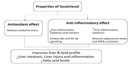
Substance use among Students — The Road to Oblivion
— Nandini Chatterjee MD, FRCP (Glasgow), FICP Professor, Department of Medicine, IPGME&R and SSKM Hospital, Kolkata 700020 and Hony Editor, JIMA
Substance abuse is an expanding public health problem which is of global concern. Tobacco, alcohol, cannabis and stimulant drug use is widely prevalent among students from various fields including the medical students. WHO reports a burden of worldwide substance use of around 2 billion alcohol users, 1.3 billion smokers and 185 million drug users. An estimated figure of about 20-40 per cent among students has been quoted by various studies. Most strikingly abuse of drugs among physicians has been estimated to be about 30 to 100 times the rate in the general population.
Why are medical students prone to addictions ?
The most common reason reported in the studies for using such substances was relief from psychological stress (>70%) for academic performance as well as fierce competition.
Peer pressure and experimental use is also important as community acceptance is a common prerogative among the young.
Easy availability (42.6%) of various drugs is a significant determinant of substance abuse among medical students, urban background, family history of substance abuse, higher economic status, depressionand low self-esteem have been associated with increased addictive tendencies among undergraduate students.
What are the different substances in common use ?
There is abundant use of alcohol, tobacco, tranquilizers and psychedelics among medical students.
Cannabis, opioids and stimulants, such as amphetamines are also prevalent among undergraduate students.
A large Canadian Study has revealed that the lifetime self-reported prevalence rates were 45.6% (95% CI, 44.0%-47.2%) for cannabis, 8.3% (95% CI, 7.4%-9.2%) for NPS, and 6.8% (95% CI, 5.9%-7.7%) for cigarettes while past-month excessive alcohol use was documented in 46.4%
These numbers reflect the magnitude of the problem and arouses concern about the implications for their personal health, training and clinical practice.
What are the Consequences of Substance Abuse?
There are far-reaching consequences on lifestyle patterns, mental health and productivity.
Academic performance is significantly affected, as depicted by a study, which found a negative correlation between substance abuse and academic achievement.
Inaccessibility, irritability,defensive behaviour, anxietyand mood swings are some of the manifestations. These ultimately lead to emotional exhaustion depersonalization and burnout. Also the risk of future cardiovascularand respiratory diseases, and psychiatric disorders are compounded. Is there enough data ?
The magnitude of this situation is not well elucidated as there is an inhibition in coming out with the problem of dependance or impairment. A systematic and multiinstitutional surveillance system is the need of the hour.
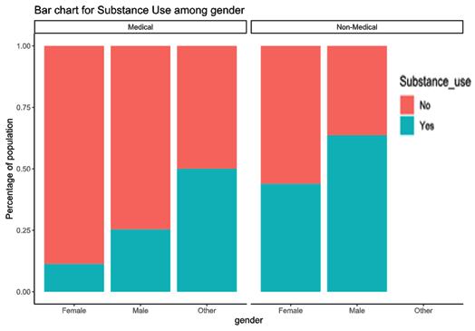
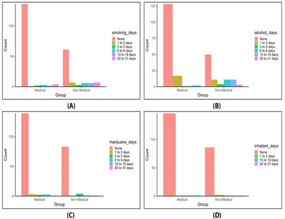
Existent literature reviewed reveals that substance abuse among undergraduate students in India is a complex issue with multiple contributing factors and significant consequences and serious health risks. This issue presents an unique study of comparison between the students of a reputed non medical college and medical students. Younger age, male gender and higher socioeconomic classes had a greater association with addictions. It was noted that substance abuse in relation to alcohol and tobacco was much more in non-medicos than their medico brothers but incidence of stimulant drug use and sedatives were similar. Awareness about the health consequences is projected as a a determinant factor.
What is the way forward ?
Hand holding is the key to success.
Counseling services, both individual and groupbased, are effective interventions to deal with this delicate situation. It is often difficult to bring out the proper history of impairment. Students should be encouraged to talk about their impediments and insecurities2.
Educational campaigns and awareness programs focusing on the risks and consequences of substance abuse have shown promise. Collaboration between educational institutions, healthcare providers, and community organizations is vital in implementing comprehensive prevention programs. Also development of strategies for support of the affected students, which is woefully lacking in our set ups, is to be devised. Further research is warranted to characterize the nationwide patterns of substance use among medical students, and to identify the predispositions so that protective factors may be reinforced effectively.
FURTHER READING
1Deb KS, Sinha VK, Kumar R, Sinha DN — Substance use among college students. Indian Journal of Psychological Medicine 2019; 41(6): 553-64.
2Kumar M, Verma M, Das P — A study on prevalence of substance abuse among college students. Indian Journal of Community Medicine 2020; 45(4): 488-91.
3Bahji A, Danilewitz M, Guerin E, Maser B, Frank E — Prevalence of and Factors Associated With Substance Use Among Canadian Medical Students. JAMANetw Open 2021; 4(11): e2133994. doi:10.1001/jamanetworkopen.2021.33994
4Kumar R, Goel NK, Sharma MK — Impact of substance abuse on academic achievement among college students. Industrial Psychiatry Journal 2021; 30(1): 76-82.
Original Article
Evaluating MBBS Students Perception on Transition from Classroom to Online Teaching during COVID-19 Pandemic — A Cross Sectional Survey
Background : With the rapid expansion of technology, Online teaching has emerged as a substitute mode of education and is rapidly becoming one of the most effective ways to impart education especially during this pandemic. This study was done to evaluate the perception of MBBS students on the transition from classroom to online teaching during COVID-19 pandemic.
Methods and Results : The present study is a cross-sectional, exploratory survey-based design. MBBS students (first year to final year) from all the medical colleges in Visakhapatnam were included in the study. The designed questionnaire was validated by a committee of faculties of the Department of Pharmacology and was sent through WhatsApp to all the participants. A duration of 1.5-2 months was set to collect the response, review and analyze. The results of our study indicated that maximum number of students (62%) felt it was a wise decision to opt for Online classes during COVID-19 pandemic to ensure the continuity of the curriculum. After analyzing the content, we found out that though there were benefits like flexibility & convenience (74.4%), going multiple times through recorded videos, improvement in technical advantage (50.1%) and cognitive skills (35.3%), online classes also had their fair share of drawbacks like technical difficulties (81.7%), lack of peer interaction (50.7%) and continuous Online classes.
Conclusion : Online teaching was the only and effective alternative available to the traditional method of teaching to avoid any lapses in the MBBS curriculum during COVID-19 pandemic. However, online teaching can never be a replacement in the Medical Education System where the courses are mostly in favour of practical expertise. This study is done to identify the hindrances in the course of Online learning and corrective measures to overcome them and also to be prepared to face such a situation with ease anytime in the future.
Key words :Pandemic, Medical students, Online teaching, Feedback.
Online teaching also known as e-learning is the usage of internet for educational purposes. The practice of Online learning started before 150 years1 In the mid 19th century “correspondence courses” was the name given to Distance Education2. The first Online course was given in the year 1981 and in the ensuing year, the Western Behaviour Sciences Institute prepared the first Online program3.The use of Online learning has been extensively approved off in higher education and is quite popular in recent decades4. The success of Online classes demands a huge responsibility on the part of students as well as on the faculty. The nature and characteristics of students determine the outcome of Online classes 5 . The
Department of Pharmacology, Gayatri Vidya Parishad Institute of Health Care and Medical Technology, Andhra Pradesh 530048
1MD, Assistant Professor
2PhD, Professor and Corresponding Author
3MD, Associate Professor
4MSc, Assistant Professor
5MD, Associate Professor, GIMSR, Visakhapatnam, Andhra Pradesh 530045
Received on : 15/03/2023
Accepted on : 01/04/2023
[J Indian Med Assoc 2023; 121(7): 15-9]
Editor's Comment :
The COVID pandemic made us realise that being physically present in the classroom is no longer the only option for learning and the online teaching prototype gave structure to preserve students' learning throughout challenging moments. For e-learning to be implemented successfully, attention must be paid to the hurdles to digital learning.
responsibility of the faculty should be to integrate multiple forms of media like Video, Audio, Text and Animation in their lectures so that students with different preferences can find the content more engrossing6,7.
Educational institutions all over the World have been shut down owing to the COVID-19 pandemic, eventually forcing them to suspend the classes. So, most of them have decided to continue with their classes using Online mode of instruction. Even though it is a very convenient mode of education considering the present situation, it may be quite challenging as the whole idea is novel not only to the faculty but also for the
121, No 7, July 2023Journal
students. Converting a successful classroom course to an online course is not a simple process and many faculties have stepped in not knowing the difficulties involved in it because it is the need of the hour. Likewise, students will have problems with logging on, technical difficulties, network problem, difficulty in submitting the assignments in the allotted time, etc, and the faculty must be prepared well in advance to rectify them. There are doubts in the minds of the many faculty about how smooth the transition has been from classroom to online teaching and whether the students are able to cope up with the sudden change that has happened due to the COVID pandemic. This has motivated us to conduct this study to deal with the situation accordingly and find out better ways in which it can be handled.
This study is conducted by taking feedback from MBBS students to evaluate their perception on the transition from classroom to Online teaching and also to compare the effectiveness of traditional and modern teaching methods. This study is also aimed at finding out problems the students might be facing, so as to overcome them if a similar situation arises in the future. In doing so it is expected that this can inculcate an effective discussion between the management and the faculty to provide the best opportunities for the students so that they can be benefited the most from the ongoing Online classes. The administration should look into matters of concern like budget, faculty workload, number of students in Online classes, scheduling of Online classes, good network connection for uninterrupted classes, etc, without which Online teaching and learning will fail8.
AIMS, OBJECTIVES AND HYPOTHESIS
To evaluate the perception of MBBS students on the transition of classroom to Online teaching during COVID-19 pandemic
Objectives :
•To evaluate MBBS students perception on transition of classroom teaching to Online teaching.
•To compare the effectiveness of traditional and modern teaching methods.
•To assess the pitfalls in the ongoing Online classes so as to overcome and improvise them if a similar situation arises in future
MATERIALS AND METHODS
Study Design, Population and Method :
The present study was a cross-sectional, exploratory survey-based design. The eligible participants included were 1st year to final year MBBS
students from all the four medical colleges of Visakhapatnam. The questionnaire was designed and validated by a committee of faculties of the Department of Pharmacology and through peer review. The prevalidated questionnaire comprised of questions related to the Socio-demographic status, experience of Online classes, traditional and modern methods of teaching, benefits and drawbacks of online teaching and Online assessment patterns were sent through WhatsApp. Duration of 1.5-2 months was set to collect the responses.
Sample Size :
By keeping a confidence level of 95% and error of 5% with total population of 2500, though the sample size needed was N=330, we ended up collecting responses from N=744 participants.
Statistical Analysis :
Collected data was imported to SPSS from the generated excel sheet of Google form. Frequency analysis was run. Chi square test was used to analyse and test the relationships between categorical variables.
RESULTS
The results of our study include data on demographic variables, feedback regarding Online classes, Online assessment, its benefits and drawbacks.
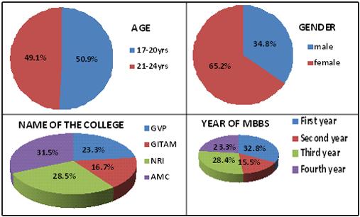
The mean age of participants was 20.46 with SD±1.323. The range of age group was between 17-24 years and majority of the participants were females. Data was collected from four medical colleges, consisting of one government college from which 31.5% responded, remaining 68.5% were from private college. Among different phases of MBBS curriculum, phase 1 students responded more as compared to other phases as we believe first year students are more curious to gain knowledge of novel things and provide their valuable feedback as they are new in the college (Fig 1).
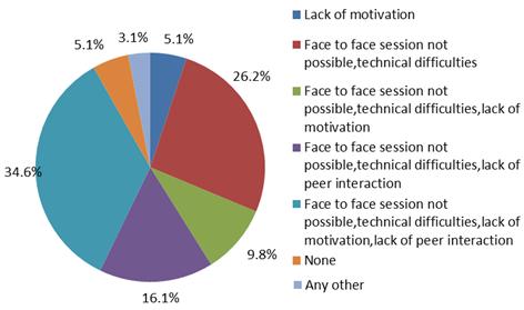
Students’ feedback on Online classes showed both positive and negative responses. Around 62% students agreed that conducting Online classes during this pandemic was a wise decision as it helped in covering the syllabus with no break in the continuity of the curriculum. On the contrary, 75.2 % felt that Offline classes are better because this mode makes them more confident and holds their attention for longer time. Majority (80.8%) students believed that they were having continuous Online classes which made them hard to concentrate and so 58.6% students wanted gap between the classes and 41.4% were content with current schedule. Importance for clearing doubts was
not being given in Online classes, so 44.5% said that one or two classes should be allotted per week for it. Higher percentage (72.6%) used mobile phone as a platform for attending Online classes as compared to other devices because it is easy to access, cheaper and portable (Table 1).
48.8% students agreed that conducting MCQs and short answers as an assessment pattern makes them more confident for exams because it is easy to score marks, less tiring, time saving and helps them to prepare for postgraduate entrance. But 66.9% responded that Online assessment is not the best way to evaluate students for the reason that there is a high scope of malpractice (Table 2).
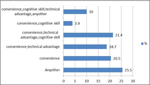
The results of our study showed that 74.4% preferred online classes because they opined that it has flexible plan and is convenient as they can visualize the lecture at any time, any place and provides faster virtual communication. Additional benefits were technical advantage and improved cognitive skills (Fig 2).
The major obstacle for Online learning was technical difficulty (81.7%) which was even more for students residing in rural areas, concluding that if India needs to show progress towards Online education it should concentrate on improving its digital literacy and networking facilities. Lack of face to face session was also the major concern among students (71.5%) (Fig 3).
DISCUSSION
The Corona Virus pandemic has generated changes in the education – knowledge process in advanced educational institutes and has manipulated the communication involving teachers and students. The first Online course was given in the year 1981 and in the ensuing year, the Western Behaviour Sciences
Institute prepared the first Online program3. The use of Online learning has been extensively approved off in higher education and is quite popular in recent decades4. The nature and characteristics of students determine the outcome of Online classes 5 . The objective of this research was to evaluate the perception of MBBS students on the transition from
Fig 1 — Demographic distribution of students (in percentage) Table 1 — Feedback on online classes (N=744)
Live online classes, online assessment, seminar104
Live online classes, Recorded videos, online assessment466.2
Live online classes, seminar375.0
Anyother375.0
Live online classes, Recorded videos, online assessment, seminar91.2
Live online classes, recorded videos91.2
Recorded videos, online assessment70.8 Which assessment pattern makes you confident for exams? MCQs262 35.2
MCQs, Short answers101 13.6
Short answers, Long answers9312.5 Not being conducted8311.1 Short answers76 10.2
Long answers304.0
MCQs, Long answers141.9
MCQs, Short answers, Long answers8511.5 Are you being motivated enough with online assessment? Yes15320.6
Partially motivated249 33.5
answered334.4 Online assessment is not the best way to assess a student.
curriculum during pandemic which was analogous to our study (62%). 81.7% students of our study complained of technical issues which was not in support of a study done in Dakshina Kannada and Udupi District (47.1%)9. The present study showed nearly half of the participants believed that Online classes improved technical skills as compared to face to face classes which was reinforced by a study done by T Muthuprasad11.
classroom to Online teaching during COVID-19 pandemic.
The result of our study showed that most of the participants were females, which was in correspondence with a previous study9. In the same study, 82% participants found classroom education more useful than Online classes as it was easier to grasp the topic contents in Classroom Education System. Similarly, our study also found three fourth participants polling in favour of offline classes, but it was contradictory to a retrospective study 10 where 1st year medical students clearly established that Online training is an identical or enhanced learning experience than classroom sessions. In a study done by T Muthuprasad, et al11, about 70% participants were ready to OPT Online classes for the continuity of
Mobile was used as a device for attending Online classes by 77.6% students followed by 13% and 9.4% students who used Laptop and Tablet, respectively in our study, which was relatively more than the results from a former study11 where 57.98% students used Smart Phone as a device followed by 35.8% and 4.98% used Laptop and Tablet respectively. About frequency and duration of class, in a study done during COVID pandemic11, 48% of students required a break of 15 min gap in between 2 classes, as opposed to our study
Fig 2 — Opinion of students regarding benefits of online classes (in percentage)
Fig 3 — Opinion of students regarding drawbacks
121, No 7, July 2023Journal
(28.4%). Nature of Online exam was yet another variable which was analysed and (35.2%) in the present study said objective assessment model makes them more confident for an exam which was not in line with the findings of a study by Muthuprasad (2021)11. A little more than a quarter percentage of participants (27%) recommended subjective questions for assessment, which was more as compared to above study by Muthuprasad, et al where only 3.9% accepted it as an assessment tool. The same study claimed 60% respondents agreed that there is no possibility of face to face sessions and in our study 72.3% agreed the same. In a study conducted by Sean Smith12 20% agreed that instant response from both peers and instructors is possible with traditional training, whereas 50.7% agreed for peer interaction and 71.5% agreed for face to face interactions in our study. 44.5% from the current study agreed for live classes to clear doubts which were far less as compared to a previously done study 11 (84%). Structure of Online classes was analyzed and the preference pattern in our study was 85.6% preferred live Online classes and (9.5%) recorded videos which, when compared to above study 17.9% preferred live classes and recorded videos (54.4%) .
In a retrospective study done by Laura M10 students suggested that increased management and individual attention with web based content enhanced their general assessment of learning experience compared to face to face classroom sessions. Similar to our study where majority agreed Online mode is flexible and convenient. On comparing improvement in learning skill, it was found that in one study12, 5% students agreed use of Computer and internet as a better knowledge experience, where as in our study 35.3% agreed for improved cognitive skill with Online mode.
CONCLUSION
Online learning has helped students to become independent learners by improving their technical knowledge, cognitive skills and also gave them opportunities to explore latest learning applications. In this study, we have tried to analyze feedback of students for both Online and offline classes. Beside the benefits of Online mode, the obstacles which students faced were discussed in this study such as Online classes could not be accessed by every student due to poor network issues, lack of Smart Phones which could lead to demographic based discrepancy with respect to quality of education and no face to face interactions leading to lack of motivation for attending those classes. As such there is need to recognize the obstacles that appear in the way of
accepting Online learning and the remedial measures to overcome it.
Limitations :
The major limitation of this study was that the data was obtained from students studying in medical colleges located only in one city in Andhra Pradesh. Thus the results cannot be generalized to the entire nation’s Higher Education System. Also, the parameters included in the questionnaire could be self limiting as the faculty involved in designing of questionnaire had very less previous experience of e learning. Due to lack of data pertaining to feedback about e learning and one time cross sectional design of the study, comparison of data before and after the start of e learning could not be made.
REFERENCES
1Saiyad S, Virk A, Mahajan R, Singh T — Online teaching in medical training : establishing good online teaching practices from cumulative experience. Int J App Basic Med Res 2020; 10: 149-55.
2Christner T — Review of Classroom of One: How Online Learning is Changing Schools and Colleges. Library Journal 2003; 128(1): 130-1.
3Harasim L — Shift happens: Online education as a new paradigm in learning. Internet and Higher Education 2000; 3: 41-61.
4Oncu S, Cakir H — Research in online learning environments: Priorities and methodologies. Computers & Education 2011; 57(1): 1098-108.
5Picciano AG — Beyond student perceptions: Issues of interaction, presence, and performance in an online course. Journal of Asynchronous Learning Networks 2002; 6(1): 2140.
6Kumar S, Black EW — Online education and virtual schooling.In :Ciccomascolo LE, Sulivan EC . The dimensions of physical education .1st ed. Burlington, MA : Jones & Bartlett Learning 2013; 243-51.
7Duffy P — Engaging the YouTube Google-eyed generation: Strategies for using web 2.0 in teaching and learning. The Electronic Journal of e-Learning 2008; 6: 119-30.
8Berge Z — Barriers to online teaching in post secondary institutions : Can policy changes fix it ? Journal of Distance Learning Administration 1998; 1(2) . www.westga.edu/distance/Berge12.html.
9Kulal A — A study on perception of teachers and students toward online classes in Dakshina Kannada and Udupi District. Asian Association of Open Universities Journal 2020; 15(3): 285-96.
10Laura M — Schimming. Measuring medical student preference: a comparison of classroom versus online instruction for teaching PubMed. J Med Libr Assoc 2008; 96(3): 217-22.
11Muthuprasad T — Students’ perception and preference for online education in India during COVID -19 pandemic. Social Sciences & Humanities Open 2021; 1-11.
12Smith S — The positive and challenging aspects of learning online and in traditional face to face classrooms : A Student Perspective. Journal of Special Education Technology 2005; 20(2): 52-9.
Original Article
Prevalence of Rheumatological Manifestations in HIV Infected Patients in a Tertiary Care Centre In Eastern India : A Prospective Observational Study
Agnibho Mondal1, Dolanchampa Modak2, Subhasish Kamal Guha3
Background : Highly active, viral suppressive Anti-retroviral Therapy (ART) has significantly increased the life expectancy of HIV infected patients, which increases precipitation of age related diseases and comorbidities. Nowadays, Rheumatological Manifestations are being recognized as an important contributor of morbidity in PLHIV.
Aims : The aim of our study was to determine the prevalence of Rheumatological Manifestations in ART naive PLHIV and during the first six months of ART initiation.
Settings and Design : The prospective observational study was conducted in the School of Tropical Medicine, Kolkata.
Materials and Methods : Newly diagnosed ART naive HIV infected patients were recruited. Clinically and Serologically for Rheumatologic Manifestations and followed up period for next six months.
Statistical analysis used : Statistical analysis was performed R version 4.0.2 and p<0.05 was considered significant.
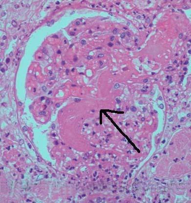
Results : We recruited 106 ART naive patients and followed them up for six months after initiation of ART. Fortythree ART naïve patients (40.6%) had Rheumatological Manifestations. The most common Rheumatological condition was HIV arthralgia (28.3%) followed by rheumatoid Arthritis (3.8%), Systemic Lupus Erythematosus (SLE) (2.8%), Osteoarthritis (0.9%), Myositis (1.9%), Psoriatic Arthritis (0.9%) and Reactive Arthritis (0.9%). Large Joint pain (Knee, Ankle, Hip in decreasing order) was the most common (38.7%) presenting symptom. Musculoskeletal adverse drug reaction of ART occurred in 6.6% patients over the period of six months. HIV clinical stage and CD4 count had no predictive role for the Rheumatological Manifestations. All participants were asymptomatic at the end of six months follow-up.
Conclusions : Timely assessment and management of Rheumatologic Manifestations along with ART initiation may result in favourable outcome in PLHIV.
Key words :HIV, PLHIV, ART naive, Rheumatological manifestations.
As of 2020, approximately 37.7 million people are living with HIV (PLHIV) all over the world 1 Availability of effective combination Anti-retroviral Therapy (ART) has increased life expectancy in People Living with HIV (PLHIV)2,3. The availability of ART has led to the changing spectrum of Rheumatological conditions4. ART regimens may themselves have Rheumatological adverse effects5, which need to be carefully distinguished. The World Health Organization now recommends initiation of ART in PLHIV as early as possible regardless of CD4 count6. HIV arthralgia, Fibromyalgia, Rheumatoid Arthritis, Avascular Necrosis, Systemic Lupus Erythematosus,
Department of Tropical Medicine, School of Tropical Medicine, Kolkata 700073
1MD (Tropical Medicine), Senior Resident
2 MD (Tropical Medicine), Associate Professor and Corresponding Author
3MD (Tropical Medicine), Professor
Received on : 06/07/2022
Accepted on : 23/11/2022
[J Indian Med Assoc 2023; 121(7): 20-3]
Editor's Comment :
Rheumatological manifestations are important contributors of morbidity in HIV infected patients.
The most common manifestation in Anti-retroviral Therapy naïve patients is HIV arthralgia.
Early diagnosis and treatment Rheumatological conditions along with Anti-retroviral Therapy has positive impact on quality of life by reducing Rheumatological morbidity in HIV infected patients.
Polymyositis, Vasculitis, Osteomyelitis are some common Rheumatological Manifestations in HIV seropositive individual. Increasing life expectancy of PLHIV precipitating Rheumatological Manifestations along with aging which is responsible for significant morbidity needed further attention than ever.
MATERIALS AND METHODS
This prospective observational study was performed in the ART centre of a Tertiary Care Hospital of Eastern India from July, 2019 to May, 2020. We recruited adult ART naïve patients. Clinical assessment was performed
121, No 7, July 2023Journal of the Indian Medical Association
before the initiation of ART and all participants were evaluated for presence of Rheumatological conditions. Specific investigations were performed in cases of suspected Rheumatological Manifestations. Autoimmune profile was also performed when needed which included Rheumatoid Factor (RF), Anti-Cyclic Citrullinated Peptides (anti-CCP) and Anti-nuclear Antibodies (ANA). Diagnosis of HIV arthralgia was done primarily by exclusion.
ART was initiated in all participants and they were subsequently followed up for six months. Monthly assessment was performed in all participants except those who were lost to follow up. All patients with Rheumatological Manifestations received standard treatment for the condition as seen fit by the treating physician. Rheumatologic diseases were diagnosed according to the guidelines of American College of Rheumatology7-9
Ethical clearance was obtained from the Clinical and Research Ethics Committee of the institution. Informed consent was obtained from all study participants before recruitment to the study.
The statistical analysis was performed using the R software package version 4.0.2 by the R Foundation for Statistical Computing. The comparison of findings was done by T-test for continuous variables and McNemar’s test for categorical variables. Between group analysis was done by Fisher’s exact test. Regression models were used to test the predictive value of variables. A p<0.05 was taken as significant during the analysis.
RESULTS
A total number of 106 patients were recruited to the study. Four patients were lost to follow up and the rest were followed up for a duration of six months.
Among the 106 participants, 75 (70.8%) had one or more opportunistic infections before initiation of ART. Coinfection with Hepatitis B was found in three patients. Majority of the study participants (69.8%) had WHO clinical stage 3 or 4 disease at baseline10.
During the final follow-up at the end of six months, several laboratory parameters showed significant improvement (p<0.05) including ESR, CRP, Total bilirubin, Conjugated bilirubin, Albumin, SGOT, SGPT, Alkaline Phosphatase and CD4 count (Table 1).
The mean CD4 count at baseline was 211 cells/µL and it was 332 at the end of six months follow-up (p<0.001). Immune Reconstitution Inflammatory Syndrome occurred in 5.7% patients (Table 2).
Forty-three patients (40.6%) were found to have Rheumatological Manifestations before initiation of ART.
Table 1 — Laboratory parameters at baseline and at the end of six months follow-up
ParametersBaseline
Table 2 — CD4 count in relation to the Rheumatological Manifestations
Rheumatological conditions Mean Median CD4 countCD4 count (cells/µL)(cells/µL)
HIV arthralgia199178
Psoriatic arthritis307307
Reactive arthritis4646 Myositis113113
Osteoarthritis309309
Rheumatoid arthritis284318
Systemic Lupus Erythematosus266366
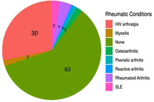
The most common Rheumatological condition was HIV Arthralgia (28.3%).Other conditions included Rheumatoid Arthritis (3.8%), Systemic Lupus Erythematosus (SLE) (2.8%), Osteoarthritis (0.9%), Myositis (1.9%), Psoriatic Arthritis (0.9%) and Reactive Arthritis (0.9%) (Fig 1).
Large (Knee, Ankle, Hip in decreasing order) Joint pain was the most common presenting symptom (38.7%) among the study participants while other
Fig 1 — Proportion of rheumatological conditions at baseline (n=106)
2023Journal
symptoms like skin rash, morning stiffness and muscle weakness was present in 2.8%, 1.9% and 1.9% participants respectively.
ANA was positive in 5.7%, Rheumatoid Factor was positive in 3.8% and anti-CCP was positive in 3.8% study participants.
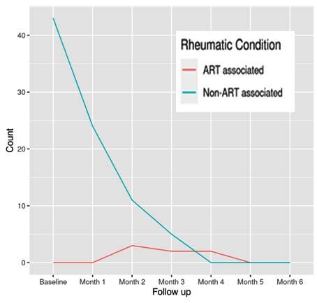
Regression model was used to determine the predictive value of different parameters on the Rheumatological Manifestations. P<0.05 on multivariate analysis was taken as significant in this case. Presence of joint pain, sites of joint involvement and less than one month of symptoms were found to be predictive of HIV arthralgia on multivariate analysis. Muscle weakness for more than one month was predictive of myositis whereas joint pain for more than one-month, high C-reactive Protein (CRP) and high Platelet Count was predictive of Osteoarthritis. The significant predictors of Rheumatoid Arthritis were joint pain for more than three months along with RF and anti-CCP. Similarly, parameters with predictive value for SLE included skin rash, joint pain for more than one-month, female sex, low haemoglotherapy. However, musculoskeletal adverse drug reaction developed in 6.6% patients over the period of six months. At the end of six months all participants were symptom-free (Fig 2).
DISCUSSION
In our study the prevalence of Rheumatological Manifestation of HIV was found to be 40.6% in ART naïve population. Most common presentation was joint pain 38.7%) and the most common Rheumatological condition was HIV Arthralgia (28.3%).
Although clinical assessment was found to have predictive role in diagnosing Rheumatological conditions, no such association was found with the clinical stage of HIV or the CD4 count of the patients. Presence of opportunistic infections also had no predictive role.
Improvement of laboratory parameters over the period of six months could be attributed to resolution of opportunistic infections, treatment of comorbidities as well as improved clinical status of the patients after initiation of ART.
Although ANA was positive in 5.7% patients at baseline, only 50% of them were diagnosed with SLE and the rest had no Rheumatological condition. However, all the patients with positive RF and antiCCP were diagnosed with Rheumatoid Arthritis. The loss to follow-up was only 3.8% and the adherence to the ART regimen was 98%. At the end of six-months follow-up all participants were free of opportunistic infections and the increase of CD4 count
of rheumatological conditions over the six months follow-up period
was significant (p<0.001). During the follow-up period of six months, only 6.6% patients developed musculoskeletal adverse reactions due to ART, none of them requiring a change of ART regimen.
All participants with Rheumatological Manifestations responded well to therapy and at the end of six months all of them were free of symptoms. Although deformity persisted in patients with Rheumatoid Arthritis.
Another study similar to ours conducted in Eastern India found the prevalence of Rheumatological Manifestation to be 63.3% in ART naïve PLHIV as well as within 6 weeks of ART initiation11. Another study conducted in India found the prevalence of Rheumatological Manifestations to be 46.7%12. The prevalence of Rheumatological Manifestations vary widely between the studies with some studies showing a prevalence as low as 3.8%13. In comparison, the prevalence of rheumatologic conditions was 40.6% in our study.
Our study was limited by the sample size and the short duration of follow-up which was inadequate to detect relatively uncommon Rheumatological Manifestations as well as Rheumatological Manifestations that may happen after six months of ART. We also could not assess cytokines to evaluate its predictive role in Rheumatological Manifestations. A baseline HIV viral load could not be obtained as well.
Although Rheumatological Manifestation is prevalent (40.6%) in ART naïve PLHIV, timely administration of ART as well as adequate
Fig 2 — Trends
Vol 121, No 7, July 2023Journal of the Indian Medical Association
management can resolve the conditions. Although ART regimens themselves may cause musculoskeletal adverse reactions, the prevalence is generally low (<4%).
The HIV clinical stage or the CD4 count had no significant predictive role in Rheumatological Manifestations. Other studies on the Rheumatological Manifestations of HIV also support this finding14,15 Although RF and anti-CCP is associated with Rheumatoid Arthritis, specific diagnosis of SLE was reached in only 50% patients with positive ANA.
Our study suggests that all ART naive PLHIV should be assessed for Rheumatological Manifestations and timely management of these conditions in addition to ART may result in a favourable outcome.
ACKNOWLEDGEMENTS
We would like to express our thanks to the members of the Department of Tropical Medicine, ART centre and laboratory personnel of the institution. We are grateful to Dr Madhuchanda Mandal, Dr Arijit Mallik and Sanchayita Roy who have helped us in the various steps of our studies.
REFERENCES
1UNAIDS. Global HIV & AIDS statistics — Fact sheet [Internet]. UNAIDS. 2020 [cited 2021 Jan 10]. Available from: https:// www.unaids.org/en/resources/fact-sheet
2Phanuphak N, Gulick RM — HIV treatment and prevention 2019: current standards of care. Curr Opin HIV AIDS 2020; 15(1): 4-12.
3Teeraananchai S, Kerr S, Amin J — Life expectancy of HIVpositive people after starting combination antiretroviral therapy: a meta-analysis. HIV Med 2017; 18(4): 256-66.
4Vega LE, Espinoza LR — Human immunodeficiency virus infection (HIV)-associated rheumatic manifestations in thepreand post-HAART eras. Clin Rheumatol 2020; 39(9): 251522.
5Reveille JD, Williams FM — Rheumatologic complications of HIV infection. Best Pract Res Clin Rheumatol 2006; 20(6): 1159-79.
6World Health Organization — Consolidated Guidelines on HIV Prevention, Testing, Treatment, Service Delivery and Monitoring: Recommendations for a Public Health Approach [Internet]. Geneva: World Health Organization; 2021 [cited 2021 Oct 1]. (WHO Guidelines Approved by the Guidelines Review Committee). Available from: http:// www.ncbi.nlm.nih.gov/books/NBK572729/
7Hochberg MC, Altman RD, April KT — American College of Rheumatology 2012 recommendations for the use of nonpharmacologic and pharmacologic therapies in osteoarthritis of the hand, hip, and knee. Arthritis Care Res 2012; 64(4): 465-74.
8Singh JA, Saag KG, Bridges SL — American College of Rheumatology Guideline for the Treatment of Rheumatoid Arthritis: ACR RA TREATMENT RECOMMENDATIONS. Arthritis Rheumatol 2016; 68(1): 1-26.
9Singh JA, Guyatt G, Ogdie A — American College of Rheumatology/National Psoriasis Foundation Guideline for the Treatment of Psoriatic Arthritis. Arthritis Rheumatol 2019; 71(1): 5-32.
10World Health Organization — Interim WHO clinical staging of HVI/AIDS and HIV/AIDS case definitions for surveillance: African Region. World Health Organization; 2005.
11Kole A, Roy R, Kole D — Musculoskeletal and rheumatological disorders in HIV infection: Experience in a tertiary referral center. Indian J Sex Transm Dis AIDS 2013; 34(2): 107.
12Saigal R, Chakraborty A, Yadav RN — Rheumatological Manifestations in HIV-Positive Patients: A Single-Center Study. Adv Ther 2020; 37(10): 4336-45.
13Parperis K, Abdulqader Y, Myers R — Rheumatic diseases in HIV-infected patients in the post-antiretroviral therapy era: a tertiary care center experience. Clin Rheumatol 2019; 38(1): 71-6.
14de Filippis LG, Scibilia G, Caliri A — Rheumatic symptoms in patients with human immunodeficiency virus are related to levels of tumor necrosis factor-alpha but not to viral load. Int J Tissue React 2005; 27(1): 9-13.
15Kulthanan K, Jiamton S, Omcharoen V — Autoimmune and rheumatic manifestations and antinuclear antibody study in HIV-infected Thai patients. Int J Dermatol 2002; 41(7): 417–22.
Disclaimer
The information and opinions presented in the Journal reflect the views of the authors and not of the Journal or its Editorial Board or the Publisher. Publication does not constitute endorsement by the journal.
JIMA assumes no responsibility for the authenticity or reliability of any product, equipment, gadget or any claim by medical establishments/institutions/manufacturers or any training programme in the form of advertisements appearing in JIMA and also does not endorse or give any guarantee to such products or training programme or promote any such thing or claims made so after.
— HonyEditor
Original Article
A Study of Laboratory
Determinants and Clinico-pharmacological Correlates of Vasculotoxic Snake Bites in a Tertiary Care Hospital in West Bengal
Background : Snake bite is an often neglected but lethal disease in a tropical country like India. There is a dearth of data regarding true magnitude of vasculotoxic Snake bite in a tertiary care facility catering a predominantly rural area relying heavily on agriculture. The objective of our study, therefore, was to identify the burden and determinants of outcome of a vasculotoxic Snake bite poisoning in this specific setting.
Methodology : A cross-sectional study was undertaken at Burdwan Medical College and Hospital, West Bengal over a period of 1 year which recruited 127 cases of Snake bite poisoning and their Epidemiological, Clinical, Biochemical and Haematological parameters were collected in a pre-designed case record form at admission. The data regarding time delay in admission, duration of hospital stay, pre-referral treatment and definitive anti-snake venom serum therapy were also collected.
Results : Commonest bites were vasculotoxic in nature (48.65%) and affects 31-40 years age group (33.85%) who typically presented with fang marks, local swelling, pain and bleeding from bite site mostly in the lower limbs. Mortality was 8 and complications developed in 27.56% patients, systemic hypotension and Acute Kidney Injury being the commonest duo. A bite-to-hospital delay of 2 to 6 hours is noted in majority (49.60%) and a 2/3rd mortality observed when admitted after 12 hours. The mean total count, ESR, CRP, LDH, CPK, Serum Urea and Creatinine were raised and statistically significant in patients with complications.
Conclusion : Renal function deterioration is one of the earliest signs of development of complications in vasculotoxic poisoning and decision delay, prompt institution of ASV are key determinants of improved prognosis [J Indian Med Assoc 2023; 121(7): 24-8]
In a developing economy like India, the poor reporting and under reporting of neglected health problems can have detrimental effect. Even WHO has perceived the importance of Snake bite as an area of serious concern and a neglected problem of tropical countries and working on a strong mandate to develop a comprehensive plan for effective Snake bite management1
Researchers have revealed that over 8 lakhs of Indian population died due to Snake bite envenomation during 2001 to 2014 with age standardized snake bite
1MD, Associate Professor, Department of Pharmacology, Manipal Tata Medical College, Manipal Academy of Higher Education, Manipal, Karnataka 576104
2MD, DM, Senior Resident, Department of Neurology, Nil Ratan Sircar Medical College and Hospital, Kolkata 700014
3MD, Professor, Department of Medicine, Burdwan Medical College and Hospital, Burdwan 713104
4MD, Assistant Professor, Department of Medicine, Burdwan Medical College and Hospital, Burdwan 713104
5 MD, Professor, Department of Forensic Medicine and Toxicology, Manipal Tata Medical College, Manipal Academy of Higher Education, Manipal, Karnataka 576104 and Corresponding author
Received on : 18/09/2022
Accepted on : 16/12/2022
Editor's Comment :
Acute renal insult and hypotensive shock are common complications of vasculotoxic snake bite in rural agricultural terrains.
Haematological and electro-renal parameters appear to be strong predictors of imminent development of renal complications in this scenario.
Eliminating decision delay and rapid referral is key to prevent vasculotoxic deaths.
death rate at 4.8 per 1,00,000 population2. Even the Standard treatment guidelines of Government of India mentioned a serious lacuna that exists between the number of Snake bite deaths reported from direct survey and official data3. Even previous studies in West Bengal have pointed towards underreporting of Snake bite cases in rural areas due to inaccessibility4. Many studies focus on profiling of cases but miss out the clinical, biochemical and hematological correlation with outcomes.
Due to less availability of literature in this particular agrarian area, this study was a novice attempt on the part of the researchers with an objective to identify determinants of outcome with Clinical, Hematological, and Biochemical correlates in vasculotoxic Snake bite
Vol 121, No 7, July 2023Journal of the Indian Medical Association
patients in a hospital setting which forms the novelty of the study.
MATERIAL AND METHODS
The study was conducted at Department of Medicine, Burdwan Medical College and Hospital in Burdwan district, West Bengal from 01-09-2016 to 3108-2017.The medical college is a tertiary care teaching hospital and caters to Burdwan, Birbhum, some parts of Bankura districts of West Bengal & parts of adjoining Jharkhand state.
Inclusion criteria :
(1)All patients coming to Emergency and Medicine Department with a definite history of Snake bite.
Exclusion criteria :
(1)Any patients who showed s signs and symptoms of neurotoxic Snake bite like ptosis, Ophthalmoplegia, Dysphagia or present in a comatose condition with developing respiratory paralysis.
(2)Any asymptomatic patient who did not develop any signs and symptoms after 24 hours of observation to exclude nontoxic bites.
(3)History of systemic kidney disease, any Hematological disorders, Gout and any Rheumatological disease.
(4)History of surgery or major trauma in previous month with anticoagulant intake.
It was an institution based observational study and cross-sectional in nature which proceeded after IEC approval vide Memo no.BMC/PG/459. 127 patients were included in the study based on inclusion and exclusion criteria. The confidentiality of the patients was well maintained abiding by the ethical guidelines. 35 patients were grouped under patients with complications out of which 8 succumbed during treatment.
The structured case record form had three parts:
The first part included the basic epidemiological parameters of Age, Sex, Education, Place of Bite, Time of Bite, Season of Bite, delay in presentation to hospital and any prereferral treatment received.
The second part included the clinical parameters presence of Local swelling, Bleeding, Pain at bite site, oozing of tissue fluid, any other bleeding site, presence of gangrene, Blood pressure, treatment given, total dosage of Anti-snake venom during treatment and outcome.
The swelling at the site of bite was graded as mild when confined to bite site, moderate when it involved less than half of involved limb and severe when it involved more than half of involved limb or presence of
cellulitis, tissue necrosis and gangrene. The development of gangrene, compartment syndrome, acute kidney injury and severe hypotension due to internal hemorrhage were included under complications. The patients who died during the course of treatment or suffered from complications were compared with those not having any.
The third part consists of laboratory parameters of individual patients collated after detailed study of the case sheets and included whole blood clotting time in 20 min, complete hemogram and renal function test parameters viz serum urea, serum creatinine, serum uric acid and other laboratory parameters of LDH, CPK, CRP, serum sodium and potassium which was taken at the time of admission.
All data were tabulated in Microsoft excel sheet and analyzed with the descriptive statistics, Freusing statistical software SPSS version 23. P value was calculated by chi-square test and p value less than 0.05 was considered statistically significant.
RESULTS
Our study found 6.76 total Snake bites per 1000 admission /ER visits during the study period and excluding the nontoxic bites it was 4.94 poisonous Snake bites per 1000 admission/ER visits. Out of overall 261 patients who had presented with Snake bite, 127 patients (48.65%) presented with predominantly Vasculotoxic variety which formed our study sample.Bites due to non-poisonous snakes was seen in 85 (32.65%) patients and neurotoxic bites in 49(18.77%) of cases. No cases of myotoxic snake bites found (Table 1).
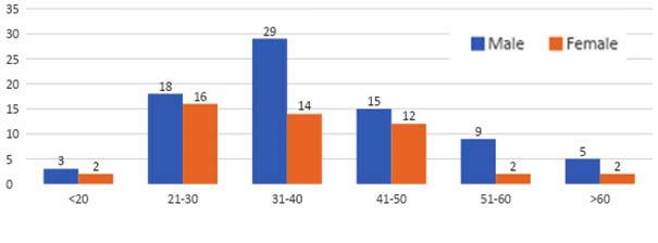
Our study revealed that over sixty percent of population belong to age group of 20-40 years with a male female ratio of 1.56:1. Snake bites cases was maximum in age group of 31-40 years (43;33.85%) followed by 21-30 years (34;26.77%)(Fig 1).
In our study, majority of the patients with vasculotoxic Snake bites presented with fang marks which presented as two distinct hemorrhagic puncture wounds (111; 87.4%) which often was accompanied by local swelling (72;56.69%) mostly in the lower limbs. Bleeding of bite site was observed where blood oozed out continuously even after wiping was observed in 38.58% cases (Table 2).
Table 1 — Total no of snake bite cases and Sex distribution
Table 2 — Presenting features at time of admission
Presenting featuresNo of patients
Distinct Fang Marks111
Local swelling64
Bleeding from bite site49
Pain at Bite site37
Bleeding from other sites11
Blistering08
A total of 35 patients presented with complications with few patients developed multiple complications. Systemic hypotension was seen in 21 cases. A total of 64 patients presented with swelling with severe swelling was seen in 17 patients with 14 patients (82.35%) developed complications. Moderate swelling was seen in 26 patients with 19 patients (73.07%) developed complications. Only two patients developed complications from 21 patients who had mild swelling at bite site. Compartment syndrome and gangrene developed in two of the patients each who had presented late and received traditional treatments before coming to hospital. 54.28% of patients developed acute kidney injury as a complication. In 8 patients succumbed during the course of treatment (Table 3).
In our study the most convenient bed side test done was 20-minute whole blood clotting test which came positive in 94 patients (74.01%) and complications was observed in 33 patients (35.10%) which was statistically significant.
Majority of the patients (49.60%) arrived late with a delay of 2 to 6 hours followed by 32 patients (25.19%) who arrived the hospital within 2 hours. 4 patients who arrived after 12 hours succumbed during treatment showed a 2/3rd mortality.
Out of the total 127 patients, patients (90; 70.86%) had used pressure bandage on the affected limb before coming to hospital while 19 (14.96%) used traditional patch by indigenous healers. 17 patients directly availed the health facility without any pre-treatment (Table 4).
Majority of the patients in our study (53;41.73%) required 5 to 10 vials of reconstituted lyophilized polyvalent anti-snake venom serum while 39 (30.70%) of patients required 11 to 20 vials. Only 6 patients required more than thirty vials. There was no significant difference between mean Hemoglobin value between patients with complications and patients without complications. The mean total leucocyte count and ESR was 10992.86 and 58.22 in patients with complications respectively and the result was statistically significant compared to uncomplicated cases.
The mean serum urea and creatinine were significantly raised in patients with complications but the change in serum uric acid level was not significant. The mean serum lactate dehydrogenase and serum creatinine phosphokinase showed higher levels. The mean serum potassium was 5.36±0.56 mEq/L in patients with complications.
The mean duration of hospital stay in patients who developed complications was 8.18 days with standard deviation of 2.78 days. Only 2 patients who developed extensive necrosis with gangrene and 2 patients who had to undergo fasciotomy due to compartment syndrome had a longer hospital stay of 15 days and 18 days respectively. Eight patients succumbed to Acute Kidney Injury and Disseminated Intravascular coagulation (Table 5).
DISCUSSION
In our study the hospital admission due to Snake bite was lower than study done at Maharashtra where it remained between 8.45 and 13.31 per 1000 admissions5.
Table 3 — Complications in vasculotoxic snake bites patients
ComplicationsNumber of patients affected
Gangrene with extensive necrosis02
Acute Kidney Injury19
Systemic Hypotension21
Disseminated intravascular coagulation04
Compartment syndrome02
Table 4 — Time delay and treatment received before reaching hospital Time taken toNo of PressureTight TraditionalNo pre-Mortality reach hospital cases bandagetourniquet herbal referral after Snake bite (n=127) Immobilization patch treatment < 2 hours32270230 2-6 hours634905112
Fig 1 — Age and sex preponderance in predominantly vasculotoxic snake bites
Table 5 — Hematological, Biochemical parameters and hospital stay
ParametersMean Value ± SDP value
Patients withoutPatients with complicationcomplication
Hemoglobin (g/dl)11.59 ± 1.4611.63 ± 1.560.892
Total Leucocyte Count8327.78 ± 1445.2710992.86 ± 3497.93<0.0001
In an earlier epidemiological survey done in the same district two decades earlier, reflected about deaths due to poisonous snake bites ranged between 5.28 to 31.75 per 1 lakh population6
Since we had taken into account only vasculotoxic bites, the mortality was mostly due to late arrival of patients due to decision delay and preventable complications could have been managed by timely administration of Anti-snake venom serum7.
Although in our study the vasculotoxic variety was predominant but it was quite lower than the study done in Paschim Midnapore district where it constituted 80% of Snake envenomation4.
We observed the non-toxic Snake bite cases who come with a definite history of Snake bites and species identification is doubtful often present with anxiety and increased heart rate and without any other symptoms. They get relieved when kept under observation in Casualty.
In our study a higher male preponderance was observed which slightly differed from an earlier study done in Paschim Midnapur district where female to male ratio was 1.07:1 but was quite similar to study done at Burdwan4,6
The age group 31-40 years were mostly affected in our study differed from previous study at Burdwan where 21-30 years was the predominant age group6. This could be attributed to the fact that the young age group were now less prone to exposure to snakes due to non-involvement in cultivation and growing employment in other sectors.
The patients who arrived late invariably developed the complications and size of swelling was directly proportional to time delay. Since the fangs of vipers are solid and cylindrical it could penetrate the dress
material easily, fang marks was a prominent feature in our study.
The presenting signs on arrival in our present study were comparable to an earlier study done at Odisha where the commonest symptom was local pain (41.6%) followed by oozing from bite site in 19.1% of vasculotoxic bites8. Researchers in India had suggested that 4255% of patients who presented after 6 hours developed complications9,10
Our study pointed out a marked increase in patient confidence on modern medical treatment than traditional healers as compared with an earlier study done in the same district two decades back where 65.47% went for traditional healers and only 22.14% received hospital treatment 6 . This could be attributable to social awareness campaign by Government and other allied sectors. Pressure bandage by a saree/dupatta/dhoti were commonly observed in patients who presented at casualty.
Patients who required more than thirty vials had a longer stay and increased mortality. Early initiation of Anti-snake venom serum was more important than cumulative doses a patient received.
Our findings were comparable to studies in India where Total Leucocyte Count, C-reactive protein, creatinine phosphokinase levels and lactate dehydrogenase levels were always elevated in vasculotoxic Snake bite11,12. Platelet count, serum creatinine phosphokinase and lactate dehydrogenase levels had been advocated in standard treatment guidelines which helped to monitor the patients with vasculotoxicity3
Serum creatinine and urea levels which represent compromised renal function were quite evident and early indicator of patients developing Acute Kidney Injury. Earlier researchers have proved 61.5% of patients with primary fibrinogenolysis and 38.5% with DIC developed renal failure and coagulation abnormality were commonly noted in vasculotoxic Snake bites13. The earlier study done to demonstrate effects of Viperidae venoms on renal structure and function had clearly showed increase in creatinine levels in sublethal doses in animal experiments. The authors had stated that severe hypotension, hemolysis, DIC and direct cytotoxic effect play a significant role in pathogenesis of Acute Renal Failure14
Vol 121, No 7, July 2023Journal of the Indian Medical Association
CONCLUSION
Vasculotoxic Snake bite requires serial monitoring of all crucial parameters with an aim to prevent the progression to Acute Kidney Disease. Early reporting and preventing decision delay is absolutely critical and require a multi-sectorial approach. Awareness campaigns must be strengthened in susceptible areas. The major limitation of the study was its crosssectional nature and more prospective studies could substantiate the management protocol.
Ethical clearance : Obtained prior to study
Conflict of interest : None
Funding : Self-funded
Acknowledgement : The authors are indebted to Burdwan Medical College and Hospital, West Bengal
REFERENCES
1http s://www.who.int/news/item/25-05-2018-snakebiteenvenoming-member-states-provide-who-with-clearmandate-for-global-action; accessed on 05-07-2022
2Suraweera W, Warrell D, Whitaker R, Menon G, Rodrigues R, Hang Fu S, et al — Trends in snakebite deaths in India from 2000 to 2019 in a nationally representative mortality study. eLife 2020;9: e54076: 1-37
3Standard treatment guidelines of Snake Bite. Ministry of health and family Welfare https://nhm.gov.in/images/pdf/guidelines/ nrhm-guidelines/stg/Snakebite_Full.pdf. Accessed on 06/07/ 2022
4Manaa K, Ghosh R, Gantaita K, Sahaa K, Paruaa P, Chatterjeea U, et al — Incidence and treatment of snakebites in West Bengal, India. Toxicology Reports 2019; 6: 239-43.
5Inamdar IF, Aswar NR, Ubaidulla M, Dalvi SD — Snakebite: Admissions at a tertiary health care centre in Maharashtra, India S Afr Med J 2010; 100: 456-8.
6Hati AK, Mandal M, De MK, Mukherjee H, Hati RN — Epidemiology of snake bite in the district of Burdwan, West Bengal. J Indian Med Assoc 1992; 90(6): 145-7.
7Chakraborty S, Banerjee P, Hazra R, Maity S, Banerjee S, Sarkar N — A retrospective study on snakebite and its outcome from a referral-cum-teaching hospital of Kolkata, India. Saudi J Health Sci 2020; 9: 130-5.
8Mishra A, Mohanty SN, Rastogi P — Study on Snake Bite Poisoning in a Tertiary Care Hospital in Rural Odisha, India. J Punjab Acad Forensic Med Toxicol 2019; 19(1): 1-5.
9Patil BT — Snake Bite Induced Acute Renal Failure: A study of Clinical Profile and Predictors of Poor Outcome. 2012 Jun: 1 (2-3): 59-65.
10Attappan G, Balaji MV, Navaneethan U, Thirumalaikolundu Subramanian — P Acute Renal Failure in Snake Envenomation: A Large Prospective Study. Saudi J Kidney Dis Transpl 2008: 19: 404-10.
11Kandasamy S, Gopalakrishnan S, Venkatesan M, Ramakrishnan M — The clinical and biochemical profile of snakebite patients- A hospital based comparative study of envenomed and non-envenomed victims. International Journal of Biochemistry and Biotechnology 2014: 2169-3048 3(2): 511-5.
12Patil SL, Kaveri VB —To study the utility of serum LDH values as a marker of hemotoxicity in snake bite victims in HSK hospital: An observational study. Indian Journal of Basic and Applied Medical Research 2017; 6(2): 331-47.
13Vijeth SR, Dutta TK, Shahapurkar J — Correlation of renal status with hematologic profile in viperine bite. Am J Trop Med Hyg 1997; 56(2): 168-70.
14Chugh KS, Pal Y, Chakravarty RN, Datta BN, Mehta R, Sakhuja V, et al — Acute renal failure following poisonous snakebite. Am J Kidney Dis 1984; 4(1): 30-8.
Ifyouwanttosendyourqueriesandreceivethe responseonanysubjectfromJIMA,pleaseuse the E-mail or Mobile facility.
Know Your JIMA
Website:https://onlinejima.com
For Reception:Mobile : +919477493033
For Editorial:jima1930@rediffmail.com Mobile : +919477493027
For Circulation:jimacir@gmail.com
Mobile : +919477493037
For Marketing: jimamkt@gmail.com
Mobile : +919477493036
For Accounts: journalaccts@gmail.com
Mobile : +919432211112
For Guideline:https://onlinejima.com
ER, PR HER2-neu Study in Breast Cancer Patients of Southern Rajasthan
Namita Goyal1, Gunjan Bhatia2
Background : Breast Carcinoma is the most common malignancy in Indian females, It is also the most common Female Cancer World wide with estimated incidence of around 25% among all Cancers. For targeted therapy breast carcinomas are further classified on the basis of their molecular profile. This molecular classification is becoming the gold standard for complete characterization of Breast Cancer. But in resourse limited settings Tumor markers ie, ER, PR and HER2, are routinely available in Breast Cancer specimens, are reliable, inexpensive and useful for therapeutic decision making.
Aims & Objective : To evaluate ER, PR HER2-neu status of patients with Breast Carcinoma. To improve prognostic value and aim at targeted therapy.
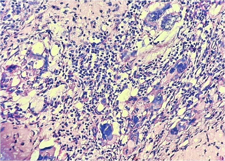
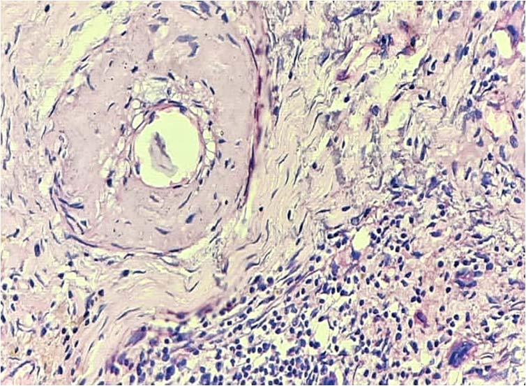
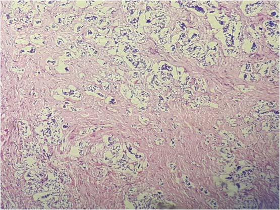
Material and Method : The study was conducted on 100 proven Breast Cancer patients of Udaipur and surrounding tribal belt attending Mahrana Bhupal Government Hospital, Udaipur. IHC was done on fully automated IHC instrument leica bond max and slides were prepared. Slides were examined and scoring was done by allred method for ER/PR and for Her2 scoring was done according to ASCO/CAP scoring staining pattern.
Results : Patients were divided in four major groups on the basis of IHC ie, ER/PR+ & Her2+, ER/PR+ & Her2 -ve, Triple negative and Her2 Overexpressed. Hormone positivity was seen among 54.63% cases and most of them were grade II histolgically. Most of the patients were in 41 to 60 years of age group.
Conclusion : IHC markers are helpful in guiding for treatment protocols in Breast Cancer patients and help in stratifying the patients in different risk group according to their prognosis.
Key words :IHC, Breast cancer.
Breast carcinoma is the most common malignancy in Indian females with age adjusted rate as high as 25.8 per 100,000 women and mortality rate around 12.7 per 100,000 women. It is also the most common Female Cancer world wide with estimated incidence of around 25% and 2.3 million newly diagnosed cases in year 20201. The increasing incidence and decreased 5 year survival rate has been attributed to change in lifestyle, late marriage, decreased breast feeding, lack of screening, late presentation, delayed and incomplete treatment.
Surgery has been the mainstay of treatment but now due to increasing awareness and availability about targeted therapy, efforts must be put to prolong the survival and improve the outcome. For targeted therapy Breast Carcinomas are further classified on the basis of their molecular profile.This molecular classification is becoming the gold standard for complete characterization of Breast Cancer.
Department of Pathology, RNT Medical College, Udaipur, Rajasthan 313001
1MD (Pathology), Senior Professor
2MD, Assistant Professor and Corresponding Author
Received on : 28/06/2022
Accepted on : 12/08/2022
[J Indian Med Assoc 2023; 121(7): 29-32]
Editor's Comment :
Breast carcinoma is one of the most common malignancy and ER PR and Her2 testing helps in determining the prognosis and giving targeted therapy to patients.
However, for molecular classification gene expression profiling, a high end technology is required so Clinicians usually rely on clinicomorphological pattern and readily available tumor markers which act as surrogate marker for molecular profiling.
These Tumour markers ie, ER, PR and HER2, are routinely available in Breast Cancer specimens, are reliable, inexpensive and useful for therapeutic decision making.
Immuno-histochemistry for these Tumour markers has been very important for deciding prognosis, predicting response to therapy and evaluating residual Tumour cells in post treatment cases.
AIMS AND OBJECTIVES
To evaluate ER, PR HER2-neu status of patients with Breast Carcinoma .
To improve prognostic value and aim at targeted therapy.
121, No 7, July 2023Journal
MATERIALS AND METHODS
The study was conducted on 100 proven Breast Cancer patients of Udaipur and surrounding tribal belt attending Mahrana Bhupal Government Hospital, Udaipur.
Both the specimen and prepared blocks were accepted for study of Tumour markers in Breast carcinoma patients. Detail clinical history and relevant clinical information was recorded in pre-designed performa.
IHC was done on fully automated IHC instrument leica bond max and slides were prepared (Table 1).
Slides were examined and scoring was done by allred method for ER/PR and for Her2 scoring was done according to ASCO/CAP scoring staining pattern.
Patient were followed up quarterly for one year regarding treatment and further workup, records were maintained (Fig 1).
RESULTS
We included a total of 100 patients in our study, out of which only one patient was Male rest all were Female this was an expected finding as the incidence of Breast Carcinoma in males worldwide is only 0.5 to 1%4. Out of total 100 cases results were acceptable in 97cases as in three cases due to tissue loss during processing results were rejected.
Most common age group involved is 40-60 years with 80.41% patients falling in this age group. Most of the Tumours were Invasive Ductal Carcinoma with No special type (95.8%) and Grade II was predominant histological grade.
We divided our patients in four major groups on the basis of IHC ie, ER/PR+ & Her2+, ER/PR+ & Her2 -ve, Triple negative and Her2 Overexpressed. Hormone positivity was seen among 54.63% cases and most of them were grade II histolgically.
One year follow-up was done for completion of treatment, recurrence and mortality which showed
That 8 patients died during 12 months, out of which 3 had stopped treatment in between while rest 5 were either on treatment or completed the Chemotherapy cycles. 4 out of these 5 patients were triple negative on IHC. Mortality in Triple Negative (TN) group was highest and all 8 patients were in age group of 61 and above.
DISCUSSION
The incidence of Breast Cancer is increasing Globally, with an extra surge in Asian countries, especially in pre-menopausal women. Breast Cancers are multifaceted disease with different morphologies and biological behaviors. Gene expression profiling studies have identified at least four categories of Breast Cancer: Luminal A, Luminal B, HER2 overexpressing, and basal-like or Triple Negative (TN) 2 . These molecular categories have been correlated with Immuno-histochemical (IHC) biomarkers3
58.1±14.759.9±12.8
Histological grade :
In our study most of the patients were in the age of 41-60 years which is also noted in many previous studies as most Indian studies have recorded median ages ranging from 48-53 years4-6. In comparison, the median age at diagnosis for Cancer of the Breast in the US is 61 years7
It can be assumed that the actual age of onset of Breast Carcinoma in the Indian patient is lower by well over a decade& this younger age of onset of Breast Cancer can be explained by racial differences. Table1
Fig 1 — Pie diagram for %incidence in
121, No 7, July 2023Journal
Now-a-days targeted therapy is the mainstay for treatment of Cancers & IHC and molecular studies are required for diagnosis, prediction, treatment and prognostication of cancers at any site8
Breast cancer has been divided into six molecular subtypes : Luminal A, Luminal B, basal like, HER2 like, normal epithelial like and claudin low9 However, The IHC surrogates for the molecular subtypes are: Luminal A (ER+ or PR+ or both, HER2 neu negative), Luminal B (ER+ or PR+ or both, HER2 neu+) or (ER+, low PR+, HER2-neu, high Ki67), basal like (ER-, PR-, HER2 neu±), HER2-neu+ (ER-, PR-, HER2-neu+). Any degree of Hormone receptor positivity makes the patient ideal candidate for Hormone therapy.
In our study 54.63% cases were positive for Hormone receptor out of which most were grade II (81.13%) this was in concordance with Kumar, et al however, the median age was lower than their study. Amongst these patients all were positive for ER however 17.8% were negative for PR.
ER expression has been labeled as a good prognostic and predictive biological marker through various studies and is associated better overall survival compared to ER negative Tumours.
However, independent prognostive and predictive role of PR expression irrespective of ER has been matter of great debate. ATAC (Arimidex, Tamoxifen, Alone or in Combination) adjuvant trial compared the efficacy of tamoxifen with that of the aromatase inhibitor anastrazole showed that patients with ER + /PR + Tumours had a lower recurrence rate than those with ER+/PR- tumors (7.6% versus 14.8%, respectively)11
Triple Negative Breast Carcinomas mainly of high histologic grade (grade III), showed high mitotic index and are found more frequently in pre-menopausal women. In our study median age of this group was 56±2 years with most of them falling in histological grade III.
Study conducted by Umemura and colleagues found that combined estrogen receptor-negative and HER2-negative Tumours constitute 19% of cases (11 of 58 Breast Cancer cases) & were associated with high expression of p53, vimentin and EGFR and these tumours showed the highest ki-67 Labeling Index and lowest expression of cyclinD1 when compared with other tumour groups12
The overall survival was least in this subgroup with 12.12% mortality in 12 month follow-up period so triple negative group has the worst overall and disease-free survival while overall survival was good in Hormone positive & HER2 negative subgroup which is in concordance with preivous studies13-15
Her2 Overexpression has both prognostic and predictive implications and the incidence of Her2 Overexpression is around 15-30% in Invasive Breast Cancers16. Trastuzumab was approved as part of a treatment regimen containing doxorubicin, cyclophosphamide, and paclitaxel for the adjuvant treatment of women with node-positive, HER2 overexpressing Breast Cancer.
In our study Her 2 score 3 as per CAP guidelines was reported as positive, score 2 was given equivocal and as National Comprehensive Cancer Network (NCCN) guidelines panel recommended that less than 3+ overexpression of HER2-neu by IHC should be additionally examined by FISH or other in situ hybridization methods so these cases were advised for further FISH testing, while score 0 and 1 was reported as Negative.
We had a total of 21.61% cases in this subgroup. When compared to Hormone positive subgroup patients in this group has higher histological grade and higher stage.
Though Recent publications have shown that newer molecular classification of Breast Cancer have greater prognostic value & Subtyping Breast Cancer using microarrays for gene expression analysis is the ideal method for such molecular classification but the availability and cost of these test are genuine constrains.
For such instances IHC-based classification systems are very useful and has been shown to correlate well with intrinsic classification using gene expression microarrays.
IHC system has it’s own limitation as there is intralaboratory and interlaboratory variation in ER results because fixation, antigen retrieval, and staining methods may differ among laboratories17
Similary discordance among Her2 results generated in different laboratories from the same specimen has also been reported18 Limitations of the study :
Study was conducted on a small group of patients and follow up period was also short . Her2 euqivocal caeses were followed for their FISH results but due to cost constrains most were either lost during follow up or such cases ended up for routine Chemotherapy due to absence of definiative HeR2 status.
Conclusion :
The biology and complex genomic intricacies of Breast Carcinoma has categorized it into different molecular subtypes but for countries with limited resources IHC can still be considered a valuable tool
Vol 121, No 7, July 2023Journal of the Indian Medical Association
for Clinicians which is simple, inexpensive, easy to interpret,reliable, reproducible and readily available.
REFERENCES
1Global cancer statistics 2020: GLOBOCON Estimates of Incidence and mortality worldwide for 36 cancer in 185 cancers
2Perou CM, Sørile T, Eisen MB — Molecular portraits ofhuman breast tumours. Nature 2000; 406(6797): 747-52.
3Carey LA, Perou CM, Livasy CA — Race, breast cancersubtypes, and survival in the Carolina Breast Cancer Study. Journal of the American Medical Association 2000; 295(21): 2492-502.
4Rajan G, Culas TB, Jayalakshmy PS — Estrogen and progesterone receptorstatus in breast cancer: A cross sectional study of 450 women in Kerala,South India. World J Surg Oncol 2014; 12: 120.
5Mukherjee G, Lakshmaiah KC, Vijayakumar M, Prabhu JS, Telikicherla D,Sridhar TS, et al — Analysis of clinicopathological characteristics of Indianbreast cancers shows conservation of speci c features in the hormonereceptor sub-types. J Integr Oncol 2016; 5: 159.
6Kumar RV, Panwar D, Amirtham U,Premalata CS, Gopal C, Narayana SM, et al — Estrogen receptor, Progesteronereceptor, and human epidermal growth factor receptor-2 status in breastcancer: A retrospective study of 5436 women from a regional cancer center inSouth India. South Asian J Cancer 2018; 7: 7-10.
8Krishnamurthy S, Poornima R, Challa VR, Goud YG — Triple negative breast cancer our experience and review. Indian J Surg Oncol 2012; 3: 12-6.
9Goldhirsch A, Winer EP, Coates AS, Gelber RD, Piccart Gebhart M, Thürlimann B, etal—Personalizing the treatment of women with early breast cancer: Highlights of the St. Gallen International Expert Consensus on the Primary Therapy of Early Breast Cancer 2013. Ann Oncol 2013; 24: 2206-23.
10Dunnwald LK, Rossing MA, Li CI — Hormone deceptor status, tumor characteristics, and prognosis: a prospective cohort of breast cancer patients. Breast Cancer Res 2007; 9(1): 6.
11Dowsett M, Cuzick J, Wale C, Howell T, Howell T, Houghton J, et al — Retrospective analysis of time to recurrence in the ATAC trial according to hormone receptor status :an hypothesis –generating study. J Clin Oncol 2005; 23: 75127.
12Umemura S, Takekoshi S, Suzuki Y, Saitoh Y, Tokuda Y, Osamura RY — Estrogen receptor-negative and human epidermal growth factor receptor 2-negative breast cancer tissue have the highest Ki-67 labeling index and EGFR expression: gene amplification does not contribute to EGFR expression. Oncol Rep 2005; 14: 337-43.
13Carey LA, Perou CM, Livasy CA, Dressler LG, Cowan D, Conway K, et al — Race, breast cancer subtypes, and survival in the Carolina Breast Cancer Study. JAMA 2006; 295: 2492-502.
14Dent R, Trudeau M, Pritchard KI, Hanna WM, Kahn HK, Sawka CA, et al — Triple-negative breast cancer: clinical features and patterns of recurrence. Clin Cancer Res 2007; 13: 442934.
15Onitilo AA, Engel JM, Greenlee RT, Mukesh BN — Breast Cancer Subtypes Based on ER/PR and Her2 Expression: Comparison of Clinicopathologic Features and Survival; Clinical Medicine & Research 2009; Volume 7, Number 1/2: 4-13.
16Burstein HJ — The distinctive nature of HER2-positive breast cancers. The New England Journal of Medicine 2005; 353(16): 1652-4. [PubMed] [Google Scholar].
17Rhodes A, Jasani B, Barnes DM, Bobrow LG, Miller KD— Reliability of immunohistochemical demonstration of oestrogen receptors in routine practice: interlaboratory variance in the sensitivity of detection and evaluation of scoring systems. J Clin Pathol 2000; 53: 125-130.
18Roche PC, Suman VJ, Jenkins RB, Davidson NE, Martino S, Kaufman PA, et al — Concordance between local and central laboratory HER2 testing in the breast intergroup trial N9831. J Natl Cancer Inst 2002; 94: 855-7.
Original Article
Implementation of Interventions Using School-based Posbindu Module and Applications to Prevent the Risk of Non-communicable Diseases to High School Students in Jakarta
Sri Widodo1, Agus Suwandono2, Sri Achadi Nugraheni3, Henry Setiawan4
Background : Non-communicable Diseases (NCDs)(PTM) are the main cause of death, accounting for 36 million (63%) of all deaths that occur worldwide. About 29 million (80%) occur in developing countries. The increasing prevalence of Non-communicable Diseases cannot be separated from the risk factors that cannot be avoided by the Indonesian people which begin to occur when they are teenagers. If teenagers never do physical activity and regulate their diet, they will be at risk of developing NCDs such as obesity and diabetes mellitus. In the present study, we aimed to dentify behavioral risk factors for preventing NCDs for high school students in DKI Jakarta.
Materials and Methods : In this a quasi-experimental method with a non-equivalent control group pretest and posttest design study. The total sample is 220 students in four DKI Jakarta schools were included. The test used univariate data analysis with distribution frequency, bivariate with chi square test and multivariate with multiple logistic regression.
Results : The intervention carried out by providing modules and School-based PTM Posbindu Applications can improve Students’ PTM Risk Prevention Behavior. Students’ dietary attitudes have the greatest influence on NonCommunicable Disease Risk Prevention Behavior in Students, namely 1.683.
Conclusion : The results of the present study indicated the School-based Posbindu PTM Modules and Applications improve Students’ Non-communicable Disease Risk Prevention Behavior. Attitudes Eating patterns, physical activity attitudes and student self-efficacy affect the risk prevention behavior of non-communicable diseases. The most dominant variable influencing the risk prevention behavior of non-communicable diseases is the attitude of students’ eating patterns. The need to further develop the Posbindu that already exists in the school setting as a guide for implementing the School Posbindu to improve PTM Risk Prevention Behavior for Students.
[J Indian Med Assoc 2023; 121(7): 33-9]
Key words :Interventions School-Based, Posbindu Module, Prevent Risk, Non-communicable Diseases.
Non-Communicable Diseases (NCDs) are the main cause of death for 36 million (63%) of all cases of death that occur worldwide. Around 29 million (80%) actually occur in developing countries. The increase in NCD deaths in the future is projected to continue to occur at 15% (44 million deaths)1
The increasing prevalence of NCDs is due to : risks that are not avoided by Indonesian society that begin to occur during adolescence. The prevalence of smoking at the age of 10-18 years is increasing every year, according to the 2013 Basic Health Research, the prevalence of smoking at the age of 10-18 years is
1Doctoral Program, Faculty of Public Health, Diponegoro University, Semarang, Indonesia and Corresponding Author
2Faculty of Public Health, Diponegoro University, Semarang Indonesia
3Faculty of Public Health, Diponegoro University, Semarang Indonesia
4Faculty of Public Health, Diponegoro University, Semarang Indonesia
Received on : 03/01/2023
Accepted on : 25/06/2023
Editor's Comment :
Non-communicable diseases are the major causes of morbidity & mortality in the modern world. Preventive strategy beginning at school level are desirable to curb the growing incidence of NCD. This study gives positive outcome with the school based Posbindu Module.
7.2%, and based on the report on the results of the Basic Health Research in 2018 it has increased to 9.1%. If teenagers never do physical activity they will be obese, in this condition the closest noncommunicable disease can be affected is diabetes mellitus2,3.
According to the results of the Global School-based Student Health Survey (GSHS) survey in 2015. It is known that the current lifestyle of adolescents is at risk for NCDs in the future. The unhealthy eating pattern is where in one day teenagers do, including eating Fast Food (53%), less consumption of Vegetables and Fruit (78.4%), Soft Drinks (28%), lack of Physical
121, No 7, July 2023Journal
Activity (67.9%), Ever Smoked (22.5%) and Consumed Alcohol (4.4%)4
Various efforts from the Government through the Ministry of Health have been carried out to improve Student Health Behavior by issuing policies on guidelines for fostering Clean and Healthy Living Behavior, one of which is implemented in schools. Efforts to change student behavior are a shared responsibility so cooperation from various parties is needed, therefore based on a joint decree of four ministries a school health business development team was formed with the hope that the four ministries would synergize with each other to improve the health of students5,6
As time goes by, the educational needs of students and monitoring of student health are not enough just to do conventionally so far what has been done is only to provide direct health education face-to-face, but along with high mobility, a breakthrough is needed by utilizing technological advances. The rapid development of technology can be used to assist education, increase knowledge, and disseminate information to become an alternative in the use of health promotion media. The use of smartphones is currently very widespread used by teenagers as a medium of communication and information. The survey results stated that 8.7% of adolescents were smartphone users equipped with an android system7
In the process, android-based applications via smartphones can be used as a media for health promotion to assist adolescents in increasing knowledge and implementing a healthy lifestyle. So that it is expected to be a medium to prevent the occurrence of pre-diabetes among adolescents8 Smartphone devices can help collect, organize, store and present information. That way the smartphone is not only for communication, it can be used for cameras, games (games), social media, learning media for students9
Utilization of current technological developments is very possible to develop a system that replaces the role of a health worker, seeing the growing development of Android smartphones currently allows it to be used as a health education medium, a tool for screening symptoms of NCDs and monitoring health checks for students. Smartphones besides being used as a communication tool, have also become a necessity for modern society, so they are very well known among the general public . android based is very simple and easy to carry10
The purpose of this study was to identify behavioral risk factors for preventing NCDs for high school students in DKI Jakarta.
MATERIALS AND METHODS
This type of quantitative research uses a quasiexperimental method (quasi- experimental ) with a nonequivalent control group pretest and posttest design.
Samples Selection :
The research was conducted in four schools in the DKI Jakarta area and the determination of the sample size in this study was determined by random sampling.The number of samples is 220 students in four schools in DKI Jakarta. Sampling was done by means of purposive sampling. Inclusion and exclusion criteria: The students included in the inclusion criteria in this study were : (1) students who were willing to participate in the study by signing the consent form, (2) male and female students aged 15-18 years, (3) students having android-based smartphones. (Special treatment group). The students who met one of the following exclusion criteria could not participate in this study : (1) students who did not follow all the intervention procedures, (2) students who withdrew from the study. Written consent was obtained from each student before sampling and the study was approved by the Health Research Ethics Committee of Diponegoro University of Public Health No. 556/EA/ KEPK-FKM/2019.
Interventions used :
The interventions carried out were providing School Posbindu Modules to Students and Teachers in implementing Posbindu in Schools and providing Health Education with the Web-based School Posbindu Application which contains Information on School Posbindu, Information on NCDs, Education on Healthy Diets, Physical Activities, Videos on Dietary Patterns & Physical Activity, Diabetes Mellitus Risk factor test.
Statistical Analysis :
SPSS 22 version were used for statistical analysis. Chi square test was used for analysis of the mutation data and P value <0.05 and Multivariate withmultiple logistic regression.
RESULTS
Analysis of Trends in Research variables based on Time and Groups :
Based on the Table 1, students’ knowledge of eating patterns increased in the intervention group. The results showed that the knowledge of students’ eating patterns significantly increased after being given treatment. Although at the beginning of the pretest the knowledge of the students in the control group was in a higher position, but after being given higher treatment
121, No 7, July 2023Journal
the intervention group experienced an increase compared to the control group.
Attitudes Students’ eating patterns increased in the intervention group. The results showed that the attitude to eating patterns significantly increased after being given treatment. Although at the beginning of the pretest, the students’ eating habits in the control group were in a higher position than the intervention group at the time of the pretest, but after being given treatment the intervention group experienced an increase compared to the control group.
Knowledge of students’ physical activity experienced an increase in the intervention group. The results showed that the knowledge of students’ physical activity significantly increased after being given treatment. At the beginning of the pretest, the knowledge of students’ physical activity in the intervention group had seen an increase and it continued consistently, even in the second pretest, the increase was high.
Table 1 — Overview of Descriptive Statistics Differences in Average Score Variables in the Intervention and Control groups
Knowledge of DietPretest 9.241,4969.501,537
Knowledge
Attitudes Students’ physical activity increased in the intervention group. The results showed that the attitude of students’ physical activity significantly increased after being given treatment. At the beginning of the pretest the attitude of physical activity of students in the intervention and control groups has seen an increase, but the increase in the intervention group is higher than the control group.
Knowledge of Periodic Health Check-up increased in the intervention group. The results showed that the knowledge of Periodic Health Examination significantly increased after being given treatment. At the beginning of the pretest the knowledge of Periodic Health Examination in the control group was very high, but consistently the intervention group showed an increase in knowledge after being given treatment.
The attitude of the Periodic Health Check-up increased in the intervention group. The results showed that the attitude of the Periodic Health Examination significantly increased after being given treatment at the start of the pretest. The attitude of the Periodic Health Check-up in the control group was very high but then decreased. Different things were shown by
2.181.3422.101.381
the intervention group that consistently showed improvement after being given treatment.
Students’ self-efficacy increased in the intervention group. The results showed that students’ self-efficacy significantly increased after being given treatment. At the beginning of the pretest the self-efficacy of students in the control group was higher but then decreased. Different things were shown by the intervention group that consistently showed improvement after being given treatment.
Teacher mentoring has increased in the intervention group. The results of the study show that mentoring Teacher significantly increased after being given treatment . Although the initial measurement of the intervention group and control group experienced an increase, but after the teacher mentoring treatment the intervention group had a higher increase than the control group.
Monitoring Teachers experienced an increase in the intervention group. The results showed that
121, No 7, July 2023Journal
monitoring Teacher significantly increased after being given treatment. Although the initial measurement of the intervention group and control group experienced an increase, after the monitoring intervention was carried out,Teacher the intervention group had higher improvement than the control group .
The role of adolescent cadres has increased in the intervention group. The results showed that the role of adolescent cadres significantly increased after being given treatment . Although the initial measurement of the intervention group and control group experienced an increase, after being given treatment the role of adolescent cadres in the intervention group was higher than the control group.
Behavior Risk prevention NCDs have increased . The results showed that the non-communicable disease risk prevention behavior in the intervention group significantly increased after being given treatment. Although at the initial measurement the intervention group and control group experienced an increase, after the intervention the intervention group experienced an increase in contrast to the control group it decreased.
Table 2 — Results of Cross Tabulation Analysis and Chi Square Test of Relationship between Independent Variables and Bound Variables
Dietary
Remarks
Analysis of the relationship of independent variables on noncommunicable disease risk prevention behavior :
Based on the following Table 2, it can be seen that the variables that influence the Non-Communicable Disease Risk Prevention Behavior are eating patterns P value 0,001, physical activity 0,000, attitudes and students’ self-efficacy 0,000.
Multivariate Analysis of the Effect of Independent Variables on Non-Communicable Disease Risk Prevention Behavior :
The results of the multivariate analysis of the influence of independent variables related to the NonCommunicable Disease Risk Prevention Behavior of Students can be seen in Table 3 . Variables of eating pattern attitude, physical activity attitude and students’ self-efficacy affect the Non-Communicable Disease Risk Prevention
Behavior in Students.
The magnitude of the influence of each variable on the Non-Communicable Disease Risk Prevention Behavior of Students can be seen in the Exp column (B). The variable that has the greatest influence on Non-Communicable Disease Risk Prevention Behavior in Students is the student’s dietary attitude, which is 1,683, meaning that students who have a good eating pattern have the opportunity to have better Risk Prevention Behavior. NCDs is 2 times greater than students whose eating patterns are not good. The R2 value obtained from the regression model is 0.009, so it is known that all research variables affect Risk
Table 3 — Multivariate Analysis of the Effect of Independent Variables on Student
121, No 7, July 2023Journal
Prevention Behavior non-communicable disease is 0.9% and the remaining 99.1% is influenced by other variables.
DISCUSSION
The results showed that the knowledge of students’ eating patterns significantly increased after being given treatment. Although at the beginning of the pretest the knowledge of the students in the control group was in a higher position, but after being given higher treatment the intervention group experienced an increase compared to the control group. Students who lack good knowledge tend to have problems with being overweight which are at risk of becoming NCDs. This is in accordance with the results of a study conducted by Dragana Milosavijevic, et al on high school students in Croatia regarding knowledge of eating habits showing that overweight people experience noncommunicable diseases at a young age11
The students’ eating habits increased in the intervention group. The results showed that the attitude to eating patterns significantly increased in the measurement after being given treatment. Although at the beginning of the pretest , the students’ eating habits in the control group were in a higher position at the time of the pretest, but after being given treatment the intervention group experienced an increase compared to the control group. The attitude of students’ good eating patterns can be seen from their regular breakfast habits.
This is in accordance with research conducted by Jannina Viljakainen, et al on a number of adolescents in Finland which states that adolescents who eat breakfast irregularly tend to experience higher body weight, as well as the results found that adolescents who eat dinner irregularly and do not often consume fruits will being overweight and at risk of noncommunicable diseases such as Diabetes Mellitus12
The intervention that has been carried out is expected to be more effective in increasing students’ knowledge of eating patterns, the existing and accessible school Posbindu application can improve attitudes and serve as a reminder for students to maintain their diet.
Knowledge of students’ physical activity increased in the intervention group. The results showed that the knowledge of students’ physical activity significantly increased after being given treatment. At the beginning of the pretest the knowledge of physical activity of the intervention group students had seen an increase and it lasted consistently even in the second pretest the increase was high.
Through the intervention in the form of the Posbindu School Application, it has been proven to increase students ‘knowledge of physical activity. In this application, students can get information easily, in addition to the above, they also get physical activity education counseling, either directly or indirectly, which was carried out during the intervention period. Even though students have a good diet, if the physical activity carried out is not optimal, it will be a risk factor for non-communicable diseases in students. As research conducted by Furong Xu on adolescents in the United States showed the results that respondents tended to consume healthier foods, but they did not pay attention to the physical activity that was carried out causing obesity in adolescents and at risk of developing NCDs such as Diabetes Mellitus13
Students’ physical activity increased in the intervention group. The results showed that students’ physical activity attitudes significantly increased in the pretest to posttest 2 measurements. At the beginning of the pretest, students’ physical activity attitudes in the intervention and control groups had seen an increase, but the increase in the intervention group was higher than the control group.
The attitude of students’ physical activity can be seen from the habits they show in their daily activities, this is in accordance with research conducted by Robert G McMurray on a number of adolescents in England which states that a strong relationship with high-intensity physical activity watching TV, playing video games causes overweight in adolescents in England14
Knowledge of Periodic Health Checkup increased in the intervention group. The results showed that the knowledge of Periodic Health Examination significantly increased after being given treatment. At the beginning of the pretest the knowledge of Periodic Health Examination in the control group was very high, but consistently the intervention group showed an increase in knowledge after being given treatment.
Periodic Health Checks on Students need to be improved considering that there are still many students with poor knowledge resulting in students not doing regular health checks. Health checks in schools need to be carried out related to the lifestyle and activities carried out by teachers and students at the school. The current trend tends to be a trend that requires students, both male and female, to have an ideal body15.
The attitude of the Periodic Health Check-up increased in the intervention group. The results showed that the attitude of the Periodic Health Examination
Vol 121, No 7, July 2023Journal
significantly increased after being given treatment. At the beginning of the pretest, the attitude of the Periodic Health Check-up in the control group was very high but then decreased. Different things were shown by the intervention group that consistently showed improvement after being given treatment.
The attitude of students’ periodic health checks is an awareness where they feel the importance of conducting regular health checks. There is a big challenge to increase students’ awareness so that they will later avoid non-communicable diseases. This is in accordance with research conducted by Tahira Sadiq, et al on a number of students in Rawalpindi and Islamabad which stated that medical and non-medical students are aware of the importance of regular health checks16
Students’ self-efficacy increased in the intervention group. The results showed that students’ self-efficacy significantly increased after being given treatment. At the beginning of the pretest the self-efficacy of students in the control group was higher but then decreased. Different things were shown by the intervention group that consistently showed improvement after being given treatment.
Adolescents with a high level of self-efficacy will tend to have a stronger effort in implementing a healthy lifestyle when compared to adolescents who have lower self-efficacy. With good self-efficacy can support confidence and self-confidence to want to try. This can be an inherent factor and motivates oneself to perform a behavior. research conducted by Arturo Direito with the application of the Improving FITness (AIMFIT) application as a form of media that helps in making lifestyle modifications to increase physical activity in adolescents. In this study there was a significant increase in the level of self-efficacy, satisfaction, enjoyment and fitness towards use and acceptance for adolescents17
Interventions carried out on Teacher proven to increase the roleTeacher do assistance. The results of the study show that mentoring Teacher significantly increased after being given treatment. Although the initial measurement of the intervention group and control group experienced an increase, but after the teacher mentoring treatment the intervention group had a higher increase than the control group.
The results of this study are in accordance with what was done by Lauran H Smith on adolescent students in Appaalchia who stated that the approach to adolescent students is an effective approach to changing the lifestyle of students in their school environment18
Teachers have an important role in encouraging students to prevent the risk of NCDs by supervising and monitoring these students. This is in accordance with research conducted by Erin K Eliassen which states that teachers have a role not only limited to being teaching staff for students. Teachers are also responsible for being student role models in carrying out these actions, the teacher can not only admonish students for snacking carelessly, but can also invite students to sit down to eat together and discuss what proper and nutritious food to eat19
Interventions carried out on youth cadres are proven to increase their role in helping efforts to improve health in schools. The results showed that the role of adolescent cadres significantly increased after being given treatment . Although the initial measurement of the intervention group and control group experienced an increase, after being given treatment the role of adolescent cadres in the intervention group was higher than the control group.
Based on the results of the research above, cadres must be fostered, guided, and supported by more skilled and experienced supervisors and given better training and knowledge. They should be able to know when and where to seek medical help for conditions they can not manage on their own. The training program does not only affect the knowledge but also the skills of the youth school cadre participants20
Youth cadres need to get support and motivation to carry out their roles so that efforts need to be made to increase the role of the school youth cadres. Based on Social Cognitive Theory, human behavior is influenced by personal, environmental, and behavioral factors. One of the environmental factors that influence human behavior is motivation21
Behavior of risk prevention NCDs in both groups have increased . The results showed that the noncommunicable disease risk prevention behavior in the intervention group significantly increased after being given treatment. Although the initial measurement of the intervention group and control group experienced an increase, after the intervention of teacher mentoring the intervention group had a higher increase than the control group which actually decreased.
Students’ understanding of NCDs is obtained through knowledge and this is in line with research conducted by Gamage in Sri Lanka which found that the proportion of students aged 17-19 years who have good knowledge of NCDs is 43% 10 . A person’s understanding is obtained through knowledge22
The limitations of this research are that the need assessment activities and the implementation of the
Vol 121, No 7, July 2023Journal of the Indian Medical Association
intervention were carried out in schools collecting students and teachers, many experienced problems related to the time of the study during the COVID-19 period in DKI Jakarta, the number of samples determined was not representative of the area of high school in DKI Jakarta because it only involved four areas. School in DKI.
CONCLUSION
Interventions that have been carried out by providing school-based PTM Posbindu modules and Applications and School Posbindu applications can improve Students’ Non-Communicable Disease Risk Prevention Behavior. Attitudes Students’ eating patterns, physical activity attitudes and students’ selfefficacy affect the risk prevention behavior of noncommunicable diseases. The most dominant variable influencing non-communicable disease risk prevention behavior is students’ eating patterns. The need to further develop the existing Posbindu in the school setting as a guide for the implementation of Posbindu in schools to improve the Non-Communicable Disease Risk Prevention Behavior of Students.
ACKNOWLEDGMENTS
The present study was financially supported by Respaty Indonesian University of Public Health. Conflict of Interests : All the authors declared that there is no conflict of interest
REFERENCES
1Ministry of Health RI, 2014. Efforts to Control NonCommunicable Disease Risk Factors. Director General of P2PL, Directorate of Non-Communicable Disease Control Jakarta.
2Riskesdas 2013. Basic Health Research (RISKESDAS) 2013. Jakarta; 2013.
3Ministry of Health RI, 2013. Principles of Prevention of NonCommunicable Diseases (PTM) and Regulations. [Online] Accessed March 25, 2019.
4RI 2015 Research and Development Center for Health Risk Behavior in Middle and High School Students in Indonesia. Jakarta: Indonesian Ministry of Health;
5Ministry of Health RI, 2014. Efforts to Control NonCommunicable Disease Risk Factors. Director General of P2PL, Directorate of Non-Communicable Disease Control, Jakarta.
6Minister of National Education, Minister of Health, Minister of Religion MDN. Joint Decree of the School Health Business Development Team. 2003.
7Muflih M, Hamzah H, Puniawan WA — Smartphone Use and Social Interaction in Adolescents at Sma Negeri I Kalasan Sleman Yogyakarta. Idea Nurs J 2017; 8(1): 12-8.
8American Diabetes Association — Strategies for Effective Screening, intervention and follow-up. 2017. p 62-9.
9Seifert T — Pedagogical Applications Of Smartphone Integration in Teaching: Lectures, Pre-Service Teachers and Pupils’ Perspectives. 2015;
10Dayer L, Heldenbrand S, Anderson P, Gubbins PO MB — Smartphone Medication Adherence Apps: Potential Benefits To Patients And Providers. J Am Pharm Assoc 2013; 53(2).
11Milosavljeviæ D, Mandiæ ML, Banjari I — Nutritional knowledge and dietary habits survey in high school population. Coll Anthropol 2015; 39(1): 101-7.
12Viljakainen J, Figueiredo RADO, Viljakainen H, Roos E, Weiderpass E, Rounge TB — Eating habits and weight status in Finnish adolescents. Public Health Nutr 2019; 22(14): 261724.
13Xu F, Cohen SA, Greaney ML, Greene GW — The association between US adolescents’ weight status, weight perception, weight satisfaction, and their physical activity and dietary behaviors. Int J Environ Res Public Health 2018;15(9).
14McMurray RG, Harrell JS, Deng S, Bradley CB, Cox LM, Bangdiwala SI — The influence of physical activity, socioeconomic status, and ethnicity on the weight status of adolescents. Obes Res 2000; 8(2): 130-9.
15Coal. Adolescent development. Sari Pediatr 2010; 12: 21-9.
16Sadiq T, Asim M, Aziz SA — Awareness among medical and non-medical students about the practice of periodic medical examination. Jiimc [Internet] 2017; 12(2): 116-9. Available from: http s://www.bibliomed.org/mnsfulltext/133/1331486455962.pdf?1633835208
17Direito A, Jiang Y, Whittaker R, Maddison R — Smartphone apps to improve fitness and increase physical activity among young people: Protocol of the Apps for IMproving FITness (AIMFIT) randomized controlled trial. BMC Public Health [Internet] 2015; 15(1): 1-12. Available from: http://dx.doi.org/ 10.1186/s12889-015-1968-y
18Smith LH, Holloman C — Comparing the Effects of Teen Mentors to Adult Teachers on Child Lifestyle Behaviors and Health Outcomes in Appalachia. J Sch Nurs 2013; 29(5): 386-96.
19Eliassen EK — The impact of teachers and families on young children’s eating behaviors. YC Young Child 2011; 66(2): 849.
20Sharma S, Taneja M — The effect of training on employee performance. Int J Recent Technol Eng 2013; 5(4).
21Glanz K, Viswanach RB — Health Behavior and Health Education. Health Behavior and Health Education. Theory, Research, and Practice. San Francisco; 2008. 67-96 p; 22AU Game, Jayawardana PL — Knowledge of noncommunicable diseases and practices related to healthy lifestyles among adolescents, in state schools of a selected educational division in Sri Lanka. BMC Public Health 2017; 18(1): 1-9.
Original Article
Establishment of Haematological Reference Intervals for Healthy Adults in Karamsad at Shree Krishna Hospital
Jigisha Kanubhai Mer1, Faruq Ibrahim Mulla2, Monica Gupta3
Background : The Complete Blood Count (CBC) is the most frequently ordered diagnostic tests in medicine. Cellular components of peripheral blood can be evaluated by Complete Blood Count examination. In era of evidencebased medicine, interpretation of laboratory result requires Reference Interval (RI) or cut-off values for diagnostic accuracy. While establishing physiologically normal values, inherent variables like Gender, Age, Occupation, Body build, genetic background and environment (altitude) are more problematic. This study aims to establish the normal haematology RI for CBC.
Aims : To establish hematological reference intervals of CBC parameters for healthy adults.
Settings and Design : The cross-sectional study with posteriori sampling carried out at Pathology laboratory.
Methods and Material : Retrospective and prospective study including healthy individuals came for routine health check-up during a period from January, 2018 to March, 2022. Total 592 individuals after applying the inclusion and exclusion criteria are included.
Results : All parameters of CBC show significant P value by Shapiro–Wilks test, except MCHC. Mann-witney U test applied to retrieve the reference interval for Males and Females.
Conclusions : The current study revealed a significant gender-based difference in RI and also differ from currently used Reference Interval. As compare to other studies also shows significant difference. As per CLSI guideline and present study, each laboratory should establish their own reference interval.
Key words :Complete Blood Count, Reference Interval.
Health is necessarily a relative and goal-oriented concept1. However, to say that health is relative implies that the condition of individuals must be related to something and for this Reference Interval (RI) is used to compare the value of individuals laboratory results2 RI plays a great role in patient diagnosis, management, disease prognosis, monitoring of response to therapy and in monitoring possible adverse reactions to therapy3 According to the Clinical and Laboratory Standards Institute (CLSI) guideline each laboratory should establish its own RI, as number of factors affect haematological values in apparently healthy individuals like gender, age, occupation, body build, genetic background and environment (altitude). The Complete Blood Count (CBC) is the most frequently ordered diagnostic tests in medicine. The CBC is used to determine quickly whether a patient is anemic or infected and to estimate the blood’s ability to coagulate normally. The concept of reference intervals was
Department of Pathology, Pramukhswami Medical College-Bhaikaka University, Karamsad Anand, Gujarat 388325
1MD (Pathology), Third Year Resident
2MD (Pathology), Professor and Corresponding Author
3MD, DNB, Professor and Head
Received on : 02/03/2023
Accepted on : 16/03/2023
[J Indian Med Assoc 2023; 121(7): 40-3]
Editor's Comment : Each laboratory should establish their own reference interval as suggested by CLSI guideline.
introduced by International Federation of Clinical Chemistry (IFCC). According to CLS Iguideline the establishment of a reference interval of a laboratory to be done by testing at least 120 samples from nondiseased individuals for each gender and age group4,5 This study aims to establish the normal haematology reference intervals for CBC for healthy adult of Shree Krishna Hospital, Karamsad, Anand, Gujarat in NABL accredited laboratory.
Complete blood count parameters included in this study are:
Aim : To establish haematological reference intervals of Complete Blood Count parameters for healthy adults at Shree Krishna Hospital, Karamsad.
Objectives : To determine whether the currently used reference interval do represent the adult population in the city.
MATERIALS AND METHODS
The cross-sectional study with posteriori sampling was carried out at the NABL accredited Central Diagnostic Laboratory, Department of Pathology, Shree Krishna Hospital and Pramukh Swami Medical college, Karamsad, a tertiary centre in Karamsad, Anand. The present study is a retrospective (152 cases) and prospective study (440 cases) including healthy normal individual, who came for health check-up at Shree Krishna Hospital during a period from January, 2018 to March, 2022. Total of 592 normal healthy individuals were identified after applying the inclusion and exclusion criteria and were included for establishing the RI. The proposed number was above the CLSI guidelines for the establishment of RI, which recommends a minimum of 120 participants.
Inclusion Criteria :
(1)Male and Female
(2)Age group : Above18 years
Exclusion Criteria :
(1)Pathophysiological States - Renal Failure, Cardiac Diseases, Chronic Respiratory Diseases, Liver Diseases, Malabsorption Syndromes, Malignancies and Hematological Disorders which included anaemias.
(2)Systemic Diseases – Hypertension and Diabetes Mellitus.
(3)Replacement or Supplementation Therapy eg, Thyroxine, Insulin.
(4)Modified Physiological States - Pregnancy, Psychological and Mental Disorders.
(5)The paediatric age group (below 18 years) was excluded since the haematological parameters have a different reference range from adults.
A volume of 2 ml blood was collected in EDTA vacutainer and was processed within 2 hours of collection for the CBC in automated haematology analyser Sysmex XN 350 or Sysmex XN 550. Instruments were installed after doing validation tests like IQ (Installation Qualification), OQ (Operational qualification), PQ (Performance Qualification), carry over and calibration on installation. The controls were checked at different concentrations (Level 1, Level 2 and Level 3). Daily two level of the total three levels
controls were run in the machine.
Statistical Analysis :
The analysis was informed entirely by CLSI guidelines which recommend the use of 97.5 percentile and 2.5 percentile formed the upper and lower limit of reference range respectively of the population and associated group comparisons based either on parametric or non-parametric statistics depending on whether the distribution of the data is Gaussian or nonGaussian. Shapiro–Wilks test was used to evaluate data distribution. The mean, median, Standard Deviation (SD), range, 2.5 and 97.5 percentile were subsequently evaluated. P value of less than 0.05 was considered statistically significant (null hypothesis is rejected), it indicates that the population does not show a “Normal distribution and there is significant genderbased difference in RI. Differences between males and females were evaluated using the Mann–Whitney U test
OBSERVATION AND RESULTS
Total 592 individuals (144 Females and 448 Males) were evaluated and gender wise partitioning of subjects was done. All nine parameters of Complete Blood Count were evaluated for their distribution by Shapiro-Wilk test and significant P value of <0.05 was applied. When parameters distributed normally than reference range is decided by mean ± 2 Standard Deviation (SD). In case of skewed distribution median, Interquartile range (IQR) and 2.5-97.5 percentile were used as reference range. The non-parametric Mann-Whitney U test was applied to all parameters to check for any significant difference between Male and Female subgroups.
The Table 1 and 2 shows that all CBC parameters have significant P value <0.05, except MCHC (P value >0.38). All parameters reject null hypothesis except MCHC, suggesting that all parameters except MCHC have significant gender-based difference.
DISCUSSION
The present study has comparable parameters and has mild variation in Reference Interval with currently used Reference Interval for Hb, RBC and HCT. Rest all parameters like MCV, MCH, MCHC, RDW, TLC and PLT have significant difference in reference interval in comparison to currently used Reference Interval. In present study derived value for PLT count has lower limit value on higher side for both Male and Females (Table 3).
The present study shows the significant difference in RI of Platelet Count in comparison to other study. The values for HB, HCT, MCHC and RBC count of
Vol 121, No 7, July 2023Journal of the Indian Medical Association
Table 1 — Reference interval for different parameters Shapiro-Wilk test
Table 2 — Ranges for parameters with difference in male and female subpopulations
Table 3 — Comparison of obtained reference interval with currently used reference interval
ParametersCurrently used ReferencePresent study Range from Standard Text reference range Book Dacie 11th edition6
present study is comparable to all three studies Siraj et al, Sehgal, et al and Rahar, et al. MCV shows
significant difference in RI. Value of TLC is comparable to Sehgal, et al and Rahar, et al MCH also shows significant difference as compare to other study as shown in Table 4. Severalfactors can affect the RI, which include age, sex, weight, environment, race and ethnic origin, biorhythms, pregnancy, nutritional state, lifestyle, medication, tobacco and alcohol consumption7 (Table 4).
121, No 7, July 2023Journal
Table 4 — Comparison of the obtained reference interval with other studies
ParametersPresent study, 2022, Sehgal,Siraj, et al 2015,Rahar, et al 2022, Karamsad, Anand et al, 2020, Asmara, Eritrea9 Delhi10 (592 samples) Mumbai8 (591 samples)(123 samples) (Sysmex XN 350) or(100 samples)(Backman coulter :(Sysmex XN-1000) Sysmex XN 550)(Sysmex XE-2100)AU 480 chemistry system)
The present study suggests that there is a significant gender-based difference in CBC parameters and has mild variation from currently used Reference Interval. As compare to other studies, present study shows significant difference between laboratory parameters. This difference suggest that each laboratory should establish their own reference interval as suggested by CLSI guideline.
REFERENCES
1Gräsbeck R. The evolution of the reference value concept. Clin Chem Lab Med (CCLM) 2004; 42(7): 692-7.
2Gary L — Horowitz, Establishment and Use of Reference Values.In: Ann. M. Gronowski, Ph.D, editor. Tietz Textbook of clinical chemistry and molecular diagnostics, 5th ed. United States of America: Elsevier publishers; 2012.p. 95-116
3Wayne PA — CLSI Defining, Establishing, and Verifying Reference Intervals in the Clinical Laboratory-Approved Guideline. CLSI Document EP28-A3C. Third edition. Available online: http://shop. clsi. org/site/Sample_pdf/ EP28A3C_sample. pdf (accessed on 19 October 2010). 2008.
4How to Define and Determine Reference Intervals in the Clinical Laboratory; Approved Guideline—Second Edition. NCCLS document C28-A2 (ISBN 1-56238-406-6). NCCLS, 940 West
Valley Road, Suite 1400, Wayne, Pennsylvania 19087-1898, USA 2000.
5Homme AG. Briggs C, Culp N, Davis B, d’Onofrio G, Zini G, Machin SJ — ICSH guidelines for the evaluation of blood cell analysers including those used for differential leucocyte and reticulocyte counting. Int J Lab Hematol 2014; 36(6): 613-27.
6Imelda Bates, Reference Ranges and Normal values. In: Mitchell Lewis, editor. Dacie and Lewis Practical Haematology, 12th ed. China: Elsevier publishers; 2017.p.8-16.
7Abdullah DA, Mahmood GA, Rahman HS — Hematology Reference Intervals for Healthy Adults of the City of Sulaymaniyah, Iraq. Int J Gen Med 2020 Nov 25; 13: 1249-54
8Sehgal KK, Tina D, Choksey U, Dalal RJ, Shanaz KJ — Reference range evaluation of complete blood count parameters with emphasis on newer research parameters on the complete blood count analyzer Sysmex XE-2100. Indian J PatholMicrobiol 2013; 56(2): 120.
9Siraj N, Issac J, Anwar M, Mehari Y, Russom S, Kahsay S, Frezghi H — Establishment of hematological reference intervals for healthy adults in Asmara. BMC Research Notes 2018; 11(1): 1-6.
10Rahar S, Kumar V, Rao S, Gupta D — Haematology reference range evaluation for novel research parameters on the complete blood count analyzersysmex XN-1000. Hamdan Medical Journal 2022; 15(2): 83. 43
Original Article
Histomorphological Changes in Breast Cancer following Neoadjuvant
Therapy and its Prognostic Implication
for Patient Management — Study of 36 Cases in a Tertiary Care Hospital
Background : Neoadjuvant Therapy is the systemic administration of therapeutic agent before definitive surgery. Neoadjuvant Therapy (NAT), specially Neoadjuvant Chemotherapy (NACT) is a standard procedure in locally advanced breast cancers to reduce the tumor size & down stage the disease leading to increase in the chances of successful resection. Following NAT there are various histomorphological changes in the breast cancer, both in the tumor cells & stroma. Our aim of the study was to evaluate the various histomorphological changes in breast cancer following neoadjuvant chemotherapy and its prognostic implications.
Materials and Methods : The study was conducted over a period of 4 years which included 36 cases of breast cancer receiving NACT. Various histomorphological parameters such as pathological response, presence of residual tumor, its grading, Tumor Infiltrating Lymphocytes (TILs), stromal changes such as fibrosis, collagenization, microcalcification, hemosiderin laden macrophages were studied.
Result : In our study patients were between 30-70 years of age. The most common histomorphological changes were nuclear enlargement and pleomorphic nuclei(28 cases, 77.7%). Pathological Complete Response (PCR) & Tumor Infiltrating Lymphocytes (TIL) seen in 8 & 16 cases (77.7%, 22.22%) respectively, in which both Overall Survival (OS) and Disease Free Survival (DFS) were seen at greater incidence. Statistical analysis was done by software IBM SPSS version 20.0.
Discussion & Conclusion : Pathological evaluation of post NACT surgical samples in breast cancer is extremely important for assessing treatment response. Prognostic value in post NACT breast cancer was directly related to tumor staging after NACT, TIL and pathological response. To conclude histopathological examination of the tumor bed to assess the residual tumor is the gold standard for assessing the response to NACT in breast cancer.
[J Indian Med Assoc 2023; 121(7): 44-8]
Key words :Neoadjuvant Therapy, Breast Cancer, Pathologic Response, Residual Tumor.
According to GLOBOCAN 2020 female breast cancer is the most common cancer (11.7%) surpassing the lung cancer1. Neo Adjuvant Therapy (NAT) is an important modality for treatment of breast cancer. NAT is defined as systemic administration of therapy prior to surgical removal of a breast tumor2 Initially NAT was used only for locally advanced inoperable breast carcinoma. But nowadays NAT has also been used for the treatment of early stage breast cancers as well with different applications as follows : (i)In advanced inoperable carcinomas to decrease in tumor size and downstage the disease to make It
1MD (Pathology), Associate Professor, College of Medicine and Sagore Dutta Hospital, Kolkata 700058
2MD (Pathology), Senior Demonstrator, Department of Pathology, Rampurhat Government Medical College & Hospital, Rampurhat 731224
3MD (Physiology), Senior Demonstrator, Department of Physiology, Rampurhat Government Medical College & Hospital, Rampurhat 731224
4MD (Pathology), Associate Professor, Department of Pathology, Rampurhat Government Medical College & Hospital, Rampurhat 731224 and Corresponding Author
Received on : 22/12/2022
Accepted on : 29/05/2023
Editor's Comment :
Thorough knowledge of the histopathological changes both in the tumor cells and the stroma of post NACT Breast cancer specimensis of ardent need for the effective and planned regimen of therapy to enhance both the overall and disease free survival in these patients.
operable for better surgical outcome3
(ii)In early stage breast carcinoma to shrink the tumor and thus allowing breast conserving surgery 4 .
(iii)In clinically node negative breast cancer patients with unfavorable tumor profiles in whom adjuvant systemic therapy is predicted, neoadjuvant therapy prior to surgery reduces the extent of axillary surgery5.
(iv)Basing on the pathological response to NAT and the residual tumor burden it provides prognostic in formations such as decrease risk of distant metastasis, rate of DFS & OS6. The gold standard for assessing the response to NAT is pathological evaluation of surgical samples following NAT7
On histopathological examinations of post NAT samples Pathologic Complete Response (PCR) and Residual Cancer Burden (RCB) are the two most
No 7, July 2023Journal
important parameters which are independently associated with improved survival outcomes8,9
NAT includes Neoadjuvant Chemotherapy (NACT), radiotherapy and targeted hormonal therapy. In our study we have analyzed the response predominantly to NACT in breast cancer patients.
OBJECTIVE
The objective was to study the various histomorphological changes in breast cancer (in the tumor cells and stroma) in response to NACT& correlation of the prognostic markers such as OS and DFS with the histomorphological parameters.
MATERIALS AND METHODS
(A)Study design, duration & place of study – It was a retrospective study conducted over a period of 4 years from August, 2018 to August, 2022 in a tertiary care hospital of West Bengal.
(B)Inclusion Criteria — Breast cancer patients who had received NAT & having pre-NAT biopsy report along with post-NAT follow updetails were included in the study.
(C)Exclusion Criteria — Breast cancer patients in whom pre NAT biopsy report & follow-up details not available were excluded from the study.
(D)Detailed clinical history regarding clinical presentation, radiological findings, pre NAT biopsy diagnosis, NAT therapy cycles were taken from clinical records.
(E)Specimen handling —
i)Gross examination : Post NAT Modified Radical Mastectomy (MRM) and Breast Conservative Surgery (BCS) specimens were examined in fresh state after receiving to identify and measure the tumor bed ie, the tissue encompassing the original tumor site10. In cases of complete response to NAT it is difficult to identify any grossly visible lesion. In cases of partial response or no response to NAT, size and number of any residual disease foci are examined. The distance of the tumor bed or residual tumor, from the surgical margins were noted in case of BCS specimens.
ii)Fixation, Grossing & Sectioning : Fixation done in 10% formalin and sections were taken. Following the Food Drug Administration (FDA) recommendations (a minimum of 1
block per centimeter of pre-NAT tumor size or at least 10 blocks in total whichever is greater) sectioning is done in MRM/BCS specimens11. In our study axillary lymph node sampling were done following the standard operating procedures12.
iii)Tissue Processing : All the sectioned tissue bits were processed, stained by Hematoxylin & Eosin (H&E) Stain and examined underthe microscope.
iv)Histological Examination : Things looked for -
(a)Presence of any residual invasive cancer component,histologic subtype & grade. Presence of in-situ component, lymphovascular invasion, necrosis, calcification, number of positive lymph nodes, and status of surgical margins (in BCS) were also noted.
(b)Stromal changes such as hyalinization, foamy macrophages, lymphocytes,multinucleated giant cells, hemosiderin-laden macrophages, necrosis and microcalcification were studied.
v)Pathological staging after NAT were done based on the TNM staging system (8th. Edition of AJCC)
(F)StatisticalAnalysis— Soft ware SPSS version 20.0 was used for data analysis and all thedata were represented as number (n) and percentage (%) and compared by X2 test as applicable.
(G)P Value — <0.05 was considered statistically significant.
(H) Ethical Clearance — Ethical clearance was taken from Institutional Ethical committee (Figs 1-4).
RESULT
In our study a total of 36 cases of breast cancer receiving NACT were studied. Various histo-
Fig 1 — Photomicrograph showing tumor cells with pleomorphic, bizzare nuclei & TILs in breast cancer following NACT (H&E,100X)
In our study PCR was seen in 8 cases (22.22%) & rest 28 cases showed PPR(pathological partial response)-(6 cases) & PNR (pathological no response)- (22 cases) respectively.
Most common type of residual cancer seen was Invasive Carcinoma of No Special Type (27,75%)(Tables 2&3). In our cases pre-NAT TIL seen in 16 cases. OS & DFS free survival seen in 11 cases. Post NAT stromal lymphocytes seen in 11 cases out which7 cases had PCR (Table 4).
DISCUSSION
morphological changes seen were as follows. Nuclear enlargement- 28 cases (77.7%), hyperchromasia- 26 (72.22%), pleomorphic nuclei- 28 (77.7%), karyorrhexis / karyolysis- 13 (36.11%). Stromal response - fibrosis in 12 cases (33.33%), collagenization-3 cases(8.33%), calcification in 5 cases(13.88%), lymphocytic infiltrate in 11 cases (30.55%), foamy histiocytes in 2 cases (5.55%), giant cells in 3 cases (8.33%) and Hemosiderin laden macrophages in 2 cases (5.55%)(Table 1).
Neo Adjuvant Therapy (NAT) is defined as administration of therapeutic agents prior to definitive surgery in Breast cancer patients. NAT may be Neoadjuvant Chemotherapy (NACT) or radiotherapy or targeted hormone therapy. In contrast to other studies in which some breast cancer patients received endocrine therapy and targeted therapy korde LA, et al7 in our study, We have analyzed the breast cancer patients who received NACT. A total of 36 cases of post NACT breast cancer patients were studied. There were various morphologic alterations both in breast cancer cells and in the stroma following NACT. Different studies have shown the benefits of NACT in breast cancer13
In our study the age of the patients range from 30-70 years, similar to study by Cheryl Sarch Phillipose, et al14
In our study clinical findings such as tumor size, quadrant involved, axillary lymph node status along with pre-NAT radiological findings (mammography & USG), pre-NAT biopsy diagnosis, hormonal status (ER, PR, HER-2 neu)were all collected from clinical records. Pathologic response following NAT were defined as follows :
According to Chevallier System15
PCR- Disappearance of all the tumor or DCIS in breast with no Invasive Carcinoma and negative lymph node. PPR- presence of invasive carcinoma alongwith stromal alterations.
Fig 2 — Photomicrograph showing hyalinization of the vessel wall & stroma along with TILs in breast cancer following NACT (H&E,100X)
Fig 3 — Photomicrograph showing downgraded residual tumor in breast cancer& calcification (inset) following NACT (H&E, 100X)
121, No 7, July 2023Journal
Table 1 — Nuclear changes and stromal response in carcinoma breast following NACT (n=36)
A.Nuclear changes
NumberPercentage(%)
i)Nuclear enlargement 2877.77%
ii)Hyperchromasia 2672.22%
iii) Pleomorphic nuclei 2877.77%
iv)Increased nucleocytoplasmic ratio2363.88%
v)Karyorrhexis/lysis13 36.11%
B.Stromal response
NumberPercentage(%)
i)Fibrosis/desmoplasia1233.33%
ii)Elastosis/collagenization03 8.33 %
iii) Calcification0513.88%
iv)Foamy histiocytes025.55 %
v)Giant cells038.33 %
vi)Hemosiderin laden macrophages02 5.55%
vii) Lymphoplasmacytic infiltrate 1130.55%
Table 2 — Pathologic response in carcinoma breast following NACT(n=36)
Response NumberPercentage(%)
i)PCR0822.22%
ii)PPR/PNR28 77.77%
a)Histologic type -Invasive carcinoma2775% No special type
Table 3 — Effect of NACT on carcinoma breast comparison with other studies
Effect Sethi, et al Sheereen, et al In our study n=40(%) n=39(%) n=36(%)
PCR4/40 (10%)7/39 (17.6%)8/36 (22.22%)
PPR12/40 (30%)6/39 (15.4 %)6/36 (16.66%)
PNR24/40 (60%)26/39 (66.7%)22/36 (61.0%)
Table 4 — Correlation of disease free survival with RDTILs & pathologic response (n=36)
1) Presence of RD TILYes = 119<0.001 No = 2512<0.001 2) PCRYes = 88< 0.001 No =-2828< 0.001
PNR- little modification in the originaltumor appearance.
According to AJCC16 treatment Effect in the Breast:
-No definite response to NAT.
-Probable or definite response to NAT.
-No residual invasive carcinoma is present in the breast.
Treatment Effect in the lymphnode :
-No definite response.
-Probable or definite response.
-No lymph node metastasis seen only fibrous scarring.
According to this PCR was defined as ypTo/Tis ypNo.
Similar to other studies17 in our study there were various nuclear and cytoplasmic alteration seen which include nuclear enlargement,nuclear Karyorrhexis, pyknosis, cytoplasmicvacuolization etc. Among these nuclear enlargement and pleomorphism were the most common findings.
In PCR microscections show only stromal fibrosis, giant cells, lymphocytes and microcalcifications.
DCIS were seen 9 cases (25%) (Table 2) similar to study by DhanyaVasudevan, et al17.
Out of 36 in 8 cases (22.22%) PCR was seen, similar to the study by Sheereen, et al18and Sethi, et al19 (Table 3).
In our study in 20 cases (55%) axillary lymph nodes, show tumor deposit (Table 2), majority of them (18 cases) show tumor grading similar to Pre-NAT tumor grading.
Prognostic significance of histomorphological parameters.
(a)Tumor staging after NAT :
More than 15 different systems have been proposed in the last 30 years for categorizing NAT response20. Comprehensive systems include clinical (pre-NAT) and pathological (Post-NAT) stage, tumor grade, hormonal status (ER, PR, HER-2 status) (i.eneobioscoring staging system)21 or tumor bed area, cellularity of residual invasive cancer, ki-67 labeling index, number of positive nodes and size of the largest metastasis (ie, residual proliferative cancer burden)22. Whatever may be the tumor staging system, parameters that are associated with improved DFS and OS) are included in it.
(b)TILs-Evaluation of TILS in the post-NAT breast tissues having residual disease is gradually gaining increasing importance23. In our study it was seen higher RD TIL levels were significantly associated with improved DFS & OS. This suggest RD-TIL indicates heterogenous immune responses to NACT and an independent prognostic implications to markers of tumor response24
(c)Pathologic response– As described earlier PCR is a well established end point of NACT. PCR is an useful prognostic marker as it is independently associated improved survival outcomes compared with patients without PCR similar to study by Cortazar P, et al8, in our study patients with PCR had shown better DFS & OS.
Vol 121, No 7, July 2023Journal
Limitation of the Study :
The limitation of the study includes (1)Small sample size
(2)Data on re evaluation of biomarker status, ER, PR, HER -2 after NAT was not available .
CONCLUSION
Since NAT is now an established effective treatment for breast cancer, the number of post NAT specimens has recently increased. As NAT cause diverse range of histological alterations a thorough knowledge of the cytologic & stromal changes rendered by therapy is necessary & extremely important for correct diagnosis, grading of tumor leading to an effective & planned regimen of therapy. This ultimately leads to better clinical outcome & effective patient management. To conclude pathological evaluation of residual disease is the most essential component of post -NAT breast specimens.
Funding : No financial support received Conflicts of Interest : none.
REFERENCES
1Deo SVS, Sharma J, Kumar S — GLOBOCAN 2020 Report on Global Cancer Burden: Challenges and Opportunities for Surgical Oncologists. Ann Surg Oncol 2022; 29: 6497-500. https://doi.org/10.1245/s10434-022-12151-6.
2Masood S — Neoadjuvant chemotherapy in breast cancers. Womens Health (Lond). 2016 Sep;12(5):480-491. doi: 10.1177/ 1745505716677139. PMID: 27885165; PMCID: PMC5373271.
3National Comprehensive Cancer Network. NCNN clinical practice guidelines in Oncology- breast cancer. 2020. Version 4.
4Franceschini G, Di Leone A, Natale M, Sanchez MA, Masett R — Conservative surgery after neoadjuvant chemotherapy in patients with operable breast cancer. Ann Ital Chir 2018; 89: 290.
5Vugts G, Maaskant-Braat AJ, Nieuwenhuijzen GA, Roumen RM, Luiten EJ, Voogd AC — Patterns of Care in the Administration of Neo-adjuvant Chemotherapy for Breast Cancer. A Population-Based Study. Breast J 2016; 22(3): 316-21. doi:10.1111/tbj.12568
6Greenwell K, Hussain L, Lee D, Bramlage M, Bills G, Mehta A, et al — Complete pathologic response rate to neoadjuvant chemotherapy increases with increasing HER2/CEP17 ratio in HER2 overexpressing breast cancer: analysis of the National Cancer Database (NCDB). Breast Cancer Res Treat 2020; 181: 249-54.
7Korde LA, Somerfield MR, Carey LA, Crews JR, Denduluri N, Shelley Hwang E, et al — Neoadjuvant Chemotherapy, Endocrine Therapy, and Targeted Therapy for Breast Cancer: ASCO Guideline. Journal of Clinical Oncology 2021; 39(13): 1485-505. doi: 10.1200/JCO.20.03399
8Cortazar P, Zhang L, Untch M, Mehta K, Constantino JP, Wolmark N, et al — Pathological complete response and longterm clinical benefit in breast cancer: the CTNeoBC pooled analysis [published correction appears in Lancet. 2019 Mar 9;393(10175):986]. Lancet 2014; 384(9938): 164-72. doi:10.1016/S0140-6736(13)62422-8
9Symmans WF, Peintinger F, Hatzis C, Rajan R, Kuerer H, Valero V, et al — Measurement of residual breast cancer burden to
10Viale G — Characterization and clinical impact of residual disease after neoadjuvant chemotherapy. Breast 2013; 22: S88-91.
11Cortazar P, Geyer CE — Pathological complete response in neoadjuvant treatment of breast cancer. Ann Surg Oncol 2015; 22: 1441-6.
12Fitzgibbons P, Connolly J, Bose S. Pathologists CoA — Protocol for the examination of resection specimens from patients with invasive carcinoma of the breast. College of American Pathologists Version: Breast Invasive Resection, 2020. 4.4.0.
13Sahoo S, Lester SC — Pathology of breast carcinomas after neoadjuvant chemotherapy. Arch Pathol Lab Med 2009; 133: 633-42.
14Philipose CH, Umashankar T, Gatty RC — A HistoMorphological Study of Changes in Neoadjuvant Chemotherapy in Breast Malignancies. JCDR 2019; 13(3): EC15-EC18.
15Chevallier B, Roche H, Olivier JP, Chollet P, Hurteloup P — Inflammatory breast cancer. Pilot study of intensive induction chemotherapy (FEC-HD) results in a high histologic response rate. Am J Clin Oncol 1993; 16(3): 223-8.
16Cho SY, Park SY, Bae YK, Kim JY, Kim EK, Kim WG, et al — Standardized Pathology Report for Breast Cancer. J Breast Cancer 2021; 24(1): 1-21. doi:10.4048/jbc.2021.24.e5
17Vasudevan D, Jayalakshmy PS, Kumar S, Mathew S — Assessment of Pathological Response of Breast Carcinoma in Modified Radical Mastectomy Specimens after Neoadjuvant Chemotherapy. Int J Breast Cancer 2015; 2015: 536145. doi:10.1155/2015/536145
18Sheereen S, Lobo FD, Kumar B, Kumar MS, Reddy SG, Patel W, et al — Histopathological Changes in Breast Cancers Following Neoadjuvant Chemotherapy: Implications for Assessment of Therapy-Induced Cytological and Stromal Changes for Better Clinical Outcome and Effective Patient care. Asian J Oncol 2018; 4: 61-8.
19Sethi D, Sen R, Parshad S, Khetarpal S, Garg M, Sen J — Histopathologic changes following neoadjuvant chemotherapy in locally advanced breast cancer. Indian J Cancer 2013; 50(1): 58-64
20Mrkonjic M, Berman HK, Done SJ, Youngson B, Mulligan AM — Breast specimen handling and reporting in the postneoadjuvant setting: challenges and advances. J ClinPathol 2019; 72: 120-32.
21Mittendorf EA, Vila J, Tucker SL, Chavez-MacGregor M, Smith BD,Symmans WF, etal—The neo-bioscore update for staging breast cancer treated with neoadjuvant chemotherapy: incorporation of prognostic biologic factors into staging after treatment. JAMA Oncol 2016; 2: 929-36.
22Sheri A, Smith IE, Johnston SR, A’Hern R, Nerurkar A, Jones RL, et al — Residual proliferative cancer burden to predict long-term outcome following neoadjuvant chemotherapy. Ann Oncol 2015; 26: 75-80.
23Dieci MV, Radosevic-Robin N, Fineberg S, van den Eynden G, Ternes N, Penault-Llorca F, etal— Update on tumor-infiltrating lymphocytes (TILs) inbreast cancer, including recommendations to assess TILs in residual disease after neoadjuvant therapy and in carcinoma in situ: a report of the International Immuno-Oncology Biomarker Working Group on Breast Cancer. Semin Cancer Biol 2018; 52: 16-25.
24Criscitiello C, Bayar MA, Curigliano G, Symmans FW, Desmedt C, Bonnefoi H, et al —A gene signature to predicthigh tumorinfiltrating lymphocytes after neoadjuvantchemotherapyand outcome in patients with triple-negative breast cancer. Ann Oncol 2018; 29(1): 162-9.
Original Article
Comparison of Substance Abuse among Medical Students in Eastern India with Other Undergraduates : An Analytical Study
Background : Substance abuse is a global concern in today’s world. Substances like tobacco, alcohol, cannabis, hashish and various modernmedicines have been greatly abused by students and youngsters in all the realms of the world despite being aware of its ill effects.
Objectives : To determine the prevalence of substance abuse among the medical students and compare the data with an esteemed non-medical college in Kolkata.
Materials and Methods : It is a cross-sectional analytical study conducted via online survey. We conducted this study among students ofmedical colleges and an esteemed non-medical college in Kolkata. Data was collected after obtaining approval from the Institutional Ethics Committee.
Students pursuing MBBS and other professional courses constitute our study population. Using Google forms, a structured questionnaire was created, designed and validated in-house and was distributed online via emails and WhatsApp links to obtain the desired information.
Results : Through demographic analysis of the study shows out of a total study sample of 229, 65% (148) of them were from medical colleges and rest were non-medicos. Sixty-eight percent of the non-medical and forty-eight percent of the medical students were females and the remainder were male students. Studying the pattern of substance abuse, non-medicos are significantly more likely to abuse smoking and alcohol than medicos. We might conclude from this that medical students who are well versed in drug abuse are less likely to become addicted than non-medical students. Since marijuana, inhalants and other lesser-known drugs are less common, their abuse patterns did not differ much between study groups.
Conclusion : We need further research to get an idea about the patterns of substance abuse in Eastern India and recognize the protective means so as to end the menace of substance abuse in the youngsters.
[J Indian Med Assoc 2023; 121(7): 49-53]
Key words :Drugs, Substance Abuse, Medical students, Alcohol, Addiction.
The youth are becoming the soft target of drug dependence in all over the world, which is mainly due to the exposure and easy access of drugs for the youngsters, especially college going students1-3. It has become a worldwide curse and more and more collegegoers are becoming addicted to drugs. Medical students, though are helping patients to come out of drug dependency, at times are themselves getting addicted to drugs unfortunately1, as if getting infected by the disease itself, against which they are fighting.
1MBBS, Intern, Department of Pharmacology, ESI-PGIMSR, ESIC Medical College & Hospital, Joka, Kolkata 700104
2MD, Associate Professor, Department of Pharmacology, AIIMS, Kalyani 741245
3LLB Student, Department of Law, St. Xaviers University, Kolkata 700016
4Final Year MBBS Student, Department of Pharmacology, ESIPGIMSR, ESIC Medical College & Hospital, Joka, Kolkata 700104
5MBBS, MD, Professor, Department of Pharmacology, ESIPGIMSR, ESIC Medical College & Hospital, Joka, Kolkata 700104 and Corresponding Author
Received on : 22/01/2023
Accepted on : 12/02/2023
Editor's Comment :
Substance abuse rates among undergraduate medical students in Eastern India are relatively lower compared to undergraduate students in other streams. This suggests that medical students may have a better understanding of the harmful effects of substance abuse due to their education and exposure to healthcare practices. The rigorous and demanding nature of medical education may act as a deterrent to substance abuse among undergraduate medical students. The heavy workload and the need for focus and dedication in their studies leave them with less time and inclination for engaging in substance abuse behaviors.
They are exposed to very high levels of work-related stress1, facing the sickness, pain, death, gore and gruesome parts of the life at a regular basis, disturbed sleep and social life and thus are prone to fall for the trap of getting addicted easily. Moreover, Milkman 1999 explained how the knowledge of pharmacology, medicine and biochemistry of drugs cause them to be comfortable with the usage of the same. Thus, we are doing a comparative study to test this hypothesis about
Vol 121, No 7, July 2023Journal of
whether medical students are prone to fall for drug addiction than non-medical college goers for having a sound knowledge about drugs in the former pupils. In addition to harmful effects of drug dependency on an individual’s life, it might parallely affect proper learning and patient care by young to-be doctors1 and increase the dangers exponentially. There are not much research work done to prove this and how the medical and nonmedical students of eastern zone of India are falling easy prey to the clutches of addiction and who has the upper hand. This study will be a detailed comparison and snapshot of the prevalence of the menace called drug abuse among the youth of two esteemed institutions in Eastern India. According to WHO there is a worldwide psychoactive substance use of around 2 billion alcohol users, 1.3 billion smokers and 185 million drug users1. Also, studies around the world including India have been found to have a prevalence rate of 20-40% among students of various fields including MBBS4,5. Studies have also shown that they have an inclination towards tobacco and alcohol use then other drugs and male are more prone to abuse than females6-12
AIMS AND OBJECTIVES
To determine the prevalence of substance abuse among the medical students in a medical college of Kolkata.
To compare and analyse the extent of substance abuse with an esteemed non-medical college in Kolkata.
MATERIALS AND METHODS
It is an observational, cross-sectional, analytical study. The data was collected over a period of 2 months from August to October by online questionnaire in Google forms provided to the official college groups in WhatsApp. After obtaining approval from the Institutional Ethics Committee of the institutes, the study was conducted for a period of 5months. Only students pursuing undergraduate courses were included in the study and doctors, faculty members, internees and other staff members of the college were excluded from the study, since we mainly wanted to focus on the collegegoers. A pre-designed and prevalidated structured proforma was used to collect the required information. Briefly, the themes under which the questions were included demographic details and patterns of substance abuse (name, duration, frequency and amount). Proper confidentiality of subjects was maintained and identity of the students were never disclosed. Chi-square test and logistic regression analysis was used to analyze the
association of various categories and overcome the bias of multiple variables.
Statistical Analysis : Analysis of the data was done in JAMOVI software (Version 2.318.0). Descriptive statistical methods were used to demonstrate demographic pattern of two populations Appropriate statistical test like Wilcoxon rank sum test was used to compare numerical variables. Chi square test was used to compare the categorical variables. A Receiver Operator Characteristic (ROC) curve is plotted to demonstrate the true positive rate against the false positive rate.
RESULTS
This study was carried out in two institutions, one being a tertiary care hospital and medical college (ESI, Joka) and other being an esteemed non-medical institution based in Kolkata. A total of 229 students participated in this analytical study of which 148 were medical and 81 were non-medical students (Table 1). Demographic pattern showed majority of Non-medical students being female (68%) in contrast with medical where female was almost equal in proportion with male (48%). Median age was slightly higher in Non-medical participants than Medical (p-value <0.001, Wilcoxon rank sum test). Family income of the medical students was comparatively lower. 66% medical students had family income of less than 5 Lakh in comparison with 20.6% in non-medical group (p-value <0.001, Pearson Chi-Square Test). Most students from both the groups resided in cities.Upon studying the pattern of substance abuse, there is a significantly high inclination of non-medicos over medicos, especially
Table 1 — Demographic parameters of study participants
Characteristic
Gender :
p-value N = 148N = 90
Female71 (48%)57 (63%)
Male75 (51%)33 (37%)0.038 1
Other2 (1.4%) 0 (0%)
Age : Median (25th, 75th Percentile)20 (19, 21)22 (20, 24)<0.001 2
Annual Income :<0.001 3
Less than 2.5 lakhs51 (34%)10 (12%)
2.5- 5 L33 (22%)7 (8.6%)
5 - 10 L29 (20%)24 (30%)
More than 10 L33 (22%)36 (44%)
Don’t know2 (1.4%)4 (4.9%)
Not willing to disclose09 (excluded from analysis)
Home Location :0.068 1
Rural34 (23%)12 (13%)
Urban114 (77%) 78 (87%)
Statistical Tests Used : 1 Fisher’s exact test; 2 Wilcoxon rank sum test; 3 Pearson’s Chi-squared test
121, No 7, July 2023Journal
towards smoking and alcohol.(p<0.001).
Family income of the students in both colleges varied between 2.5L to >10L. Maximum students from both the population resided in cities. Substance abuse was significantly notified (<0.05) in higher income group population in both the colleges (Table 1). Number of students with frequency of substance use in last 30 days A) Smoking Tobacco, B) Alcohol, C) Marijuana and D) Inhalant in all groups was depicted in Fig 1. But in both the populations, majority of substance abusers were male students except in one where female predominance is observed among non medical student population (Fig 2).
Most students from both the groups resided in cities.Upon studying the pattern of substance abuse, there is a significantly high inclination of non-medicos over medicos, especially towards smoking and alcohol (p<0.001).
However, drugs like marijuana, inhalants and other lesser-known drugs do not have much difference in abuse patterns between the study groups (Table 2). Chi square test and Fisher Exact T test was used to demonstrate that.
Significantly high pattern of dependency on smoking and alcohol (p<0.001) was seen in the non-
medical students while the pattern of marijuana, inhalants and other drugs (cocaine, speed, LSD, zaladine, ecstasy, angel dusts, downers, heroin, opium) was more or less equal in both the study groups (Table 2). Compared to medical students, prevalence of substance use among both males and females were higher in non-medical students. Number of medical students not having any addictions were more in all types of drugs than non-medical students. More nonmedical students had the habit of occasional smoking and taking inhalants for 12 days per month than medical students, while the latter were prone to be occasional users of alcohol and marijuana for 1-2 days a month than the nonmedicos.
Table 3 shows a Logistic Regression model where age, gender, annual income and group (Medical/Non-medical) were statistically significant predictors. Similar finding was evident in earlier tables and figures also. Males are more likely to use substance (OR = 2.47, 95% CI = 1.15 –5.28) than female counterpart. Students with family income more than 10 lakh are more likely to use substance than below 2.5 lakh group (OR = 4.26, 95% CI = 1.6 – 11.28).
Fig 1 — Number of students with frequency of substance use in last 30 days: (A) Smoking Tobacco, (B) Alcohol, (C) Marijuana and (D) Inhalant
Fig 2 — Gender wise distribution of students with substance use in Medical and Non-medical group.
Table 2 — Substance use pattern among the Medical and Non-medical students
Statistical Tests Used : 1Pearson’s Chi-squared test; 2 Fisher’s exact test
Overall Accuracy of the model was 0.777 with Sensitivity of 0.66 and Specificity of 0.826.ROC curve of the model (AUC = 0.808). Receiver Operator Characteristic (ROC)curve was constructed to demonstrate the true positive rate against the false positive rate. ROC curve of the model (AUC = 0.808) (Fig 3).
DISCUSSIONS
Drug abuse is a global concern, and the younger generation are the most vulnerable to fall in its clutches. Substance abuse has grown its roots down to all the professions, age groups and gender, be it medical students who have an idea of its ill effects or no-medical ones who are not that enlightened about it. In our study, the average age was found to be 20 to 24 in the two groups which
almost corresponds to that of Nasirzadeh, et alin which it was found to be 25.04±3.29 in a study group of population aged 18 to 29 in the year 2013 13 Nasirzadeh, et al also included the educational qualifications of drug users to shed a light on the effect of education and knowledge to aware people of its consequences and it simulates with our research since we have included study groups pursuing undergraduate courses. Another study by Gupta, et al showed that maximum students (53.5%) of study population in their study were alcohol abusers followed by smoking whereas our study shows similar kind of pattern12 Gupta, et al included only male students where our studies differed since both the colleges from which we included our study population were co-educational institutions. Their study also included other factors like parent’s occupation, familial addiction, and perceived reason of substance abuse etc. but unfortunately it could not be included in our study because of limitation of time and resources. Similar pattern of high alcohol dependency has also been shown in study by Rai D, et al having up to 90% current alcohol abuse in Western countries which much higher than in students of Indian subcontinent and proposed it might be mostly due to western influence in our country14. They also concluded that in UK, Medical Schools Council (MSC) and General Medical Council (GMC) have overseen the development of guidance towards medical students and unfortunately there is still no clear provision in Indian medical school system to positively rehabilitate impaired students and physicians14. But in simulation with studies of both Rai D, et al and De Debasmita, et al it can be safely concluded that male dominance is there in almost all college going students addicted to
Table 3 — Logistic Regression model to represent the log odds of ‘Substance Use 'in respect to ‘No Substance Use’ Model Coefficients – Substance Use
: (Reference – Female)
location : (Reference – Rural)
Fig 3 — ROC Curve of Logistic Regression model (AUC = 0.808)
Vol 121, No 7, July 2023Journal of the Indian Medical Association
drugs, especially alcohol and smoking and is even consistent with other reports from India1,14. Our study also corroborates with Gupta, et al that non-medicos are more inclined to drug abuse than medical students and it strengthens our hypothesis that the knowledge of drug and its harmful effects curb the expectancy of getting addicted.
Limitations of the study : Further study of drug abuse patterns in eastern India and North-East (where injectables are more common) is needed to get an idea of its prevalence among the youngsters of India and to recognize and identify the important determinants so as to nip it in the bud. Also, the study sample of the two populations had much differences which might fail to give a clearer idea. Leading questions were also unavoidable since it was done on online basis based on google forms.
CONCLUSION
The study findings have exposed that the nonmedicos are more prone to the abuse than medicos, though the latter are not totally immune to the effects of addiction. This is a major public health concern. Further research has to be done in eastern India to know about the professional courses and even the postgraduation courses and to identify the determinants and risk factors.
Ethical Clearance : Taken from institutional ethics committee
Source of Funding : Nil
Conflict of Interest : Nil
REFERENCE
1De Debasmita & Mukhopadhyay, Kaushik &Barkondaj, Bikramjit& Chattopadhyay, Chandan. (2021). Reigniting an Old Debate: Does Substance Abuse Exist among Medical Students in a Tertiary Care Center of Kolkata during COVID-19 Pandemic? A Cross-sectional Descriptive Study. Bengal Physician Journal. 8. 10.5005/jp-journals-10070-7067.
2Bohigian GM, Croughan JL, Sanders K, Evans ML, Bondurant R, Platt C — Substance abuse and dependence in physicians: the Missouri Physicians’ Health Program. South Med J 1996; 89(11): 1078-80.
3Jennifer Bennett and Diarmuid O’Donovan, Substance misuse by doctors, nurses and other healthcare workers journal. Current Opinion in Psychiatry 2001; 14: 195-9.
4The global burden of substance abuse. [accessed on January 11, 2014]. Available from: http://www.who.int/ substance_abuse/facts/global_burden/en/
5Chen C-Y, Lin K-M — Health consequences of illegal drug use. Curr Opin Psychiatry 2009; 22: 287-92.
6Meressa K, Mossie A, Gelaw Y — Effect of substance abuse on academic achievement of health officer and medical students ofJimma University, Southwest Ethiopia. Ethiop J Health Sci 2009; 19: 155-63.
7Epidemiologic trends in drug abuse. Proceedings of the Community Epidemiology working Group. National Institute on Drug Abuse. [accessed on January 11, 2014]. Available from: http://www .drugabuse.gov/sites/default/files/ cewgjune09vol1_web508pdf .
8Jagnany VK, Murarka S, Haider S, Kashyap V, Jagnany AK, Singh SB, et al — Pattern of substance abuse among the undergraduate medical students in a medical college hostel. Health Popul Perspect Issues 2008; 31: 212-9.
9Kumari R, Nath B — Study on the use of tobacco among male medical students in Lucknow, India. Indian J Community Med 2008; 33: 100-3.
10Ramakrishna GS, Sankara Sharma P, Thankappan KR — Tobacco use among medical students in Orissa. Natl Med J India 2005; 18: 285-9.
11Chatterjee T, Haldar D, Mallik S, Sarkar GN, Das S, Lahiri SK — A study on habits of tobacco use among medical and nonmedical students of Kolkata. Lung India 2011; 28: 5-10.
12Gupta S, Sarpal SS, Kumar D, Kaur T, Arora S — Prevalence, pattern and familial effects of substance use among the male college students - a North Indian Study. JClinDiagnRes 2013; 17: 1632-6. National institute on alcohol abuse and alcoholism. Alcohol facts and statistics. [accessed on March 10, 2014]. Available from: http://www.niaaa.nih.gov/alcohol-health/ overview-alcohol-consumption/alcohol-facts-and-statistics .
13Nasirzadeh M, Eslami AA, Sharifirad G, Hasanzadeh A — The mental health and substance abuse among youths aged 18 to 29: A comparative study. J Edu Health Promot [serial online] 2013 [cited 2023 Jan 17]; 2: 34. Available from: https:// www.jehp.net//text.asp?2013/2/1/34/115822
14Rai D, Gaete J, Girotra S, Pal HR, Araya R — Substance use among medical students: time to reignite the debate. Nat Med J India 2008; 21(2): 75-8.
Original Article
Antibiotic Susceptibility Pattern of Common Organisms against Doxycycline
Objective : The current study was conducted to determine the antibiotic susceptibility patterns of commonly isolated organisms to doxycycline and compare the results with those obtained from cefuroxime, cefixime, and cefpodoxime.
Methods : Kirby-Bauer disc diffusion method recommended by the Clinical and Laboratory Standard Institute was used to assess the antibiotic resistance of 40 clinical isolates obtained from various sources such as skin, pus, sputum, bronchoalveolar lavage, and blood. The susceptibility of these isolates to different antibiotics was determined using disc diffusion. Additionally, the Minimum Inhibitory Concentration (MIC) values were measured using the ETest and Agar dilution methods. To compare the levels of drug resistance, the Chi-square test was utilized.
Results : The results revealed that doxycycline exhibited the highest overall sensitivity, with 95% of the isolates being susceptible. Gram-positive isolates demonstrated a higher sensitivity rate of 97% to doxycycline compared to gram-negative isolates,which showed a sensitivity rate of 89%. In contrast, cefixime showed limited effectiveness, with only 13% of isolates being susceptible. Gram-positive isolates displayed a low sensitivity rate of 3%, while gram-negative isolates exhibited a slightly higher sensitivity of 44%. Cefuroxime and cefpodoxime demonstrated moderate sensitivity rates, with 23% and 15% of isolates being susceptible, respectively. Gram-positive isolates displayed a sensitivity rate of 26% for cefuroxime and 6% for cefpodoxime, while gram-negative isolates exhibited a sensitivity rate of 11% for cefuroxime and 44% for cefpodoxime.
Conclusion : Doxycycline had higher disk diffusion and agar dilution sensitivity (95%) than cephalosporins (Cefixime, Cefuroxime, and Cefpodoxime), indicating stronger broad-spectrum action. The resistance rates of cephalosporins ranged from 75% to 85%. Nearly 65% of cephalosporin-resistant isolates were responsive to doxycycline, demonstrating higher effectiveness. Doxycycline stands out as an effective treatment option for various infections, including skin, upper respiratory tract, and blood infections.
[J Indian Med Assoc 2023; 121(7): 54-60]
Key words :Doxycycline, Disk Diffusion method, Minimum Inhibitory Concentration, Cephalosporins, In-vitro study.
Antibiotic resistance has become a global public health concern, posing significant challenges to the treatment of bacterial infections. The emergence and spread of multidrug-resistant pathogens have led to limited therapeutic options and increased morbidity and mortality rates 1 . Consequently, continuous
1MBBS, MD (General Medicine), DM (Cardiology), Senior Consultant, Shilpa Medical Centre, Mumbai
3MBBS, MD, Medical Advisor, Dr Reddys Laboratories and Corresponding Author
4MBBS, MBA, Cluster Head - Acute 1, Dr Reddys Laboratories
5MBBS, MD, Medical Advisor, Dr Reddys Laboratories
6B Pharm, MBA, Clinical Research Specialist, Dr Reddys Laboratories
7BHMS, PGDCR, Team lead Clinical research, Dr Reddys Laboratories
8MBBS, MD, Cluster Head - Clinical Research & Ideation, Dr Reddys Laboratories
9MBBS, MS, FRCPS, Head, Medical Affairs, Dr Reddys Laboratories
Received on : 30/06/2023
Accepted on : 06/07/2023
Editor's Comment :
Conducted under controlled laboratory conditions, the in vitro technique consistently demonstrated the Doxycycline’s potent activity. Toxicity was not observed even at higher doses. These promising results indicate the potential for repurposing Doxycycline as an affordable and accessible treatment option for infectious diseases, especially in resource-limited regions. Doxycycline should be considered as a first-line treatment option especially for skin and respiratory infections, particularly when dealing with S aureus and S epidermidis isolates.
monitoring of antibiotic susceptibility patterns is crucial to guide appropriate empirical therapy and combat the growing threat of antimicrobial resistance2
Doxycycline, a member of the tetracycline class of antibiotics, has been widely used in the treatment of various bacterial infections3. It exhibits a broad spectrum of activity against both gram-positive and gram-negative bacteria, including some atypical pathogens. Doxycycline’s mechanism of action involves the inhibition of bacterial protein synthesis by binding to the 30S ribosomal subunit4. It is known for
Vol 121, No 7, July 2023Journal of the Indian Medical Association
its favorable pharmacokinetic profile, including excellent tissue penetration and a long half-life, making it an attractive choice for the treatment of many infections1
Cephalosporins, another class of antibiotics, have also been extensively used in clinical practice. They are bactericidal agents that target bacterial cell wall synthesis by binding to Penicillin-binding Proteins (PBPs). Cefuroxime belongs to the second generation of cephalosporins, while cefixime and cefpodoxime are part of the third generation4. These drugs exhibit a broad spectrum of activity against many gram-positive and gram-negative bacteria5
Several studies have investigated the antimicrobial susceptibility patterns of doxycycline and cephalosporins individually. However, limited research has compared the susceptibility profiles of these antibiotics against commonly encountered bacterial pathogens6. Understanding the comparative efficacy of these antibiotics is essential for selecting appropriate treatment regimens, especially in areas with a high prevalence of multidrug-resistant organisms7,8.
The study design involved collecting clinical isolates from various sources. Standard microbiological techniques were employed to identify the isolated organisms, and the antibiotic resistance levels were determined following the guidelines set by the Clinical and Laboratory Standards Institute (CLSI).
This was done by using either the disc diffusion or broth microdilution methods9. Data on the zone of inhibition or Minimum Inhibitory Concentration (MIC) was recorded and analyzed statistically10
This study aimed to assess the antibiotic susceptibility patterns of commonly isolated organisms against doxycycline and compare use it with the cephalosporins cefuroxime, cefixime, and cefpodoxime. By evaluating the susceptibility of these antibiotics against a range of bacterial isolates, we can better understand their effectiveness and make informed decisions regarding empirical therapy.
MATERIALS AND METHODS
Materials (Kindly provide Materials details)
Methodology
The antibiotic resistance profile of 40clinical isolates obtained from the skin, pus, sputum, bronchoalveolar lavage, and blood was determined using the KirbyBauer disc diffusion technique2. This was done in line with the recommendations made by the Clinical and Laboratory Standard Institute (CLSI). Additional Minimum Inhibitory Concentration (MIC) values were determined with the use of the E-Test and the agar dilution procedure11. Among 40 clinical isolates, 70%
(n=28) were isolated from Skin infections/Pus, 20% (n=8) were isolated from upper respiratory infections ie, sputum and bronchoalveolar lavage and 10% (n=4) were isolated from blood specimens. A significant threshold of less than 0.05 was set for the P test, and the Chi-square test was employed to compare the levels of resistance among the different antibiotics.
RESULTS
Disk Diffusion Method :
The study evaluated multiple drugs against 40 kinds of isolates consisting of different microorganisms.
Cefixime (5 mcg) —
Cefixime exhibited varying levels of effectiveness against the tested isolates. Saureus(MRSA) showed limited susceptibility to cefixime, with zone sizes ranging from 4 mm to 14 mm. S. aureus(MSSA) strains displayed better susceptibility, with zone sizes ranging from 6 mm to 22 mm. S. epidermidis MR, Moraxella catarrhalis, and S. typhi isolates showed varying susceptibility, with zone sizes ranging from 2 mm to 26 mm. However, some isolates of S. epidermidis, H influenza, Acinetobacter baumanni, Streptococcus mitis, Enterococcus faecium, and E. coli showed no zone of inhibition, indicating resistance to cefixime.
Cefuroxime(30mcg)—
Among S. aureus strains, both MRSA and MSSA, cefuroxime showed significant effectiveness. The zone sizes ranged from 18 mm to 36 mm, indicating susceptibility of the isolates to cefuroxime. S. epidermidis MR, S. epidermidis and Moraxella catarrhalis isolates exhibited varying levels of susceptibility, with zone sizes of 14 mm, 18 mm and 20 mm, respectively. Notably, H. influenza and Acinetobacter baumanni isolates from sputum showed resistance to cefuroxime, as indicated by the absence of a zone. Streptococcusmitis,Enterococcusfaecium, and some S typhi isolates also displayed resistance to cefuroxime, with no zone observed. On the other hand, E coli from sputum exhibited susceptibility to cefuroxime, with a zone size of 20 mm.
Cefpodoxime(10mcg)—
Cefpodoxime exhibited varying effectiveness against the tested isolates. S aureus strains, both MRSA and MSSA, showed mixed susceptibility to cefpodoxime. MRSA isolates had zone sizes ranging from 2 mm to 24 mm, indicating a mixed response to cefpodoxime. MSSA isolates displayed zone sizes ranging from 4 mm to 22 mm, also indicating a mixed response. S. epidermidis MR and Moraxella catarrhalis isolates exhibited resistance to cefpodoxime, as indicated by
121, No 7, July 2023Journal
the absence of a zone. Additionally, H influenza, Acinetobacter baumanni, S typhi, Enterococcus faecium, and Streptococcus mitis isolates showed resistance to cefpodoxime, with no zone observed. On the other hand, S epidermidis and E coli isolates displayed susceptibility to cefpodoxime, with zone sizes of 10 mm and 20 mm, respectively.
Doxycycline(30mcg)—
Doxycycline exhibited overall effectiveness against the tested isolates. Both MRSA and MSSA strains of S. aureus displayed susceptibility to doxycycline, with zone sizes ranging from 16 mm to 36 mm. S epidermidis MR, Moraxella catarrhalis, S typhi, Acinetobacter baumanni, Streptococcus mitis, Enterococcus faecium, and E coli isolates also showed susceptibility to doxycycline, with zone sizes ranging from 20 mm to 36 mm. H influenza and S. epidermidis isolates exhibited mixed susceptibility, with zone sizes ranging from 10 mm to 36 mm. However, some isolates of S epidermidis and H influenza showed lower zone sizes, indicating decreased susceptibility.
DoxycyclineAPI(30mcg)—
Doxycycline API exhibited overall effectiveness against the tested isolates. Both MRSA and MSSA strains of S aureus displayed susceptibility to doxycycline API, with zone sizes ranging from 16 mm to 24 mm. S epidermidis MR, Moraxella catarrhalis, S. typhi, Acinetobacter baumanni, Streptococcus mitis, Enterococcus faecium, and E coli isolates also showed susceptibility to doxycycline API, with zone sizes ranging from 20 mm to 26 mm. H influenza and Sepidermidis isolates exhibited mixed susceptibility, with zone sizes ranging from 10 mm to 22 mm. However, some isolates of S. epidermidis and H. influenzashowed smaller zone sizes, indicating limited susceptibility.
Table 1 demonstrates the comparison of sensitivity. Out of the total isolates tested 95% (n=38) were sensitive to doxycycline. Among the gram-positive bacteria subset, 97% showed sensitivity, while among the gram-negative bacteria subset, 89% exhibited sensitivity. The results were significantly different (p<0.05) when compared to cephalosporins.
The susceptibility rates of different antibiotics were assessed for bacteria are depicted in Tables 1 and 2. Comparison of sensitivity among different specimen types of doxycycline demonstrated high efficacy, with susceptibility rates of 93% for the skin and tissue samples, 88% for respiratory samples, and 100% for blood samples. Cefpodoxime showed relatively lower susceptibility rates across all specimen types,
indicating potential limitations in treating infections in these areas. The p-value (p>0.05) indicates a statistically significant difference in antibiotic sensitivity among the specimen types.
MIC :
SRL01 ( S aureus MRSA) from a skin swab in Table 3 and 4 exhibited a very high resistance with a value greater than 256 mcg/ml when treated with cefixime. Similarly, SRL24 (S aureus MRSA) from a swab also demonstrated a high resistance with a value greater than 256 mcg/ml. Also, SRL30 ( S epidermidis MR) from pus, SRL31 (S epidermidis MR) from a skin abscess, and SRL40 ( H influenza) from sputum were resistant to cefixime with values of more than 256 mcg/ml.
Similarly, in the case of cefuroxime, isolate SRL02 ( S aureus MSSA) from a skin swab exhibited susceptibility with a value of 0.5 mcg/ml. SRL25 (S aureus MRSA) from a skin swab also showed susceptibility, but with a higher value of 24 mcg/ml. However, SRL30 (S epidermidis MR) from pus demonstrated high resistance to cefuroxime, with a value of 128 mcg/ml.
For cefpodoxime, SRL02 (S aureus MSSA) from a skin swab displayed susceptibility with a value of 0.125 mcg/ml. In contrast, isolating SRL25 (S aureus MRSA) from a skin swab in the second table demonstrated high resistance with a value greater than 256 mcg/ml.
Analyzing the antibiotic Doxycycline, isolate SRL02 ( S aureus MSSA) from a skin swab showed susceptibility with a value of 0.38 mcg/ml. SRL25 (S aureus MRSA) has a skin-swab-inhibited susceptibility but with a slightly higher value of 0.5 mcg/ml. Notably, an isolate of SRL30 (S epidermidis MR) from pus demonstrated high susceptibility with a value of less than 0.016 mcg/ml.
When Doxycycline API was tested using the agar dilution method, SRL02 (S. aureus MSSA) from a skin swab, was found to be susceptible at 0.5 mcg/ml. Similarly, isolating SRL25 (S aureus MRSA) from a skin swab in the second table exhibited susceptibility, but with a lower value of 0.125 mcg/ml. Isolate SRL30 (S epidermidis MR) from pus also demonstrated susceptibility, but with a value of 16 mcg/ml.
The analysis of antimicrobial susceptibility revealed significant differences in sensitivity rates among the tested antibiotics. Doxycycline exhibited high overall sensitivity, with 95% of isolates being sensitive. Cefixime showed low sensitivity rates, with only 13% of isolates being sensitive. Cefuroxime and cefpodoxime also demonstrated relatively low sensitivity rates. The statistical analysis confirmed a
121, No 7, July 2023Journal
Table 1 — Disk Diffusion
Data
SlIsolateIsolate
NoIDSource(5
1SRL01S.aureus (MRSA)Skin Swab4 mm18 mm6 mm22 mm22 mm
2SRL02S.aureus (MSSA)Skin Swab22 mm36 mm22 mm22 mm20 mm
3SRL03S.aureus (MRSA)Skin Swab22 mm32 mm24 mm26 mm22 mm
4SRL04S.aureus (MSSA)Skin Swab6 mm22 mm12 mm22 mm16 mm
5SRL05S.aureus (MSSA)Skin Swab12 mm28 mm12 mm24 mm22 mm
6SRL06S.aureus (MSSA)Skin Swab16 mm36 mm18 mm22 mm22 mm
7SRL07S.aureus (MSSA)Skin Swab4 mm14 mm6 mm20 mm22 mm
8SRL08S.aureus (MSSA)Skin Swab4 mm18 mm6 mm24 mm18 mm
9SRL09S.aureus (MSSA)Skin SwabNo Zone10 mm6 mm34 mm24 mm
10SRL10S.aureus (MSSA)Skin SwabNo Zone6 mm4 mm28 mm24 mm
11SRL11 S.aureus (MSSA)Skin Swab6 mmNo ZoneNo Zone22 mm22 mm
12SRL12S.aureus (MSSA)Skin SwabNo Zone12 mmNo Zone22 mm22 mm
13SRL13S.aureus (MSSA)Pus14 mm24 mm18 mm22 mm22 mm
14SRL14 S.aureus(MRSA)PusNo Zone8 mm2 mm16 mm16 mm
15SRL15 S.aureus(MRSA)PusNo Zone8 mmNo Zone22 mm22 mm
16SRL16
17SRL17
18SRL18
S.aureus(MRSA) Skin SwabNo Zone12 mmNo Zone34 mm28 mm
S.aureus(MRSA) Skin SwabNo Zone8 mm4 mm24 mm22 mm
S.aureus(MRSA) Skin SwabNo Zone12 mm4 mm24 mm24 mm
19SRL19S.aureus (MRSA)Skin Swab4 mm12 mm6 mm22 mm22 mm
20SRL20S.aureus (MRSA)Skin Swab4 mm14 mm6 mm30 mm20 mm
21SRL21S.aureus (MRSA)Skin SwabNo Zone8 mmNo Zone22 mm22 mm
22SRL22S.aureus (MRSA)Skin SwabNo Zone8 mmNo Zone26 mm22 mm
23SRL23S.aureus (MRSA)Skin Swab14 mm26 mm14 mm22 mm21 mm
24SRL24S.aureus (MRSA)Skin SwabNo Zone6 mmNo Zone36 mm24 mm
25SRL25S.aureus (MRSA)Skin SwabNo Zone10 mm2 mm22 mm21 mm
26SRL30S. epidermidis (MR)PusNo Zone14 mmNo Zone26 mm26 mm
27SRL31S.epidermidis (MR) Skin Abscess2 mmNo ZoneNo Zone10 mm2 mm
28SRL32S.epidermidisPus6 mm18 mm10 mm20 mm20 mm
29SRL33Moraxella catarrhalisSputum10 mm20 mm18 mm24 mm22 mm
30SRL37S.aureus MSSASputum8 mm36 mm18 mm24 mm22 mm
31SRL40 H.influenzaSputumNo ZoneNo ZoneNo Zone36 mm22 mm
32SRL41Acinetobacter baumanniSputumNo ZoneNo ZoneNo Zone18 mm16 mm
33SRL42S.typhi Blood20 mm22 mm22 mm24 mm20 mm
34SRL43S. typhi Blood26 mm20 mm22 mm24 mm20 mm
35SRL44S.typhi Blood22 mm20 mm20 mm22 mm20 mm
36SRL45Acinetobacter baumanniSputumNo ZoneNo ZoneNo Zone22 mm22 mm (Sputum)
37SRL46Streptococcus mitis Bronchoalveolar No ZoneNo ZoneNo Zone20 mm20 mm Lavage
38SRL47 H.influenzaSputumNo ZoneNo ZoneNo Zone12 mm10 mm
39SRL48 Enterococcus faeciumBloodNo ZoneNo ZoneNo Zone22 mm24 mm
40SRL54 E.coliSputum20 mm20 mm20 mm22 mm20 mm
significant variation in sensitivity rates among the tested antibiotics (p >0.05).
The sensitivity rates of four antibiotics (doxycycline, cefixime, cefuroxime, and cefpodoxime) were assessed based on Minimum Inhibitory Concentration (MIC) thresholds (Fig 1). Doxycycline (95%), cefuroxime (23%), and cefpodoxime (15%) were the three most sensitive drugs overall (Tables 2 & 4). However, cefixime showed the lowest sensitivity rate (13%) (Table 4). Significant differences were observed in sensitivity rates between the tested antibiotics. These findings highlight
Table 2 — Statistical Analysis
Comparison of Sensitivity Antibiotic OverallGram PositiveGram Negativep-alue SensitivitySensitivitySensitivity (n=40)(n=31) (n=9)
Doxycycline(30mcg) 38(95%)30(97%)8(89%)
Cefixime(5mcg) 6(15%) 2(6%) 4(44%) p<0.05
Cefuroxime(30mcg) 12(30%)8(26%)4(44%)
Cefpodoxime(30mcg) 6(15%) 2(6%) 4(44%)
Comparison of Sensitivity among different specimen types
Specimen Type Doxycycline Cefixime CefuroximeCefpodoxime p-value (30mcg)(5mcg)(30mcg)(30mcg)
the variable susceptibility patterns among isolates and emphasize the need to consider clinical guidelines when interpreting these results (Tables 3&4).
DISCUSSION
The present study aimed to investigate the antibiotic susceptibility profile of clinical isolates to commonly used antibiotics. A total of 40 isolates were included in the analysis, and their sensitivity to doxycycline,
cefixime, cefuroxime, and cefpodoxime was evaluated4,12. The results provided valuable insights into the effectiveness of these antibiotics against the tested isolates and shed light on the variation in susceptibility patterns among gram-positive and gramnegative organisms12. Doxycycline demonstrated notable effectiveness in skin and respiratory samples, showing larger zone sizes against S aureus (MSSA) and S epidermidis MR isolates compared to cefuroxime, cefixime, and cefpodoxime. Its broadspectrum activity against various bacteria, both Grampositive and Gram-negative, contributes to its superior performance.
A recent study observed a new radiopharmaceutical of 99 mTc-doxycycline that was developed for accurate infection diagnosis of S aureus. In vitro and in vivo studies demonstrated high radiolabeling yield, binding to bacterial cells, stability, accumulation at infection sites, and renal excretion13. Another study investigated the effects of doxycycline on influenza virus infection and its underlying mechanisms7. The findings revealed that doxycycline can weaken the pathogenicity of the virus by inhibiting matrix metalloproteinases present in neutrophils14
The findings of this study revealed that doxycycline exhibited the highest overall sensitivity of 95% among the tested antibiotics. This suggests that doxycycline can be considered a potential therapeutic option for the treatment of infections caused by the tested isolates. Furthermore, when examining the sensitivity patterns based on the gram stain classification, grampositive isolates demonstrated a higher susceptibility rate (97%) to doxycycline compared to gram-negative isolates (89%). This disparity in susceptibility may be attributed to differences in the cell wall structure and mechanisms of antibiotic resistance between grampositive and gram-negative bacteria12
In contrast to the favorable results observed for
doxycycline, cefixime showed limited effectiveness, with only 13% of isolates being susceptible3. This suggests that cefixime may not be the most suitable option for treating infections caused by the tested isolates. Gram-positive isolates exhibited a particularly low sensitivity rate of 3%, indicating that cefixime may not be effective against this group. However, gram-negative isolates showed a slightly higher sensitivity rate of 44%, suggesting potential utility for cefixime in treating certain gram-negative infections11
Cefuroxime and cefpodoxime, two other antibiotics evaluated in this study, demonstrated moderate sensitivity rates. Cefuroxime showed a susceptibility rate of 23%, while cefpodoxime showed a rate of 15%. Gram-positive isolates were more likely to be sensitive to cefuroxime (26%) than to cefpodoxime (6%), which suggests that cefuroxime may work better against grampositive organisms. Gram-negative isolates, on the other hand, were more likely to be sensitive to cefpodoxime (44%) than to cefuroxime (11%), which suggests that cefpodoxime may be a better choice for treating gram-negative infections4
This study has limitations including a small sample size and a limited number of antibiotics evaluated the study did not investigate the efficacy of doxycycline in clinical situations or explore its precise mechanisms of action.
The study provides a comprehensive analysis of doxycycline’s antimicrobial effectiveness across various skin and respiratory infections. Conducted under controlled laboratory conditions, the in vitro technique consistently demonstrated the drug’s potent activity, even at higher doses without toxicity. These promising results indicate the potential for repurposing doxycycline as an affordable and accessible treatment option for infectious diseases, especially in resourcelimited regions. A comprehensive understanding of antibiotic susceptibility patterns will aid in optimizing antibiotic therapy and combating the challenge of antibiotic resistance. Further research and randomized controlled trials are warranted.
CONCLUSION
The study provided valuable insights into the antibiotic susceptibility profile of clinical isolates. Doxycycline demonstrated the highest overall sensitivity, while cefixime showed very limited
Vol 121, No 7, July 2023Journal of the Indian Medical Association
effectiveness. Cefuroxime and cefpodoxime exhibited moderate sensitivity rates, with variations observed between gram-positive and gram-negative isolates.Considering the findings, doxycycline should be considered as a first-line treatment option especially for skin and respiratory infections, particularly when dealing with S. aureus and S. epidermidis isolates.
Conflict of interest : Dr Mangesh Tiwaskar is a Senior Consultant at Shilpa Medical Centre. Dr Rashmi Khadapkar is the technical expert at Agilus Diagnostic Ltd. Rest of the authors are employees at Dr Reddy’s Laboratories.
ACKNOWLEDGMENT
The authors would like to thank ‘NeoCrest Life Sciences Consulting Private Limited’ for their medical writing support.
REFERENCES
1Gharavi MJ, Zarei J, Roshani-Asl P, Yazdanyar Z, Sharif M, Rashidi N — Comprehensive study of antimicrobial susceptibility pattern and extended spectrum beta-lactamase (ESBL) prevalence in bacteria isolated from urine samples. Sci Rep [Internet] 2021; 11(1): 1-11. Available from: https:// doi.org/10.1038/s41598-020-79791-0
2Chanda W, Manyepa M, Chikwanda E, Daka V, Chileshe J, Tembo M, etal— Evaluation of antibiotic susceptibility patterns of pathogens isolated from routine laboratory specimens at Ndola Teaching Hospital: A retrospective study. PLoS One 2019; 14(12).
3Garg P — Role of Doxycycline in the Management of Dengue Fever. 132–6.
4Christophy R, Osman M, Mallat H, Achkar M, Ziedeh A, Moukaddem W, et al — Prevalence, antibiotic susceptibility and characterization of antibiotic resistant genes among carbapenem-resistant Gram-negative bacilli and yeast in intestinal flora of cancer patients in North Lebanon. J Infect Public Health [Internet] 2017; 10(6): 716-20. Available from: https://doi.org/10.1016/j.jiph.2016.10.009
5Savanur SS, Gururaj H — Study of antibiotic sensitivity and resistance pattern of bacterial isolates in intensive care unit setup of a tertiary care hospital. Indian J Crit Care Med 2019; 23(12): 547-55.
6Webster RG, Peiris M, Chen H, Guan Y — H5N1 outbreaks and enzootic influenza. Biodiversity 2006; 7(1): 51-5.
7Dangi T, Jain A — Influenza virus: A brief overview. Proc Natl Acad Sci India Sect B - Biol Sci 2012; 82(1 MAJOR HUMAN VIRAL): 111–21.
8Algammal AM, El-Sayed ME, Youssef FM, Saad SA, Elhaig MM, Batiha GE, et al — Prevalence, the antibiogram and the frequency of virulence genes of the most predominant bacterial pathogens incriminated in calf pneumonia. AMB Express [Internet]. 2020; 10(1). Available from: https://doi.org/ 10.1186/s13568-020-01037-z
9Dagnew M, Yismaw G, Gizachew M, Gadisa A, Abebe T, Tadesse T, et al — Bacterial profile and antimicrobial susceptibility pattern in septicemia suspected patients attending Gondar University Hospital, Northwest Ethiopia. BMC Res Notes [Internet] 2013; 6(1): 1. Available from: BMC Research Notes
10Jia HR, Zhu YX, Liu X, Pan GY, Gao G, Sun W, et al — Construction of Dually Responsive Nanotransformers with Nanosphere-Nanofiber-Nanosphere Transition for Overcoming the Size Paradox of Anticancer Nanodrugs. ACS Nano 2019; 13(10): 11781-92.
11Madkhali OA, Sivagurunathan Moni S, Sultan MH, Bukhary HA, Ghazwani M, Alhakamy NA, et al — Formulation and evaluation of injectable dextran sulfate sodium nanoparticles as a potent antibacterial agent. SciRep[Internet] 2021; 11(1): 1-12. Available from: https://doi.org/10.1038/s41598-02189330-0
12Worku M, Belay G, Tigabu A — Bacterial profile and antimicrobial susceptibility patterns in cancer patients. PLoS One [Internet] 2022; 17(4 April): 1-17. Available from: http://dx.doi.org/ 10.1371/journal.pone.0266919
13Ýlem Özdemir D, Asikoglu M, Ozkilic H, Yilmaz F, Hosgor Limoncu M, Ayhan S. 99mTc Doxycycline hyclate: a new radiolabeled antibiotic for bacterial infection imaging. Journal of Labelled Compounds and Radiopharmaceuticals 2014; 57(1): 36-41.
14Narasaraju T, Fong C, Lal SK, Chow VT — Repurposing of doxycycline to attenuate influenza virus pathogenesis via inhibition of matrix metalloproteinases in neutrophils. In Drug Repurposing for Emerging Infectious Diseases and Cancer. 2023; (pp. 529-542). Singapore: Springer Nature Singapore.
Original Article
Outcome of COVID-19 in Type 2 Diabetics : A Cross Sectional Study
Background : Presences of diabetes mellitus and cardiovascular diseases has been associated with increased risk of COVID-19 disease severity and worsening outcomes, including high mortality. Different therapeutics option for treating COVID-19 can further affect glucose metabolism. To understand a few perspectives of relationship between diabetes and COVID 19, the present study was conducted to explore the outcome of COVID-19 in type 2 diabetics.
Methods: A cross sectional multicentric study was conducted on outdoor and indoor T2DM patients diagnosed with COVID-19 who were assessed for their demographic data, metabolic measures and duration of diabetes. Outcomes were assessed using ACTT version 2 WHO Ordinal scales on day 10 and day 15 of symptoms onset. Association between COVID-19 outcome and various factors like glycemic statuses, BMI, ongoing anti diabetes therapy and concomitant use of statin and anti-platelet drugs were explored.
Results : A total of 248 diagnosed COVID-19 diabetic patients were included for the study. Hospitalization was noted in 82.25% cases, while 20.97% cases needed mechanical ventilation. In 27 deaths were noted. Significant positive association was observed only for BMI (p=0.05) at Day 15, while rest association measures were nonsignificant. Drug Usage pattern suggested maximum use of metformin, followed by glimiperide and insulin. Other antidiabetic agents prescribed included canagliflozin, voglibose, linagliptin, sitagliptin, teneligliptin and vildagliptin.
Conclusion : Tight glycaemic control and management of cardiovascular risk factors are imperative for COVID-19 patients with T2DM. Co-existence of COVID-19 and T2DM can further worsen the clinical outcomes, hence management approaches must exert caution and should be individualized.
[J Indian Med Assoc 2023; 121(7): 61-4]
Keywords : COVID-19, Type 2 Diabetes Mellitus, Outcomes, ACTT version 2 WHO Ordinal scales.
Presences of diabetes mellitus and cardiovascular diseases has been associated with increased risk of COVID-19 disease severity and worsening outcomes, including high mortality. Defect in glucose homeostasis, inflammation, immune dysregulation and activation of the Renin–Angiotensin–Aldosterone System (RAAS) were potential contributors which linked COVID-19 and Diabetes Mellitus (DM). Tight glycemic control during the COVID-19 pandemic and prevention of micro and macrovascular complications of diabetes would have been crucial in diabetic patients
1M Pharm, PhD Scholar, Department of Pharmacology, Calcutta School of Tropical Medicine, Kolkata 700073
2DM, Senior Resident, Department of Clinical Pharmacology, Calcutta School of Tropical Medicine, Kolkata 700073 and Corresponding Author
3MD, Associate Professor, Department of Medicine, Malda Medical College and Hospital, Malda
4MD, Assistant Professor, Department of Medicine, R G Kar Medical College & Hospital, Kolkata 700004
5MD, Professor, Department of Medicine, R G Kar Medical College & Hospital, Kolkata 700004
6DM, Senior Endocrinologist, Department of Endocrinology, Joshi CLinic, Mumbai 400050
7DM, Professor, Department of Pharmacology, Netaji Subhash Medical College and Hospital, Bihar 801103
Received on : 15/01/2023
Accepted on : 15/06/2023
Editor's Comment :
Tight glycaemic control and management of cardiovascular risk factors are imperative for COVID-19 patients with T2DM. Different therapeutics option for treating COVID-19 can further affect glucose metabolism. Co-existence of COVID-19 and T2DM can worsen the clinical outcomes, hence management approaches must exert caution and should be individualized.
for prevention of severe courses of COVID-19. Postulations suggest that hyperglycemia directly increases SARS-CoV-2 replication in human monocytes, which is further sustained by glycolysis via the production of mitochondrial reactive oxygen species. Activation of hypoxia-inducible factor 1α also plays critical role to support viral proliferation 1 According to various studies hyperglycemia or a history of Type 1 DM (TIDM) and Type 2 DM (T2DM) were independent predictors of morbidity and mortality in patients with SARS. Diabetic patients represented higher categories of SARS-CoV-2 infection severity compared to non-diabetics. Need for medications and hospitalizations were thus increased in presence of poor glycemic control which also predicted increased mortality. SARS CoV 2 infection caused increase in Reactive Oxygen Species (ROS) production. Increase in ROS and IL 6 production further cause rise in insulin
Vol 121, No 7, July 2023Journal of the Indian Medical Association
resistance and RAAS activation leading to hyperglycemia and vascular endothelial damage2
As a part of management, insulin and dipeptidyl peptidase 4 (DPP4) inhibitors were safer alternatives in diabetic patients suffering from COVID-19. In patients at high risk of severe disease it is better to de-prescribe metformin and sodium– glucose cotransporter 2 inhibitors3. Furthermore, obesity - a common comorbid condition associated with T2DM – has also been linked. Studies suggested that obese population having phenotype of chronic inflammation have high risk of developing COVID 19 related inflammations4. Different therapeutics option under investigation to treat COVID19 including corticosteroid affect glucose metabolism. This requires careful approach particularly in patients with DM and needs to give importance on frequent structured blood glucose monitoring and personalized adjustment of anti-diabetes medications. To understand a few perspectives of relationship between diabetes and COVID 19, the present study was conducted to explore the outcome of COVID-19 in T2DM patients.
MATERIALS AND METHODS
A cross sectional multicentric study was conducted in few tertiary care hospitals of Kolkata and Mumbai. Permission to conduct the study was obtained from the institutional ethics committee prior commencement. Written informed consent was obtained from participants consenting to be the part of this study. All consenting outdoor and indoor T2DM patients diagnosed with COVID-19 [Real Time Polymerase Chain Reaction (RT PCR) positive patients] were included for the study. Those not willing to participate or not able to comprehend the purpose of the study were excluded.
Demographic data along with comorbidities and duration of diabetes was obtained using a prestructured data collection form. Metabolic measures like glycosylated hemoglobin (HbA1C), Fasting and Postprandial Plasma Glucose (FPG, PPPG), Low Density Lipoprotein (LDL) and Body Mass Index (BMI) was obtained at baseline. Outcomes were assessed using ACTT version 2 WHO Ordinal scales on day 10 and day 15 of symptoms onset. Association between COVID-19 outcome and glycemic statuses, BMI, ongoing anti diabetes therapy and concomitant use of statin and anti-platelet drugs were explored.
Data were statistically analyzed. Categorical data were presented as numbers and percentages, while continuous data were presented as mean ± standard deviation (SD). Measures of association was analyzed using Pearson’s correlation coefficient. Different levels
were expressed at 95% confidence interval. A P value of less than 0.05 has been considered statistically significant. All statistical analyses for various measures were performed using various statistical software packages like Statistical Package for the Social Sciences (Windows version 21.0; SPSS Inc, Chicago, IL, USA) and Microsoft Excel.
RESULTS
A total of 248 diagnosed COVID-19 diabetic patients were included for the study. Male: female ratio for the included study population was 1.2:1, with 55.6% male predominance. The mean age was observed as 59.17 ± 14.45 years, with majority belonging to the age band of 50-60 years. The mean diabetes duration was noted to be 6.08 years. Comorbidities noted were hypertension (34.67%), followed by hypothyroidism (16.93%), chronic kidney disease (11.29%) etc. Metabolic measures such as HbA1C, FPG, PPPG, LDL and BMI were observed. For a total of 248 patients included, hospitalization was noted in 82.25% cases, while 20.97% cases needed mechanical ventilation. 27 deaths were noted. (Table 1)
Clinical Status Using ACTT Ordinal Scale was assessed on day 10 and day 15, and further outcomes were adjudged based on the status. (Table 2) Day 10 Table 1 — Patient Characteristics
[n (%)]
Vol 121, No 7, July 2023Journal of
and day 15 clinical status was also associated with the baseline glycemic indices like HbA1C%, FPG, PPPG and LDL and BMI. Significant positive association was observed only for BMI (p=0.05) at Day 15, while rest association measures were nonsignificant (Table 3).
Drug Usage pattern suggested maximum use of metformin, followed by glimepiride and insulin. Other antidiabetic agents prescribed included canagliflozin, voglibose, linagliptin, sitagliptin, teneligliptin and vildagliptin (Table 4). However, negative correlation of COVID-19 outcomes with metformin usage, insulin
intake was observed though not statistically significant. No significant association was observed between COVID-19 outcomes and concomitant use of statin or antiplatelet drugs.
DISCUSSION
COVID-19 pandemic emerged infectious disease management as a global concern. Clinicians discovered that treating COVID-19 involved not just antimicrobials but also treatments for the disease’s comorbidities. Glycaemic crisis has been common hindrance during COVID-19 infection or its management. The risk of hypoglycemia has been high in patients treated with sulfonylurea, meglitinides, and insulin. Interactions between these agents and other drugs like hydroxychloroquine used for COVID-19 treatment even increased the risk of hypoglycemia5-7
Clinical Status Using ACTT Ordinal ScaleOn Day 10On Day 15
not requiring supplemental oxygen - requiring ongoing medical care (COVID-19 related or otherwise)
Also, COVID-19-associated hypothalamic-pituitaryadrenal axis hyperactivation, catecholamine surge, and cytokine circulation could cause hyperglycemia. Corticosteroids and vasopressors raised plasma glucose 3,8. Studies suggest that acute stress like COVID-19, increases insulin resistance and decreases pancreatic reserve9. In COVID-19 patients, elevated glucagon, epinephrine, cortisol, and cytokines raise plasma glucose via increased hepatic glucose synthesis from glycogenolysis and reduced gluconeogenesis, or reduced insulin-mediated uptake. Furthermore, hyperglycemia causes endothelial dysfunction, catabolism, procoagulation, immunological dysfunction, sympathetic nervous system activation, platelet activation, acid–base disturbances, proinflammatory cytokine production, mitochondrial dysfunction, electrolyte, and fluid shift3,8
Elevated glucose levels directly promote SARSCoV-2 replication in human monocytes, and glycolysis maintains replication by producing mitochondrial reactive oxygen species and activating hypoxiainducible factor 1α10 Hyperglycemia may promote virus growth. Accordingly, hyperglycemia or a history of T1DM or T2DM independently predicted morbidity and death in COVID-19 subjects. Comorbid T2DM in mice infected with MERS-CoV caused a dysregulated immune response and significant lung pathology.[11] Thus, coexistence of T2DM increased SARS-CoV-2 infection severity and poor glycemic management predicts more hospitalizations, medicines, and death12 The present study was noted similar findings where poor glycemic control was associated with Day 10 and Day 15 outcome.
Initial investigations indicated increased severity of COVID-19, induced by SARS-CoV-2 infection, in diabetics. COVID-19 may potentially cause
Table 2 — Clinical Status Using ACTT Ordinal Scale
Vol 121, No 7, July 2023Journal of the Indian Medical Association
hyperglycemia. Hyperglycemia and other risk factors may modify immunological and inflammatory responses, predisposing individuals to severe COVID19 and perhaps fatal results. The major entrance receptor for SARS-CoV-2 is ACE2, part of the Renin–Angiotensin–Aldosterone System (RAAS). DPP4 may also bind. Preliminary studies demonstrate that glucose-lowering DPP4 inhibitors do not affect SARSCoV-2 susceptibility13. Sodium–glucose cotransporter 2 (SGLT2) inhibitors are not indicated for COVID-19 patients due to their pharmacological properties. Acute glycaemia is best managed with insulin. In our study we could not found any significant association between glucose lowering agent usage. It could be due to small sample size and as this study was not longitudinal, which exists as a limitation of the study.
CONCLUSION
During the COVID-19, tight glycemic control and management of cardiovascular risk factors are imperative for patients with T2DM. Co-existence of COVID-19 and T2DM can further worsen the clinical outcomes, hence careful management approaches are warranted.
Source of Support : Nil
Conflict of Interest : None Declared.
REFERENCES
1Drucker DJ — Coronavirus infections and type 2 diabetes–shared pathways with therapeutic implications. Endocr Rev 2020; 41: 457-70.
2Yang J — Prevalence of comorbidities and its effects in patients infected with SARS-CoV-2: a systematic review and meta-analysis. Int J Infect Dis 2020; 94: 91-5.
3Samajdar SS, Dasgupta S, Tripathi SK, Joshi SR — Management of Hyperglycemia in In-hospital COVID-19 Patients: A Review. Bengal Physician Journal 2020; 7(2): 31-4.
4Cuschieri S, Grech S — Obesity population at risk of COVID19 complications. Global Health, Epidemiology and Genomics 2020; 5: e6: –6.
5International Hypoglycaemia Study Group Minimizing hypoglycemia in diabetes. Diabetes Care 2015; 38(8): 158391.
6Wright AD, Cull CA, Macleod KM, Holman RR — UKPDS Group Hypoglycemia in Type 2 diabetic patients randomized to and maintained on monotherapy with diet, sulfonylurea, metformin, or insulin for 6 years from diagnosis: UKPDS73. J Diabet Complicat 2006; 20(6): 395-401.
7Budnitz DS, Lovegrove MC, Shehab N, Richards CL — Emergency hospitalizations for adverse drug events in older Americans. N Engl J Med 2011; 365(21): 2002-12.
9Kazakou P, Lambadiari V, Ikonomidis I, Kountouri A, Panagopoulos G, Athanasopoulos S, et al — Diabetes and COVID-19; A Bidirectional Interplay. Front. Endocrinol 2022; 13: 780663.
10Codo AC, Davanzo GG, Monteiro LB, de Souza GF, Muraro SP, Virgilio-da-Silva JV et al — Elevated Glucose Levels Favor SARS-CoV-2 Infection and Monocyte Response through a HIF-1á/Glycolysis-Dependent Axis. CellMetab 2020 Sep 1; 32(3): 437-46.e5.
11Kulcsar KA, Coleman CM, Beck SE, Frieman MB — Comorbid diabetes results in immune dysregulation and enhanced disease severity following MERS-CoV infection. JCI Insight 2019; 4(20): e131774.
12Critchley JA — Glycemic control and risk of infections among people with type 1 or type 2 diabetes in a large primary care cohort study. Diabetes Care 2018; 41: 2127-35.
13Scheen AJ — DPP-4 inhibition and COVID-19: From initial concerns to recent expectations. Diabetes Metab 2021; 47(2): 1012-13.
Case Report
A
Young Female with Renal Cortical Necrosis Treated with Intravenous Immunoglobulin in a Resource Poor Setting : A Case Report
Renal Cortical Necrosis (RCN) is a rare cause of Acute Kidney Injury (AKI) characterized by diffuse or patchy ischemic coagulation necrosis of the cortex. One of the important causes is Acute Thrombotic Microangiopathy (TMA). It is very crucial to diagnose & treat it early to avoid morbidity & mortality. There is limited therapy available in resource poor setting. Here we report a case of TMA with Renal Cortical Necrosis (RCN) with poor response to Plasmapheresis & Steroid, treated with Intravenous Immunoglobulin (IVIG). To the best of our knowledge, this is the first report of successful treatment of adult patient of Dialysis dependent Renal Cortical Necrosis (RCN).
[J Indian Med Assoc 2023; 121(7): 65-7]
Key words :Intravenous Immunoglobulin (IVIG), Thrombotic Microangiopathy (TMA) Ab-Antibody, Antinuclear Anti-body (ANA), Anti Complement Factor H (anti CF-H).
Thrombotic Microangiopathy (TMA) is one of the most important causes of Renal Cortical Necrosis (RCN) and Rapidly Progressive Renal Failure (RPRF). It can be classified into Primary (hereditary or acquired) & Secondary TMA. Acquired Thrombotic Thrombocytopenic Purpura (TTP) is an Autoimmune Disorder caused by inhibitory effects of autoantibodies on ADAMTS13 (cleaves vWF multimers that are secreted from Vascular Endothelial Cells) thereby decreasing enzymatic activity and includes classical acquired primary idiopathic TTP (ADAMTS13 deficient), hemolytic-uremic syndrome [Shiga toxin-mediated (HUS)], atypical HUS (drugmediated TMA and complement-mediated) 1-4 . Other causes are Systemic Infections, Disseminated Malignancy, Severe Preeclampsia, Hemolysis Elevated Liver Enzymes and Low Platelets (HELLP) Syndrome, Malignant Hypertension, Autoimmune Disorders [eg, Systemic Lupus Erythematosus (SLE), Systemic Sclerosis (SS), Catastrophic Antiphospholipid Syndrome (CAPS)] and Hematopoietic Stem-cell (HSCT) or Organ Transplantations.
There are limited treatment options available till date for Acute Cortical Necrosis, which in most cases caused by TMA. Plasmapheresis, Steroid & Dialysis are the only options available in a resource poor setting. If not treated early, Acute Cortical Necrosis lead to long term Dialysis dependency, morbidity and mortality.
CASE REPORT
A 30-year female presented with Rapidly Progressive Renal Failure (RPRF) with anuria without extrarenal organ
Department of Nephrology, Saveetha Medical College Hospital, Chennai, Tamil Nadu 600077
1MBBS, MD (Medicine), DM (Nephrology) Final Year Trainee and Corresponding Author
2MD, DM (Nephrology), Professor and Head
3MD, DM (Nephrology), Associate Professor
Received on : 16/06/2022
Accepted on : 06/07/2022
Editor's Comment :
In a resource poor setting, complement & genetic studies may be not always possible, so we should avoid delay in treatment.
In a case of Acute TMA with cortical infarct, IVIG may help if there is partial or failure to response to standard therapy.
involvement features, no fever, diarrhea, any history suggestive of active source of infection, no features of Autoimmune Disease or recent drug history or vaccination, recent or current pregnancy or abortion. She didn’t have any history of previous Comorbidities. On evaluation she was found to have Hypertension, pallor and normal systemic examination. She found to have deranged Renal parameters with Serum Creatinine 10 mg/dl, serum urea 206 mg/dl, Metabolic Acidosis, Severe progressive anemia with mild Thrombocytopenia with peripheral smear no any evidence of Hemolysis or Schistocytes, raised serum LDH, mild indirect hyperbilirubinemia with normal liver enzymes, normal coagulation parameters with low C3, normal C4, negative immunological markers & Anti Phospholipid Antibodies. Her imaging of the abdomen shown normal renal anatomy, no evidence of obstruction & normal color doppler of renal vessels. She got started on Hemodialysis, through right Internal jugular vein hemodialysis catheter. Renal Biopsy was done which was suggestive of focal areas of cortical tissues coagulation necrosis with ghost outlines of Glomeruli and Tubules. The glomeruli were blood-less with organizing thrombus in afferent arterioles and fibrinoid necrosis of capillary loops. There was no segmental sclerosis/double contouring/endocapillary hypercellularity/crescent. Tubules was showing moderate tubular injury with sloughing of epithelial cells and regenerative atypia of lining epithelial cells nuclei. Interstitium shown inflammation to <10% of biopsied cortex & IFTA <10%. Arterioles & medium sized vessels
Vol 121, No 7, July 2023Journal
shown fibrinoid necrosis of vessel wall & thrombi in varying stage of organization. These changes were suggestive of a diagnosis of acute TMA with patchy cortical necrosis (Figs 1-4). We considered the possibility of atypical HUS, Anti CF-H antibody had been sent, as this is one of most common cause of this kind of presentation & complete genetic analysis was not possible due to economical restraint. She was started on alternate day Plasmapheresis (PLEX) with pulse steroid Injection Methylprednisolone 500 mg for 3 days, then Tablet Prednisolone 1mg/kg/day. She responded to this partially & Urine output improved, but she remained dialysis dependent with severe progressive anemia requiring blood transfusion. She then received IV Immunoglobulin (IVIG) 2 gm/kg total. Her clinical course improved following this and within 48 hours her renal parameters started improving with no further need for dialysis or blood transfusion. She is currently off dialysis & hematological & renal parameters improving with S Creatinine approximately 2 mg/dl. Repeat Renal biopsy not done, as patient refused in view of clinical &
biochemical recovery. Her anti CF-H antibody report was reported as negative. After patient follow-up, to prognosticate risk of recurrence & ESRD, genetic panel for analysis of aHUS was sent & result came after 1 month, as no abnormal gene detected.
PERSPECTIVE
This young female mother of two kids, with Acute Cortical Necrosis, in a resource poor setting in rural South India with logistical challenges to do complete complement workup, ADAMTS13 activity and nonavailability of costly drugs, with risk of mortality & ESRD, was managed successfully by following clinical course and response in arriving at a probable etiological diagnosis & early treatment initiation.
Fig 2 — Tubular injury and sloughing of the tubular lining cells (H&E, 100x)
Fig 3 — Fibrinoid necrosis (H&E, 400x)
121, No 7, July 2023Journal
DISCUSSION
Renal Cortical Necrosis is one of the rare but critical causes of irreversible renal failure. It can be histologically classified as diffuse and focal or patchy. There are various etiologies mainly classified as Obstetric and Nonobstetric causes, eg, Pre-eclampsia, Septic abortion, Placenta previa, Sepsis, Gastroenteritis, Snake Bite, Pancreatitis, Shock, Drugs, Organophosphorus poisoning, SLE, APLA Syndrome, vasculitis, TMA & other causes of RPRF6. TMA is one of important causes of Acute Renal Cortical Necrosis, which should be suspected and treated early. TMA is clinically suspected in cases presenting with Microangiopathic hemolytic anemia, anemia with thrombocytopenia with normal coagulation profile and end organ damage, commonly renal and brain, but other organs like lung, heart also can be involved. TMA can be primary or secondary. Primary causes are TTP, HUS, Secondary causes may be malignant hypertension, drugs, autoimmune disorder, pregnancy related complication or abortion. Anuria with dialysis dependency are bad prognostic markers for case of TMA with cortical necrosis, if there is delay in diagnosis or treatment. There are few case reports of partially reversible Acute Renal Cortical Necrosis7
In this instance, we describe a case of Acute Renal Cortical Necrosis presenting with RPRF with anuria, dialysis dependent, found to have TMA, negative workup for common causes of Secondary TMA. The most common cause in in this setting was atypical HUS. Though ADAMTS13 level, antibody test against ADAMTS 13, genetic workup and antibody against complement factor like CF-H Antibody should be done in each patient, but it will take lots of time (mostly month in our setting) and requires resources & cost to treat such cases, with limited success rate. We sent genetic analysis test & anti CF-H antibody analysis. This case was refractory to treatment to standard therapy. She had partial response to standard of care treatment for TMA (PLEX with pulse steroid and oral steroid,) and Hemodialysis. We kept a possibility of antibody mediated aHUS & started treatment with IVIG, without wasting time to wait for result. Though her anti CF-H Antibody was negative & because of poor resources, we could not do complete antibody panel for complement gene. In this case she responded to treatment with IVIG, probably secondary to antibody against complement. Though we got the report of genetic analysis showing no abnormal gene detected against aHUS.
There is no case report to the best of our knowledge till date in adult dialysis dependent case of Acute Cortical Necrosis. In one case report in a pediatric age group, the patient of TMA of unknown etiology, was treated with IVIG,
after failed response to standard therapy. But child had developed dialysis dependency, ESRD and had undergone Renal Transplantation5
Secondary TMA needs to be evaluated for assessment of complement activation, where alternative complement pathway will be dysregulated & C3 low & C4 will be normal. If there is evidence of complement activation, Eculizumab (complement C5 inhibitor) needs to be initiated. Other secondary causes need to be need to ruled out.
CONCLUSION
In a case of Acute TMA with cortical infarct and probable diagnosis is secondary TMA (aHUS secondary to antibody mediated against complement factor) and secondary causes has been properly evaluated and ruled out, IVIG may help if there is partial or failure to response to therapy. In a resource poor setting, complement & genetic studies may be not always possible and the use of IVIG when standard of care fails might prove to be a reasonable option and use of limited resources when a full work up for TMA might not be possible.
ACKNOWLEDGEMENTS
IVIG treatment was funded by Tamil Nadu State Health Insurance. I don’t have any conflict of Interest.
REFERENCES
1Prakash J, Singh VP — Changing picture of renal cortical necrosis in acute kidney injury in developing country. World Journal of Nephrology 2015; 4(5): 480-6. https://doi.org/ 10.5527/wjn.v4.i5.480
2Palma L, Sridharan M, Sethi S — Complement in Secondary Thrombotic Microangiopathy. Kidney international reports. 2021; 6(1): 11-23. https://doi.org/10.1016/j.ekir.2020.10.009
3McFarlane PA, Bitzan M, Broome C, Baran D, Garland J, Girard LP, et al — Making the Correct Diagnosis in Thrombotic Microangiopathy: A Narrative Review. Canadian Journal of Kidney Health and Disease 2021; 8: 20543581211008707. 4https://doi.org/10.1177/20543581211008707
5Blasco M, Guillén E, Quintana LF, Garcia-Herrera A, Piñeiro G, Poch E, et al — Thrombotic microangiopathies assessment: mind the complement. Clinical Kidney Journal 2020; 14(4): 1055-66. https://doi.org/10.1093/ckj/sfaa195
6Ito S, Okuyama K, Nakamura T — Intravenous gamma globulin for thrombotic microangiopathy of unknown etiology. Pediatr Nephrol 2007; 22: 301-5. https://doi.org/10.1007/s00467-0060326-9
7Manisha S, Swarnalata, Meenakshi S, Michelle P — Renal cortical necrosis in tropics, Saudi journal of kidney diseases and transplantation 2020; 4(24): 725-30
8Tan W, Abd Ghani F, Seong Lim CT — Partially reversible acute renal cortical necrosis secondary to hyperhomocystenemia — A case report and literature review. India J Nephrology 2019; 29: 288-90. http://www.indianjnephrol.org/ text.asp?2019/29/4/288/258667
Case Report
A Common Complication in an Uncommon Circumstance
Sowkarthick K S1, Raaj Vishnu2, Dhivyaramani Leelakrishnan3, Lijo Varghese4
Mitral Valve Stenosis is a prevalent healthcare problem in developing countries and affects the young population. Severe Mitral Stenosis which is calcific or with associated mitral regurgitation requires amitral valve replacement. A Stuck mitral valve is a complication after the mitral valve replacement due to sub-therapeuticanti-coagulant. We present a case of a Stuck mitral valve with Ortner’s Syndrome, resolved by managing the causative etiology. To conclude, maintaining adequate anti-coagulation levels after the postvalve replacement is of paramount importance and a Stuck valve is a dreadful complication that needs to be addressed immediately. [J Indian Med Assoc 2023; 121(7): 68-9]
Ortner’s Syndrome also known as a Cardio-vocal Syndrome, was first described in 1897 by Norbert Ortner,1 is a clinical condition with hoarseness of voice attributable to left recurrent Laryngeal Nerve Palsy1. In the latter phase of the Mitral Valve Replacement Surgery, a Stuck Mitral Valve is a common complication2. In Mitral Stenosis, Ortner’s Syndrome usually occurs due to enlargement of the left atrium causing compression on the recurrent Laryngeal Nerve3,4. We report this as a case of a Stuck mitral valve with Ortner’s Syndrome.
CASE REPORT
A 48-year-old lady, s/p mitral valve replacement (mechanical valve) 7 years ago on anticoagulant and diuretics presented to the Emergency Room with complaints of breathing difficulty for one week.
On a primary survey, she had an increased work of breathing, tachypnea with bilateral crepitation plus tachycardia of 125/min and Blood Pressure of 160/80 mmHg. Her GCS was 15/15 with a Random Blood Sugar of 135 mg/dl.
She was started on non-invasive ventilation with FiO2 50%. Furosemide bolus followed by aninfusion dose was started. Electrocardiography showed the left bundle branch block. A screening bedside Echocardiography revealed a decreased mitral valve movement suggestive of a stuck mitral valve with the increased diastolic mitral gradient of 30mmhg (peak) and bilateral diffuse B profile suggestive of Acute pulmonary edema.
An expert opinion from the Cardiology team was sought and the patient was initiated on Thrombolysis with streptokinase after obtaining necessary consent (2.5 lakh unit bolus followed by 1 lakh unit per hour infusion for 24 hours). Furthermore, a bedside Chest X-ray showed an upper lobe consolidation for which intravenous antibiotics were given. The patient was
Department of Emergency Medicine, KMCH Institute of Health Sciences and Research, Coimbatore, Tamil Nadu 641014
From the heart to the voice, unexpected links remind us to listen closely and consider the broader impact of cardiovascular conditions.
admitted to the emergency intensive care unit. She was weaned off non-invasive ventilation.
On day two, an echo-cardiogram showed a movement of the mitral valve with a gradient of 8 mm Hg (peak) and a freely mobile thrombus of 8x5 mm attached to the valve was noted. The patient was noted to have hoarseness of voice, when questioned retrospectively she said that it is a long-standing problem. A video laryngoscopy done by an Otolaryngologist revealed a left uncompensated vocal cord palsy. Computed Tomography of the Neck and Thorax revealed uncompensated left vocal cord palsy and cardiomegaly with dilated pulmonary artery, the size being 3.09cm (normal range 2.7cm in females). However, the size of the right and left atriums was within normal limits.
The diagnosis of Ortner’s Syndrome was established, voice rehabilitation and speech therapy were initiated. Meanwhile, anticoagulant was continued and coagulation parameters were monitored. Target international normalized ratio was maintained. On day eight, the patient was discharged with advice to continue anticoagulant and speech therapy. On review after a month, the hoarseness of voice was completely resolved (Figs 1-4).
DISCUSSION
The complication of Prosthetic Valve Replacement is endocarditis, Thrombosis and Heart Failure. Thrombosis can occur at any phase of the postvalve Replacement Surgery. Trans-thoracic echocardiography, transesophageal Echocardiography, cine-fluoroscopy, or cardiac Computed Tomography should be done to confirm the diagnosis 2 . Echocardiographic signs of obstructive Prosthetic Valve Thrombosis are reduced valve mobility, presence of thrombus, abnormal transprosthetic flow, central prosthetic regurgitation, elevated trans-prosthetic gradients and a reduced effective prosthetic area 5 . An emergency valve replacement is recommended for obstructive prosthetic valve thrombosis in critically ill patients without a contraindication to surgery. Management of non-obstructive thrombosis depends 68
imaging of the Neck and Thorax for confirmation and an etiological search. Expecting the spontaneous recovery of vocal cord mobility is the treatment of choice if there is no transection of the cord. However in Ortner’s syndrome, complete resolution occurs if the primary causative etiology is treated11,12.
CONCLUSION
A Stuck mitral valve is a rare complication of postmitral valve replacement and Ortner’s syndrome initially diagnosed in the background of the same is even rarer. Hoarseness occurs both due to life-threatening and non-life-threatening etiologies. In the presence of underlying Cardiovascular Disease, the search for the etiology is not complicated. The importance of therapy for vocal cord palsy should be educated to the patient to prevent aspiration and the proper guidance should be given regarding strict compliance to anticoagulation therapy.
mainly on the occurrence of a thromboembolic event and the size of the thrombus. Surgery should be considered for a large (>10 mm) non-obstructive prosthetic valve thrombus that is complicated by embolism or persists despite optimal anticoagulation2
A prospective observational study done by Bade, et al among 34 hemodynamically unstable patients with a bileaflet stuck mitral valve has concluded that Thrombolysis with streptokinase was safe and successful with low mortality6
Hoarseness results from a wide range of causes, one is Vocal Cord Palsy. Neurologic causes of Laryngeal Paralysis can be supranuclear, nuclear, high vagal lesions, and low vagal or recurrent laryngeal lesions. Carcinoma cervical esophagus, Carcinoma Thoracic Esophagus, Aortic Aneurysm, Thyroid Surgery, Thyroid Cancers, Mediastinal Lymphadenopathy, Intrathoracic Surgery and Idiopathic are the causes of a low vagal or recurrent laryngeal nerve lesion.8
Literature have got a few case series in which Vocal Cord Paralysis was the initial presentation of lifethreatening Cardiovascular Disorders 9,10. The diagnosis of Ortner’s syndrome requires an indirect Laryngoscopy and Computed Tomography or Magnetic Resonance
REFERENCES
1Klee K, Eick C, Witlandt R, Gawaz M, Didczuneit-Sandhop B — Unilateral recurrent nerve palsy and cardiovascular disease–Ortner’s syndrome. Journal of Cardiology Cases 2017; 15(3): 88-90.
2Vahanian A, Beyersdorf F, Praz F, Milojevic M, Baldus S, Bauersachs J, et al — ESC/ EACTS Guidelines for the management of valvular heart disease: developed by the Task Force for the management of valvular heart disease of the European Society of Cardiology (ESC) and the European Association for Cardio-Thoracic Surgery (EACTS). European Heart Journal 2022; 43(7): 561-632.
3Guduri P — A Rare Case of Mitral Stenosis with Ortner’s Syndrome-A case Report.
4Shariff RE, Abidin HA, Kasim SS — Lessons of the month: A forgotten classic: Delayed diagnosis of mitral stenosis presenting initially as Ortner’s syndrome. Clinical Medicine 2020; 20(6): e267.
5Roudaut R, Serri K, Lafitte S — Thrombosis of prosthetic heart valves: diagnosis and therapeutic considerations. Heart 2007; 93(1): 137-42.
6Bade AS, Shaikh SS, Khemani H, Singh G, Bansal NO — Thrombolysis is an effective and safe therapy in stuck mitral valves with delayed presentation as well as hemodynamically unstable patients: a single centre study. CardiologyResearch 2018; 9(3): 161
7Shahul HA, Manu MK, Mohapatra AK, Magazine R — Ortner’s syndrome. Case Reports 2014; 2014: bcr2013200950.
8Dhingra PL — Diseases of Ear,Nose,Throat and Head and neck surgery-7th edition,2018.RELX India private limited.
9Yakubu PD, Khanna NN, Edafe EA, Rao S, Abubakar AB — Aortic arch aneurysm presenting as ortner’s syndrome. Archives of Medicine and Surgery 2019; 4(2): 49.
10Erdogan H, Arslan FZ, Cengiz A, Arslan S, Durmaz MS — A Rare Cause of Ortner’s Syndrome: Pulmonary Artery Aneurysm, 2017.
11Kaipuzha RR, Pulimoottil DT, Bakshi SS, Gopalakrishnan S — Ortner’s syndrome: Looking back at this rarity. Journal of Clinical and Preventive Cardiology 2018; 7(4): 168
12Al Kindi AH, Al Kindi FA, Al Abri QS, Al Kemyani NA — Ortner’s syndrome: Cardiovocal syndrome caused by aortic arch pseudoaneurysm. J Saudi Heart Assoc 2016; 28(4): 266-9.
Fig 1 — Pre-lysis TTE Showing Stuck Mitral Valve
Fig 3 — Left Vocal Cord Palsy with Right Vocal Cord Abducted
Fig 2 — Post-lysis TTE Showing Mobile Thrombus on the Valve
This 11 year old girl presented to the OPD with this rash on her face. Her mother said that this rash had been present for the last three months. She had been treated for acne and Seborrhic dermatitis. But the rash is persistent and she has burning sensation when going out in the sun.
Questions :
(1) What is the probable diagnosis?
(2) What is the treatment of this condition?
(3) What is the aetiology of this disease?
Answers :
(1)This facial skin patch, with a clear expanding scaly border, is most suggestive of Tinea (Tines Faciei)
(2)Treatment of Tinea is by anti-fungals, topical or oral, singly or in combination. Presently, in India, there is widespread resistance to single antifungals and thus, dual therapy has to be used. Topical preparations include creams of azoles like Clotrimazole (1%) or Sertaconazole (2%), Allylamines like Terbinafine or other drugs like Amorolfine. Oral drugs include terbinafine, Griseofulvin or Fluconazole. Of them, some like
1Consultant Physician, Kolkata
Editor's Comment : Tinea can have myriad presentations Presently in India, dermatophyte infections have become notoriously recalcitrant.
Excess animal contact is one of the reasons of Tinea corporis
Fluconazole are used once weekly while others like Terbinafine are to be taken daily. Duration of therapy is generally taken as 4—6 weeks. But recently in India, dermatologists had to use prolonged therapy and higher doses.
(3)Infection caused by dermatophytes: fungi multiplying within keratinized tissue. Three main groups are Trichophyton, Epidermophyton and Microspora. Some of these are Zoophilic fungi and are transmitted to humans by animals like cattle, dog or pet Guinea Pigs.
REFERENCES
1Baumgardner DJ — Fungal infections from human and animal contact. J Patient Cent Res Rev 2017; 4: 78-89.
Short Communication
Personal Physician to His Holiness, Lord Buddha
Kaushik Ghosh1
Jivaka Kumarvaccha was an esteemed Physician and Surgeon, who not only served as the Personal Physician to Lord Buddha, but also left a legacy of ethical and methodical medical practice. Centuries prior to Hippocrates, Jivaka had already established fundamental principles of treatment and emphasized the separation of religion from medicine. In light of his accomplishments, we remember him as the Father of Medicine and honor his contribution to the field.
[J Indian Med Assoc 2023; 121(7): 71-2]
Key words :Jivaka, Lord Buddha, Father of Medicine, Personal Physician.
It was an early morning in late 5 th century BC. The premises of Taxila Mahabihar was deluged in Sunbeams. All the disciples of Athreya Punarvasu, the famous Ayurvedacharya in ancient India stood tense and waited for their turn. It was the final day of their examination after seven years of rigorous training on eight System of Human Physiology. It was the day to testify their Mahaguru too. No onewas able to answer a question satisfactorily. Mahaguru Athreya advised them to ask for the divine help to Dhanyantwari, the Vedic God of Medicine. The Taxila Mahabihar was the first documented University of Worldly repute in ancient times. It had produced many scholars like Athreya Punarbasu. Athreya was claimed as the first author of Ayurvedic book. He asked his disciples to go to the nearby Himalaya terrine forest and return with some plants having medicinal properties by dawn. Every disciple was supposed to bring the sample parts of plants which they would have thought to have medicinal property and describe the uses in front of Athreya.
Towards evening most of the disciples came back. But his favourite Jivaka was yet to come. Everyone brought different parts of trees and was eager to explain their usefulness. At last, Jivaka returned from forest in twilight. He had nothing in his hand. Mahaguru asked him, “What had happened to you Jivaka? Couldn’t you find any tree with medicinal values”! Jivaka bowed his head down; with folded hands he replied to Athreya patiently , "Gurudeva, I hadn’t found any plant without medicinal property”!
Athreya heaved a sigh of relief to find a person who could serve the country men as the greatest physician in his times.
1MD, FRCP, Associate Professor, Department of Medicine, Murshidabad Medical College & Hospital, Berhampore 742101 and Corresponding Author
Received on : 12/04/2023
Accepted on : 16/06/2023
Hundred years before Hippocrates was born Jivaka described the ways to diagnose ‘a patient with disease’ instead of describing a disease a patient could suffer from. Unfortunately, the West was merely aware of this legend who was regarded as one of the sixteen Arahants, who, according to Buddhism, achieved spiritual enlightenment during life time.
Much of his early life is known from different religious descriptions written centuries after his death but there is a lack of scientific authentication in most cases. Jivaka is known by the Buddhists as the Personal Physician to Lord Buddha. There is a pathetic story behind his birth. He was born of a courtesan named Salavati in the then republic Vaishali. According to the inscription in early part of The Vinaya Pitaka or Mahabhargav which is regarded as most canonical1 she abandoned her unwanted child in a garbage dump outside the city of Rajgriha.
While en route, Prince Abhaya found the newborn baby alive in astringent condition; he rescued the baby and gave him a new name ‘Jivaka’ connoting Life. Jivaka was brought up by the prince in royal premises. After he came to know about his true identity, he felt shattered and engrossed himself in learning medicine. He was sent to Taxila Mahabihar to fulfill his mission.
He was the contemporary of Siddhartha Gautama, the founder of Buddhism. His practice of medicine and incorporation of traditional methods into practices is worthy of recounting in present days too. Both of them lived and worked in the same area of Northern India and Sub-Himalayan terrain. Jivaka came across Siddhartha at the juncture of an era which would have been recounted in the following several decades as memorandum of Humanity and Sacrifice. Jivaka, enriched with his talent and wisdom, had taken care of terrestrial body of Lord Buddha and myriads of monks. By his virtue and royal patronage of king Bimbisara, Jivaka Kumarvaccha became a wealthy person in his time. He was a saint in soul and would believe in Sacrifice and Charity. Although
Vol 121, No 7, July 2023Journal
Lord Buddha never permitted him to join the Sangha, but he regarded him as one of the most enlightened disciples.
The stories inscribed in Vinaya Pitaka had glorified him as a Physician with divine power. He treated king Bimbisara suffering from Fistula; cured intractable Headache of a merchant’s wife and did some kind of Neurosurgery2. He has even cured Lord Buddha at least twice and voiced of hygienic apparel and foods for the monks. Lord Buddha was convinced to see his irrefutable explanation regarding necessity of fresh clothes and foods rather than collecting from dead bodies. Ajatashatru who was infamous for killing his father king Bimbisara was also a patronage of Jivaka. Jivakas’ indifference in politics assured his spiritual journey through the path of practicing medicine and devotion. He introduced Ajatashatru with Lord Buddha which had a religious significance in history of India.
Before the death of Lord Buddha in Kushinagar his devotees asked Him, “ Bhagavâ! what would be thereafter? Bhagava replied, “Suññatâ” (Nothing). It was his last sermon. The life of Siddhartha itself is greatest teaching of Humankind. Sacrifice is the most enlightened principle among his eightfold path of salvation. He was a congregant of Karma Yoga or worshiper of work too. Lord Buddha attained Nirvana at age of eighty years after suffering from intestinal infection1,3. Every action and words of Siddartha was hailed as divine and no doubt at juncture of a changing era. He is regarded as Buddha or Enlightened one. His Humanly Suffering; Sacrifice; Sermons; Sangha all protrude as greatest examples of mankind.
Here appeared Jivaka Kumarbacha as personal Physician of Lord Buddha. Jivaka was considered as the most down trodden person of the Society. His life as a student, trainee and healer was not only exemplary in his time but in modern times also. His devotion to the subject and treating patient earned respect from all walks of the Society. He accomplished many cortical surgeries and stressed on the fact that surgery comprises of three stages. Pre-operative and Postoperative care were of equal importance as procedure of surgery itself. He advised seven months to lie on same side to his wealthy busy patient after surgery knowingly that seven days was enough! After seven days, patient stood up and said that he did enough. He could not do it anymore!
After Nirvana of Lord Buddha, Jivaka Kumarbacha was the instrumental on organizing first Buddhist council with the king Ajatashatru. Life and sermon of Lord Buddha were taught and memorized and recited over centuries before these had been texted. Mahabhargav, the early and medicinal part of Vinaya Pitaka was written in Pali language in 4thcentury BC2. Other parts had been texted centuries later. Though factual dilution may have a
biasness of accountability of Factswith passage of time, but the strong tradition of mesmerizing Lord Buddhas’ Sutras or Sermon which are regarded highly divine might have been changed. During his last days Jivaka had written his Sanhita or medical manuscripts which still a treasure for us. We got Charak and Sushruta as legendary figures in Ayurveda or surgery before him in ancient India.
From a prodigal son of a royal harlot to become the greatest Physician in the Age of Buddha was not an easy journey. From the common people to the courtiers of the kings, his movement was unfettered. Resultantly he experienced the petty, hateful politics of almost all states. But he was alearner from dawn to dusk. He learned from Tathagata about the ultimate truth of humanity. After acquiring mammoth properties, peoples’ love and jealousy, he returned to the forester; to the tree king, which was greatest wealth in his life.
Jivaka Kumarvaccha had dedicated his life in pursuit of truth; in union with the God in his own ways of serving Humankind. Hundred years before Hippocrates in Greece, he was instrumental to put our profession in highest regarded place 4 . The only difference from Hippocrates was his unwillingness to establish a school for the continuation of his teaching. Perhaps this saint thought it was useless to establish his teaching separately from the divine teaching of the Lord Himself. Therefore, as the illegitimate son of a courtesan as well as the greatest Physician and Teacher of his time, Jivaka Kumarvaccha chose to remain an Arahant, or a Protector of Buddha’s physical body, rather than be identified as the Father of Medicine.
Conflict of Interests : None
Acknowledgement : The author has written this manuscript in memory of his father Sri Prokash Ghosh in a bereaved heart who encouraged him to write about the great soul in his last days of illness.
REFERENCES
1Chen TS, Chen PS — Jivaka, physician to the buddha. Journal of Medical Biography 2002; 10(2): 88-91. doi:10.1177/ 096777200201000206
2Chen TS, Chen PS — The death of buddha: A medical enquiry. JournalofMedicalBiography 2005; 13(2): 100-3. doi:10.1258/ j.jmb.2005.04-04
3Recent literature the genuine works of Hippocrates . translated from the greek, with a preliminary discourse and annotations. by Francis Adams, LL.D., surgeon. Two volumes in one. New York: Win. Wood & Co.. The Boston Medical and Surgical Journal 1891; 125(6): 147-147. doi:10.1056/ nejm189108061250609
4Wright JC. South Asia - Y. ousaka, M. Yamazaki and K. R. Norman (comp.): Index to the vinaya-pitaka. VII, 700 pp. Oxford: The Pali jext Society, 1996. £22.50. - K. R. Norman: Collected papers. vol. vi. XVII, 284 pp. Oxford: The Pali Text Society, 1996. £18.95. Bulletin of the School of Oriental and African Studies 1997; 60(2): 379-82. doi:10.1017/ s0041977x00036739
View Point
Growing Homophobia due to Monkeypox Outbreak in 2022
Mariam Ansar1, Arkaketan Chatterjee2, Sumit Roy Chowdhury3
Homophobia in the context of certain diseases has always been prevalent ever since the AIDS epidemic in the 1980s. With the outbreak of Monkeypox in 2022, there were several misconceptions regarding its transmission which led to stigmatisation of Homosexuals. Through this article we examine the established mechanisms of transmission of this virus and hence attempt to dispel such misconceptions which cause a significant psychological impact on this population. [J Indian Med Assoc 2023; 121(7): 73]
Key words :Monkeypox, Homophobia, Social medicine, Communication.
The emerging multi-country Monkeypox outbreak in 2022 has exacerbated the stigma revolving around Homophobia and diseases associated with them.United Nations’ Aids agency denounced the homophobic and racist reports on Monkeypox spread in May1. The modes of monkeypox virus transmission needs emphasis so that we can battle this growing fear in people due to misinformation and adequate response to the current outbreak can be made in an effective manner.
Monkeypox (MPX) is a zoonotic orthopoxvirus that was first isolated from a patient with suspected Smallpox infection in the Democratic Republic of the Congo (DRC) in 19702. According to latest World Health Organisation (WHO) reports this year, there have been 3413 laboratory confirmed cases and one death from 50 countries/ territories in five WHO Regions3
WHO released a public health advise for Homosexual, Bisexual and other men who have sex with men and emphasised the need to stop stigmatising people as any human could get this disease and pass it onto another regardless of their sexuality. Transmission can occur from animal to human, human to human and from contaminated environments to humans. It is found to spread via direct contact with someone infected with a rash or scab, or contact with objects including clothing, beddings or surfaces used by them. It can spread via respiratory droplets, bodily fluids including pus, oral, anal, and vaginal secretions4
Monkeypox is not a ‘Gay Disease’, although it is more frequently being diagnosed in this community in recent times because of several reasons. Around 40% of the homeless youth belong to the Lesbian, Gay, Bisexual, Transgender (LGBT) community due to strong rejection from their families. These people are more likely to have depression, use illegal drugs and have unsafe sex. Racism and economic burden for this community is another important factor why they are susceptible to the
1MBBS, Junior Resident, Department of General Medicine, Medical College and Hospital, Kolkata 700073 and Corresponding Author
2MBBS, Junior Resident, Department of General Surgery, Medical College and Hospital, Kolkata 700073
3MD, Senior Resident, Department of Neuroanaesthesiology and Critical Care, All India Institute of Medical Sciences, New Delhi 110029
Received on : 15/07/2022
Accepted on : 27/07/2022
spread of infections. Monkeypox rashes resemble skin lesions in sexually transmitted diseases including herpes, syphilis and the generalised lymphadenopathy resembles diseases like Acquired Immunodeficiency Syndrome (AIDS). Because of the positive health seeking behaviour in homosexuals due to their pre-existing high risk of sexually transmitted diseases, Monkeypox cases have been found to be diagnosed in them more frequently at sexual health clinics 5. Studies have shown that the discrimination of Homosexuals in 1980’s fuelled the AIDS pandemic as well 6
Due to the growing number of Monkeypox cases, the authors feel the urgent need for good quality education among the masses for disease burden reduction. Multiple social factors impact the health behaviour of Homosexuals in our community. It is our responsibility to make them feel safe to seek health facilities whenever required. Schools and workplace education and behavioural modification should be ensured to prevent criminalising them.
Isolation of confirmed patients and local confinement of suspected cases, regular soap/alcohol based hand wash, disinfection of clothes and surfaces, wearing protective personal equipment (including Gloves, Masks, Gowns, Googles), keeping active lesions covered with clothing and most importantly proper knowledge has shown to decrease spread of this disease and is the best way to curb the Worldwide outbreak of Monkeypox.
REFERENCES
1The Guardian. https://www.theguardian.com/world/2022/ may/23/un-denounces-homophobic-and-racist-reporting-onmonkeypox-spread . Accessed 27 June 2022
2Moore M, Zahra F. Monkeypox. 2022 May 22. In: StatPearls [Internet]. Treasure Island (FL): StatPearls Publishing; 2022 Jan.
3World Health Organization. https://www.who.int/emergencies/ disease-outbreak-news/item/2022-DON396. Accessed 27 June 2022
4Centers for Disease Control and Prevention. https:// www.cdc.gov/ poxvirus/monkeypox/about.html. Accessed 27 June 2022
5World Health Organization. https://cdn.who.int/media/docs/ default-source/hq-hiv-hepatitis-and-stis-library/public-healthadvice-for-msm-on-monkeypox-22-may-2022.pdf?sfvrsn= 7648499_29&download=true . Accessed 27 June 2022
6American Psychological Association. https://www.apa.org/ pi/aids/resources/exchange/2012/04/discriminationhomophobia. Accessed 27 June 2022
Drug Corner
A Review on the Role of 21st Century Vitamin E : Tocotrienol in Nonalcoholic Fatty Liver Disease
Vimal Pahuja1,
Anish Desai2
Nonalcoholic Fatty Liver Disease (NAFLD) is an increasingly prevalent condition associated with significant morbidity and mortality, closely linked to cardiovascular disease, malignancies, and cirrhosis. The role of oxidative stress in NAFLD’s development and progression is well recognized. Despite the growing burden of NAFLD, there is no established pharmacotherapy. Apart from weight loss and lifestyle modifications, alpha-tocopherol, a specific form of Vitamin E, is currently recommended for steatohepatitis patients due to its ability to alleviate oxidative stress in NAFLD. Another member of the vitamin E family, tocotrienol (T3), possesses additional anti-inflammatory, antioxidant and anti-fibrotic attributes beyond those of alpha-tocopherol, making it a promising candidate for managing NAFLD. Tocotrienols have shown 40-60 times higher antioxidant activity than alpha-tocopherol. T3s can penetrate into tissues that have saturated fatty layers, such as the liver, more efficiently due to their unsaturated side chain.This review provides an overview of NAFLD management, focusing on the potential benefits of tocotrienol supplementation. Clinically, tocotrienols have shown normalization of hepatic echogenic response and reduction in fatty liver inhibition (FLI), Homeostatic Model Assessment for Insulin Resistance (HOMA-IR), serum hs-CRP, tumour necrosis factoralpha (TNF- α), Alanine Transferase (ALT), Asparagine Transferase (AST), Malondialdehyde (MDA) parameters. Mechanistically, tocotrienol demonstrates improvements in lipid metabolism, protection against liver steatosis, and the reduction of mitochondrial and endoplasmic reticulum stress, inflammation, and liver fibrosis. In conclusion, tocotrienol shows promise as a potential therapeutic agent for NAFLD.
[J Indian Med Assoc 2023; 121(7): 74-8]
Key words :NAFLD, Tocotrienols, Antioxidants, Anti-inflammatory, Risk factors.
Nonalcoholic Fatty Liver Disease (NAFLD) is a prevalent disease with a global occurrence of 2530% in the adult population, making it the most common chronic liver disease and a major cause of liver transplantation in the Western world. In populations with Type 2 Diabetes Mellitus (T2DM) and/or morbid obesity, the prevalence of NAFLD is even higher, ranging from 50-90%1. In India, NAFLD is prevalent between 9-32% of the general population, with a higher occurrence in individuals with overweight or obesity and those with diabetes or prediabetes2. NAFLD is a heterogeneous disease that can progress from simple steatosis (NAFL, fat accumulation in the liver is >5% of hepatocytes) to nonalcoholic steatohepatitis (NASH; steatosis, hepatocyte ballooning and focal inflammation) and to liver fibrosis, cirrhosis and Hepatocellular Carcinoma (HCC) in the absence of alcohol consumption; the suggested daily limit is less than 20g for women and less than 30g for men3. NASH patients have a high risk of mortality due to liver
1MD, Diploma (Diabetes), Full time Consultant & Metabolic Physician, Dr L H Hiranandani Hospital, Powai, Mumbai 400076
cirrhosis, HCC, cardiovascular disease, and extrahepatic cancers, making it a silent epidemic closely linked to obesity and T2DM 1 . The occurrence of NAFLD is significantly higher among patients with obesity and diabetes, ranging from 70% to 90%.NAFLD is anticipated to become the primary reason for liver transplantation worldwide by 20304. Fig 1 depicts the progression of NAFLD.
Risk factors :
Obesity, particularly central abdominal obesity (waist circumference >102 cm in men, >88 cm in women), insulin resistance, diabetes mellitus, hypertension, and hypertriglyceridemia are major risk factors for NAFLD. It has been suggested that there is ethnic variation in the distribution of NAFLD. While the cause of this ethnic variation is unknown, both lifestyle and genetic predisposition may be contributing factors5. Studies have shown that NAFLD and NASH are more common in men, although women tend to develop NAFLD later in life6
Pathogenesis :
The pathophysiology of NAFLD is a complex process characterised by the accumulation of lipids in liver cells, which causes fatty infiltration. There are several pathways that can lead to the development of
hepatic steatosis. These include increased free fatty acid supply to hepatocytes, which can occur due to increased fat intake or increased lipolysis from adipose tissue7
Once hepatic steatosis has developed, the liver becomes more sensitive to inflammatory stimuli. There are two mechanisms that are believed to play a pivotal role in the pathogenesis of Non-alcoholic Steatohepatitis (NASH)8. The first mechanism involves oxidative stress and lipid peroxidation, which can result in cell death. The second mechanism involves proinflammatory and cytokine-mediated cell injury, which can also lead to cell death. These processes can result in fibrogenesis and collagen turnover, leading to hepatic fibrosis and, eventually, cirrhosis8 Management of NAFLD :
The mainstay of treatment for NASH and NAFL is lifestyle changes involving diet and exercise. This includes reducing calories, achieving moderate weight loss, and adjusting the types of nutrients consumed3 While there is currently no established pharmacotherapy for NASH, it is important to focus on the composition of the diet rather than simply restricting calories. Emphasizing the importance of carefully modulating the macro and micronutrient content of the diet is important. The Mediterranean Diet (MD) is a dietary option that can effectively lead to weight loss and provide metabolic benefits for individuals with NAFLD9. This diet is characterised by low levels of saturated fat and cholesterol and high levels of polyunsaturated fatty acids, carbohydrates, and fiber. Numerous cross-sectional and longitudinal studies have demonstrated the positive impact of MD on NAFLD9,10.
There is a clear inverse relationship between coffee consumption and the risk of liver disease. A meta-
analysis was conducted on 16 studies that involved more than 3,000 coffee consumers and over 13,000 non-consumers11. The pooled results indicated that coffee consumption significantly reduces the risk of hepatic fibrosis and cirrhosis. Furthermore, coffee intake has been linked to a lower risk of Hepatocellular Carcinoma (HCC). Another systematic review and meta-analysis involved 18 cohorts with more than 2 million participants, as well as 8 case-control studies comprising 1,825 cases and 4,625 controls12. The study discovered that an additional 2 cups of coffee led to a 35% decrease in the risk of HCC7
Apart from lifestyle modifications, the existing treatment options for individuals with NAFLD include the use of insulin sensitisers such as metformin and thiazolidinediones, weight loss medications like orlistat and sibutramine, and bariatric surgery, which is a viable option for morbidly obese patients7. However, liver transplantation remains the only definitive curative treatment option for end-stage cirrhosis7
Vitamin E :
Vitamin E has gained significant attention as a possible treatment for NAFLD due to its antiinflammatory and antioxidant properties 13. It also possesses anti-fibrotic properties in nonalcoholic steatohepatitis patients14. Vitamin E is a group of fatsoluble antioxidants that includes two types: tocotrienols (T3s) and tocopherols (TFs). Both types have four isomers, namely α (alpha), β (beta), δ (delta), and γ (gamma)15. Several meta-analysis reports have shown that vitamin E, particularly á-tocopherol (αTF), can enhance the metabolic profile, liver enzyme levels, and liver pathology in individuals with NAFLD13,16-18 Vitamin E has emerged as a potentially effective therapeutic strategy for NASH patients by targeting components related to oxidative stress. Guidelines
Vol 121, No 7, July 2023Journal of the Indian Medical Association
such as American Association for the Study of Liver Diseases (AASLD), European Association for the Study of the Liver (EASL), Asian Pacific Association for the Study of the Liver (APASL) have recommended lifestyle modifications as the first-line therapy for patients with NAFLD, and in pharmacotherapy for patients with advanced fibrosis or cirrhosis, vitamin E is the recommended agent. So, along with alphatocopherol, tocotrienol supplementation can also be explored for the same. However, despite the efficacy of α -tocopherol, the potential of tocotrienol (T3), another powerful member of the Vitamin E family, remains largely unexplored in NAFLD treatment15. Role of Tocotrienols (T3s) — The Vitamin E of 21st century :
Tocotrienols are unsaturated forms and have an isoprenoid side chain, which makes them more easily absorbed and better able to penetrate tissues with saturated fatty layers, such as the liver and brain19 Tocotrienols have been found to possess unique properties, such as powerful neuroprotective, anticancer, antioxidant, and cholesterol-lowering properties that differ from tocopherols20
α-Tocotrienol is 40-60 times more effective than αtocopherol in countering lipid peroxidation of liver microsomes21. On the other hand, tocopherols are naturally lipophilic, which means they protect polyunsaturated fatty acids compounds such as lipoproteins, cellular membranes, and fat deposits from peroxidation reactions19. Interestingly, studies have shown that tocotrienols are more effective than tocopherols in reducing oxidative stress and inflammation 22 . Tocotrienols act as free radical scavengers, reducing oxidative stress in metabolic disorders and protecting cellular functions 23 Tocotrienols have demonstrated superior anti-fibrotic effects compared to tocopherols in pre-clinical studies 24 . Since orally treated tocotrienols are mostly metabolised inside the liver, supplementing with tocotrienols has been shown to improve liver function in obese patients and increase the energy expenditure of the whole body through improved oxidation of hepatic fatty acids 25 . It has been observed that even micromolar amounts of tocotrienol can suppress
HMG-CoA reductase activity, a hepatic enzyme responsible for cholesterol synthesis22. In addition, tocotrienols have been found to possess more potent antioxidant properties than α-tocopherol26,27. Due to the unsaturated side chain, tocotrienols can penetrate into tissues that have saturated fatty layers, such as the brain and liver, more efficiently22. Fig 2 depicts the reduction in FLI, and HOMA-IR parameters in tocotrienols group as compared to tocopherols, based on study conducted by Pervez et al. Table 1 presents the clinical trials demonstrating the effect of tocotrienol on NAFLD patients.
Anti-inflammatory activity :
Extensive research has been conducted on the anti-inflammatory properties of tocotrienols, with very promising results. Inflammation is closely linked to the activation of the transcription factor NF-κB32,33, but tocotrienols have been found to suppress the expression of various mediators of inflammation, including TNF-α, IL-1, IL-6, IL-8, inducible nitric oxide synthase, and cyclo-oxygenase 222. They have also been shown to suppress the STAT3 cell-signaling pathway, which is also involved in inflammation34. In a study, tocotrienols administered to streptozotocininduced diabetic rats for 10 weeks significantly prevented the behavioral, biochemical, and molecular changes associated with diabetes through the suppression of NF-κB signaling pathway activation35 Additionally, non-toxic concentrations of tocotrienol have been found to attenuate TNF-α-induced NF-κB activation in human chronic myeloid leukemia cells, which are key steps in the development of inflammation 32 . Fig 3 depicts the properties of tocotrienol in management of NAFLD.
Antioxidant activity :
Oxidative stress is characterised by the production
*p<0.001 versus baseline in the tocotrienol group.
#p<0.001 versus baseline in the tocopherol group, study conducted by Pervez et al
Fig 2 — Mean FLI and HOMA-IR at baseline, 24- and 48-weeks.
Vol 121, No 7, July 2023Journal of the Indian Medical Association
Table 1 — Effects of tocotrienol administration in patients with NAFLD
Study designNo ofTreatment GroupsResults References participants (n)
Randomized87
Treatment : tocotrienol twice Normalization of hepatic echogenicMagosso, et al placebo-controlled daily for 1 year (oral).response versus placebo(2013)28 clinical trialPlacebo control groupWorsening of steatotic grade in 2 cases in the placebo group, none in the T3 group.
Randomized,71
Treatment : tocotrienol twice dailyReduction inserumAST, hs-CRP,Pervez, et al double-blind, placebo- for 12 weeks (oral). MDA and FLI score versus placebo(2018)29 controlled pilot studyPlacebo control : sucroseNo adverse effect detected.
Randomized,71
Treatment : tocotrienol twice dailyReduction in FLI, HOMA-IR, hs-CRP,Pervez, et al placebo-controlled trial for 24 weeks (oral).IL-6, TNF-α, MDA, AST, ALT. (2020)30 Placebo control:sucroseNo adverse events reported.
Randomized100
Treatment : tocotrienol (γT3) orReduced FLI, HOMA-IR, serum hs-CRP,Pervez, et al double-blindtocopherol (αTF) twice dailyReduced body weight, IL6, TNFα (2022)31 active-controlled trialfor 24 and 48 weeks (oral).
of reactive species surpassing the capacity of antioxidant defense, which results in DNA damage and disruptions in cellular function36 Vitamin E is widely acknowledged as one of the most powerful natural antioxidants (Fig 3)37. The hydroxyl group of tocochromanols aromatic ring is credited with their antioxidant properties, as it donates hydrogen to scavenge free radicals or reactive oxygen species (ROS)38
Conclusion :
Despite extensive research and its high prevalence, the treatment of Non-alcoholic Fatty Liver Disease (NAFLD) remains an unmet medical need. Tocotrienols demonstrates a potential agent for managing NAFLD. Studies indicatethat tocotrienols effectively safeguard against steatosis, inflammation, oxidative stress, and fibrosis, which are all associated with the progression of NAFLD. However, the level of protection varies depending on the severity of the condition and the duration of treatment. Considering its demonstrated protective effects on metabolic abnormalities in various clinical trials, tocotrienols could also serve as a preventive measure against the development of NAFLD.
ACKNOWLEDGMENT
Garg from IntelliMed Healthcare Solutions for providing all the support for drafting the manuscript.
REFERENCES
1Polyzos SA, Chrysavgis L, Vachliotis ID, Chartampilas E, Cholongitas E — Nonalcoholic fatty liver disease and hepatocellular carcinoma:Insights in epidemiology, pathogenesis, imaging, prevention and therapy. SeminCancer Biol 2023; 93: 20-35.
2Duseja A — Nonalcoholic fatty liver disease in India - a lot done, yet more required! Indian J Gastroenterol 2010; 29(6): 217-25.
3Munteanu MA, Nagy GA, Mircea PA — Current Management of NAFLD. Clujul Med 2016; 89(1): 19.
We want to acknowledge Nirav Bhatia and Manish
4Kanwar P, Kowdley KV — The Metabolic Syndrome and Its Influence on Nonalcoholic Steatohepatitis. ClinLiverDis 2016; 20(2): 225-43. Available from: https://pubmed.ncbi.nlm.nih.gov/ 27063266/
Fig 3 — The pharmacological properties of tocotrienol for the management of NAFLD
Vol 121, No 7, July 2023Journal of the Indian Medical Association
5Williams CD, Stengel J, Asike MI, Torres DM, Shaw J, Contreras M, et al — Prevalence of nonalcoholic fatty liver disease and nonalcoholic steatohepatitis among a largely middle-aged population utilizing ultrasound and liver biopsy: a prospective study. Gastroenterology 2011; 140(1): 124-31.
6Browning JD, Szczepaniak LS, Dobbins R, Nuremberg P, Horton JD, Cohen JC, et al — Prevalence of hepatic steatosis in an urban population in the United States: impact of ethnicity. Hepatology 2004 Dec [cited 2023 May 26]; 40(6): 1387-95. Available from: https://pubmed.ncbi.nlm.nih.gov/15565570/
7Malek A — Non-alcoholic fatty liver disease (NAFLD). J BMANA. 2022; 1(1): 41-7. Available from: https://bmanaj.org/ abstract.php?article_id=30&sts=2
8Dowman JK, Tomlinson JW, Newsome PN — Pathogenesis of non-alcoholic fatty liver disease. QJM An Int J Med 2010; 103(2): 71.
9Kesse-Guyot E, Ahluwalia N, Lassale C, Hercberg S, Fezeu L, Lairon D — Adherence to Mediterranean diet reduces the risk of metabolic syndrome: a 6-year prospective study. Nutr Metab Cardiovasc Dis 2013; 23(7): 677-83.
10Thoma C, Day CP, Trenell MI — Lifestyle interventions for the treatment of non-alcoholic fatty liver disease in adults: a systematic review. J Hepatol 2012; 56(1): 255-66.
11Liu F, Wang X, Wu G, Chen L, Hu P, Ren H, et al — Coffee Consumption Decreases Risks for Hepatic Fibrosis and Cirrhosis: A Meta-Analysis. PLoS One. 2015 Nov 10 [cited 2023 May 25];10(11).
12Kennedy OJ, Roderick P, Buchanan R, Fallowfield JA, Hayes PC, Parkes J — Coffee, including caffeinated and decaffeinated coffee, and the risk of hepatocellular carcinoma: a systematic review and dose–response metaanalysis. BMJ Open 2017; 7(5): e013739.
13Amanullah I, Khan YH, Anwar I, Gulzar A, Mallhi TH, Raja AA — Effect of vitamin E in non-alcoholic fatty liver disease: a systematic review and meta-analysis of randomised controlled trials. Postgrad Med J 2019; 95(1129): 601-11.
14Harrison S — Vitamin E and vitamin C treatment improves fibrosis in patients with nonalcoholic steatohepatitis. Am J Gastroenterol 2003; 98(11): 2485-90.
15Chin KY, Ekeuku SO, Chew DCH, Trias A — Tocotrienol in the Management of Nonalcoholic Fatty Liver Disease: A Systematic Review. Nutrients 2023; 15(4).
16Vadarlis A, Antza C, Bakaloudi DR, Doundoulakis I, Kalopitas G, Samara M, et al — Systematic review with meta-analysis: The effect of vitamin E supplementation in adult patients with non-alcoholic fatty liver disease. J Gastroenterol Hepatol 2021; 36(2): 311-9.
17Chin KY, Pang KL, Soelaiman IN — Tocotrienol and its role in chronic diseases. Adv Exp Med Biol 2016; 928: 97-130.
18Abdel-Maboud M — The efficacy of vitamin E in reducing non-alcoholic fatty liver disease: a systematic review, metaanalysis, and meta-regression. Therap Adv Gastroenterol 2020; 13.
19Ahsan H, Ahad A, Siddiqui WA — A review of characterization of tocotrienols from plant oils and foods. J Chem Biol 2015; 8(2): 45-59.
20Sen CK, Khanna S, Roy S — Tocotrienols: Vitamin E beyond tocopherols. Life Sci 2006; 78(18): 2088-98.
21Abdullah A, Atia A — The role of tocotrienols in liver health and disease. Current Topics in Pharmacology 2018; 22: 135-422018.
22Ahsan H, Ahad A, Iqbal J, Siddiqui WA — Pharmacological potential of tocotrienols: a review. Nutr Metab (Lond) 2014; 11(1).
23Ali SF, Nguyen JCD, Jenkins TA, Woodman OL — TocotrienolRich Tocomin Attenuates Oxidative Stress and Improves Endothelium-Dependent Relaxation in Aortae from Rats Fed a High-Fat Western Diet. Front Cardiovasc Med 2016 Oct 17;3.
24Jiang F, Liao Z, Hu LH, Du YQ, Man XH, Gu JJ, et al — Comparison of antioxidative and antifibrotic effects of αtocopherol with those of tocotrienol-rich fraction in a rat model of chronic pancreatitis. Pancreas 2011; 40(7): 1091-6.
25Zhao L, Fang X, Marshall MR, Chung S — Regulation of obesity and metabolic complications by gamma and delta tocotrienols. Molecules 2016; 21(3): 344.
26Serbinova E, Kagan V, Han D, Packer L — Free radical recycling and intramembrane mobility in the antioxidant properties of alpha-tocopherol and alpha-tocotrienol. Free Radic Biol Med 1991; 10(5): 263-75.
27Serbinova EA, Packer L — Antioxidant properties of átocopherol and á-tocotrienol. Methods Enzymol 1994; 234(C): 354-66.
28Magosso E, Ansari MA, Gopalan Y, Shuaib IL, Wong JW, Khan NAK, et al — Tocotrienols for normalisation of hepatic echogenic response in nonalcoholic fatty liver: A randomised placebo-controlled clinical trial. Nutr J 2013; 12(1).
29Pervez MA, Khan DA, Ijaz A, Khan S — Effects of deltatocotrienol supplementation on liver enzymes, inflammation, oxidative stress and hepatic steatosis in patients with nonalcoholic fatty liver disease. Turkish J Gastroenterol 2018; 29(2): 170-6.
30Pervez MA, Khan DA, Slehria AU, Ijaz A — Delta-tocotrienol supplementation improves biochemical markers of hepatocellular injury and steatosis in patients with nonalcoholic fatty liver disease: A randomized, placebo-controlled trial. Complementary Therapies in Medicine 2020; 52: 102494.
31Pervez MA, Khan DA, Mirza SA, Slehria AUR, Nisar U, Aamir M — Comparison of delta-tocotrienol and alpha-tocopherol effects on hepatic steatosis and inflammatory biomarkers in patients with non-alcoholic fatty liver disease: A randomized double-blind active-controlled trial. Complement Ther Med 2022; 70: 102866.
32Kwang SA, Sethi G, Krishnan K, Aggarwal BB — γ-tocotrienol inhibits nuclear factor-êB signaling pathway through inhibition of receptor-interacting protein and TAK1 leading to suppression of antiapoptotic gene products and potentiation of apoptosis. J Biol Chem 2007; 282(1): 809-20.
33Shirode AB, Sylvester PW — Synergistic anticancer effects of combined γ -tocotrienol and celecoxib treatment are associated with suppression in Akt and NFκB signaling. Biomed Pharmacother 2010; 64(5): 327-32.
34Bachawal SV, Wali VB, Sylvester PW — Combined γtocotrienol and erlotinib/gefitinib treatment suppresses Stat and Akt signaling in murine mammary tumor cells. Anticancer Research 2010; 30(2): 429-37.
35Kuhad A, Chopra K — Attenuation of diabetic nephropathy by tocotrienol: Involvement of NFkB signaling pathway. Life Sci 2009; 84(910): 296-301.
36Birben E, Sahiner UM, Sackesen C, Erzurum S, Kalayci O — Oxidative stress and antioxidant defense. World Allergy Organ J 2012; 5(1): 9-19.
37Peh HY, Tan WSD, Liao W, Wong WSF — Vitamin E therapy beyond cancer: Tocopherol versus tocotrienol. Pharmacol Ther 2016; 162: 152-69.
38Yoshida Y, Saito Y, Jones LS, Shigeri Y — Chemical Reactivities and Physical Effects in Comparison between Tocopherols and Tocotrienols: Physiological Significance and Prospects as Antioxidants. J Biosci Bioeng 2007; 104(6): 439-45.
Vol 121, No 7, July 2023Journal of the Indian Medical Association
Image in Medicine
Bhoomi Angirish1, Bhavin Jankharia2
Quiz 1
Hysterosalpingogram of a 29 year female who presented with recurrent pregnancy loss.
Questions :
(1)What is the diagnosis ?
(2)Name the Mullerian duct anomalies?
Answers :
(1)Hysterosalpingogram shows two widely divergent endometrial cavities with one cervical canal suggestive of bicornuate unicollis uterus.
(2)Mullerian duct anomalies are congenital abnormalities that occur due to complete agenesis, defective fusion or resorption failure of Mullerian (paramesonephric) ducts. These are classified as :
Class I : Uterine agenesis / hypoplasia.
Class II : Unicornuate uterus
Class III : Uterus didelphys
Class IV : Bicornuate uterus
Class V : Septate uterus
ClassVI : Arcuate uterus
Class VII : In utero diethylstilbestrol (DES) exposure ( T-shaped uterus)
Quiz 2
MRI brain of a 19 year old female who presented with low grade fever and convulsions.
Questions :
(1)What is the diagnosis ?
(2)What are the differential diagnosis of ring enhancing lesions?
Answers :
(1)Few well defined peripherally enhancing lesions which appear hypointense on T2W and FLAIR images with surrounding perilesional edema are seen in bilateral cerebral hemispheres. These imaging findings are suggestive of multiple tuberculomas.
Department of Radiology, Picture This by Jankharia, Mumbai, Maharashtra 400004
1MD, DNB (Radiology)
2MD, DMRD (Radiology)
(2)The common differential diagnosis of ring enhancing lesions are – Other infections (such as neurocysticercosis, cerebral toxoplasmosis, cerebral abscesses), cerebral metastases, CNS lymphoma.
Letters to the Editor
[The Editor is not responsible for the views expressed by the correspondents]
Family Physicians : Have we lost them?
SIR, — Once Upon a time, society was known to have a good number of Family Physicians who were part and parcel of the healthcare system of family structure in Indian society. A Family Physician is an aptly trained & skilled clinician who has an expert knowledge of a wide range of common health problems which are small, transient and more prevalent. A Family Physician attends patients with his private practice in a local community and takes care of primary and less common but life threatening and treatable emergencies also in patients of all age group and in their family. Family Physician provide healthcare services based on best scientific evidences. These evidences are integrated sensitively, skilfully and after appropriate search for prevailing patterns of disease. This way Family Physician comprehends an effective management of diseases on OPD basis at a very affordable costing to end user to keep them healthy.
In last few decades concept of Family Physician or general practice is thinned. Times are gone of family Physician as once society used to this grass root level Doctor . And now the scene is reversed as society has ample number of Consultants/Specialists and super specialists. Time is ripe to have high end techno-savvy and intervention oriented super physicians and super Surgeons. But in process of becoming a high-end Doctor, this concept of Family Physicians has seen a downtrend. The Doctor attending families on a regular basis was a cushion between the consultant and home Remedy Treatment protocol. He was a cushion between early diagnostic treatment and high-end investigation in hospitals. He was a cushion between small and trivial surgical repair works and high-end surgical interventions. He was a cushion between early detection of crucial disease and late-stage expensive treatment. He was a cushion between economical, cure protocol and expensive corporate hospital protocol. He was instrumental in keeping a family Profile of his patients and families and patterns to detect disease early enough. He was instrumental in attending the patient all through the day and get a proper treatment done at a very affordable price. Due to lost Family Physician now society has lost faith in Doctor in general. Battery of investigation and referral to consultants have made holes in the pocket of the patients. Remaining damage has been done by mushrooming of corporate hospitals. High end Hospitals have their sole Motto of money making only and getting their company listed to stock exchanges or to concentrate on growth in topline & bottomline in their balance sheet. Poor status of Government hospitals with respect to human resources and availability of medicines have been a long-lasting stand and leading to poor societal status of health providing services. Need of the hour is to revive this institution of Family Physician in society where people can avail basic
and primary treatment at minimal cost. Many MBBS graduates who don’t get postgraduate admission may opt to have a Family Physician establish their practice in small town area which attract rural population for their market needs. Many quacks and medical shop owners are having a shadow practice in this way only in rural areas. Such half cooked Medical man have generated a pattern of malpractice which leads to delayed diagnosis and delayed referral to higher centres in case of major diseases costing much to the end user.
Religious or Societal institutions may come forward who have community halls where in a room for daily OPD can be spared. These rooms may accommodate Family Physician on salary or on rental basis to extend Sharing of knowledge and medicines. This way, society will incorporate Family Physician who can serve to have early diagnosis of critical situations and refer critically ill patients to higher centres. Primary Health centres in Government sector are barely fulfilling their duties because of lack of human resources and infrastructure and above all their remote locations and difficult access. Recruited Doctors join these remote PHCs and leave as there is no residential Building or other institutions. Neither it’s possible to have all facilities at such PHCs. A residential House and good salary with the team of paramedics will enhance the moral of new graduates to work in such grave situations at such remote postings. Neither Doctors nor school teachers join for such rural postings and this situation continues to serve sequentially. Need is to recruit such fresh medical Graduates who are from a rural background and are willing to serve in their home district or in Hometown or even in their PSC village where from they can look after even their farmland as well. The understanding of Medical Profession as a cash crop has ruined the whole structure of medical education as its very cost driven affair nowadays. One me ask why and how? The answer is simple. No doubt Private Medical Colleges have cashed in, on demand of social status lying behind a medical degree to their ward and generate high profile on intellectual, social and financial scale in a single shot. Social reformation in field of medical education must be incorporated, one day or the other, as a student from low Socio-economic strata or from peasant society will generate in altogether different societal and familial bondage which at this moment, we are losing. Family Physician is an axis around which family and secondaryTertiary level hospital run around. Nowadays, rushing into a corporate hospitals for small and trivial ailments, ultimately leads to too expensive outcome. And thereby generating an uproar that Doctors are dacoits and looters. That’s the wrong narrative. And this is because of possibility of lost entity of Family Physician. Let’s turn around and move to our local Family Physician.
President, IMA, Indore Dr Anil Bhadoria
Dengue & Platelets
SIR, — The Dengue fever is now slowly become a pandemic in the third world countries. It can spread to human beings by a bite of an infected Aedes mosquito. It has two variants I.e. Aedes aegypti and Aedes albopictus. About four billion people, nearly 1/ 2 of the world’s humans live in areas with a risk of dengue. Zika, Chikungunya and other viruses are additionally spread by using these mosquitoes.
Dengue is caused with the aid of one of any four related viruses: Dengue virus 1,2,3 and 4 for this reason, an individual can be contaminated with dengue virus as many as four instances in his or her lifetime.
These mosquitoes normally lay eggs near standing water in containers that preserve water, like buckets, bowls, animal dishes, flower pots, and vases. These mosquitoes decide upon to bite people, and stay both indoors and outside near people. Mosquitoes that unfold dengue chew throughout the day time. Mosquitoes grew to become infected when they bite a person infected with the virus. Infected mosquitoes can then spread the virus to different people thru bites.
A pregnant girl already contaminated with dengue can omit the virus to her fetus throughout pregnancy or around the time of birth. To date, there has been one documented document of dengue unfold through breast milk. Because of the advantages of breastfeeding, moms are encouraged to breastfeed even in areas with hazard of dengue.
Rarely, dengue can be unfolding via blood transfusion, organ transplant, or through a needle stick injury.
Symptoms :
•Mild symptoms of dengue can be burdened with other illnesses that cause fever, aches and pains, or a rash.
The most frequent signs and symptoms of dengue are fever with any of the following :
•Nausea, vomiting
•Rash
•Aches and pains (Eye pain, typically behind the eyes, muscle, joint, or bone pain)
Symptoms of dengue typically remaining 2-7 days. Most people will get better after about a week.
•Warning symptoms and signs and symptoms of extreme dengue normally commence in the 24-48 hours after your fever has long past away.
The extreme signs of dengue are as following :
•Belly pain, tenderness
•Vomiting (at least three times in 24 hours)
•Bleeding from the nose or gums
•Vomiting blood, or blood in the stool
•Feeling tired, restlessness or irritable
Treatments :
There is no unique medicine to treat dengue, deal with the signs of dengue.
•Rest as a great deal as possible
•Take paracetamol pill to control fever and relieve pain.
•Do not take aspirin
•Drink plenty of fluids to remain hydrated. Drink water or drinks with introduced electrolytes.
•For moderate symptoms, care for an ill infant, child, or household member at home.
Symptoms of dengue can come to be severe inside a few hours. Severe dengue is medical emergency. It requires immediate clinical care at a nursing home or hospital.
Dengue Testing :
To realize the presence of dengue virus in blood, you want to bear some definitive exams which includes;
•Dengue NS1 Antigen
•Immunoglobulin M (IgM)
•Immunoglobulin G (IgG)
•Dengue RNA PCR Test
Engue Vaccine :
There is a growing public fitness need for nice preventive interventions towards dengue, a disease caused by using four viruses, termed serotypes 1-4. A safe, fine and low cost dengue vaccine in opposition to the 4 strains would characterize a foremost strengthen for the control of the sickness and ought to be an important device for achieving the WHO intention of decreasing dengue morbidity by means of at least 25% and mortality with the aid of at least 50% by 2020. One dengue vaccine has been licensed, Dengvaxia (CYD-TDV), developed by means of Sanofi Pasteur. Approximately 5 extra dengue vaccine candidates are in medical development, with two candidates (developed with the aid of NIH/Butantan and Takeda) now in Phase III trials.
CYD-TDV was first licensed in Mexico in December 2015 for use in persons 9-45 years of age living in endemic areas, and is now licensed in 20 countries. CYD-TDV given as a three dose series on a 0/6/12-month schedule.
Platelets:
Platelets are the smallest formed factors in blood, a disk shaped, non-nucleated aspect with fragile membrane produced in the bone marrow through fragmentation of megakaryocytes. A normal human being has a platelet remember between 1.5 lakhs and four lakhs. The low platelet counts in the blood brought about via the Dengue virus destruct the capability to blood clot and make it unable to fight infections. Individuals having dengue has experienced a considerably fall in platelet count to around twenty to forty thousands.
The platelet remember is low due to Dengue virus because of the following:
•Suppression of bone marrow, consequences drop in the production of platelets.
•The blood cells, which are affected by way of the dengue virus has the ability to harm platelets, so the healthy platelets are also damaged.
Vol 121, No 7, July 2023Journal of the Indian Medical Association
•Now the antibodies which are produced at some stage in this time caused a big destruction on platelets.
Molecular Response of Platelets to Dengue :
Platelets are anucleated blood cells that are derived from the bone marrow. The alpha granules contents of platelets are rich in cytokines such as CXC Motif Chemokine Ligand, CCL 53, and transforming growth factor TGF. The release of contents into the plasma can be caused by the activation of platelets. The human immunodeficiency virus HIV H1N1 is one of the most common causes of platelet activation. The release of the cytokines CCL8 and CCL5 promotes the development of a pro inflammatory state along with the recruitment of other immune cells to the site of the infection. The interaction between Platelets and Monocytes and Neutrophils increases inflammation. The key regulators of the replication and propagation of several viruses in the host are known as platelet factor PF4 CCL5 and fibrinopeptides. Studies show that CXCL4 can increase the risk of HIV1 infections. Data from our lab shows that CXCL4 has a significant effect on the interferon IFN pathway and on the DENV replication in monocytes. Increased IFN production and suppression of DENV and JEV in monocytes can be achieved by blocking the CXCL4 signalling.
There is a key role played by platelets in the suppression of the disease. There are regulatory roles for cytokines, which are released from alpha granules. The CCR1CCL2 axis plays an important role in the pathogenesis of the disease while the CCR1CCL5 axis has a protective role. There was a year in 2014, a study suggests that low levels of CCL5 and high levels of CXCL8 could be a marker for severe Dengue disease. There is an increase in the expression of Eselectin on the endothelial cells after the DENV infection. Eselectin and Pselectin help in the attachment of the platelets to the endothelial cells. Pselectin is expressed on the surface of activated platelets and promotes interaction of platelets with monocytes and neutrophils. The cells of the
erythrocytes that are involved in the production ofCXCL8 IL6 CXCL10 and CCL5 are found in the body of a person. The cells contribute to the total cytokine pool. These cytokines help in increasing the permeability of the blood vessels in the body, which in turn leads to inflammation and the development of the disease.
Conclusion :
Now, an eminent physician discovered that using fluids and platelets in an illogical way can do more harm than good to patients. A real catastrophe has occurred after the platelet infusion, the body’s inflammatory response increases, and the patient’s condition becomes very serious due to the release of cytokines, causing a cytokine storm in the body’s immune system, leading to cardiac arrest, heart failure, etc. So, Judicial use of fluid, plasma & platelets should be done for the betterment of patients.
FURTHER READING
1Anamika Singh PB — Role of Platelet Cytokines in Dengue Virus Infection. Frontiers in Cellular and Infection Microbiology. 2Ayo Y Simon MR — Dengue virus binding and replication by platelets. (2019, November 21). Blood, ASH Publication, 2020; 378-85.
3CDC - Centre for Disease Control and Prevention. (2021, December 16). Retrieved from www.cdc.gov: https:// www.cdc.gov/dengue/vaccine/index.html
4Garcha DS — E TIMES (2019, November 21). . Retrieved from timesofindia.indiatimes.com: https://timesofindia.indiatimes.com/ life-style/health-fitness/health-news/dengue-tests-how-todiagnose-dengue-fever/articleshow/71650337.cms
5Rana, D. D. (2016). LYBRATE. Retrieved from lybrate.com: https://www.lybrate.com/topic/why-platelet-count-decreasesin-dengue/f2e02bcb3f84848333fb59fddd8a8 685#:~:text=Platelet%20count%20in%20dengue%20decreases% 20as%20it%20suppresses,leads%20to%20massive% 20destruction%20of%20platelets%20in%20dengue.
1MBBS (Cal), DPH (Cal), Dr Amitabha Bhattacharya1 M PHIL RMTS (CSTM, WBUHS) Anwesh Ghosh2 2BSc Microbiology, MSc Forensic Science, M PHIL RMTS Student (CSTM)