Understanding the Changing Landscape of Primary Headache Disorders in India
In recent years, the prevalence and management of primary headache disorders in India have undergone significant transformations, influenced by shifting demographics, advancements in clinical understanding, and evolving healthcare systems. This editorial aims to elucidate the changing scenario of primary headache disorders in India, focusing on demographic trends, clinical case presentations, diagnostic challenges, and management strategies, supported by pertinent statistics and bibliographic references.
Demographic Trends and Epidemiological Insights :
Statistical analyses from recent population-based studies have revealed noteworthy trends in the prevalence and distribution of primary headache disorders across different demographic segments in India. For instance, a study conducted by Ray, et al (2017) reported a prevalence rate of migraine at 23.3% in Eastern India, with a higher prevalence among females (28.4%) compared to males (17.9%). Similarly, Gupta and Bhatia (2015) found a migraine prevalence of 18.4% in a tertiary care hospital setting, further emphasizing the substantial burden of migraine in the Indian population. These statistics underscore the need for targeted interventions tailored to specific demographic groups.
Clinical Case Presentations and Phenotypic Variability :
Clinical case presentations offer valuable insights into the diverse manifestations and complexities of primary headache disorders encountered in Indian clinical practice. Statistical analyses of case series provide data on the distribution of headache phenotypes, associated symptoms, and treatment responses. Lakshmi, et al (2007) described the clinical profile of headache in a tertiary care referral center in South India, highlighting the prevalence of migraine (64.3%) and tension-type headache (30.2%) among patients. Such statistics underscore the heterogeneity of primary headache disorders and the importance of individualized management approaches.
Diagnostic Challenges and Loopholes :
Despite advances in diagnostic criteria and neuroimaging technologies, several challenges persist in the accurate diagnosis of primary headache disorders in India. Aggarwal, et al (2005) noted common diagnostic pitfalls such as under-recognition of migraine variants and misdiagnosis of secondary headaches. Limited access to specialized investigations further exacerbates diagnostic challenges, leading to delays in appropriate management. These loopholes underscore the need for enhanced clinician education and improved diagnostic infrastructure to facilitate timely and accurate diagnosis.
Management Strategies and Treatment Outcomes:
Effective management of primary headache disorders necessitates a comprehensive approach encompassing pharmacological, non-pharmacological,
and lifestyle interventions. Statistical analyses of treatment outcomes provide insights into the efficacy and safety of various therapeutic modalities. Kulkarni, etal (2015) evaluated the impact of headache disorders on public health in Karnataka State, emphasizing the need for evidence-based management strategies to alleviate the burden of headache disorders. Such statistics inform healthcare policy and resource allocation decisions, ensuring optimal patient care.
FURTHER READINGS
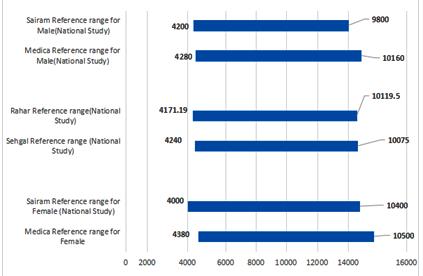
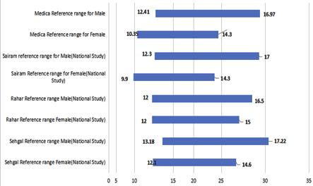
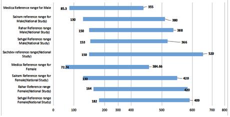
1Ray BK, Paul N, Hazra A, Ghosal MK, Ray J, Choudhury D, et al — Prevalence, burden, and risk factors of migraine: a community-based study from Eastern India. Neurology India 2017; 65(6): 1280-8.
2Gupta R, Bhatia MS — Migraine prevalence in a tertiary care hospital. Neurology India 2015; 63(3): 382-6.
3Lakshmi BV, Kameshwar Prasad G, Ravishankar K — Clinical profile of headache in a tertiary care referral centre in South India.
Hony Editor, JIMA Sanjoy Banerjee
Original Article
Post COVID-19 Rhino-Orbito-Cerebral Mucormycosis : Retrospective Clinical Observational Study & Analysis of the Patients Presenting in Kheda District, Gujarat, India
Background : Mucormycosis is a rare, life threatening fungal infection having an increased incidence during this COVID-19 pandemic, especially in the second wave in India. The state of Gujarat leads in the number of rhino-orbitocerebral mucormycosis cases Post COVID-19 infection.
Aims and Objectives : Rhino-orbito-cerebral fungal infections are being reported as a post COVID-19 sequelae. This observational study explores correlation between mucormycosis, diabetes-mellitus and corticosteroid therapy, with the aim to understand disease pattern, predisposing factors, presenting features and outcomes with surgical and anti-fungal therapy.
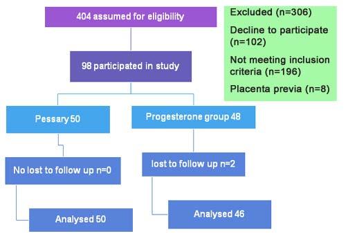
Materials and Methods : This retrospective clinical analysis includes data collection of 50 patients from an Otorhinolaryngology hospital located in Kheda district of Gujarat, India from 1st April, 2021 to 31st May, 2021. All these were post COVID-19 patients presented after varying number of days postinfection and had undergone indoor treatment at various hospitals.
Results : Of the 51 patients, 14 were from Nadiad itself, rest were from peripheral areas and aged between 20 and 75 years. All the patients had diabetes mellitus Pre-COVID except one and majority underwent corticosteroid medications and supplemental oxygen therapy during COVID treatment. Mucormycosis infection was observed with palatal involvement in 26 patients (50.98%) and 10 patients (19.60%) with eye involvement.
Conclusion : Close correlation was observed between invasive rhino-orbito-cerebral mucormycosis, diabetes mellitus and corticosteroids administration in COVID-19 positive patients. Possible follow up and larger sample size will be needed to justify this results more.
[J Indian Med Assoc 2024; 122(5): 14-8]
Key words :COVID-19 Sequelae, Mucormycosis, Diabetes Mellitus, Corticosteroids, Black Fungus.
Assessment of placement of human pathogen named 2019-nCoV was done by Coronaviridae Study Group (CSG) which then classified viruses and taxons of the Coronaviridae family. CSG recognizes that this virus forms a clade to human prototype and bat causing Severe Acute Respiratory SyndromeRelated Coronavirus (SARS-CoV-2) on the basis of phylogeny and taxonomy.
1MBBS, MS (ENT) Associate Professor, Department of Otorhinolaryngology, Dr N D Desai Medical College & Hospital, Gujarat 387003 and Corresponding Author
Viral infections like COVID-19 are akin to immunosuppressive states making the patient more prone for superadded infections like Mucormycosis.
Strict Control of Diabetes & early surgical intervention in the patient of Mucormycosis resulted in better patient outcomes. Besides Lyophilized Amphoterin the newer Anti Fungals like Posaconazole & Isavuconazole exhibited equally good efficacy in treating patients of Mucormycosis with lesser side effects & cost.
Dominant pandemic SARS-CoV-2 associated pneumonia, stroke, kidney dysfunctions and vascular thrombosis had afflicted and succumbed more than millions of people worldwide in few years. Recent viral storm in India noticed severe devastating co-infection named Mucormycosis, a “Black fungus” caused by Mucorales species in patients who are recovering from COVID-192. Duration of time in which this infection occurred was couple of days to weeks from COVID recovery. Involvement of maxillofacial area has led to worse outcome during a deep COVID crisis in immunocompromised patients with uncontrolled diabetes and subsequent corticosteroid therapy2-4. Tissue necrosis is the sign of mucormycosis5 along with severe pain,
Vol 122, No 05, May 2024Journal of the Indian Medical Association
necrotic ulcer at palate, ocular swelling, visual problems as blurring and/or loss of vision, cough and shortness of breath are all associated clinical signs and symptoms2,6. Lethality was increased with rhinocerebral (brain and sinus) involvement7.
Alarming rise in mucormycosis cases in postCOVID-19 phase has stressed to think some triggering causative factors beyond the steroid use and immunocompromised status by diabetes mellitus or other diseases8. Diagnosis by MRI, superintendence of contributory factors, Functional Endoscopic Sinus Surgery (FESS)5, conservative management by antifungal drugs and surgical debridement are the best treatment protocol2. The aim is to analyse 51 cases of mucormycosis for correlating between causative factors to help clinicians workout through the evolving disease pattern, considering the poor prognosis of the disease and its short time spread.
MATERIALS AND METHODS
This is a retrospective, uni-centric study of 51 cases of rhino-cerebro-orbital mucormycosis treated between April till June and were followed up till the end of July, 2022. The study followed compliance with Helsinki statement and exemption was made due to its retrospective nature. Standard informed consent to participate and publish were obtained for every patient.
All these patients were treated at ENT hospital in Nadiad, Gujarat, India. Maxillary sinusitis, headache, necrosis of palatal bone/mucosa or acute loss of vision are some of the common complaints patients get presented with. All but five of the total patients were known cases of previous COVID-19 infection. Every patient was treated with corticosteroids as a part of a standard COVID-19 drug regime. Exclusion criteria had patients with history of chemotherapy, granulocytopenic patients, radiotherapy, history of medication-related osteonecrosis of jaw, osteoradionecrosis or those on other immuno-modulator drugs.
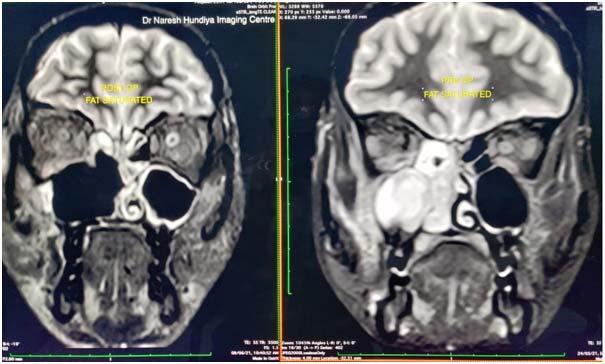
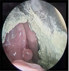
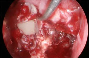
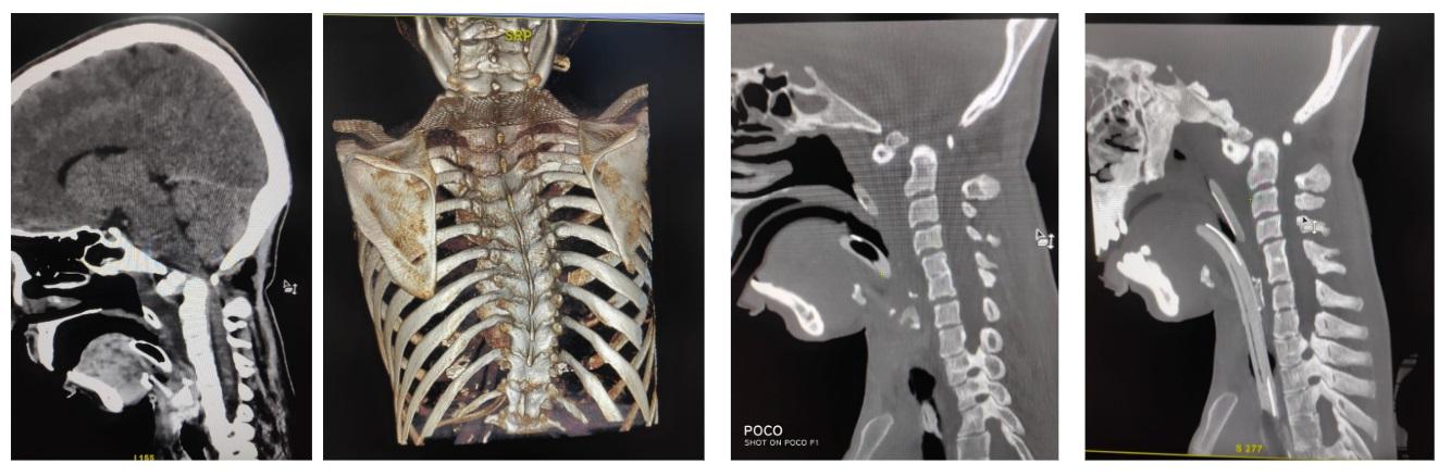
Routine blood investigations, ECG, chest Xray, CT scan and MRI of the face (including orbits) and brain were done (Fig 1).
Sinusitis with mucosal thickening of ethmoidal and maxillary sinuses, facial swelling, sudden dental pain
and teeth mobility, headache, ophthalmoplegia, epiphora, edema of extraocular muscles, orbital cellulitis, etc were few clinical features that the patients presented with. Ophthalmology reference was taken for those patients having orbital extension.
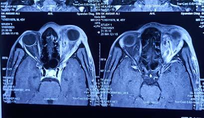
Punch biopsy from the oral cavity especially the necrotic palatal part and/or collection of infected tissue/ nasal discharge is/are sent for (KOH test) to screen for any fungal hyphae. KOH test is a rapid method of identifying the presence of fungal hyphae or yeast compared to the fungal culture which takes weeks for reporting and thus was done for all our patients. Histopathologic examination as well as fungal and bacterial culture were also done from the tissue samples. From surgical debridement to maxillectomy (partial, subtotal, total) till orbital exenteration, the surgical intervention varies depending on the orbital involvement.
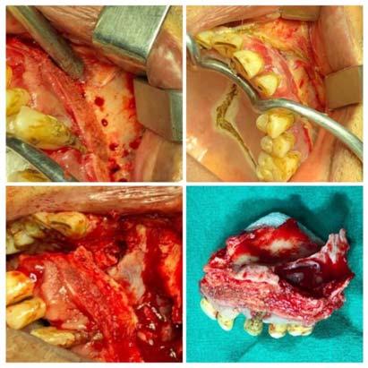
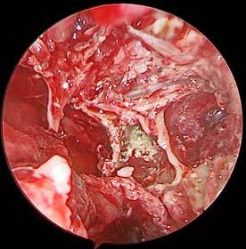
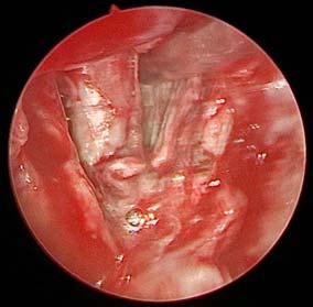
Once Mucormycosis was confirmed, within 72 hours, surgery was performed as well (Fig 2). Post surgery, either Amphotericin B therapy was instituted if it was available and if it was not available then either orally Posaconazole (200 mg q8h) was administered or Isavuconazole at a loading dose of 372 mg orally every 8 hours and maintenance dose of 372mg orally once a day. Inj Amphotericin B was administered at 35 mg/kg. Daily renal function tests had to be performed due to possible nephrotoxicity of Amphoterecin B. Those who could afford, liposomal Amphotericin B injections were given which had lesser renal complications. Alteration in dosage was adjusted according to renal tolerance. Amphotericin B was not available in private clinic/hospitals but only available in government hospitals. Those who were ready to get
Fig 1 — Pre-operative and Postoperative coronal MRI slice
admitted in Government hospital for further treatment were sent to Government hospitals for Amphotericin B while rest others were subject to oral medicaments.
RESULTS
Data was collected from ENT Hospital indoor and outdoor case records. Of the 51 patients, 14 were from Nadiad itself, rest were from peripheral areas and aged between 20 and 75 years with a mean age of 49.74 years. This study was performed from the start of April 2021, with most of the patients seen and operated in May and few in June and they all were followed up till July end. In the study, there were 43 male patients while eight were female patients. Out of the 51 patients in the study, 40 (78.43%) patients had diabetes mellitus pre-covid and all the diabetic patients underwent steroid therapy. Supplemental oxygen was provided to 24 (47.05%) patients. For those having involved maxillary sinus, FESS was performed; those having palatal or alveolar bone involved, thorough debridement / maxillectomy was performed while those having eye involvement, orbital exenteration was performed.
Fortynine patients (96.01%) presented with
involvement of maxillary sinus. Mucormycosis infection was observed with palatal involvement in 26 patients (50.98%) and 10 patients (19.60%) with eye involvement. Of the 26 patients having palatal bone involvement, 23 patients have undergone maxillectomy. Three patients developed intracranial extensions. All but two patients having mucormycosis, received steroid therapy during the COVID treatment. By the end of July till the last follow up, it was observed that 12 patients were deceased out mainly due to other causes like six patients refused treatment, two patients died due to brain haemorrhage, one patient died due to abdominal distension, 2 patients had sudden cardiac arrest, one had hemoptesis due to hypovolemia. Of the 39 surviving, 32 are disease free while seven patients had been showing some osteomyelitic changes and are currently undergoing the therapy but are medically doing fine.
DISCUSSION
Mucormycosis is a aggressive and lethal infection caused by Mucoraceae belonging to class of Zygomycetes9,10. The fungus has a likeliness to affect the nasal mucosa and mostly seen in immunosuppressive conditions like diabetes, ketoacidosis, solid organ transplant, severe burns, etc. Germination is seen in the nasal and paranasal sinuses ultimately involving the palate, orbit and brain causing death11 Mucormycosis in patients can be also contributed to excessive and long term use of steroids12
Decrease of phagocytic function, diabetic ketoacidosis and fungal heme-oxygenase elevating iron uptake for metabolism are some pathogenic mechanisms responsible for aggression of the fungal disease13.
There have been few case reports published presenting co-relation between COVID-19, diabetes and steroids14-17. Moorthy published a case study having 18 patients which is by far, the largest case series on role of COVID-19, steroid and uncontrolled diabetes causing mucormycosis in 2021.4 Of the 18 patients, 16 patients received steroids. Out of these 16 patients, 15 were diabetic as well. Blindnesss truck
Fig 2 — Collage representing resection with debridement of the maxillary lesion and lastly providing an obturator
12 of the 18 patients and orbital exenteration was carried out in seven patients. The fungi in 16 patients was noted as mucormycosis, one patient had as per gillosis while one patient had a mixed fungal infection. Eleven of the patients survived, six died and one got lost to follow-up.
Basically, in susceptible hosts, standard defense mechanisms slows down. Suppose, in diabetic ketoacidosis, the serum pH is acidic causing dissociation of free iron from the sequestering proteins. The liberation of free iron leads tofaster fungal growth. Further invasion of the fungus is caused by mechanisms like neutropenia or functional defects because of corticosteroids or hyperglycemia or acidosis due to diabetic ketoacidosis. Sequentially, adherence and damage to the endothelial cells because of fungus allows fungal angioinvasion and thrombosis of vessels causing necrosis of tissue by fungal infection18
In India till May 2020, 66.8% of the COVID-19 cases were males19. Rhino-cerebro-orbital (44-49%) was the type most commonly found, followed by cutaneous (10-19%), pulmonary (10-11%), disseminated (6-11%) and gastrointestinal (2-11%)20. But in our case study, all the cases occurring with COVID-19, diabetes and steroids led to rhino-cerebro-orbital mucormycosis. Patients usually presented with headache, fever, unilateral facial swelling, orbital cellulitis with the presence of palpebral oedema, chemosis, ptosis and ophthalmoplegia21. The prognosis is poor with about 33.3%-80% being the overall mortality rate21,22. CT scan is usually the first diagnostic tool to check the status of sinuses, although best way to detect extrasinus spread is using Magnetic Resonance Imaging (MRI)9. A definitive diagnosis of mucormycosis as the causative species is achieved only by histological examination of the biopsy specimen. Culture and KOH examination may be used only as a suggestive tool for noting the presence of mucormycosis.
We feel the acute increase to be due to infection with COVID-19 contributing in more than one way. Firstly, the reduced numbers of T lymphocytes, CD4 + T and CD8 + T cells suggestive of immune dysregulation may alter innate immunity leading to secondary fungal infections 23 . Secondly, the pathogenesis of COVID-19 kind of mimics the spectrum of Thrombotic Microangiopathies (TMA) leading to angioinvasion and endothelial damage much like that of mucormycosis, aggravating the disease24. Thirdly, glucocorticoids have been used extensively to reduce hospital stay and mortality related to COVID19. In most of the protocols fortreating moderate to
severe cases of COVID-19 infection, Dexamethasone and methylprednisolone have both been used25, 26 Due to the immuno-suppressive nature of glucocorticoids, patients become more susceptible to secondary infections.
Due to the high mortality rate, need of early intervention by aggressive surgical debridement, systemic antifungal medications and management of underlying illness are much essential for a better rate of survival. A standard blanket protocol of steroid administration for COVID-19 infection may need to be evaluated again and an emphasis on strict blood sugar control during and after COVID-19 infection should be put.
Both central ciliary and retinal artery occlusion can be caused by mucormycosis27. Orbital exenteration can be done to reduce the disease burden and prevent the intracranial spread in cases where blindness is delayed even if having a clear radiological picture of involvement of the orbital cavity. Some cases even mandate partial or total maxillectomy. Repeated surgical debridement may be needed for local control of the disease and an aggressive surgical approach seems to improve patient survival. It is important to note that once the diagnosis is suspected, all immunosuppressive therapy should be reduced or discontinued if it is possible and Amphotericin B should be therapeutically started28
CONCLUSION
This clinical observational survey includes few points to be considered in priority are possible avoidance of glucocorticoids in mild COVID-19 cases (without hypoxemia) or maintained doses of glucocorticoids in critical cases of COVID. Antifungal medications, surveillance of immuno-compromised status of diabetes mellitus in all patients, screening of COVID-19, early diagnosis of fungal co-infection, focus on triggering contributor factors and timely required treatment are a valuable means to control disease and its severe outcome.
Funding :
The authors did not receive support from any organization for the submitted work.
Compliance with Ethical Standards :
The authors have no conflicts of interest to declare that are relevant to the content of this article.
Informed consent had been signed by all the patients but this being a retrospective study, it doesn’t require ethical approval.
No new research has been done on any human beings or animals.
Vol 122, No 05, May 2024Journal
REFERENCES
1Coronaviridae Study Group of the International Committee on Taxonomy of Viruses. The species Severe acute respiratory syndrome-related coronavirus: classifying 2019-nCoV and naming it SARS-CoV-2. Nat Microbiol 2020; 5: 536-44. https:/ /doi.org/10.1038/s41564-020-0695-z
2Sen M, Lahane S, Lahane TP, Parekh R, Honavar SG — Mucor in a Viral Land: A Tale of Two Pathogens. Indian J Ophthalmol 2021; 69(2): 244-52. doi: 10.4103/ ijo.IJO_3774_20.
3Anna Skiada — Review Epidemiology and Diagnosis of Mucormycosis: An Update. J Fungi 2020; 265(6): 1-20.
4Moorthy A, Gaikwad R, Krishna S, Hegde R, Tripathi KK, Kale PG, et al — SARS-CoV-2, Uncontrolled Diabetes and Corticosteroids-An Unholy Trinity in Invasive Fungal Infections of the Maxillofacial Region? A Retrospective, Multi-centric Analysis. J Maxillofac Oral Surg 2021; 20(3): 1-8. doi: 10.1007/s12663-021-01532-1
5Maini A, Tomar G, Khanna D, Kini Y, Mehta H, Bhagyasree V — Sino-orbital mucormycosis in a COVID-19 patient: A case report. Int J Surg Case Rep 2021; 82: 105957. doi: 10.1016/ j.ijscr.2021.105957.
6Sarkar S, Gokhale T, Choudhury SS, Deb AK — COVID-19 and orbital mucormycosis. Indian J Ophthalmol 2021; 69: 1002-4.
7Balai E, Mummadi S, Jolly K, Darr A, Aldeerawi H — Rhinocerebral Mucormycosis: A Ten-Year Single Centre Case Series. Cureus 2020; 12(11): e11776. DOI 10.7759/ cureus.11776
8Garg D, Muthu V, Sehgal IS, Ramachandran R, Kaur H, Bhalla A, et al — Coronavirus Disease (COVID-19) Associated Mucormycosis (CAM): Case Report and Systematic Review of Literature. Mycopathologia 2021; 186(2): 289-98.
9Ferguson BJ — Mucormycosis of the nose and paranasal sinuses. Otolaryngol Clin North Am 2000; 33(2): 349-65.
10Uçkay I, Chalandon Y, Sartoretti P, Rhoner P, Berney T, Hadaya K, et al — Invasive zygomycosis in transplant recipients. Clin Transplant 2007; 21(4): 577-82. doi: 10.1111/j.13990012.2007.00684.x
12Gonzalez BDG, Garaa R, Gil F — Mucormycosis of head and neck:report of five cases with different presentations. J Cranio Maxillo Facial Surg 2012; 40: 584-91. doi: 10.1016/ j.jcms.2011.10.015.
14Mekonnen ZK, Ashraf DC, Jankowski T — Acute invasive rhino-orbitalmucormycosis in a patient withCOVID-19associated acute respiratory distress syndrome. Ophthalmic Plast Reconstr Surg 2021; 37(2): e40-e80. Doi:10.1097/ IOP.0000000000001889.
15Mehta S, Pandey A — Rhino-orbital mucormycosis associated with COVID-19.Cureus 2020; 12(9): e10726. doi:10.7759/ cureus.10726
16Werthman-Ehrenreich A — Mucormycosis with orbital compartment syndrome in a patient with COVID-19. Am J Emerg Med 2021; 264. e5-264.e8.Doi:10.1016/ j.ajem.2020.09.032
17Waizel-Haiat S, Guerrero-Paz JA, Sanchez-Hurtado L, CallejaAlarcon S, Romero-Gutierrez L — A Case of Fatal RhinoOrbital Mucormycosis Associated With New Onset Diabetic Ketoacidosis and COVID-19. Cureus 2021; 13(2): e13163. DOI 10.7759/cureus.13163.
18Spellberg B, Edwards Jr J, Ibrahim A — Novel perspectives on mucormycosis:pathophysiology, presentation, and management. Clin Microbiol Rev 2005; 18(3): 556-69. https:/ /doi.org/10.1128/CMR.18.3.556-569.2005.
19Chanda A — COVID-19 in India: transmission dynamics, epidemiological characteristics, testing, recovery and effect of weather. Epidemiol Infect 2020; 148: e182.doi:10.1017/ S0950268820001776.
20Arnaiz-Garcýa ME, Alonso-Pena D, Gonzalez-Vela MC, GarcýaPalomo JD, Sanz-Gimenez- Rico JR, Arnaiz-Garcýa AM — Cutaneous mucormycosis: report of five cases and review of the literature. J Plast Reconstr Aesthet Surg 2009; 62(11): e434-41.
21Scheckenbach K, Cornely O, Hoffmann TK, Engers R, Bier H, Chaker A, et al — Emerging therapeutic options in fulminant invasive rhinocerebral mucormycosis. Auris Nasus Larynx 2010; 37(3): 322-8.
22Jung SH, Kim SW, Park CS, Song CE, Cho JH, Lee JH, etal— Rhinocerebral mucormycosis: consideration of prognostic factors and treatment modality. Auris Nasus Larynx 2009; 36(27): 274-9.
23Gangneux JP, Bougnoux ME, Dannaoui E, Cornet M, Zahar JR — Invasive fungal diseases during COVID-19: we should be prepared. J Mycol Med 2020; 30: 100971. https://doi.org/ 10.1016/j.mycmed.2020.100971
24Sweeny JM, Barouqa M, Krause GJ, Gonzalez-Lugo JD, Rahman S, Gil MR — Evidence for secondary thrombotic microangiopathy in COVID-19. medRxiv preprinthttps:// doi.org/https://doi.org/10.1101/2020.10.20.20215608.
25Government of India Ministry of health and family welfare Directorate general of health sciences (2020). Clinical management protocol for COVID-19. https:// www.mohfw.gov.in/pdf/Clinical Management Protocolfor COVID19. pdf.Accessed: July7, 2020
26The Recovery Collaborative Group.Dexamethasone in Hospitalized Patients with COVID-19.N Engl J Med 2021; 384(8): 693-704. http://doi.org/https://doi.org/10.1056/ NEJMoa2021436
27Luo QL, Orcutt JC, Seifter LS — Orbital mucormycosis with retinal and ciliary artery occlusions. Br J Ophthalmol 1989; 73(8): 680-3. https://doi.org/10.1136/bjo.73.8.680
28Spellberg B, Walsh TJ, Kontoyiannis DP, Edwards J Jr, Ibrahim AS — Recent advances in the management of mucormycosis: from bench to bedside. Clin Infect Dis 2009; 48(12): 1743e1751.
Original Article
A Prospective, Observational Study of Serum Triglyceride and Cholesterol Level as Markers of Dengue Severity in Children in a Tertiary Care Hospital
Some Suvra Bose1, Soumyakanti Panda2, Manas Kumar Mahapatra3, Amit Kumar Das4
Background : Dengue related morbidity and mortality results from shock and hemorrhagic manifestations that occur predominantly in critical phase of illness due to severe capillary leakage. Several clinical and laboratory parameters have been studied to predict possibility of Severe Dengue. This study aims to evaluate serum cholesterol and triglyceride level as markers of Dengue severity in children.
Materials and Methods : This prospective observational study includes confirmed cases of dengue infected children of 1 month to 12 years old. Cases were grouped as Dengue Fever without Warning Signs (DF-WS), Dengue Fever with Warning Signs (DF+WS) and Severe Dengue (SD) fever. Serum triglyceride, Cholesterol and other relevant investigations were recorded with changing clinical severity and at recovery stage.
Result : Eighty four children were included in the study of which 53% belongs to 5 to 10 years age group. Mean duration of fever was 4.4 days. Eleven children (13.1%) were admitted in a critical stage. We had 37(44%), 44(52.4%), 3(3.6%) children with DF+WS, DF-WS and SD respectively at first evaluation, which subsequently progressed to 43(51.1%), 30(35.1%), 9(10.7%) children respectively with 2 death (2.3%). We noticed fall in mean serum cholesterol level in SD (Mean=104.9mg%) compared to D+WS (Mean=140.6 mg%) and DS-WS (Mean=158.4 mg%). However, triglyceride level increased in SD (Mean=214 mg%), compared to D+WS (Mean=97.6 mg%) and D-WS (Mean=60.6mg%).
Conclusion : Decreasing serum cholesterol and increasing triglyceride values can be taken as a surrogate marker of Dengue severity along with the clinical severity classification. [J Indian Med Assoc 2024; 122(5): 19-23]
Key words :Dengue in Children, Lipid Profile in Dengue, Triglyceride in Dengue, Cholesterol in Dengue.
Dengue is a mosquito born disease caused by Dengue virus (Flavivirus) which affects people globally across all age groups with a seasonal outbreak. About 5 million people Worldwide gets affected by Dengue infection annually, of which approximately five thousands Dengue related deaths reported in the year 2023 from 80 countries/territories and five WHO regions: Africa, Americas, South-East Asia, Western Pacific and Eastern Mediterranean regions 1,2 . Dengue illness has varied clinical presentations starting from undifferentiated Dengue fever, Classical Dengue fever, and Dengue hemorrhagic fever to Dengue shock syndrome 3. WHO (2009) classified dengue cases as per clinical severity as Dengue Fever without Warning Signs (DF-WS),
Department of Pediatric Medicine, Dr B C Roy Postgraduate Institute of Paediatric Sciences, Kolkata 700054
1MD, Associate Professor and Corresponding Author
2DNB, Senior Resident
3DNB, Senior Resident, Department of Pediatric Medicine, Medical College, Kolkata 700073
4MD, Professor
Received on : 10/01/2024
Accepted on : 01/02/2024
Editor's Comment :
The success of Dengue fever management lies on early detection of risk factors and judicious fluid management as till today there is no definite chemotherapeutic agents.
Dengue Fever with Warning Signs (DF+WS) and severe Dengue fever (SD)4. Mortality in Dengue illness increases when fatal complications like hemorrhagic manifestations and shock develop due to immune mediated vascular damage and altered permeability along with thrombocytopenia, coagulopathy and multi organ dysfunction precipitate. The success in the management of complicated dengue lies on its early detection and initiation of proper therapy. Several biochemical and radiological abnormalities can pick up the early signs of critical phase of Dengue fever before its clinical manifestation. Cholesterol and triglyceride play an important role in the pathophysiology of Dengue fever right from its receptor mediated cellular entry, virus multiplication to fresh invasion to a new target host cell5,6. Thus alteration in serum cholesterol and triglyceride is expected in critically ill Dengue patients and probably surrogate
Vol 122, No 05, May 2024Journal of the Indian Medical Association
markers for impending adverse outcome. Various studies have found that total cholesterol level decreases in critical phase of Dengue patients7-9 Biswas, et al observed that every 10 mg/dl drop in serum cholesterol and LDL in Dengue patient since admission risk the development of DHF and DSS by about 9% and 12% respectively 10 . Alteration in triglyceride level is not uniform among the previous studies. There are paucity of studies in children with Severe Dengue and altered cholesterol and triglyceride level. So the aim of our study is to determine the correlations between serum cholesterol and triglyceride level with Dengue severity in a child suffering from Dengue illness. Alteration in serum cholesterol and triglyceride levels can be taken as an additional marker for Dengue severity so that early intervention can be planned.
MATERIALS AND METHODS
This was a prospective study conducted at Dr B C Roy Postgraduate Institute of Pediatric Sciences from 1st September, 2020 till 30th November, 2021 amid COVID-19 pandemic and covering 2 monsoons at Kolkata. The monsoon was chosen as there is an annual surge during this time at Kolkata, but during 2020 due to COVID-19 outbreak and nationwide closure of schools we got very minimum cases, whereas we got most of the cases of our study from monsoon of 2022 and also we extended our study period from 12 months to 14 months. Written informed consent was obtained from parents willing to participate in the study group after obtaining approval from Institution Ethics Committee (IEC,memo no: BCH/ME/PR/2960 dated 09/12/2020). We have strictly excluded COVID-19 patients from our study group and they had a separate isolation ward.
Inclusion Criteria :
All the febrile children attending OPD and admitted at IPD with following —
(1)Diagnosed as a confirmed case of Dengue as per WHO 2009 case definition4
(2)Age : 1 month to 12 years.
Exclusion Criteria :
(1)Children with pre-existing liver diseases & nephrotic syndrome.
(2)Children with prior documented abnormal lipid profile.
(3)Children with history of familial dyslipidemia.
(4)Children with COVID-19 infection or any other co-infections like Malaria, Typhoid, Scrub to minimize bias factor.
OBSERVATION
All admitted children satisfying inclusion criteria were included in the study. Dengue infection diagnosed by serological test (NS1 and IgM by Mac ELISA) as per WHO guideline and COVID-19 by (RAT or RT-PCR for SARS COVID-19). Demographical profile of all subjects enrolled was recorded in a self-made printed data collection sheet. On first contact clinical evaluation and necessary investigations done. Blood sent for CBC, LFT, Lipid profile, Chest X-ray and USG abdomen as required case to case and recorded in data sheet. Dengue cases were grouped as per WHO 2009 clinical severity classification guidelines as Dengue Fever without Warning Signs (DF-WS), Dengue Fever with Warning Signs (DF+WS) and Severe Dengue (SD) fever4. They were managed as per national Dengue management protocol. Serum lipid and other relevant investigations repeated with changing clinical severity and finally at recovery phase. So, we recorded three sets of investigational reports along with total duration of hospital stay with clinical outcome.
All data were plotted in Microsoft office 365 excel sheet for statistical representation. Categorical variables are expressed as number of patients and percentage of patients. Mean and SD was used for categorical variables. Chi-square test used to assess the strength of association between Cholesterol and Triglycerides with Dengue severity. The statistical software SPSS version 26 has been used for the analysis. Data representation and result analysis done using proper statistical methods. P value <0.05 was considered statistically significant
RESULTS
Out of 7974 children got hospitalized during our study period, 84 (1.05%) children met the inclusion criteria for Dengue illness and were selected for our study. We found that 5 to 10 years age group children (n =45; 53%) were mostly affected by Dengue illness of the cohort of 84 children. Sex ratio of the entire study population was Boys : Girls = 45:39. We recorded maximum admission from rural area 45 (53.6%), followed by urban slum 26 (31%) and urban/suburban 13 (15.5%). Mean duration of fever was 4.4 days.
Out of total 84 subjects, 73 (86.9%) got admitted in febrile stage and 11 (13.1%) was admitted in a critical stage; which were further distributed as per WHO clinical severity. Thus, at the time of first evaluation we had 37 (44%), 44 (52.4%), 3 (3.6%) children with DF+WS, DF-WS and SD respectively. Among the children admitted in febrile stage (n=73), 29 (39.7%)
122, No 05, May 2024Journal
had warning sign (DF+WS) and out of 11 children admitted in critical stage 3 (27.3%) had Severe Dengue (SD)(Table 1). The baseline serum cholesterol level at the time of admission among children admitted in febrile stage was recorded as 152.5 mg%, 160 mg% and 135.8mg% respectively among D+WS, D-WS, SD. The serum triglyceride was 62mg%, 57.1mg% and 92.2 mg% respectively among D+WS, D-WS and SD (Tables 2 & 3).
The clinical distribution according to disease severity changed subsequently as the disease progressed. Amongst the children with D+WS, 6 (6/ 37; 16.2%) were developed Severe Dengue and out of 44 children with D-WS, 2 (4.5%) developed Severe Dengue and 12 (27.3%) developed warning signs. Hence, the distribution changed to 43 (51.1%), 30 (35.1%), 11 (13.1%) children with DF+WS, DF-WS and SD respectively. Two (2/84; 2.3%) children with Severe Dengue died in critical stage (Table 1).
We recorded mean serum cholesterol and triglyceride level among these 3 clinical group during critical stage and found that there was significant dip in mean serum cholesterol level in Severe Dengue (Mean= 104.9mg%) group compared to D+WS (Mean=140.6 mg%) and DS-WS (Mean=158.4 mg%). But the changes in triglyceride level among the 3 clinical groups was opposite and the mean triglyceride level increased to maximum level among children with SD (Mean=214 mg%), compared to D+WS (Mean=97.6 mg%) and D-WS (Mean=60.6 mg%).
Table 1 — Demography clinical severity, and outcome of Dengue patients (n=84)
FactorsFrequency
GenderMale: n (%)45 (55.6%)
Female: n (%)39 (46.4%)
Age1-4 years: n (%)32 (38%)
5-10years: n (%)45 (53%)
> 11 years: n (%)7 (9%)
Locality Urban: n (%)13 (15.6%)
Urban slum: n (%)26 (31%)
Rural: n (%)45 (53%)
Duration of fever (days): mean (sd)4.4 (0.8)
Clinical stageFebrile stage: n (%)73 (86.9%) at admissionCritical stage: n (%)11 (13.1%)
Clinical severity at theD+WS: n (%)37 (44%) time of admissionD-WS: n (%)44 (52.4%)
SD: n (%)3 (3.6%)
Clinical severity at theD+WS: n (%)43 (51.1%) time of dischargeD-WS: n (%)30 (35.1%) (excluding 2 deaths)SD: n (%)9 (10.7%)
Duration of hospital stay (days): mean (sd)7.4 (1.3)
Mortality: n (%)2 (2.3%)
D= Dengue; WS= Warning Sign; SD= Severe Dengue; sd = standard deviation
Table 2 — Serum cholesterol level at various stages of Dengue illness (n=84)
Analysis of serum Cholesterol at Febrile stage of Dengue illness
ClinicalNumber Meansd Maximum Minimum Median stage (mg/dl) D+WS37 152.512178134155 D-WS301608.7180137160 SD6135.86.6147128135
Analysis of serum Cholesterol at Critical stage of Dengue illness
ClinicalNumber Meansd Maximum Minimum Median stage (mg/dl)
Table 3 — Serum Triglyceride level at various stages of Dengue illness (n=84)
Analysis of serum Triglyceride at Febrile phase of Dengue illness
ClinicalNumber Meansd Maximum Minimum Median stage (mg/dl)
Analysis of serum Triglycerides at Critical phase of Dengue illness
ClinicalNumber Meansd Maximum Minimum Median stage (mg/dl) D+WS43 97.6281864193 D-WS30 60.617903564 SD11214.671.830492236
Analysis of serum Triglyceride at recovery phase of Dengue illness
ClinicalNumber Meansd Maximum Minimum Median stage (mg/dl)
Following recovery we again recorded serum cholesterol and triglyceride among the 3 groups and found serum cholesterol (Mean=133.2 mg%) and triglyceride (Mean=153.4 mg%) level restored to normal values in Severe Dengue group. (Table 2 &3) Mean duration of hospital stay was 7.4 days. Two children died from Dengue Shock Syndrome (DSS) and multi organ dysfunction and rest all recovered.
DISCUSSION
In the present study 84 (1.05%) children got enrolled out of 7974 children admitted in pediatric ward during
122, No 05, May 2024Journal
the study period of 15 months, which was much less compared to previous years, probably due to COVID19 pandemic, which caused prolonged school closure and restricted public gatherings and events with enhanced health consciousness. Thus there was significant drop in infectious diseases. Children mostly affected belonged to 5-10 years age group which was quite similar to another study from Eastern India by Purakait R, et al11. The male : female ratio of our study population was 1.15:1 closely matching with another study by Nayak R, et al from Odisha12, however, in another study by Prabhuraj A et al. the female patients outnumbered male patient with a sex ratio of M:F:: 0.7:113. The knowledge of gender demography in disease epidemiology is helpful in executing public health prevention programs14,15. We found that 45 children got admitted from rural area which is a reversal of previous concept that Dengue is an urban-centric disease and Nayak, et al. At Odisha also had a similar observation. This paradigm shift may be due to rapid urbanization of rural areas.
In our study, 73 patients (86.9 %) got admitted in febrile stage of Dengue and 11 (13.1%) were in critical stage of illness. Among 84 patients, 43 (51.1%) had Dengue Fever with Warning Sign (DF+WS), 30 (35.1%) had Dengue Fever without Warning Sign (DFWS) and 11 (13.1%) had Severe Dengue (SD) infection (Table 1). A study from Odisha in 2016 by Nayak, et al showed out of 97 total hospitalized cases, 84 (86.59%) were non severe and 13 (13.40%) were Severe Dengue12. But in our study dengue without warning sign cases were much less in comparison to their study which may be due to pandemic situation where only serious dengue cases sought hospital admission.
In our study the mean (sd) serum cholesterol level at febrile stage was around the normal range in all clinical groups which was 152.5(12), 160 (8.7), 135.8 (6.6) mg/dl respectively among Dengue Patients with Warning Sign (D+WS), without Warning Sign (D-WS) and Severe Dengue (SD). But with changing severity serum cholesterol level drops in critical stage of illness and the mean value was 140.6 (13.4), 158.4 (4.7), 104.9 (10.7) mg/dl among dengue with warning sign (D+WS), Dengue without Warning Sign (D-WS) and Severe Dengue (SD) fever respectively. The drop is highest in patients with Severe Dengue and also the 2 unfortunate children who succumbed. With recovery serum cholesterol level returned to normal range with a slow recovery in Severe Dengue group and the mean (sd) value was 147.7 (13.9), 159.8 (7.9), 133.2 (9.3) mg/dl among Dengue with Warning Sign (D+WS), without Warning Sign (D-WS) and Severe Dengue (SD) fever respectively (Table 2).
On the contrary, serum triglyceride level which was near normal range at the time of first contact, increased 2 to 3 times during critical stage in patients with Severe Dengue and Dengue with Warning Signs. The mean (sd) value of serum triglycerides was 62(13.7), 57.1(12.5), 92.2(25.4) among D+WS, D-WS and Severe Dengue respectively at admission, which increased to 214.6 (71.8) in Severe Dengue group during critical stage with death of 2 children (Table 3).
A meta-analysis of seven studies by Lima WG, et al, showed that total cholesterol and LDL were significantly lowered in severe grade of DHF16. But they didn’t found similar association with other lipid components like Triglycerides, HDL, VLDL whereas the present authors found that serum triglycerides level increases with Dengue severity. The exact mechanism behind this disproportionate lipid components alteration with Dengue severity is not clear, but few research work postulates that damage to liver cells in severe dengue is responsible for lowered serum cholesterol along with increased cholesterol leakage due to enhanced capillary permeability 17,18. Many other studies suggest excessive lipid utilization by replicating Dengue virus19. Serum triglyceride level increases along with other inflammatory markers like Ferritin, LDH, IL-6 as it happens in other clinical conditions like MISC or MAS.
Dengue illness during monsoon is a clinical, challenge to treating pediatrician almost every year. The rapidity of clinical deterioration and sudden death provokes trepidation both to doctor and parents. Thus there is a requirement of early predictors of Severe Dengue which will alert and enable us for early meticulous intervention. Studies found that use of NO/ IL-6 as early predictors but they are not readily available in resource poor settings20. However, both serum cholesterol and triglyceride level estimation at admission and its serial progression will help early diagnosis of severe and deadly Dengue.
The strength of the study was, more number of serious ill patients incorporated in the study group, with single primary investigator reducing interpretation bias and all laboratory investigations were done in same institute with strict adherence to national guidelines of Dengue management.
Limitation(s) :
The weakness of the study is small sample size due to COVID-19 pandemic. This study didn’t correlate subtypes of cholesterol with Dengue severity and serum lipid levels with the outcome.
122, No 05, May 2024Journal of the Indian Medical Association
CONCLUSION
This study has demonstrated that serum cholesterol significantly decreases with severity of Dengue fever along with increase in serum triglyceride level which signals impending Dengue illness associated catastrophe.
Conflict of interest : None
Funding : None
Contribution : SP: concept of the study and design data collection; MKM: review of literature, data collection and statistical analysis; SSB: writing manuscript, statistical analysis, guarantor; AD: writing manuscript, editing, manuscript review.
Acknowledgement : We all acknowledge the contribution of all staffs of Department of Pediatrics BC Roy Hospital for facing the challenge during Dengue outbreak and COVID-19 pandemic. We sincerely solicit the co-operation of patient and patient party.
REFERENCES
1Brady OJ, Gething PW, Bhatt S, Messina JP, Brownstein JS, Hoen AG, et al — Refining the global spatial limits of dengue virus transmission by evidence-based consensus. PLoS Neglected Tropical Diseases 2012; 6(8): e1760.
2World Health Organization (21 December 2023). Disease Outbreak News; Dengue- Global situation Available at : https:/ /www.who.int/emergencies/disease-outbreak-news/item/ 2023-DON498.
3John DV, Lin YS, Perng GC — Biomarkers of severe dengue disease- A review. Journal of Biomedical Science 2015; 22(1): 83.
4World Health Organization. Regional office for South-East Asia[homepage on Internet]. New Delhi: Trend of Dengue cases and CFR in SEAR Countries. c2009 [cited 2012 October 23]. Available from:http://www.searo.who.int/en/Section10/ Section332/Section2277.html
5Gutsche I, Coulibaly F, Voss JE, Salmon J, Dalayer J, Ermonval M, et al — Secreted dengue virus nonstructural protein NS1 is an atypical barrel-shaped high-density lipoprotein. Proceedings of the National Academy of Sciences 2011; 108(19): 8003-08.
6Cui L, Lee YH, Kumar Y, Xu F, Lu K, Ooi EE, et al — Serum metabolome and lipidome changes in adult patients with primary dengue infection. PLoS Neglected Tropical Diseases 2013; 7(8): e2373.
7Heaton NS, Perera R, Berger KL, Khadka S, Lacount DJ, Kuhn RJ, et al — Dengue virus nonstructural protein 3 redistributes fatty acid synthase to sites of viral replication and increases cellular fatty acid synthesis. Proceedings of the National Academy of Sciences 2010; 107(40): 1734550.
8Mackenzie JM, Khromykh AA, Parton RG — Cholesterol Manipulation by West Nile Virus Perturbs the Cellular Immune Response. Cell Host & Microbe 2007; 2(4): 229-39.
9Soto-Acosta R, Mosso C, Cervantes-Salazar M, PuertaGuardo H, Medina F, Favari L, et al — The increase in cholesterol levels at early stages after dengue virus infection correlates with an augment in LDL particle uptake and HMGCoA reductase activity. Virology 2013; 442(2): 132-47.
10Biswas HH, Gordon A, Nunez A, Perez MA, Balmaseda A, Harris E — Lower low-density lipoprotein cholesterol levels are associated with severe dengue outcome. PLoS Neglected Tropical Diseases 2015; 9(9): e0003904. PMID:26334914.
11Purkait R, Basu R — The changing clinico-demographic profile of dengue infection in children: a hospital-based study from eastern India. Int J Community Med Public Health 2020; 7(5): 1901-6.
12Nayak R, Panda P, Padhy, P Mishra K G — Paradigm Shift in Socio-Demographic Profile of Dengue Infection: A Hospital Based Cross-Sectional Study. J Family Med Prim Care 2021; 10(6): 2405-10.
13Prabhuraj A, Kumaravel K, Rekha VA, Nithiyapriya A, Sampathkumar P, Anand MV — A study of serum lipid profile changes in children with dengue haemorrhagic fever and its correlation with severity in a tertiary care hospital. Journal of Clinical and Diagnostic Research 2020; 14(8): 10-3. https:/ /doi.org/10.7860/jcdr/2020/44837.13913
14Hammond SN, Balmaseda A, Pérez L, Tellez Y, Saborío SI, Mercado JC, et al — Differences in dengue severity in infants, children, and adults in a 3-year hospital-based study in Nicaragua. Am J Trop Med Hyg 2005; 73(6): 1063-70. PMID: 16354813.
15Dunham C, Fealk MH, Sever WE — Following severe injury, hypocholesterolemia improves with convalescence but persists with organ failure or onset of infection. Crit Care 2003; 7(6): R145.
16Lima WG, Souza NA, Fernandes SOA, Cardoso VN, Godói IP — Serum lipid profile as a predictorof dengue severity: A systematic review and meta-analysis. Rev Med Virol 2019; 29(5): e2056. https://doi.org/10.1002/rmv.2056.
17Lee LK, Gan VC, Lee VJ, Tan AS, Leo YS — Clinical relevance and discriminatory value of elevated liver aminotransferase levels for dengue severity. PLoS Negl Trop Dis 2012; 6: e1676. doi: 10.1371/journal.pntd.0001676 PMID: 22679523.
18Berg J, Tymoczko J, Stryer L — The complex regulation of cholesterol biosynthesis takes place at several levels. Biochemistry. New York: WH Freeman. 2002.
19Wills BA, Oragui EE, Dung NM, Loan HT, Chau NV — Size and charge characteristics of the protein leak in dengue shock syndrome. J Infect Dis 2004; 190: 810-18. PMID: 15272410.
20Valero N, Espina LM, Añez G, Torres E, Mosquera JA — Short report: increased level of serum nitric oxide in patients with dengue. The American Journal of Tropical Medicine and Hygiene 2002; 66(6): 762-64.
Original Article
Mortality amongst COVID-19 Patients in Relation to their Vaccination Status
Balaji Selvaraju1, Praveen Kumar M2, Lawrence P2
Background : The second wave of COVID-19 occurred in March, 2021 in India causing large numbers of severe infections and also the vaccination drive was started during January, 2021. Previous studies proved that various factors were associated with mortality
Aims and Objectives : This study was conducted with the objective of estimating the prevalence of mortality and also the factors associated with it, especially in relation to the vaccination status
Materials and Methods : A cross-sectional study was conducted in a tertiary care hospital among the COVID-19 patients during the months of June and July 2021. A semi-structured questionnaire was used containing the basic details about the patient and the relevant clinical details. A universal sampling method was employed and those patients who were below the age of 18 years were excluded. Data analysis was done using SPSS 21.
Results : A total of 226 patients were included in the study. Most of the study subjects were in the age group of >45% (73.5%) and the majority were males. Nearly 42% of them had any one of the co-morbidities and only 11.5% of them were vaccinated against COVID-19. The prevalence of mortality was 21.2% and the factors associated were age, co-morbidity status, duration of hospital stay, disease severity and the vaccination status.
Conclusion : Vaccination against COVID-19 had less risk of mortality even though other factors could influence it. Hence further research is needed to explore other factors that might affect both morbidity and mortality.
Key words :COVID-19, Mortality, Vaccination Status, Second Wave.
COVID-19 was declared a public health emergency by the World Health Organization because of its rapid spreading crossing the international border from Wuhan city of China to all other countries globally1,2 There occurs burden all over the world both in terms of health and wealth due to the ill effects of this disease causing significant mortality and morbidity2,3,10. Based on the WHO data the mortality rate during the pandemic varies from country to country ranging from 0.1% to 25%4,5. As of March, 2022, there are 446 million cases and 6 million deaths occurred reported all over the world5. In India, the second wave of COVID pandemic occurred in March, 2021 with large numbers in terms of severity at hospitals. Although there is no definitive treatment for COVID-19, steroids have been used based on the experience with influenza and SARSCoV. But vaccination will be effective in not preventing but at least reducing the disease severity11. In India, vaccination drives are started from January, 2021 with
1MBBS, MD, Senior Resident, Department of Community Medicine, Srinivasan Medical College and Hospital, Dhanalakshmi Srinivasan University, Trichy, Tamilnadu 621112 and Corresponding
Author
2MBBS, MD, Assistant Professor, Department of General Medicine, Trichy SRM Medical College Hospital & Research Centre, Trichy, Tamil Nadu 621105
Received on : 10/10/2022
Accepted on : 28/10/2022
[J Indian Med Assoc 2024; 122(5): 24-7]
Editor's Comment :
There are various risk factors which have been linked with mortality among COVID-19 patients. Effective vaccination against COVID-19 could help in not only preventing the emergence of disease among high risk individuals but also helps in reducing the mortality among the population affected by COVID-19.
initial priority given to the health care and frontline workers which was later extended towards the elderly population and people aged above 45 years. COVID vaccination was provided for all people 18 years of age and above also as of June 20215-8. Significant immunity was observed in both previously infected and naive subjects which varies from 92% in documented infection, 92% in severe disease, and 87 % in case of hospitalized patients13-17. Previous studies showed that numerous factors9 were associated with mortality in COVID-19. We conducted this study to find the association between the vaccination status and mortality amongst the COVID patients and also the other factors of mortality.
MATERIALS AND METHODS
A cross-sectional study was conducted in a Medical College and Hospital situated in Tamil Nadu, South India. The study populations were all the patients with RT-PCR proven COVID-19 infection and admitted to
122, No 05, May 2024Journal
the Hospital. Those patients who were in the age group less than 18 years were excluded from the study since the vaccination was not approved for that age group. The study duration was between June, 2021 and July, 2021.Ethical approval was obtained from the institutional ethical committee and informed consent was obtained from the study participants
A semi-structured questionnaire containing the basic patient details, co-morbidity status, vaccination status and the severity of the disease was used. The severity of the disease was assessed using the guidelines given by the Ministry of health and family welfare for the management of COVID patients12. Universal sampling method was employed based upon which all those patients’ admitted during June and July months of 2021 were taken. Based on the exclusion criteria, a total of 227 patients were included in the study.
Data were collected and entered in the MS-Excel and analyzed using the SPSS software version 21. Descriptive statistics were used to determine the frequencies of the study variables and to construct pie charts and bar charts. Association between the various factors especially vaccination status and mortality was analyzed by Chi-square test of proportion.
RESULTS
The study included 226 patients admitted during the two months in the tertiary care hospital which contains all the patients with all forms of mild, moderate, and severe COVID infection. The patient basic details and clinical details in which more than two-thirds of the study subjects were above the age of 45 years (Table 1). The majority (58.8%) were males and 93 participants (41.2%) had any one of the comorbidities. With regards to the number of days of hospital stay, more than four-fifth of the study subjects were under hospital admission less than 7 days.
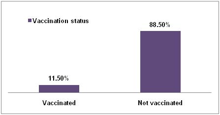
The vaccination status of the subjects in which only 26 participants had been vaccinated which contributes
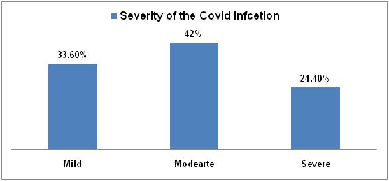
only 11.5% of the total study population (Fig 1). The severity of the COVID infection in which the majority (42%) had the moderate form of the disease and only 24.4% had a severe form of the disease (Fig 2).
The study subjects with the age more than 45 years had higher mortality when compared to those aged less than 45 years (Table 2). Also those who have had any of the co-morbidity and those who had severe forms of disease had higher risk of mortality. The study subjects with hospital stay of less than 7 days and those who were vaccinated were at less risk of death when compared to the others. The above findings were found to be statistically significant at P value less than 0.05. There was no gender association found with the mortality status of the study subjects.
DISCUSSION
The results of our study showed that the overall mortality of the admitted patients during the study period was 21.2 % which is nearly equal to a similar study conducted in New Delhi by Muthukrishnan J, et al13 which was depicted as 28.4 % of patients died. Also in the present study, those who were belonged to mild and moderate forms of disease never died. This clearly explains that only severe forms of the COVID infection were associated with mortality as shown in Table 2. These findings were consistent with the various other studies which explained the similar findings15-19
Table 1 — Basic characteristics of study subjects (N-226)
Fig 2 — Severity of COVID infection of the study subjects (N-226)
Fig 1 — Vaccination status of study subjects (N-226)
122, No 05, May 2024Journal
In the present study, we identified there is an increased risk of mortality with increasing age and also the presence of morbidity in the patients affected by COVID-19. These findings were supported by a study conducted in a similar setting by Rastad, et al19 which showed that the presence of diabetes had more risk of developing death. But in our study, we considered only the presence of co-morbidity as a factor that was significant. Previous studies conducted during trials and the real world data depicted that there is less possibility of having the severe forms of the disease and thus low mortality among those who are vaccinated against the COVID-1920-22. Our study showed less mortality rate among those who were under hospital stay of less than 7 days. The vaccine effectiveness of the AstraZeneca (ChAdOx1 nCoV-19) vaccine has shown to be as 80% in terms of the Hospitalization 23 COVISHIELD vaccine has been proved to be effective in preventing the infection by 80-94%24. The current study finding of risk of hospitalization is also supported by a study done by Carrillo-Vega MF, et al25
Table 2 — Association between the study variables and mortality status (N-226) Characteristics Dead (N- 48)Live (N-178) TotalChi-squareP value N-226 value
No of hospital stay : <7 days29 (15%) 164(85%)193 30.501 <0.05* >7 days 19(57.5%)14(42.5%)33
Vaccination status : Not Vaccinated 48(64.8%)26(35.2%)747.923 <0.05** Vaccinated 0(0%) 152(100%)152
*significant at p value <0.05 (Chi square test)
**Significant at p value <0.05 ( Likelihood ratio)
severity duration of hospital stay should be considered while predicting mortality among the COVID-19 infected patients
Source of Fund : Nil
Conflicts of Interest : Nil
REFERENCES
1Chen J, Qi T, Liu L, Ling Y — Clinical progression of patients with COVID-19 in Shanghai, China. Journal of Infection 2020; 80: 1-6 doi: 10.1016/j.jinf.2020.03.004
In the present study, the vaccinated individuals though a very small proportion might fall into the severe forms of the disease but they never end their life. This clearly explains the importance of vaccination helps in reducing the severity of the disease and thereby reducing mortality. There are many limitations in the current study. One of them is that we didn’t take into account the number of doses of vaccination and the type of vaccines since the effectiveness differs based upon these facts. Also, the study was done during the initial days immediately after the vaccination drive comes into action and hence many of the population would not be vaccinated. And since it was performed in a hospital the findings cannot be externally validated to the general population.
CONCLUSION
Thus the findings of the study imply that vaccination is as effective in reducing mortality and also the factors such as increasing age, presence of co-morbidity,
2Chen N, Zhou M, Dong X — Epidemiological and clinical characteristics of 99 cases of 2019 novel coronavirus pneumonia in Wuhan, China: a descriptive study 2020; 395: 507-13. doi: 10.1016/S0140-6736(20)30211-7.
3Wu F, Zhao S, Yu B — A new coronavirus associated with human respiratory disease in China 2020; 579: 265-9. doi10.1038/s41586-020-2008-3
4Lippi G, Sanchis-Gomar F, Henry BM — Coronavirus disease 2019 (COVID-19): the portrait of a perfect storm, 2020; 8: 497. doi: 10.21037/atm.2020.03.157.
5Mohapatra PR, Mishra B — Regulatory approval of COVID-19 vaccine for restricted use in clinical trial mode, 2021; 21: 599-600. doi- 10.1016/S1473-3099(21)00045-1
6Estimating mortality from COVID-19. https://www.who.int/ news-room/commentaries/detail/estimating-mortality-fromcovid-19 Accessed 23 March 2022.
7Kashte S, Gulbake A, El-Amin — COVID-19 vaccines: rapid development, implications, challenges and future prospects. Human cell, 2021; 34(3): 711-33. doi- 10.1007/s13577-02100512-4
8Bhuyan A — India begins COVID-19 vaccination amid trial allegations, 2021; 397(10271): 264. doi: 10.1016/S01406736(21)00145-8.
Vol 122, No 05, May 2024Journal of
9Acharya KP, Ghimire TR, Subramanya SH — Access to and equitable distribution of COVID-19 vaccine in low-income countries. npj Vaccines, 2021; 6(1): 1-3. doi- 10.1038/s41541021-00323-6.
10Bhosale S, Kulkarni AP — Is a problem shared, a problem halved? Not always! The novel coronavirus COVID-19 outbreak, 2020; 24(2): 88. doi: 10.5005/jp-journals-1007123365
11Dixit SB, Zirpe KG, Kulkarni AP — Current approaches to COVID-19: therapy and prevention, 2020; 24(9): 838. doi: 10.5005/jp-journals-10071-23365.
12Clinical Guidance For Management Of Adult Covid-19 Patients https://covid.aiims.edu/clinical-guidance-for-management-ofadult-covid-19-patients Accessed on March 23, 2022.
13Muthukrishnan J, Vardhan V, Mangalesh S — Vaccination status and COVID-19 related mortality: A hospital based cross sectional study, 2021; 77: 278-82. doi-10.1016/ j.mjafi.2021.06.034
14Rastad H, Karim H, Ejtahed H-S — Risk and predictors of inhospital mortality from COVID-19 in patients with diabetes and cardiovascular disease, 2020; 12(1): 57. doi- 10.1186/ s13098-020-00565-9.
16Mehraeen E, Karimi A, Barzegary A — Predictors of mortality in patients with COVID-19ea systematic review, 2020; 40: 101226. doi: 10.1016/j.eujim.2020.101226.
17Eumann Mesas A, Cavero-Redondo, Aparecido Sarria Cabrera M — Predictors of In-Hospital COVID-19 Mortality: A Comprehensive Systematic Review and Meta-Analysis Exploring Differences by Age, Sex and Health Conditions, 2020; 15(11): e0241742 doi: 10.1371/journal.pone.0241742.
18Trecarichi EM, Mazzitelli M, Serapide F — Clinical characteristics and predictors of mortality associated with COVID-19 in elderly patients from a long-term care facility, 2020; 10(1): doi: 10.1155/2022/5904332.
19Polack FP, Thomas SJ, Kitchin N — Safety and efficacy of the BNT162b2 mRNA COVID-19 vaccine. 383(27): 2603-2615. doi: 10.1056/NEJMoa2034577
20Dagan N, Barda N, Kepten E — BNT162b2 mRNA covid-19 vaccine in a nationwide mass vaccination setting, 2021; 384(15): 1412-23. doi: 10.1056/NEJMoa2101765
21Bhimraj A, Morgan RL, Shumaker AH, — Infectious diseases society of America guidelines on the treatment and management of patients with COVID-19. 2020. doi: 10.1093/ cid/ciaa478.
22Haas EJ, Angulo FJ, McLaughlin JM — Impact and effectiveness of mRNA BNT162b2 vaccine against SARSCoV-2 infections and COVID-19 cases, hospitalisations, and deaths following a nationwide vaccination campaign in Israel: an observational study using national surveillance data, 2021; 397(10287): 1819-29 doi: 10.1016/S0140-6736(21)009478.
23Lopez Bernal J, Andrews N, Gower C — Effectiveness of the Pfizer-BioNTech and Oxford-AstraZeneca vaccines on covid- 19 related symptoms, hospital admissions, and mortality in older adults in England: test negative case-control study, 2021; 373: n1088 doi: 10.1136/bmj.n1088
24Hyams C, Marlow R, Maseko Z — Effectiveness of BNT162b2 and ChAdOx1nCoV-19 COVID-19 vaccination at preventing hospitalisations in people aged at least 80 years: a test negative, case-control study. 21(11): 1539-48 doi: 10.1016/ S1473-3099(21)00330-3
25Carrillo-Vega MF, Salinas-Escudero G, García-Peña C — Early estimation of the risk factors for hospitalization and mortality by COVID-19 in Mexico 2020; 15(9): e0238905. doi: 10.1371/ journal.pone.0238905.
Original Article
A Comparative Study of Effect of Mindfulness-based Stress Reduction on Psychological Stress & Quality of Life in Patients of Rheumatoid Arthritis with Waitlist Control Group
Background : For centuries, contemplative cultures have used meditation as a practice. Its medicinal effects have just recently been investigated, but the results point to a wide range of advantages. Rheumatoid Arthritis (RA), an autoimmune condition harm the body’s joints and cause joint pain. Apart from jeopardizing the patient physically it also affects the psychological well-being. Given the research linking mindfulness to better immune indicators, mindfulness training may lessen disease-related stress in RA patients by boosting their immune system thereby improving their perceived stress as well as Quality of Life (QoL).
Aims and Objectives : To examine the effects of standardized Mindfulness-based Interventions (MBI) on psychological stress and QoL in a tertiary care hospital of eastern India.
Materials and Methods : 60 patients of RA were selected by purposive random sampling and divided into cases and waitlist controls comprising 30 patients in each group. The cases received MBI over a period of 6 months. Psychological Stress was estimated by Depression, Anxiety, Stress Scale (DASS) 21 and QoL by WHO QoL-BREF among both the groups at baseline, 4 months and 6 months post-intervention.
Results : Significant reduction of depression, anxiety and stress score was found in case group at 4th and 6th months. For the control group, it was not significant. The score was found to improve significantly in cases in the psychological domain of WHO QoL-BREF in the case group in 4th and 6th month. Scores in controls did not change significantly.
Conclusion : MBI caused a decrease in the depression, stress, and anxiety scores; while improving the psychological well-being of RA patients.
[J Indian Med Assoc 2024; 122(5): 28-32]
Key words :Rheumatoid Arthritis, Mindfulness-Based Stress Reduction, Quality of Life, Depression-Anxiety-Stress.
TDepartment of Psychiatry, IPGME&R and SSKM Hospital, Kolkata 700020
1MD (Psychiatry), Senior Resident and Corresponding Author
Editor's Comment :
Rheumatoid Arthritis patients suffer from various psychological issues leading to a poorer Quality of Life. Mindfulness intervention can be a novel approach towards the holistic management of Rheumatoid Arthritis along with uplifting of the Quality of Life. Although, whether the intervention helps in decreasing the disease process of Rheumatoid Arthritis is a subject of research.
he practice known as Mindfulness-based Stress Reduction (MBSR) uses mindfulness to assist patients with pain and other life difficulties that were first challenging to treat in a medical setting. In order to help individuals become more mindful, the MBSR program- which integrates yoga, body awareness and mindfulness meditation was developed at the University of Massachusetts Medical Center in the 1970s by Professor Jon Kabat-zinn 1,2 Controlled clinical research on meditation has been conducted and the results3 show that it could have positive benefits, including lowering stress levels, promoting relaxation, and enhancing Quality of Life4. MBSR is a secular concept despite having spiritual concepts at its root5
2MD, RMO cum Clinical Tutor, Department of Psychiatry, Murshidabad Medical College & Hospital, Murshidabad, West Bengal 742101
3MD, DNB, Associate Professor, Department of Psychiatry, Murshidabad Medical College & Hospital, Murshidabad, West Bengal 742101
4MD (Psychiatry), Professor, Advisor, Metal Health Service & Studies, Swasthya Bjhawan, Kolkata, West Bengal 700064
5MBBS, Postgraduate Trainee
Received on : 11/08/2023
Accepted on : 10/04/2024
Rheumatoid Arthritis :
Rheumatoid Arthritis (RA) is a chronic, progressive autoimmune condition with no known cause. Persistent inflammation that mostly affects the joints in the periphery characterizes it. Although the pain and impairment may be reduced if the disorder is detected earlier and immediately and effectively treated, it often begins as an insidious symmetrical arthritis and has
122, No 05, May 2024Journal
an unexpected and variable course.
RA often starts as a state of prolonged cellular activity that results in immune as well as autoimmunity complexes in the joints and other organs where it appears. The synovial membrane is where the illness first manifests itself and there, swelling and congestion allow immune cells to invade. The three phases of RA development include an initiation phase (induced by nonspecific inflammation), an amplifying phase (resulting in activation of T cell) and a chronic inflammatory phase along the tissue damage6.
Rheumatoid Arthritis and Stress :
The risk of experiencing different types of psychological distress is raised in people with Rheumatoid Arthritis7 According to a study conducted in 2010 in Michigan, USA, the authors found that stress plays a definite role in exacerbation of RA in addition to traumatic or stressful life experiences that occurred before the beginning of the disease8
Patients with RA and systemic lupus erythematosus were studied in experimental research that focused on acute-phase reactivity in the SLE (Stress-Response Systems)9. The Hypothalamic-Pituitary-Adrenal (HPA) axis, Autonomic Nervous System (ANS) and the immune system were evaluated at three levels of physiology in patients with SLE as well as RA. Although the baseline levels and reactivity of the ANS and HPA axis as well as experimentally produced stress were inconsistent, the authors did discover some signs of altered immune functioning in patients when compared to controls. A history of abuse and depression are quite frequent in people with rheumatologic disease and they have been related to changes in the immune and stress responses10,11
The findings from the current study of Stress consequences on SLE as well as RA point to a larger body of research that includes both animal models as well as the clinical investigations of other rheumatic conditions, which is consistent with the findings in these disorders. Numerous forms of stress have been seen in animal models to develop Arthritis12 Meditation holds the promise in lowering the stress related to emotional is associated with RA, which is why more and more people with RA are turning to complementary/alternative therapies13. This research investigates whether a Mindfulness-based Stress Reduction for Rheumatoid Arthritis patients would effectively lessen psychological distress and enhance well-being.
MATERIALS AND METHODS
Study Participants : Participants were based in a hospital, cross
sectional study among Rheumatoid Arthritis patients and demographically & clinically matched controls who have attended the outpatient clinic of Rheumatology Department, IPGME&R, Kolkata in between March, 2017 to June, 2018. Purposive sampling was done to include 60 diagnosed cases of RA from Rheumatology OPD who were of 18 to 55 years of age and who have given consent. They were split into two groups, each with 30 patients. One group who received MBSR were cases; rest were waitlist controls. Patients who have not met the age criteria, who have Mental Retardation, or substance abuse disorder, had scheduled major surgery or already been participated in another major trial were excluded from the study. The IPGME&R, Kolkata, Ethics Committee reviewed the procedure before approving it. Each participant in the study-a patient or a control subject provided their informed permission.
Instruments :
• DASS21 (Depression, Anxiety and Stress scale) : A screening method for determining, classifying and evaluating patients’ levels of stress, anxiety and depression. The three subscales of the exam are represented by these 3 negative emotional states: (1) depression, (2) anxiety and (3) stress14
• WHO QoL (BREF) : It is a self-administered questionnaire with 26 items that is a summary of the WHO QoL-100 scale. These scales evaluate the subjective reactions to various life situations based on assessments over the previous two weeks. In addition to overall well-being, it encompasses four areas: environment, social relationships, physical health, and psychological health. Each item receives a score ranging from 1 to 5. Better grades correspond to a higher QoL15. Bengali version was used.
• Mindfulness-based Stress Reduction (MBSR) : It is a structured, patient-centered educational method that utilizes mindfulness meditation training. The program’s prototype was created by the Stress Reduction Clinic at the University of Massachusetts Medical Centre16
Study Technique : After obtaining informed consent from both cases and controls, the following parameters were assessed before the start of intervention for both the groups. These were DASS 21 for assessing psychological stress and WHO QoL (BREF) for measuring the Quality of Life. The mindfulness-based stress reduction was started in the case group. The groups were divided into 3 groups each comprising of 10 patients.They received 8 sessions weekly for 2 months, two monthly booster sessions for next 2 months and then for 2 months
122, No 05, May 2024Journal
maintenance programme where they were advised to practice MBSR as a homework assignment. Patients were followed up through phone calls and visit. Each patient was followed up for 6months. 2 patients were lost to follow-up. The waitlist control group received the intervention at the end of the study. The abovementioned parameters were re-applied at the end of the 4th month and end of the sixth month on both cases and controls. The data collected by above means was analysed and compared by suitable statistical techniques and the results were interpreted accordingly. Treatment from Rheumatology department was not hampered.
RESULTS
Data were put into a Microsoft Excel spreadsheet and evaluated statistically by SPSS 25.0 (Statistical package for social sciences)17. Baseline parameters were recorded for 60 patients who were clinically diagnosed Rheumatoid arthritis patients and randomized to case or control group in 1:1 ratio. After excluding 2 patients who dropped out from the study with no follow up at 4 months, a total of 58 eligible patients were analyzed for baseline and outcome parameters. Of these 58 patients, 28 belonged to the case group and 30 belonged to the control group. All of the research variables were compared between the two groups.
For the purpose of evaluating the effectiveness of the intervention, the adjusted mean change from baseline at 4 and 6 months was calculated 2 (treatment group) X 2 (time) linear mixed model for repeated measures with Mindfulness Based Stress Reduction and utilizing group contrasts to compare the control group. P-value <0.05 was taken as being significant.
(1) Socio Demographic Variables :
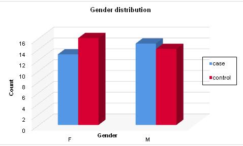
Gender : There were 13 females and 15 males in case group while control group had 16 females and 14 males. Chi-square test showed no difference in gender distribution [S2(1) =0.06, p =0.792, v= 0.03]. Overall female : male ratio was 1:1 (Fig 1).
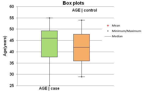
Age : The average age of cases was 43.79±7.78 years. The average age of controls was 41.67±7.51 years. An independent samples t-findings test’s showed no noticeable variations in the mean ages. (Fig 2).
(2) Psychological Stress : [DASS 21]
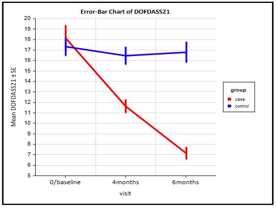
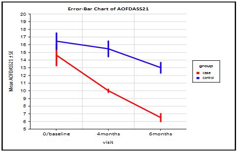
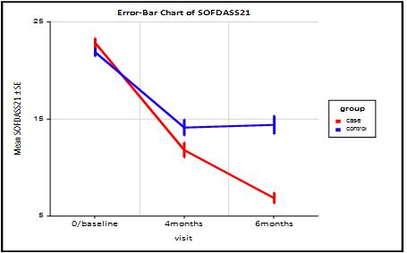
Results : Baseline means among the variables in the two groups were not significant (Table 1). During follow up at 4th& 6th month, the change was significant in DASS 21 scores in case group. When the change among the two groups were calculated, DASS 21
scores was found to be remarkable (Table 1, Figs 3, 4 & 5).
(3) Quality of Life Variables : [WHO QoL-BREF]
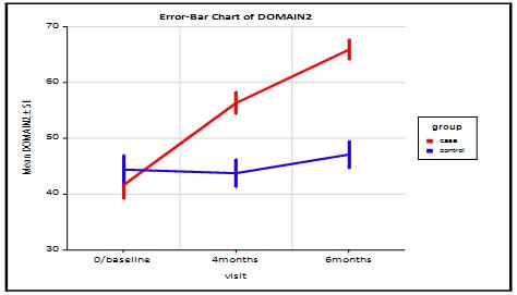
The baseline means among the variables among the two groups were not significant. At 4th & 6th month, the change was significant in Psychological &
Fig 1 — Bar Graph showing Distribution of Gender Across Cases and Control (F-Female, M-Male)
Fig 2 — Box Plot Showing Mean, Median and Range of Age In Two Groups
Fig 3 — Line Plot with Standard Error Bars for Depression Score of DASS 21
122, No 05, May 2024Journal
Table 1 — Change in Sub-items of Dass Sacle at Fourth& Sixth Month
Variables Change amongChange amongChange amongChange amongChange amongChange among Case GroupControl Groupthe Two GroupsCase GroupCase Groupthe Two Groups Over at 4thmonthat 4thmonthOver Time [ P Value]at 6thmonthat 6thmonthTime [P Value] [P Value][P Value][Calculated By Mixed[P Value][P Value][Calculated By Mixed Model Analysis of Variance]Model Analysis of Variance]
Environmental Domain in the case group. Among the control group, the change was significant in Environmental Domain.When the change among the two groups were calculated, Psychological Domain score was found to be significant (Table 2, Fig 6).
DISCUSSION
Mindfulness can have positive effects on Rheumatoid Arthritis (RA) patients, both in terms of their physical symptoms and psychological well-being. Rheumatoid arthritis is a chronic autoimmune disease that affects the joints, causing pain, stiffness and inflammation. It can also lead to fatigue, anxiety and depression, affecting the overall Quality of Life of those living with the condition. In our study,there were 13 females and 15 males in the case group while the control group had 16 females and 14 males. The chisquare test showed no difference in gender distribution. Overall female: male ratio was 1:1. The average age of cases was 43.79±7.78 years. The average age of controls was 41.67±7.51 years. An independent samples t-test resulted in no apparent variation in the mean ages. MBSR was found to improve depression, anxiety and stress scores of the cases in the 4th and 6th month compared to controls in DASS Scoring. Similarly, the psychological domain of WHOQoL-BREF was found to improve in cases in comparison to the control group. The above results were consistent with
Table 2 — Change In Quality of Life Variables [WHO QoL-BREF] At Sixth Month
Variables Change amongChange amongChange amongChange amongChange amongChange among Case GroupControl Groupthe Two GroupsCase GroupCase Groupthe Two Groups Over at 4thmonthat 4thmonthOver Time [P Value]at 6thmonthat 6thmonthTime [P Value] [Calculated [P Value][P Value][Calculated by Mixed Model[P Value][P Value] by Mixed Model Analysis of Variance] Analysis of Variance]
Fig 6 — Line Chart of Domain 2 or Psychologicaldomain
Fig 5 — Line Chart of Stress Scores of DASS 21 eith Standard Error Bars
Fig 4 — Line Chart with Error Bars Showing Mean of Anxiety Score of DASS 21
Vol 122, No 05, May 2024Journal of
some previous studies such as Teasdale, et al 200018, Morone, et al 200819, Witkiewitz, et al20, Desrosiers et al21. However, it’s important to note that mindfulness should be considered a complementary approach alongside conventional medical treatments prescribed by healthcare professionals. Patients should always consult with their doctors before incorporating mindfulness practices into their treatment plans.
Limitations :
•We were only able to recruit a modest number of patients due to the resources available to the recruitment team. For future investigations, it will be important to have a larger sample size.
•The disease activity along with the antirheumatoid medications received by both groups was not considered.
Future Directions :
Following can be thought of in the coming days to ensure a more —
(1)Study with a long-term follow-up and a moderate number of patients can be considered to see the lasting effect of MBSR.
(2)Incorporating telemedicine advice within the study to ensure proper follow-up.
(3)Conducting studies to explore the potential benefit of combining Mindfulness-based Stress Reduction with standard pharmacological treatment of RA.
(4)We can explore mindfulness and brain plasticity relations.
(5)Investigations can be done on whether mindfulness-based therapy is improving inflammatory markers in rheumatoid arthritis.
Financial Support and Sponsorship : Nil.
Conflicts of Interest : There are no conflicts of interest.
ACKNOWLEDGMENT
We would like to acknowledge all the faculty members and junior residents who helped relentlessly in conducting our research work and the patients for providing their valuable time.
REFERENCES
1Pickert K — The art of being mindful. Finding peace in a stressed-out, digitally dependent culture may just be a matter of thinking differently. Time 2014; 183(4): 40-6.
2Will A, Rancea M, Monsef I, Wöckel A, Engert A, Skoetz N — Mindfulness-based stress reduction for women diagnosed with breast cancer. Cochrane Database of Systematic Reviews 2015; 2: doi:10.1002/14651858.cd011518. ISSN 1465-1858
3Ospina MB, Bond K, Karkhaneh M — Meditation practices for health: state of the research. Evid Rep Technol Assess
(Full Rep) 2007; 155: 35-37.
4Bryant FB, Veroff J — Savoring: A new model of positive experience. Psychology Press; 2017 Sep 29; 1:
5Greeson JM, Webber DM, Smoski MJ, Brantley JG, Ekblad AG, Suarez EC, etal— Changes in spirituality partly explain healthrelated quality of life outcomes after Mindfulness-Based Stress Reduction. Journal of BehavioralMedicine2011; 34(6): 508-18
6Harrison’s Principles of Internal Medicine (18th ed.). United States: McGraw Hill.2012: 2738.
7Keefe FJ, Smith SJ, Buffington AL, Gibson J, Studts JL, Caldwell DS — Recent advances and future directions in the biopsychosocial assessment and treatment of arthritis. J Consult Clin Psychol 2002; 70: 640-55.
8Hassett A, Daniel JC — The role of stress in rheumatic diseases. 2010; 123.
9de Brouwer SJ, van Middendorp H, Kraaimaat FW, Radstake TR, Joosten I, Donders AR, et al — Immune responses to stress after stress management training in patients with rheumatoid arthritis. Arthritis Research & Therapy 2013; 15(6): R200.
10Kojima M, Kojima T, Suzuki S, Oguchi T, Oba M, Tsuchiya H, et al — Depression, inflammation, and pain in patients with rheumatoid arthritis. Arthritis Care & Research 2009; 61(8): 1018-24.
11McLean SA, Williams DA, Stein PK, Harris RE, Lyden AK, Whalen G, et al — Cerebrospinal fluid corticotropin-releasing factor concentration is associated with pain but not fatigue symptoms in patients with fibromyalgia. Neuropsychopharmacology 2006; 31(12): 2776.
12Harbuz MS, Richards LJ, Chover- Gonzalez AJ, Marti Sistac O, Jessop DS — Stress in autoimmune disease models. Annals of the New York Academy of Sciences 2006; 1069(1): 5161.
13Dube SR, Fairweather D, Pearson WS, Felitti VJ, Anda RF, Croft JB — Cumulative childhood stress and autoimmune diseases in adults. Psychosomatic Medicine 2009; 71(2): 243..
14Lovibond SH, Lovibond PF — Manual for the Depression Anxiety Stress Scales. (2nd. Ed.) Sydney: Psychology Foundation. 1995.
15Group TW — The World Health Organization quality of life assessment (WHOQOL): development and general psychometric properties. Social Science & Medicine 1998; 46(12): 1569-85.
16Santorelli S, editor. Mindfulness-based stress reduction (MBSR): standards of practice. Center for Mindfulness in Medicine, Health Care & Society, University of Massachusetts Medical School; 2014 Feb.
17IBM SPSS Statistics for Macintosh, Version 25.0.
18Teasdale JD, Segal ZV, Williams JM, Ridgeway VA, Soulsby JM, Lau MA — Prevention of relapse/recurrence in major depression by mindfulness-based cognitive therapy. Journal of Consulting and Clinical Psychology 2000; 68(4): 615
19Morone NE, Greco CM, Weiner DK — Mindfulness meditation for the treatment of chronic low back pain in older adults: a randomized controlled pilot study. Pain 2008; 134(3): 310-9.
20Witkiewitz K, Bowen S — Depression, craving, and substance use following a randomized trial of mindfulness-based relapse prevention. Journal of Consulting and Clinical Psychology 2010; 78(3): 362.
21Desrosiers A, Vine V, Klemanski DH, Nolen Hoeksema S — Mindfulness and emotion regulation in depression and anxiety: common and distinct mechanisms of action. Depression and Anxiety 2013; 30(7): 654-61.
Original Article
A
Comparative Study to Estimate Knowledge about Pneumococcal Conjugate Vaccine among Medical and Nursing Students studying at Civil Hospital Campus, Ahmedabad
Rajan Kesaji Parmar1, Mitesh K Patel2, Rajshree J Bhatt3
Background : India is committed to prevent pneumonia related deaths in children which is leading cause of vaccine preventable deaths among children under 5 globally and in India.
Aims and Objective : To know awareness about PC vaccine among Medical and Nursing students of civil hospital, Ahmedabad.
Materials and Methods : This cross sectional study was conducted on medical and nursing students of civil hospital, Ahmedabad, Gujarat. Baseline knowledge of PC Vaccine, side effects of PC Vaccine, was assessed by a self-administered structured questionnaire.
Statistical Analysis : Data analysis was done in Microsoft excel and Chi square test was applied.
Results : There were 444 respondents and among them 240 (54%) were medical students and 204 (46%) were nursing students, with male female ratio was 95:127. Awareness regarding PC Vaccine integration to national immunization schedule found significantly more in nursing students as compare to medical students, (Chi = 63.4 at P<0.05). Medical students had more knowledge about PC Vaccine as compared to nursing students. (Chi = 0.97 at P <0.05).
Conclusion : There is still gap in awareness of PC Vaccine integration to national immunization schedule among Medical students. Field visits among Nursing students have positive effects on awareness of nursing students.
Implication : Medical students should visit to this type of field activities to improve their knowledge about ongoing activities like this.
[J Indian Med Assoc 2024; 122(5): 33-4]
Key words :Knowledge, Pneumococcal Conjugate Vaccine, Medical Students, Nursing Students.
The Government of India has launched a comprehensive public health program to prevent pneumonia-related fatalities in children. Pneumonia is the primary cause of vaccine-preventable deaths in children under five worldwide and in India as well (7 per 1000 live births)1 It is intended for children in the under-5 years age range. The maintenance of high population immunity will subsequently be achieved by adding immunizations to the regular schedule at six, fourteen weeks and nine months2 The safety and efficacy profile of the PCV Vaccination is strong. For streptococcus, sero-conversion under field settings is 80% at 6 weeks, 85% at 14 weeks or more and 85% or more at 9 months. The majority of adverse responses are moderate and temporary3.
In order for PCV integration to be successful,this
1MD, Assistant Professor, Department of Community Medicine, Banas Medical College & Research Institute, Palanpur, Gujarat 385001 and Corresponding Author
2MD, Associate Professor, Department of Community Medicine, B J Medical College and Civil hospital, Ahmedabad, Gujarat 380016
3MD,Assistant Professor, Department of Community Medicine, B J Medical College and Civil hospital, Ahmedabad, Gujarat 380016
Received on : 09/12/2022
Accepted on : 23/07/2023
Editor's Comment : At all ages, vaccines save lives and keep us safe.
implies that in order to effectively communicate with the population, members of the medical and paramedical fraternities must possess comprehensive knowledge. The goal of this study is to evaluate the knowledge of the next generation of medical and paramedical professional’s domains. The purpose of this study was to find out how well-informed medical and nursing students were about the pneumococcal conjugate vaccination and how well-informed the study participants were about how the vaccine fits into the national immunization schedule4-6.
MATERIALS AND METHODS
A cross-sectional study was carried out in January and December of 2022. The investigation was conducted at the appropriate medical and nursing colleges, which were housed on Ahmedabad’s civil hospital campus. In all, 444 pupils purposive sampling was used to enroll (Third Year Part-1 Medical students = 240 and Third Year B Sc Nursing students = 204), (Male = 254, Female = 190), aged roughly 20-21 years,
122, No 05, May 2024Journal
in the study. All of the batch’s students were involved in the study, with the exception of those who weren’t there. The study did not include those who did not provide informed consent. Data was gathered through the use of structured questionnaires. The questionnaire asked questions about the utility of the PC vaccine, its integration into the national immunization schedule and its knowledge.
Table 1 — Awareness regarding PC vaccine integration to national immunization schedule among Medical and Nursing Students (N-444)
Awareness Medical (N = 240)Nursing (N = 204) χ2 p Value
Session Site 100(41)140(59)137(65.7)70(34.5) 13.58< 0.05
Table 2 — Awareness regarding PC Vaccine among Medical and Nursing students (N-444)
This campaign will help reduce child mortality from pneumonia, which is India’s biggest cause of vaccine-preventable deaths among children under five. Tables 1-3 were used to display the percentages of the results, and M S Excel 2007 was used for the analysis. Authorization to authorities from the medical college and nursing school were consulted in order to conduct the research. Since there is no active intervention in this study, ethical approval was not necessary. Following the acquisition of informed consent, students were enrolled with the understanding that their participation in the study would be entirely voluntary and that there would be no negative consequences.
DISCUSSION AND RESULT
MR VaccineMedical (N = 240)Nursing (N = 204) χ2 p Value
Combination of Vaccine 24(11.5)216(88.5)74(36.3)130(63.7) 19.42< 0.05
personnel, as they are the bridge population between public and professional health team.
Conflict of Interest : None.
REFERENCES
Every year in India, 500 000 children under the age of five pass away from diseases that can be prevented by vaccination; of them, 15% are pneumonia-related deaths.There was a statistically significant difference.(p value < 0.05 and chi square = 13.58) An evaluation of students’ knowledge at an Egyptian university found that, while medical students tended to be more aware of the risks and contraindications of vaccines, overall, students’ knowledge was lacking more knowledgeable than their peers8 It is imperative that no child be left behind for this integration to be successful. The current campaign is run at schools and outreach centers, in addition to fixed venues sessions.It is therefore expected of the teachers to emphasize the value of immunization 7 During immunization, a 0.5 ml dose of the polysaccharide conjugate vaccine was administered intramuscularly at the anterolateral portion of the right thigh. Medical students’ knowledge ranged from 59.8% to 74.5%, whereas nursing students’ knowledge varied from 25.5% to 40.2%.
CONCLUSION
PC Vaccination integration may have been more successful with better use of health education message especially in medical and para-medical
2MR Campaign — The state second in coverage. March 5, 2017. The Hindu.
3Palanisamy B, Gopichandran V, Kosalram K — Social capital, trust in health information, and acceptance of vaccination campaign in Tamil Nadu: A case–control study. J Postgrad Med 2018; 64: 212-9.
4Noni EM — The SAGE working group on Vaccine hesitancy. Vaccine hesitancy: Definition, scope and determinants. Vaccine 2015; 33: 4161-4.
5Heidi L — Missing the signals: India’s anti-vaccination social media campaign. The Vaccine Confidence Project. March 2017. [homepage of the Vaccine Confidence Project: London School of Tropical Medicine on the Internet].
6Sachiko O, Ligia P, Mary Q — Exploring pathways for building trust in vaccination and strengthening health system resilience. BMC Health Serv Res 2016; 16(Suppl 7): 639-44.
7Sreedevi A — Vaccination campaign: A trust deficit? JPostgrad Med 2018; 64(4): 202-3.
8Abd Elaziz KM, Sabbour SM, Dewedar SAA — Vaccination campaign in an Egyptian University: vaccine uptake and knowledge and attitudes of students. Vaccine 2010; 28(47): 7563-8. doi: 10.1016/j.vaccine.2010.08.053. Epub 2010 Aug 24.
9Kaur K — A Study to Assess The Level of Knowledge Regarding PCV Vaccine Among Mothers of under 15 years Children In Rural Area: Bathinda, Punjab. IOSR Journal of Nursing and Health Science (IOSR-JNHS). e-ISSN: 23201959.p- ISSN: 2320-1940, Volume 8, Issue 1, Ser. X. (JanFeb 2019), PP 01-05.
Original Article
Biological Reference Intervals for Hematological Parameters Including Novel Research Parameters in Population of Eastern India
Ashvini Sengupta1, Saikat Pal2
Background : Biological Reference Intervals (BRI) are used for a comparative decision-making process by describing the distribution of results derived from healthy reference population. Biological reference intervals should be derived from the local population to take into account regional variations.
Aims and Objectives : Our aim was to establish a biological reference interval for routine hematological parameters along with novel hematology parameters from health check-up of young working in-house staff of our hospital.
Materials and Methods: This is a cross sectional study carried out in our hospital using samples collected for routine health check-up of hospital staff using the Mindray BC-6800 Plus automated analyser from December, 2022 to June, 2023.
Results : A total of 474 male participants and 464 female participants were included and reference intervals for reportable and non-reportable novel hematologic parameters established.
Discussion : The study has established comparability of parameters with other Indian and International studies and shows difference in values from other population in India specifically for platelet parameters supporting the findings of Inherited Macrothrombocytopenia. We have also established the reference ranges of additional Novel Research hematological parameters with increasing utility in modern hematology practice and intend to study them further for their utility in diagnostic and therapeutic management of patients.
[J Indian Med Assoc 2024; 122(5): 35-9]
Key words :Biological reference ranges, Inherited Macrothrombocytopenia, Novel hematological parameters.
Biological reference intervals (BRI) are used for a comparative decision-making process by describing the distribution of results derived from healthy reference population. The values aid in the interpretation of laboratory results for patient care1-3 Most common parameter used by clinicians for management, diagnosis and monitoring of their patients is a CBC3
Hematologic interpretations rely on the BRI derived from population to be served. Although age and sex are the two most common categorizing criteria for BRI, variations are also seen during physiological conditions like pregnancy or exercise4-6
Only few clinical laboratories have the resources to establish BRIs as they require data from healthy local population. As reference values in-use are used from authoritative textbooks or western references5, adopting from dissimilar population without considering local ethnicity may lead to mismanagement with increase in cost and risk patient safety. Therefore the need to establish hematological BRIs in the eastern population of the country.
Medica Superspecialty Hospital, Kolkata 700099
1MBBS, MD (Path), Director, Department of Laboratory Services and Corresponding Author
2MBBS, DNB (Micro), DNB Resident
Received on : 20/02/2024
Accepted on : 30/04/2024
Editor's Comment :
The eastern population is ethnically different from other population in our country especially in platelet parameters which is also called as Bengal Macrothrombocytopenia. So, the need to establish reference ranges in our population for routine parameters as well as for many novel hematology parameters available in our analyser, so as to use them as reference points for further studies in this field.
AIMS AND OBJECTIVES
Aims : To establish a biological reference interval for routine hematological parameters from blood samples collected from yearly health checkup of young working (<40 years) in-house population of our hospital.
Objectives : Establish biological reference interval for reportable hematologic parameters (WBC, Neu%, Lym%, Mon%, Eos%, Bas%, RBC, HGB, HCT, MCV, MCH, MCHC, RDW-CV, RDW-SD, PLT, MPV, PDW, PCT) along with novel research hematological parameters available for evaluation (P-LCC, P-LCR, HFC#, HFC%, WBC-D, WBC-N, TNC-D, TNC-N, NLR, Micro#, Micro%, Macro#, Macro%, PLT-I, PLR, PDWSD, Neu-X, Neu-Y, Neu-Z, Lym-X, Lym-Y, Lym-Z, MonX, Mon-Y, Mon-Z) for reference for future studies.
MATERIALS AND METHODS
(1)A cross sectional study was carried out in our hospital of apparently healthy adults of both sexes of hospital staff from December, 2022 to June, 2023.
122, No 05, May 2024Journal
(2) Inclusion into the study was based on the following criteria : aged <40 years adults; subject to health check up with normal records of vital signs and physical examination.
(3) Exclusion criteria : age >40 years, having comorbidities.
(4)As per CLSI recommendation6 a sample size of minimum of 120 participants is required. Total of 938 persons, 474 males and 464 females were recruited for the study.
(5) Sample collection : 3-ml venous blood was collected from antecubital vein under aseptic conditions into Dipotassium Ethylene Diamine tetra acetic acid (K 2 EDTA) Vacutainer tube (Becton Dickinson, PL6 7BP, UK) for hematological analysis.
(6) Laboratory assays : The whole blood EDTA samples were analysed within two hours using Mindray BC-6800 Plus automated hematology analyzer (Mindray Bio-medical electronics Co, Ltd, Shenzhen, China), for both reportable (for invitro diagnostic use in clinical laboratories) and nonreportable/ research use hematologic parameters.
Reportable parameters : The parameters used in this study are white Blood Cell Count (WBC), Neutrophil percentage (Neu%), Lymphocyte percentage (Lym%), Monocyte percentage (Mon%), Eosinophil percentage (Eos%), Basophil percentage (Bas%), Red Blood Cell Count (RBC), Hemoglobin (HGB), Hematocrit(HCT), Mean Cell Volume (MCV), Mean Cell Hemoglobin (MCH), Mean Cell Hemoglobin Concentration (MCHC), Red Cell Distribution WidthCoefficient of Variation (RDW-CV), Red Cell Distribution Width-Standard Deviation (RDW-SD), Platelet Count (PLT), Mean Platelet Volume (MPV), Platelet Distribution Width (PDW), Plateletcrit (PCT). Non-reportable/Research Use only parameters: Platelet-Large Cell Count (P-LCC), Platelet-Large Cell Ratio (P-LCR), High Fluorescent Cell percentage (HFC%), White Blood Cell Count -DIFF (WBC-D), White Blood Cell Count-WNB (WBC-N), Total Nucleated Cell Counts-DIFF (TNC-D), Total Nucleated Cell Counts-WNB (TNC-N), Neutrophil-to-lymphocyte Ratio (NLR), Microcyte percentage (Micro%), Macrocyte percentage (Macro%), Platelet-tolymphocyte ratio (PLR), Platelet Distribution Width Standard Deviation (PDW-SD), mean neutrophil distribution-side scatter intensity (Neu-X), Neutrophil distribution-side fluorescent light intensity (Neu-Y), mean neutrophil distribution- forward scatter intensity (Neu-Z), mean Lymphocyte distribution- side scatter intensity (Lym-X), mean Lymphocyte distribution-side fluorescent intensity (Lym-Y), mean Lymphocyte distribution- forward scatter intensity (Lym-Z), mean
monocyte distribution-side scatter intensity (Mon-X), mean Monocyte distribution-side fluorescent light intensity (Mon-Y), mean Monocyte distributionforward scatter intensity (Mon-Z).
The principles used by the analyzer for measurement are :
• Sheath flow impedance method, laser scatter and SF Cube cell analysis technology (3D analysis using information from scatter of laser light at two angles and fluorescence signals) for cell differentiation and counting;
• Colorimetric method for HGB measurement.
Well-trained experienced laboratory personnel performed all tests according to Standard Operating Procedures (SOPs).
(7) Quality control : The analyzer is calibrated annually and daily Internal Quality Control (IQC) run as per QC protocol with 3 level (low, normal and high) commercially available controls with daily monitoring using LJ charts and Westgard rules. Laboratory is also enrolled in a proficiency testing program for hematology.
(8) Statistical analysis : Quantitative data has been represented as mean & SD and qualitative data has been represented as percentages. 97.5 percentile and 2.5 percentile formed the upper and lower limit of reference range respectively of the population. The biological reference interval is Mean±2*SD for those parameters whose p-value for normality test (Shapiro Wilk’s test) is greater than 0.05 (p-value>0.05). The group comparisons were based either on parametric or non-parametric statistics depending on whether the distribution is normal or not. For comparison of each of the parameters between two groups (across males and females), the non-parametric Mann-Whitney test is being used.
RESULTS
Demographic characteristics: A total of 474male participants and 464 female participants were included in the study.
Table1 shows the reference intervals for reportable and non-reportable novel hematologic parameters of our study population.
Table 2 shows comparison between our study and other Indian and International studies.
Figs 1-3 show comparative representation of 3 key parameters between various studies.
DISCUSSION
Biological reference ranges for WBC counts across all Indian and International studies are comparable with appropriate differential counts (Fig 1).
The Hemoglobin reference ranges are comparable with the Indian multi-centric study7 but slightly less than the international study values especially the lower 36
Vol 122, No 05, May 2024Journal of the Indian Medical Association
Table 1 — Biological Reference ranges of Males and Females in Eastern Indian Population – Reference range (Mean± 2*SD) and Percentile (2.5 - 97.5). Mann-Whitney test for 2 groups used for comparison
BRIMale (N=474)Female (N=464)
TestsReference rangeReference range
WBC4.28-10.164.38-10.50
NEU# 2.25-6.42.01-7.25
LYM#1.10-3.81.19-3.37
MON#0.23-0.720.23-0.68
EOS#0.03-0.850.03-0.86
BAS#0.01-0.08 0-0.06
IMG# 0-0.050-0.03
NEU%42.1-74.343.3-76.6
LYM%17.3-45.3 16.67-44.6
MON% 4.0-9.73.40-9.44
EOS% 0.6-12.70.40-9.67
BAS% 0.1-1.010.1-0.8
IMG%0-0.7 0-0.4
RBC4.14-5.3823.65-5.12
HGB12.4-16.910.3-14.3
HCT37.9-51.132.5-43.4
MCV78.7.3-99.773.4-96.8
MCH25.4-33.123.21-31.9
MCHC31.4-35.030.4-34.3
RDW-CV12.5-15.3 12.3-16.44
RDW-SD 39.14-50.038.7-50.3
PLT85.3-35572.74-384.66
MPV9.1-16.48.65-16.20
PDW15.7-17.015.4-16.9
PCT0.14-0.340.14-0.42
P-LCC 44.50-121.546.15-135.8
P-LCR20.2-68.2 16.55-68.21
HFC# 0-0.0530-0.06
HFC%0-0.7 0-0.84
WBC-D 4.38-10.474.43-10.9
WBC-N 4.28-10.164.38-10.50
TNC-D 4.38-10.474.43-10.9
TNC-N 4.28-10.164.38-10.50
NLR0.96-4.060.98-4.5
MICRO#0.02-0.260.02-0.54
MICRO% 0.48-5.00.4-11.6
MACRO#0.09-0.290.05-0.19
MACRO% 1.68-6.21.05-4.7
PLT-I81.3-355.092.6-380.4
PLR38.4-189.742.66-215.5
PDW-SD10.2-29.09.55-28.2
NEU-X 301.6-397.3306.53-397.7
NEU-Y404.6-500.3416.3-514.8
NEU-Z1553.8-1832.51549.3-1824.0
LYM-X 92.0-105.991.1-105.4
LYM-Y 665.4-785.7685.1-797.4
LYM-Z 921.6-981.1927.9-987.8
MON-X 199.1-223.8203.2-227.9
MON-Y 962.3-1122.6982.6-1149.5
MON-Z1201.9-1309.51223.4-1321.1
limits and also lower than studies from Mumbai8 , Chennai10and Delhi9, the possible explanation could be prevalence of Thalassemia traits in the eastern population. The RBC count reference ranges also show wider range possibly both due to prevalence of traits and smoking reflected also in other RBC parameters like HCT and MCV. RDW the newly reportable
parameter shows slightly higher upper limit values compared to international studies but is comparable with other Indian studies (Fig 2).
Platelet counts are conspicuously lower than the values from all the studies, Indian and International due to the unique finding of Macrothrombocytopenia1315 in the eastern population characterized by decreased platelet count (mild to moderate thrombocytopenia), macroplatelets (giant platelets) and no bleeding manifestations. This also corroborates with difference in other novel platelet parameters values like PDW and PCT from the other studies (Fig 3).
Earlier called as Harris Syndrome seen in mediterranean population and also called as Asymtomatic Constitutional Macrothrombocytopenia (ACMT) 13 is now called as Inherited Macrothrombocytopenia as it is linked to genetic molecular sequences. It is also referred to as Bengal Macrothrombocytopenia15 by many hematologists in the southern and western parts of the country as they have specially found it is patients from Bengal. All novel platelet parameters like MPV, PDW, PCT also show increased values compared to all the other studies which corroborates with Inherited Macrothrombocytopenia, thus emphasizing the
Fig 2 — Distribution of Reference range of HGB across Indian and International Studies
Fig 1 — Distribution of Reference range of WBC across Indian and International Studies
122, No 05, May 2024Journal
importance of establishing the region-specific reference intervals in the laboratory reporting system for our ethnic population.
Here, we have also established the reference ranges of additional Novel research hematological parameters with increasing utility in modern hematology practice like Platelet-large Cell Count (P-LCC), Plateletlarge Cell Ratio (P-LCR), High Fluorescent Cell Percentage (HFC%), White Blood Cell Count -DIFF (WBC-D), White Blood Cell Count-WNB (WBC-N), Total Nucleated Cell Counts-DIFF (TNC-D), Total nucleated cell Counts-WNB (TNC-N), Neutrophil-toLymphocyte Ratio (NLR), Microcyte Percentage (Micro%), Macrocyte percentage (Macro%), Platelet-to-lymphocyte ratio (PLR), Platelet Distribution Width Standard Deviation (PDW-SD), mean neutrophil distribution-side scatter intensity (Neu-X), neutrophil distribution-side fluorescent light intensity (Neu-Y), mean neutrophil distribution- forward scatter intensity (Neu-Z), mean lymphocyte distribution- side scatter intensity (Lym-X), mean lymphocyte distribution-side fluorescent intensity (Lym-Y), mean lymphocyte distribution- forward scatter intensity (Lym-Z), mean monocyte distribution-side scatter intensity (Mon-X), mean monocyte distribution-side fluorescent light intensity (Mon-Y). mean Monocyte distribution - forward scatter intensity (Mon-Z). Few of these like NLR, PLR have already found utility in COVID patients. These can be studied further for evaluation of various diseases.
CONCLUSION
We have established the region-specific reference intervals in the laboratory reporting system for our ethnic population which is different from other population in India and also corroborates with findings of other studies from Eastern region especially in platelet parameters. We have also established the reference ranges of additional Novel Research hematological parameters with increasing utility in modern hematology practice and intend to study them further for their utility in diagnostic and therapeutic management of patients.
REFERENCES
1Aytekin M, Emerk K — Accurate reference intervals are required for accurate diagnosis and monitoring of patients. eJIFCC 2008; 19(2): 1-5.
2Katayev A, Balciza C, Seccombe DW — Establishing reference intervals for clinical laboratory test results: Is there a better way? Am J Clin Pathol 2010; 133(2): 180-6.
3Jacob EA — Complete Blood Cell Count and Peripheral Blood Film, Its Significant in Laboratory Medicine: A Review Study. Am J Lab Med 2016; 1(3): 34-57. Higgins, Chris. (2012). An Introduction to Reference Intervals (1)-Some Theoretical
Considerations. Point of Care. 11. 2–5. 10.1097/ POC.0b013e318246a59c.
4Zeh CE, Odhiambo CO, Mills LA — Laboratory Reference Intervals in Africa. 2012.
5Imelda bates- Reference ranges and normal values. In Barbara J. Bain, Imleda Bates, Michael A. Laffan- Dacie Lewis Practical Haematology, 12th Edition, page 8-17
6EP28 Defining, Establishing, and Verifying Reference Intervals in the Clinical Laboratory, 3rd Edition, CLSI
7Sairam S, Domalapalli S, Muthu S, Swaminathan J, Ramesh VA, Sekhar L, et al — Hematological and biochemical parameters in apparently healthy Indian population: defining reference intervals. Indian J Clin Biochem 2014; 29(3): 290-7.
8Sehgal KK, Tina D, Choksey U, Dalal RJ, Shanaz KJ — Reference range evaluation of complete blood count parameters with emphasis on newer research parameters on the complete blood count analyzer Sysmex XE-2100. Indian J Pathol Microbiol 2013; 56: 120-4.
9Rahar S, Vijay K, Swati R, Deepika G — Haematology reference range evaluation for novel research parameters on the complete blood count analyzer sysmex XN-1000. Hamdan Medical Journal 2022; 15(2): 83-8.
10A R S, Parameaswari PJ, Shanthi B, Revathy C, Parijatham BO — The reference intervals for the haematological parameters in healthy adult population of chennai, southern India. J ClinDiagn Res 2012; 6(10): 1675-80.
11Ritesh S, Tiwari AK, Shalini G, Vimarsh R, Sethi M — Establishing biological reference intervals for novel platelet parameters (immature platelet fraction, high immature platelet fraction, platelet distribution width, platelet large cell ratio, platelet-X, plateletcrit, and platelet distribution width) and their correlations among each other. Indian J Pathol Microbiol 2014; 57(2): 231-5. doi: 10.4103/0377-4929.134676.
12Kim H, Hur M, Kim SW, Moon HW, Yun YM — Reference intervals for clinically reportable platelet parameters on the Mindray BC-6800Plus hematology analyzer. Clin Chem Lab Med 2020; 58(10): e213-e215.
13Kunjali Naina HV, Nair SC, Daniel D, George B, Chandy M — Asymptomatic constitutional macrothrombocytopenia among West Bengal blood donors. The American Journal of Medicine 2002; 112(9): 742-3. doi: 10.1016/s0002-9343(02)01114-2.
14Edupuganti HS, Krishnamurthy V — Prevalence of constitutional macrothrombocytopenia in the immigrants of Northern and Eastern states of India. Department of Pathology, JSS Medical College, JSS Academy of Higher Education and Research, Mysuru, Karnataka, India, Year 2020; 63(4): 593-6.
15Ali S, Ghosh K, Shetty S — Differential expression of genes involved in Bengal macrothrombocytopenia (BMTCP). Blood Cells Mol Dis 2015; 55(4): 410-4.
Fig 3 — Distribution of Reference range of PLT across Indian and International Studies
Original Article
Clinical Outcome of Arabin Cervical Pessary in Women at Risk of Preterm Birth in Indian Scenario
Background : Preterm birth complications are the leading cause of neonatal morbidity and mortality. This study was designed to ascertain the role of Arabin cervical pessaries in the management of women at risk of spontaneous preterm birth.
Methods : It was a Randomized Controlled Trial, on pregnant women with 12-24 weeks of pregnancy with either history of >2 spontaneous preterm birth or cervical length <25 mm with or without a history of spontaneous preterm birth. Women were randomly assigned to Arabin cervical pessary group (50) or the progesterone group (46). The primary outcome was the preterm birth rate at <37 weeks and <34 weeks. Secondary outcomes were any maternal side effects along with the neonatal outcome.
Results : The preterm birth rate <34 weeks gestation was less frequent, 8/50(16%) in the pessary group versus 16/46 (34.78%) in the progesterone group, with a relative risk of 0.46, the p-value of 0.042. Preterm birth rate <37 weeks in pessary group was 41/50 (82%) versus 41/46 (89.13%) in progesterone group, relative risk 0.92, p-value 0.32. In the pessary group, there was a significant reduction in further threatened preterm labour 38% versus 60.87% in the progesterone group (p=0.025) which subsequently reduces the use of tocolytics.
Conclusion : The study shows that cervical pessary can be considered an affordable, safe and reliable option for reducing the rate of early spontaneous preterm birth in singleton pregnancies in women at risk of spontaneous preterm birth.
Key words :Arabin Pessary, Cervical Pessary, Preterm Birth.
Preterm birth and complications associated with prematurity contribute maximally to neonatal mortality. The global rate of preterm birth is quite variable, it ranges from 5% to 18% among various countries1. India is leading among the top fifteen nations contributing to two-thirds of the world’s preterm babies. More than 80% of premature births occur between 32 to 37 weeks of gestation. The management cost of premature babies is massive which further enhances if the birth weight is less than 1000 grams2 Approximately 40-45% of premature babies are born because of spontaneous preterm labour which is more than iatrogenic early induction of labour whether for an obstetrical or medical reason1 So early prediction and effective management strategy of spontaneous preterm labour will play a vital role in bringing down
Department of Obstetrics and Gynaecology, SN Medical College, Agra, Uttar Pradesh 282002
1MS (Obstat and Gynaecol), Professor
2 MS (Obstat and Gynaecol) Assistant Professor and Corresponding Author
Arabin cervical pessary is a safe and economical alternative to natural micronized progesterone for the management of threatened preterm labour. Arabin cervical pessary can be used as convenient, one time treatment and has shown encouraging results in terms of prolongation of gestational age.
the rate of preterm birth and cutting back its economic load.
Despite robust research to develop an effective screening tool for early prediction of preterm labour, we are still dependent on short cervical length on transvaginal sonography before 28 weeks of gestation. The gradual decrease in cervical length and the presence of funneling further increase the risk of preterm labour. In women, at high risk of preterm labour either on cervical assessment or history alone cervical cerclage is the treatment of choice for a long time. For placement of cervical cerclage, women require hospital admission, operation theatre and general anesthesia with conflicting results in success. Various formulations of progesterone are also used in different doses and routes like vaginal, oral and intramuscular for the management of preterm labour. Various studies have quoted that more exposure to progesterone during
No 05, May
pregnancy can act as a triggering factor for some chronic conditions in fetuses that may appear later in adult life. A higher level of maternal progesterone will elevate the fetal progesterone levels, which adversely affect the progesterone target cells in the fetal pituitary, testis of a male fetus and developing fetal reproductive system3
Cervical cerclage and progesterone have some pros and cons. So, there is a need and space for the introduction of a novel approach in form of Arabin cervical pessary which appears effective and safer than cervical cerclage and progesterone. This study aims to assure the efficacy and safety of Arabin cervical pessary in the prevention of preterm birth in the Indian population.
MATERIALS AND METHODS
This study was conducted in the Department of Obstetrics and Gynaecology. The study was executed as a randomized controlled trial with allocation concealment. The purposive sampling method was used to recruit study participants from November, 2019 to June, 2021 who had consulted inpatient and Outpatient Departments. We enrolled pregnant women, having 12 to 24 weeks of pregnancy with a past history of spontaneous preterm birth, second-trimester abortions, traumatic obstetrics delivery, multiple dilatation and curettage, antepartum hemorrhage, multifetal gestation and polyhydramnios.
The recruited participants were called for an initial TVS assessment of cervical length and then reassessed every two weeks for any cervical changes. Screening of infections from the lower genitourinary tract was done by vaginal swab culture, endocervical culture, and urine culture in all the recruited participants.
Inclusion Criteria :
•All asymptomatic pregnant women of 12-24 weeks of gestation with a history of >2 spontaneous preterm birth
•All pregnant women of 12-24 weeks of pregnancy with or without a history of one spontaneous preterm birth with cervical length <25 mm
Exclusion Criteria :
•Active preterm labour
•Clinical evidence of chorioamnionitis
•Vaginal bleeding
•Preterm premature rupture of membranes
•Evidence of fetal compromise or fetal death
•Lethal fetal malformation
•Multiple gestations
•Silicon allergy
Material :
Arabin Cervical Pessary (Fig 1) is a soft, flexible, perforated and dome shape made up of silicon. Its inner diameter is to be fitted as high as possible around the cervix and its outer diameter is to hold the pessary against the vaginal wall to support the pelvic floor. Arabin cervical pessary rectifies the cervicovaginal angle by rotating the cervix to approach the posterior vaginal wall. It is available in three sizes small, medium, and large. The commonly used ones are small and medium sizes; small should be preferred for a singleton pregnancy, thin-built and short-statured women whilst medium and large should be offered to taller women and edematous cervix respectively.
Group Distribution :
Participants who had signed the informed written consent were randomly assigned into two groups by using the simple randomization sealed envelope method. All the sealed opaque envelopes having assigned group cards were numbered sequentially along with the signatures on the back of envelopes, prepared by persons not involved in the trial. Once a patient has consented to enter for trial, the envelope was assigned and opened by trained medical personnel independent of the trial.
Group1— was prescribed with insertion of Arabin cervical pessary and post-procedural TVS to ensure correct placement of Pessary.
A preliminary examination to evaluate the cervical length, with a special note on the presence or absence of funneling through TVS was done. Judicious screening of any form of genitourinary infection was done by performing vaginal swabs, endocervical swabs, and urine cultures. Treatment of infections with specific antimicrobial drugs was completed before insertion of pessary.
Insertion :
The patient was laid in the dorsal lithotomy position and the shoulder support with a pillow. The gel-covered
Fig 1 — Arabin Cervical Pessary
122, No 05, May 2024Journal
pessary was folded with the smaller diameter directed upwards so that the pessary can be placed toward the top of the posterior fornix. While inserting the pessary it remains folded until the upper vaginal fornix is approached, then slide as high as possible with the smaller diameter surrounding the cervix. The correctly placed pessary was not perceived by the patient and to ensure this we asked the patient to stand up and walk a few steps. If the patient complained of any pain or uneasiness, the size and placement were reassessed. A post-insertion TVS was done in every patient to ascertain the correct placement of the pessary.
Removal :
The pessary was removed at 37 weeks of gestation. Pessary removal before 37 weeks was considered in patients with active preterm labour not responding to tocolysis, in case of severe vaginal bleeding, premature rupture of membranes, or suspect of chorioamnionitis.
Primary Outcome :
Prolongation of gestational age in terms of delivery >37 weeks, 34-37 weeks, 34-28 weeks, and <28 weeks
Secondary Outcome :
•Subsequent threatened preterm labour episodes and use of tocolytics (Tablet Nifedipine 20 mg twice daily)
•Premature rupture of membranes (PROM)
•Complications such as expulsion, infection, discharge, and bleeding
•Neonatal outcomes in terms of neonatal infections (sepsis), necrotizing enterocolitis, respiratory distress syndrome, hyperbilirubinemia and neonatal death
Statistical Analysis :
The data are communicated as percentages, mean with Standard Deviation (SD). Mean ± Standard Deviation was calculated for all quantitative data. Categorical variables were displayed by using frequency measures. The Chi-square test was used for group comparison. Mean, standard deviation, p-value, and Relative- Risk were calculated. The p-value <0.05 is the measure of significance. The SPSS 22.0 version (Chicago, Inc, USA) Window software was used for statistical analysis.
RESULTS
The study flow is depicted in Fig 2. We recruited 404
Cervical
eligible pregnant women during the study period for primary assessment out of which 102 women had declined to participate in the trial due to personal constraints, 196 women had not fulfilled the inclusion criteria and 8 women were diagnosed as placenta previa. A total of 98 women had consented to participate in the trial after satisfying the inclusion criteria. Two patients did not complete the study protocol from the progesterone group. So, in the final analysis, we have 50 patients in the pessary group and 46 patients in the progesterone group.
Both the groups were comparable in terms of demographic characteristics as shown in Table 1. In the pessary group, the mean age was 29.14 years, and 28.54 years in the progesterone group. The mean BMI in the pessary group was 23.76±3.62 and in the progesterone group was 23.85±3.69. In the pessary group, the mean gestational age at the time of enrollment was 23.70±3.62 weeks and in the progesterone, the group was 23.70±3.69 weeks. Most of our study participants did not have a past history of preterm birth in any of the groups. In the pessary group, the mean cervical length was 1.49 cm and in the progesterone group, it was 1.33 cm. Funneling was
Fig 2 — Flowchart showing the distribution of patients in study groups
Table 1 — Demographic profile of patients
122, No 05, May 2024Journal
noted in 38(76%) patients at the time of enrollment in the pessary group and 30 (65.22%) patients in the progesterone group. Various morbidities during the course of pregnancy in the study period were described in Table 2. Subsequent threatened labour episodes were present in 4(8%) cases in the pessary group while it was reported by 12(26.09%) patients in the progesterone group. The statistical difference is significant (p=0.017) in both groups in terms of the occurrence of subsequent threatened labour episodes. In the pessary group 19 (38%) patients and the progesterone group 28(60.87%) patients, needed tocolytic therapy in the form of tablet nifedipine 20 mg two times a day and the difference was statistically significant with the p-value 0.025. Bleeding episodes were reported by 2(4%) patients in the pessary group and 2(4.35%) patients in the progesterone group. Premature rupture of membranes was found in 2(4%) patients in the pessary group and it was found in 6(13.04%) patients in the progesterone group. Vaginal discharge was reported by all the patients of the pessary group 50(100%) and 18(39.13%) patients in the progesterone group. We have to remove pessary in 7 (14%) patients on medical grounds and 11(22%)patients needed repositioning of pessary without its removal. The mean duration of hospital stay due to various morbidities was 3.2 days in the pessary group and 4.5 days in the progesterone group.
Table 2 — Maternal morbidities in the antenatal period
Pessary ProgesteroneP value Chi-square group (N=50) group (N=46) value
Subsequent threatened preterm labour episodes
8%(04)26.09%(12)0.0175.64
Tocolytic therapy in form of Tab-Nifedipine 20mg bd 38%(19)60.87%(28)0.0255.01
Corticosteroid treatment for fetal lung maturation 94%(47)84.78%(39)0.1392.181
Bleeding in pregnancy 4%(02)4.35%(02)0.9320.007
Premature rupture of membranes : 4%(02)13.04%(06)0.1092.565
30-32 weeks00% 66.67%
32-35 weeks04% 33.33%
Side effects : - Vaginal discharge
100%(50)39.13%(18)
- Pessary repositioning without removal22%(11)- Pessary removal(medical indication) 14%(07)-
Maternal hospital stay (mean)3.2 days4.5 days
spontaneously at this gestational age. In the pessary group, 9(18%) patients successfully continued their pregnancy beyond 37 weeks of gestation and 5(10.87%) attain that milestone in the progesterone group. In the pessary group mean gestational age of delivery was 35.08 weeks and in the progesterone group it was 33.76 weeks. In the progesterone group, the preterm birth rate at <34 weeks was significantly more (p=0.042) but no significant difference was detected in both groups in terms of spontaneous preterm birth rate at <37 weeks of gestation (RR=0.92; p=0.32 at 95%CI).
Table 3 gives the details of the pregnancy outcome in terms of the gestational age of delivery. After pessary insertion,a maximum number of patients that are 33(66%) successfully carry their pregnancy up to 3437 weeks of gestation and in the progesterone group the maximum number of patients delivered at 34-37 weeks of gestation that are 25(54.35%).
Two patients (4%) underwent spontaneous delivery before 28 weeks of gestation in the pessary group and in the progesterone group 2(4.35%) patients delivered spontaneously before the completion of 28 weeks. In the pessary group, 6(12%) patients spontaneously delivered between the gestational age of 28-34 weeks and in the progesterone group, 14(30.43%) patients delivered
The distribution of cases according to fetal/neonatal outcomes was elaborated in Table 4. Neonatal death in the pessary group was seen in 2(4%) babies and the progesterone group, it was in 3(6.52%) babies (p0.58). Necrotizing enterocolitis was diagnosed in 3(6%) babies in the pessary group and in the progesterone group, it was in 4(8.70%) babies (p=0.614). Respiratory distress syndrome was observed in 10(20%) babies in the pessary group and the progesterone group was observed in 14(30.43%) babies (p=0.243). Treatment of sepsis was given in 6(12%) babies in the pessary group and it was needed in 7(15.22%) babies of the progesterone group (p=0.646). In the pessary group, hyperbilirubinemia was seen in 7(14%) babies, and in the progesterone group, it was seen in 10 babies (p=0.326). Statistical differencewas not significant in
Table 3 — Pregnancy outcomes in terms of delivery
Pessary ProgesteroneP value Chi-square group (N=50)group (N=46) value
With the completion of this trial, we can figure out that a significant number of patients who were managed with Arabin cervical pessary were able to carry their pregnancy beyond 34 weeks of gestation as compared with patients treated with progesterone. The spontaneous preterm delivery rate at <34 weeks was significantly higher in the progesterone-treated patients (RR=0.04; p=0.042 at 95%CI). We can speculate that Arabin cervical pessary is helpful in prolongation of gestation and thus decreasing the rate of early prematurity. In the literature search, we found the study performed by Le Dang Khoa, et al observed that spontaneous preterm delivery at <34 weeks of gestation had occurred in 24(16%) patients in the pessary treated arm versus 33(22%) in the patients managed with progesterone (RR=0.73,95%CI= 0.46-1.18, p0.24)4. However, the overall perinatal outcome was not significantly different.Rodolfo C Pacagnella, et al conducted a study using vaginal progesterone and cervical pessary in combination and observed that even with the use of combination therapy prolongation of gestational >37 weeks and the neonatal outcome were not significantly improved over with the sole use of vaginal progesterone. They also inferred that the spontaneous preterm birth rate <34 weeks was significantly low and this finding was more prominently observed in the group of primiparous women presented with cervical length <25mm (RR=0.59,95%CI 0.370.94; p-0.02)5
Goya, et al6 and Saccone, et al7 procured dissimilar outcomes, inferring that the Arabin cervical pessary could be more helpful in prolongation of gestational age if during antenatal care we optimally select the patients by incorporating TVS and cervical length monitoring in women at high-risk of preterm birth. Although,based on past obstetrics history, just 11% of the screened participants were at high risk of preterm birth in the study of Goya, et al6 found a preterm birth rate with a relative risk of 0.24(0.13-0.43) at 34 weeks
of gestation. However, the study conducted by Saccone, et al7 is different in terms of participant recruitment. They screened all pregnant women with an asymptomatic singleton gestation with short cervical length on transvaginal sonography without a past history of spontaneous preterm birth. They found a significant reduction in the preterm birth rate at <34 weeks of gestation with a relative risk of 0.48(0.24-0.95). The difference in the outcomes of cervical pessary in various studies might be due to different inclusion criteria for participant recruitment. A more comprehensive analysis is required, whether the study claiming a higher preterm birth rate had recruited participants with one or more risk factors for preterm birth as compared to studies showing a low preterm birth rate like 9.4% by Hui, et al8
Le Dang Khoa, et al4 concluded in their study that overall neonatal outcome was significantly better in women managed with a cervical pessary, especially in terms of Respiratory Distress Syndrome (RDS) and neonatal sepsis. Although maternal outcomes were not showing any significant difference.
A higher rate of vaginal discharge was observed in women managed with cervical pessary without any other significant morbidity. However, a large number of patients with cervical pessary complaints of vaginal discharge but none of the studies found any increase in the rate of cervicovaginal infection. Nicolaides, et al9 had performed high vaginal swab culture and did not report any increase in the rate of cervicovaginal infections with the use of a cervical pessary. The largest randomized controlled trial was performed by Norman, et al10 to evaluate the role of vaginal progesterone in the reduction of spontaneous preterm birth in highrisk pregnant women. This trial demonstrated that there was no significant reduction in the preterm birth rate in the vaginal progesterone group versus the placebo group and concluded the poor efficacy of vaginal progesterone in improving the perinatal outcome. Although, this study was conducted on a very diverse study population and was not sufficiently powered to infer any significant deference. Relatively there is fewer research publication on Arabin cervical pessary, with heterogeneity in outcomes, pointing toward the need for further exploration. The Arabin cervical pessary, a non-hormonal, non-invasive, acceptable and easily
122, No 05, May 2024Journal
demountable novel approach is creating its place in the management of pregnant women at risk of preterm birth.
CONCLUSION
In the global scenario, premature birth is a prominent cause of morbidities in the neonatal period as well as the leading cause of neonatal death. Premature birth also has long-term implications in form of various neurological morbidities in children. For a long time, we have solely depended on cervical cerclage and different progesterone formulations for managing preterm labour. Both of the modalities have their advantages and limitations, leaving space for the search for some safer yet effective novel mode of treatment like cervical pessary. We analyzed the effectiveness and safety profile of Arabin cervical pessary in comparison to progesterone. Pessary insertion is a non-invasive, outpatient procedure that alleviates the need for cervical stitches and any form of anesthesia. Pessary seems economical for the patient. With the use of pessary recurrence rates of threatened preterm labour episodes and hospital stays were significantly reduced which makes it more acceptable for the patients,socially as well as financially. However, we could not observe any significant reduction in the spontaneous preterm delivery rate below 37 weeks of gestation in the pessary group but a significant reduction was demonstrated in less than 34 weeks of gestation. We could not observe any prominent maternal and neonatal side effects with the use of a pessary. After analyzing the data, we can say that Arabin cervical can be opted for as a safe, reliable and cost-effective device for reducing the rate of early spontaneous preterm birth in singleton pregnancies in women at high risk of preterm birth. In developing countries like India, which carry the major load of preterm babies, there is an urgent need for low-cost, low-technical prevention modalities that can be introduced easily by various types of community health practitioners at the periphery, to reduce the burden of preterm birth and associated neonatal morbidities. To establish the role of pessary in the
management of preterm labour, large multicentric randomized trials will be needed.
REFERENCES
1Dehaene I, Scheire E, Steen J — Obstetrical characteristics and neonatal outcome according to etiology of preterm birth: a cohort study. Archives in Gynecology and Obstetrics 2020; 302: 861-71.
2Zainal H, Dahlui M, Soelar SA — Cost of preterm birth during initial hospitalization: A care provider’s perspective. PLoS One 2019; 14(6): e0211997. http://doi.org/10.1371/ journal.pone.0211997
3Siemienowicz K J, Wang Y, Mareckova M — Early pregnancy maternal progesterone administration alters pituitary and testis function and steroid profile in malefetuses. Scientific Reports 2020; 10: 219-20. https://doi.org/10.1038/s41598-020-78976x
4Khoa LD, Nguyen LK, Nguyen LMT — Cervical pessary vs vaginal progesterone for the prevention of preterm birth in women with twin pregnancies and a short cervix: an economic analysis alongside a randomized controlled trial; https:// doi:10.1002/uog.20848
5Pacagnella RC, Mol BW, Pinheiro AB — A randomized controlled trial on the use of pessary plus progesterone to prevent preterm birth in women with short cervical length (P5 trial). BMC Pregnancy and Childbirth 2019; 19: 442. https:// doi.org/10.1186/s12884-019-2513-2
6Goya M, Pratcorona L, Merced C — Pesario Cervical para Evitar Prematuridad (PECEP) Trial Group. Cervical pessary in pregnant women with a short cervix (PECEP): an open-label randomised controlled trial. Lancet 2012; 379: 1800-6. Doi:10.1016/ S0140-6736(12)60030-0
7Saccone G, Maruotti GM, Giudicepietro A — Italian Preterm Birth Prevention (IPP) Working Group. Effect of cervical pessary on spontaneous preterm birth in women with singleton pregnancies and short cervical length: A randomized clinical trial. JAMA 2017; 318(23): 2317-24. doi: 10.1001/ jama.2017.18956
8Hui SYA, Chor CM, Lau TK, Lao TT — Cerclage pessary for preventing preterm birth in women with a singleton pregnancy and a short cervix at 20 to 24 weeks: a randomized controlled trial. Am J Perinatol 2013; 30(04): 283-8. Doi: 10.1055/s0032- 1322550.Epub 2012 Aug 8.PMID:22875662
9Nicolaides KH, Syngelaki A, Poon LC — A randomized trial of a cervical pessary to prevent preterm singleton birth. The New England Journal of Medicine 2016; 374(11): 1044-52. Doi: 10.1056/NEJMoa1511014
10Norman JE, Marlow N, Messow CM — OPPTIMUM study group. Vaginal progesterone prophylaxis for preterm birth (the OPPTIMUM study): a multicentre, randomised, double-blind trial. Lancet 2016; 387: 2106-16. http://dx.doi.org/10.1016/ S0140-6736(16)00350-0.
Original Article
Routine Oxytocin Infusion versus Discontinuation during Active Phase of Labour : Does it Make a Difference in Outcome — A Prospective Longitudinal Study
A randomized prospective study was carried out at a large tertiary maternity care center in West Bengal where 132 non risk laboring mothers at term receiving IV oxytocin for induction or augmentation recruited to study the difference in feto-maternal outcome when oxytocin was deliberately stopped during active labour. Cases were randomly divided into 2 groups, those received oxytocin infusion till the end of labour (n1= 66) & those whose oxytocin drip was stopped at 5 cm cervical dilatation with uterus contracting actively (n2=66). All the 132 cases were monitored on WHO partographic paper allowing a trial for vaginal delivery maximum for 8 hours. The maximum dose of oxytocin required to complete the labour barring the third stage administration was 30 iu. Overall, fetal distress occurred in 24 (18.2%) cases; 104 (78.8%) cases had vaginal delivery and 28 (21.2%) cases required Emergency Caesarean Section; mean duration of active labour was 216.88 ± 88.99 minutes when all 132 (n) cases were considered. It was 189.66 ± 80.27 min for n1 cases (oxytocin continued) & 244.09 ± 89.52 minutes for n2 cases (oxytocin discontinued). For n1 group incidence of fetal distress was 20 (30.3%) cases, Hyperstimulation 5 (7.6%) cases, Augmentation failure 4 (6.06%) cases, Caesarean section 22 (33.3%) cases, Sick Neonatal Care Unit (SNCU) admission was 6 (9.09%) cases. The corresponding figures for n2 group were 4(6.06%), NIL, NIL, 6(9.1%) and 1(1.5%). The study showed a significant reduction of fetal distress, hyper-stimulation,emergency caesarean section and SNCU admission when oxytocin drip was deliberately stopped during active labour but at the cost of prolongation of first stage of labour by nearly one hour.
[J Indian Med Assoc 2024; 122(5): 46-9]
Key words :Oxytocin, Active labour, Oxytocin drip: continued/discontinued.
Oxytocin is the most frequently used drug in the labour room to hasten the labour process to allow a trial for a vaginal delivery in a slowly progressing labour; to prevent postpartum hemorrhage and the third stage bleeding. The credit for its discovery goes to Sir Henry Dale in 1906, who first described the posterior pituitary extract contracts the uterus during labour when administered intravenously1. Subsequently, the drug was used in the subcutaneous and intravenous route by Alek Bourne in 1927 and Theobald, et al in 1948 to study its safety and efficacy5. Some cases of ruptured uterus were reported which restricted its use before the birth of the baby. But the drug again came to limelight when O’Driscoll published the article “Active Management of Labour” in BMJ in 1973 stressing the routine use of oxytocin in all primigravidae to complete a labour process within 12 hours and significantly reducing caesarean section rate to
Department of Obstetrics and Gynaecology, Bankura Sammilani
Medical College and Hospital, Bankura, West Bengal 722102
1MBBS, DGO, MD, DNB, Professor and Corresponding Author
2MBBS, 2nd Year Postgraduate Trainee
3MBBS, MS, Senior Resident
4MBBS, MD (Obstat & Gynaecol), Specialist Medical Officer
Received on : 04/03/2024
Accepted on : 22/04/2024
Editor's Comment :
Use of oxytocin during labour should be carefully monitored. Injudicious use of oxytocin may cause complications like hyper stimulation, fetal distress, even rupture uterus. Oxytocin does reduce the duration of first stage of labour and when its infusion is stopped during established active labour, the labour does progresses well though a bit slowly with a significant decrease in complications.
4.8%; where the only indication to stop oxytocin infusion was fetal distress 2 . Since then oxtocin became a routine drug to augment a slow progressing labour and standardized labour ward practices established in different countries4. But till date, not a single guideline in this regard has been found to be superior to the other to have an optimum feto-maternal outcome as well as optimum Caesarean section rate.
It is now established that IV oxytocin drip in doses 5 to 40 mIU/min during labour in a primi-para is safe although fetal hypoxia, uterine tachysystole & hyperstimulation are recognized complication & hence each and every case needs to be monitored frequently. Question has arisen that if we stop oxytocin drip during active labour - does it make a difference? Here, we present a study on laboring mother with continuous oxytocin drip versus when oxytocin is stopped during
No 05, May
active labour in terms of duration of labour, number of caesarean sections, neonatal outcome, incidence of hyper-stimulation.
MATERIALS AND METHODS
Ours is a large tertiary care centre situated in a district town of the state of West Bengal, India offers undergraduate degree to 200 students and postgraduate degree in Obstetrics and Gynaecology to 15 trainees each year. The number of deliveries conducted is more than twenty thousand a year, including seven thousand plus Caesarean sections. The current study is a prospective randomized trail spanning 8 months (34 weeks). Only in-house cases were selected observing strict inclusion/exclusion criteria.
We recruited 132(n) women carrying singleton term pregnancy presenting vertex with a cervical dilatation of less than or equal to 4 cm (<4 cm) and all of them needed an Oxytocin infusion for the purpose of induction or augmentation. Women having risk factors associated with increased Cesarean Delivery eg, Multiple pregnancy, diabetes, cervical incompetence harboring an encirclage suture, pregnancy induced hypertension, pre-eclampsia, elderly primi-gravida >35 years, post cesarean pregnancy were excluded in our study. Cervical ripening with intracervical prostaglandin E2 gel (maximum two doses 500 microgram each 6 to 8 hours apart) was accepted in the study. The cases were arbitrarily divided into two groups when cervical dilatation reached 5cm showing a blind eye to parity and period of gestation (POG). Thus each group n1 & n2 consisted of 66 laboring mothers having synto drip. When the cervical dilatation reaches 5 cm n1 group had continued drip till delivery whereas for n2 group the synto drip was stopped and a Ringer lactate drip started at the rate of 40 drops/min till delivery. The dose of oxytocin used was according to recommendation of Danish society ie, 5 units oxytocin diluted in 500 ml isotonic saline & was initiated 3.3 mIU/min and increased every 20 mins by 3.3 mIU until a regular uterine contraction of 3 to 5 /10 mins lasting for >40 secs <60 secs was achieved. The maximum dose of oxytocin used in the study was 30 mIU/min3 where oxytocin @ 30mIU/min given for 30 mins but patient failed to achieve “Active labor” the cases were declared “Induction failure” & proceeded for Emergency Cesarean section. Cases where after initial successful induction cervical dilatation did not progress for more than two hours the condition was marked as “Arrest of Labour” & went for Emergency Cesarean section. All the 132 cases were monitored by postgraduate
students on individual partographic paper recommended by WHO. The only deviation was 2 hourly p/v examination instead of 4 hourly (as envisaged by WHO) to have an easier plotting on partograph. We allowed a trial of vaginal delivery for a maximum of 8 hours. starting from the active phase provided there was no feto-maternal distress and therefore logically we had to ignore to some extent the significance of alert line & action line. The trial & methodology was duly approved by our Institutional Ethics committee.
We have categorized the cases according to their parity; the neonates were categorized as normal & hypoxic with low Apgar score who needed Sick Neonatal Care Unit (SNCU) admission; the labor events are categorized as duration of active labor& occurrence of complication eg, fetal distress, hyperstimulation, augmentation failure; mode of delivery as normal &cesarean.Finally these events were analysed under continued oxytocin infusion group (n1) vis-a-vis oxytocin discontinued group (n2). Descriptive statistics were expressed in terms of mean ± SD, median, range of numerical data & ratio & proportion for categorical data. Analytical statistics were done by chi-square t-test, ANOVA & Fischer’s exact test; P-value less than 0.05 taken as statistically significant.
ANALYSIS AND RESULTS
Among 132 term pregnancies, the number of cases that crossed 37 weeks was 32 (24.2%), those above 38 weeks was 27 (20.5%), above 39 weeks was 37 (28%) & finally, above 40 weeks was 36 (27.3%). Total dose of oxytocin required 20 to 30 units in 60 (45.5%) cases; 10 to 20 units in 46(34.8%) cases & 5 to 10 units 26 (19.7%) cases for induction/ augmentation of labor. Over all, fetal distress occurred in 24 (18.2%) cases. In 5 (3.8%) cases there was augmentation failure. Twenty-eight (21.2%) cases underwent emergency cesarean section & 104 (78.8%) undergone normal delivery. Mean duration of active labor (4 cm cervical dilatation to delivery) was 216.88±88.99 minutes, all 132 (n) cases being considered.
Table 1 shows the analysis and division of cases according to parity where oxytocin drip was continued till the completion of labor (n1) & where oxytocin was discontinued (n2) at 5 cm cervical dilatation.
Table 2 shows the analysis of cases (n1 & n2) according to weeks of gestations. Here, the null hypothesis is there is no association between parity and study groups; also there is no association between period of gestation and study groups. The alternative hypothesis is there is statistical association between
122, No 05, May 2024Journal
the parity,period of gestation and selected group of cases. Since P values both in Table 1 & Table 2 are >0.05 the study failed to reject the null hypothesis and there is no statistical association between parity and period of gestation among the study cases (n1 & n2) is a valid statement.
Table 3 shows the analysis of labor outcome and perinatal events between oxytocin continued (n1) and oxytocin discontinued (n2) groups.
The difference of occurrence of fetal distress, hyperstimulation, augmentation failure, Cesarean Section, SNCU admission are obvious between two groups. Fetal distress occurred in 20 (30.3%) cases in the oxytocin continued group compared to only 4 (6.06%) cases in the discontinued group. There was no case of hyper-stimulation in oxytocin discontinued group, but 5 (7.6%) cases had hyper-stimulation with oxytocin continued group(n1). Even with continued oxytocin drip 4 (6.06%) cases failed to reach the 2nd stage of labor indicating an augmentation failure whereas no such case was reported where oxytocin drip was discontinued during active labor. Continued oxytocin drip (n1) reported 22 (33.3%) cases of cesarean section as against only 6 (9.1%) cases when oxytocin was discontinued during active labor. Duration of active labour where there was successful vaginal delivery with oxytocin augmentation was 189±80 mins against 244±89 mins when oxytocin augmentation stopped at the onset of active labour.Number of SNCU admission was 6 (9.09%) when oxytocin drip continued throughout labour against only 1 (1.5%) case when oxytocin drip was stopped at the onset of active labour.
Therefore, the adverse labour events and Caeserean Section rate was definitely higher when oxytocin drip was continued during labour than when it was stopped at the onset of active labour. But, the duration of active phase of labour was nearly 60 mins (1 hour) shorter among the oxytocin continued group of cases (n1) than among the discontinued group of cases (n2).
DISCUSSION
There was no debate about the fact that oxytocin is an essential drug in a labour room. Once we declare a mother that she is in labour and place her on the labour table, obviously, the next point of discussion would be when would labour be completed and how the baby is doing and when to resolve to an Emergency Caesarean Section leaving the hope of a normal delivery. Use of oxytocin in pharmacological doses and the response of the uterus and the fetus are the two major factors in answering these queries. Since its routine use in the 1970s after O’Driscoll’s pioneering
Table 1 — Association of Parity versus Group (n1 & n2) Parity OxytocinOxytocinTotal Continued Discontinued (n1)at 5 cm (n2)
Table 2 — Association of Groups (n1 & n2) with Gestational ages
GA Weeks Continued DiscontinuedTotal at 5 cm
Table 3 — Analysis of labour outcome and Perinatal events
Total no of cases OxytocinOxytocin n = 132 continued (n1)discontinued (n2)
(7.6%)NIL
(33.3%)
work on active management oxytocin has proved itself a reasonably safe drug barring few instances of hyperstimulation and fetal distress causing an increased emergency cesarean section rate. Few unfortunate incidence of ruptured uterus would show cases not properly monitored, lack of knowledge regarding signs
No
of rupture uterus or an abusive use. The WHO partograph shows that once the cervix dilates 4 cm with effective uterine contraction the laboring mother should attain her second stage within 6 hours and any slowness of the progress of labour should be corrected by an artificial rupture membrane and judicious use of oxytocin. Our study shows that oxytocin in doses of 5 to 30 milli units/min significantly shortens the first stage of labour with cases of hyper-stimulation/fetal distress occurring in 7.6% & 30.3% cases which was reduced to nil & only 6.06% by stopping oxytocin infusion at >5 cm cervical dilatation with active uterine contraction but at the cost of a prolonged first stage by nearly one hour. Also, our study shows a fairly higher emergency caesarean section rate of 33.3% when oxytocin continued throughout labour against only 9.1% when it is stopped in established active labour. There was no perinatal mortality in our study although Sick Neonatal Care Unit admission was 9% in oxytocin continued group against 1.5% in the discontinued group and there was nil incidence of ruptured uterus even among multipara indicates the fact that oxytocin is safe with standard intra-natal care. Previous authors Bor, et al in 2015 reported a 12% incidence of hyper-
stimulation when oxytocin was continued throughout labour against only 2% when it was discontinued in active labour and a caesarean section rate of 22% in the oxytocin continued group against 15% in oxytocin discontinued group3; overall, our study indicates that stoppage of oxytocin infusion is a valid option in a busy labour care centre which ensures a reasonable safety with optimum feto-maternal outcome.
REFERENCES
1Clark S, Belfort M, Saade G, Hankins G, Miller D, Frye D, Meyers J — Implementation of a conservative check listbased protocol for oxytocin administration: maternal and newborn outcomes. American Journal of Obstetrics and Gynecology 2007; 197(5): 480-1- 480.e5.
2O’Driscoll K,Stronge JM, Minogue M — Active management of labour. Br Med J 1973; 3(5872): 135-7.
3Bor P, Ledertoug S, Boie S, Knoblauch NO, Stornes I— Continuation versus discontinuation of oxytocin infusion during the active phase of labour: a randomised controlled trial. BJOG 2016; 123: 129-35, 4Johnson N, van Oudgaarden E, Montague I, McNamara H. The effect of oxytocin induced hyperstimulation on fetal oxygen. British Journal of Obstetrics & Gynaecology 1994; 101(9): 805-7.
5Steer PJ — Oxytocin should not be used to augment labour: FOR: thereis too much risk for too little benefit. BJOG: DEBATE LANCET: 2015: october.PP1543.
Ifyouwanttosendyourqueriesandreceivethe responseonanysubjectfromJIMA,pleaseuse the E-mail or Mobile facility.
Know Your JIMA
Website:https://onlinejima.com www.ejima.in
For Reception:Mobile : +919477493033
For Editorial:jima1930@rediffmail.com
Mobile : +919477493027
For Circulation:jimacir@gmail.com
Mobile : +919477493037
For Marketing: jimamkt@gmail.com
Mobile : +919477493036
For Accounts: journalaccts@gmail.com
Mobile : +919432211112
For Guideline:https://onlinejima.com
Original Article
Experiences from First 100 Cases of COVID Associated Rhino-orbitoCerebral Mucormycosis Epidemic Treated from a Tertiary Care
Centre in Eastern India : An Otorhinolaryngologist Prospective
Background : Early clinical presentations of Rhino-orbito-cerebral Mucormycosis (ROCM) are often underappreciated. Awareness and recognition of clinico-radiological features that may be more suggestive of mucormycosis can expedite management in this rapidly progressive disease.
Materials and Methods : All patients of Rhino-orbito-cerebral Mucormycosis (ROCM) admitted to our facility from 24th May, 2021 to 24th August, 2021 are studied. Clinical, radiological and surgical findings are recorded and analysed.
Observation and Results : Our experiences of dealing with first 100 cases presented in less than 3 months' time amidst COVID-19 pandemic with the hope of defining demography of population at risk, early diagnosis and recommendations for management of mucormycosis are summarised in details.
Conclusion : Strong clinical suspicion is mainstay for early diagnosis. Anti-fungal medications are the mainstay of management along with surgical debridement. MRI is essential tool for diagnosis, evaluating progression and surgical planning. Regular endoscopic follow-ups are vital in detecting remnant and thus reducing recurrences.
[J Indian Med Assoc 2024; 122(5): 50-7]
Key words :Mucormycosis, COVID-19, Invasive Fungal Sinusitis, Eye Infection Fungal, Magnetic Resonance Imaging, Debridement.
Viral pandemics are definite threat to civilization worldwide, but it’s not unique. Many viral epidemics has made its mark in past few decades.
SARS-CoV-2 was a novel one, completely unknown to humanity and its understanding is still evolving. But an epidemic caused by fungi is never seen before in
1MS (ENT), Professor, Department of Otorhinolaryngology & Head-Neck Surgery, Calcutta Medical College & Hospital, Kolkata 700073
2MS (ENT), Consultant, Department of Otorhinolaryngology & Head-Neck Surgery, Calcutta Heart Clinic and Hospital, Kolkata, West Bengal 700106 and Corresponding Author
3MS (ENT), Senior Resident, Department of ENT and Head Neck Surgery, RG Kar Medical College and Hospital, Kolkata 700004
4 MS (ENT), Assistant Professor, Department of Otorhinolaryngology & Head-Neck Surgery, IPGME&R and SSKM Hospital 700020
5MS (ENT), Professor, Department of Otorhinolaryngology & Head-Neck Surgery, IPGME&R and SSKM Hospital, Kolkata 700020
6MD (Pathology), Associate Professor, Department of Pathology, Bankura Sammilani Medical College and Hospital, Bankura, West Bengal 722102
7MS (Ophthalmology), Associate Professor, Department of Ophthalmology, IPGME&R and SSKM Hospital, Kolkata 700020
8MS, MCh (Plastic Surgery), Associate Professor, Department of Plastic Surgery, IPGME&R and SSKM Hospital, Kolkata 700020
9MDS, Clinical Tutor, Department of Dentistry, IPGME&R and SSKM Hospital, Kolkata 700020
10MD (Pathology), Associate Professor, Department of Pathology, IPGME&R and SSKM Hospital, Kolkata 700020
Received on : 06/08/2022
Accepted on : 12/10/2023
Editor's Comment :
High clinical suspicion is the mainstay of early diagnosis and treatment.
Immunocompromised patients specially post COVID are most susceptible.
Start Lyposomal Amphotericin in proper dose just after preliminary tests- MRI and smear for KOH. Early surgical debridement after stabilisation of patients. A multidisciplinary team comprising ENT specialist, Neurologist, Opthalmologist, Endocrinologist and Faction maxillary surgeon must take charge of mucor ward.
medical history. Mucor is not completely unknown to medical science, rather a quite common fungi found almost everywhere but are rarely pathogenic to immuno-competent individuals. Mucormycosis is caused by fungus of Mucorales order, most common genera causing human infection are Rhizopus,Mucor and Lichtheimia accounting for about 80% of cases1,2
Mucormycosis has long been known to affect immunocompromised patients particularly those with uncontrolled diabetes 4 But several factors are responsible for the spike in incidence of Rhino-orbitocerebral Mucormycosis (ROCM) in era of COVID-19 pandemic, even in non-immuno-compromised patients. The prevalence of Mucormycosis in India is about 80 times the prevalence in developed countries, being approximately 0.14 cases per 1000 population5. We here share our experiences of dealing with the first
122, No 05, May 2024Journal
100 cases presented to our institute in less than 3 months’ time amidst COVID-19 pandemic with the hope of defining the demography of the population at risk, early diagnosis and recommendations for management of mucormycosis.
MATERIALS AND METHODS
Study Setting & Design :
This prospective descriptive study was carried out in the Department of Otorhinolaryngology & Head Neck Surgery, IPGME&R & SSKM Hospital, which is the apex tertiary care hospital in West Bengal, India.
AIMS AND OBJECTIVE
(1)Describe epidemiology and risk factors for COVID-19 associated mucormycosis.
(2)Elaborate clinical spectrum of Rhino-orbitalcerebral Mucormycosis (ROCM)
(3)Formulate surgical approach.
Patient Selection and Protocol :
All suspicious patients of Rhino-orbito-cerebral Mucormycosis (ROCM) admitted to our facility from 24thMay, 2021 to 24th August, 2021 were included in our study. Every patient is evaluated at presentation with detailed history, clinical signs, ENT, ophthalmic, and neurological examination to assess extent of disease along with imaging and electro physiological studies. Around 23 patients where operative interventions could not be done due to poor general health, underlying associated comorbid conditions, unfit for general anaesthesia or early death prior operative intervention were excluded.
Diagnostic nasal endoscopy and guided biopsies along with KOH smear was done for all the cases. The diagnosis was made by KOH smear on finding fungal filaments of the Mucorales order in presence of appropriate clinical and radiological signs. COVID Associated Mucormycosis (CAM) was defined as occurrence of Mucormycosis in a subject with current/ previous diagnosis of COVID-19 (RT-PCR/Rapid antigen test positive for SARS-CoV-2 or CT/signs suggestive of COVID pneumonia). Cases were considered as Early CAM when Mucormycosis was diagnosed within 2 weeks, intermediate CAM when they occurred after 2 weeks to 4 weeks & late CAM when diagnosed after 4 weeks of COVID-19 diagnosis.
All subjects on admission underwent Magnetic Resonance Imaging (MRI) of Paranasal Sinuses (PNS), orbits & brain, both plain [T1, T2/T2 Fluid Attenuation Inversion Recovery Sequence (FLAIR), T2 Fat Suppression (FS), Gradient Echo Sequence (GRE), Diffusion Weighted Imaging (DWI)] along with contrast studies and Magnetic Resonance Angiography (MRA) of brain vessels as per need except in cases with
contraindications. Radiological findings were studied in details to identify areas of involvement and pattern of spread for surgical planning.
Debridement of necrotic tissue and involved areas through endoscopic or open approach along with orbital clearance or exenteration were done as indicated. and sent for KOH smear, culture and HPE.
ANALYSIS AND RESULTS
A total of first 100 Rhino-orbito-cerebral Mucormycosis patients attending our tertiary care institute were analysed in this study. All were admitted in our dedicated Mucormycosis ward, all of their data were documented. Except for 4 patients of simultaneous pulmonary mucormycosis no other forms were encountered in this study. About 27 cases were referred from other centres untouched to our centre, out of which 19 were operated elsewhere before admission.
Our study group had an age distribution of patients ranging from 18 years to 71 years. Commonest age group was 41-50 years with 41 patients followed by 51 - 60 years with 36 patients. 63 of them were male and 37were female (male: female:1.72:1).There is a slight muslim predominance with 57 cases. 76 patients were from rural background. 59 patients belonged to lower middle class.
All the patients had a history of COVID-19 either prior or during presentation except in 9 cases where symptoms suggested of SARS-CoV-2 defined illness, however, did not have any previous RT-PCR reports. None were vaccinated for COVID-19. Early CAM was observed in 14cases, intermediate CAM in 55 cases late CAM in 47cases. Twenty-six cases had evidence of SARS-CoV-2 associated pneumonia. 43 patients had history of prior hospital admission for COVID. 26 patients had been treated with oxygen therapy (via nasal cannula/face mask/non rebreathing mask) eighter in hospital setup or untrained home setup. 16 patients required prior invasive ventilation for COVID management.
At time of presentation all 100 patients were diabetic, 57 of them were known diabetic patients and 43 has developed T2DM while undergoing COVID management. 78 patients had history of steroid intake during SARS-CoV-2 infection and 22 subjects did not have definite history or documentation of steroid use. Among these 41 cases, 17 had documented uncontrolled hyperglycemia even with low dose short duration corticosteroid therapy. 2 patients were receiving dialysis prior COVID and 7 patients received intermittent dialysis during COVID management. Hypertension was present in 44 patients out of whom 23 patients acquired it during COVID management.
122, No 05, May 2024Journal
We had1 patient each for bronchogenic carcinoma, low grade non-Hodgkin lymphoma and renal transplant. No chemotherapy or HIV patients were encountered in the study group. About 44 patients suffered from COVID-19 pneumonia prior mucormycosis (Figs 1&2).
Unilateral nasal blockade and discharge, often blackish and serosanguinous was present in 54 patients. Significant observations included occurrence of upper jaw tooth pain and loosening of teeth in 31 cases and blackish discoloration over palate and palatal necrosis in nearly 21 cases.Unilateral cheek swelling, pain, eye lid swelling, lid ulcerations, blackish discolorations in around 26 cases.
Among orbital findings, extraocular movement abnormalities were seen in 53 patients, of which isolated lateral rectus involvement was seen in 3 subjects. Diminution of vision was observed in 49 cases. Bilateral extra-ocular movement restriction was observed only in 8 cases. Unilateral complete ophthalmoplegia was seen in 47 cases. Optic nerve involvement was observed in 33 cases which was unilateral in 25 cases and bilateral in 8 cases.
Among diverse spectrum of neurological symptoms, headache was observed in53 and proptosis/ptosis was seen in 49 and facialnumbness in 44 were most common. Clinical objective signs of unilateral trigeminal nerve involvement (sensory more than motor) were noticed in 37 cases. Two subjects had signs of bilateral trigeminal nerve involvement. Facial nerve involvement in mucormycosis was seen in 11 subjects. Dysarthria and double vision were reported in 7 and 5 subjects respectively. Nasal regurgitation of food (liquids more than solids) and nasalintonation of speech were found in 9 cases. 16 developed disorientation during course of the illness. Focal deficit in form of limb weakness and seizures were noticed in 6 and 2 cases respectively.
Diagnostic nasal endoscopy was done in all suspected cases on presentation for visual examination, sample collection and later for surgical follow-ups. About 46 had traces of characteristic blackish eschar seen on DNE. A significant number of cases had only pale mucosa without any characteristic feature suggestive of mucor, about 37 of them. Rest were obscured with crusting and serosanguinous or purulent discharge. Endoscopic deep tissue specimen was collected in all the cases. Immediate KOH mount was prepared which on microscopical examination
showed broad aseptate branched fungal hyphae in favour of Mucorales species except in 3 cases where aspergillus species detected. Fungal culture was positive in 36 cases only (Fig 4).
MRI mostly showed a continuous mass involving various areas of nose & paranasal sinus progressing to orbit or cranium, of variable low to intermediate intensity with areas of non-enhancement. Maxillary sinus was involved in 63 cases. Findings ranged from mucosal thickening to complete opacity in the entire maxillary sinus with or without air pockets. Ethmoid sinus was the next most common sinus involved followed by Sphenoid sinus and frontal being the least commonly involved sinus. Maxillary sinus with sphenoid sinus together was the most common multisinus involvement. Bony erosion was amongst the common presentation found almost all the cases except for the few initial cases. Of that, thinning/erosion of lamina papyracea was the most common finding. About 21 cases had hard palate erosion ranging from minor erosion to perforation. Anterior wall of maxilla & frontal bone was not involved in any of our cases. Septal perforation was suggested in 23 cases.
In Fig 5, MRI cut from patient with significant involvement of left intra & extra conal orbital space, left nasolacrymal duct, left infra trmporal fossa extending to check, with thrombosis of left superior ophthalmic vein and widening of left cavernous sinus without any significant involvement of any of the sinuses.
Involvement of the posterior wall of the maxilla was specially considered in all cases as guided by review of literature. It was found that there was necrotic debris behind posterior wall of maxilla in pterygopalatine fossa in most of the cases, even when posterior wall of maxilla looked intact which was seen in around 8 cases.
The orbit was involved 47 of our patients, of which 10 patients had only thickening of the extraocular muscles, 34 had preseptal cellulitis or intraorbital abscess, in 37 patients there was involvement of the intraconal compartment, 16 cases had radiological involvement of the optic nerve. Periorbital oedema, chemosis, exophthalmos and variables degrees of ulceration, crusting
No 05, May
& necrosis were seen in all cases of orbital involvement. Among extraocular muscles, involvement of medial rectus was most commonly observed in 27 subjects followed by inferior rectus involvement in 26 cases. Orbital soft tissue involvement was observed in 30 subjects. Orbital apex was invaded by mucormycosis in 23 cases. About 14 cases had bilateral involvement.
For all 47 patients in whom the orbit was involved, there was definitive evidence of involvement of the pterygopalatine region except in 14 cases where no necrotic tissues were found after exploration of the pterygopalatine fossa. Involvement of the pterygopalatine region along with infiltration of the posterior periantral fat planes with soft tissue attenuation was present in 79 patients on MRI. There was also a contiguous extension from this to infratemporal fossa in 16 patients.
The pre-maxillary tissue was oedematous & cellulitic in 13 patients. Ulceration, necrosis & frank Eschar of the overlying cheek skin was seen in 6 cases. Breech in anterior wall of maxilla was not evident in any of our cases. Zygomatic bone was involved in 2 case.
Skull base osteomyelitis is a rare complication, usually seen relatively late in the course of the disease because angio-invasive nature of fungus which facilitates extensive spread of infection into the deep soft tissues through the perivascular channels even before bone destruction. Skull base involvement in form of clivus and greater wing of sphenoid was found in 14 and 19 cases.
Intracranial involvement was found in 27 patients, of which 22 had only cavernous sinus thrombosis. Around 9 frontal lobe abscesses and 2 temporal lobe abscesses were seen. No occipital lobe involvement was seen. 6 patient had multiple abscesses suggestive of mycotic emboli. Though meningeal involvement was seen in 29 cases, no sign of meningitis was clinically found in these cases possibly due to focal involvement. Direct fungal invasion to brain parenchyma was seen in 5 cases with evidences of stroke in 19 cases.
Stroke correlated well with the side where internal carotid was involved/more involved (in case of bilateral ICA involvement) and most common observation was presence of watershed infarction (superficial and deep territory) with 4 patients having ACA/MCA and MCA/PCA territory watershed infarction. Among the stroke patients, Internal Carotid Artery (ICA) at the level of cavernous sinus was encased by mucormycosis in 10 cases, while MR angiography showed normal calibre of ICA on the affected side in 1 subject.
Internal carotid artery thrombosis was seen in 4 cases. Bilateral ICA involvement was observed in 2 subjects. Additionally, 4 subjects demonstrated ICA involvement without any evidence of infarction.
Average hospital stay for patients were 26 to 37 days. Operative timings were not considered as multiple and multilevel procedures were performed in these cases in ongoing COVID-19 pandemic requiring multiple biosafety protocols. Every patient was packed post-surgery with Amphotericin B impregnated packs which was removed after 2 nd days. Suction and irrigation are done under endoscopic guidance on 2nd day. Repeat MRI is performed after 10 days unless any deterioration of symptoms or endoscopic evidence of residual disease is seen. Patients are followed up weekly till adequately healed mucosalised cavities are achieved mostly by weekly endoscopies and if needed by MRI. Five patients were re-operated (3 debridement and 2 exenteration) for residual or progressive disease within 10 days of first procedure. Despite all our efforts, 32 patients expired eighter due to the disease or underlying comorbid complications. Rest of the patients are under stages of mucosalhealing process with no residual or recurrences with minimum 3 months of endoscopic follow-up.
DISCUSSION
The term Rhino-orbital-cerebral Mucormyco-sis refers to the entire spectrum of disease caused by mucorales when it involves nose, sinuses, orbit and brain which is often discussed in continuum1,3-5. In COVID times, reuse of disposable masks, contaminated rebreathers, contaminated water
Fig 3 — Management protocol in a suspected case of ROCM
chambers of oxygen supplies and contaminated suction equipments are also implicated due to reduced availability in hospital & excessive use by non-medical personals without proper sterilisation control.
Diabetes is the single most common underlying condition in ROCM 3-5 along with other immuno compromising conditions like burns, AIDS, severe trauma and organ transplant patients on immunosuppressants or multiorgan failure are at risk for lowering immunity and initiation of mucormycosis3-5 Another factor that may predispose to mucormycosis is the use of antifungal prophylaxis or treatment, which is effective against Aspergillus but not Mucorales (voriconazole and echinocandins)5. The COVID-19 pandemic, with unchecked steroid usage, in association with diabetes and irrational use of multiple antibiotics & antifungals make the land fertile for unusual fungal growth, as evidenced by recent surge of mucormycosis6,7 COVID-19 patients are more vulnerable because of compromised immune system with decreased CD4+ and CD8+ lymphocytes6,7
Susceptible patients get infected by inhaling spores into nasal and sinus mucosa or oral cavity. This fungus is known to be angio-invasive; it digests elastic tissue and penetrates vessel wall, causing arteritis and intramural spread. The fungus invades vessels wall, causing mechanical and toxic damage to the intima leading to thrombosis. These thromboses cause emboli and vascular obstruction responsible for tissue necrosis. This produces the characteristic blackish necrotic eschar on which fungus thrives and invades further.The fungus also causes osteitis and osteomyelitis of sinus wall along with pressure necrosis and subsequent erosion leading to extension into orbit & anterior skull base.Various elements lead to orbital extension, primarily due to anatomical relationship to paranasal sinuses; a paper-thin bone of the lamina papyracea separates the orbit from ethmoid sinuses and the direct spread of infectious organisms which permits due to lack of valves in the inferior and superior ophthalmic veins in the midface. Colonization of pterygopalatine fossa and resulting involvement of infraorbital nerve may
explain the initial retro-orbital pain and paraesthesia in this region. In association with the marked orbital inflammatory process in a patient with mucormycosis optic nerve infarction may develop following the presentation. Due to central location of the sphenoid sinus, the infection can easily spread to the surrounding skull base structures like cavernous sinus, sella, internal carotid artery, meninges, clivus, prepontine cistern, rarely basilar artery, etc. Hematogenous spread occurs by formation of mycotic emboli and thrombus due to extension of fungal hyphae. The fungus usually involves more proximal portions of the vessels causing vascular damage in large areas. The thrombus or the embolileads to cerebral infarction. This spread could lead to vasculitis and eventually meningitis or cavernous sinus thrombosis. Perineural spreadoccurs along the nerves and their for a minae resulting in cranial nerve palsies and anterior skull base spread. The infection can also spread by olfactory nerves through the cribriformplate, into the anterior skull base.
A clinician must consider mucor with high index of suspicion and sound clinical judgement to pursue invasive testing early to reach a diagnostic consensus. Vague symptoms like fever, nasal blockage or discharge, headache, facial pain with swelling, upper jaw tooth pain and loosening of teeth, hemiplegia or stroke, cognitive dis-order etc can be primarily seen & often misled or delay diagnosis. Signs of deep-seated retro-orbital orbital pain, facial hypoesthesia, abrupt vision loss, ophthalmoplegia, chemo-sis, proptosis, ptosis and periorbital cellulitis usually draws the patient to serious medical attention (Fig 3).
PAS staining showing branched hyphae suggestive of mucormycosis from sample collected by diagnostic nasal endoscopy at x400 magnification (Fig 6). Postoperative histopathological examination showing mucor infiltration in bone from intra-operative sample collected from region of lamella papyracea (Fig 7).
A CT scan is the most widely used imaging modality for evaluating usual sinus disease or bony erosion but MRI is superior in delineating extent of disease, bone erosion & identifying tissue necrosis 4,8. Multiple sequences of MRI provide complete overview of the disease process specially asset of T1, T1 with contrast, T2, T2 fat suppressed and on need basis MRA or DW studies are crucial for proper surgical planning. Nevertheless, MRI and CT scan may be normal in limited cases, especially at beginning of infection, which reinforces the necessity for a careful endoscopic nasal evaluation and biopsy in ‘at risk’ patients.
Fig 4 — Diagnostic nasal endoscopic picture in a case of mucormycosis
Fig 5 — MRI cut from patient with significant involvement of left intra & extra conal orbital space
MRI of sinuses and orbits usually do not show predictable patterns as seen in our cases with most of the cases showing iso to hypo-intense appearance on T2W images. T2W signal or enhancement patterns are variable and thus not reliable markers for invasive fungal infection. Extra-sinus extension in forms of fat stranding in the premaxillary, retro-maxillary fat, orbital fat stranding and altered fat in pterygopalatine fossa is more important to suggest invasive fungal infection on imaging, in the appropriate clinical setting.
Meticulous nasal endoscopy is the crux of diagnosis; idea is to identify discolouration (often a black necrotic turbinate), granulation, ulceration or crusts in nose, most frequently near the middle turbinate,septum and more rarely inferior turbinate along with necrotic tissues. Dried blood resembling black necrotic tissue or a purulo-sanguineous exudate with thin, pink and watery presentation and with an uncomfortable smell may be detected on the nasal septum, turbinates and palate. Endoscopic guided biopsy specimen usually yields better sample. If possible, MRI is to be done prior biopsy, as minimal debridement done for biopsy can be misinterpreted as inflammation by MRI.
Definitive diagnosis is based on histopathological and microbiological examination. In tissue specimen, mucorales presents as broad aseptate hyphae, 10-50 μm large, with right-angled branching. Cultures may be negative even if histopathology shows the characteristic organism. The rates of successful tissue cultures are only 33-50% probably because of hyphae damaging during manipulation, suboptimal standard culture conditions and prolonged antifungal pretreatment. Agents causing mucormycosis can contaminate specimen or colonise airway, so isolation of fungi in culture do not necessarily prove infection. Interpreting the culture results in context of patient’s
actual clinical condition is necessary to determine initiation of antifungal therapy. Serum tests like 1,3-beta -D-glucan aspergillus galactomannan assay which are increasingly being used for suspected invasive fungal infections are characteristically negative for mucormycosis as the do not share same cell wall components. However, a simple direct microscopic smear with KOH offers a quick reliable diagnosis to initiate management.
Anti-fungal medications are the mainstay of management along with surgical debridement3,4,9 Prompt and aggressive surgical debridement of all necrotic tissue is of paramount importance for a successful outcome. The survival rates are different depending on therapeutic approach: AMB alone (61%); surgery alone (57%); and AMB plus surgery (70%)9
High-dose lAMB (5-10 mg/kg/d iv), for a minimum of 6 to 8 weeks, is considered to be the gold standard3,4 Drug is to be started with highest tolerable dosage and not escalated gradually. cAMB was more commonly used (1 mg/kg per day iv) due to lower costs and availability, especially in view of epidemic situation and out of proportion demand. lAMB is better tolerated, and higher doses can be achieved with reduced risk of nephrotoxicity. Renal function and electrolytes are monitored in all forms of AMB therapy. Poscanazole (POSA) and isavuconazole (ISAV) are approved by the FDA and the European Medicine Agency (EMA) for treatment of mucormycosis. POSA and ISAV can be given orally or intravenously (2x300mg/day 1, followed by 1×300 mg/day for POSA, respectively, 3×200 mg/ days 1-2, followed by 1×200 mg/day for ISAV).
Management of persistent resistant hyperglycemia & diabetic ketoacidosis were major challenge to our medical team. Impaired renal function which includes increased potassium wasting and hypokalaemia, metabolic acidosis due to distal renal tubular acidosis & polyuria due to diabetes insipidus were common with use of amphotericin B. Acute renal failure, renal transplant & single kidney patient needed special attention regarding dose adjustment. Early initiation of antifungal, its maintenance fighting metabolic complications and renal replacement therapies have major role in deciding prognosis.
Early and adequate debridement enormously reduce fungal & inflammatory load. Fungus continue to grow in necrotic soft tissue elements, thrombotic vessels and also in dead osteomylitic bone where amphotericin does not reach. Debridement helps better penetration of antifungals. Adequate debridement also prevents direct extension into orbital or cerebral. In
Fig 6 — PAS staining showing branched hyphae suggestive of mucormycosis from sample collected
Fig 7 — Postoperative histopathological examination showing mucor infiltration in bone
122, No 05, May 2024Journal
case of lack of response to medical modalities, it is the way to stop progression of infection and buy time for medicinal therapies to work.
Wherever possible all procedures were performed in single sitting possibly through endoscopic endonasal route for disease clearance in nose, sinuses, peri sinus areas, orbital, skull base & approachable cranial cavity through endonasal route. One must ensure that all cavities are properly marsupalized for drainage of left out sinuses and cavities after the surgery. Surgical planning also involves preparation of a corridor for easy endoscopic follow-up for recurrence, minor suction-clearance process for crust removal and amphotericin irrigation. When the disease is confined to sinuses, radical endoscopic approach is considered and unlike usual FESS, mucosa is taken off with disease except in uninvolved areas, wide meatotomies and turbinectomies were performed even on suspicion of fungus involvement. Normal posterior part of bony septum was only removed in cases with bilateral ethmoidal and sphenoidal involvement for surgical access except for devitalised necrotic brittle bone from septum in 19 cases. Palatal involvement was involved in 21 cases and were removed endoscopically in 5 cases and as a part of open maxillectomy in 16 cases. Some structures are invariably involved like turbinate & medial wall of maxilla, and are removed eighter for disease or more extensive surgical approaches. Endoscopic modified denker’s approach was used in every endonasally done case. Endoscopic transmaxillary approach involving removal of posterior wall of maxilla was done whenever MRI showed disease in pterygomaxillary or temporal fossa region, or else recurrent, residual or advanced cases. Endoscopic transprerygoid approach is performed to explore pterygopalatine fossa in all our cases and contrary to usual consensuses no mucor or necrosis was identified in 14 cases. Most of the times maxillary artery and sphenopalatine artery was found thrombosed & it was traced laterally till blood flow is established. Root of pterygoid wedge was drilled in all the cases with exposure of the vidian nerve as medial limit and maxillary nerve representing the lateral limit, which can be drilled deep to reach intra-petrosal ICA. The maxillary nerve is followed from the roof of maxillary sinus to the infraorbital fissure.
Intra-operative picture showing pus being drained from orbit after incising the periorbita following endoscopic danker approach for maxillectomy, ethmoidectomy and medial orbital wall resection with traces of blackish eschar along orbital floor (Fig 8). Intraoperative picture following endoscopic extended medial maxillectomy with exposure of necrotic
pterygoid wedge and pterygomaxillary fossa following removal of posterior wall of maxilla (Fig 9). Intraoperative picture during endoscopic orbital clearance showing exposure of optic nerve and surrounding necrotic tissues (Fig 10).
Orbital exenteration is done were done in 22 cases in association with department of ophthalmology for extensive orbital disease with no visual potential & ophthalmoplegia, where the disease is limited to orbit without or minimal extension to cavernous sinus. The decision to exenterate lies with treating multidisciplinary team because there is no firm consensus regarding the indications and timing of exenteration. Orbital medial wall resection was done whenever T2W fat suppressed images directed intraorbital disease and globe sparing orbital clearance was done in25 cases under endoscopic guidance. Every effort was made while debridement to preserve vision and spare nerves and muscle wherever possible. Orbit was opened for inspection by endoscopic medial orbital wall removal in 7 redo cases with visual disturbance and found to have only inflammatory oedema.With angled telescopes & debriders, globe sparing orbital clearance is quite rewarding in expert hands. It’smuch less radical, allows for an early aesthetic appearance even if visual potential fails, and does not subject the patient for any further complex costly reconstructive procedure. It also helps in direct trans-nasal cauterization of ophthalmic artery as it emerges from optic foramen under better magnified visualization of the orbital involvement.
Irrigation of orbit and sinuses with amphotericin B (1 mg/ ml) increases local concentration of the drug and has been shown to improve outcomes. Retrobulbar injection of amphotericin B can be given in patients who are unable to undergo aggressive surgical debridement (1 ml of 3.5 mg/ml with the antecedent retrobulbar injection of anaesthetics)4.
Open trans-facial faciomaxillary approaches in various forms of maxillectomy were devised in extensive case with pan sinus and extra sinus involvement with periorbital, orbital & facial involvement difficult to approach through endonasal route in association department of maxillofacial & plastic surgery whenever needed. If there is dural involvement, then additional transcranial surgery is done in a team approach with the neurosurgeons. These surgeries included craniofacial exposure and resection to include the sinonasal and intracranial areas. 4 cases of frontal lobe abscess were drained in association with department of neurosurgery in same sitting by open craniotomy approach.14,5 & 2 external maxillectomy, external fronto-ethmoidectomy and external fron toethmoido-sphenoidecomy were performed
Fig 8 — Endoscopic Intra-operative picture showing pus being drained from orbit after incising the periorbita
Fig 9 — Intra-operative picture following endoscopic extended medial maxillectomy with exposure of bilateral maxillary sinuses and anterior face of sphenoid with sphenoid sinus in view
respectively. There were fewer instances when the fungus in brain was not easily approachable without significant morbidity and risk of mortality or there were multiple intracerebral abscesses and, in these situations. We decided to rely on medical treatment alone. We do not recommend any reconstructions in same sitting in view of high possibility of recurrence, for adequate follow-up and for not unduly prolonging the already extensive surgery.
Also, there is a special need for adequate sterilisation protocols of endoscopic micro instruments housing several joints and screws which might fungal particles in view of an implantable fungal disease in question in an epidemic situation. Endoscopic approach provides enlarged closeup & wherever needed angled views for far reached spaces around the corners for precise disease clearance. Though we have devised open approaches for extensive diseases with skin involvement or orbital exenteration but still advocate the role of combined endoscopic clearances of deeper confines.
Though our limited knowledge of this dreaded rare disease has boasted several times in this pandemic, we got here with loss of many life & left many with severe morbidity. But this disease once considered almost fatal has seen a steady rise in survival rate by our combined effort in treating, learning from seniors and colleagues & importantly educating our junior colleagues. We sincerely hope that we don’t have to see this nightmare again. We hope knowledge gathered in this venture will aid early diagnosis & precise management of Rhino-orbito-cerebral Mucormycosis.
CONCLUSION
Strong clinical suspicion is the mainstay for early diagnosis. Maximal tolerable medical therapy is to be initiated on clinical grounds even if microbiological proof
Fig 10 — Intra-operative picture during endoscopic orbital clearance showing exposure of optic nerve and orbital apex
couldn’t be obtained. MRI is essential tool for diagnosis, evaluating progression and surgical planning. Adequate debridement aims a radical approach till normal bone and normally bleeding tissues are identified. Regular endoscopic follow-ups are vital in detecting accidental remnant or early recurrent disease. Healthy epithelisation of surgical site along with normalisation of biochemical & radiological parameters dictates end point of antifungal therapy.
REFERENCES
1Marisa ZR, Gomes, Lewis RE, Kontoyiannis DP — Mucormycosis Caused by Unusual Mucormycetes, NonRhizopus, -Mucor, and -Lichtheimia Species. Clinical Microbiology Reviews 2011; 24: 411-45.
2Paltauf A — Mycosis mucorina. Archiv F Pathol Anat 1885; 102: 543-64.
3Government of India. Directorate General of Health Services. COVID 19 Associated Mucormycosis. CD Alert. June, 2021. Available from https://www.ncdc.gov.in.
4Cornely OA, Alastruey-Izquierdo A, Arenz D, Chen SC, Dannaoui E, Hochhegger B, et al — Global guideline for the diagnosis and management of mucormycosis: an initiative of the European Confederation of Medical Mycology in cooperation with the Mycoses Study Group Education and Research Consortium. The Lancet Infectious Diseases 2019; 19: 405-21.
5Skiada A, Pavleas I, Drogari-Apiranthitou M — Epidemiology and Diagnosis of Mucormycosis: An Update. JFungi 2020; 6: 265.
6Chen Z, Wherry JE — T cell responses in patients with COVID19. Nature Review Immunology 2020; 20: 529-36.
7Sen M, Lahane S, Lahane TP, Parekh R, Honavar SG — Mucor in a Viral Land: A Tale of Two Pathogens. Indian J Ophthalmol 2021; 69: 244-52.
8Therakathu J, Prabhu S, Irodi A, Sudhakar SV, Yadav VK, Rupa V — Imaging features of rhinocerebral mucormycosis: A study of 43 patients. The Egyptian J of Radiology and Nuclear Med 2018; 49: 447-52.
9Roden MM, Zaoutis TE, Buchanan WL, Knudsen TA, Sarkisova TA, Schaufele RL, et al — Epidemiology and outcome of zygomycosis: a review of 929 reported cases. Clin Infect Dis 2005; 41: 634-53.
Case Series
A Case Series of Non-traumatic Brown-Sequard Syndrome with Rare and Diverse Etiologies
This case series presents a rare collection of Non-traumatic Brown-Sequard Syndrome (BSS) cases from India, showcasing the diverse and unusual etiologies contributing to this condition. BSS is characterized by a unique pattern of sensory and motor deficits resulting from spinal cord hemisection. While typically associated with trauma, these cases highlight non-traumatic origins. The series includes cases with varied etiologies such as inflammatory disorders, spinal cord hematomata and infarction adding to the understanding of BSS’s pathophysiology. The clinical presentations, diagnostic challenges and management strategies are discussed, emphasizing the need for a comprehensive approach to diagnosing and treating BSS. This series highlights the importance of considering rare and diverse etiologies in the evaluation of non-traumatic BSS.
Key words :Non-traumatic Brown-Sequard Syndrome.
Brown-Sequard Syndrome (BSS) is a relatively uncommon neurological condition due to the hemi section of the spinal cord first described by Charles Eduard Brown Sequard in 1850. Clinical symptoms include motor weakness, loss of joint position, vibration sense on the same side of the lesion and loss of pain and temperature sense on the opposite side of the lesion. It is broadly classified as traumatic and non-traumatic based on aetiologies. The most common causes are spinal cord trauma and spinal disc herniations 1. Nontraumatic causes can be divided into compressive and non-compressive which are further very rare. In this series of 4 nontraumatic cases with different aetiologies.
CASE 1
A 22-year-old gym trainer presented with a 12-day history of progressive left leg weakness and numbness in the right leg in July, 2018. No back pain, radicular pain, or bladder issues were reported. Similar mild symptoms occurred in February and April, resolving within 7-10 days. The patient had no major trauma, smoking, or regular alcohol consumption and he was neither diabetic nor hypertensive.
Physical examination revealed normal mental functions, speech and cranial nerves. Motor examination showed Grade 3/5 power in the left lower limb with brisk reflexes, while the right side exhibited normal power and reflexes. Plantar reflexes were extensor on the left and
Department of Neurology, Lokmanya Tilak Municipal General Hospital and Medical College (Sion Hospital), Mumbai, Maharashtra 400022
1DNB (Neurology), Assistant Professor and Corresponding Author
2DM (Neurology), Additional Professor and Head
3DNB (Neurology), Senior Registrar
Received on : 20/02/2024
Accepted on : 09/04/2024
[J Indian Med Assoc 2024; 122(5): 58-61]
Editor's Comment :
This case series highlights the rarity and diversity of nontraumatic BSS aetiologies, with a particular focus on the diagnostic challenges and atypical presentations in these cases.
Diligent clinical examination with appropriate investigations is of paramount importance for early diagnosis and prompt appropriate treatment. These exceptional cases advance our knowledge of nontraumatic BSS.
flexor on the right.
Sensory examination showed absent vibration and proprioception on the left below the T10 dermatome, with a 75% loss of pin-prick and thermal sensations on the right below the T10 dermatome. Vibration and proprioception sensations were preserved.
A diagnosis of Brown-Sequard Syndrome was made, and an MRI of the cervical spine was obtained to rule out various differentials, such as disc herniation, epidural hematoma, cystic diseases, infection, low-grade glioma, neuromyelitis optica and Multiple Sclerosis (MS).
The MRI revealed multiple patchy hyperintensities in the cervical and dorsal spinal cord and the conus medullaris, with gadolinium enhancement at T10 and T11 (Fig 1). Brain MRI revealed multiple T2-FLAIR hyperintense lesions without diffusion restriction, meeting Revised McDonald’s Criteria for relapsingremitting MS as shown in Fig 2.
Further evaluation eliminated trauma and other differentials through MRI, abnormal Cerebrospinal Fluid (CSF) analysis with mild pleocytosis and positive tests for CSF oligoclonal bands and raised IgG index confirming the diagnosis of multiple sclerosis. The serum antibody tests for neu-romyelitis optica and MOG were negative. The patient received methylprednisolone and was advised Disease-modifying therapy (DMRD), resulting in significant motor strength improvement.
CASE 2
In May 2019, a 64-year-old man with diabetes, hypertension and Myelodysplastic syndrome experienced sudden left lower limb weakness, which worsened over several days. He ultimately lost mobility in the left limb and experienced numbness in both lower limbs, with urinary retention. Subsequently, he developed right lower limb weakness.
Examinations revealed intact cognitive and cranial nerve functions. He exhibited heightened muscle tone, with left lower limb motor function at grade 1/5 and exaggerated deep tendon reflexes. Sensory examination showed reduced pain and temperature sensation on the right side below T4 and decreased joint position and vibration sensation on the left side. Follow-up exams indicated right lower limb motor function at grade 2/5 and left lower limb at 1/5, with extensor plantar reflexes and no additional sensory changes.
The diagnosis was Brown-Sequard Syndrome, a partial spinal cord injury causing one-sided motor and sensory deficits. Possible differentials included disc herniation, epidural hematoma, low-grade glioma, metastatic spread, infection, and bleeding diathesis with spinal cord hematoma.
Laboratory tests showed haemoglobin at 8.4 mg/dL, a platelet count of 48,000, WBC count of 4,400, INR of 1.1, prothrombin time of 11 seconds and APTT of 29.6/26 seconds.
Spinal MRI revealed intraparenchymal haemorrhage in the cervicodorsal cord from T2 to T4 with surrounding long-segment intraparenchymal T2 hyperintensity from C6 to T6 suggestive of spinal cordoedema was observed as shown in Figs 3 & 4. Patient managed conservatively as it was intra parenchymal oedema with correction bleeding diathesis with platelet infusion therapy. Patient improved slowly with residual left leg weakness however he was able to walk with support.
CASE 3
The 80-year-old male patient presented with a twoday history of progressive right leg weakness and difficulty walking. He also experienced numbness in his left leg accompanied by pain. Over the past day, he developed urinary retention, necessitating Foley catheterization and he couldn’t stand or walk without support. Additionally, he complained of paraesthesia in his right forearm and had suffered from lower back pain radiating to the right leg for the past 4 to 5 years, with progressive intensity. He has a known history of ischemic heart disease and underwent Coronary Artery Bypass Grafting (CABG) in 1993. He has been taking Clopidogrel and aspirin and has a past history of hypertension but no diabetes.
On examination, there was increased tone in the right lower limb with pyramidal-type weakness (3/5), hyperreflexia, and an extensor plantar response. Sensory examination revealed contralateral pain and temperature loss below T12-L1 (distal to the knee), while proprioception and vibration were preserved. Sensation was decreased in the lumbar and sacral dermatomes. These clinical findings suggested a partial BrownSéquard Syndrome on the right at the T12 level.
In light of sub-acute right lower limb weakness and Brown-Sequard-like sensory disturbances, infectious and inflammatory causes were initially considered. However, laboratory studies, including hematologic, biochemical, and immunologic investigations, returned normal results. Cerebrospinal fluid analysis from a lumbar puncture was unremarkable. Although the MRI of the head didn’t show any obvious abnormalities, diffusion-weighted imaging of the spinal cord revealed restricted diffusion (Fig 5) in the thoracic spinal cord, suggesting of infarct.
CASE 4
A 46-year-old female with a history of hypothyroidism presented to the emergency room with a 3 day history of sudden-onset motor weakness in her left side with right leg paraesthesia’s. She denied any recent trauma, infection, dizziness, or changes in vision. On examination, she exhibited 3/5 strength in the left upper limb and lower limb while maintaining normal strength in the right side. Temperature and pain sensory deficits were noted in the right leg,extending to a truncal sensory level at the chest. Additionally, proprioception was diminished in the left leg. Patellar and ankle reflexes were brisk on the left side. Her vision was normal, fundus examination was normal with No RAPD.
Magnetic Resonance Imaging (MRI) of the spine revealed a T2 hyperintense signal within the upper cervicothoracic cord, centred at the C3-T1 level, with mild cord expansion as shown in Fig 6.
Notably, MRI of the brain displayed no abnormalities. Spinal fluid analysis yielded normal results, with no pleocytosis and a normal IgG index, and no presence of oligoclonal bands. She was also evaluated for vascular aetiologies. Chest Computed Tomography (CT) scan was negative for aortic dissection and echocardiography were normal. Her ANA ESR and CRP level was normal. Her serum NMO (AQP4) antibody titres were found to be significantly elevated, supporting the diagnosis of Neuromyelitis optica. High-dose intravenous methylprednisolone was initiated, leading to some improvement in symptoms after several days of therapy. Subsequently, the patient was started on immunosuppressant and neuro-rehabilitation. She recovered well on follow up.
122, No 05, May 2024Journal
DISCUSSION
Brown-Sequard Syndrome (BSS) resulting from nontraumatic etiologies is an unusual clinical entity. Cases of non-traumatic BSS without compressive factors are even rarer. In this discussion, we explore the diverse etiologies and clinical manifestations of non-traumatic BSS, shedding light on the diagnostic challenges posed by this condition.
Traditionally, BSS is predominantly associated with traumatic spinal cord injuries, most commonly resulting from penetrating and non-penetrating trauma. Nonpenetrating traumatic causes include vertebral fractures and atypical disc herniations. On the other hand, nontraumatic BSS can be categorized into compressive and non-compressive etiologies. Compressive causes encompass conditions such as syringomyelia, hematomyelia, spinal epidural hematoma and meningioma, while non-compressive causes include inflammatory myelopathies, decompression sickness, and vasculitis induced by heroin abuse2
Inflammatory Myelopathies : Non-traumatic BSS due to inflammatory myelopathies is a notable subset. While idiopathic transverse myelitis, Multiple Sclerosis (MS), and Neuromyelitis Optica Spectrum Disorder (NMOSD) are common aetiologies, they typically manifest as complete transections of the spinal cord. However, in some cases, patients exhibit atypical clinical manifestations with unilateral involvement mimicking BSS. MS is a chronic neurological disorder primarily affecting young females, diagnosed using MacDonald’s criteria. Instances of MS presenting as BSS are exceptionally rare but have been documented in various case reports 3 . NMOSD, which usually presents with features like optic neuritis and longitudinally extensive transverse myelitis, is increasingly recognized for its atypical and rare presentations, including BSS4
Spinal Cord Infarction : Non-compressive partial BSS due to spinal cord infarct is another intriguing nontraumatic aetiology. Spinal cord infarction is relatively uncommon, accounting for a small fraction of vascular neurological pathologies. The specific hemi-cord localization seen in these cases raises questions about
atypical spinal cord infarcts. MRI findings in spinal cord infarction can vary, with spinal cord swelling and T2 abnormalities developing over time and gadolinium enhancement appearing at a later stage. Timely diagnosis and repeated MRI can enhance the sensitivity and specificity in patients suspected of acute spinal cord ischemia. Recently there are case reports of spinal cord infarctions presenting as BSS5
Hematomyelia : Non-traumatic hematomyelia is an extremely rare condition that can present as a BrownSéquard type of syndrome. Its diagnosis relies on precise MRI imaging. This condition may be associated with various factors such as blood dyscrasias, bleeding disorders and other underlying conditions6
CONCLUSION
In Conclusion, this case series highlights the rarity and diversity of non-traumatic BSS aetiologies, with a particular focus on the diagnostic challenges and atypical presentations in these cases. Diligent clinical examination with appropriate investigations is of paramount importance for early diagnosis and prompt appropriate treatment. These exceptional cases advance our knowledge of non-traumatic BSS.
REFERENCES
1Rekand T — Brown–Sequard described a rare, but important syndrome. Spinal Cord 2013; 51: 257.
2McCarron MO, Flynn PA, Pang KA — Traumatic BrownSéquard-plus syndrome. Arch Neurol 2001; 58(9): 1470-2.
3Ralot TK, Singh R — Brown-Séquard Syndrome as a First Presentation of Multiple Sclerosis. Malays J Med Sci 2017; 24(4): 106-110.
4Agrawal A, Lourdusamy D, Agarwal A, Holland NR — Neuromyelitis optica spectrum disorder presenting as BrownSequard syndrome. Edorium J Neurol 2015; 2: 1-4.
5Meng YY, Dou L — Spinal cord infarction presenting as Brown-Séquard syndrome from spontaneous vertebral artery dissection: a case report and literature review. BMC Neurol 2019; 19(1): 321.
6Lee JH, Park KS, Lee DS, Park MS — Spontaneous Spinal Subdural Hematoma causing Brown-Séquard Syndrome with Thrombocytopenic Myelodysplastic Syndrome. Korean J Spine 2015; 12(3): 213-6.
Case Report
Auto-amputated Ovarian Cysts in Infants : Current Status
Chandrasen K Sinha1, Ravindar Anbarasan2, Ramnik Patel3, Ashwini Joshi4, Abraham Cherian5
Auto-amputated Ovarian Cyst (AOC) is a rare finding in the pediatric population especially in infants, but it must be considered for the incidental finding of absence of the fallopian tube or ovary in patients who undergo surgery for any reason. A fetal pelvic or ovarian cyst may predispose to torsion and subsequent auto-amputation either in utero or in the post natal period. Most patients are asymptomatic with an antenatal pickup and can be a cause for concern. Expectant management is advocated for smaller cysts (<4 cm) but surgery (laparoscopy or laparotomy) is diagnostic and curative.
[J Indian Med Assoc 2024; 122(5): 62-4]
Key words :Auto-amputation, Infants, Laparoscopy, Laparotomy, Ovarian Cyst, Ovarian Torsion.
Auto-amputated Ovarian Cyst (AOC) in infants and/ or the finding of an absent fallopian tube or ovary is rare and is probably due to torsion of the cyst pedicle1 The auto-amputated structure may then reattach to another surface or often become free floating, possibly with calcifications. We present our experience of 3 cases from three tertiary care hospitals and a review of the literature.
MATERIALS AND METHODS
A systematic search of the databases (PubMed, Medline, Scopus, Google scholar) was performed using MeSH terms: neonate, infant, ovary and auto-amputation. The search was expanded by entering other terms (eg, ovarian surgery, torsion, child) to check for any missing article. The ‘related articles’ search facility in PubMed and references contained within relevant reports were also assessed as appropriate. We report three cases along with the literature review.
RESULTS
Case Reports :
Case 1 : An 11-month-old girl presented with antenatal diagnosis of right Multicystic Dysplastic Kidney (MCDK) and normal left kidney. Follow up ante-natal Ultrasonography (US) showed increase in the size of the cyst. At 33 wk scan, the diameter of the cyst was 4.3 cm. In post-natal US, the size increased to 6 cm cyst. As the scan was suggestive of MCDK, initially a conservative
1MS, FRCS, Professor, Consultant Pediatric Surgeon, St George’s University Hospital, London
3MD, MS, MCh, FRCS, Director-Professor, PGICHR and KTCGH, Rajkot 360001 Gujarat, India and Corresponding Author
4MBBS, MS, MCh, FRCS, Consultant Pediatric Surgeon/Urologist, The Royal London Hospital, London
5MS, FRCS, Consultant Pediatric Urologist, Great Ormond Street Children’s Hospital (GOSH) NHS Foundation Trust, London
Received on : 04/02/2022
Accepted on : 10/03/2022
Editor's Comment :
The routine 20-week anomaly antenatal ultrasound scans have led to an increased detection of fetal ovarian cysts. Although most of them (<4cm) may regress spontaneously, some may grow into large cysts and undergo torsion followed by auto-amputation. However, pre- and post-natal scans may fail to identify autoamputation and findings of a freefloating amputated cystic mas in association with absence of fallopian tube and/or ovary are important clues. Laparoscopy is a safe and effective tool for the diagnosis and removal of such ovarian cysts in neonates and infants.
management was planned. US at 10 months showed further increase in the size of the cyst to 7x6x4cm cyst with septa, fluid filled levels and some solid tissue, but no renal tissue. The cyst was occupying mainly right side of abdomen. Uterus and ovaries were not seen. MAG3 scan showed no function in the right kidney. Blood investigations including tumour markers were normal Urine culture had shown E Coli infection, which was treated with nitrofurantoin. With a provisional diagnosis of right kidney cyst, a laparoscopic removal of cyst was planned through transperitoneal route. To our surprise, a large auto-amputated free lying cyst (10x10x12 cm), having 500ml fluid with debris was present in the pelvis. The cyst was removed laparoscopically after aspirating the fluid. Histopathology revealed it as a benign ovarian cyst.
Case 2 : This 10-month-old infant was diagnosed to have a cyst on antenatal US. Post-natal US revealed a cyst (6x4cm) in pelvis with absent right ovary. The cyst was not resolving and appeared complex on US (thick walled with debris). Hence, an operative removal was planned. Laparoscopy revealed the ovary was absent on that side, other side ovary was normal looking, and the cyst to be free-floating lying-in pelvis. The cyst was removed laparoscopically. Histopathology revealed it as a benign ovarian cyst.
Case 3 : A one-day-old female neonate, born at 38+1week gestation via a normal vaginal delivery, was transferred to a tertiary children’s hospital with an
No 05, May 2024Journal
antenatal scan at 35+2 weeks showing a cystic lesion in the left side of the abdomen extending into the fetal pelvis. A fluid level and some debris were noted within the cyst, raising a suspicion of a mesenteric or a duplication cyst. An abdominal X-ray done after birth was unremarkable. The postnatal scan confirmed a 3X3cm cyst in the left side of the abdomen, filled with fluid and some debris. Apart from this finding, the neonate was otherwise well. She opened her bowels normally. Her blood investigations were normal. With a strong suspicion of a mesenteric or duplication cyst, baby underwent laparotomy which revealed a 6X6cm cyst adherent to the mesentery of the descending colon. It was noted to have a stump which was torted by 540 degrees and had a band leading on to the pelvis. Further examination revealed an absent left ovary with the left fallopian tube ending in a blind stump. The right ovary, tube and uterus were normal. The cyst was resected in total. The histopathology was consistent with an AOC with no features of malignancy.
Review of Literature :
In addition to the 3 cases reported by us, our review of literature identified that in the last 34 years (1980-2013)
search in English literature, a total of 20 cases have been reported of infants (<1 year) with AOC. Three cases had a mobile abdominal mass was felt preoperatively. 4 cases were >5 cm in diameter. Only three of those cases were diagnosed as auto-amputated preoperatively. One infant had bilateral AOC, in which the histopathology showed hemorrhagic cysts of ovaries with dystrophic calcification, presumably related to intrauterine ischemic or hemorrhagic infarction. In 4 cases, initially a conservative approach with US follow up was undertaken, which showed no features of spontaneous regression, and finally operative removal was planned. Only 6 cases were operated laparoscopically and the other 14 cysts were removed by laparotomy. Interestingly, no malignancy was reported in any of these cases. More details are summarised in Table 11-3,6,13-25
DISCUSSION
AOC in infants was first reported in 1981 2 . The diagnosis and management have not been clearly established due to its rarity.
Clinically, AOC can be anticipated in cases presenting with freely mobile abdominal masses or cysts with variable locations in US3. Like ovary torsion, intracystic
Table 1 — Showing auto-amputated ovarian cysts in infants (updated from reference 2)
Author Age at diagnosis Pre-op. US featuresSize (cm)SpontaneousOp Pathology (reference) diagn. regression
hemorrhage and wall calcification is also found in ovarian auto-amputation, which suggests that auto-amputation could be a possible long-term consequence of torsion during fetal life1. Incidence of intracystic hemorrhage and torsion in ovarian cyst has been reported to be between 34% and 45%4,5 and the risk of torsion increases with increasing size of cyst 6 . Hence, many authors have recommended surgical removal of cysts of more than 5 cm in diameter7-9
Our case 1 was unique, as it was the largest autoamputated ovary cyst reported so far (10x10x12 cm), having 500ml fluid with debris. Other 7 cases, removed laparoscopically were smaller (up to 4cm in maximum diameter). We believe that size of the cyst should not be a contraindication of laparoscopic approach. As none of the cases in literature have shown malignancy, probably aspiration of a large cyst may make the operative procedure easier. Association of renal anomalies with ipsilateral Mullerian anomalies has been described in literature9, but the association of renal anomalies with ovarian auto-amputation has never been reported. The association of right sided MCDK with ipsilateral autoamputation of ovary could have embryological etiology, but it is difficult to prove. This association mislead us initially towards conservative treatment, otherwise we could have operated this baby earlier and this could be a learning lesson for future.
In a literature review, Ushakov, et al reported 25 cases of ovarian teratoma of greater omentum in adult. It was believed that auto-amputation and reimplantation of an ovarian dermoid cyst was the most common etiology of omental teratomas in those cases11. In a large literature review, Marshall reported 45 cases of ovarian enlargement in the first year of life (29 in the newborn and 16 in the remainder of the first year). Newborn ovarian lesions consisted of benign cysts of germinal or Graafian epithelial origin and granulose cell tumors, but one malignant lesion, a granulosa cell carcinoma, was reported from a premature stillborn. The infant ovarian lesions consisted of benign cysts, benign cystic teratomas, granulosa cell tumors and a single malignancy, a mesonephroma. But none of them were reported to be auto-amputated12
CONCLUSION
Laparoscopic removal of AOC is feasible and safe. No malignancy in AOC has been reported so far, which is encouraging for aspiration of large cysts during laparoscopic removal. However, as ovarian malignancy is known in even in this age group close observation and early intervention is required in these patients.
REFERENCES
1Koike Y, Inoue M, Uchida K — Ovarian autoamputation in a neonate: a case report with literature review, Pediatr Surg Int 2009; 25: 655-8.
2Kennedy LA, Pinckney LE, Currarino G, Votteler TP — Amputated calcified ovaries in children. Pediatr Radiol 1981; 141: 83-6.
3Kuwata T, Matsubara S, Maeda K — Autoamputation of fetal/ neonatal ovarian tumor suspected by a “side change” of the tumor. J Reprod Med 2011; 56: 91-2.
4Bagolan P, Giorlandino C, Nahom A — The management of fetal ovarian cysts. J Pediatr Surg 2002; 37: 25-30.
5Galinier P, Carfagna L, Juricic M — Fetal ovarian cysts management and ovarian prognosis: a report of 82 cases. J Pediatr Surg 2008; 43: 2004-9.
6Brandt ML, Luks FI, Filiatrault D — Surgical indications in antenatally diagnosed ovarian cysts. J Pediatr Surg 1991; 26: 276-82.
7Shozu M, Akasofu K, Yamashiro G — Changing ultrasonographic appearance of a fetal ovarian cyst twisted in utero. J Ultrasound Med 1993; 12: 415-7.
8Louis-Borrione C, Delarue A, Petit P, Sabiani F, Guys JM — Antenatal diagnosis of ovarian cyst: perinatal management. Arch Pediatr 2002; 9: 417-21.
9Dolgin SE — Ovarian masses in the newborn. Semin Pediatr Surg 2000; 9: 121-7.
10Farrugia M, Hiorns M, Mushtaq I — Multicystic dysplastic kidney and cystic accessory uterine cavity: a new prenatally diagnosed association. Pediatr Surg Int 2011; 27: 891-3.
11Ushakov FB, Meirow D, Prus D, Libson E, BenShushan A, Rojansky N — Parasitic ovarian dermoid tumor of the omentum—a review of the literature and report of two new cases. Eur J Obstet Gynecol Reprod Biol 1998; 81: 77-82.
12John R — Marshall. Ovarian Enlargements in the First Year of Life Review of 45 Cases. Ann Surg 1965; 161: 372-7.
13Jawad AJ, Zaghmout O, Al-Muzrakchi AD, Al-Hammadi T — Laparoscopic removal of an autoamputated ovarian cyst in an infant. Pediatr Surg Int 1998; 13: 195-6.
14Corbett HJ, Lamont GA — Bilateral ovarian autoamputation in an infant. J Pediatr Surg 2002; 37: 1359-60.
15Avni EF, Godart S, Israel C, Schmitz C — Ovarian torsion cyst presenting as a wandering tumour in a newborn: antenatal diagnosis and postnatal assessment. Pediatr Radiol 1983; 13: 169-71
16Alrabeeah A, Galliani CA, Giacomantonio M, Heifetz SA, Lau H — Neonatal ovarian torsion: report of three cases and review of literature. Pediatr Pathol 1988; 8: 143-9.
17Mordehai J, Mares AJ, Barki Y, Finaly R, Meizner I — Torsion of uterine adnexa in neonates and children: a report of 20 cases. J Pediatr Surg 1991; 26(10): 1195-9.
18Aslam A, Wong C, Haworth JM, Noblett HR — Autoamputation of ovarian cyst in an infant. J Pediatr Surg 1995; 30: 1609-10.
19Decker PA, Joseph C, Sato TT — Laparoscopic diagnosis and management of ovarian torsion in the newborn. JSLS 1999; 3: 141-3.
20Tseng D, Curran TJ, Silen ML — Minimally invasive management of the prenatally torsed ovarian cyst. J Pediatr Surg 2002; 37: 1467-9.
21Tsobanidou C, Dermitzakis G — Ovarian cyst as a pelvic mass in an infant. Eur J Gynaecol Oncol 2003; 6: 582-3.
22Amodio J, Hanano A, Rudman E — Complex left fetal ovarian cyst with subsequent autoamputation and migration into the right lower quadrant in a neonate: case report and review of the literature. J Ultrasound Med 2010; 29: 497-500.
23Zampieri N, Scire G, Zambon C — Unusual presentation of antenatal ovarian torsion: free-floating abdominal cysts. Our experience and surgical management. J Laparoendosc Adv Surg Tech A 2009; 19 Suppl 1: S149.
24Visnjic S, Domljan M, Zupancic B — Two-port laparoscopic management of an auto-amputated ovarian cyst in a newborn. J Minim Invasive Gynecol 2008; 15: 366.
25Dueck A, Poenaru D, Jamieson MA — Unilateral ovarian agenesis and fallopian tube maldescent. Pediatr Surg Int 2001; 17: 228.
Case Report
Multidisciplinary Management of Maternal Near Miss (MNM) due to Peripartum Cardiomyopathy — A Case Report
A Maternal Near Miss (MNM) case is defined by WHO as a woman who nearly died but survived a complication that occurred during pregnancy, child birth or within 42 days of termination pregnancy. Peripartum Cardiomyopathy (PPCM) is a rare type of idiopathic heart failure of unknown etiology that occurs in late pregnancy or 5 months postpartum with significant morbidity & mortality with an incidence of 2/3 cases in post-partum period. The hyperdynamic circulatory state of normal pregnancy can camouflage the heart failure. The case discussed here is a 32 year-old, G4P2L2A1, at 32 weeks of gestation with Type 2 Diabetes Mellitus (T2DM) & Hypertensive Disorders of Pregnancy (HDP) with previous 2 LSCS, presented to Emergency Room (ER) of Multispeciality Hospital with sudden onset of Shortness of Breath (SOB) with otherwise uneventful ante-natal period. She was put on elective ventilation due to persistent desaturation with Bilevel positive Airway pressure (BiPAP). FHS was localized. Echocardiography revealed severe generalized Left Ventricular (LV) wall hypokinesia, extreme tachycardia and LVEF(Ejection Fraction) of 30%. Mother crashed in ICU, ACLS was followed by LSCS. Next day, she was extubated successfully and gradually weaned from Medical Intensive Care Unit (MICU) to Maternity ward in a week. Neonates Septic Screen was positive. Excellent Neonatological care brought the neonate to oral feeds on D10. Mother & Neonate were discharged on D20. This is a unique MNM as both mother & baby were salvaged at 32 weeks of gestation with PPCM despite the crash in MICU.
[J Indian Med Assoc 2024; 122(5): 65-7]
Key words :Maternal Near Miss, Peripartum Cardiomyopathy, Neonates Septic Screen, Left Ventricular Wall Hypokinesia.
Peripartum Cardiomyopathy (PPCM) a rare type of idiopathic heart failure of unknown aetiology, 1st described in 1849 1 , is due to combined effect of hyperdynamic state of pregnancy with its unique hormonal impact and a genetic pre-disposition with variable global incidence ranging from 1 in 102 to 1 in 4000, with higher preponderance in developing countries2 and diverse clinical outcome from complete recovery to death. Risk factors include advanced maternal age, race, multiparity, multifetal pregnancy, socio-economic disparity and medical co-morbidities including Systemic Hypertension, Diabetes, Asthma and Anaemia and is often diagnosed retrospectively
The case discussion presented here is unique Maternal Near Miss (MNM) as both mother & baby were salvaged due to high index of clinical suspicion &
Department of Obstetrics and Gynaecology, Charnock Hospitals, Kolkata 700157, West Bengal
1MBBS, DGO, DNB, MNAMS, Consultant Gynaecologist and Corresponding Author
2MBBS, MS, Junior Consultant
3MBBS, DNB, FMAS, MNAMS, Visiting Consultant
4MBBS, PGDGM, FICM, ICU-in-Charge
5MBBS, MD, Visiting Consultant, Department of Anaesthesia
6MBBS, MD, Visiting Consultant, Department of Paediatrics
7MBBS, DTMH, MD, Visiting Consultant, Department of Internal Medicine
Received on : 02/03/2023
Accepted on : 12/10/2023
Editor's Comment :
Cross Speciality urgent comprehensive management by Emergency Medicine Unit, Critical Care Unit, Anaesthetist, Cardiologist, Obstetrician & Neonatal Intensive Care Unit along with paramedical staff rescued the patient with Peripartum Cardiomyopathy and her preterm baby. This whole hearted co-ordination & co-operation of Hospital administration & staff is pivotal for such a life saving medical emergency.
multidisciplinary aggressive co-ordinated approach at 32 weeks of gestation with PPCM.
CASE REPORT
A 30-year-old, G4P2L2A1, at 32 weeks of gestation with T2DM & HDP with previous 2 LSCS, presented to ER of Multispeciality Hospital with sudden onset of Shortness of Breath (SOB) with H/O regular uneventful ANC with Metformin, Labetalol & nutraceuticals. At ER, examination revealed pallor, tachypnoea with B/L diffuse coarse crepitations, SpO2-60% in room air, tachycardia, hypertension & hyperglycemia, dehydration & GCS within normal limits. ABG manifested uncompensated respiratory acidosis. FHS was localized.
Parenteral fluids, intravenous regular soluble Insulin, broad spectrum antibiotics, nebulization & BiPAP with & continuous urinary catheterization was done within 10 mins of admission at ER.
Counselling was done regarding critical condition and
Vol 122, No 05, May 2024Journal of the Indian Medical Association
guarded prognosis of mother & foetus.
After being transferred to MICU, patient desaturated further. Elective ventilation & connection to ventilator machine with PRVC mode was done.
Triple lumen central line was inserted in Right Internal Jugular Vein
Management continued with IV Fluids, Furosemide, Inotropes & Ivabradine.
PPCM was Suspected (Fig 1).
Urgent USG confirmed fetal viability.
Consensus of delivery of the fetus was made by the team to improve maternal prognosis with High Risk Informed Consent.
Patient, with all ALS, was shifted to OT through sterile corridor. Emergency LSCS was done promptly through Pfannenstiel Incision. A 1.6 kg baby boy was delivered and handed over to the Neonatal Team. Skin incision to skin apposition was completed in 15 mins. Patient was shifted to ICU where PPH was managed with uterine massage, uterotonics and 1 unit of PRBC transfusion. Antibiotic coverage was escalated to Meropenem & Teicoplanin after antibiotic sensitivity of blood culture.
She was extubated successfully after 48 hours. Noradrenaline infusion was stopped. Arterial Blood Gas (ABG) normalized. Central lines were removed. She gradually improved with nebulization & Incentive spirometry and was shifted to Maternity Ward after 4 days. Fluids were restricted to 1.2litres /day with salt restricted diet. She was stabilized on Ivabradine, Torsemide, Spironolactone, Carvedilol, Enalapril, Wafarin (Keeping INR 2-2.5), Empagliflozin & Bromocriptine.
Baby did not cry at birth, appeared cyanosed with (Heart Rate (HR)-24/min & resuscitation continued with OGT feeding was increased with supplements (as breast feeding was disallowed based on Cardiologist’s advice). By D10 of life, baby tolerated oral feeds on demand. Weight gain was appreciated (Fig 2).
Mother and baby were discharged in stable condition on D20 of baby’s life.
DISCUSSION
The current diagnostic criteria for PPCM include1 —
(1)Cardiac failure in a previously healthy woman in the late pregnancy or 5 months post-partum.
(2)Absence of a determinable etiology for cardiac failure
(3)Absence of demonstrable cardiac disease prior to last month of pregnancy
(4)Echocardiographic evidence of diminished left ventricular systolic function
Diagnostic echocardiographic criteria include Left ventricular ejection fraction <0.45 or M-mode fractional shortening <30% (or both) & end-diastolic dimension >2.7cms/m2. ECG, MRI, endomyocardial biopsy can also aid in retrospective diagnosis. Although PPCM is increasingly recognized as important cause of HF in pregnant women without pre-existing CVD, aetiology and pathophysiology of the disease are still being established. An investigation of molecular biomarkers associated with PPCM can provide valuable insight into the molecular profile of the disease and improve prognosis. Cardiac angiogenic imbalance caused by cleaved 16kDa prolactin has been hypothesized to contribute to the development of PPCM, fuelling investigation of Prolactin Inhibitors like Bromocriptine for the management of PPCM3 (Figs 3&4).
The way forward to treatment protocol in case of inadequate response to medical therapy can envisage cardiac re-synchronization, ventricular assist device and cardiac transplant.
Fig 1 — Flowchart of Management Sequence done
Fig 2 — Flowchart of Neonatal Resuscitation sequence done
REFERENCES
1Mubarik A, Chippa V, Iqbal AM — Postpartum Cardiomyopathy. [Updated 2022 Aug 8]. In: StatPearls [Internet]. Treasure Island (FL): StatPearls Publishing; 2022 Jan-. Available from: https://www.ncbi.nlm.nih.gov/books/ NBK534770/
3Arrigo M, Blet A, Mebazaa A — Bromocriptine for the treatment of peripartum cardiomyopathy: welcome on BOARD, European Heart Journal, Volume 38, Issue 35, 14 September 2017, 2680-2, https://doi.org/10.1093/ eurheartj/ehx428
months follow-up
Fig 4 — LVEF improved from 30% to 39% at 3
Fig 3 — Schematic representation of vascular, hormonal and genetic predisposition of PPCM
Case Report
Silent Beats : Spontaneous Recovery of Transient Ventricular Asystole following High Spinal Cord Injury
Susheel Kumar Malani1, Digvijay D Nalawade2, Ajitkumar Jadhav3, Pratik Wadhokar4
Background : High Spinal Cord Injuries can give rise to various complications, including cardiovascular disturbances. One rare but critical manifestation following such injuries is transient ventricular asystole, characterized by a temporary absence of ventricular contractions. This case report presents a unique instance of transient ventricular asystole following a High Spinal Cord Injury (HSCI), highlighting its rarity and the remarkable spontaneous recovery observed without invasive cardiac intervention. The importance of vigilant monitoring and comprehensive assessment in Spinal Cord Injury (SCI) patients is emphasized without delaying definitive neurosurgical management.
Case Presentation : A 29-year-old female experienced a HSCI due to a road traffic accident. Cardiopulmonary resuscitation was performed upon arrival at the Emergency Department. Subsequent evaluations revealed hypotonia in all limbs and the patient was admitted to the Intensive Care Unit. Her Initial Electrocardiogram (ECG) showed ventricular asystole, but later ECGs displayed sinus rhythm with intermittent sinus tachycardia. Imaging confirmed cervical spine dislocation and kyphoscoliotic deformity. Remarkably, the patient’s cardiac function rapidly recovered, eliminating the need for invasive cardiac treatment.
Discussion : Cardiac asystole is a rare complication after cervical spine trauma with High Spinal Cord Injuries. In this case, transient ventricular asystole likely resulted from a cervical vertebral fracture and manipulation during stabilization. The patient’s spontaneous recovery without cardiac intervention highlights the importance of avoiding unnecessary cardiac workups that may delay definitive neurosurgical management.
Conclusions : Spinal Cord Injury impacts the autonomic nervous system, leading to cardiovascular changes like bradyarrhythmia. This case emphasizes the significance of meticulous monitoring and prompt neurosurgical intervention in HSCI cases while minimizing undue focus on invasive cardiac interventions.
[J Indian Med Assoc 2024; 122(5): 68-70]
Key words :Spinal Cord Injury, Bradyarrhythmia, Ventricular Asystole, High Cervical Cord Injuries.
Cardiac asystole is a rare occurrence following Cervical
Spine Injury (CSI), observed in 15-16% of cases of high spinal cord injury approximately 1 . Though complications occur more frequently with severe cervical trauma, even low-impact injuries can also have been known to cause cardiac asystole. Furthermore, even minor fractures and cervical trauma can result in significant cardiovascular manifestations, particularly in elderly individuals and patients with congenital kyphoscoliosis or ankylosing spondylitis 2 . In some reported cases, cardiac arrest has been documented during C1 laminectomy for irreducible atlantoaxial subluxation, with spontaneous circulation returning upon interruption of the laminectomy procedure 3. This case report highlights a unique instance of transient ventricular asystole following High Spinal Cord Injury (HSCI), which spontaneously recovered without the need for
Department of Cardiology, Dr D Y Patil Medical College, Hospital & Research Centre, Pimpri, Pune, Maharashtra 411018
1MD, DM, FACC, FSCAI, FESC, Professor and Head
2MD, DM, FACC, FSCAI, Associate Professor
3MD (Med), DNB (Card), FSCAI, Assistant Professor and Corresponding Author
4MD, DM (Card), Senior Resident
Received on : 27/11/2023
Accepted on : 08/12/2023
Editor's Comment :
High spinal cord injuries frequently result in transient bradyarrhythmias caused by affection of the autonomic nervous system.
It may be deemed of utmost importance to combine prompt neurosurgical intervention with meticulous observation. In order to prevent postponing definitive neurosurgical treatment of severe spinal cord injuries, the invasive cardiac evaluation should receive the least amount of attention.
pharmacological or invasive cardiac treatment. The avoidance of such interventions prevented any potential delay in definitive neurosurgical management for the patient.
CASE PRESENTATION
A 29-year-old female had met with a road traffic accident after falling from a two-wheeler and suffering injuries to her neck. Upon reaching the emergency medical service department, she was reportedly found to have pulselessness, cold, clammy extremities and altered consciousness, and thus she received 3-5 minutes of Cardiopulmonary resuscitation. After the return of spontaneous circulation, she was intubated and shifted to the Intensive Care Unit (ICU) under the care of neurosurgery with inotropic support. She became conscious and was found to have hypotonia with zero
122, No 05, May 2024Journal
power in all four limbs. Her initial labs, chest X-rays, and spine Xrays were taken. Initial Electrocardiogram (ECG) showed tall, peaked T waves with intermittent AV dissociation and a brief episode of Ventricular Asystole (Fig 1). After initial medical stabilization with inotropic support and mechanical ventilation, the patient was assessed for a suspected neck injury, which was stabilized with a brace. ECG later on showed sinus rhythm with intermittent episodes of sinus tachycardia but no episodes of bradyarrhythmias, which were evident initially (Fig 2). Her cervical spine X-ray (Fig 3) showed atlantoaxial dislocation, fusion of C5-C6 vertebrae, and lordosis was maintained with suspicion of fracture of the dens process. Dorsal vertebrae showed evidence of congenital thoracic kyphoscoliosis. Lab parameters were normal hence, under the cardiology supervision (bedside back up with temporary pacemaker and Atropine etc) it was decided to proceed with further neuroimaging. A CT brain and spine with 3D reconstruction (Fig 4) was obtained and showed Kyphoscoliotic deformity of the thoracic spine with Os odontoideum, which is a anomaly of the second cervical vertebrae with separation of a part of the odontoid process from the body of axis vertebra (also called as dens), which sometimes may resemble like a fracture of the dens. There was evidence of an atlantoaxial dislocation with spinal cord compression and a prevertebral hematoma extending from the skull base to C6. The patient was shifted to a higher center for neurosurgical intervention, where posterior C1-C2 stabilisation was planned as a definite neurosurgical procedure.
In this case report, our patient experienced transient ventricular asystole which rapidly recovered and not needed further intervention for cardiac abnormalities of her Spinal Cord Injury (SCI). Thus, we should avoid aggressive or invasive cardiac workups, which may delay appropriate neurosurgical treatment in due course.
DISCUSSION
Cardiac asystole following cervical spine trauma is a relatively uncommon complication, observed in approximately 15-16% of patients with High Spinal Cord
3 — X-ray of cervical spine showing atlanto-axial dislocation, fusion of C5-C6 vertebrae and suspicion of fracture of the dens process with maintained lordosis
Fig 1 — Initial ECG showing tall, peaked T waves with intermittent AV dissociation and a brief episode of ventricular asystole
Fig 2 — ECG showing restoration of sinus rhythm with intermittent episodes of sinus tachycardia
Fig
Injuries (HSCI). It is believed to occur due to autonomic dysregulation following the initial injury4. However, the consensus was lacking in existing literature on the specific timing and duration of cardiac asystole following the initial trauma. In previous studies they have reported that incidence of bradyarrhythmia peaks between 2-7 days, but cardiac electrocardiographic abnormalities can persist upto 2-6 weeks after the initial injury5. Cardiac dysrhythmias can manifest as a result of Spinal Cord Injuries (SCI), with the occurrence varying based on the level and severity of the injury. Initially, these irregular heartbeats tend to be more severe in individuals with high-level SCIs, but they tend to lessen as the injury progresses. The dominance of the parasympathetic system results in neurogenic bradycardia (slow heart rate) and increased heart rate variability. The occurrence of bradyarrhythmias is a major concern during the acute phase of SCI, typically within the first 7 days. This is due to the spinal shock that follows the sudden loss of descending supraspinal input, causing an unregulated spinal sympathetic reflex circuit, thus resulting in an autonomic imbalance6
In this case, our patient experienced transient ventricular asystole, likely attributed to a cervical vertebral fracture and neck manipulation during the initial stabilization process. Interestingly, she did not require pharmacological interventions or invasive cardiac treatments such as temporary pacemaker insertion. While a literature review suggests that oral albuterol may be effective in managing bradycardia during the acute phase post-spinal cord injury and cardiac pacemaker implantation may be useful for refractory bradycardia during this phase, these interventions were not necessary in our patient’s case7
CONCLUSIONS
Spinal Cord Injury impacts the autonomic nervous system, leading to cardiovascular changes like significant bradyarrhythmia. This case emphasizes the significance of meticulous monitoring and prompt neurosurgical intervention in high Spinal Cord Injury cases, while minimizing undue focus on invasive cardiac interventions in cases of bradyarrythmias where cardiovascular disturbances are usually transient.
Funding : None
Acknowledgements : NIL
REFERENCES
1Hawayek B, Lucasti C, Patel D, Maraschiello M, Kowalski J — Cardiac asystole following high spinal cord injury: a case report. J Spine Surg 2021; 7(2): 233-7. doi: 10.21037/jss-20669. PMID: 34296038; PMCID: PMC8261558.
2Mayà-Casalprim G, Ortiz J, Tercero A — Cervical spinal cord injury by a low-impact trauma as an unnoticed cause of cardiorespiratory arrest. Eur Heart J Case Rep 2020; 4: 1-6. 10.1093/ehjcr/ytaa044.
3Shibao Y, Koda M, Nakayama K, Asada T, Sato K, Kono M, et al — A Case of Cardiac Arrest during C1 Laminectomy for Irreducible Atlantoaxial Subluxation. Case Reports in Orthopedics, vol. 2021, Article ID 6691426, 3 pages, 2021. https://doi.org/10.1155/2021/6691426.
4Kim SW, Park CJ, Kim K — Cardiac arrest attributable to dysfunction of the autonomic nervous system after traumatic cervical spinal cord injury. Chin J Traumatol 2017; 20: 11821. 10.1016/j.cjtee.2016.11.004.
5Shaikh N, Rhaman MA, Raza A — Prolonged bradycardia, asystole and outcome of high spinal cord injury patients: Risk factors and management. Asian J Neurosurg 2016; 11: 42732. 10.4103/1793-5482.146394.
6Wulf MJ, Tom VJ — Consequences of spinal cord injury on the sympathetic nervous system. Front. Cell. Neurosci. 2023. 17:999253. doi: 10.3389/fncel.2023.999253
7SCIRE Project — Acute Cardiovascular Care Following Spinal Cord Injury. 2022. Retrieved from https://scireproject.com/ wp-content/uploads/2022/02/acute-cardio_V7.pdf
Fig 4 — CT brain and spine with 3D reconstruction showing Kyphoscoliotic deformity of the thoracic spine with Os odontoideum, which is an anomaly of the second cervical vertebrae which sometimes may resemble like a fracture of the dens due to separation of a part of the odontoid process from the body of axis vertebra.
This article explores the under recognized contributions of healthcare professionals in society through the narrative of Sushena, a vaidya from the ancient Indian epic, Ramayana. It highlights the timeless and boundaryless nature of medical ethics and compassion, drawing parallels between Sushena's story and the challenges faced by modern medical professionals in India. Despite their critical role in healthcare, these professionals often lack societal acknowledgment and sometimes face violence. The article proposes the construction of a temple dedicated to Sushena in Shri Ram Janmbhumi Complex, Ayodhya as a symbolic and tangible tribute to the medical profession, urging society to value and recognize their contributions more deeply. [J Indian Med Assoc 2024; 122(5): 71-2]
Key words :Vaidyas, Ayurveda, Ancient Medicine, Modern Medicine.
The history of medicine is filled with the stories of many unrecognized heroes who have spent their lives treating and saving others. Often, their contribution to society is overlooked or forgotten over time. One such example is the story of the vaidya (doctor) in the Ramayana, an ancient Indian epic, who saved the lives of Lakshmana and others. People familiar with the story of Ramayana mostly remember that Lord Hanuman flew all the way to Himalaya and came back with the entire mountain as he could not identify the exact herb that saved lives. Despite playing a key role at a crucial point in the story, the vaidya’s name, Sushena, is rarely remembered. This reflects a common reality for many medical professionals today, who work hard and make significant contributions without widespread recognition. This story serves as a reminder of the countless individuals in medicine whose efforts are fundamental to our health, yet often remain unseen and unacknowledged. This story also highlights the rich heritage of ancient Indian medicine having expertise and resources to manage critically wounded warriors. The story of Sushena :
Ramayana, one of the two most important epics of Hinduism is the story of Lord Rama, the seventh incarnation of the god Vishnu. He along with his brother Laxmana and the vanar-sena (an army of monkeys)
1MD, PhD, Chief Diabetologist, Diacare-Diabetes Care and Hormone Clinic, Ahmedabad, Corresponding author
2FCPS, MBBS, Chief Diabetologist, Diabecity & CKS Hospital, Jaipur 3DNB, MBBS, Director, Center for Diabetes Care, Greater Noida 4MD, Medical Director, Vora Clinic, Mumbai
5MS, Secretary, Indian Medical Association
6MS, Prabhujot Hospital and Surgical Institute, Gujarat
7MD, Diabetes School, Deesa, Gujarat
8MD, Sagore Dutta Medical College, Kolkata
9MD, DM, Consultant Endocrinologist, Lilavati Hospital and Joshi Clinic, Mumbai
10MD, DNM, MNAMS, SMS Medical College and Hospital, Jaipur
11PhD, Indian Drugs Manufactures Association, Maharastra
Received on : 01/04/2024
Accepted on : 01/05/2024
was at war against Ravana (the king of Lanka) who had kidnapped Rama’s wife, Sita. During a battle Ravana’s son Meghnatha hurls a powerful weapon at Laxmana making him unconscious.
In Lanka Kand of Ram Charit Manas1, Goswami Tulsidas narrates the story when Jamuvanta advises that someone bring the vaidya Sushena from Lanka, following which Hanuman was commanded with the task to bring Sushena from his home.
Sushena then instructs Hanuman to bring a particular herb from a specific mountain, which was going to be used for saving the life of Lakshmana.
There is mention of Sushena and his role at a few places in Valmiki Ramayana as well. Lord Rama has been described speaking to Sushena in despair questioning his own power and valour and the whole purpose of war2. Sushena consoles Rama and saves the life of Laxmana using some special herb brought by Hanuman. Lord Rama praises Sushena for attending the wounded Lakshmana. He further asks him to treat Vibhishan and other warriors3
Sushena, the royal vaidya of Lanka, is summoned during a dire moment when Laxmana is near death. Despite being from the enemy's land, Sushena advises the remedy that ultimately saves Laxmana, showcasing the timeless and boundaryless nature of medical ethics and compassion. This story also highlights the rich heritage of traditional Indian medicine which existed in the form of Sushruta Samhita4 and Charak Samhita5,6 believed to have been written in 6th century BC and 2nd century BC.
Contemporary Reflections :
In modern times, the medical profession continues to be indispensable, with healthcare workers at the forefront of battling global health crises. While formal recognition, such as India's Padma Awards, exists, the societal acknowledgment of these contributions often lacks the depth and visibility it deserves.
Just like Lakshmana, thousands of lives are saved daily by the medical professionals, but their contribution to society still goes unrecognised like that of Sushena. Despite its huge population and scarcity of resources, the Indian medical system is still able to provide medical services 24 X 7 in public as well as private health care sector unlike many western countries that have a long waiting list. Doctors of Indian origin continue the tradition of dominance with their presence in various parts of the globe.
Despite their tireless, round-the-clock services under challenging conditions, instances when the modern-day Sushena fails to save a life, often lead to he/she being blamed for negligence. Medical professionals in India often face verbal abuses and
physical violence sometimes leading to death7,8. A recently published study concluded that 63% of medical practitioners in India fear of violence9. The story of Sushena reminds us of the importance of recognizing and valuing the silent, critical contributions of healthcare professionals.
Proposal for Recognition :
We propose inclusion of a temple dedicated to Sushena along with proposed temples of Shabari, Nishad and others in Shri Ram Janmbhumi complex, Ayodhya. This temple would not only honour a key figure from the Ramayana but also serve as a lasting tribute to medical professionals' invaluable role throughout history and in contemporary society. It represents a step towards acknowledging the profound impact of healthcare workers in a manner that is both symbolic and tangible.
CONCLUSION
The inclusion of a temple for Sushena would symbolize the recognition of the silent, yet monumental contributions of healthcare professionals across generations. It serves as a call to action for society to remember the heroes from our epics and recognize those heroes in our hospitals and clinics. This proposal urges a re-evaluation of how we value and recognize the medical profession, advocating for a deeper appreciation of those who dedicate their lives to healing others.
FURTHER DETAILS
1Shri Ram Charit Manas, Hindi Text with English Translation. Page - 862. Accessed online from - https://www.hariomgroup.org/ hariombooks_shastra/Ramcharitmanas/ Sri-Ram-CharitManas-Hindi-Text-with-English-Translation.pdf
2Ralph T H Griffith — Ramayana of Valmiki. Book VI, CANTO CII. Accessed online from - https://www.hariomgroup.org/ hariombooks_shastra/Ramayana/Valmiki-Ramayana-EngTranslation-Griffith.pdf
4An English Translation of The Sushrut Sanhita. Accessed online from- https://rarebooksocietyofindia.org/book_archive/ Sushruta%20Samhita%201.pdf
5Gabriel Van Loon, Charak Samhita, Handbook on Ayurveda. Accessed online from - http://www.rkamc.org.in/images/ Charaka-Samhita-Acharya-Charaka.pdf
6Charak Samhita Accessed online at - https://vedpuran.files. wordpress.com/2016/07/charaksamhitaatridevajigupt-vol-1.pdf
7Reddy IR, Ukrani J, Indla V, Ukrani V — Violence against doctors: A viral epidemic? IndianJPsychiatry 2019; 61(Suppl 4):S782S785. doi: 10.4103/psychiatry.IndianJPsychiatry_120_19. PMID: 31040474; PMCID: PMC6482703.
8Ghosh K — Violence against doctors: A wake-up call. Indian J MedRes2018; 148(2): 130-3. doi: 10.4103/ijmr.IJMR_1299_17. PMID: 30381535; PMCID: PMC6206759.
9Touhid R — Violence against doctors exposes the inequalities in Indian medicine BMJ 2023; 382; 1803 doi:10.1136/bmj.p1803
Pictorial CME
Dementia and PET Scan of
Brain
Rudrajit Paul1
PET-CT (Positron Emission Tomography CT) of brain has now emerged as an important diagnostic tool for dementia. There are two patients with varying degrees of dementia and altered behaviour. Their PET-CT of brain (FDG: 18-Fluorodeoxyglucose) are shown below :
Answers :
(1)In this Patient 1, we can see hypometabolism in the frontal and temporal lobes. In addition, there is hypometabolism in posterior cingulate gyrus (Panel B). This pattern is suggestive of fronto-temporal dementia (FTD). The hypometabolism in frontal lobes was more, compared to the temporal regions (Panel A). This is expected, as per the clinical presentation of the patient (behavioural variant of FTD). Had this been the semantic variant of FTD, temporal atrophy would have been more.
Reference : Jeong Y, Cho SS, Park JM, Kang SJ, Lee JS, Kang E, et al — 18F-FDG PET findings in frontotemporal dementia: an SPM analysis of 29 patients. J Nucl Med 2005; 46(2): 233-9.
Patient 1 : 75-year-old male, recent onset behaviour alteration and memory loss. He had significant social disinhibition. However, he was a voracious crossword puzzle solver and that ability seemed unaffected. MMSE-24/30 (Attention more affected)
(2)In this image, we can see hypometabolism and decreased FDG uptake in bilateral parieto-temporal region in an asymmetric fashion (more on left). FDG uptake in frontal lobes is normal. In panel B of this figure, we can see prominent areas of decreased activity in temporal region and cingulate gyrus. This pattern of activity is suggestive of Alzheimer’s disease. That is why, in the MMSE, memory functions are more affected.
Reference : Marcus C, Mena E, Subramaniam RM — Brain PET in the Diagnosis of Alzheimer’s Disease. Clin Nucl Med 2014; 39(10): e413-26.
1MD, MRCP, DNB, Consultant Physician, Department of Medicine, Ruby General Hospital, Kolkata, West Bengal 700107 and Corresponding Author
Received on : 02/05/2024
Accepted on : 06/05/2024
[The Editor is not responsible for the views expressed by the correspondents]
Chigger Fever or Cigarette Burn — Unraveling the Mystery of Skin Sore
SIR, — Scrub typhus, mite triggered zoonosis was described in association with red mites in China in 313 AD. Orientia tsutsugamushi was discovered in Japan by Nagayo and Ogata in the 1920s and early 1930s. This microorganism is maintained in nature via trans ovarial transmission by its vector, trombiculid mites. Eggs laid by infected mites hatch with the emergence of infected larvae (chiggers) that feed on the rat population. Both humans and rats are dead-end hosts. Human transmission occurs by the bite of the larva of trombiculid mites. Later disseminated vasculitis along with perivascular inflammation occur finally leading to significant vascular leakage leading to end - organ injury and death from multi organ failure. One of the leading cause of non - malarial febrile illness in asian subcontinent. The decision to initiate treatment should be on clinical suspicion and confirmed by serological tests at a later date. Here we describe a young male who presented with fever, typical constitutional symptoms with classical cutaneous eschar who responded well with doxycycline, a first line of treatment.
CASE REPORT
A 30-year-old male soft ware professional presented to our skin OPD with painful sore in right thigh of 5 days duration. Gave H/ O of mountain trekking 2 weeks ago. Associated with fever with chills, nausea, malaise, headache, myalgia and dry cough. General examination revealed fever, with bilateral conjunctival congestion. Systemic examination - No hepatosplenomegaly. Eyes - conjunctival hyperemia with normal retina. Skin examination - an reddish brown eschar of size 2 X 2 cm with surrounding erythema was seen in anterolateral aspect with linear tender band like erythema was seen in then anteromedial aspect of right thigh (Figs 1&2). Bilateral tender inguinal adenopathy was noted. There were no signs of meningeal irritation. No focal neurological deficits. Routine hematological investigations revealed leucocytosis with lymphopenia, high ESR. There was 2 fold rises in serum transaminases. CSF examination was normal. No alteration in renal function tests. Chest X-ray was WNL. He was treated Tab. Doxycycline 100 mgm BD orally after food for 2 weeks along with supportive measures. On 5th day, patient was symptomatically better with remission of symptoms and signs (Figs 3&4). Paired sera taken on the day of presentation and 3 weeks later showed rise in antibody titre which confirmed the diagnosis and clinical response to treatment.
DISCUSSION
This chigger fever caused by rickettsial bacterium Orientia tsutsugamushi with high antigenic variation predominantly affect young adults1. One of the common cause of acute undifferentiated febrile illness (AUFI) in endemic areas, It may be fatal at times2,3 The vector is the larval stage (chigger) of the trombiculid mite. The mites and the rodents that carry this gram negative organism serve as the major reservoirs. Scrub typhus, named after the type of vegetation that harbors the mite vector, is endemic in India. Also seen as occupational disease in rural areas. Long incubation period also observed. Clinical features arise after an incubation period of 6-21 days and manifest as fever, headache, myalgia, and gastrointestinal symptoms 4 . The bite mark ‘eschar’ is a pathognomonic sign of scrub typhus that occurs at the site of the
chigger feeding. It appears as an ulcer that enlarges and undergoes central necrosis to finally turn into a black crust. This skin lesion may appear similar to a skin burn from a cigarette4 Common sites of eschar are often in hidden areas like neck, axillary, and infraaxillary regions. In both sexes, it is usually seen below the umbilicus in the front of the body, while in women, the prominent sites were the mammary and inframammary regions5. Our patient had the lesion in front of thigh. The pathophysiology consists of vasculitis as a result of endothelial cell infection and subsequent infiltration of T cells and monocytes/macrophages around the blood vessels. If left unattended, complications like hepatitis, renal failure, meningoencephalitis, respiratory failure in the form of Acute Respiratory Distress Syndrome (ARDS), and sometimes myocarditis are reported. Rarely, CNS involvement such as acute diffuse encephalomyelitis, encephalopathy, meningitis, and sometimes deafness, cranial nerve palsies, and various eye manifestations are also seen especially in second week of the unattended infection. Usually responds to doxycycline but azithromycin, rifampicin and ciprofloxacin can be used as alternatives. DEET applied over the skin or impregnated into clothing is found to effective in preventing disease transmission. Chemoprophylaxis with weekly doxycycline especially when venturing in to endemic areas may be effective. Rodent control measures may paradoxically increase the risk of disease as chiggers lose their natural host and target humans. Rapid urbanization with change in land use is the major contributing factor towards increase in prevalence. Chigger thrives in hot and humid conditions and with the increase in global warming and high humidity especially during the monsoons, adding more to existing prevalence of infection. Trekking or camping also adds to the disease burden by increased exposure to mite bites. Despite being seen commonly, this disease is under reported and neglected in terms of research and formulation of systematic vector control policies. No vaccinations are available till recently.
CONCLUSION
This case report depicts that there should be high index of clinical suspicion when patients report from endemic areas. Prompt relevant investigations with early drug institution helps in clearance of the disease which may be fatal at times.
REFERENCES
1Janardhanan J, Trowbridge P, Varghese GM — Diagnosis of scrub typhus. Expert Rev Anti Infect Ther 2014; 12(12): 1533-40.
2Devasagayam E, Dayanand D, Kundu D, Kamath MS, Kirubakaran R, Varghese GM. The burden of scrub typhus in India: A systematic review. PLoS Negl Trop Dis 2021; 15(7)
3Bonell A, Lubell Y, Newton PN, Crump JA, Paris DH — Estimating the burden of scrub typhus: A systematic review. PLoS Negl Trop Dis 2017; 11(9)
4Rajapakse S, Rodrigo C, Fernando D— Scrub typhus: pathophysiology, clinical manifestations and prognosis. Asian Pac J Trop Med 2012; 5(4): 261-4.
5Munegowda KC, Nanda S, Varma M, Bairy I, Vidyasagar S — A prospective study on distribution of eschar in patients suspected of scrub typhus. Trop Doct 2014; 44(3): 160-2.
1MD, DNB, MNAMS, Rajesh Rajagopalan1 Senior Consultant, Dermato Venereologist, Department of Dermatology, KMCH Speciality Hospital, Erode, Tamilnadu 638009
Fig 4 — Resolving erythema after doxycycline
Fig 3 — Resolving eschar
Fig 1 — Eschar on thighFig 2 — Linear band like erythema on thigh