


Authorship : Mere Names on the Byline ?
— Nandini Chatterjee MD, FRCP (Glasgow), FICP Professor, Department of Medicine, IPGME&R and SSKM Hospital, Kolkata 700020 and Hony Editor, JIMA
Scientific publication is an essential tool for propagation of knowledge and dissemination of newer evidences into the medical community. It is through published literature that humanity comes to know about disease profiles around the world, newer diagnostic and therapeutic modalities and eventual outcomes. However, any research nowadays is accomplished by the conjoint efforts of many individuals, through multidisciplinary teams, acquiring information from diverse origins. There is also a scholarly hierarchy in research projects where students function under the guidance of seniors. It becomes important to give credit to each and every person involved. Thus gone are the days when a single person would write a paper on a discovery he or she has made his was in vogue till the 1920s after which multiple authorship took over.
The manuscript prepared for documenting work also involves multiple persons in the analysis and interpretation of data followed by drafting , revisionand final approval before publication.
The International Committee of Medical Journal Editors (ICMJE) has developed four definite criteria for authorship , all of which need to be satisfied for an individual to be designated as author . It is mandatory that the group of authors should be able to take public responsibility for their work and vouch for the accuracy and integrity of the work of co-authors.
The ICMJE Criteria also help to distinguish authors from other contributors who are designated asnon - author contributors.
We should keep in mind that there certain actions like, supervision of a research group or administrative support; technical assistance, proofreading and fundacquisition which are not considered criteria for authorship. These individuals need to be acknowledged at the end of the article .
Also it is a delicate task to determine the order in which authors are listed on the byline. This has to be a collective decision by the authors through a consensus and not by the editors of journals.
The quality and quantity of effort put into the research and manuscript preparation is to be taken into account and this decision should be taken even before the work has actually begun by allotment of specific responsibilities. Collective discussion is very important in avoiding dissent and bitterness.
Vol 121, No 2, February 2023Journal of the Indian Medical Association 13
Of all the authors, the corresponding author plays a vital role in communicating with the journal, responding to queries about the publication during the review process and also afterwards should there be any need for additional data. All documents, like the details of authorship, ethics committee approval, clinical trial registration andrelevant disclosures alsoneed to be furnished by the corresponding author.
It is a common occurrence in reality that the names on the byline do not reflect the strict recommendations mentioned above. Many a time, the guest authors and gift authors find pride of place in the authorship lineup.
The guest authors are usually influential people whose inclusion in the byline may bea way to increase the credibility of the publication .
Gift authors are people who feature in the list due to some personal rapport,senior authors might want to reward someone who has helped them in the past or gratify co-workers to maintain cordial relations with them.At times junior authors may put a senior colleague’s name with the hope of favourable consequences regarding review and publication.
What are themotivations behind publications and why is authorship important ? Communication to the scientific world about ones own research work is the most important , but at times it may be a pressure to increase chances of getting research grants, promotions in academic career or tenure positions. All this leads to a large number of authors in publications, a condition called hyperauthorship.
However it should be kept in mind that though an authorship may bring reputation to a person, it also entails responsibilities of defending the intellectual content of the manuscript, concede any error publicly and in case of fraud state publicly its extent and nature and why it occurred.
Awareness about the criteria and responsibility of authorship is becoming very essential nowadays. The author should know, understand and adhere to recommendations laid down by ICMJE. Some journals require the corresponding author to specifically indicate the contributions of individual authors in the manuscripts so that there is no ambiguity about their inclusion and order of placement . This also leads to abiding to publication ethics. The Committee On Publication Ethics (COPE) has guidelines, advisories for authors as well as journal editors for guidance regarding various responsibilities of authors. Flowcharts for detecting authorship problems, should disputes arise , are also readily available. All information about recommendations and guidelines of ICMJE and COPE are available on the public domain and may be downloaded for personal upgradation and knowledge before one embarks on the next manuscript preparation.
FURTHER READING
1Helgesson G, Eriksson S — Responsibility for scientific misconduct in collaborative papers. Medicine, Health Care and Philosophy 2017; https://doi.org/10.1007/s11019-0179817-7
2ICMJE — The new ICMJE recommendations (August 2013). Retrieved from http://www.icmje.org/news-and-editorials/ new_rec_aug2013.html
3ICMJE Recommendations for the conduct, reporting, editing, and publication of scholarly work in medical journals 2016. Retrieved from http://www.icmje.org/index.html
4Jones AH — Can authorship policies help prevent scientific misconduct? What role for scientific societies? Science and Engineering Ethics 2003; 9: 243-56. https://doi.org/10.1007/ s11948-003-0011-3
5Neill US — Publish or perish, but at what cost? J Clin Investig 2008; 118(7): 2368. https://doi.org/10.1172/JCI36371.
Vol 121, No 2, February 2023Journal of the Indian Medical Association 14
Original Article
Role of Counselling and its Impact on the Dietary Habits, Glycemic Control and Diabetic Awareness of Newly Diagnosed Type 2 Diabetes Mellitus Patients
Sangita Patel1, Varun Parmar2, Charoo Iyer3, Jesal Patel4
Background : Type 2 Diabetes Mellitus is a lifestyle disorders and it leads to complications that are life threatening which can be prevented by proper Counselling and Diet monitoring of patients.
Objective : To evaluate effect of Counselling on the Glycemic control, Dietary habits and Diabetes awareness of type 2 DM patients.
Method : A randomized clinical trial was conducted at a tertiary hospital. 96 subjects were randomized and baseline data was gathered from all patients included in the study. Out of these 48 patients were given Counselling on various aspects of Diabetes including diet, complications, medication, lifestyle modifications, exercise etc. Lab investigations and Diet calculations were done on first and 4 months later to measure the effect of Counselling on patient’s Diet and Glycemic control and Diabetes awareness.
Results : Diabetic awareness was measured in terms of number of correct responses which increased from 325 to 542 in Intervention group and from 357 to 402 in Control group. The increase in intervention group (22.60%) was more than that of the Control group (4.59%). The amount of calories in the diet of intervention and control group was respectively 2322±371 and 2334±460. Post Intervention it was 2344±400 and 2056±267respectively. Before intervention the difference in the amount of Calories, FBS, PP2BS between the 2 groups was statistically insignificant. But after intervention the difference with reference to total calories (p=0.0003), FBS (p=0.01) and PP2BS (p=0.0001) became statistically significant.
Conclusion : Counselling led to a significant improvement in the Diabetic awareness, Glycemic control and Dietary habits of patients in terms of caloric intake.
[J Indian Med Assoc 2023; 121(2): 15-21]
Key words :Counselling, Diabetes Mellitus, Diet, Glycemic control.
The prevalence of Diabetes is increasing all over the world. According to an estimate 285 million people were suffering from Diabetes in the world in 2010. 90% of them were Type 2 Diabetes Mellitus (DM) patients. The world diabetic population is estimated to reach 366 million by 20301
Management of DM includes both Pharmacotherapy and Counselling the patient about lifestyle changes. Lifestyle changes (eg, dietary regulations, exercise, self-care) are cheap, help in reducing doses of oral hypoglycaemic drugs and delay shifting of Pharmacotherapy from oral hypoglycaemic drugs to Insulin. Thus patient education, involvement and awareness about these aspects are paramount for the successful care of diabetes.
Department of Community Medicine, Medical College Baroda, Vadodara 390001
1MD (PSM), DIH, Additional Professor and Corresponding Author
2MD, Public Health Consultant, Bhansali Trust, Vadodara 385340
3MBBS, Intern
4MBBS, Intern, GCS Medical College, Ahmedabad 380025
Received on : 16/08/2021
Accepted on : 01/12/2021
Editor's Comment : Regular counselling of type 2 diabetes leads to improvement in diabetes awareness, change in choosing food items such as cutting the carbohydrate intake and this leads to overall glycemic control.
Diet, especially excessive caloric intake is a major driving force behind the escalation of obesity and Type2 Diabetes worldwide. In particular, higher dietary Glycemic Index ,Glycemic Load (GL)2,3 and trans-fats are associated with increased diabetes risk, whereas greater consumption of cereal fibre and polyunsaturated fat is associated with decreased risk. Diligent Counselling of patients, with the aim of improving their awareness and encouraging early incorporation of lifestyle changes especially dietary changes might help enhance Glycemic control, quality of life and delay disease progression.
Therefore, the present study was performed to evaluate the role of Counselling and its impact on the diabetes awareness, Dietary habits and Glycemic control of newly diagnosed Type 2 DM patients visiting a Tertiary hospital.
Vol 121, No 2, February 2023Journal of
the Indian Medical Association
15
MATERIALS AND METHODS Sampling :
From August, 2014 to December, 2015, an RCT (Randomised control trial) was conducted for which Type 2 DM patients were selected from the patients visiting the diabetic clinic and medical OPD in a tertiary care hospital. Expecting a 40% increase in the number of patients with “good Glycemic control” in the Intervention group and 10% increase in the Control group from baseline and by keeping alpha risk at 5% and power at 90%, the calculated sample size was 78,39 in each group. By adding around 20% loss to follow up, the sample size increased to 96, 48 in each group. So 96 subjects were randomized and included in the study, 48 in each group. Sample size calculation was done using software Medcalc (version 12.5.0).
Newly diagnosed patients of Type 2 DM in the age group of 25 to 65 years were included in the study. Pregnant females, patients unwilling to take part in the study, patients with diagnosis duration <1 month or >4months and those with physical deformities or severe disease other than DM were not included in the study. Patients who had changes in their pharmacological prescription before the second visit, those with past history of Ketoacidosis or severe complications eg, Nephropathy, Neuropathy or CAD were excluded.
A list of newly diagnosed patients was drawn from the medical OPD register. As number of registered patients whose diagnosis was made in the last 2 to 4 months was less than our required sample size, we continued tracing patients from the register till actual sample size of 96 was achieved. Then these 96 patients were listed and randomized using random numbers generated by the software Epi info 7. Thus, patients were divided into separate groups – intervention and control group with 48 patients in each. But 16 patients left the study and did not return for follow up (dropout rate of 16.66%), so further study was carried out with 40 patients in each group. Registration numbers of the patients selected from the diabetic clinic and medical OPD were used for personal information, clinical profile, lab diagnosis and other details.
Diabetes Awareness :
To assess and measure the baseline understanding and practice of all the participants of both the groups, they were asked to fill a questionnaire (Table 1) which was imparted in a language intelligible to the patient (Gujarati) at 0 month. After filling the questionnaire all subjects of intervention group were counselled. Questionnaire similar to previous kind was given to the patients of both the groups on a follow up visit (after 4
month) to assess and measure improvement in the awareness if any. Some of the question had one correct option and some had multiple correct answers. Patients were given 20 minutes to mark correct answers.
Diet Calculation and Lab Investigations :
Diet evaluation of the patients was done at 0 and 4th month of both the groups. Diet calculation was done by 24 hours recall method provided that pt has taken his regular diet on the previous day of Counselling. Answers of question 22 and 23 (Table 1) were derived from the diet calculation data only. For fat intake the cut off was set at 20% of total calories.
During first visit and at the 4th month Weight, Height, BMI and Blood Pressure were measured of all patients. Anthropometric measurements like Weight and Height were taken using standard techniques and standardised instruments. BMI was calculated using formula Weight in Kg/(Height in meters)². Obesity’s WHO criteria for was used to define obese. (BMI > 25 kg/m² is Overweight).
Laboratory investigations namely Post Prandial Blood Glucose (PP2BS), Fasting Blood Glucose (FBS) and Random Blood Glucose (RBS) were done in both the groups. All these investigations were done in SSG hospital only. Blood samples from the both groups were drawn under a complete aseptic precaution, after obtaining complete informed consent. For estimation of Blood Glucose a fluoride vacuumed evacuated tubes were used. Blood Glucose was measured by Glucose Oxidase-Peroxidase Enzymatic Method.
Counselling :
Components of Counselling comprised general information about Diabetes and its complications, diet modification, physical activities, medication and its side effects, danger signs and symptoms of Hypoglycaemia. Patients were first introduced to a video of about 15 minutes that contained all the above mentioned information and then the patient was counselled for 7-15 minutes. At the end of first session, take home material on Diabetes was provided to the patients in form of leaflets/ booklets. Patients were given skill based training on how to do Blood Glucose monitoring by glucometer and Uri-stick.
Diet calculation was done on first and last visit for both the groups to measure the effect of Counselling on patient’s diet. It was calculated by using a diet calculator (developed by Dr Raja Namidi, National Institute of Nutrition, Hyderabad), which uses raw material, cooked food and actual food consumption. Change in diet related practice in terms of Carbohydrate, Protein and Fat was also calculated & Post Counselling differences between two groups was measured.
Vol 121, No 2, February 2023Journal
of the Indian Medical Association
16
Ethical Issues :
The standard drug therapy that was prescribed by a Physician in the medical OPD was not changed in the both groups. Apart from this, the intervention group received additional Counselling on Diabetes. The study was approved by Institutional Ethics Committee for Human Research (IECHR). After completion of the study, patients of the control group were contacted telephonically and called for Counselling. They were then given the same Counselling and information booklets as the intervention group.
Operational Definitions :
•Type-2 Diabetes Mellitus : group of disorders characterized by variable degrees of insulin resistance, impaired insulin secretion and increased Glucose production.
CriteriaforDiagnosisofDiabetesMellitus
Symptoms of DM : Polydipsia, Polyuria and unexplained weight loss & Random Blood Glucose concentration >200mg/dl)a OR
Fasting Plasma Glucose >126 mg/dL)b OR
HbA1C > 6.5% c OR
Two-hour Plasma Glucose >200mg/dL) during an oral Glucose Tolerance Test (GTT)d
aRandom is defined as without regard to time since the last meal.
bFasting is defined as no caloric intake for at least 8 h.
cThe test should be performed in laboratory certified according to A1C standards of the */Diabetes Control and Complications Trial.
dThe test should be performed using a glucose load containing the equivalent of 75 g anhydrous glucose dissolved in water, not recommended for routine clinical use.
•Exposure / Exposed : Here “Exposure” means “Counselling. Exposed means participants who got counselled in the first visit. ie, Intervention group.
•Good Outcome (Glycemic control) : patient having FBS < 126 mg/dl and PP2BS <200 mg/dl
•Bad Outcome (Glycemic control) : patient having FBS > or = 126mg/dl or PP2BS > or = 200 mg/dl
RESULTS
In the study population mean age of intervention and control were 48.63±7.32 and 49.08±6.48 respectively. Number of females in the Intervention Group was 16 (40%), while in the control group there were 22 females (55%). Mean age of female in intervention and Control Groups were 50.43 ± 8.27 and 48.77±5.30 respectively. Mean age of male in intervention and Control Groups were 47.42 ± 6.51 and 49.44 ± 7.83 respectively.
4 patients (10%) in Intervention Group and 3 patients (7.5%) in Control Group had their age below or equal to 40 years. In the age group of 41 to 50 years, intervention group had 22 (55%) participants and control group had 21 (52.5%) participants. Intervention Group had 14 (35%) patients above the age of 50, while Control Group had 16 (40%).
Both the groups had equal number of Hindus 36 (90%) and Muslims 4 (10%). None of the participant belonged to any other religion.
Socio-economical classification of the participants was done using modified Prasad’s Classification.6 (15%) participants of an Intervention and 9 (22.5%) participants of a Control Group were from upper class. In the Intervention Group 20 (50%), 12 (30%) and 2 (5%) participants belonged to upper-middle, middlemiddle and lower-middle class respectively. Similarly in the Control Group 16 (40%) and 15 (37.5%) belonged to upper-middle and middle-middle class respectively, while none of the participants was from lower-middle class. Also none of the participants belonged to lower class in either group.
2 (5%) participants from the Intervention and 3 (7.5%) participants from the control group were uneducated. 18 (45%) and 22 (55%) participants in Intervention and Control Group respectively had completed primary schooling. In Intervention Group 19 (47.5%) and 14 (35%) from Control Group completed secondary or higher-secondary school. Two participants, one participant from the Intervention Group and one from the Control Group, were graduates.
To check if after the randomization both the groups were comparable with regards to Age, Sex, Religion, Socio-economic class and Education, difference between proportions and means for all mentioned variables of both the groups was calculated. This difference was statistically insignificant (p>0.05 for each variable).
Means of Height, Weight and BMI of Intervention group were 67.00 ± 13.12, 160.20 ± 11.35 and 26.19 ± 5.21. Means of these factors in Control Group, in that specific order, were 66.35 ± 10.69, 160.37 ± 8.94 and 25.72 ± 3.11. Systolic BP of intervention group was 135.55±17.86 and that of control group was 131.15 ± 12.59. Similarly Diastolic BP of intervention group was 82.85 ± 8.59 and that of Control Group was 81.90 ± 9.83. Difference between both groups with respect to each of these variables was measured to see if both the groups are comparable.
42.5% (n=17) patients of Intervention Group and 47.5% (n=19) patients of Control Group had their BMI in normal range, while 52.5% (n=21) of the participants of intervention group and 52.5% (n=21) participants of
Vol 121, No 2, February 2023Journal
of the Indian Medical Association
17
Control Group were above the normal limit of BMI (either overweight or obese). None of the participants in Control Group and only 2 participants of intervention group had their weight below normal. Proportion of the Hypertension in the Intervention Group was 35% (n=14), while in Control Group it was 30% (n=12). The difference was not statistically significant.
When asked about presence of Diabetes in family, only 5 (6.25%) said that at least one of their blood relative had Diabetes, out of these 4 were from Intervention Group and only one was from Control Group. Fisher’s exact test was applied to see the difference between both the groups with respect to presence of family history. Difference was not statistically significant (p <0.36)
Only 6 patients were put on mono-therapy with Metformin (MT) and all of them got allocated to the control group. 85% (n=34) participants in the Intervention Group and 80% (n=34) patients in the Control Group were prescribed Glipizide (GPZ) and Metformin. 6 patients of Intervention Group and one patient of control group were prescribed Glimiperide (GMP) and Metformin. Three drugs of Glipizide, Metformin and Voglibose (VGB) were prescribed to only one patient of control group. These drug groups were rearranged and only two groups were made according to their capability to reduce Blood Sugar – group 1 included MT or GPZ+MT which had less capacity to reduce Blood Sugar than group 2 which included patients on MT+GMP or GPZ+MT+VGB. A chi squared test was applied to see the difference between these two groups. This difference came statistically insignificant.
When patients were asked if they took their medicines regularly, 42.5% (n=17) from the Intervention Group and 47.5% (n=36) patients from the Control Group said “no”. This difference was statistically insignificant.
10 participants (25%) of the Intervention Group had one or more type of addictions majority 30 (75%) of patients had none. 9 participants (22.5%) of the Control Group had one or more type of addictions while majority 31 (77.5%) of patients had
none. Thus, 61 (78.25%) participants were neither alcoholic nor using any form of tobacco. None of the participants were addicted to any other substance.
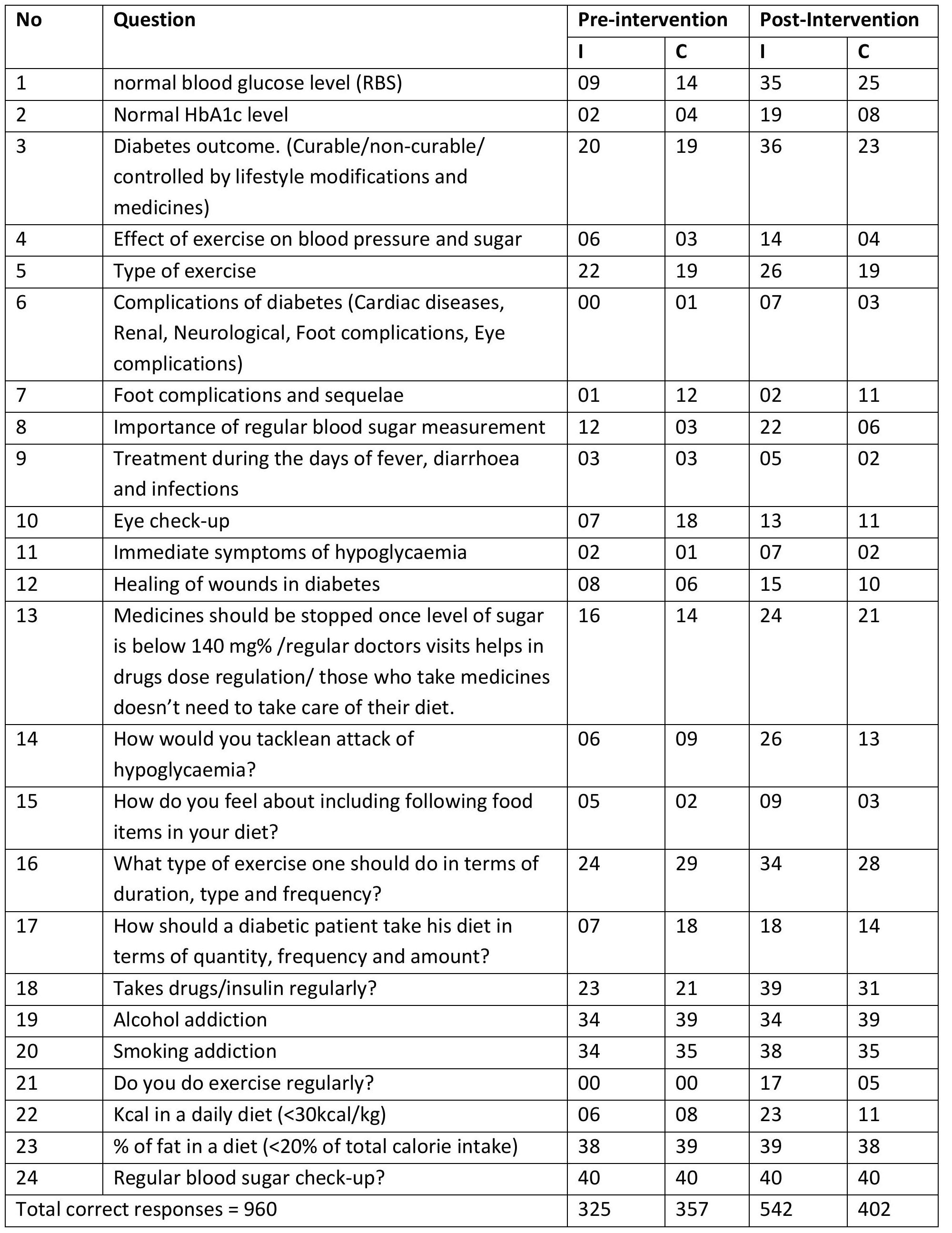
The proportions of correct responses to the questions in the questionnaire in Intervention and Control Group before after Counselling are shown in Table 1. At baseline the number of correct responses was more in Control Group than intervention group but this difference was statistically not significant. Before intervention in an Intervention Group, out of possible 960 correct responses only 325 (33.85%) were registered. This number of correct responses rose to 542 (56.45%) after intervention. Before intervention in a Control Group, out of possible 960 correct responses only 357 (37.18%) correct responses were registered, the number of correct responses rose to 402 (41.87%) on the second visit. Even though the number of correct responses in absolute terms increased in both the groups, Intervention group showed much more increase (22.60%) than the Control Group (4.59%).
At baseline patients in both Intervention and Control Group had poor knowledge about the various Table 1 — Level of Awareness in both Groups (Corrected Responses) (N=80)
Vol 121, No 2, February 2023Journal of
the Indian Medical Association
18
complications of Diabetes as well as management of hypoglycaemic episodes. Awareness about these aspects showed an increase at 4 months in both intervention and control group. Patients in both the groups at 0 months had knowledge about different types of exercises but this knowledge was not implemented as none of the patients were actually doing any regular exercise. At 4 months, the number of patients doing regular exercise increased in both Intervention and Control group, with more increase in intervention as compared to control group.
An unpaired t-test was applied to check the difference between diet of two groups. The results showed that before intervention the amount of calories (p=0.89) and fat percentage (p=0.50) both were statistically not different between the 2 groups. But after intervention the difference with reference to total Calories (p=0.0003) and Fat percentage (0.013) became statistically significant between intervention and control group (Table 2).
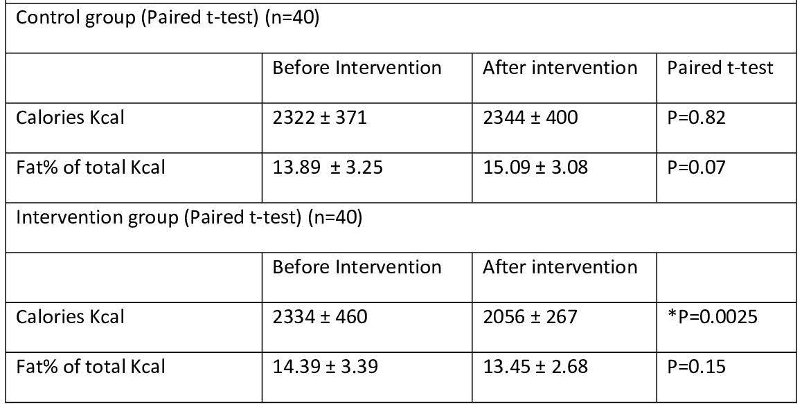
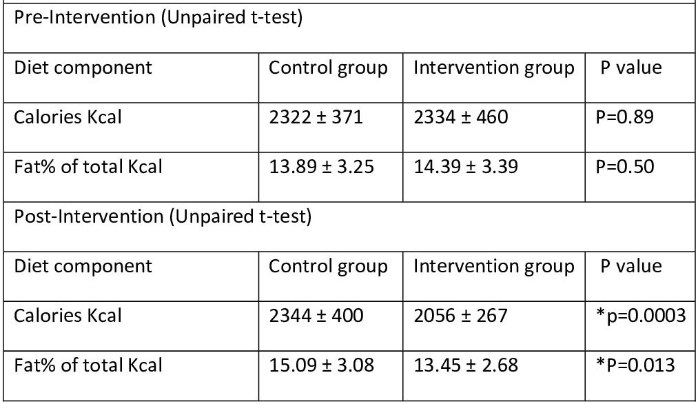
Paired t-test was used to see if there is any statistical difference in the Intervention Group as well as in the Control Group with respect to total calories intake and fat %.
The difference was statistically significant for total calories intake in the intervention group. (p=0.0025).For the fat % the difference was there in the means but the paired t-test suggested that this difference was statistically insignificant. (p=0.15) (Table 3).
The difference in the Control Group in pre and postintervention data was statistically insignificant with respect to total calories intake (p=0.82) and fat % (0.07). It is important to notice that the percentage of fat derived energy out of total energy increased in control group in second visit (Table 2).
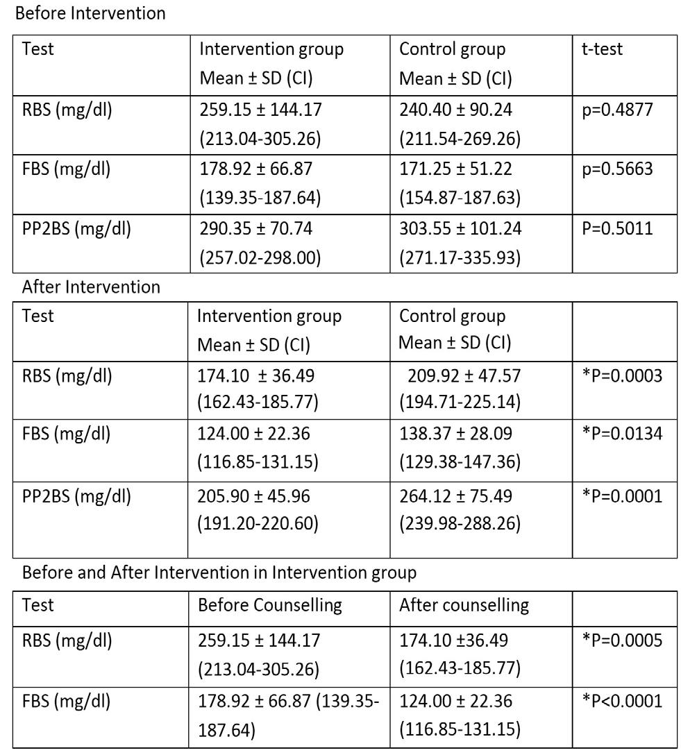
Metabolic control of Diabetes was measured by doing Plasma Glucose measurement. For this purpose RBS, FBS and PP2BS were done. Means of Blood Glucose measurements were calculated. Paired t-test in a Control Group showed significant difference for FBS (p=0.0006) but the difference with respect to RBS (p=0.06) and PP2BS (p=0.052) was statistically insignificant. For intervention group before and after difference with respect to all three parameters RBS (p=0.0005), FBS (p<0.0001) and PP2BS (p<0.0001) was statistically significant (Table 4).
After intervention 20 participants from Intervention Group and 35 participants from the Control Group had either their FBS level above the normal levels (126 mg/ dl) or their PP2BS levels above normal levels (>200 mg/dl). While before Intervention these numbers for Intervention and Control Group were 37 and 35 respectively. Thus before intervention 92.5%
participants of intervention group had poor Glycemic control and 87.5% participants of control group had poor Glycemic control.
In other words 50% patients from Intervention Group and only 12.5% patients from the Control Group could achieve good Glycemic control. Relative Risk (RR) of poor Glycemic control with respect to Counselling was 1.75. Attributable Risk (AR) of poor Glycemic control with respect to FBS in non-intervention participants was 42.86%.
Vol 121, No 2, February 2023Journal of the Indian Medical Association
Table 2 — Diet Comparison between Two Groups
Table 3 — Diet Comparison in Each Group
19
Table 4 — Effect of Councelling on the Blood Sugar Levels
DISCUSSION
Diabetes is a chronic, incurable condition that has considerable impact on the life of each individual patient. WHO projects that Diabetes will be the 7th leading cause of death in 2030. Healthy diet, regular physical activity, maintaining a normal body weight and avoiding tobacco use can prevent or delay the onset of Type2 Diabetes4. The vast majority of day-today care in Diabetes is handled by patients and/or families, so Counselling the patients to improve selfmanagement should be a central component of any effective treatment plan. Educational programs with in-home reinforcement can improve the selfmanagement of Diabetes and lead to improvement in health indicators5
In our study effect of Counselling the patients about self-management was measured with respect to the change in diabetic awareness, dietary patterns of the participants and Glycemic control.
Patients’ level of awareness about Diabetes was measured in terms of number of correct responses to the questions in Table 1. At the end of the study period the total number of correct responses increased in both the groups, but the intervention group showed greater increase than the control group and this difference was statistically significant. This implies that Counselling did improve the patients understanding about DM especially its complications, the management of hypoglycemic episodes and the importance of doing regular exercise. Several studies using such questionnaires also reported a similar positive impact of Counselling on patients knowledge about DM and its implementation in everyday life6-10.
At baseline patients in both intervention and control group had poor knowledge about the various complications of Diabetes as well as management of hypoglycaemic episodes. Awareness about these aspects showed an increase at 4 months in both intervention and control group. Patients in both the groups at 0 months had knowledge about different types of exercises, but this knowledge was not implemented as none of the patients were actually doing any regular exercise. At 4 months, the number of patients doing regular exercise increased in both intervention and control group with more increase in intervention as compared to control group.
Table 2 results shows that the difference between both the groups for total calories intake and % fat intake was statistically insignificant at the beginning of the study. The amount of K cal a diabetic takes during the whole day should be less than 2200 or 30 kcal/day. These means suggest that the caloric intake of participants in both the groups was on a higher side.
So the Counselling regarding this was very necessary. The energy derived from fat in these groups were within normal limits (<20%).
After the counselling the intervention group showed the improvement in terms of total energy intake (p=0.0025). But the Control Group did not show any improvement, rather the average Calorie intake slightly increased. Change in terms of fat in both the groups was not seen. And whatever change seen in an intervention was statistically insignificant in a group (p=0.15), while in the control group the % fat intake increased by a statistically significant amount (p=0.07). Since all the values of pre and post intervention for % fat intake are less than 20, we can say that the amount of energy derived was within normal limits.
Post Intervention difference between both the groups in terms of Calories (p=0.0003) and % Fat (p=0.013) was significant. Patients in the Intervention group were taking more healthy diet and they did modify their diet according to their needs. Since both groups were taking amount of fats within the recommended limits, there was still a scope for Carbohydrate reduction in some patients of the control group. Thus educating the patient about dietary changes did produce an improvement in their dietary patterns. This is similar to the results obtained in a study conducted by Krishnan D, Gururajan R, et al which showed that participants who received both dietary and exercise Counselling with periodic followup were generally likely to follow dietary principles more carefully and were more involved with their interactions with the Counsellor11
The effect of Counselling on Glycemic control was measured by comparing calculated means of RBS, FBS and PP2BS of the participants in both the groups. The analysis was done similar to the diet analysis. Results showed that the baseline (before Counselling) Blood Sugar in terms of RBS, FBS and PP2BS were comparable as there was no statistical difference. Paired t-test results showed improvement in all three Blood Sugar parameters in an intervention group, while the control group showed improvement in only RBS (p=0.0625) and FBS (p=0.0006) and not in PP2BS (p=0.51) levels. This might be due to improper knowledge and practice regarding the diet in a Control Group. Post intervention difference in the Blood Sugar parameters was significant for all three, RBS (p=0.0003), FBS (p=0.013) and PP2BS (0.0001). Thus there was a better Glycemic control in intervention group than Control Group (Table 3).
This is similar to result obtained in an interventional study done by Renuga E ,Vanitha Rani N, et al in 2014 in India stated that “There was a reduction in the
Vol 121, No 2, February 2023Journal
of the Indian Medical Association
20
mean FBS from baseline to the follow-up in both the groups but a statistically significant higher reduction in the mean FBS was found in the Intervention Group from baseline to the final follow-up when compared to the control group (p<0.001).” 12 In another study conducted by Ahmed MM, Degwy HME, et al statistically significant improvement was found in the mean levels of HbA1c and FBS after application of face to face diabetic education13.
Several other studies also showed that Counselling led to better Glycemic Control measured in terms of Glycated Haemoglobin(HbA1c)levels14-18. A study by Norris S L, Lau J, Jay Smith J S, et al showed that selfmanagement education improves Glycated Hb levels at immediate follow up and increased contact time increases the effect. The benefit declines 1-3 months after the intervention ceases, however, suggesting that learned behaviours change over time. They also stated that further research is needed to develop interventions effective in maintaining long-term Glycemic control19
Source of Financial Support in the form of grants : Nil
REFERENCES
1WHO. Facts and figures about diabetes [Internet]. 2016. Available from: http://www.who.int/diabetes/facts/ world_figures/en/.
2Greenwood DC, Threapleton DE, Evans CEL, Cleghorn CL, Nykjaer C, Woodhead C, et al — Glycemic index, glycemic load, carbohydrates, and type 2 diabetes: systematic review and dose-response meta-analysis of prospective studies. Diabetes Care [Internet]. 2013 Dec [cited 2018 Dec 13]; 36(12): 4166–71. Available from: http://www.ncbi.nlm.nih.gov/ pubmed/24265366
3Dong J-Y, Zhang L, Zhang Y-H, Qin L-Q — Dietary glycaemic index and glycaemic load in relation to the risk of type 2 diabetes: a meta-analysis of prospective cohort studies. Br J Nutr [Internet] 2011; Dec 29 [cited 2018 Dec 13]; 106(11): 1649-54. Available from: http://www.ncbi.nlm.nih.gov/ pubmed/22017823
4Alwan A — World Health Organization. Global status report on noncommunicable diseases 2010. World Health Organization; 2011. 162 p.
5Lavelle D, Zeitoun J, Stern M, Butkiewicz E, Wegner E, Reinisch C — Diabetes Self-Management Education in the Home. Cureus [Internet]. 2016 Jul 25 [cited 2018 Dec 13]; 8(7): e710. Available from: http://www.ncbi.nlm.nih.gov/pubmed/ 27588231
6Malathy R, Narmadha M, Ramesh S, Alvin JM, Dinesh BN — Effect of a diabetes counseling programme on knowledge, attitude and practice among diabetic patients in Erode district of South India. JYoungPharm[Internet] 2011 Jan [cited 2018 Nov 15]; 3(1): 65-72. Available from: http:// linkinghub.elsevier.com/retrieve/pii/S0975148311310114
7Al-Maskari F, El-Sadig M, Al-Kaabi JM, Afandi B, Nagelkerke N, Yeatts KB — Knowledge, Attitude and Practices of Diabetic Patients in the United Arab Emirates. Baradaran HR, editor. PLoS One [Internet] 2013 Jan 14 [cited 2018 Nov 15]; 8(1): e52857. Available from: http://www.ncbi.nlm.nih.gov/pubmed/ 23341913
8Kiberenge MW, Ndegwa ZM, Njenga EW, Muchemi EW — Knowledge, attitude and practices related to diabetes among community members in four provinces in Kenya: a crosssectional study. Pan Afr Med J [Internet] 2010 [cited 2018
Nov 15]; 7: 2. Available from: http://www.ncbi.nlm.nih.gov/ pubmed/21918691
9S B, M T, G N, R M, Reddy YP — International journal of pharmacy and pharmaceutical sciences. [Internet]. Vol. 6, International Journal of Pharmacy and Pharmaceutical Sciences. IJPPS; 2014 [cited 2018 Nov 16]. 456-461 p. Available from: https://innovareacademics.in/journals/ index.php/ijpps/article/view/2023
10P P — Impact of patient counseling on Knowledge, Attitude, Practice and Quality of Life in patients with Type II Diabetes mellitus and Hypertension. Indian J Pharm Pract [Internet] 2011 [cited 2018 Nov 16]; 4(1). Available from: http:// www.ijopp.org/article/285
11Raj Gururajan DK, Srinivas Kondalasamy AH-B, Hafez-Baig A, Kondalasamy-Chennakesavan S, Wickramasinghe N, Gururajan R — The Impact of Diet Counselling on Type 2 Diabetes Mellitus: An Indian Case Study. J Diabetes Metab [Internet] 2015 Sep 29 [cited 2018 Nov 16]; 6(10). Available from: https://www.omicsonline.org/open-access/the-impactof-diet-counselling-on-type-2 diabetes-mellitus-anindiancase-study-2155-6156-1000610.php?aid=61555
12Renuga E, Sr R, Rani N V — Impact of continous patient counselling on Knowledge, Attitude, and Practices and Medication adherence of diabetic patients attending outpatient pharmacy services. 2016 [cited 2018 Nov 16];9. Available from: https://innovareacademics.in/journals/index.php/ajpcr/ article/viewFile/9386/3727
13Ahmed M, Degwy H, Ali M, Hegazy N — The effect of educational intervention on knowledge, attitude and glycemic control in patients with type 2 diabetes mellitus. Int J Community Med Public Heal [Internet]. 2015 Feb 5 [cited 2018 Dec 13]; 2(3): 302-7. Available from: http://ijcmph.com/index.php/ ijcmph/article/view/970
14Arabia Fawzy Khalil Sharaf S — Impact of Health Education On Hba1c Level Among Diabetic Patients in Al-Qassim Region [Internet]. Vol. /, Annals of Alquds Medicine. 1434 [cited 2018 Dec 13]. Available from: https://annalqudsmed.files. wordpress.com/2013/06/sharaf-impact-of-health-educatio2013-gp2.pdf
15Zibaeenezhad MJ, Aghasadeghi K, Bagheri FZ, Khalesi E, Zamirian M, Moaref AR AF — The effect of educational interventions on glycemic control in patients with type 2 diabetes mellitus [Internet]. Vol. 9. InternationalCardiovascular Research Journal 2015 [cited 2018 Dec 15]. p. 17-21. Available from: http s://www.sid.ir/En/Journal/ ViewPaper.aspx?ID=436472
16Machado M, Bajcar J, Guzzo GC, Einarson TR — Sensitivity of Patient Outcomes to Pharmacist Interventions. Part I: Systematic Review and Meta-Analysis in Diabetes Management. Ann Pharmacother [Internet]. 2007 Oct 29 [cited 2018 Nov 16]; 41(10): 1569–82. Available from: http:// www.ncbi.nlm.nih.gov/pubmed/17712043
17Roppolo E, Bonetta M, Gilli S— The Role of Physical Counselling in Patients with Type 2 Diabetes Mellitus: A Systematic Review. J Diabetes Mellit [Internet]. 2015 [cited 2018 Nov 16]; 5: 97–110. Available from: http://www.scirp.org/journal/jdmhttp:// dx.doi.org/10.4236/jdm.2015.52012http://dx.doi.org/10.4236/ jdm.2015.52012http://creativecommons.org/licenses/by/4.0/ 18Wise PH, Dowlatshahi DC, Farrant S, Fromson S, Meadows KA — Effect of computer-based learning on diabetes knowledge and control. Diabetes Care [Internet]. [cited 2018 Nov 16]; 9(5): 504-8. Available from: http:// www.ncbi.nlm.nih.gov/pubmed/3533475
19Norris SL, Lau J, Smith SJ, Schmid CH, Engelgau MM — Selfmanagement education for adults with type 2 diabetes: a meta-analysis of the effect on glycemic control. Diabetes Care [Internet]. 2002 Jul [cited 2018 Nov 16]; 25(7): 1159-71. Available from: http://www.ncbi.nlm.nih.gov/pubmed/ 12087014.
Vol 121, No 2, February 2023Journal
of the Indian Medical Association
21
Original Article
High Fear & Stress in the Quarantine Population of COVID-19 in Southern Rajasthan : A Survey
Gazala Hitawala1, Sushil Kherada2, Ravindra kumar Gehlot3, Faizaan Faizee1, Lalit Kumar Raiger4
Background : The advent of the COVID-19 pandemic has caused a significant psychological impact on the General Public, Health Care Workers, Elderly, High-risk groups, etc. Higher fear is likely among the quarantine population.
Aim of this study : To evaluate the fear and stress of individuals in quarantine; to determine the possible factors that are influencing the Psychological reactions of the individuals in quarantine compared to the general population; to provide a basis for future Government policies.
Methods : A semi-structured questionnaire that included a pre-tested, 7-item Fear of COVID-19 Scale (FCV-19S) was used for data collection. A total of 245 responses were received. Through random sampling, 50 participants each were chosen from the general and quarantine populations. p-value <0.05 was considered significant.
Results : Individuals in quarantine had a greater fear of COVID-19 compared to the general population (p=0.0059). Symptomatic fears like clammy hands (p=0.032), sleep disturbance (p=0.00026) and heart palpitations (p=0.000034) were commoner in the quarantine population. The younger age group in the quarantine population was comparatively more affected by News and Social media (p=0.00018). Getting a negative screening test resulted in lesser fear both in the quarantine (p=0.017) and general populations (p=0.002).
Conclusion : The individuals under quarantine have greater fear possibly due to stressors like transmitting the infection to family, working on the frontlines, being in high-risk groups, losing jobs, and exposure to social media. However, negative screening tests were shown to reduce the fear.
[J Indian Med Assoc 2023; 121(2): 22-6]
Key words :COVID-19, Fear of pandemic, Psychological fear, Quarantine.
COVID-19 is a new respiratory infection outbreak that started in China in December, 2019. As of 4th May, 2020, a total of 42,533 cases and 1373 deaths were reported in India and 3,435,894 cases and 239,604 deaths all around the world 1 . By 12th November, 2021, the total number of confirmed COVID cases in India rose up to 34, 414, 186 and the total deaths reached to 462, 6902 The epidemic brought not only the risk of death from the viral infection but also unbearable Psychological pressure to people in China and the rest of the world. There have been reports on the Psychological impact of the COVID-19 pandemic on the Frontline Workers3, Students4, Health Care Workers5, Elderly6, etc.
Quarantine is the separation and restriction of movement of people who have potentially been exposed to a contagious disease to ascertain if they become unwell, hence reducing the risk of them infecting others. This definition differs from isolation, which is the
RNT Medical College, Udaipur, Rajasthan, 313001
1MBBS, Intern, Department of Psychiatry
2MD, Senior Professor and Head, Department of Psychiatry
3MD, Associate Professor, Department of Anaesthesiology
4MD, Senior Professor and Head, Department of Anaesthesiology and Corresponding author
Received on : 16/09/2021
Accepted on : 30/12/2021
Editor's Comment :
COVID-19 exacerbated fears Worldwide which is higher in who quarantine. The stressors which contributed to increase fear were losing jobs, economic crisis, transimitting infection to family members,social stigma high risk for future/ life threatening, and these people need counselling/ psychological support.
separation of people who have been diagnosed with a contagious disease from people who are not sick7
The Fear of COVID-19 Scale (FCV-19S) is a reliable and valid tool to assess fear as a Psychological reaction to the COVID-19 pandemic which is proven by studies in multiple Countries8. Fear of COVID-19 Scale is a seven-item, unidimensional scale with robust psychometric properties. It has been proven that the English version of the COVID-19S is a sound unidimensional scale with robust psychometric properties that can be used with confidence among English-speaking populations 9,10. Moreover, total scores on the FCV-19S are comparable across the Country, Gender and Age which suggests that it is a good Psychometric instrument to be used in assessing and allaying fears of COVID-19 among individuals8,11 However, no detailed study comparing the mental health status of the population under quarantine with the normal population has been conducted to date.
Vol 121, No 2, February 2023Journal
of the Indian Medical Association
22
The purpose of our study is :
(1) To evaluate the fear and stress of individuals in quarantine.
(2) To determine the possible factors that are influencing the Psychological reactions of the individuals in quarantine compared to the general population.
(3) To provide a basis for future Government policies. Materials and Methods: This analytical research study was conducted at RNT Medical College, Udaipur (Rajasthan), for which the data was collected during May - June 2020. The study protocol was approved by the Institutional Ethical Committee. [RNT/STAT/IEC/ 2020/426 Dated18/05/2020]
A cross-sectional survey was conducted, using a semi-structured, pre-tested questionnaire that obtained Socio-demographic information (like gender, age, residence, educational status, occupation), medical history and information regarding the COVID screening test (RT-PCR). Moreover, a previously validated and standardized instrument, the 7-item Fear of COVID19 Scale (FCV-19S) was used to evaluate the levels of fear8,12,9. The FCV-19S includes seven items that can be subdivided into 4 items based on emotional fear reactions and 3 items based on symptomatic expressions of fear. Respondents report their symptoms using a 5-item Likert rating scale that ranges from 1 (stronglydisagree) to 5 (strongly agree), such that the total score ranges from 7 to 3511.
The questionnaire along with the consent form was distributed through digital media and the data was collected via google forms. Appropriate ethical approval procedures were followed while taking consent from subjects and also in conducting the research. A total of 245 responses were received. Using the inclusion and exclusion criteria, the respondents were subdivided into quarantine and general. The inclusion and exclusion criteria comprised of :
Inclusion criteria : Age between 18 & 60 years. The quarantine population comprised individuals who had stayed under quarantine due to history of recent travel or history of contact with a COVID-19 patient or history of contact with a COVID-19 suspected patient. The general population comprised of individuals who had not stayed under quarantine.
Exclusion criteria : The participants with preexisting or previously diagnosed mental health disorders or history of treatment for any mood/anxiety disorder or symptoms of upper respiratory tract infection or a positive screening test for COVID-19 were excluded from the study.
Statistical Analysis : Slovin’s formula [N / (1 + Ne2)] was applied to calculate the sample size for the
quarantine population in the institute. Taking the confidence level as 95% the resulting sample size for the quarantine population was 50 with a margin of error of 10%. After implementing the inclusion and exclusion criteria, 50 participants were selected in the quarantine population through random sampling and for effective comparison, 50 participants were similarly selected in the general population. The collected data were analyzed with SPSS v22.0 software (IBM Corp, Armonk, NY). The means and Standard Deviation were calculated and compared using a two-tailed t-test. pvalue < 0.05 was considered statistically significant.
RESULTS
* Most of the respondents were between the age of 18 and 30 years and majority were males. Among the sample of the quarantine population, 86% were Health Care Workers (Table 1).
The quarantine population reported to have higher rates of symptomatic fear - clammy hands, sleep disturbance, Heart palpitations. Moreover, news and Social media had a significantly higher impact on the fear of COVID-19 among the quarantine population. There was no significant difference in the average scores between the quarantine population and the general population with regards to being afraid of COVID-19 and losing life to COVID-19 (Table 2).
Overall, there was a significant difference (p=0.0059) in the fear of COVID-19 between the quarantine (14.96 ± 5.510) and the general population (12.48 ± 2.908) on the FCV-19S.
Both males and females in the quarantine population had higher fear compared to the males and females in the general population, p=0.032 and p=0.022 respectively (p < 0.05) (Table 3).
In both the general and the quarantine populations, the females showed higher levels of fear (13.35 ± 2.54 & 17.23 ± 6.56 respectively) compared to the males (11.9 ± 3.03 & 14.16 ± 4.95 respectively). However, the results were not significant.
The 2 ends of the age groups (18 - 30 years and 45
Vol 121, No 2, February 2023Journal of the Indian
Medical Association
DemographicGeneral Quarantine (n= 50)(n= 50) SexMale30 (60%)37
Female20 (40%)13
Age18 – 30 Years *34 (68%)33
31
Years14
45
Education Undergraduate 27 (54%)21
Postgraduate13 (26%)17
Others10 (20%)12
Occupation Health Care Workers19
Others31 (62%)7
ScreeningYes 16 (32%)34
test doneNo34 (68%)16
Table 1 — Demographic profile
(74%)
(26%)
(66%)
– 44
(28%)13 (26%)
– 60 Years2 (4%)4 (8%)
(42%)
(34%)
(24%)
(38%)43 (86%)
(14%)
(68%)
(32%)
23
makes me uncomfortable to think about
hands become clammy when I think of
- 60 years) showed higher fear in the quarantine population as compared to the general population (p=0.000969, p=0.0048). Whereas the 31- 44 years group showed no difference in the levels of fear between the general population and the quarantine population (p = 0.24). In addition to that, on comparing the scores for the question - “When watching news and stories about COVID-19 on Social media, I become nervous and anxious” in the 18-30 years age group, the quarantine population was more affected by news and social media compared to the general population (2.71±1.159 versus 2.0 ± 0.862, p = 0.00018).
The Postgraduates and Undergraduates in the quarantine population had higher levels of fear compared to the general population (p = 0.036, p = 0.020) while those classified as “others” (which included respondents who have done diploma course or attended high school only) had no difference in the level of fear between the quarantine and the general population (p = 0.94). Similarly, the Health Care Workers and other occupational categories showed greater fear in the quarantine population (p = 0.037, p = 0.029). The respondents who had not taken a screening test for COVID-19 had higher levels of fear in the quarantine population compared to those in the general population (p = 0.0000302) (Table 4).
On comparing respondents within the same population, those who had taken a screening test for COVID-19 showed lesser fear both in the quarantine and the general population. Since our study did not include COVID-19 positive patients, those respondents who took a screening test and received negative results eventually had significantly less fear of COVID-19 (p=0.017, p=0.002)(Table 5).
DISCUSSION
The main goal of this study is to compare the psychological reaction and fear arising from the COVID19 outbreak between the general population and the
population that stayed in quarantine and explore different factors that are influencing their levels of fear. This study indicates that the average score in terms of fear of COVID-19 was higher in the quarantine population (14.96 ± 5.51) compared to the general population (12.48 ± 2.908).
Reports indicate that various factors like unpredictability, uncertainty, seriousness of the disease, misinformation and Social isolation play a role in contributing to stress and mental morbidity13. Similarly, studies on the Psychological impact of the quarantine suggested that not being able to see friends and family members, worry of infecting their family members and confinement also play a role in the psychological effects of the quarantine population14
Vol 121, No 2, February 2023Journal of the Indian Medical Association
Category Average ScoreSDp value I
2.521.070.57 General 2.641.08 It
COVID-19Quarantine 2.541.150.04 General 2.120.96 My
COVID-19Quarantine 1.660.850.03 General 1.360.48 I am
COVID-19Quarantine 1.820.830.90 General 1.840.91 When
COVID-19Quarantine 2.681.20.002
anxiousGeneral 2.020.91 I cannot sleep
I’m
aboutQuarantine 1.720.83 <0.001 getting COVID-19 General 1.220.42 My heart races or palpitates when I think aboutQuarantine 2.021.09 <0.001 getting COVID-19 General 1.280.49
Table 2 — Average score of individual items of the FCV-19S Question
am most afraid of COVID-19Quarantine
afraid of losing my life because of
watching news and stories about
on social media, I become nervous or
because
worrying
Male Female Category Quarantine General Quarantine General Count (%)37 (74%)30 (60%)13 (26%)20 (40%) Average 14.1611.917.2313.35 SD4.953.036.562.54 p value 0.030.02
Table 3 — Average score comparison of the same sex group between the quarantine population and the general population
Average Scores QuarantineSD Q GeneralSD Gp value Age Groups : 18 – 30 years 16.24 5.3504 12.742.49< 0.001 31 – 44 years 10.774.41812.573.480.21 45 – 60 years 18.002.4497.500.7070.004 Education : Undergraduate 16.005.72312.083.3530.03 Postgraduate 15.815.78512.812.6320.02 Others 12.003.76612.103.2130.94 Occupation : Healthcare Workers14.775.43311.893.3810.03 Others 16.146.28312.842.5700.02 Screening Test Done : Yes13.715.54110.693.5720.05 No17.634.53013.322.114< 0.001
Table 4 — Average score comparison based on demographics between the general and the quarantine population
Screening Test Done AverageSDp value GeneralYes10.697 3.5720.002 No13.322.114 QuarantineYes 13.715.5410.01 No17.634.530 24
Table 5 — Screening test comparison within the same
population
When considering the fear of COVID-19 in relation to the various demographic factors, while some studies showed higher levels of fear and a greater Psychological impact on females15, others did not show a significant difference in the Psychological impact between males and females4. In our study we did not find a significant difference between the fears of the males and females within the same population. This can be due to the difference between the number of males and females both in the quarantine (males 74% females 26%) and the general population (males 60%, females 40%).
However, when we compared the males and females in the general population with the males and females in the quarantine population respectively, significantly higher levels of fear were seen in the quarantine population for both males (p = 0.032) and females (p = 0.022).
On comparing different age groups, we found that age ranges depicted a variation in the level of fear between the quarantine and the general population. While the age groups of 18-30 years (p=0.000969) and 45-60 years (p=0.004) had a much higher level of fear in the quarantine population, there wasn’t a significant difference in the level of fear in the age group of 31-44 years. This is supported by a previous study which states that the younger population are much more exposed to Social media than the middle aged and the elderly which can contribute to a greater Psychological impact16. In our study this is explained by comparing the results of the 18-30 years age group’s average score for the question - “When watching news and stories about COVID-19 on Social media, I become nervous or anxious”. This had a significantly higher average value (p = 0.00018) in the quarantine population compared to the general population. It is suggested that young people can easily trigger stress as they tend to collect information from social media17. The quarantine population were Health Care Workers and their higher levels of fear can be attributed to seeing their patients die, worry about their own safety, exhaustion due to increased duration of work and fear of other colleagues who have tested positive for COVID-194. Along with this, it is reported that the Social disconnectedness and perceived isolation can result in higher levels of anxiety and depression in the elderly6. The elderly, are at a heightened risk of the Psychosocial outcomes of the COVID-19 pandemic18, which was also observed in our study.
The educational groups (Undergraduates and Postgraduates) reported having higher fears in the quarantine population. This stems from their awareness about the gradually increasing distances between the people resulting from the quarantine, the effect of the virus on their studies and future employment4.
Occupationally, both Health Care Workers (p=0.037) and individuals from other occupations (p=0.029) had significantly higher levels of fear when staying under quarantine. The Psychological impacts on the Health Care Workers are largely supported by various studies that highlight that the Health Care Workers are afraid due to multiple reasons. Some of them include the high risk of the infection and inadequate protection from contamination, frustration, isolation, lack of contact with family5 and the fear of infecting their families or seeing their patients die3
Similarly, studies during the SARS outbreak report: acute stress in the quarantine population can be attributed to getting back in quarantine after resuming work as a Health Care Workers on the frontlines and the duration of quarantine19,14. The participants from other occupational categories who were under quarantine also showed higher fears which might be related to the future employment opportunities3, economic crisis20, and the stigma associated with COVID-19.
Getting a screening test has been attributed to lesser fear both within the quarantine (p = 0.017) and the general population (p = 0.002). Similarly, when we compared respondents between the quarantine and the general population based on whether or not they have taken a screening test for COVID-19, there was a significantly higher fear in the quarantine population (p = 0.0000302) who had not yet taken a screening test for COVID-19. But there was no significant difference in the fear of the respondents who had already taken the screening test and eventually tested negative. A screening test that confirms the negative status of the infection is helpful in reducing the stress and fear levels. Similar to this, previous studies in China found that fabricated or false reports about COVID-19 infection resulted in worse Psychological outcomes21
Our study suggests that the quarantine population has greater fears and anxiety compared to the normal population due to COVID-19 related stressors. These include close contact with a positive or a suspected patient, working on the frontlines, economic stressors and being in the high risk age groups. The symptomatic fear like disturbance in sleep, heart palpitations and clammy hands were found to be of significant intensity in the quarantine population compared to the general population.
News and Social media play a major role in the Psychological reaction of the younger individuals; measures to censor the News and Social media platforms regarding COVID-19 related information should be taken. Increasing the number of screening tests can help in flattening the curve of the infection and at the same time, it can help in reducing the stress
Vol 121, No 2, February 2023Journal of the Indian Medical Association
25
and fear among the individuals. Since isolation and quarantine can be fearful, proper counselling and support should be provided to those staying in quarantine. The availability of proper protective equipment and scales to evaluate the mental health of Health Care Workers with appropriate counselling and therapy can be helpful. High risk groups such as the elderly should be screened for mental health problems, provided Psychosocial support and Psychoeducation.
Limitations : The FCV-19S is based on Likert-scale which provides 5 choices to the respondents and it is likely that people avoid choosing the “extreme” options on the scale, because of the negative implications involved.
However, the scale is generalizable and has proven reliability and validity7
Conclusion : The emergence of COVID-19 has exacerbated fears Worldwide which is even higher in those who are staying in quarantine due to a history of travel or contact with suspected or positive patients. There are stressors which have contributed to increased fear and anxiety in the quarantine population. These stressors include situations like losing jobs, economic crisis, transmitting infection to family members, being in the high risk age group, working on the frontlines, exposure to media and societal stigma. However, negative screening test results have reduced anxiety and fear.
The mental health of the individuals in quarantine is significantly affected in the COVID-19 pandemic compared to the general population and they require attention, help and support from their families and the society. The Government should work towards providing timely Psychological services to those staying in quarantine.
REFERENCES
1Coronavirus disease (COVID-19) situation report – 105. https:/ /www.who.int/docs/default-source/coronaviruse/situationreports/20200504-covid-19-sitrep-105.pdf?sfvrsn= 4cdda8af_2 (accessed May 5, 2020)
2The current COVID-19 situation http s://www.who.int/ countries/ind/ (accessed November 15, 2021)
3Cai H, Tu B, Ma J, Chen L, Fu L, Jiang Y et al. Psychological Impact and Coping Strategies of Frontline Medical Staff in Hunan Between January and March 2020 During the Outbreak of Coronavirus Disease 2019 (COVID-19) in Hubei, China. Med Sci Monit 2020; 26: e924171. doi:10.12659/ MSM.924171
4Cao W, Fang Z, Hou G, Han M, Xu X, Dong J — The psychological impact of the COVID-19 epidemic on college students in China. Psychiatry Res 2020; 287: 112934. doi:10.1016/j.psychres.2020.112934
5Kang L, Li Y, Hu S, Chen M, Yang C, Yang BX — The mental health of medical workers in Wuhan, China dealing with the 2019 novel coronavirus. Lancet Psychiatry 2020; 7(3): e14. doi:10.1016/S2215-0366(20)30047-X
6Santini ZI, Jose PE, York Cornwell E, Koyanagi I, Nielsen L,
Hinrichsen C et al. Social disconnectedness, perceived isolation, and symptoms of depression and anxiety among older Americans (NSHAP): a longitudinal mediation analysis. Lancet Public Health 2020; 5(1): e62-e70. doi:10.1016/ S2468-2667(19)30230-0
7Centers for Disease Control and Prevention. Available from: https://www.cdc.gov/quarantine/index.html (accessed June 29, 2020)
8Lin CY, Hou WL, Mamun MA — Fear of COVID-19 Scale (FCV-19S) across countries: Measurement invariance issues. Nurs Open 2021; 8(4): 1892-908. doi:10.1002/nop2.855
9Winter T, Riordan BC, Pakpour AH — Evaluation of the English Version of the Fear of COVID-19 Scale and Its Relationship with Behavior Change and Political Beliefs. Int J Ment Health Addiction 2020; https://doi.org/10.1007/s11469-020-00342-9
10Mahmood QK, Jafree SR, Qureshi WA — The Psychometric Validation of FCV19S in Urdu and Socio-Demographic Association with Fear in the People of the Khyber Pakhtunkhwa (KPK) Province in Pakistan. Int J Ment Health Addiction (2020). https://doi.org/10.1007/s11469-020-00371-4
11Ahorsu DK, Lin CY, Imani V, Saffari M, Griffiths MD, Pakpour AH — The Fear of COVID-19 Scale: Development and Initial Validation. Int J Ment Health Addict 2020; Mar 27: 1-9. doi: 10.1007/s11469-020-00270-8.
12Sakib N, Bhuiyan AKMI, Hossain S, Mamun FA, Hosen I, Abdullah AH — Psychometric Validation of the Bangla Fear of COVID-19 Scale: Confirmatory Factor Analysis and Rasch Analysis. Int J Ment Health Addict 2020; 1-12. doi:10.1007/ s11469-020-00289-x
13ZandifarHYPERLINK “http s://www.sciencedirect.com/ science/article/abs/pii/S1876201820300988” A., HYPERLINK “https://www .sciencedirect.com/science/article/pii/ S1876201820301775”Badrfam R. Iranian mental health during the COVID-19 epidemic. Asian Journal of Psychiatry. 2020; 51:101990
14Hawryluck L, Gold WL, Robinson S, Pogorski S, Galea S, Styra R — SARS control and psychological effects of quarantine, Toronto, Canada. Emerg Infect Dis 2004; 10(7): 1206-1212. doi:10.3201/eid1007.030703
15Wang C, Pan R, Wan X, Tan Y, Xu L, Ho CS — Immediate Psychological Responses and Associated Factors during the Initial Stage of the 2019 Coronavirus Disease (COVID-19) Epidemic among the General Population in China. IntJEnviron Res Public Health 2020; 17(5): 1729. doi:10.3390/ ijerph17051729
16Srivastava K, Chaudhury S, Bhat PS, Mujawar S — Media and mental health. Ind Psychiatry J 2018; 27(1): 1-5. doi:10.4103/ipj.ipj_73_18
17Ahmed MZ, Ahmed O, Aibao Z, Hanbin S, Siyu L, Ahmad A — Epidemic of COVID-19 in China and associated Psychological Problems. AsianJPsychiatry 2020; 51: 102092. doi:10.1016/ j.ajp.2020.102092
18Pfefferbaum B, North CS — Mental Health and the Covid-19 Pandemic. N Engl J Med 2020; 10.1056/NEJMp2008017. doi:10.1056/NEJMp2008017
19Brooks SK, Webster RK, Smith LE, Woodland L, Wessely S, Greenberg N, et al — The psychological impact of quarantine and how to reduce it: rapid review of the evidence. Lancet 2020; 395(10227): 912-20. doi:10.1016/S01406736(20)30460-8
20Ayittey FK, Ayittey MK, Chiwero NB, Kamasah JS, Dzuvor C — Economic impacts of Wuhan 2019-nCoV on China and the world. J Med Virol 2020; 92(5): 473-5. doi:10.1002/jmv.25706
21Zhou, SJ., Zhang, LG., Wang, LL — Prevalence and sociodemographic correlates of psychological health problems in Chinese adolescents during the outbreak of COVID-19. Eur Child Adolesc Psychiatry 2020; 29: 749-58 (2020). https:// doi.org/10.1007/s00787-020-01541-4
Vol 121, No 2, February 2023Journal of
the Indian Medical Association
26
of the Indian Medical Association
Original Article
Morbidity Pattern among the Farm House Residents in Vijayapur District, Karnataka — A Cross Sectional Study
Sandeep Gurunath Yankanchi1, Rekha Udigiri2
Background : Agriculture Workers have a multitude of health problems, a fact which is often forgotten because of widespread misconception that occupational health is mainly concerned with industry and industrialized countries. The health problems of workers in agricultural field may be accidents (Snake and insect bites), toxic hazards (chemical exposure and insecticide poisoning), physical hazards (extreme conditions and solar radiation) and respiratory problems (farmer’s lung and occupational asthma).
Objectives : To study the morbidity pattern among the Farm house residents.
Material and Methods : A cross sectional study was conducted among the farm house residents in rural areas of Vijayapura district. A Sample of 450 farm house residents were interviewed by pre-structured proferma containing information regarding Socio demographic profile, present and past six months morbidities. In each Taluka, the selection of households was done by considering villages as the Primary Sampling Unit (PSU). PSUs were selected with probability proportional to size sampling and 5 households in a selected PSU were selected by random sampling.All characteristics were summarized descriptively, Chi-square ( χ2) test was employed to determine the significance of differences between groups for categorical data.
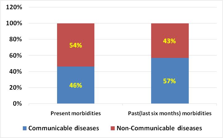
Results : The findings of the present study among Farm dwellers in the rural area of vijayapura district revealed that majority at the time of study were having Anaemia followed by Respiratory Infection and majority of Farm dwellers in past six months were having Dental carries as a morbidity followed by Respiratory infection.
Conclusion : The present study concludes that overall majority of the Farm house residents presently suffering from Non-communicable Diseases (54%) followed by Communicable Diseases (46%).
[J Indian Med Assoc 2023; 121(2): 27-32]
Key words :Morbidity pattern, Farm house, Household, Agriculture.
Agriculture is an art/practice of cultivating land. Agriculture sector occupies a key position in our Country. It provides employment to about 65% of the working population of India. Agricultural Workers constitute by far the largest segment in the unorganized sector. Agriculture workers constitute the most neglected classes in the Indian Rural structure. Their income is low and irregular. They do not possess any skill and training and have no alternate employment opportunities1
Agriculture is essential for good health as it produces food, fibre and materials for shelter along with medicinal plants. It is also an important source of livelihood in many of the middle and lower income countries2 Agriculture as an occupation differs from another occupation in that, workers work in the open fields, exposing themselves to extremes of climates and also there are no ‘Labour laws’ in practice. The
Department of Community Medicine, BLDE-DU Shri B M Patil Medical College Hospital & Research Centre, Karnataka 586103
1MD, Assistant Professor and Corresponding Author
2MD, Professor
Received on : 10/01/2022
Accepted on : 07/04/2022
Editor's Comment :
Agriculture is the backbone of the country as Agriculture sector occupies a key position in our country so farm house residents health is utmost important.
health problems of workers in agricultural field may be accidents (Snake and insect bites), toxic hazards (chemical exposure and insecticide poisoning), physical hazards (extreme conditions and solar radiation) and respiratory problems (farmer’s lung and occupational asthma)3.
According to the Karnataka Land Revenue (Amendment) Act, 2015 :
“Farm Buildings” or “Farm house” means a house attached to a farm and constructed in a portion of an agricultural land, used for the residence of the agriculturist or used for the purpose of keeping Agricultural equipment’s and tethering cattle. The house shall be used by a farmer for his own use and it shall not be let out for commercial activities to any individual or agency. “Amendment of section 95.- Inside section 95 of the principal Act, - (a) after sub-section (1) state Farm building or Farm house so erected shall
Vol 121, No 2, February
2023Journal
27
not be more than ten percent of his holding subject to a maximum of such extent of land as may be prescribed4 The Farm house workers are so remotely dispersed in Rural area that the health services may not reach them. Data regarding morbidity pattern among Farm house dwellers is very sparse. Community based study can only reflect the true picture of morbidity pattern in a given Community. Hence the present study was undertaken to explore the morbidity pattern among the Farm house residents of Vijayapura District.
MATERIALS AND METHODS
This was a descriptive cross-sectional study conducted among the Farm house residents of Vijayapura District. The study was done over a period of one year (June, 2017 – May, 2018).
After obtaining ethical clearance from the Institutional Ethical Committee the study was conducted in Vijayapura District. Geographically Vijayapura District has been divided into five Talukas, namely Vijayapur, Indi, Sindgi, Basavana Bagevadi and Muddebihal. Within each Taluka, the selection of households was done in different stages considering villages as the Primary Sampling Unit (PSU)5 Villages, where the number of households was less than 5 were not considered in the selection of samples and removed from the list. Allocation of the total sample population of 384 (~400) in Farm households is done in proportion to their population. Households have been selected in two stages. PSUs were selected with Probability Proportional to Size (PPS) sampling and 5 households in a selected PSU were selected by random sampling.
The List of Households Staying in Farm was taken from the Government Primary Health Centre and chits containing the head of the family were made. Total 5 chits from each village were selected randomly and included in the study.
From each household four participants randomly were interviewed regarding morbidity pattern. If any selected household did not contain 4 participants, was excluded and new household was selected randomly. The Household members were reached with the help of ASHA / Health worker of PHC which helped to develop rapport with people staying in the household. The purpose and overview of the study was explained at the time of the interview and interviewers were informed that their participation was entirely voluntary, their anonymity would be assured and consent was taken.
Distribution of Sample :
Mean number of person per household (HH) = 4(on the basis of pilot observation in a nearby village)
Hence, Total number of HH in Farm houses = 400/ 4 = 100
Mean number of HH in farm houses per village = 4.7 (~ 5) (on the basis of pilot observation in a nearby village)
Total number of PSU (Villages) = 100/4.7 = 21
The sample size was calculated based on the formula. n= z2pq/d2 Due to lack of information on morbidity among the farm house residents in the study area, the calculation was based on the assumption of prevalence to be 50%. Assuming a confidence level of 95% and at a precision of 5%, the total sample size was 384 farm house residents. A round of sample of 384 (~400) was taken for the study, but the collected sample size was 450. The Study was conducted in Vijayapura District, situated in the Northern part of Karnataka. Farming and agriculture related business is the main occupation for many people in the district. People residing in Farm houses for less than 6 months were excluded from the study. Investigation like Haemoglobin estimation by using Mission HB instrument And Blood Sugar Estimation by Using Accu-Chek Active Glucometer. All characteristics were summarized descriptively, Chi-square (χ2) test was employed to determine the significance of differences between groups for categorical data. Data were analysed using SPSS software v.23.0.
RESULTS
A total of 450 were the study participants, majority of male participants belonged to age group of 41-50 (21.4%) years and female participants belonged to age group of 11-20 (21.7%) years. The major proportion of males (97.8%) and female participants (96.8%) belonged to Hindu religion. 58.1% of male and 62.9% female participants belonged to nuclear family followed by 29.7% male and 25.8% female participants belonged to joint family. The majority of male (49.8%) and female (48.9%) participants were illiterates. More than 50% of the participants belonged to class V Socio-economic status (Table 1).
Among Study participants (n=450), 66% were presently suffering from various diseases, Among which majority of the participants 54% were having Non Communicable Diseases like Anaemia, Hypertension, Diabetes Mellitus, Accidents, Arthritis and 46% were having Communicable Diseases like Gastro-enteritis, Respiratory infections, Dental caries, Fever.
Majority of participants 58% in the last six months had suffered from various diseases, Among which majority of the participants 57% were having Communicable Diseases like Gastro-enteritis, Respiratory infections, Dental caries and Fever. 43%
Vol 121, No 2, February 2023Journal of the Indian Medical Association
28
Table
variables
were having Non Communicable Diseases like Hypertension, Diabetes Mellitus, Accidents, Arthritis, Scorpion bite, Snake bite,Cataract, Corneal scar, Hearing loss, Skin diseases (Fig 1).
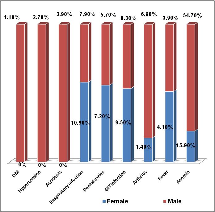
We observed current morbidity status of participants, majority of the male participants (54.7%) and female participants (15.9%) were diagnosed as anaemia. Respiratory infection in 7.9% of males and 10.9 % females’ participants.Accidents were reported among 3.9% of male participants only (Fig 2).
The maximum proportion of participants were suffering presently from Noncommunicable Diseases like Anaemia (127) arthritis (18), Accidents (9), Hypertension (5) and Diabetes mellitus (2) followed by Communicable disease like Respiratory infection (49), Gastrointestinal infection (40), Dental caries (29),
Fever (18). Non-communicable was the commonest condition associated with the participants of 41-50 years age group who were illiterate and lived in nuclear families. The statistically significant association was observed between present morbid conditions with related to age, sex, type of family, educational status, occupation (Table 2).
In our study, we recorded past 6 month morbid condition also, 18.6 % of the female participants and 13.5% of male participants reported Dental caries. H/o known case of Hypertension was present among 4.3% and 1.3% of male and female participants respectively. Similarly H/o of Diabetes Mellitus was present among 3.7% and 1.3% of male and female participantsrespectively. Scorpion bite (7.7%) and Snake bite (4.1%)
Vol 121, No 2, February 2023Journal
of the Indian Medical Association
Parameters Male FemaleTotal N%N%N% Age : <10198.33114.05011.1 11-203615.74821.78418.7 21-3041 17.93114.07216.0 31-4040 17.54620.88619.1 41-5049 21.43917.68819.6 51-6023 10.0198.6429.3 61-7093.973.2163.6 >70125.200.0122.7 Religion : Hindus224 97.821496.843897.3 Muslims52.273.2122.7 Type of family : Nuclear133 58.113962.927260.4 Joint68 29.75725.812527.8 Three Generation28 12.22511.35311.8 Educational Status : Illiterate114 49.810848.922249.3 Primary81 35.48036.216135.8 Secondary3113.53013.66113.6 PUC And Above31.331.461.3 Occupation : Student43 18.86931.211224.9 Labour104.494.1194.2 Household Activities62.6188.1245.3 Farmer170 74.212556.629565.6 SE-Status : Class Iv111 48.510145.721247.1 Class V118 51.512054.323852.9 Total229100.0221100.0450100.0
1 — Distribution of respondents according to Socio-demographic
Fig 1 — Proportion of present and past morbidities (last six months) among study participants
29
Fig 2 — Gender-wise distribution of present morbid status of the respondents
reported more among females compared to male participants (Fig 3).
During last six months morbid condition, the majority of participants had Communicable disease like Dental caries (72) followed by Respiratory infection (49), Gastro-intestinal infection (19) fever (9) followed by Non-Communicable disease like Accidents (20), Scorpion bite (27), Snake bite (12), Arthritis (12), Hypertension (10), Diabetes Mellitus (9), Cataract (9), Skin Disease (9), Corneal Ulcer (3), Hearing Loss (3). Communicable Diseases were the commonest condition associated with the participants of 31-40 years of age group who were illiterate and were farmers living in nuclear families and maximum number of females suffered from Communicable Diseases compared to male’s participants. This statistically significant association was observed between last six months morbid conditions and Age, Sex, Education,
Occupation & Socio-economic status (Table 3). DISCUSSION
Farming is a lifelong occupation. The farmers live near the Farm land and are often exposed to the environmental hazard throughout their life. Agricultural work tends to be a family occupation and all members of a family are involved in field activities.Rapid technological development in the Agricultural Sector has tremendously improved in last 25 years. The new innovations have increased production. They have also given rise to new variety of problems related to safety and health. Broadest and most extensive exposure to injury, diseases are suffered by Agricultural Workers.
In present Study, Maximum proportion of participants had Anaemia (36.9%) followed by Respiratory infection (9.3%), Gastrointestinal infection (8.9%), Dental caries (6.4%), Fever (4%), Arthritis (4%), Accidents (2%), Hypertension (1.5%), Diabetes Mellitus (0.6%). Majority of male participants (54.7%)
(in numbers)
Vol 121, No 2, February 2023Journal of
the Indian Medical Association
Parameters Communicable Non-X2 & diseases CommunicableP Valve diseases Age : <10193 37.289 10-202711< 0.0001 21-302124 31-402729 41-502346 51-60924 61-70412 >70612 Sex : Male62132 43.119 Female7429< 0.0001 Religion : Hindus129158 2.443 Muslims730.1180. Type of Family : Nuclear8881 8.137 Joint38540.0171. Third Generation1026 Education : Illiterate8198 10.696 Primary3353 0.0135. Secondary207 Puc and Above23 Occupation : Student37 741.994 Labour12 8<0.0001 Household Activities82 Farmer79144 Se Status : Class Iv7267 3.799 Class V6494 0.0513 TOTAL136161 Note: *significant at 5% level of significance (p<0.05)
Table 2 — Association of socio-demographic variables with present morbid conditions (in numbers)
ParametersCDNCDX2 & P value Age: <10141033.079 10-203113 <0.0001 21-30245 31-403628 41-503426 51-60714 61-7038 >70010 Sex : Male69705.906 Female8044 0.0151 Religion : Hindus144112 0.6392 Muslims52 0.4240 Type of Family : Nuclear98690.9239 Joint37310.63.1 Third Generation1414 Educational status : Illiterate8067 10.801 Primary3839 0.0128 Secondary288 Puc And Above30 Occupation : Student39 2313.545 Labour12 00.0036 Household Activities59 Farmer9382 SE Status : Class Iv65686.635 Class V8446 0.01 TOTAL149114 Note : *significant at 5% level of significance (p<0.05) 30
Table 3 — Association of socio-demographic variables with past morbid conditions
reported with Anaemia compared to female participants (15.9%) and maximum number of females suffered from Respiratory infection compared to males participants respectively. Accidents were reported among 3.9% of male participants only. Verma V, et al, in Rural area of Allahabad District of Uttar Pradesh observed that Overall, most prevalent diseases were linked to Ocular, Musculoskeletal, Psychological system, Gastrointestinal System and Dental Disorder affecting 274 (68.5%), 239 (59.75%), 119 (29.75%), 100 (25%), 94 (23.5%), of elderly respectively. The prevalence of Anaemia (43%), Under-nutrition (38.5%) and Respiratory problems (16%) were more in Rural aged6. Sharma D, et al, in Rural area of north India reported the morbid condition like Musculoskeletal problems (56.5%), Hypertension (25%0, Cataract (37%), Dental problems (33%), Asthma (4%), Corneal opacity (1.5%) and a significantly higher proportion of women suffered from Musculoskeletal problems (females: 66.7% versus males: 42.7%), Hypertension (females: 48% versus males: 32.7%), Diabetes (females: 7.8% versus males: 3.6%), while chronic Obstructive Pulmonary Disease (males: 14.3% versus females: 0.4%) was observed more in men7. According to DLHS IV survey Karnataka, reported that prevalence
of morbidity in Rural area was mainly injury (3.4%), acute illness (5.7%) and chronic illness (5.3%)8 Gupta SK, et al, in Rural area of Madhya Pradesh found that the prevalence of Anaemia was 42%. However, the prevalence of Anaemia was high in females (82%) compared to males (18%)9
The proportion of Anaemia was more in males compared to female participants probably due to walking barefoot in field area may leads to worm infestations. The percentage of females suffering from Respiratory infection was more compared to male participants would be due to use of smoke forming challahs in Farm house. Accidents were reported among the male participants probably due to use of farming tools & machineries.
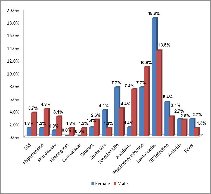
Among Last six months morbidity condition, majority of participants had Dental caries(16%) followed by Respiratory infection (9.3%), Accidents (4.4%), Scorpion bite (6%), Gastrointestinal infection (4.2%), Snake bite (2.7%), Fever (2%), Arthritis (2.7%), Hypertension (2.9%), Diabetes Mellitus (2.6%), Cataract (2%), Skin Disease (2%), Corneal Ulcer (0.7%), Hearing Loss (0.7%). A study done by Rahman SJ, et al in a Rural block of Jorhat District, Assam revealed that, (54.25%) of the farmers suffering from Respiratory Tract Infections followed by Musculoskeletal problems (23.25%) and Gastrointestinal Tract Ailments (11.75%)10. According to Hameed S, et al Study reported that the proportion of Arthritis, Anaemia and Obesity were significantly higher among females than among male participants11
Ahmeed SM, et al in his study showed, there was significant association found between Fever, Skin, Eye, Ear problems with Age and Gender12. A study done by Kulkarni RR, et al in a Rural area of Belgaum District observed majority of Agriculture Workers were having Dental caries (25.50%), Dental stains (21.75%), followed by Musculoskeletal System (21.75%) and Respiratory System (19%)13 Kansal S, et al, in Rural community of Eastern Uttar Pradesh revealed that
Vol 121, No 2, February 2023Journal of the Indian Medical Association
Fig 3 — Gender-wise distribution of past (last six months) morbid status of the respondents
31
Respiratory diseases (18%) followed by Fever (15.4%), GIT Diseases (11.4%), Bone and Joint problems contributed the principle cause of morbidity in the study population14
CONCLUSION AND RECOMMENDATIONS
The Farm house workers are a special group, who are remotely dispersed in Rural areas which makes that the health services may not accessible to them. The present study concludes that overall majority of the Farm house residents presently suffering from Noncommunicable Diseases (54%) followed by communicable Diseases (46%). Among last –six months morbidity status, majority suffered from Communicable Diseases (57%) followed by Noncommunicable Diseases (43%).
Farmers should be advised regarding use of protective measures like using masks for protection from hazards of fertilizers and pesticides, by wearing gloves while handling manure, by wearing long boots while walking in the fields. Also training for the use of agricultural equipment, as per instruction manuals.
REFERENCES
1Agrawal S — An Introduction to health systems: Text book of Public Health and Community Medicine: Department of community medicine, Armed forces medical College, Pune in collaboration with WHO India office; 347-52.
2Hawkes C, Ruel M — The links between agriculture and health: an intersectoral opportunity to improve the health and livelihoods of the poor. BullWorldHealthOrgan 2006; 84(12): 984-90.
3Suryakantha AH — Occupational health: Community medicine with recent advances. 3rded.New Delhi: Jaypee Brothers Medical Publishers (P) Ltd; 2014; 220-44.
4The Karnataka Land Revenue (Amendment) Act, 2015 [Internet] 2018 [Accessed 17August2018]; Available from: http://kredlinfo.in/solargrid/land%20revenue%20act.pdf
5Census (2011), Registrar General of India, Government of India, 2016. (Cited 2 nd sept 2016. Available from: www.censusindia.gov.in
6Verma V, Prakash S, Parveen K, Shaikh S, Mishra N — A comparative study of morbidity pattern in elderly of rural and urban areas of Allahabad district, Uttar Pradesh, India. Int J Community Med Public Health 2016; 3(5): 1152-6.
7Sharma D, Mazta SR, Parashar A — Morbidity Pattern and Health Seeking Behavior of Aged Population residing in Shimla Hills of North India/ : A Cross Sectional Study. J Fam Med Primary Care 2013; 2(2): 188-93.
8International Institute for Population Sciences. District Level Household and Facility Survey -4 State Fact Sheet Karnataka. 2014; 1–8.
9Gupta S, Agarwal S, Gupta V, Kaushal R, Jain A, Khare N — Prevalence of Anemia among rural population living in and around of rural health and training center, Ratua Village of Madhya Pradesh. Muller J Med Sci Res 2014; 5(1): 15-8.
10Rahman SJ, Das BR, Nath G — Health seeking behavior of farming community in rural area of Titabor block in Jorhat district. Int J Community Med Public Health 2017; 4(10): 3854-8.
11Hameed S, Kumar N, Naik PM, Sachidananda K, Prasanna KS — Morbidity patternamong the elderly population in a rural area of Dakshina Kannada, Karnataka- A cross sectional study. Natl J Community Med 2015; 6(2): 222-5.
12Ahmed S M, Tomson G, Petzold M, Kabir Z N — Socioeconomic status overrides age and gender in determining health seeking behaviour in rural Bangladesh. Bull World Health Organ 2005; 83(2): 109-11
13Kulkarni R, Shivaswamy M, Mallapur MD — Health seeking behavior of rural agricultural workers: A community-based cross-sectional study. Int J Med Public Health 2013; 3: 33-7.
14Kansal S, Kumar A , Singh IJ, Mohapatra SC — A Study On Morbidity Pattern In Rural Community Of Eastern Uttar Pradesh. Indian J Prev Soc Med 2008; 39(3): 184-8.
Ifyouwanttosendyourqueriesandreceivethe responseonanysubjectfromJIMA,pleaseuse the E-mail or Mobile facility.
Know Your JIMA
Website:https://onlinejima.com
For Reception: Mobile : +919477493033
For Editorial:jima1930@rediffmail.com
Mobile : +919477493027
For Circulation: jimacir@gmail.com
Mobile : +919477493037
For Marketing:jimamkt@gmail.com
Mobile : +919477493036
For Accounts: journalaccts@gmail.com
Mobile : +919432211112
For Guideline:https://onlinejima.com
Vol 121, No 2, February 2023Journal
of the Indian Medical Association
32
Original Article
Study of Effectiveness of Convalescent Plasma-therapy in Moderate to Severely ill COVID-19 Patients
Manoj Saluja1, Prakarsh Sharma2, Sidharth Sharma3, Saurabh Chittora4, Deelip Meena5, Gaurav Bhargava5
Background : Convalescent Plasma-therapy, a classic adaptive immunotherapy used in the treatment of SARS, MERS and 2009 H1N1 pandemic with acceptable efficacy and safety in the past. Convalescent Plasma-therapy was taken into consideration in management of COVID-19 disease during the initial days of pandemic but was withdrawn later due to its doubtful beneficial role. This study aims to explore the beneficial role of Convalescent plasma and to determine whether Convalescent Plasma-therapy holds a second chance in treating SARS-CoV-2.
Methods : This cross-sectional observational study includes 82 cases of moderate to severely ill COVID-19 patients who received Convalescent Plasma-therapy and 41 controls who didn’t. regular monitoring of Total Leukocyte Count (TLC), PaO2/FiO2 (PaO2 is partial pressure of Oxygen in arterial blood, fractional inspired oxygen (P/F ratio), Neutrophil to Lymphocyte Ratio (N/L ratio) inflammatory markers, respiratory rate, oxygen saturation, ABG and Radiological Imaging was done for comparative analysis
Results : In case group 39 patients (47.56%) were on oxygen mask, 17 patients (20.73%) on Non-invasive Ventilation (NIV), 9 Patients on Non-rebrether Mask (NRM) (10.97%), 16 patients (19.51%) on room air, 1(1.21%) on High Flow Nasal Cannula (HFNC) initially. After 7th day of Convalescent Plasma-therapy 49 patients (59.75%) were on room air which suggests significant improvement in mode of ventilation in case group as compared to Control Group. Mean respiratory rate in case group was 30.46 Cycles Per Minute (CPM) initially and 24.7 CPM on day 7th of Plasma-therapy which is statically significant.
Conclusion : Plasma-therapy is effective if given in early stage of disease and Convalescent Plasma donors having adequate antibody titre. [J Indian Med Assoc 2023; 121(2): 33-7]
Key words :Convalescent plasma-therapy, COVID-19, P/F ratio, Mode of ventilation.
An epidemic of Severe Acute Respiratory Syndrome coronavirus-2 (SARS-CoV-2) emerged in Wuhan, China. It was named as Coronavirus Disease 2019 (COVID-19) by World Health Organization (WHO). This epidemic spread Globally at great pace and within 3 months it was declared a pandemic by WHO on March 11, 2020. As of now on August, 2021, 21.9 crore cases with 45 lakh deaths have been recorded worldwide. India also has its fair share with 3.3 crore cases and 4.4 lakh deaths owing to this pandemic1-3. SARS-CoV2 transmits through inhalation or direct contact with droplets of infected people with an incubation period ranging from 2 to >14 days.
Convalescent Plasma (CP) therapy, a classic adaptive immunotherapy, is used in prevention and treatment of many infectious diseases. Convalescent plasma delivers passive immunity in form of neutralizing antibodies.
Department of Medicine, Government Medical College, Kota, Rajasthan 324005
1MBBS, MD (Medicine), Senior Professor & Unit Head
2MBBS, MD (Medicine), 3rd year Postgraduate Resident and Corresponding Author
3MBBS, MD (Medicine), 3rd year Postgraduate Resident
4MBBS, MD (Medicine), Asisstant Professor
5MBBS, MD (Medicine), Senior Resident
Received on : 09/01/2022
Accepted on : 05/12/2022
Editor's Comment :
In the present times when mutated variant strains are emerging at a phenomenal pace, mRNA vaccine based on specific protein antigens may not conquer immunity against newer variants whereas convalescent plasma with its natural antibodies carries potential to offer broad immunity against all variants.
Plasmatherapy instead of being outrightly excluded from therapeutic armamentarium against Corona Virus, needs a re-evaluation as afresh so that its therapeutic potential may be exploited for the benefit of Corona Virus victims.
Convalescent Plasma is donated by recovered cases of COVID-19. It is the acellular component of blood that contains antibodies which specifically recognizes SARS-CoV-2. These antibodies are thought to exert an antiviral effect by suppressing virus replication. Virus-specific antibodies from recovered persons are often the first available therapy for an emerging infectious disease, till new antivirals and vaccines are being developed4-6.
Convalescent Plasma is relatively safe, with comparable risk to that of non-immune plasma. Known general risks of Plasma-therapy includes allergic reactions, Transfusion-Associated Circulatory Overload (TACO), and Transfusion-Associated Acute Lung Injury (TRALI). On August 23, 2020, the US FDA granted Emergency Use Authorization (EUA) of CP in
Vol 121, No 2, February 2023Journal of the
Indian Medical Association
33
hospitalized individuals with COVID-197-11
However, some recent studies show no benefit of Plasma-therapy in COVID. In may 2020, ICMR had started a study regarding the efficacy of Plasmatherapy in COVID patients known as PLACID trial12 This study showed no role of Convalescent Plasmatherapy in disease progression and mortality. There are some limitations made out from this study. Most of the Plasma donors had only mild disease and around 2/3rd of these donors had median titre value of 1:40 which is way lesser than FDA recommended 1:160 neutralising titre. In some donor’s antibody titres were not measured due to unavailability of antibody titre kit or faulty kits. From several studies it is observed that there is a positive correlation between magnitude of neutralising antibody response and disease severity in recovered COVID-19 patients13-14. Convalescent Plasma-therapy used in the treatment of SARS, MERS, and 2009 H1N1 pandemic with acceptable efficacy and safety in the past.
MATERIALS AND METHODS
This study is a cross-sectional observational study performed during a period of July, 2020 to Jan, 2021 at Government Medical College, Kota and attached hospitals. Subjects falling in inclusion criteria were labconfirmed RT-PCR positive for nasopharyngeal swab according to CDC criteria. Moderate to severely ill admitted patients were included.
This study includes 82 cases of moderate to severely ill COVID-19 patients who received convalescent Plasma-therapy and 41 controls of moderate to severely ill COVID-19 patients who did not received Convalescent Plasma-therapy. Moderately ill COVID-19 patients were those who had Respiratory Rate between 24-30 per minute and spo2 of 90-94% on room air. Severely ill defined as respiratory rate >30 and SpO2 of <90% on room air.
Patients who were asymptomatic or with mild symptoms, pregnant & lactating women, having known hypersensitivity to blood products and recipients of immunoglobulins in past 30 days were excluded. Patients who were critically ill PaO2/ FiO2 <100 or in shock requiring vasopressors to maintain a Mean Arterial Pressure (MAP) of >65 mm Hg or MAP of <65 mm Hg were also excluded.
After admission, each patient was monitored till the end of hospital stay/ demise by a multispecialty team. Temporal assessment of the patient’s profile was ensured by regular monitoring of vitals, daily assessment of the patients, and serial blood biochemistry, inflammatory markers, ABG and radiological imaging. Baseline parameters were taken before giving Convalescent Plasma-therapy (Day 0) and
data was collected after giving Plasma-therapy on day 3, day 7 and comparative analysis was done.
Convalescent Plasma Donors :
Potential donors must have had documented SARS-CoV-2 infection (either nasopharyngeal swab positivity or serologic positivity), be symptom-free for at least 14 days and meet standard blood donor eligibility requirements. Currently, individuals who themselves were treated with Convalescent Plasma for their own COVID-19 illness are not allowed to donate blood products, including Convalescent Plasma, for 3 months. Donations can occur as frequently as weekly for several months following clearance of infection before antibody titres begin decreasing15
Statistical Methodology :
Statistical analysis was performed using Statistical Package for Social Science (SPSS) Version 22.0. Quantitative Continuous variables data were expressed as Mean ± Standard Deviation whereas Quantitative discrete variables data were expressed as frequencies are expressed as number (%). The Qualitative data were expressed in Medians with interquartile ranges. The student’s t-test and χ2-test were used to compare the difference for means between two or more than two groups or to compare categorical variables, while continuous variables were compared using the MannWhitney U test. All statistical tests were two-tailed. Statistical significance was taken as p<0.05.
RESULTS
This study included 82 participants who received Plasma-therapy. Patients who were critically ill were excluded. There were 41 patients in the Control Group which received the same treatment as of case group except Convalescent Plasma. Out of 82 patients of case group, 60 was male and 22 were females with mean age of 55.6±14.6 years (age range 26-85). 75 patients (91.46%) got discharged and 7 patients (8.54%) died. Co-morbidities (pre-existing illness) were present in 45 patients (54.88%).
Table 1 shows no statistical difference among the case group and Control Group patient with respect to gender and age. Further the both Case and Control group found statistically similar on the basis of outcome and co-morbidity.
It is observed form Table 2 that there is no statistical difference among the case group and control group patient with respect to Total Leukocyte Count (TLC), Oxygen saturation, duration of hospitalisation and inflammatory markers measured on Day 0, Day 3, Day 7.
In case group N : L value increases on day 3 from the baseline and significantly decreases on day 7. However, in Control Group it decreases on day 3 &
Vol 121, No 2, February 2023Journal of
the Indian Medical Association
34
day 7 respectively. There is significant improvement in P: F value in both the groups.
Further, both Case and Control Group found statistically similar on the basis of saturation % and inflammatory markers.
Case group shows significant improvement in mode of ventilation and respiratory rate as compared to controls.
Table 3 shows comparison among variables day 0, day 3 and day 7 in case group before and after giving Convalescent Plasma-therapy.
Table 4 shows comparisons among variables in control group.
Fig 1 shows no significant difference in mean PF value post Plasma-therapy in cases when compared to control group.
Fig 2 shows significant difference in change of mode of ventilation post Plasma-therapy in cases as compared to controls.
DISCUSSION
Our study explores the effectiveness of Convalescent Plasma-therapy in moderate to severely ill COVID-19 patients. We have taken 82 cases of COVID-19 and each of these were given 200-400ml of CP and effect of this was noted in different variables at 3rd and 7th day. 41 controls were taken which includes the COVID-19 patients who received all standard care except Convalescent Plasma.
The variables included were as follows : -
1. TLC count [effect noted as decrease or increase in TLC count], 2. N/L ratio (effect noted as whether there
is decrease or increase in this ratio), 3. P/F ratio (with the help of ABG we have calculated Pao2 and then we calculated Pao2/Fio2 ratio), 4. Oxygen Saturation (SpO2), 5. Mode of ventilation, 6. Respiratory rate and 7. Duration of hospital stay.
Previous studies have reported the use of Convalescent Plasma transfusion in treatment of various infections. Convalescent Plasma obtained from COVID-19 recovered patients who had established humoral immunity against the virus, contains high neutralizing antibodies. These antibodies are capable of neutralizing SARS-CoV-2 and eradicating the pathogen from blood circulation and Lung tissues. In our study we have included the donors who were recently recovered from COVID-19 and had high titres of neutralizing antibodies.
We have found from our study that in cases there is significant improvement in all these variables in form of decreased TLC count, decrease in N/L ratio, increased P/F ratio, decrease in Respiratory Rate, improvement in Saturation and change in Mode of ventilation (patients wean off from oxygen support) on day 3 and day 7 after giving Convalescent Plasma-therapy when no comparison was made to Control group.
To increase the validity of our study we have also included 41 controls who received all standard care except Convalescent Plasma. We have compared the
Vol 121, No 2, February 2023Journal of the Indian Medical Association
Demographic FactorsCase Group Control GroupT test Chi squareP value (N=82)(N=41) test Gender : Male 60 (73.17%)29 (70.73%) 0.080.77 Female22 (26.83%) 12(29.27%) Age, year : Mean ± SD55.6±14.656.8± 16.70.399 0.689 Outcome : Discharged 75(91.46%)36(87.80%)0.4160.51 Expired 7(8.54%)5(12.20%) Comorbidity : COPD 1(1.21%)3(7.31%)3.230.07 CVA0 (0%) 2(4.87%)0.1580.69 HTN29(35.36%)16(39.02%)0.6760.41 T2DM 24(29.27%))15(36.58%) 0.0510.47 IHD3(3.63%)2(4.87%)0.5040.477 CKD1(1.21%)2(4.87%)2.4460.117 Hypothyroidism 5(6.05%)4(9.75%)0.0510.47 Asthma 1(1.21%) 0(0%) 1.5370.215 Obesity2 (2.42%) 0(0%) 0.0510.47 Post Renal Transplant 1(1.21%) 0(0%) 1.5370.215 None 37(45.12%)18(43.90%)0.0160.899 Values are presented as number (%) or Mean ± SD
Table 1 — Comparison between COVID-19 Patients with CP treatment (case group) and COVID-19 patients without CP treatment (control group)
Fig 1
35
Fig 2
Table 3 — Correlation between clinicopathologic factors of case group before & after convalescent plasma-therapy using one way ANOVA
While on comparing with Control Group (41 in number) the difference is significant only in improvement in Respiratory Rate and change in Mode of Ventilation. Otherwise, there is no significant difference in TLC count, N/L ratio, P/F ratio, and saturation between Case and Control group.
In Case group 39 patients (47.56%) were on Oxygen mask, 17 patients (20.73%) on NIV, 9 Patients on NRM (10.97%), 16 patients (19.51%) on room air, 1(1.21%) on HFNC initially. After 7 th day of convalescent Plasmatherapy 49 patients (59.75%) were on room air which suggests significant improvement in mode of ventilation in case group as compared to control group. Similarly mean Respiratory Rate in Case group was 30.46 CPM initially while in Control group it was 27.32 CPM and this difference was statistically significant. On day 7 mean respiratory rate was 24.7 CPM in case group while it was 23.07CPM in control group and the difference was statistically insignificant. It implies that there is significant improvement in respiratory rate in case group.
Comparison with other studies :
baseline characteristics of both case and control group. Both groups are almost similar in age and sex characteristics. These controls were selected from those COVID-19 patients who had not received Convalescent Plasma due to unavailability of donor or cross matched Plasma or who didn’t give the consent.
A large observational study finds the usefulness of Convalescent Plasma for treatment of COVID-19 patients. It shows that 7-day mortality and 30-day mortality were lower in those patients who received Convalescent Plasma within 3 days of onset of symptoms. The conceived trial from Netherlands was terminated early because they could not find any effect on mortality at 60 days, hospital stay or severity at 15 days. A randomised control trial of 103 patients
Vol 121, No 2, February 2023Journal of the
Indian Medical Association
Clinicopathologic FactorsCase Group Control GroupT test Chi squareP value (N=82) (N=41) test TLC value on Day 0Mean ± SD9.09 ± 4.17 10.05±6.101.026 0.30 TLC value on Day 3Mean ± SD10.78 ± 5.439.50±4.351.313 0.192 TLC value on Day 7Mean ± SD9.32 ± 4.269.32±4.9501.0 NL Ratio value on Day 0Mean ± SD11.97 ± 10.43 10.65±12.300.352 0.725 NL Ratio value on Day 3 Mean ± SD17.27 ± 19.2 10.18±13.872.104 0.03 NL Ratio value on Day 7 Mean ±SD 12.91 ± 11.93 7.95±10.782.243 0.02 PF value on Day 0Mean ± SD193.05 ± 92.64 253.94±90.893.458 0.00 PF value on Day 3 Mean ± SD239.54±117.64300.39±103.762.8090.00 PF value on Day 7Mean ± SD 333.18±149.09392.25±144.212.0940.00 Mode of VentilationO2 MASK 39(47.56%)21(51.41%) 19.207 0.00 on Day 0NIV 17(20.73%)1(2.44%) RA16(19.51%)18(43.90%) NRM9(10.97%)0 (0%) IMV0(0%) 1(2.44%) HFNC 1(1.21%)0 (0%) Mode of VentilationO2 MASK 37(45.12%)20(48.78%) 17.733 0.01 on Day 3NIV 12(14.63%)0 (0%) RA19(23.17%)20(48.78%) NRM11(13.41%)0 (0%) IMV2(2.42%)1(2.44%) HFNC 1(1.21%)0 (0%) Mode of VentilationO2 MASK 19(23.17%)11(26.83%)5.2810.25 on Day 7NIV 7(8.53%)1(2.44%) RA49(59.75%)28(68.29%) NRM6(7.31%)0 (0%) IMV1(1.21%)1(2.44%) HFNC0 (0%)0 (0%) Respiratory Rate (CPM) value on Day 0Mean ± SD30.46± 3.36 27.32±2.815.148 0.00 Respiratory Rate (CPM) value on Day 3Mean ± SD27.68 ± 4.0724.73±3.244.042 0.00 Respiratory Rate (CPM) value on Day 7Mean ± SD24.70 ± 4.8223.07±5.271.714 0.08 Saturation% on Day 0Mean ± SD 93.72 ± 3.96 93.07±3.380.899 0.37 Saturation% on Day 3Mean ± SD 94.87 ± 3.18 94.53±2.140.618 0.53 Saturation% on Day 7Mean ± SD 95.29 ± 2.35 95.68±2.420.859 0.39 Duration of HospitalizationMean ± SD10.162±6.199.27±3.840.793 0.43 Inflammatory Markers Normal 2(2.42%) 0(0%) 1.0170.06 Raised 80(97.58% 41(100%) Values are presented as number (% or Mean ± SD
Table 2 — Comparison between COVID 19 Patients with CP treatment (case group) and COVID 19 patients without CP treatment (control group)
ClinicopathologicDay 0Day 3Day 7F TestP value factors Pre-Plasma Post PlasmaPost Plasma TLC value9.09 ± 4.1710.78 ± 5.439.32 ± 4.26 3.1860.043 NL Ratio11.97 ± 10.4317.27 ± 19.212.91 ± 11.93 3.1720.043 PF value193.05 ± 92.64 239.54±117.64 333.18±149.09 28.066 0.000 Respiratory Rate 30.46± 3.3627.68 ± 4.0724.70 ± 4.8239.833 0.000 Saturation% 93.72 ± 3.9694.87 ± 3.1895.29 ± 2.355.2149 0.006 Values are presented as number (%) or Mean ± SD 36
with severe COVID-19 in China shows no effect of Convalescent Plasma on time to clinical improvement. However, in that trial a subgroup of 45 patients with severe disease showed clinical improvement. One retrospective observational study conducted in South-West China explored the potential efficacy and safety of Convalescent Plasma treatment in 8 critically and severely ill patients which suggest early administration of Convalescent plasma may beneficial in improvement of clinical features. In a study conducted by ICMR (placid trial) did not show any benefit of giving Convalescent Plasma transfusion in disease progression and mortality. By using proper Convalescent Plasma collection with high neutralising antibody titre and timing of giving Plasma-therapy might hasten it being a more potential COVID-19 treatment.
Table 4 — Correlation between clinicopathologic factors of control patients at different days using one way ANOVA
Limitations of our study :
There are some limitations of this study. First, except for Convalescent Plasma, patients also received other standard care like antiviral treatment despite the uncertainty of the efficacy of the drug used. These antivirals might contribute to the recovery of patients or synergize with the therapeutic effects of Convalescent Plasma. Most of the patients received glucocorticoids which might interfere with Immune System and can cause delay in viral clearance16,17 Second is small sample size of the study group. Despite of these limitations our study shows Convalescent Plasma might be a beneficial option for treating moderate to severely ill COVID-19 patients.
CONCLUSION
It is observed from this study that there is improvement in Lung Function (respiratory rate and mode of ventilation) whereas no significant effect on duration of hospital stays, laboratory parameters and mortality benefit. As there is development of variant SARS-CoV-2 strains, Convalescent Plasma donated by variant strain affected population may prove beneficial. Furthermore, evaluation and studies are required to see the long-term benefits like prevention of restrictive pattern and Fibrosis of lung by Convalescent Plasma-therapy. Plasma-therapy still holds a chance if given in early stage of disease and Convalescent Plasma donors having adequate antibody titre.
REFERENCES
1Zhou P — A pneumonia outbreak associated with a new coronavirus of probable bat origin. Nature 2020; 579: 270-3. https://doi.org/10.1038/s41586-020-2012-7
2Chen N — Epidemiological and clinical characteristics of 99 cases of 2019 novel coronavirus pneumonia in Wuhan, China: A descriptive study. Lancet 2020; 395: 507-13. doi: 10.1016/ S0140-6736(20)30211-7. Epub 2020 Jan 30. PMID: 32007143; PMCID: PMC7135076.
3World Health Organization, Coronavirus disease (COVID-19) Pandemic. https://www. who.int/emergencies/diseases/ novel-coronavirus-2019. Accessed 11 March 2020.
4Casadevall A, Pirofski L — The convalescent sera option for containing COVID-19. J Clin Invest, 10.1172/JCI138003.
5Convalescent plasma antibody levels and the risk of death from COVID-19, New Eng J Med 2021; 10.1056/ NEJMoa2031893.
6Agarwal A — Convalescent plasma in the management of moderate COVID-19 in adults in India: open label phase II multicentre randomised controlled trial (PLACID trial), Brit Med J 2020; 371:m3939 http://dx.doi.org/10.1136/bmj.m3939.
7Simonovich VA — A randomized trial of convalescent plasma in severe COVID-19 pneumonia. New Eng J Med 2020; 10.1056/NEJMoa2031304.
8Libster R — Early high titer plasma therapy to prevent severe COVID-19 in older adults. New Eng J Med 2021; 10.1056/ NEJMoa2033700.
9Joyner MJ — Early safety indicators of COVID-19 convalescent plasma in 5000 patients. J Clin Invest 10.1172/ JCI140200.
10Chen — SARS-CoV-2 neutralizing antibody LY-CoV555 in outpatients with Covid-19. N Engl J Med 2020: 10.1056/ NEJMoa2029849.
11Bandyopadhyay, Mondal T, Mondal S — A Brief Outline of COVID 19 Specific Therapy in Hospitalised Patients In India Syamasis. J Indian Med Assoc 2020; 118(7): 28-33] https:// onlinejima.com/read_journals.php?article=447
12BMJ 2020; 371 doi: https://doi.org/10.1136/bmj.m3939 (Published 22 October 2020) Cite this as: BMJ 2020;371:m3939
13Gontu A, Srinivasan S, Salazar E — Limited window for donation of convalescent plasma with high live-virus neutralizing antibody titers for COVID-19 immunotherapy. Commun Biol 2021; 4: 267. https://doi.org/10.1038/s42003021-01813-y
14Legros V, Denolly S, Vogrig M — A longitudinal study of SARSCoV-2-infected patients reveals a high correlation between neutralizing antibodies and COVID-19 severity. Cell Mol Immunol 2021; 18: 318-27 (2021). https://doi.org/10.1038/ s41423-020-00588-2
15Joyner MJ, Carter RE, Senefeld JW, Klassen SA, Mills JR, Johnson PW, et al — Convalescent Plasma Antibody Levels and the Risk of Death from Covid-19. N Engl J Med 2021; 384(11): 1015-27. doi: 10.1056/NEJMoa2031893. Epub 2021 Jan
16Raju R, Prajith V, Biatris PS — Therapeutic role of corticosteroids in COVID-19: a systematic review of registered clinical trials. Futur J Pharm Sci 2021; 7: 67. https://doi.org/ 10.1186/s43094-021-00217-3
17Beigel JH — What is the role of remdesivir in patients with COVID-19? Curr Opin Crit Care 2021; 27(5): 487-92. doi: 10.1097/MCC.0000000000000866. PMID: 34353998; PMCID: PMC8416929.
Vol 121, No 2, February 2023Journal
of the Indian Medical Association
Clinicopathologic factorsDay 0Day 3Day 7F TestP value TLC value10.05±4.359.50±4.359.32±4.95 0.2160.805 NL Ratio 10.65±12.3010.18±13.877.95±10.78 0.5580.573 PF value 253.94±90.89300.39±103.76392.25±144.21 15.300.000 Respiratory Rate27.32±2.8124.73±3.2423.07±5.2712.1530.000 Saturation% 93.07±3.3894.53±2.1495.68±2.42 9.5890.000 Values are presented as number (%) or Mean ± SD. 37
of the Indian Medical Association
Original Article
Study of Hindrances in Sustaining — Body Donation Programme in COVID-19 Era
Sayantan Roy1, Hironmoy Roy2, Ananya Biswas3, Anwesa Pal4, Kuntala Ray5, Prasanta Chakraborty6
Rationality : During the COVID-19 pandemic, there has been widespread confusion regarding the acceptance of donated bodies, especially those of COVID-19 unknown status. The present study aims to (1) quantify the fall in the body donation in COVID pandemic times & (2) explore the perception of the recipients of body donation (Anatomy faculties) and the facilitators of body donation (NGO members), regarding the problems they faced.
Materials and Methods : A cross sectional study using a pre designed study schedule was conducted over period of two months. 30 Anatomy faculties from 3 Medical Colleges in Kolkata and 20 members from 2 NGOs were interviewed. The actual numbers of the body donation received has been documented from record in Anatomy Departments and actual numbers of the body donation facilitated by NGOs has been obtained from record maintained in NGO office. The responses were analysed accordingly.
Results : There was significant decline in body donation activity in all the Medical Colleges during COVID times. 80% Anatomy faculties and 60% NGO members recommended posthumous RTPCR tests for COVID-19 unknown bodies. 93.33% faculties and 60% NGO members also wanted the Government to publish directives for posthumous RTPCR tests in donor’s body. Most faculties (83.33%) were unsure as to whether embalming fluid can neutralize COVID-19 virus or not.
Conclusion, Limitation & Future Scope : This study explored the hesitations, its causes and remedial measures of hindrances in body donation program in COVID times. Being an ICMR STS project, authors needed to wrap up it by only two months time; but it can be used as the pilot to continue same in larger scale. Further researches are required to assess the lethality of embalming fluid on COVID-19 virus in this perspective.
[J Indian Med Assoc 2023; 121(2): 38-40]
Key words :COVID-19, Body donation, Lockdown times.
In the process of the “Body donation” after death, usually the body of the deceased is brought in the Department of Anatomy by his/her legal heirs. So the faculties/staff of the Anatomy Department remains in the recipient end of the Body Donation Programme, where the Society remains in donor end of the process. There are many Social Welfare Originations who promote the Body donation service and they actively participate to coordinate the procedure.
According to a Government of India directive, embalming was debarred for known COVID positive bodies. So naturally for those bodies, donations have been halted. Donation of non-COVID deaths were attempted to avoid due to confusions, lack of knowledge, evidences among the recipients & donors1
Department of Anatomy, IPGME&R and SSKM Hospital, Kolkata 700020
1MBBS Student (Phase 3 Part 2), IPGME&R and SSKM Hospital, Kolkata 700020
2MD, Professor and Corresponding Author
3MD, Associate Professor
4MD, Assistant Professor
5MD, Associate Professor, Department of Community Medicine
6MD, Demonstrator
Received on : 20/10/2022
Accepted on : 07/12/2022
Editor's Comment :
The study demonstrated a significant fall of body donation in COVID-19 times. The cause of hindrances of the COVIDuntested bodies was mainly lack of evidence of lethality of embalming fluid on Sars-CoV-2 virus and resulting fear of transmission from the Covid unknown cases. In the present day it is true that the COVID pandemic has passed away, but lessons we learnt can be utilized in future administrative planning.
Earlier studies revealed that most Universities refused Body donations and stopped all dissection research and training during that period2-4. Even we can see that there is much confusion prevailing around the question - whether embalming fluid can neutralize SARS-CoV-2 virus or not. Only if it does, the COVID19 unknown bodies can be accepted directly without performing any posthumous test; in literatures as were searched for5-8
Objective :
(1)To compare the frequency of ‘Body donation’ activities held in the Anatomy Departments of Medical Colleges during Covid-times with pre-Covid time.
(2)To explore the perception of ‘recipients of body donation’ in accepting Body donation of COVID-19 unknown cases.
Vol 121, No 2, February 2023Journal
38
of the Indian Medical Association
MATERIALS AND METHODS
Type of study : Field Operational Research.
Study Design : Cross sectional observational study.
Study duration : This was an ICMR- STS (Short term studentship) project. After obtaining necessary approvals from ICMR, Institutional Ethics Committee; data collection was done in 2 months of time period within the months of August & September 2022.
Study setting : The study was conducted under purview of Department of Anatomy of IPGME&R, Kolkata.
Study population : faculties of the Department of Anatomy(s) who were in the recipients end of the “body donation” programme and Exclusion criteria: Who did not give informed written consent to participate in the study.
Sampling technique : Non-randomized feasibility sampling
Sample size : Considering the feasibility, data was collected from 30 faculties from three (3) different Medical Colleges of Kolkata.
Study technique :
Data was collected by
(a)Analysis of the maintained records in the office
(b)Face to face interviews with the participants.
Materials used :
(a)An interview guide
(b)Informed consent form
(c)Records maintained in Anatomy Departments(s)
Data collection procedures :
After approval from ICMR, necessary permissions were taken from Institutional Ethics Committee (IPGME&R/IEC/2022/309 dated 30/06/ 2022), HODs, President of ASI- WB Chapter, NGO officials. Data on numbers of Body donation accepted by Medical College- has been obtained from the register maintained in the Department(s). For feasibility, period of March, 2019 to February, 2020 has been considered as “pre-COVID times” & period of March 2020 to February, 2021 has been considered as body donation in “COVID period”. Perception of the recipients of Body donation- has been obtained by the face to face indepth interview with the faculties in Department of Anatomy; using the interview guide after obtaining informed consents from them.
Data analysis procedures:
The data collected was checked for completeness and consistency. Then the data was entered in Microsoft Office Excel 2010 (Microsoft Corporation, Redmond, WA, USA), SPSS version 20 and analysed.
RESULTS
In total thirty faculties of Anatomy Departments of three different Medical Colleges of Kolkata have finally participated in the study. The records have well shown the vivid fall of the Body donation in the COVID period than usual times (Table 1/Fig 1).
On interviewing with the faculties, it became evident that during the COVID-19 times majority (83%) had no knowledge regarding the lethality of the embalming fluid of SARS-CoV-2 virus; so they (76%) would to prefer for the COVID RTPCR test with 72 hrs of validity and if needed for the posthumous RTPCR testing (10% opined) to overcome hesitation to accept the body of COVID-19 unknown cases (76.6%)(Table 2)
DISCUSSION
The numbers of Body donation in a Medical College depends upon its location, social reputation as well as the trend of donation from its service area9,10. The difference in findings can be explained by the fact that trends in Body Donations vary a lot from time to time, and from institute to institute. Similar trends were observed by some researchers in Organ donations and Organ transplantations11,12. However, no research in our knowledge has yet been conducted to find out the status of Body donations in West Bengal in COVID-
Vol 121, No 2, February 2023Journal
Pair Monthly body MeanSTDT value (pairedSignificance donationDeviation sample t test)(2 tailed) Medical College 1Pre-Covid times 7.332.8078.3320.000 Covid times 0.170.389 Medical College 2Pre-Covid times 5.502.2768.370.000 Covid times 0.000.00 Medical College 3Pre-Covid times 5.831.801 10.6820.000 Covid times 0.080.289
Table1, Fig 1 — Comparison of ‘acceptance of body donation’ in preCOVID with COVID times
39
From above diagram it becomes evident that during Covid times, there was significant decrease of body donation in all the three Medical Colleges.
19 pandemic era. Majority of the faculties (83.33%) confessed they don’t know whether embalming fluid can kill COVID-19 virus or not and that more research is required in this field. Now that COVID-19 cases are regressing, adequate measures must be taken by the authorities to remediate the gaps, so that proper coordination exists in the noble process of Body donations, and the hindrances in body donations are gradually erased.
CONCLUSION & FUTURE SCOPE
Table 2 — Perceptions of the faculties of Anatomy departments Perception of AnatomyType of responseNo of faculties [N=30] respondents(%)
What was your perceptionto accept directly4 (13.33%) on receiving the offer forTo prefer for the Covid RTPCR report,23 (76.67%) body donation in Covid if negative, to accept unknown cases – during COVID pandemic?to refuse directly 3 (10%)
What was your acceptableAfter death it would be carried on3 (10%) time-window, for the Covid24 hr from the time of body donation3 (10%) RTPCR test in these cases?48 hr from the time of body donation 1(3.33%) 72 hr from the time of body donation23 (76.6%)
According to you, who would From donor’s family21 (70%) to arrange for the RTPCR test,From recipient department7 (30%) in these cases?
The study demonstrated significant fall of body donation in COVID-19 times. The cause of hindrances of the Covid-untested bodies was mainly lack of evidences of lethality of embalming fluid on SARS-CoV-2 virus and resulting fear of transmission from the Covid unknown cases. In present day it is true that the Covid pandemic has passed away, but lessons we learnt can be utilized in future days administrative planning.
What is your knowledged onIt inactivate surely5 (16.67%) lethal potency of embalming fluidIt not inactivates0 (0%) to inactivate COVID-19 virus?I don’t know25 (83.33%)
REFERENCES
1Ravi KS — Dead body management in times of COVID-19 and its potential impact on the availability of cadavers for medical education in India. Anatomical sciences education 2020; 13(3): 316
Limitations :
Time- it was a Short term studentship project; so within a span of two months data collects were done. For which authors were compelled to restrict in smaller group of population. In future days, same study can be carried on in larger group of populations spanning all over the state even the country. Secondly, the lethality of embalming fluid on SARS-CoV-2 virus was also felt needed evidence. The work can be carried on in liaison with Department of Microbiology.
ACKNOWLEDGEMENT
The researchers are sincerely indebted to Indian Council of Medical Research to grant their kind permission for the Short Term Studentship Program project. The authors convey their gratefulness to the members of the (a) Institutional Ethics Committee of the institute, (b) Prof Abhijit Bhakta, the President of the Anatomical Society of India West Bengal Chapter, (c) Head of the Department of Anatomy of IPGME&R, Kolkata; Medical College Kolkata & NRS Medical College
Kolkata, Prof. Soma Saha of Department of Anatomy, Medical College, Kolkata; for providing necessary permission & kind help to carry on the work. The author expresses their gratitude to all the participants who provided their valuable time to allow them interviewed.
Conflict of Interest : None declared for Source of fund (grants) : Self
2Manzanares Céspedes MC, Dalmau Pastor M, Simon de Blas C, Vázquez Osorio MT — Body Donation, Teaching, and Research in Dissection Rooms in Spain in Times of COVID 19. Anatomical Sciences Education 2021; 14(5): 562-71.
3Singal A, Bansal A, Chaudhary P, Singh H, Patra A — Anatomy education of medical and dental students during COVID-19 pandemic: a reality check. Surgical and Radiologic Anatomy 2021; 43(4): 515-21.
4Okafor IA, Chia T — COVID-19: Emerging considerations for body sourcing and handling. A perspective view from Nigeria. Anatomical Sciences Education 2021; 14(2): 154-62.
5Singh V, Pakhiddey R — Acceptance of donor bodies and their embalming during COVID-19 period: A challenge to anatomists. Journal of The Anatomical Society of India 2020; 69(3): 125.
6Rodic N, Tahir M — Positive postmortem test for SARS-CoV-2 following embalming in confirmed COVID-19 autopsy. American Journal of Clinical Pathology 2021; 155(2): 318-20.
7Quondamatteo F, Corzo-Leon DE, Brassett C, Colquhoun I, Davies DC, Dockery P, et al — Neutralisation of SARS-CoV2 by anatomical embalming solutions. Journal of Anatomy 2021; 239(5): 1221-5.
8Shchegolev AI, Tumanova UN — Persistence of SARS-COV2 in deceased patients and safe handling of infected bodies. Bulletin of Russian State Medical University 2021; 3: 5-11.
9Srimani P, Mazumdar A, Majumdar S— Analysis of cadaver population: a retrospective study in a medical teaching institution in West Bengal. Int J Anat Res 2017; 5(1): 3530-4.
10Sadhu AN, Meyur R, Kundu B, Biswas S, Chakraborty SU — Trends in body donation for medical education: 10 year retrospective study. Indian J Basic Appl Med Res 2013; 2: 1089-92.
11Ahmed O, Brockmeier D, Lee K, Chapman WC, Doyle MM — Organ donation during the COVID 19 pandemic. American Journal of Transplantation 2020; 20(11): 3081-8.
12Lentine KL, Vest LS, Schnitzler MA, Mannon RB, Kumar V, Doshi MD, et al — Survey of US living kidney donation and transplantation practices in the COVID-19 era. Kidney International Reports 2020; 5(11): 1894-905.
Vol 121, No 2, February 2023Journal of
the Indian Medical Association
40
Original Article
Dyslipidemia in Patients with Tuberculosis and Diabetes Mellitus
Siddhartha Pandey1, Sarita Bajaj2, Virey Vireshwr Goyal3
Aims : To calculate the prevalence of Dyslipidemia in patients with Mycobacterium Tuberculosis infection (Mtb) and Diabetes Mellitus (DM).
Materials and Methods : A study done on 294 patients infected with Mtb were enrolled in the study. Patients infected with Tuberculosis (TB) were selected from Medicine and Pulmonary Medicine Departments in MLN Medical College Prayagraj. Patients were segregated into two groups on the basis of glucose tolerance; Abnormal Glucose Tolerance (AGT) and Normal Glucose Tolerance (NGT).
Results: A total of 294 patients with TB were taken in the study, 143 patients in AGT group and 151 patients in NGT group. The average age of the patients in AGT group was 47.91 years. Among AGT and NGT, Dyslipidemia was found to be statistically significant among AGT group (p=0.01). Mean Serum Triglycerides (209.8, p<0.004) and LDL Cholesterol (144.3, p<0.0001) in AGT group.
Conclusion : Patient with TB DM are more prone for Dyslipidemia and atherogenicity.
Key words :Dyslipidemia, Tuberculosis, Diabetes, Atherosclerosis.
According to The Global Tuberculosis Report 2019, majority of TB cases in 2018 were in the SouthEast Asian region (44%). Eight countries contributed for two thirds of the Global total: India (27%), China (9%), Indonesia (8%), the Philippines (6%), Pakistan (6%), Nigeria (4%), Bangladesh (4%) and South Africa (3%)1.
Insulin Resistance (IR) has been implicated in various diseases, such as Cardiovascular Disease (CVD) 2 , Hypertension 3, Polycystic Ovarian Syndrome (PCOS)4, Type 2 Diabetes (T2DM) 5, Obesity 6, and Metabolic Syndrome (MetS) 3. The prevalence of MetS in India has been documented to be from 11% to 41% across this vast country with numerous Socio-cultural varieties7
The population in both Urban and Rural areas have increase chances of having DM. A recent review found that DM ranged from 3% to 12% across different Rural areas of the country with an expected rate of increase of 2.0 per 1000 population per year8.
Interestingly, the causative agent of TB, Mycobacterium tuberculosis(Mtb), has been shown to rely heavily on host derived lipids for its survival9-11 Mtb induces the formation of lipid-loaded foamy macrophages, similar to atherosclerotic lesions and exploits these cells as its primary niche for replication. Several studies have identified high Cholesterol levels as risk factor for TB12-14 and reducing Cholesterol
Department of Medicine, MLN Medical College, Moradabad, Uttar Pradesh 211002
1MD, Senior Resident and Corresponding Author
2DM, Former Professor and Head
3MD, Junior Resident
Received on : 31/01/2022
Accepted on : 11/04/2022
[J Indian Med Assoc 2023; 121(2): 41-3]
Editor's Comment : Lipids plays an important role in development of both diabetes and tuberculosis. Hence it may be beneficial to control lipid profile in patients with TB-DM and further research is required to know its effect on treatment in patients with TB-DM.
levels using statins was beneficial in Mtb infected macrophages, mice15
MATERIALS AND METHODS
A Total of 294 patients infected with Mtb were enrolled in the study. Patients of TB were selected from Medicine and Pulmonary Medicine Departments in MLN Medical College Prayagraj. All Consecutive adults with TB infection determined Bacteriologically, Histologically, clinically or Radiologically were recruited after informed consent. Patients with serious life threatening TB infection , pregnant women, patients on steroid therapy, those unwilling for study, MDR / XDR TB, HIV, Patient taking lipid lowering agents were excluded.
Informed consent was taken and demographic characteristics, anthropometry and details of the diagnosis of Tuberculosis were obtained. Patients with self-reported history of previous diagnosis of Diabetes and those on treatment with anti-diabetic agents were considered to have DM and no need to have any further testing other than A1C. Patients of DM not previously diagnosed, samples were drawn for A1C and on a subsequent day had an Oral Glucose Tolerance Test (OGTT) on a fasting state. For all the patient’s Serum Fasting Lipid Profile was drawn to assess lipid status. Patients were grouped into, either having AGT or NGT. AGT group included patients with Impaired Glucose Tolerance (IGT) and DM.
Vol 121, No 2, February 2023Journal of the Indian Medical Association
41
RESULTS
A total of 294 patients with TB were taken. Of them 143 patients had AGT group, and 151 patients had NGT group. The mean age of the patients in AGT group was 47.91 years while in NGT group was 37.05 years. Majority of patients were male in both the groups.
Maximum patients of AGT and NGT group had a BMI in normal range value ie, 46.8% and 52.3% respectively. On comparison the odds of Glucose intolerance were less in underweight patients (OR=0.45; 95% CI 0.20-1.01; p=0.05) whereas Obese patients had higher odds of Glucose Intolerance (OR = 4.20; 95% CI 0.86-20.4; p=0.05).
Among AGT and NGT, Dyslipidemia was found to be statistically significant among AGT group (p=0.01). Correlation between age and Dylipidemia was calculated and was found to be equal in both the groups( correlation coefficient 0.28 in AGT and 0.27 in NGT group; p<0.001) suggesting age as a nonconfounding variable in our study. Mean Serum Triglycerides was 209.8 mg/dl in AGT group and 170.93 mg/dl in NGT group (p <0.004). The LDL Cholesterol in AGT group was 144.3 mg/dl and 97.03 mg/dl in NGT group(p<0.0001) (Tables 1&2 and Figs 1&2).
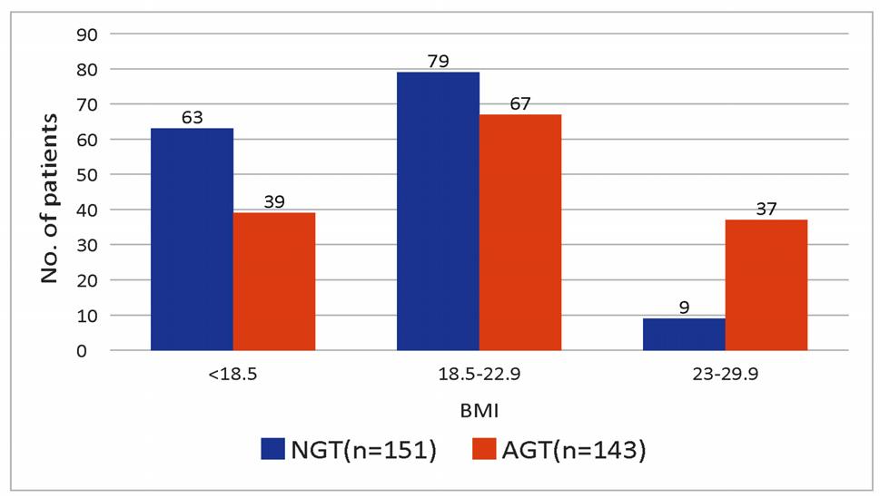
DISCUSSION
A total of 294 TB patients with DM were taken up for study and status of Lipid Profile was determined at the time of admission. It was found out that mean age in patients with TB and AGT group was significantly higher (47.91 years) as compared to those with TB alone (37.05years, p=0.0001). Gadallah, et al 16 in a nationwide populationbased study found that screening for DM among 1435 TB patients with no history of DM detected 30 new cases of DM, with a case detection rate of 2.09%. It was found out that majority of patients in AGT group had Dyslipidemia as compared to NGT group which was statistically significant (p=0.01). Mean Serum Triglycerides level and LDL Cholesterol were (209.82 ± 68.03, p<0.004) and (144.32± 24.83, p<0.0001) respectively in AGT group. In this study HDL values were lower and VLDL values were higher in AGT group even though they were not statistically significant. In a similar study done by Vrieling, et al17 on 177 patients it was observed that DM patients had Dyslipidemia as evidenced by high levels of VLDL, Triglycerides and low HDL Cholesterol. They concluded that TB-DM patients possess a distinctive Plasma Lipid Profile with pro-atherogenic properties. Several studies conducted in the past showed
Vol 121, No 2, February 2023Journal of the
Indian Medical Association
(Values in mg/dl) Lipid profile AGT(n=143) NGT(n=151)p value S Cholesterol (mean ± SD)182.35 ±32.26157 ± 42.37 0.15 S Triglycerides (mean ± SD)209.82 ± 68.03170.93 ±85.19 0.004 LDL Cholesterol (mean ± SD)144.32 ±24.8397.03 ±41.690.0001 HDL Cholestrol (mean ±SD)59.01 ± 27.9466.20 ± 26.03 0.18 VLDL (mean ±SD)45.25 ± 5129.69 ± 11.89 0.23
CharacteristicsAGT(n=143) NGT(n=151) 2 p -Value Age : <50 years 70(48.9%)123(81.45%)34.41 <0.001 >50 years 73(51.1%)28(18.54%) Gender : Male 100(69.9%) 92(60.9)2.60.11 Female 43(30.1%)59(39.1%) Residence : Rural100(69.9%)64(42.4%)22.59 <0.001 Urban 43(30.1%)87(57.6%) Education : Below High school 124(86.7%)110(72.8%)8.690.003 Above High school 19(13.3%)41(21.2%) Age (Years) 47.91 ± 15.3237.05 ± 13.580.0001 Hb (g/dl)10.66± 1.6110.53 ± 1.58 0.08 Serum creatinine (mg/dl)1.28 ± 0.52 0.95 ± 0.290.0001
Table 2 — Association of dyslipidemia in patients with TB and DM
Table 1 — Baseline characteristics
Fig 1 — BMI and glucose intolerance
42
Fig 2 — Association of dyslipidemia in patients with TB and DM
the benefits of using statin therapy for decreasing the incidence of TB in elderly population with DM. A study done by Lee, et al18 among 27,958 patients age >65 years and were followed up from 1998 to 2009. A total of 13,981 Type 2 diabetic patients were chosen. Calcium channel blocker, beta blocker, and statin users exhibited a reduced independent association after controlling for age, sex, other co-morbidities, and medications, with risk ratios of 0.76 (95 percent CI, 0.58-0.98), 0.72 (95 percent CI, 0.58-0.91) and 0.76 (95 percent CI, 0.60-0.97), respectively. Calcium channel blocker, beta blocker, and statin medication may reduce TB infection in older Taiwanese patients with Type 2 Diabetes, according to the researchers. Lai, et al19 did another trial with 8098 new TB cases and 809 800 control individuals. Statin users had a lower chance of active Tuberculosis, according to the researchers. Statin use for more than 90 days in a year was linked to the lowest unadjusted risk of Tuberculosis (RR 0.74; 95 percent CI 0.63 to 0.87). After correcting for individual confounders (RR 0.66; 95 percent CI 0.56 to 0.78) and DRS adjustment, the protective effect of active TB persisted (RR 0.66; 95 percent CI 0.56 to 0.78) (RR 0.62; 95 percent CI 0.53 to 0.72). The result of their study showed that patients with Dyslipidemia had reduced chances of infection with active Tuberculosis who were on stations, and the duration of stations offered prevention from Tuberculosis. In a similar study by Liao, et al20 to identify 8,236 patients above 20 years with recent infection of Pulmonary Tuberculosis. Odds Ratio (OR) of Pulmonary Tuberculosis was 0.67 for patients who had been taking statins (95% CI 0.59, 0.75). The adjusted ORs of Pulmonary Tuberculosis were 0.87 (95% CI 0.69, 1.10) for patients with total duration of statins use <3 months, 0.77 (95% CI 0.58, 1.03) for 36 months, and 0.59 (95% CI 0.51, 0.68) for >6 months, compared with subjects who never used statins. They concluded statins use correlates with a small but statistically significant risk reduction of Pulmonary Tuberculosis. The protective effect is stronger for prolonged use of statins.
CONCLUSION
In this study it was observed that patients with TBDM are more prone for Dyslipedimia as compared to patients with TB alone.
Limitation : This was a small population study.
REFERENCES
1World Health Organization. Global tuberculosis report 2019. Availaible from: https://apps.who.int/iris/bitstream/handle/ 10665/329368/9789241565714-eng.pdf?ua=1.
2Ginsberg HN — Insulin resistance and cardiovascular disease. J Clin Invest 2000; 106: 453-8.
3Natali A, Ferrannini E — Hypertension, insulin resistance, and the metabolic syndrome. Endocrinol Metab Clin North Am 2004; 33: 417-23.
4Park KH, Kim JY, Ahn CW, Song YD, Lim SK, Lee HC — Polycystic ovarian syndrome (PCOS) and insulin resistance. Int J Gynaecol Obstet 2001; 74: 261-7.
5Reaven GM — Banting lecture role of insulin resistance in human disease. Diabetes 1988; 37: 1595-13.
6Kahn BB, Flier JS — Obesity and insulin resistance. J Clin Invest 2000; 106: 473-8.
7Khan Y, Lalchandani A, Gupta AC, Khadanga S, Kumar S — Prevalence of metabolic syndrome crossing 40% in Northern India: Time to act fast before it runs out of proportions. Journal of Family Medicine and Primary Care 2018; 7(1): 118-5.
8Misra P, Upadhyay RP, Misra A, Anand K — A review of the epidemiology of diabetes in rural India. Diabetes Res Clin Pract 2011; 92: 303-8.
9Russell DG, Cardona PJ, Kim MJ, Allain S, Altare F — Foamy macrophages and the progression of the human tuberculosis granuloma. Nat Immunol 2009; 10(9): 943-8.
10Lee W, VanderVen BC, Fahey RJ, Russell DG — Intracellular Mycobacterium tuberculosis exploits host-derived fatty acids to limit metabolic stress. J Biol Chem 2013; 288(10): 6788800.
11Kim MJ, Wainwright HC, Locketz M, Bekker LG, Walther GB, Dittrich C, etal—Caseation of human tuberculosis granulomas correlates with elevated host lipid metabolism. EMBO Mol Med 2010; 2(7): 258-74.
12Soh AZ, Chee CB, Wang YT, Yuan JM, Koh WP — Dietary cholesterol increases the risk whereas PUFAs reduce the risk of active tuberculosis in Singapore Chinese. J Nutr 2016; 146(5): 1093-100.
13Martens GW, Arikan MC, Lee J, Ren F, Vallerskog T, Kornfeld H — Hypercholesterolemia impairs immunity to tuberculosis. Infect Immun 2008; 76(8): 3464-72.
14Martens GW, Vallerskog T, Kornfeld H — Hypercholesterolemic LDL receptor-deficient mice mount a neutrophilic response to tuberculosis despite the timely expression of protective immunity. J Leukoc Biol 2012; 91(6): 849-57.
15Parihar SP, Guler R, Khutlang R, Lang DM, Hurdayal R, Mhlanga MM, et al — Statin therapy reduces the mycobacterium tuberculosis burden in human macrophages and in mice by enhancing autophagy and phagosome maturation. J Infect Dis 2014; 209(5): 754-63.
16Gadallah M, Amin W, Fawzy M, Mokhtar A, Mohsen A — Screening for diabetes among tuberculosis patients: a nationwide population-based study in Egypt. African Health Sciences 2018; 18(4): 884-7.
17Vrieling F, Ronacher K, Kleynhans L, van den Akker E, Walzl G, Ottenhoff THM, et al — Patients with Concurrent Tuberculosis and Diabetes Have a Pro-Atherogenic Plasma Lipid Profile. E Bio Medicine 2018; 32: 192-9. Available from https://doi.org/10.1016/j.ebiom.2018.05.011.
18Lee MY, Lin KD, Hsu WH, Chang HL, Yang YH, Hsiao PJ, et al Statin, Calcium Channel Blocker and Beta Blocker Therapy May Decrease the Incidence of Tuberculosis Infection in Elderly Taiwanese Patients with Type 2 Diabetes. Int J Mol Sci 2015; 16(5): 1369-16.
19Lai CC, Lee MTG, Lee SH, Hsu WT, Chang SS, Chen SC, et al Statin treatment is associated with a decreased risk of active tuberculosis: an analysis of a nationally representative cohort. Thorax 2016; 71(7): 646-6.
20Liao KF, Lin CL, Lai SW. Population-Based Case-Control Study Assessing the Association between Statins Use and Pulmonary Tuberculosis in Taiwan. Front. Pharmacol 2017; 8: 597. Available from:doi: 10.3389/fphar.2017.00597.
Vol 121, No 2, February 2023Journal of the
Medical Association
Indian
43
Original Article
Profile of Patients with Hypoglycemia Presenting to the Emergency Medicine Department of a Tertiary Care Hospital
Dhivyaramani Leelakrishnan1, Kingsly Robert Gnanadurai2
Background : Hypoglycemia is a common treatable endocrinological emergency. This study is aimed at profiling patients with Hypoglycemia presenting to the Emergency Department.
Materials and Methods : This is a prospective study aimed to know the clinical profile of patients more than 18years of age presenting with Capillary Glucose Random Blood Sugar value less than 70mg/dl from the period of September, 2018-May, 2020. Results: 123 participants were included in the study among which 69.1% of them were known Diabetes Mellitus with mean duration of 6.9 years. Using the American Diabetes Association (ADA)/ European Association for Study of Diabetes (ESAD) definitions 71.5% presented with severe Hypoglycemia. Neuroglycopenic symptoms were the most common presenting symptom among the diabetics however, autonomic symptoms were the most predominant symptom among non-diabetic population. Drug induced Hypoglycemia was the most common cause among diabetic population accounting for 43.5% and among non-diabetic group 28.9% it was probably due to food intake secondary to fasting. Modified Clarke’s score of more than 4 indicated impaired awareness of Hypoglycemia.
Conclusion : Education about the prevention of hypoglycemia needs to be initiated from the Emergency Room. Spontaneous Hypoglycemia indicates impaired glucose tolerance. Development of risk stratification score helps in accurate disposition of patients from Emergency Room.
[J Indian Med Assoc 2023; 121(2): 44-8]
Key words :Modified Clarke’s score.
Diabetes Mellitus is the most common Metabolic Disorder which presents to the Emergency Department with life threatening emergencies and are important contributors to morbidity and mortality. Hypoglycemia is the most common diabetic emergency and is associated with adverse outcomes1. According to the Endocrine Society Guidelines Hypoglycemia is defined as a documented alert value of less than 70mg/dl; while the American Diabetes Association (ADA) stresses that a documented value of 54mg/dl whether symptoms are present or not, denotes clinically important Hypoglycemia2
The International Hypoglycemia Study Group proposed the levels of Hypoglycemia, which included level 1 is glucose alert value of 70 mg/dL or less, level 2 is glucose level of 54 mg/dL is sufficiently low to indicate serious, clinically important Hypoglycemia and level 3 being severe Hypoglycemia, as defined by the ADA denotes severe cognitive impairment requiring external assistance for recovery3 Hypoglycaemic coma occurs when Blood Sugar is less than 50mg/dl4
Spontaneous Hypoglycemia a condition referred as the occurrence of hypoglycemia without diabetes, is
Department of Emergency Medicine, Bangalore Baptist Hospital, Coimbatore. Karnataka 560024
1MBBS, DNB, Senior Resident
2MBBS, MD, PGDMLE, Head and Corresponding Author
Received on : 19/01/2022
Accepted on : 20/05/2022
Editor's Comment : Diabetes Mellitus being the most prevalent noncommunicable disease the complications of treatment should be identified in emergency room. Prompt recoginition prevents the adverse events and helps to eliminate other mimics Hypoglycemic episodes among non-diabetic population may indicate impaired glucose tolerance hence complete evaluation is needed.
a puzzling clinical issue. Reactive Hypoglycaemia a entity of spontaneous Hypoglycemia5 is defined as postprandial Hypoglycemia seen in non-diabetic individual6 But in our Indian background, Sepsis was more commonly associated with Spontaneous Hypoglycemia, however an episode of Spontaneous Hypoglycemia is indicator of Pre-diabetes state7
The body activates defence mechanisms against a hypoglycemic episode by hormonal counter regulation and increased behavioural response, this interplay mechanisms are compromised in advanced Diabetes Mellitus resulting in defective adrenomedullary response and defective neural and sympathetic responses which leads to unawareness of hypoglycemic symptoms. These are components of Hypoglycemia Associated Autonomic Failure (HAAF)8. Scoring systems namely Gold and Clarke’s score are used to assess the impaired awareness of Hypoglycemia in recurrent episodes of Hypoglycemia, it is evident that high scores individuals have more
Vol 121, No 2, February 2023Journal of
the Indian Medical Association
44
neuroglycopenic symptoms than autonomic symptoms9
MATERIALS AND METHODS
This is a prospective observational study using a semi -structured questionnaire done in Emergency Room of a Tertiary Care Teaching Institute in Bangalore. All patients above the age of except pregnant women, presenting with Hypoglycemia that is Random Capillary Blood Sugar value less than 70mg/dl to the Emergency department from September, 2018-May, 2020 were included.
Data and Statistical Analysis :
The results were averaged (mean ± Standard Deviation) for each parameter for continuous data the percentage for categorical data using student T test, Chi square test and multivariate logistic regression was calculated. In all the above test a “p” value of less than 0.05 was accepted as indicating statistical significance.
OBSERVATIONS
Among 123 patients included in the study,85 were already diagnosed Diabetes Mellitus among these patients 95.3% had Type 2 Diabetes Mellitus. The mean duration of Diabetes Mellitus was 6.9 years. According to the ADA/ European Association for Study of Diabetes (ESAD) definition 71.5% presented with severe Hypoglycemia (Fig 1).
The maximum number of Hypoglycemic episodes occurred among 60-69 years of age in diabetic population at 27.6% as compared to 23.7% episodes occurred among 30-39 years in non-diabetic population. The mean age of occurrence of hypoglycemic episode was 58.2±17 years.52.8% of hypoglycaemic episodes occurred in females In 31.8% among diabetic population hypoglycemic episodes occurred in early morning hours. Among non-diabetic population 39.5% had presented with hypoglycemic episode in timeline of 12pm-6pm (Table 1).
Among the diabetic group 43.5% presented with altered sensorium,34.1% presented with both dizziness and loss of consciousness, 24.7% presented with fever, 22.4% presented with sweating and among non-diabetic population 39.5% presented with sweating, 36.8% with dizziness and 34.2% presented with altered sensorium.
Among 123 participants on comparing the onset of symptoms with different ranges of Glucose Random Blood Sugar (GRBS) values 54.1% presented with altered sensorium within
GRBS range of 20-30mg/dl. Autonomic symptoms were the presenting symptom in patients with GRBS more than 30 mg/dl.
Most common cause for a hypoglycemic episode among the diabetic was drug induced Hypoglycaemia out of which 32.5% occurred in Oral Hypoglycemic Agents (OHA) users and 20.3% occurred in both oral hypoglycemic and insulin users. It was evident that sulphonylureas and insulin mixtard were the common offending agents. Among the non-diabetic group 28.9% hypoglycemic episodes were due to inadequate food intake, followed by Liver Cell Failure at 18.4%. Sepsis accounted for 15.8% cases of hypoglycemia, among the focus of Sepsis, Urosepsis ranked first followed by Pneumonia which included the COVID 19 infection also (Table 2).
Among the 85, who had already existing Diabetes Mellitus, 33% had a hypoglycemic episode in the past 12 months and all had Modified Clarke’s score of more than 4. The association between onset of Hypoglycemia and Modified Clarke’s scoring was significant.
Among 123 patients 60% were admitted ,44.7% were discharged against medical advice. Among nondiabetic population 28.9% went discharge against medical advice. Died in Emergency Room was 2.6% among non-diabetics.
ParameterDiabetic Non-diabeticTotalP value (N=85)(N=35)(N=123)
N%N%N%
AGE20-29 years44.7%615.8%108.1% <0.001
30-39 years00.0%923.7%97.3%
40-49 years67.1821.11411.4
50-59 years1618.8410.52016.3
60-69 years2832.9615.83427.6
70-79 years2124.7410.52520.3
80-89 years1011.812.6118.9
Gender Male43 50.61539.55847.20.254
Female42 49.42360.56552.8
Vol 121, No 2, February 2023Journal
of the Indian Medical Association
Table 1 — Demographic Characteristics
45
Fig 1 — Distribution of Diabetic Status among Hypoglycemia
DISCUSSION
In our study Hypoglycemia was more common in known diabetics than non-diabetic population which is attributed to the fact that most of the patients were on medications for Diabetes Mellitus.
The prevalence of Type 2 Diabetes Mellitus is more than Type 1 Diabetes Mellitus and this was evidenced by the fact that 95.3% were diagnosed Type 2 Diabetes Mellitus and 4.7% were Type 1 Diabetes Mellitus.This was consistent with a retrospective study done by Jordi Caballero, et al at Hospital Universitario de Bellvitge in Emergency Room 81.9% episodes occurred in type 2 diabetic and 16.1% occurred in Type 1 Diabetic10
Many studies on Hypoglycemia used the study definition of Hypoglycemia as Glucose Random Blood Sugar (GRBS) <40mg/dl. Hence our data of level 2 Hypoglycemia according to International Hypoglycemia Group classification being the most common at 71.5% was inconsistent (Fig 2).
27.6% of total hypoglycemic episodes occurred in the elderly age group of 60-69 years however, among non-diabetics 23.7% episodes occurred in a young age group of 30-39 years. These findings were almost consistent with a study done by Ohenhen Oluwatoyin Abisoye, et al at a Tertiary Health Institution at Nigeria in which among diabetics 38% episodes occurred in 55-64 years of age and 31% occurred in 65-74 years of age11.
Among non-diabetic group there were 27 episodes in less than 60 years of age which is consistent with findings done by
Krishnaraj, et al in 2010 in which total 108 participants, 61 episodes occurred in less than 65 years of age12
In our study the mean age among diabetic group was 63.9 years which was similar with study done by Satish, et al on clinical profile of Hypoglycemia in which the mean age of patients ranged from 61.6 years13. The mean age among non-diabetic population was 46.1 years. This was comparable with the finding done by Santra, et al in which the mean duration among non-diabetics is 42.84 years14
52.8% participants were female, however among diabetic group the gender distribution was approximately equal among both genders. In our study among non-diabetes 60.5% were females, which was similar to a study done by Ekpebegh et al among 26 non-diabetic patients out of which 65.4% were females15.
31.8% participants among known Diabetics Mellitus,had an episode during early morning hours which was in conformity with a study done by Ohenhen Oluwatoyin Abisoye, et al among diabetic patients, 51.7% had hypoglycemic episodes in the early morning hours11. In our study among the 38 non-diabetics, 39.5% had a hypoglycemic event in noon hours. This is a new finding as no much studies had been done on the time of presentation of non-diabetic hypoglycemia which needs further studies.
Vol 121, No 2, February 2023Journal of the Indian Medical Association
Causes Diabetic Non-diabeticTotalP value (N=85)(N=38)(N=123) N%N%N% Inadequate Food Intake14 16.5%1128.9%2520.3%0.112 Sepsis29 34.1%615.8%3528.5% 0.037 Liver Cell Failure0 0.0%718.4%75.7% <0.001 Drug Induced (OHA/Insulin)37 43.5%12.6%3830.9% <0.001 Alcohol Intoxication1 1.2%513.2%64.9% 0.004 Other Causes Carcinoma Duodenum0 0.0%25.2%11.6% 0.001 Idiopathic0 0.0%513.2%54.1% Insulinoma0 0.0%12.6%10.8%
Table 2 — Percentage of Causes of Hypoglycemia
46
Fig 2 — Association of GRBS with Symptoms of Hypoglycemia
The mean GRBS value among diabetic was 39.7 mg/dl, this value was similar with the study conducted by Satish et al in which the mean GRBS was 37.12 mg/dl13
40.7% patients presented with altered sensorium which indicates that neuroglycopenic symptoms are the most common presenting symptoms of Hypoglycemia which was consistent with study done by Kumar, et al in which 59.3% presented with decreased conscious levels2
Among 123 participants on comparing the onset of symptoms with different ranges of GRBS values 54.1% presented with a neuroglycopenic symptom, altered sensorium within range of 20-30mg/dl. However autonomic symptoms such as sweating were common in patients with GRBS value >30mg/dl. This implies the importance of counter regulatory hormones in the pathophysiology of Hypoglycemia, which helps to counsel the patients regarding various symptoms onset.
Another than Diabetes Mellitus, among 51% systemic Hypertension was associated comorbidity, which was similar to the study done by Caballero et al who found in 73.3% systemic hypertension was the most associated comorbidity10.
Among the non-diabetic population malignancy was the most common underlying comorbid conditions. However, in the studies done on non-diabetic Hypoglycaemia, have emphasized on the fact that Hypoglycemia can present as undiagnosed malignancy.
Among non-diabetic group autonomic symptom such as sweating were common at 39.5%. This finding in conformity with a study done Gharbi, et al among 40 non-diabetic patients, 82.5% presented with autonomic symptoms16
In our study among 123 participants,30.9% had a hypoglycemic event secondary to anti-diabetic medication, this finding is consistent with the study done by Kumar, et al among 320 diabetics out of which 59.81% were diagnosed drug-induced hypoglycemia2.
Among the drug-induced Hypoglycemia 32.5% were OHA user’s, followed by 20.3% were on OHA’s and insulin, this was consistent with the study done by Ohenhen Oluwatoyin Abisoye, et al 60.7% were on OHA drugs and 32.6% were both OHA plus insulin users12. Among OHA user’s 39% occurred among the diabetics who were on monotherapy, out of which 14.6% were on sulphonylureas closely followed by 13.8% on biguanides which was similar to the study conducted by Eren, et al among 225 patients,64 subjects were using oral hypoglycemic drugs out of
which 43 were on sulphonylureas17.
In our study among the non-diabetic group 28.9% of hypoglycemic event was due to inadequate food intake due to fasting among 32% of participants followed by Liver Cell Failure. This was inconsistent with the study done by Ekbepegh, et al in which the main cause for Hypoglycemia was hypercortisolism attributed to underlying retroviral status of the patient15
In our study by correlating the most common time of presentation of Hypoglycemia being the noon hours and the most common cause being inadequate food intake secondary to fasting the possibility of underlying impaired glucose tolerance among non-diabetic group was found. This is a new finding and further studies are needed regrading Hypoglycemia among nondiabetic population.
In this study among 28.5% the cause of hypoglycemic event was sepsis, out of which 34.1% was in the diabetic group which is inconsistent to the fact that infections are the most common cause for Hypoglycaemia among non-diabetes12. This can be due to a smaller number of non-diabetic patients were included in this study. However, considering the focus of Sepsis 32.4% was diagnosed urosepsis, which is consistent with the study done by Indu, et al in which among the 10 patients diagnosed Hypoglycemia secondary to infection most common was urosepsis diagnosed in three patients18. Also among the causes of sepsis 5.9% had hypoglycemic event due to COVID19 during the pandemic.
38.5% of them had a previous episode of Hypoglycemia among the 85 already diagnosed Diabetes Mellitus in past 12 months and all had a Modified Clarke’s score of more than four indicating that there was impaired awareness of hypoglycemic symptoms and thus this episode is a recurrence of Hypoglycemia. This was correlating with study done by Geddes, et al showed strong association between Modified Clarke’s score and impaired awareness thus recurrent Hypoglycemia9
In 55.3% patients the outcome from Emergency Room was admission for further management It was also noted that in non-diabetic group discharged against medical advice was most common outcome. There were two deaths,with sepsis accounting for 1.6%. This is consistent with the study done by Makoto, et al among patients with Sepsis had increased mortality among hypoglycemic group19
Limitations :
•It is a single center study.
•Difficulty in taking history from the patient himself, more reliability on the attender’s history which
Vol 121, No 2, February 2023Journal
of the Indian Medical Association
47
can be a source for information bias.
• Among the non-diabetic with hypoglycemia most of them went discharge against medical advice which attributes to missing out the etiology.
CONCLUSION
71.5% presented at level 2 Hypoglycemia and above, but considering the non-diabetics level 1 Hypoglycemia was the commonest. Neuroglycopenic symptoms occurred in Capillary Blood Sugar levels of 20-40 mg/dl while autonomic symptoms were the presenting symptom above 30mg/dl. Sulphonylurea induced hypoglycemia was the most common cause for hypoglycemia among the diabetics. In the nondiabetic group, it was evident that more hypoglycemic episodes occurred during noon hours which raise the concern for pre-existing impaired glucose tolerance. Hypoglycemia among the non-diabetic population needs further studies. Hypoglycemia was also the presentation for COVID-19 in diabetic population. The Modified Clarke’s scoring system evaluated in patients proved that a score of more than 4, indicates impaired awareness and recurrent Hypoglycaemia. The overall death was 2.6% in Emergency Room due to Multiorgan Failure due to Sepsis.
REFERENCES
1Umpierrez G, Korytkowski M. Diabetic emergencies— ketoacidosis, hyperglycemic hyperosmolar state and hypoglycaemia. Nature Reviews Endocrinology 2016; 12(4): 222.
2Kumar JG, Abhilash KP, Saya RP, Tadipaneni N, Bose JM — A retrospective study on epidemiology of hypoglycemia in Emergency Department. IndianJournalof Endocrinologyand Metabolism 2017; 21(1): 119.
3International Hypoglycaemia Study Group — Glucose concentrations of less than 3.0 mmol/L (54 mg/dL) should be reported in clinical trials: a joint position statement of the American Diabetes Association and the European Association for the Study of Diabetes. Diabetes Care 2017; 40(1): 155-7.
4Lu Z, Liu J, He Q, Chakraborty A, Zhu T — Analysis of Risk Factors for Hypoglycemic Coma in 194 Patients with Type 2 Diabetes. Medical science monitor: International Medical Journal of Experimental and Clinical Research 2017; 23: 5662
5Vihonen H, Kuisma M, Nurmi J — Hypoglycemia without diabetes encountered by emergency medical services: a retrospective cohort study. Scandinavian Journal of Trauma, Resuscitation and Emergency Medicine 2018; 26(1): 12.
6Akirov A, Grossman A, Shochat T, Shimon I — Mortality among hospitalized patients with hypoglycemia: insulin related and noninsulin related. The Journal of Clinical Endocrinology & Metabolism 2017; 102(2): 416-24.
7Sako A,Yasunaga H, Matsui H, Fushimi K, Hamasaki H, Katsuyama H, et al — Hospitalization with hypoglycemia in patients without diabetes mellitus: A retrospective study using a national inpatient database in Japan, 2008-2012. Medicine 2017; 96(25)
8Davis SN, Duckworth W, Emanuele N, Hayward RA, Wiitala WL, Thottapurathu L, et al — Effects of severe hypoglycemia on cardiovascular outcomes and death in the Veterans Affairs Diabetes Trial. Diabetes Care 2019; 42(1): 157-63.
9Geddes J, Wright RJ, Zammitt NN, Deary IJ, Frier BM — An evaluation of methods of assessing impaired awareness of hypoglycemia in type 1 diabetes. Diabetes Care 2007; 30(7): 1868-70
10Caballero-Corchuelo J,Guerrero-Pérez F, de la Jordana PG, Pérez-Maraver M — Analysis of the characteristics of patients with diabetes mellitus who attend a tertiary hospital emergency department for a hypoglycemic event. Endocrinología, Diabetes y Nutrición (English ed.) 2019; 66(1): 19-25.
11Ohenhen Oluwatoyin Abisoye, Uwameiye Oseribhor. Prevalence and Presentation of Diabetes-Related Hypoglycemia Amongst Persons with Diabetes in a Tertiary Health Instition in Nigeria. International Journal of Diabetes and Endocrinology. Special Issue: Hypoglycemia in Diabetes 2020; 5(3): 34-40. doi: 10.11648/j.ijde.20200503.
12Nirantharakumar K, Marshall T, Hodson J, Narendran P, Deeks J, Coleman JJ, et al — Hypoglycemia in non-diabetic inpatients: clinical or criminal?. PLoS One 2012; 7(7): e40384.
13Clinical profile of patients presenting with hypoglycemia in a tertiary care hospital, Satishkumar,Balbir singhverma,Dinesh kumar Paripex-indian Journal of Research –Volume 9,Issue 2,Feburary 2020.
14Santra G, Roy D — A study on etiological spectrum of hypoglycemia in nondiabetic patients in a rural medical teaching institution. International Journal of Health & Allied Sciences 2018; 7(2): 83
15Ekpebegh C — Aetiologyof SpontaneousHypoglycaemia in a South African Hospital. Turkish Journal of Endocrinology & Metabolism 2017; 21(2)
16Gharbi R, Chaker F, Chihaoui M, Danguir S, Yazidi M, Rejeb O, Slimane H — The contribution of fast test in the diagnostic approach of hypoglycemia in non-diabetic patients. In18th European Congress of Endocrinology 2016; 13 (Vol. 41). BioScientifica.
17Eren SH, Caliskan HM, Kilicli F, Korkmaz I, Acibucu F, Dokmetas HS — Etiologies of patients admitted to emergency department with hypoglycemia. Scientific Research and Essays 2013; 5(12): 1479-82.
18Indu KC — Causes of Hypoglycemia in Hospitalized Diabetic Patients Referred to Endocrine Department of a Tertiary Level Hospital of India. Medical Journal of Shree Birendra Hospital 2016; 15(1): 26-31.
19Furukawa M, Kinoshita K, Yamaguchi J, Hori S, Sakurai A— Sepsis patients with complication of hypoglycemia and hypoalbuminemia are an early and easy identification of high mortality risk. Internal and Emergency Medicine 2019; 14(4): 539-48.
Vol 121, No 2, February 2023Journal
of the Indian Medical Association
48
A
Original Article
Study on Sexually Transmitted Infections Using Syndromic
Approach among Patients Attending Suraksha Clinic at a Tertiary Care Hospital of West Bengal
Suman Das1, Shatanik Mondal2, Pijush Kanti Mandal3
Introduction : A proper understanding of different aspects of Sexually Transmitted Infections (STIs) in different regions of a country like India is very essential for STI control.
Objectives : To assess pattern of STIs using Syndromic approach among patients attending at Suraksha Clinic and to find out treatment seeking behaviour among them.
Material and Method : A cross-sectional study was conducted at a Tertiary Care Hospital of West Bengal. This retrospective study was done selecting all patients who attended the Clinic for first time from 1st May, 2020 to 30th April, 2021. Relevant records were reviewed. Descriptive statistics and chi-square test were used.
Observations : In present study Suraksha Clinic attendees were mainly comprised of female (95.8%); while 65.8% and 64.2% of patients were suffering from Lower abdominal pain and Cervicovaginal discharge respectively. Nearly one-third of them (37.9%) were suffering from more than one disease. About one-fourth of patients (28.2%) accessed the STI/RTI/Suraksha Clinic directly. The treatment seeking behaviour was significantly associated with gender and education of patients.
Conclusions : This Tertiary Care Hospital based study highlighted higher prevalence of certain STIs among attendees to Suraksha Clinic as well as lower proportion of male and directly walk-in patient.
[J Indian Med Assoc 2023; 121(2): 49-51]
Key words :Suraksha Clinic, Syndromic approach, Treatment seeking behaviour.
Group of illnesses which are caused by infections, transmitted by sexual contact through exchange of semen, vaginal fluid, blood and other fluids, are called Sexually Transmitted Infections (STIs); it also transmitted by direct contact with the affected body areas of people with STIs. No doubt that it leads to huge Psycho-social consequences both at the individual as well as at the community levels1. It also plays an important role in the acquisition and transmission of HIV2. Besides its contribution to the substantial burden of mortality and morbidity, have made it as one of the major public health problems affecting both developing and developed countries1,3
The pattern of STIs is found usually to vary from region-to-region, especially in large nations like India3 In our country, the assessment and management of patients is still largely based on syndromic approach suggested by the National AIDS Control Organization (NACO)4 Thus the information regarding the profile of STIs relies essentially on syndromic diagnosis5.
However, West Bengal State AIDS Prevention &
Department of Community Medicine, Malda Medical College and Hospital, Malda, West Bengal 732101
1MD, Senior Resident and Corresponding Author
2MD, Assistant Professor
3MD, Associate Professor, Department of Medicine
Received on : 31/01/2022
Accepted on : 20/02/2022
Editor's Comment :
The local epidemiology of STIs addressing lower male attendees to Suraksha clinic and higher prevalence of certain STIs, would be better explored through research with help of grass-root Health workers.
Control Society (WBSAP&CS) is going to ensure better access of STI/RTI related services to the people in need by establishing the ‘Suraksha Clinics’ at all those hospitals which are not covered by STI Services under NACP-IV, as new initiative. Therefore, new STI clinics are going to be established at Sub-division Hospitals & State General Hospitals also. At present, total 46 (fourty six) STI Clinics are currently running by WBSAP&CS in this State6
In order to plan and implement successful targeted interventions and to combat problem of STIs, availability of current baseline information of STIs in the various parts of the country is essential. Literature review shows that information regarding Suraksha Clinic as well as pattern of STIs is scare in West Bengal. So, present study was planned among patients attending at Suraksha Clinic, to generate valuable information regarding the pattern of STIs using syndromic approach and to find out treatment seeking behaviour of them.
MATERIAL AND METHOD
A retrospective study with cross-sectional design
Vol 121, No 2, February 2023Journal of
the Indian Medical Association
49
was conducted with help of the relevant records of STI patients at Suraksha Clinic of aTertiary Care Hospital of West Bengal over the period of past one year from 1 st May, 2020 to 30 th April, 2021. Complete enumeration technique was followed to select all study subjects who visited the Clinic for first time during study period. Data were collected through careful review process of cunsellor’s patient register and clinical records of STI patients.
Data were collected after scientific review and ethical approval of the synopsis by Institutional Ethics Committee. Confidentiality and anonymity of patients’ information was assured. Patients were categorized mainly as per the NACO guidelines7 into Genital Ulcerative Disease-Herpetic (GUD H), Genital Ulcerative Disease Nonherpetic (GUD NH), Cervicovaginal discharge, Lower abdominal pain and Urethral discharge.
Collected data were checked for completeness and consistency and then the data were entered in the computer on Excel data sheets (Microsoft Excel, 2013). The principles of descriptive statistics were applied to organise and present the data in tables and diagrams. Proportions in relation to different outcome variables were also calculated. Data were analysed using Statistical Package for Social Sciences [IBM Corp. Released 2011. IBM SPSS Statistics for Windows, Version 20.0. Armonk, New York, United States]. Appropriate statistical tests such as Chisquare test were applied.
OBSERVATIONS
Overall 4348 patients visited at Suraksha Clinic of the Tertiary Care Hospital in West Bengal during study duration, while 2132 study subjects visited for the first time. These patients with first time visit at the Suraksha Clinic were included in present study. Among the total patients more than half were (57.7%) were belonging to 25 to 44 years age group, nearly one-twentieth (4.2%) were male. Overall three-fourth of patients (75.8%) were formally educated while 2.9% were illiterate and 21.3% were non-formally educated. Majority of the patients (71.8%) were referred and 28.2% accessed the STI/RTI clinic directly.
Majority of them were suffering from lower abdominal pain (65.8%) followed by Cervicovaginal discharge (64.2%), GUD-Non herpetic (3.1%), GUD-Herpetic (2.8%)(Table 1). On further analysis, it was noted that 808 (37.9%) patients had more than one STI. Only two study subjects were found to be HIV-positive.
Addressing treatment seeking behaviour among patients whether suraksha clinic was accessed directly or referred, it was explored that
the behaviour was significantly associated with gender (<0.001) and education (<0.001) of patients (Table 2).
DISCUSSION
Present study revealed that major issues among study subjects were Lower abdominal pain (65.8%) and Cervicovaginal discharge (64.2%). Corroborating with present study Cervicovaginal discharge was noted as most common problem among patients at STI-clinic of a Tertiary Care Hospital of Rajasthan (38%)4 Sharma S, Tiwari S, Paliwal V, et al noted in a study of patterns of Sexually Transmitted Diseases using a Syndromic approach from a Tertiary Care Hospital of the Northern India that vaginal/cervical discharge (13.4%) was remarkable problem5. In addition to this, among females of STI-clinic, Cervicovaginal discharge constituted the maximum proportion of cases at a Tertiary Care
Vol 121, No 2, February 2023Journal
of the Indian Medical Association
Characteristics Frequency Percent Age (years) : <2043520.4 20-24390 18.3 25-441231 57.7 >44763.6 Gender : Male904.2 Female 204295.8 Education : Illiterate632.9 Non-formally educated455 21.3 Class I - Class IV460 21.6 Class V - Class VIII 107850.6 Class IX and above763.6 Direct walk-in/ Referred : Direct walk-in 60228.2 Referred153071.8 Syndromic diagnosis* : Cervicovaginal discharge 136964.2 GUD-Non herpetic663.1 GUD-Herpetic592.8 Lower abdominal pain 140265.8 Urethral discharge170.8 Other STIs251.2
: * Multiple responses
Table 1 — Distribution of study subjects according to certain characteristics (n=2132)
Note
Direct walk-in
Chi-square [No (%)][No (%)] test Age (years) : <24 years242 (30.1)563 (69.9)2.128 (1) 0.145 >24 years360 (27.1)967 (72.9) Gender : Male81 (90.0)9 (10.0)176.899 (1) <0.001 Female 521 (25.5)1521 (74.5) Education : Illiterate and Non-formally educated110 (21.2)408 (78.8)16.550 (1) <0.001 Others 492 (30.5)1122 (69.5) 50
Table 2 — Association between treatment seeking behaviour and certain characteristics of patients
Referred
Hospital of the Northern India (61.04%)5 and at a Tertiary Care Hospital of Eastern India (29.9%)8. However, Genital Ulcer Disease-herpetic was reported as most common problem among patients of STI-clinic at a Tertiary Care Hospital of North India (21.75%)3, at a Tertiary Care Hospital of Eastern India8 as well as among male patients at a Rural-based Tertiary Care Center (24.37%)9. Again a study on patterns of Sexually Transmitted Diseases using Syndromic approach from a Tertiary Care Hospital of the Northern India explored that the overall most common STI was balanoposthitis (39.62%)5
Lower abdominal pain was noted in very lower proportion than present study (65.8%) at a Tertiary care Hospital of Rajasthan (2.3%)4, at a Tertiary Care Hospital of the Northern India (2.66%)5. Such variation in observation might be explained by the way of diagnosis (ie, syndromic approach, investigation based), Socio-economic diversity across the country, sampling technique of different studies and comparatively less male participants in present study.
In present study only two study subjects were found to be HIV-positive. On the contrary, newly diagnosed HIV cases were noted 0.4% in such a study ata tertiary care hospital of Rajasthan4 as well as HIV prevalence was reported as widely varied proportion (2.48% to 10.59%) in different studies at STI-clinic in tertiary care hospital across the country3,5,8,9
In present study majority of the patients (71.8%) were referred. On the contrary, Suvirya S, Singh R and Senthamizh P, et al in a study regarding treatment seeking behaviour of STI clients in a Tertiary Care Centre of North India found that majority of the patients (84.7%) accessed the STI/RTI clinic directly and only 15.3% were referred10
Pearson’s Chi-square test revealed in present study that treatment seeking behaviour had a significant association with gender and education but not with age of patients. The study regarding treatment seeking behaviour of STI clients in a Tertiary Care Centre of North India showed similar significant association with age, gender and education of patients10.
CONCLUSIONS
This Tertiary Care Hospital based study highlighted higher prevalence of certain STIs among attendees to Suraksha Clinic, among whom majority were female and referred patient. The treatment seeking behaviour was significantly associated with gender and education of patients.The local epidemiology of STIs would be better understood through research with help of grassroot health workers. Such further exploration regarding STIs may help us to implement STI control programs successfully as well as to address the Global efforts to combat HIV/AIDS.
Recommendations : Information, Education and Communication (IEC) techniques need to be strengthened to spread awareness about STI-clinics among general population as well as to increase utilization of such clinics at Tertiary Care Centre by them through direct walk-in. Besides proper training of the Health Care Providers, condom promotion, partner notification and partner management, persuasive counselling to attendees may motivate their peer group to avail the Suraksha Clinic facilities provided by the NACO.
Acknowledgement : The authors are grateful to all faculties and staffs of Suraksha Clinic of Malda Medical College, West Bengal for their their sincere cooperation during conduction of the study.
Source of Support : Self-funded by Researchers.
Conflict of Interest : Nil
REFERENCES
1Suvirya S, Shukla M, Pathania S, Banerjee G, Kumar A, Tripathi A — Stigma associated with sexually transmitted infections among patients attending suraksha clinic at a tertiary care hospital in northern India. Indian J Dermatol 2018; 63: 469-74.
2UNAIDS Report on the Global AIDS Epidemic; 2013. Available from: http://www.files.unaids.org/en/media/unaids/ contentassets/documents/epidemiology/2013/gr2013/ UNAIDS_Global_Report_2013_en.pdf. [Last assessed on 2021 Mar 28].
3Banger HS, Sethi A, Malhotra S, Malhotra SK, Kaur T — Clinicoepidemiological profile of patients attending Suraksha Clinic of tertiary care hospital of North India. Indian J Sex Transm Dis 2017; 38: 54-9.
4Nyati A, Gupta S, Jain SK, Yadav D, Patidar BL, Sharma M — A retrospective study of the pattern of sexually transmitted infections from a tertiary care hospital ofRajasthan. Indian J Sex Transm Dis 2017; 38: 147-51.
5Sharma S, Tiwari S, Paliwal V, Mathur DK, Bhargava P — Study of patterns of sexually transmitted diseases using a syndromic approach in the era of human immunodeficiency virus from a tertiarycare hospital of the Northern India. Indian J Sex Transm Dis AIDS 2015; 36(2): 158-61.
6West Bengal State AIDS Prevention & Control Society. ‘Suraksha Clinic’- Sexually Transmitted Infections (STI) testing and treatment Centre. Available from: http:// wbsapcs.wbhealth.gov.in/pages/display/12-sti- [Last assessed on 2021 Mar 28].
7National AIDS Control Organization, Ministry of Health and Family Welfare. Operational Guidelines for Programme Managers and Service Providers for Strengthening STI/RTI Services. New Delhi: Government of India; 2007. p.18.
8Sarkar S, Shrimal A, Das J, Choudhury SR — Pattern of sexually transmitted infections: A profile from a sexually transmitted infections clinic of a tertiary care hospital of eastern India. Ann Med Health Sci Res 2013; 3: 206-9.
9Vora R, Anjaneyan G, Doctor C, Gupta R — Clinicoepidemiological study of sexually transmitted infections in males at a rural-based tertiary care center. Indian J Sex Transm Dis AIDS 2011; 32(2): 86-9.
10Suvirya S, Singh R, Senthamizh P, Sharma V — Treatment seeking behaviour of STI clients in a tertiary care centre of North India: A cross sectional study. Indian J Sex Transm Dis AIDS 2016; 37(1): 7-11.
Vol 121, No 2, February 2023Journal
of the Indian Medical Association
51
Original Article
Aesthetic Reconstruction of Scrotum after Founiers Gangrene using Laterally based Medial Thigh Fasciocutaneous Flap
Aravind Lakshmana Rao1, Vishnu PS2
Introduction : Fournier’s Gangrene is a rapidly progressive necrotising faciitis affecting the genital region, perineum, perianal region and the abdominal wall. It can have an adverse effect on the functional and psychological aspects of the patient. Many flaps are available for the defect coverage.
Aim : The aim of this prospective study was to analyse the effectiveness of laterally based medial thigh flap for scrotal reconstruction. This is a fasciocutaneous flap just below the Scrotum which covers moderate to large sized scrotal defects successfully.
Methods and Materials : This study was done at a teaching hospital in South India on patients with major scrotal defects secondary to Fournier’s Gangrene. Ten patients with major scrotal defects secondary to Fournier’s gangrene were subjected to reconstruction of scrotum by medial thigh fasciocutaneous flap.
Results and Conclusions : All ten flaps survived and provided a durable and aesthetic cover for exposed testes. Only one flap had necrosis of distal 2cm, which was successfully managed by secondary suturing. The donor areas were primarily covered with placement of a small graft near the base of the flap, which healed well with an inconspicuous scar. In conclusion, the laterally based medial thigh fasciocutaneous flap provides excellent closure for extensive scrotal skin loss. It is a robust flap, easy to mobilise, rarely undergoes necrosis, provides a sensate cover with excellent aesthetic results. The technique is simple, less time consuming and can be easily incorporated by plastic Surgeons and General Surgeons.
[J Indian Med Assoc 2023; 121(2): 52-5]
Key words :Fournier’s Gangrene, Fasciocutaneous flap, Scrotal reconstruction, Medial thigh flap.
Fournier’s Gangrene is an acute, rapidly progressive and potentially fatal, infective necrotising fasciitis affecting the external Genitalia, perineal or perianal regions1. Fournier’s gangrene was first described by Jean Alfred Fournier in 18832. Injury to the perineal area continues to be the most frequent reason for the entry of bacteria that mediates the infectious process3
Fournier’s Gangrene presents with major soft tissue loss and may even be lethal4. Since the testes have an independent blood supply, they are spared and remain exposed5. Fournier’s Gangrene can lead to an extensive skin loss involving the Scrotum, Penis, Thighs and Lower abdomen. After thorough surgical debridement, major scrotal and perineal defects along with exposed testes have to be dealt by Plastic Surgeons6
Different surgical procedures were used to cover the exposed testes. Earlier methods for testicular salvage were: covering with skin grafting, burying them
Father Muller Medical College and Hospital, Mangalore, Karnataka
575002
1MCh (Plastic Surgery), Professor, Department of Plastic Surgery
2MS, Senior Resident, Department of General Surgery and Corresponding Author
Received on : 31/01/2022
Accepted on : 11/05/2022
Editor's Comment :
Aggressive debridement and appropriate antibiotic cover followed by regular dressings is the mainstay of management in Fornier’s Gangrene involving scrotum. Durable and aesthetic cover of the exposed testicles is the need of the hour after proper debridement.
Laterally based Medial thigh flap is a suitable option to cover the exposed testicles and also gives good aesthetic outcome.
underneath the medial thigh skin, tissue expansion of adjacent tissues and use of local fasciocutaneous or musculocutaneous flap4.
Reconstruction of the Scrotum is important for Functional, Cosmetic and Psychological reasons7
The scrotal region requires durable coverage as it is an essential gland that produces sperm and is the representative organ of masculinity8. Early scrotal coverage with single-staged sensate flap that provides complete and adequate protection of exposed testicles is the ideal choice9.
The aim of the present study was to evaluate the effectiveness of laterally based medial thigh fasciocutaneos flap in reconstruction of Scrotum in patients with Fournier’s Gangrene.
MATERIALS AND METHODS
This prospective study involves ten patients of
Vol 121, No 2, February 2023Journal of the Indian Medical Association
52
Fournier’s Gangrene undergoing laterally based medial thigh fasciocutaneous flap for reconstruction of Scrotum. Ethical Committee clearance was taken from the institution before commencement of the study. All patients were referred from the Department of General Surgery after thorough debridement and proper antibiotic treatment. The patients were aged between 38 and 62 years with the average being 51.3. Among the subjects, seven also had involvement of other areas like perineum, perianal region and lower abdomen along with exposed testes. Five patients had associated Diabetes Mellitus. Three with extensive perianal and lower thigh involvement had undergone Diversion Colostomy to accelerate wound healing. Consent was taken from all the subjects for inclusion in the study.

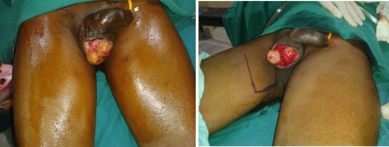
Pre-operative Preparation :
Antibiotics were administered as per Culture and Sensitivity Reports. Diabetes was controlled using insulin therapy. Thrice daily saline dressing was done to reduce the bacterial colonisation of the wound before Surgery. All the subjects were catheterised using Foley’s indwelling catheter. Groin area was inspected to rule out bacterial or fungal infection. Bowel preparation was done in all except those who had Colostomy.
Surgical Technique :
Patient was made to lie down in supine position and a folded linen or sand bag was placed below the same thigh for comfortable access to medial aspect of thigh. Thorough saline wash was given to scrotal, perineum and perianal region. Wound margin was freshened (Figs 1a & 1b). Dimensions of the defect were measured in two dimensions. Template was designed as per the dimensions of the defect. Before making incision for the flap, reverse planning was done several times using the template, to confirm the reach of the flap. Template was placed just below the defect over the medial thigh. The flap margins were marked. The superior margin of the flap corresponded with inferior border of the defect. The medial vertical margin was also marked as per template. The inferior margin was marked parallel to the superior margin. Lignocaine and adrenaline injection was infiltrated beneath the
flap to reduce bleeding. The flap elevation was started from medial to lateral direction. Deep fascia was incised; tagging sutures were taken between deep fascia and dermis to prevent shearing of septocutaneous perforators. Flap was raised above adductor muscle of the thigh. Septo-cutaneous perforators, while elevating the flap were cauterised using bi-polar diathermy. After sufficient elevation of the flap, it was inset to the scrotal defect. A dependent suction drain was used routinely in all cases. In the donor area, distal portion was primarily closed and proximal part near the base of the flap was grafted. Bolster dressing was applied over skin graft. Remaining defects over thigh, perianal, or lower abdomen were either closed primarily or skin grafted. Padded scrotal dressings were applied (Figs 2a & 2b).
Postoperative Care :
The patients were positioned in the supine position to avoid compression and traction on the flap. Flap was inspected on second day; however, the graft bolster dressing was kept undisturbed. Graft dressing was opened on fifth day and staples were removed and patient was discharged with regular follow-up. All the cases were followed for six months (Figs 3a, 3b & 3c).
RESULTS
Ten laterally based medial thigh fasciocutaneous flaps were performed to reconstruct Scrotum in patients
Vol 121, No 2, February 2023Journal
of the Indian Medical Association
Fig 1a — Scrotal defect
Fig 1b — Flap marking
Fig 2a — Elevation of the flap
53
Fig 2b — Flap inset
with Fournier’s Gangrene. The subjects were followed up for six months. The wounds healed satisfactorily in all patients included in the study. There was partial flap necrosis of two cm in one patient requiring debridement and secondary suturing. Minor graft loss over the donor area was noticed in one patient, which healed by secondary intention in three to four weeks. Sutures were removed after two weeks. Flap fused well with the native Scrotum and suture line was inconspicuous after six weeks. The suture line mimicked the normal anatomical median raphe in those patients, where half of the Scrotum was reconstructed by flap. The crude touch and pressure sensation over the flap was almost equal to that of native Scrotum. Normal hair growth was noticed on the flap after four weeks. All subjects were happy about the cosmetic appearance of the Scrotum. The details of patients are mentioned in Table 1.
DISCUSSION
Many surgical options have been used to reconstruct exposed Testes in Fournier’s Gangrene. Reconstruction of the Scrotum is vital for functional, aesthetic and psychological reasons6 . The ideal reconstructive approach would seem to incorporate the following flap features: A single stage procedure, excellent flap reliability, sensate flaps with potential for normal function, minimal donor-site morbidity and simplicity6
Scrotal cover using split skin grafting leads to graft loss and subsequently shrinkage of Testicles 4 Aesthetically, skin graft is not acceptable to the patients and skin graft will remain as an anaesthetic patch. Moreover, normal hair growth will not be present over grafted skin. The placement of Testes in medial

AgeSiteFlap
thigh leads to exposure of Testes to core body temperature and may affect Spermatogenesis. Other issues like dragging pain while walking and cycling and later leading on to Testicular Atrophy has been reported10
The problems associated with this procedure are constant pain caused by mechanical trauma and Testicular Atrophy11. Laterally based medial thigh flap is a reliable fasciocutaneous flap, which can be performed in reasonable short operative time for defects of any size. The donor scar and grafted areas are hidden in the medial aspect of the thigh.
The fasciocutaneous flap of the medial thigh has rich blood supply due to the presence of branches of the femoral artery (internal and circumflex pudendal), making the flap very safe even in diabetic and vasculopathic patients11
Flap should be planned immediately at the lower border of the defect; the inferior margin of the defect forms the superior border of the flap. This step prevents the skin bridge between the defect and the flap. The width of the flap corresponds to the width of the defect.
The minor drawback of this technique is that, the inset of the flap is deficient at the root of the Scrotum. Postoperatively, patient experiences serous discharge from this part for a period of two to three weeks. Some patients also complained of mild dragging pain in this region due to attachment of the flap medially. Flap division and inset done under Local Anesthesia relieved both the above problems.
44Scrotum8 X 5 cmNil18
42Scrotum & penis9 X 4 cmNil13
38Scrotum7 X 5 cmNil10
48Scrotum & abdomen8.5 X5.5 cm2cm flap necrosis16
56Scrotum & perineum7.5 X 5 cmNil18
61Scrotum, perianal & ABD9.5 X 6 cmPartial graft loss12
58Scrotum & penis8 X 6.5 cmNil19
54Scrotum6 X 3.5 cmNil11
50Scrotum, perineum & penis8.5 X 5 cmNil14
62Scrotum, ABD & perineum9 X 5.5 cmNil16
In the surgical technique explained by Ayad, et al6 , the dominant pedicle is located over the apex of the femoral triangle 6-8 cms below the inguinal ligament. There is a need for Doppler examination before starting the procedure to locate the pedicle. After elevation of the flap from distal to proximal direction, careful dissection is required to prevent the injury to the pedicle. Our technique does not require either Doppler examination or
Vol 121, No 2, February 2023Journal of the
Indian Medical Association
Fig 3a — Defect exposing testesFig 3b — After flap coverFig 3c — Follow up after 6 months
Table — Patient demographics of Fournier’s Gangnrene in our hospital
dimension)Complications Hospital stay
54
of the Indian Medical Association
meticulous dissection of the flap. Mageed, et al4 have explained their technique of anteromedial fasciocutaneous thigh flap in which the flap is proximally based and longitudinally oriented and the base of the flap was drawn on the anteromedial aspect of thigh at the level of the inguinal crease. The vascular supply to this flap is the rich suprafascial plexus of vessels present at the anteromedial thigh. However, this technique requires bilateral flaps to cover larger defects.
Pudendal thigh flap provides good quality skin and good support. Even though there is need for bilateral flaps, primary closure of the donor area is an additional advantage of this flap10. Fasciocutaneous thigh flaps can cover medium to large-size defects. The pedicled antero lateral thigh perforator flap is another good alternative for reconstruction of extensive defects of penoscrotal area2. Fasciocutaneous medial thigh flap was sufficient for the defects in our study group. The advantages of our technique are :
•Single stage reconstruction of scrotal defect of any size using one flap only.
•Restoration of normal skin color and hair on the Scrotum, as the flap is designed from adjacent area.
•Preservation of sensation over the flap.
•Donor area is hidden in the medial aspect of the Thigh.
•Durable cover to the exposed Testicles.
•Good aesthetic result.
CONCLUSION
Early recognition and diagnosis, followed by Emergency Surgery, are the keys to treating these cases and for prevention of Systemic Sepsis, Potential Organ Failure and Death12. The laterally based medial thigh fasciocutaneous flap provides excellent cover for
extensive losses of scrotal skin. It is a very robust flap, can be easily mobilized, rarely undergoes Necrosis, and provides sensate cover and excellent Aesthesis. The technique is simple, less time consuming, does not require complex flap planning, and hence can be easily incorporated by Plastic Surgeons and General Surgeons as well.
REFERENCES
1Smith GL, Bunker CB, Dinneen MD — Fournier’s gangrene. Br J Urol 1998; 81: 347-55.
2Lin CT, Chang SC, Chen SG, Tzeng YS — Reconstruction of perineoscrotal defects in Fournier’s gangrene with pedicle anterolateral thigh perforator flap. ANZ J Surg 2016; 86(12): 1052-5.
3Eke N — Fournier’s gangrene: a review of 1726 cases. Br J Surg 2000; 87: 718-28.
4El- Mageed MA — Evaluation of the Anteromedial Thigh Fasciocutaneous Flap for Scrotal Reconstruction. Egypt J Plast Reconstr Surg 2007; 31(2): 149-55.
5Hallock GG — Scrotal Reconstruction Following Fournier Gangrene Using the Medial Circumflex Femoral Artery Perforator Flap. Ann Plast Surg 2006; 57(3): 333-5.
6Ayad WM, Al-Shahat OA, Zaied E, Albanooby T, Taman E — Egypt J Plast Reconstr Surg 2011; 35(1): 129-32.
7Yu P, Sanger JR., Matloub HS, Gosain A, Larson D — Anterolateral Thigh Fasciocutaneous Island Flaps in Perineoscrotal Reconstruction. Plast Recosntr Surg 2002; 109 (2): 610-6.
8Mauro V — Fasciocutaneous flap of the inner thigh for scrotal reconstruction in Fournier syndrome. Rev Bras Cir Plást 2011; 26(4): 707-9.
9Hallock GG — Scrotal reconstruction following Fournier’s gangrene using the medial thigh fasciocutaneous flap. Ann Plast Surg 1990; 24: 86-90
10Mopuri N, O’Connor EF, Iwuagwu FC — Scrotal reconstruction with modified pudendal thigh flaps. J Plast Reconstr Aesthet Surg 2016; 69(2): 278-83.
11Monteiro E, Carvalho P, Costa P, Ferraro A — Inner Thigh Lift Flap for Fournier gangrene of the scrotum. Plast Reconstr Surg 2002; 110(5): 1372-3.
12Tomar SS — Fournier’s gangrene - Not a stigma for surgery with newer surgical reconstruction of scrotum. Indian J Surg 2010; 72(Suppl 1): 339-40.
Disclaimer
The information and opinions presented in the Journal reflect the views of the authors and not of the Journal or its Editorial Board or the Publisher. Publication does not constitute endorsement by the journal.
JIMA assumes no responsibility for the authenticity or reliability of any product, equipment, gadget or any claim by medical establishments/institutions/manufacturers or any training programme in the form of advertisements appearing in JIMA and also does not endorse or give any guarantee to such products or training programme or promote any such thing or claims made so after. —
HonyEditor
Vol 121, No 2, February 2023Journal
55
Review Article
Off-Pump Coronary Artery Bypass Grafting Surgery : A Narrative Review
Mansour Jannati1, Fatemeh Jannati2
Coronary Artery Bypass Grafting (CABG) Surgery is considered the standard treatment for revascularization in Coronary Artery Disease. The Off-Pump Coronary Artery Bypass Graft (OPCAB) which evades the use of Cardiopulmonary Bypass (CPB) became a popular CABG procedure, due to the adverse effect of CPB. Hypothetically, OPCAB may improve the rates of perioperative Myocardial Injury, Neurocognitive Impairment, Stroke and Mortality. However, some studies showed no superior outcomes for OPCAB compared to on-pump CABG. This study aims to evaluate the short-term and long-term outcomes of OPCAB versus On-Pump CABG. The results showed that OPCAB is probably a safe procedure for patients, especially for those with high-risk conditions. OPCAB procedure showed superior short-term outcomes in terms of Myocardial Infarction, Systemic Inflammatory Response, Postoperative Cognitive Dysfunction, Renal Failure, Stroke, Pulmonary Complication, Postoperative Transfusion, Hospital stay length and infection than On-pump CABG. However, regarding long-term outcomes, Off-Pump CABG had a higher rate of incomplete revascularization and repeat revascularization and a higher risk of long-term mortality as well as lower graft patency. Furthermore, the result showed that the higher experience of the surgeons in OPCAB improves the outcome of the Surgery.
[J Indian Med Assoc 2023; 121(2): 56-61]
Key words :Off-Pump Coronary Artery Bypass Grafting Surgery, OPCAB, Short-term Outcomes, Long-term Outcomes.
Coronary Artery Disease (CAD) is the main reason for death Globally and AHA estimated that CAD leads to more than 360,000 deaths each year 1 Coronary Artery Bypass Grafting (CABG) considers the main standard treatment for CAD2. The first CABG was done in the 1960s as the only treatment method for Ischemic Heart Disease 3 . Developments in Cardiopulmonary Bypass (CPB) have improved Cardiac Surgery; however, the non-physiological effect of CPB leads to organ dysfunction and tissue injury. In addition, cerebrovascular events related to CPB are a significant problem4,5. As the number of patients undergoing CABG increases, solutions to reduce the alleged adverse effects of CPB due to aortic manipulation and clamping have been evaluated.Therefore, Off-Pump CABG (OPCAB) became popular in the 1990s6. In the United States, OPCAB peaked in 2002 and almost 25% of all CABG in operations were done Off-Pump6
In Japan, in 2000, and after the emersion of the stabilizer, OPCAB has become a prevalent practice for CABG, and now about 65% of CABG cases are done by OPCAB7
1MD, Cardiovascular Surgeon, Department of Cardiovascular Surgery Ward, Namazee Hospital, Shiraz University of Medical Sciences, Shiraz, Fars, Iran and Corresponding Author
2MD, General Physicion, Department of Student Research Committee, Shiraz University of Medical Sciences, Shiraz, Iran
Received on : 17/08/2022
Accepted on : 23/10/2022
Editor's Comment :
The comparison of Off-Pump CABG and On-Pump CABG has yielded mixed results.
Off-Pump procedure is safer with lesser short term complications though long term outcomes like incomplete revascularization, repeat revascularization and poor graft patency is more in off pump revascularization.
After many years of using the OPCAB procedure and publishing many trials and meta-analyses about the benefits of this method, there is still controversy on the outcomes and advantages of Off-Pump CABG. The present review summarizes the most recent and relevant data about this method and evaluates the shortterm and long-term outcomes of Off-Pump CABG.
MATERIALS AND METHODS
In this review study, all articles on Off-Pump Coronary Artery Graft surgerywere evaluated from the year 2000 to 2021. Articles were searched through databases such as PubMed, Web of Science, Scopus, Google Scholar, Science Direct and Cochrane library using the keywords “Off-Pump Coronary Artery Graft Surgery “, “OPCAB”, “Short-term outcomes” and” longterm outcomes”. A total of 167 articles were extracted in an initial search. After reviewing the abstract of these articles, finally, 68 articles that met the necessary criteria of the present review were selected and evaluated completely.
Vol 121, No 2, February
2023Journal of the Indian Medical Association
56
OPCAB in High-Risk Patients :
In a meta-analysis by Kowalewski, et al, it was shown that the odds of Cerebral Stroke are significantly decreased in OPCAB in comparison to conventional CABG in high-risk patients. Furthermore, they showed that there is a significant association between patients’ risk profile and benefits from OPACB in Myocardial Infarction (MI), Death and Cerebral Stroke, proposing that in high-risk patients, OPCAB must be toughly considered. Therefore, they concluded that OPCAB improves the short-term outcomes in high-risk patients8 In a prospective randomized trial study on 411 patients, it was found that OPCAB decreases primary mortality as well as morbidity in high-risk patients9
In Kerendi, et al study it was revealed that the riskadjusted outcomes for mortality and morbidity endpoints in high-risk patient populations were superior after OPCAB versus On-Pump CABG10.The result of Rastan, et al study on high-risk patients demonstrated that patients with reduced left ventricular function and multi-risk patients benefit from OPCAB in terms of perioperative mortality and morbidity. Furthermore, in patients with major extracardiac risk factors, OPCAB reduced the rate of Perioperative Stroke. In individuals with Pulmonary or renal Dysfunction, OPCAB resulted in a reduction in the failure of the organ. OPCAB resulted in a lower need for transfusion in high-risk populations11
Morbidity and Mortality after OPCAB :
In a study on 118,140 CABG procedures, that 11,717 cases of them were OPCAB,the use of the OPCAB method was related to a reduction in risk-adjusted operative mortality from 2.9% with the On-Pump method to 2.3% in the OPCAB group. Also, the risk-adjusted morbidity rate was reduced from 14.15% with On-Pump CABG to 10.62% in OPCAB group12. Bittner, et al evaluated 57 multivessel diseases OPCABG casesfor 2 years and reported that Off-Pump CABG can be done with rational low morbidity and mortality in high-risk patients13. Vettath, et al also showed that Off-Pump CABG resulted in an improvement in the mortality rate over time14. Takagi, etalstudied eight medium- to largesize RCTs including 8780 patients with long-term followup OPCAB versus on-pump CABG and revealed that OPCAB rises long-term (5 years or more) mortality in comparison to On-Pump CABG (ONCAB)15. In another study, they demonstrated that OPCAB is related to an increase in very long-term, 10 years and more, all-cause mortality in comparison to ONCAB16
OPCAB and Systemic Inflammatory Response : The systemic inflammatory response is probable after CABG using CPB which comprises the
complement and Leukocyte activation, proinflammatory cytokines releasing Nitric Oxide metabolism changes, and an increase in the free radical’s Oxygen production that may lead to organ dysfunction and coagulation conditions17. Schulze, et al showed that the OPCAB revascularization process and not using CPB significantly diminishes the Systemic Inflammatory Response Syndrome and the need for primary catecholamine. This can help to the improvement of Organ Function, also improvement of recovery from surgery 18 . Nesher, et al study demonstrated that Serum cytokine levels (IL-6, IL-8 and IL-10) in the OPCAB method were lower in comparison tothe on-pump method19. In another study, OPCAB attenuated the systemic release of IL-8 and IL-10, whereas no difference was seen in the release of IL-6, CRP and Neutrophils. Signs of cytokine uptake were seen in the Lungs by OPCAB20. In Meng, et al study, the analyses presented that the concentration of IL-10 was significantly lower after OPCAB in comparison to on-pump CABG. But, for IL-6 and IL-8 no significant differences were observed in the two groups21
Postoperative Cognitive Dysfunction in OPCAB :
Cerebral Dysfunction including Postoperative Cognitive Dysfunction (POCD), delirium and stroke after Cardiac Surgery remains a devastating problem, especially in the older age group.They may happen as a result of cerebral emboli,inflammation, or hypoperfusion22.
In Sun et al. study, a Meta-analysis of 13 randomized controlled trials on 2326 patients it was demonstrated that the occurrence of POCD was significantly lower following OPCAB than On-Pump in 1-2 weeks and at 3 months after Surgery23. In a study by Szwed, et al, it was shown that anaorticOPCAB significantly reduced the rate of postoperative cognitive dysfunction and delirium compared to conventional OPCAB24. Schmitz, et al also found that postoperative neurocognitive function significantly improves by OPCAB25.In another study, Off-pump CABG decreased postoperative Neuropsychological dysfunction in elderly cases in comparison to on-pump CABG26 However, in Lund, et al study, long-term cognitive function after OPCAB was similar to on-pump coronary Artery Bypass Grafting Surgery27. Similarly, Marasco et al meta-analysis indicated no significant neurocognitive advantage while using OPCAB in comparison to on-pump CABG28
Postoperative Renal Dysfunction and OPCAB :
Postoperative Renal Dysfunction is a significant complication of CABG and is associated with the
Vol 121, No 2, February 2023Journal
of the Indian Medical Association
57
patient’s clinical condition, CPB-related events, renal hypoperfusion, hypotension,hypothermia, microemboli, and stimulation of the inflammatory response29,30
According to Umit, et al study, the Off-Pump CABG offers excellent renal protection and a significantly lower risk of Renal Dysfunction compared with conventional CABG31. Abu-Omar et al in a study on 1580 patients showed that Off-pump CABG is associated with a decrease in postoperative renal damage32. Rocha, et al reported that OPCAB is related to improving in-hospital renal outcomes in patients with moderate renal failure, but is not related to the longterm cumulative occurrence of end-stage renal failure which needs permanent Dialysis33
Postoperative Pulmonary Dysfunction and OPCAB:
One of the prevalent difficulties after CABG is pulmonary complications associated with surgical procedures, anaesthesia procedure effects, and Cardiopulmonary bypass pump 34
A study by Staton et al. reported that OPCAB was associated with a greater decrease in postoperative respiratory compliance compared to the on-pump method.Moreover, OPCAB resulted in superior gas exchange and earlier extubation35. According to Raja et al. study, based on available evidence from RCTs, OPCAB reduces Postoperative Pulmonary Dysfunction and is more effective than On-Pump CABG for Chronic Obstructive Pulmonary Disease (COPD) patients36 Silva, et al study also revealed that patients in the OPCAB group had higher improvement in pulmonary function after Surgery than On-Pump group37. However, in Montes, et al study Off-pump CABG in comparison to the On-Pump method did not result in important protection from postoperative pulmonary dysfunction38
OPCAB and Postoperative Arterial Fibrillation :
Postoperative Atrial Fibrillation (AF) is a common arrhythmia that happens after CABG. OPCAB is considered to be a less invasive technique and is believed to reduce the rate of AF, however, there is inconsistency in obtained data.Athanasiou, et al demonstrated a reduction in the occurrence of postoperative AF with Off-Pump CABG techniques. However, they suggested that the results should use with caution29. Lewiki et al. reported no difference between the incidence of AF after OPCAB and ONCAB surgeries. Böning et al. also showed no alteration in the rate of postoperative AF by using OPCAB39. In the Junior, et al study, Off-pump CABG did not decrease the occurrence of postoperative AF. They also defined age >70 years old and preoperative AF as risk factors
for postoperative AF40.
Incomplete Revascularization in OPCAB :
In this Cohort Ji, et al on 1,349 patients with a triple-vessel lesion, the occurrence of Off-Pump incomplete revascularization was 19.9%41. In a report on 41,139 patients with the left main and three-vessel lesion, the rate of incomplete revascularizationin 6,367 OPCAB patients was about 29.0%42. In a SYNTAX trial by Head, et al, the incomplete revascularization rate was 36.8% after OPCAB 43 . Another study including 7,427 OPCAB cases and 7,128 on-pump CABG cases demonstrated that the incomplete revascularization rate of the Off-Pump method was 13.3%44.
Repeat Revascularization and OPCAB :
Takagi, et al analysis showed that OPCABG may raise the rate of repeat revascularization by 38% over ONCABG45.In a meta-analysis study by Zhou, et al, it was found that OPCABG increases the rate of repeat revascularization in comparison to ONCAB at 1-year follow-up, but not at 5-year follow-up46
Graft PatencyAfter OPCAB :
Hattler et al. study demonstrated significantly lower graft patency for OPCAB than On-Pump CABG47. Parolari, et al study in a meta-analysis of randomized studies showed a reduction in postoperative graft patency of Coronary Artery Bypass Grafts done by OPCAB methods48. Another study by Houlind, et al found that graft patency after Off-Pump CABG was lower than the On-Pump method even with heparinization protocols49. However, Puskas, et al reported that graft patency was similar for OPCAB and on-pump CABG at 30 days and one year50. Hu, et al also revealed no significant differences in graft patency between OPCAB and ONCAB GROUPS in Triple Vessel Coronary Artery Disease patients51.
Conversion in OPCAB :
Regardless of the popularity of OPCAB and its benefits, in a small number of patients, conversion to on-pump CABG may be required due to reasons including hemodynamic disturbance, physical difficulty in grafting, ischemia, or arrhythmias. Chakravarthy et al. reported that conversion in OPCAB is related to significant mortality raise. Increased left ventricular enddiastolic pressure, women’s gender and pre-operative need for Intra-aortic balloon are indicators of amplified mortality risk in conversion52. According to Tariq, et al study, emergency conversion from OPCAB to on-pump is the most disastrous occurrence leading to higher morbidity and mortality. Conversion as a result of
Vol 121, No 2, February 2023Journal
of the Indian Medical Association
58
arrhythmias was the main cause (rate: 9%) and patients with higher New York Heart Association status and Chronic Obstructive Pulmonary Disease had a higher risk of emergency conversion. Therefore, they suggested that the decision for OPCAB should be made with caution for each patient53. In Keeling, et al study, intra-operative conversion from OPCAB to on-pumpwas reported to be a morbid incident. They recommended that elective ONCAB should be selected for those with a higher risk for conversion. They also found that older age, preoperative intraaortic balloon pump placement, ejection fraction <35%, increasing number of involved vessels, Heart Failure and emergent procedural condition were independent markers for conversion54
OPCAB and Surgeons’ Experience :
The important role of Surgeons’ experience in OPCAB was highlighted frequently by professionals in the field. Benedetto, et al showed that mortality was reduced after OPCAB when done by high-volume surgeons in high-volume OPCAB centers and in contrast the mortality risk increased when OPCAB was done by surgeons in low-volume centers55. In the Glance et al. study it was found that for On-Pump CABG Surgery, higher surgeon case volumes are related to lower rates of mortality56. In another study by Chen, et al, it was shown that by Increasing the experience of a surgeon the rate of blood transfusion was reduced by about 33%57. Hsu, et al evaluated the proficiency of surgeons for OPCAB by the following quality indicators, the revascularization index, and the conversion rate. They reported that a revascularization index of >1.4 and a conversion rate of <5% show the proficiency of surgeons for OPCAB 58.Hemil, et al reported that the occurrence of emergency conversion during OPCAB has reduced with raising surgeons’ experience; however, in these patients, the rate of morbidity remained unaffected59
Short-term Outcomes of OPCAB :
In a meta-analysis, by Reston, et al the results showed that rates of perioperative myocardial Infarction, Reoperation for bleeding, Renal failure, Stroke and Mortality were lower after OPCABG than after CABG. They also showed a decrease in hospital stay length, AF and wound infection related to OPCABG, however, the differences were not statically significant60. In a study by Elmahrouk, et al on 450 patients, it was found that the rate of early postoperative AF and Renal Failure was decreased in the Off-Pump group. But, no statistically significant difference was observed in neurologic complications, AMI, or early mortality between Off- or On-Pump groups61. Gao, et al evaluated the short-term outcome
in 318 high-risk patients who underwent CABG and found that the OPCAB group had significantly decreasedventilator support time, Postoperation ICU time, operative mortality and morbidity62. Guan, et al performed a meta-analysis and systematic review on 32,354 patients and reported a significant benefit from OPCAB in terms of 30-day mortality, MI, stroke, renal failure, Infection, Pulmonary complication, Postoperative transfusion, and reoperation bleeding. They showed no significant difference in AF and Neurological Dysfunction 63 . Kowalewski, et al performed a meta-analysis on 100 studies (19,192 patients) and demonstrate a significant decrease in the odds of cerebral stroke in the OPCAB group compared with on-pump CABG8
Long-term Outcomes of OPCABG :
In Luo, etalstudy on seven RCTs and 9,128 patients, in long-term follow-up OPCAB had a significantly higher rate of revascularization (OR=1.45; p=0.04) than onpump CABG64.Chikwe et al. evaluated long-term results on 6,950 who underwent Off-Pump CABG and reported a rise in repeat revascularization, incomplete revascularization and mortality at 10 years in comparison to ONCAB65. A meta-analysis by Takagi, et al on 22 studies, and more than 100,000 patients, disclosed that OPCAB is probably related to worse longterm survival (5years or more) compared with ONCAB66
In another study on 13 studies including 13,234 patients, Off-pump CABG increased the risk of all-cause death and repeat bypass surgery at long-term, more than 4-year follow-up67. In a study on 5,203 patients by Kim, et al it was reported that for overall mortality, patients who underwent Off-Pump CABG had a significantly higher risk of mortality (HR: 1.43; p<0.0001) in comparison to those who underwent ONCAB68
CONCLUSION
OPCAB is probably a safe substitute for On-Pump CABG for patients, especially with high-risk conditions. OPCAB procedure showed superior short-term outcomes in terms of Myocardial Infarction, renal Failure, Stroke, Pulmonary Complication, Postoperative Transfusion, Hospital stay length and Infection than On-Pump CABG. Also, it was shown that the OPCAB revascularization process significantly diminishes the systemic inflammatory response syndrome. In addition, OPCAB significantly reduced the rate of early postoperative cognitive dysfunction compared to conventional OPCAB. However, regarding long-term outcomes Off-Pump CABG had a higher rate of incomplete revascularization and repeat revascularization and a higher risk of long-term mortality
Vol 121, No 2, February 2023Journal
of the Indian Medical Association
59
as well as lower graft patency. Furthermore, the result showed that the higher experience of the surgeons in OPCAB improves the outcome of the surgery.
REFERENCES
1Shaefi S, Mittel A, Loberman D, Ramakrishna H — Off-Pump Versus On-Pump Coronary Artery Bypass Grafting-A Systematic Review and Analysis of Clinical Outcomes. J Cardiothorac Vasc Anesth 2019; 33(1): 232-44.
2Vilahur G, Badimon JJ, Bugiardini R, Badimon L — Perspectives: The burden of cardiovascular risk factors and coronary heart disease in Europe and worldwide. European Heart Journal Supplements 2014; 16(suppl_A): A7-A11.
3Konstantinov IE. Robert H — Goetz: the surgeon who performed the first successful clinical coronary artery bypass operation. Ann Thorac Surg 2000; 69(6): 1966-72.
4Warren OJ, Smith AJ, Alexiou C — The inflammatory response to cardiopulmonary bypass: part 1—mechanisms of pathogenesis. J Cardiothorac Vasc Anesth 2009; 23(2): 22331.
5Baufreton C. Role of surgical factors in strokes after cardiac surgery. Arch Cardiovasc Dis 2010; 103(5): 326-32.
6Bakaeen FG, Shroyer AL, Gammie JS — Trends in use of offpump coronary artery bypass grafting: Results from the Society of Thoracic Surgeons Adult Cardiac Surgery Database. J Thorac Cardiovasc Surg 2014; 148(3): 856-3, 64 e1; discussion 63-4.
7Kuwahara G, Tashiro T — Current Status of Off-Pump Coronary Artery Bypass. Ann Thorac Cardiovasc Surg 2020; 26(3): 125-32.
8Kowalewski M, Pawliszak W, Malvindi PG — Off-pump coronary artery bypass grafting improves short-term outcomes in high-risk patients compared with on-pump coronary artery bypass grafting: Meta-analysis. J Thorac Cardiovasc Surg 2016; 151(1): 60-77 e1-58.
9Lemma MG, Coscioni E, Tritto FP — On-pump versus offpump coronary artery bypass surgery in high-risk patients: operative results of a prospective randomized trial (on-off study). J Thorac Cardiovasc Surg 2012; 143(3): 625-31.
10Kerendi F, Morris CD, Puskas JD — Off-pump coronary bypass surgery for high-risk patients: only in expert centers? Curr Opin Cardiol 2008; 23(6): 573-8.
11Rastan AJ, Walther T, Falk V — [Coronary artery bypass grafting on the beating heart in high-risk patients]. Herz 2007; 32(6): 483-90.
12Cleveland JC, Jr., Shroyer AL, Chen AY, Peterson E, Grover FL — Off-pump coronary artery bypass grafting decreases risk-adjusted mortality and morbidity. Ann Thorac Surg 2001; 72(4): 1282-8; discussion 8-9.
13Bittner HB, Savitt MA — Off-pump coronary artery bypass grafting decreases morbidity and mortality in a selected group of high-risk patients. Ann Thorac Surg 2002; 74(1): 115-8.
14Vettath MP, Ravisankar M, Kopjar T, Kannan AV, Gangadharan N. Off-Pump Coronary Artery Bypass Grafting Improves Early Clinical Outcomes Including Operative Mortality. Heart Surg Forum 2018; 21(3): E151-E7.
15Takagi H, Hari Y, Mitta S — A meta-analysis of >/=5-year mortality in randomized controlled trials of off-pump versus on-pump coronary artery bypass grafting. J Card Surg 2018; 33(11): 716-24.
16Takagi H, Ando T, Mitta S, group A. Meta-Analysis Comparing >10-Year Mortality of Off-Pump Versus On-Pump Coronary Artery Bypass Grafting. Am J Cardiol 2017; 120(11): 19338.
17Raja SG, Berg GA — Impact of off-pump coronary artery bypass surgery on systemic inflammation: current best available evidence. J Card Surg 2007; 22(5): 445-55.
18Schulze C, Conrad N, Schutz A — Reduced expression of systemic proinflammatory cytokines after off-pump versus conventional coronary artery bypass grafting. Thorac Cardiovasc Surg 2000; 48(6): 364-9.
19Nesher N, Frolkis I, Vardi M — Higher levels of serum cytokines and myocardial tissue markers during on-pump versus offpump coronary artery bypass surgery. J Card Surg 2006; 21(4): 395-402.
20Rasmussen BS, Laugesen H, Sollid J — Oxygenation and release of inflammatory mediators after off-pump compared with after on-pump coronary artery bypass surgery. Acta Anaesthesiol Scand 2007; 51(9): 1202-10.
21Meng F, Ma J, Wang W, Lin B — Meta-analysis of interleukin 6, 8, and 10 between off-pump and on-pump coronary artery bypass groups. Bosn J Basic Med Sci 2017; 17(2): 85-94.
22Goto T, Maekawa K — Cerebral dysfunction after coronary artery bypass surgery. J Anesth 2014; 28(2): 242-8.
23Sun JH, Wu XY, Wang WJ, Jin LL — Cognitive dysfunction after off-pump versus on-pump coronary artery bypass surgery: a meta-analysis. J Int Med Res 2012; 40(3): 852-8.
24Szwed K, Pawliszak W, Szwed M — Reducing delirium and cognitive dysfunction after off-pump coronary bypass: A randomized trial. J Thorac Cardiovasc Surg 2021; 161(4): 1275-82 e4.
25Schmitz C, Weinreich S, Schneider R — Off-Pump versus on-pump coronary artery bypass: can OPCAB reduce neurologic injury? Heart Surg Forum 2003; 6(3): 127-30.
26Baba T, Goto T, Maekawa K — Early neuropsychological dysfunction in elderly high-risk patients after on-pump and off-pump coronary bypass surgery. J Anesth 2007; 21(4): 452-8.
27Lund C, Sundet K, Tennoe B — Cerebral ischemic injury and cognitive impairment after off-pump and on-pump coronary artery bypass grafting surgery. Ann Thorac Surg 2005; 80(6): 2126-31.
28Marasco SF, Sharwood LN, Abramson MJ — No improvement in neurocognitive outcomes after off-pump versus on-pump coronary revascularisation: a meta-analysis. Eur J Cardiothorac Surg 2008; 33(6): 961-70.
29Athanasiou T, Aziz O, Mangoush O — Do off-pump techniques reduce the incidence of postoperative atrial fibrillation in elderly patients undergoing coronary artery bypass grafting? Ann Thorac Surg 2004; 77(5): 1567-74.
30Loganathan S, Nieh CC, Emmert MY — Off-pump versus onpump coronary artery bypass procedures:postoperative renal complications in an Asian population. Ann Acad Med Singap 2010; 39(2): 112-6.
31Arslan U, Calik E, Tekin AI, Erkut B — Off-pump versus onpump complete coronary artery bypass grafting: Comparison of the effects on the renal damage in patients with renal dysfunction. Medicine (Baltimore) 2018; 97(35): e12146.
32Abu-Omar Y, Taghavi FJ, Navaratnarajah M — The impact of off-pump coronary artery bypass surgery on postoperative renal function. Perfusion 2012; 27(2): 127-31.
33Rocha RV, Yanagawa B, Hussain MA — Off-pump versus on-pump coronary artery bypass grafting in moderate renal failure. J Thorac Cardiovasc Surg 2020; 159(4): 1297-304 e2.
34Mali S, Haghaninejad H — Pulmonary complications following cardiac surgery. Arch Med Sci Atheroscler Dis 2019; 4: e280-e5.
35Staton GW, Williams WH, Mahoney EM — Pulmonary outcomes of off-pump vs on-pump coronary artery bypass surgery in a randomized trial. Chest 2005; 127(3): 892-901.
36Raja SG, Dreyfus GD — Impact of off-pump coronary artery bypass surgery on postoperative renal dysfunction: current best available evidence. Nephrology (Carlton) 2006; 11(4): 269-73.
Vol 121, No 2, February 2023Journal
of the Indian Medical Association
60
of the Indian Medical Association
37e Silva AM, Saad R, Stirbulov R, Rivetti LA — Off-pump versus on-pump coronary artery revascularization: effects on pulmonary function. Interact Cardiovasc Thorac Surg 2010; 11(1): 42-5.
38Montes FR, Maldonado JD, Paez S, Ariza F — Off-pump versus on-pump coronary artery bypass surgery and postoperative pulmonary dysfunction. J Cardiothorac Vasc Anesth 2004; 18(6): 698-703.
39Boning A, Diegeler A, Hilker M — Preoperative atrial fibrillation and outcome in patients undergoing on-pump or off-pump coronary bypass surgery: lessons learned from the GOPCABE trial. InteractCardiovascThoracSurg 2015; 20(1): 74-8.
40Bohatch Junior MS, Matkovski PD, Di Giovanni FJ — Incidence of postoperative atrial fibrillation in patients undergoing onpump and off-pump coronary artery bypass grafting. Rev Bras Cir Cardiovasc 2015; 30(3): 316-24.
41Ji Q, Zhao Y, Zhu K — Impacts of incomplete revascularization following off-pump coronary artery bypass grafting on clinical outcomes of patients with triple-vessel lesions: insights from a single-center study of propensity-matched data. Journal of Thoracic Disease 2019; 11(4): 1325-35.
42Omer S, Cornwell LD, Rosengart TK — Completeness of coronary revascularization and survival: Impact of age and off-pump surgery. J Thorac Cardiovasc Surg 2014; 148(4): 1307-15 e1.
43Head SJ, Mack MJ, Holmes DR, Jr — Incidence, predictors and outcomes of incomplete revascularization after percutaneous coronary intervention and coronary artery bypass grafting: a subgroup analysis of 3-year SYNTAX data. Eur J Cardiothorac Surg 2012; 41(3): 535-41.
44Benedetto U, Caputo M, Patel NN, et al. Long-term survival after off-pump versus on-pump coronary artery bypass graft surgery. Does completeness of revascularization play a role? Int J Cardiol 2017; 246: 32-6.
45Takagi H, Mizuno Y, Niwa M — A meta-analysis of randomized trials for repeat revascularization following off-pump versus on-pump coronary artery bypass grafting. Interact Cardiovasc Thorac Surg 2013; 17(5): 878-80.
46Zhou P, Zhu P, Xiao Z — Meta-Analysis of Repeat Revascularization of Off-Pump and On-Pump Coronary Artery Bypass Surgery. Ann Thorac Surg 2018; 106(2): 526-31.
47Hattler B, Messenger JC, Shroyer AL —Off-Pump coronary artery bypass surgery is associated with worse arterial and saphenous vein graft patency and less effective revascularization: Results from the Veterans Affairs Randomized On/Off Bypass (ROOBY) trial. Circulation 2012; 125(23): 2827-35.
48Parolari A, Alamanni F, Polvani G — Meta-analysis of randomized trials comparing off-pump with on-pump coronary artery bypass graft patency. Ann Thorac Surg 2005; 80(6): 2121-5.
49Houlind K, Fenger-Grøn M, Holme SJ — Graft patency after off-pump coronary artery bypass surgery is inferior even with identical heparinization protocols: results from the Danish On-pump Versus Off-pump Randomization Study (DOORS). The Journal of Thoracic and Cardiovascular Surgery 2014; 148(5): 1812-9. e2.
50Puskas JD, Williams WH, Mahoney EM — Off-pump vs conventional coronary artery bypass grafting: early and 1year graft patency, cost, and quality-of-life outcomes: a randomized trial. JAMA 2004; 291(15): 1841-9.
51Hu S, Wang X, Song Y, Lu F — Graft patency in off-pump and conventional coronary artery bypass grafting for treatment of triple vessel coronary disease. Chin Med J (Engl) 2003; 116(3): 436-9.
52Chakravarthy M, Prabhakumar D, Patil TA, George A, Jawali V — Conversion during off-pump coronary artery bypass graft surgery: A case-control study. Ann Card Anaesth 2019; 22(1): 18-23.
53Tariq K, Zia K, Mangi A — Conversion from Off to On-Pump Coronary Artery Bypass Grafting. Is it Avoidable? Cureus 2020; 12(1): e6791.
54Keeling B, Thourani V, Aliawadi G — Conversion From Off-Pump Coronary Artery Bypass Grafting to On-Pump Coronary Artery Bypass Grafting. Ann Thorac Surg 2017; 104(4): 1267-74.
55Benedetto U, Lau C, Caputo M — Comparison of Outcomes for Off-Pump Versus On-Pump Coronary Artery Bypass Grafting in Low-Volume and High-Volume Centers and by Low-Volume and High-Volume Surgeons. Am J Cardiol 2018; 121(5): 552-7.
56Glance LG, Dick AW, Osler TM, Mukamel DB — The relation between surgeon volume and outcome following off-pump vs on-pump coronary artery bypass graft surgery. Chest 2005; 128(2): 829-37.
57Chen JW, Hsu RB — Impact of surgeon experience on the rate of blood transfusion in off-pump coronary artery bypass. J Formos Med Assoc 2016; 115(3): 145-51.
58Hsu RB, Lin CH — Surgical proficiency and quality indicators in off-pump coronary artery bypass. Ann Thorac Surg 2013; 96(6): 2069-74.
59Hemli JM, Patel NC, Subramanian VA — Increasing surgical experience with off-pump coronary surgery does not mitigate the morbidity of emergency conversion to cardiopulmonary bypass. Innovations (Phila) 2012; 7(4): 259-65.
60Reston JT, Tregear SJ, Turkelson CM — Meta-analysis of short-term and mid-term outcomes following off-pump coronary artery bypass grafting. Ann Thorac Surg 2003; 76(5): 1510-5.
61Elmahrouk AF, Hamouda TE, Kasab I, Ismail MF, Jamjoom AA — Short term outcome of conventional versus off-pump coronary artery bypass grafting for high-risk patients. Journal of the Egyptian Society of Cardio-Thoracic Surgery 2018; 26(1): 57-63.
62Gao HW, Zheng Z, Hu SS — [The short-term outcomes in selected high-risk patients underwent off-pump or on-pump coronary artery bypass grafting]. Zhonghua Xin Xue Guan Bing Za Zhi 2007; 35(3): 245-7.
63Guan Z, Guan X, Gu K —. Short-term outcomes of on- vs offpump coronary artery bypass grafting in patients with left ventricular dysfunction: a systematic review and metaanalysis. J Cardiothorac Surg 2020; 15(1): 84.
64Luo T, Ni Y — Short-term and Long-term Postoperative Safety of Off-Pump versus On-Pump Coronary Artery Bypass Grafting for Coronary Heart Disease: A Meta-analysis for Randomized Controlled Trials. ThoracCardiovascSurg2015; 63(4): 319-27.
65Chikwe J, Lee T, Itagaki S, Adams DH, Egorova NN — LongTerm Outcomes After Off-Pump Versus On-Pump Coronary Artery Bypass Grafting by Experienced Surgeons. J Am Coll Cardiol 2018; 72(13): 1478-86.
66Takagi H, Umemoto T — All-Literature Investigation of Cardiovascular Evidence G. Worse long-term survival after off-pump than on-pump coronary artery bypass grafting. J Thorac Cardiovasc Surg 2014; 148(5): 1820-9.
67Thakur U, Nerlekar N, Muthalaly RG — Off- vs. On-Pump Coronary Artery Bypass Grafting Long-Term Survival is Driven by Incompleteness of Revascularisation. Heart Lung Circ 2020; 29(1): 149-55.
68Kim JB, Yun SC, Lim JW — Long-term survival following coronary artery bypass grafting: off-pump versus on-pump strategies. J Am Coll Cardiol 2014; 63(21): 2280-8.
Vol 121, No 2, February 2023Journal
61
Review Article
A Study of the Epidemiology of Childhood Blindness and Plan of Action in Bihar
Vishwa Ratan1
Vision Disorders are among the most common disabilities to affect children. Childhood Blindness is an important Public Health Problem due to inequalities in the Social and Economical conditions of Bihar State.
Moreover it’s a significant component of WHO vision 2020 program. By Public Health Interventions, the prevalence of cases of Childhood Blindness can be reduced. With this background, a study was done in assessment of the epidemiology of childhood blindness, with suggestions of plan of actions to reduce the prevalence of childhood Blindness which will lead to educational opportunities and improvement in mental and general health.
This study had limitations due to average Health infrastructures and record keeping from PHC to Medical colleges. But at the same time, still a lot of timely Neonatal Eye Care Services and proper refraction strategies have helped in prevention of Childhood Blindness.
In this study, the principals of epidemiology has been followed, as to finding the answers of “Who, Where and Why” regarding Childhood Blindness in Bihar.
The current prevalence of blindness in children is known to be around 0.6%. Despite various intervention programs. This Public Health Problem a challenge both from epidemiology and care provider point of view.
[J Indian Med Assoc 2023; 121(2): 62-3]
Key words :Strong School Health program intervention, Awareness of kitchen gardening.
Methods :
Information from search engines used included Pubmed, Google scholar. Field visits to different levels of Health infrastructure, like PHC, CHC, District Hospitals and Medical Colleges, while working in a, Govt, UNICEF sponsored project Hospital Records1
Community Based Survey :
From verbal discussion with Eye Specialists, Program Managers and Health Workers.
From Health Department Records available in State Blindness Control Cell1
Observations :
The main cause of Childhood visual impairment was Refractive Errors. Under Blindness Control Programme till date 3.5 lacs children have been examined at schools of Bihar over a period of nearly fourteen years from 1999 to 2013. The important observation being that the majority of children suffered from Refractive errors. Random sampling of 15 district records out of total 38 districts were done.
Districts with high percentage of Refractive errors
Sharsha, Supual, Madhepura, Rhotas, Kishanganj, Darbhangha, Banka, Madhubani and Sitamadhi . This in terms of percentage is 60% Districts with low
1MBBS, MS, Associate Professor, Venkateshwara Institute of Medical Sciences, Gajraula, Amroha, UP 244235
Received on : 04/10/2021
Accepted on : 08/12/2021
Editor's Comment :
School Health Check up program, Should be strengthened and implemented Seriously. Awareness about kitchen garden and use of roof tops of the hutments for growing Vegetables.
Data collection and analysis of childhood blindness should be done on an Annual basis.
percentage of refractive errors Munger, Nalanda, Nawada and Begusarai, Arrah, Gaya. This in terms of percentage is 33.33%
The findings showed that North Districts across River Ganges had more cases of Refractive Errors than Districts of South and Central Bihar.
The possible causes of high percentage in North Bihar Districts being Poverty, Malnutrition.
Yearly floods causing loss of livelihood leading to poor economic status.
The second largest cause of childhood blindness was Vitamin-A deficiency.
The Prevalence of childhood blindness due to VitA deficiency per thousand was as follows :
The OPD records on random study of District Hospitals showed that , South Bihar showed a bigger percentage of cases in comparison to North Bihar.
Vol 121, No 2, February 2023Journal of the Indian Medical Association
62
YearPR 2000 0.40 2005 0.25 2010 0.02
The possible cause being rich fruit plantation like papaya, mangoes, which are consumed in large children population. Practice of hut roof for agriculture purpose is very common in villages, of North Bihar, where papaya can be grown.
With intervention of Vitamin-A deficiency control program sponsored by UNICEF for nearly five years played a very crucial role in bringing down the prevalence rate of Vitamin-A deficiency cases. Moreover, this program also led to huge reduction in Gastroenteritis cases in children.
Thus breaking the vicious cycle of — Diarrhea-malnutrition- vitamin-A def- blindness2.
3. Lately with more access to Health Care Facilities, other causes too were also identified
Coloboma of Iris
Congenital Cataract Microphthalmos
Possibly, Hereditary reasons are responsible for these ocular morbidity.
Many cases of ocular trauma were also recorded.
Discussion :
The impact of Vitamin-A blindness program, supported by UNICEF, had a great impact in VitaminA deficiency both in terms of the incidence and prevalence leading to decreasing the burden of Childhood Blindness. After the program, robust immunization programme, which includes Vitamin-A administration to children had also a great impact2
The School Health Check-up Programme, which includes detection of Refractive Errors and general examination of Eye , too had been very successful. A large number of children with Refractive Errors were detected , which was eventually corrected by giving glasses, which can lead to reduction of Amblyopia in future years.
Theobservationssuggeststhat— Strengthening of refractive services at Primary and Secondary level of school going children.
Screening of Eyes at school levels, as well at health facilities like PHC, CHC, District Hospitals should be done regularly.
Provision of low vision devices at low or free should be done.
Plan of Actions/ strategy to be adopted for control of childhood Blindness.
Establishment of Paediatric Ophthalmic Department in Medical and District Hospitals. Low vision clinics should be provided at District Hospitals.
Training programs in Paediatric Ophthalmology should be carried out regularly1
Conclusions :
Regular small survey should be carried out by Ophthalmologist in their respective areas, which will go a long way in prevention of Childhood Blindness. The observations should be documented and published. There should be continuum of health promotion of Health programs along with Rehabilitation. A comprehensive eye care approach in control of Childhood Blindness should be a priority by the health planners and providers.
Conflict of Interest : No financial support
REFERENCES
1Wadhwani M, Vashist P, Singh SS, Gupta V, Gupta N, Saxena R — Prevalence and causes of childhood blindness in India: A systematic review. Indian J Ophthalmol 2020; 68(2): 3115. Published online 2020 Jan 20. doi: 10.4103/ijo.IJO_2076_18
2UNICEF Supported field visits as State program officer (Control of Blindness).
Vol 121, No 2, February 2023Journal
of the Indian Medical Association
63 Submit Article in JIMA - Online See website : https: // onlinejima.com Any queries : (033) 2237-8092, +919477493027; +919477493033
Review Article
A Study of the Urethral and Stretched Penile Lengths in the Adult, Indian, Male Population
Rishi Amritlal Grover1, Hiren Vaidya2, Hamir Rajatiya3, Mohua Moitra4
There are few contemporary studies about the normal Urethral and Penile Length and the paucity of data about the same is surprising. This paper aims to add to the existing sparse anatomical data about the same. 264 male patients (age 18-82 years) admitted at a Tertiary Care Center, for any non-urological indication, between January, 2019 and April, 2020, who fulfilled the inclusion and exclusion criteria, were included in the study. The methodology was based on direct measurement of the Stretched Penile Length (penopubic junction to tip of glans) and estimating the Urethral Length by measuring the exposed segment of an indwelling Foley catheter and subtracting it from the total catheter length (measured from the base of balloon to the ‘Y’ junction). The mean Urethral Length was 17.8 cm (14-30 cm) and the mean Stretched Penile Length was 7.8 cm (5-12 cm). There was no correlation between the age, BMI and Urethral Length or the Stretched Penile Length.
[J Indian Med Assoc 2023; 121(2): 64-6]
Key words :Male Urethral Length, Stretched Penile Length.
The indwelling Urethral Catheter is an ubiquitous device and transurethral access is almost mandatory for Endoscopic Surgeries in Urology. While there are a multitude of studies on penile length in children and adolescents1-6 and adults7-9, there are very few contemporary studies about the Urethral Length10,11 and the paucity of data about the same is surprising. This paper aims to add to the existing sparse anatomical data.
MATERIALS AND METHODS
264 male patients at a Tertiary Care Center, admitted for any non-urological indication, between January, 2019 and April, 2020, who fulfilled the inclusion and exclusion criteria, were included in the study.
Inclusion Criteria :
All male patients admitted at a Tertiary Referral Center for any non-urological indication.
Only patients who already had an indwelling catheter were included.
Exclusion Criteria :
Age less than 18 years
Any prior history of Transurethral, Urinary Bladder,
1MS, DNB (Urology), Urologist, Department of Urology, Icon Hospital, Surat, Gujarat 395002
2MS (Surgery), Associate Professor, Department of Surgery Surat Municipal Institute of Medical Education and Research, Surat, Gujarat 395010 and Corresponding Author
3MS (Surgery), Consultant Surgeon, Department of Surgery, Sadbhavna Trust Hospital, Bhavnagar, Gujarat 364290
4MD, PSM, Professor, Department of Community Medicine, Medical College Baroda, Vadodara, Gujarat 390001
Received on : 31/01/2022
Accepted on : 13/03/2022
Editor's Comment : This study adds to the scant global data on normal urethral length and almost non-existent data on the normal,adult, penile length in the Indian subcontinent. The findings can be invaluable for optimizing size of urethral devices and especially for customizing penile implants for the Indian population.
Prostate or Penile Surgery (except circumcision).
H/o prior traumatic urethral catheterisation or instrumentation
Any patient where such history was not available, could not be elicited or where immobility or altered sensorium precluded weighing or accurate measurements.
Similarly, any patients with visible orthopaedic prosthetic devices like external fixators were also excluded to avoid skewing of the BMI estimation.
Any patient having a urethral catheter other than a Foley catheter eg, Nelaton catheter or infant feeding tube etc.
Any patient with an endocrine condition (previously known or evident on examination) which could affect development of the genitalia.
Any patient where examination showed anomalies of the external Genitalia or Urethra like hypospadias or epispadias.
No patient was catheterised solely for the purpose of the study. History and informed consent was obtained from the patient and/or an attendant and the penile measurements were all taken with the subjects in supine posture. The brand of the indwelling catheter was noted. The catheter was then held upright without
Vol 121, No 2, February
2023Journal of the Indian Medical Association
64
traction and the flaccid penile length noted. The penis was then stretched gently and the catheter was marked at the level of the external Urethral Meatus using an indelible marker (mark ‘C’). Using a commercially available steel ruler, the Stretched Penile Length was then noted by resting the base of the ruler perpendicular from the pubic symphysis. The length of the catheter from the ‘Y’ junction of the catheter (marked ‘B’) to this marking was measured. The length of the Urethra was estimated by subtracting the length ‘BC’ from the length ‘AB’.
Thereafter, the patients were weighed taking care to see that the urine collecting bags were emptied before doing so and the height was measured in erect posture.
Markings were as follows :
A (notional) : The base of the Foley catheter balloon (which rests at the bladder neck)
B (actual) : The ‘Y’ junction of the Foley catheter
C (actual) : The marking on the catheter at the level of the external Urethral meatus
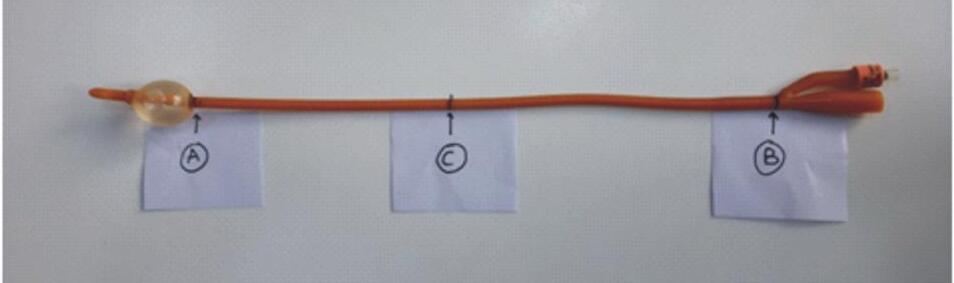
Thus the Urethral Length was calculated as follows:
AB - BC = AC (Estimated Urethral Length)
Where AB is the distance from the Bladder neck to the ‘Y’ junction of the catheter, BC is the distance from the ‘Y’ junction till the external meatus and AC is the estimated distance from the Bladder neck till the external urethral meatus (the urethral length) (Fig 1).
X-axis: age in years, Y-axis: number of subjects
The data were tabulated, basic statistics derived and statistical analysis done for correlation between the various measurements (Tables 1-4).
RESULTS
There was no correlation between the age, BMI and Urethral Length or the Stretched Penile Length. A weak correlation was demonstrated between estimated
Urethral Length and the Stretched Penile Length. The ‘p’ value for BMI versus stretched Penile Length was significant (<0.05) but the R/PE value was less than 6, hence the correlation is not considered as significant. This correlates with previously published data10.
DISCUSSION
MRI has been used to study Urethral Anatomy and has the advantage of being non-invasive and also identifying additional conditions like inflammation, Sinuses, Fistulae or Diverticula 12. However, it is operator and software dependent. On the contrary, measurements using urethral catheters10,11 give a direct and more reliable estimate of the urethral length and are less observer dependent.
The advantage of our technique was that neither was any patient catheterised for the sole purpose of the study, nor did the measurement require removal of any indwelling catheter. While due diligence was done during data collection and all the data was collected by the same investigator, the measurements could have been affected by the degree of stretch on the penis and the amount of suprapubic fat.
Vol 121, No 2, February 2023Journal
of the Indian Medical Association
Fig 1 — Foley Catheter With Measurement Markings
Parameter Median MeanMinMax Age (years)38.541.41882 Height (cm)165 163.2132190 Weight (kg)65 63.33598 Body Mass Index (BMI)23.723.815.136.2 Catheter Size (Fr)18 16.71218 Length of Urethra (cm)17 17.81430 Stretched Penile Length (cm) 7.957.8512
Table 1 — Basic Statistics
Table 2 — Age Distribution
Table 3 — Indication for Hospitalization
65
Ortho-Orthopedics, Trauma-Any non-urological trauma, MedicalAny medical illness, Gastro-Any medical or surgical gastrointestinal or hepatobiliary pathology, Surgery-Any general surgical procedure
of the Indian Medical Association
There is no gold standard or best technique accepted for measuring Stretched Penile Length and we used the penopubic skin junction to glans tip measurements. We did record the flaccid penile girth for all subjects in this series, but did not include it in the analysis as this correlates poorly with erect measurements13. Given the location of the study and the fact that the subjects were catheterised and admitted for some pathology, measurement of erect penile girth or length was not feasible. The pubic bone to tip of glans measurement is more accurate and reliable, while the penopubic skin to glans measurement can be affected by Obesity14,15. Also, we have not taken into consideration the duration of hospital stay at the time of the study which may have had some effect on the weight. Similarly, we have also not taken into account the effect on weight of any debilitating illnesses or malignancies which the patients may have been suffering from. However, since the total number of patients falling into these categories was miniscule, we presume that these did not have a significant bearing on the final results (Table 5).
CONCLUSIONS
Our data adds to the existing scant information about the male Urethral and Penile Length available in the literature. The same may be applied to optimising the size of Urethral and/or Penile devices or implants for the Indian subcontinent.
REFERENCES
1Cinaz P, Yeþilkaya E, Onganlar YH, Boyraz M, Bideci A, Çamurdan O, et al — Penile anthropometry of normal prepubertal boys in Turkey. Acta Paediatrica 2012; 101(1): e33-6.
2Teckchandani N, Bajpai M — Penile length nomogram for Asian Indian prepubertal boys. Journal of Pediatric Urology 2014; 10(2): 352-4.
3Park SK, Ergashev K, Chung JM, Lee SD — Penile circumference and stretched penile length in prepubertal children: A retrospective, single-center pilot study. Investigative and Clinical Urology 2021; 62(3): 324.
4Park S, Chung JM, Kang DI, Ryu DS, Cho WY, Lee SD — The change of stretched penile length and anthropometric data in Korean children aged 0–14 Years: comparative study of last 25 years. Journal of Korean Medical Science 2016; 31(10): 1631-4.
5Jaiswal VK, Khadilkar V, Khadilkar A, Lohiya N — Stretched penile length and testicular size from birth to 18 years in boys from Western Maharashtra. Indian Journal of Endocrinology and Metabolism 2019; 23(1): 3.
6Soydan H, Akyol Ý, Ates F, Yilmaz O, Dursun F, Baykal K — Cross-sectional analysis of penile length in males 13 to 15 years old according to pubertal development stages. The Journal of Urology 2012; 188(4): 1319-23.
7Aslan Y, Atan A, Aydýn AÖ, Nalçacýoðlu V, Tuncel A, Kadýoðlu A — Penile length and somatometric parameters: a study in healthy young Turkish men. Asian Journal of Andrology 2011; 13(2): 339.
8Awwad Z, Abu-Hijleh M, Basri S, Shegam N, Murshidi M, Ajlouni K — Penile measurements in normal adult Jordanians and in patients with erectile dysfunction. International Journal of Impotence Research 2005; 17(2): 191-5.
9Spyropoulos E, Borousas D, Mavrikos S, Dellis A, Bourounis M, Athanasiadis S — Size of external genital organs and somatometric parameters among physically normal men younger than 40 years old. Urology 2002; 60(3): 485-9.
10Kohler T, Yadven M, Manvar A, Liu N, Monga M — The length of the male urethra. International Braz J Urol 2008; 34(4): 451-6.
11Krishnamoorthy V, Joshi PB — Length of urethra in the Indian adult male population. Indian journal of urology: IJU. Journal of the Urological Society of India 2012; 28(3): 297.
12Ryu JA, Kim B — MR imaging of the male and female urethra. Radiographics 2001; 21(5): 1169-85.
13Habous M, Muir G, Tealab A, Williamson B, Elkhouly M, Elhadek W, et al — Analysis of the interobserver variability in penile length assessment. The Journal of Sexual Medicine 2015; 12(10): 2031-5.
14Habous M, Muir G, Soliman T, Farag M, Williamson B, Binsaleh S, et al — Outcomes of variation in technique and variation in accuracy of measurement in penile length measurement. International Journal of Impotence Research 2018; 30(1): 21-6.
15Wessells H, Lue TF, McAninch JW — Penile length in the flaccid and erect states: guidelines for penile augmentation. The Journal of Urology 1996; 156(3): 995-7.
Vol 121, No 2, February 2023Journal
ParameterKohler10 Krishnamoorthy11 Aslan7 Spyropoulos9 Our study n=109422113252264 Mean Urethral Length (cm) 22.317.5517.8 Urethral Length range (cm)15 - 2914 - 22.514 - 30 Stretched Penile Length (cm) 13.712.187.8
Table 5 — Comparative Analysis
BMI versus ULUL versus SPLBMI versus SPLAge versus SPL R-0.03 -2.66%0.24 24.00% -0.13 -12.64% -0.12-11.56% R2 0.000.07%0.06 5.76% 0.02 1.60% 0.01 1.34% Coeff Alienation(K) 1.00 99.93% 0.94 94.24% 0.98 98.40% 0.99 98.66% t-0.434.00 -2.06-1.88 p value 0.67 66.68% 0.0001 0.01% 0.04 4.01% 0.06 6.07% Std Error (SE) 0.060.060.060.06 Probable Error (PE) 0.040.040.040.04 R/PE -0.646.14 -3.10-2.82 BMI : Body Mass Index, UL : Estimated Urethral Length, SPL : Stretched Penile Length
Table 4 — Statistical Analysis
66
Case Report
Stroke — Next Wave of Complications from COVID-19
Jaideep Singh1, Ayushi Gulati2, Smritikhari2, Himanshu Chaudhary3, Pritish Mahanta4, Atul Verma4, T R Sirohi5
Since the declaration of COVID-19 infection as Pandemic in March, 2020, There has been rise in Multisystem Complications apart from regular Acute Respiratory Syndrome which is hallmark of COVID-19 infection. As the second wave surge of COVID-19 has occurred, most of the patients already suffered from dyspnoea but also rare complications like CVA (Infarct and Haemorrhage) , Seizure and altered sensorium related to Hypoxic Brain Injury. COVID-19 frequently presents with a state of altered coagulability which increases the risk of pulmonary embolism and other Thrombotic events such as Cerebrovascular events. This case report is limited to Neurological complications seen in COVID-19 Infected patients. [J Indian Med Assoc 2023; 121(2): 67-8]
Key words :CVA, Acute Respiratory Distress Syndrome, Coagulability, Neurological.
The COVID-19 infection caused by SARS- CoV-2 virus has been declared Pandemic by World Health Organization since March, 2020 1. The most frequent presentation of the disease is viral pneumonia with fever and dry Cough, Acute Respiratory Syndrome due to COVID-19 infection was primary identified in Wuhan City, China on January, 20202. there is evidence of heterogeneous spectrum of Multisystem involvement due to distribution of ACE receptors over different sites in human body.
COVID-19 frequently presents with a state of altered coaguability which increases risk of Pulmonary embolism and other thrombotic events such as Cerebrovascular events in marked number of COVID patients. Patients present with altered D-dimer, Fibrinogen and Ferritin levels.
Cases discussed in this report were admitted in Lokpriya Hospital (Reg no- RMEE1900995), Meerut – A Primary care facility (COVID Section) in western UP which is primarily a Sugarcane belt of India where most Patients are Uneducated and belong to lower Socio-economic status.
Editor's Comment :
As COVID pandemic still running Globally so clinician should also aware other manifestations other than Respiratory system.
A Male patient 51 years (UHID-11106) came with c/o difficulty in Breathing x 6days , with vitals, PR 100/min, BP 132/86mmhg, RR 34/min temperature 98.4F Spo2 74% in RA. No history of any comorbidities. Patient’s COVID Antigen test was done, which came out to be positive and patient was admitted in COVID Section for further management. Patient was taken on continuous O2 support via NRBM and maintained Spo2 90-92%, Relevant investigations are-
Department of General Medicine, Subharti Medical College, Meerut, Uttar Pradesh 250005
1MD (Gen Medicine), Senior Resident and Corresponding Author
2MD (Gen Medicine), Senior Resident
3MS (Gen Surgery), Senior Resident, Department of General Surgery
4MD (Gen Medicine), 3rd Year Junior Resident
5MD (Gen Medicine), Professor
Received on : 25/08/2022
Accepted on : 01/09/2022
During the Course of treatment, On 3rd day, patient started complaining of weakness in Right Upper Limb
Vol 121, No 2, February 2023Journal of the Indian Medical Association
67
Case 1 : Hb13.7 D-Dimer 1084.37 TLC12.8LDH878 Neut.94 FERRITIN 388.18 Lympho.6 HBA1C7.1 Urea39 PT/INR 12.4/1.06 Creat1.2CRP 103.7 Na136 K3.9
Fig 1 — HRCT chest S/O CTSI 18/25.
and Lower Limb, on examination, plantars were found extensor on right side and flexor on left side, B/L pupil were normal size normal reactive. Power in Right Upper Limb was 3/5 and Lower Limb 4/5. After which immediate NCCT head (Fig 1a) was done. Which were suggestive of SUBTLE HYPODENSE AREA OF MEAN 20HU NOTED INVOLVING LEFT OCCIPITAL, THALAMUS AND CORONA RADIATA REGION/ ISCHEMIC INFARCT, patient’s blood thinner were increased after taking Neurology opinion. Patient was then managed conservatively. Patient was then discharged with stable vitals at room air with COVID RTPCR negative report., with existing Neurological deficit and mild Post COVID symptoms.
3 more Cases are discussed in this Case report showing CNS related complications in Admitted COVID Infected patients.
DISCUSSION
Even though, most common manifestation of COVID19 is Respiratory Failure, but during the Second wave Patients had constellation of Neurological manifestations like headache, vertigo, dizziness, loss of smell and taste, as mild symptoms and Major complications like Seizures, CVA (infarct / bleed) has been present in COVID-19 patients.
The pathophysiological mechanisms that underlie Cerebrovascular events in COVID-19 could potentially be related to vasculopathy 4. In addition, there is an increase of conventional Stroke risk during Sepsis 5 , comorbidities, such as Diabetes, Hypertension, Dyslipidemia enhance expression of Angiotensinconverting enzyme2 receptors in the Brain and neurotropism of SARS-CoV-2 virus6
The above cases discussed shows that without any prior Neurological history, patient presented or developed these Neurological complications, attributed to the SARSCoV2 infection. Therefore, multidisciplinary approach needed in management of COVID-19 patients.
From the beginning of COVID-19 Pandemic, potential Central Nervous System involvement has been hypothesized through various etiological mechanisms, including direct Neuroinvasion 7 , parainfectious autoinflammatory involvement8-11, endothelial dysfunction12 and indirect involvement due to altered homeostasis such as altered coagulative states that cause an increase in ischaemic hemorrhagic lesions13-18.
Further studies are needed to determine whether these Neurological complications are more due to Thrombo Inflammation caused by SARS-CoV-2 virus due to enhance expression of ACE-2 receptors in the brain or due to Prophylactic/overuse of anticoagulant therapy in hopitalised patients.
REFERENCES
1Furuse Y, Sando E, Tsuchiya N — Clusters of coronavirus disease in communities, Japan, January-April 2020. Emerg Infect Dis 2020; 26(9): 2176-9. doi:10.3201/
eid2609.202272PubMedGoogle Scholar.
2Yaghi S , Ishida K , Torres J — SARS-CoV-2 and stroke in a New York healthcare system. Stroke 2020; 51: 2002–11.doi:10.1161/STROKEAHA.120.030335 pmid:http:// www.ncbi.nlm.nih.gov/pubmed/32432996 CrossRefPubMedGoogle Scholar.
3Sweid A , Hammoud B ,Weinberg JH — Letter: thrombotic neurovascular disease in COVID19 patients. Neurosurgery 2020; 87: E4006.doi:10.1093/neuros/nyaa254 pmid:http:// www.ncbi.nlm.nih.gov/pubmed/32496534 PubMedGoogle Scholar.
4Oxley TJ , Mocco J , Majidi S — Large-vessel stroke as a presenting feature of Covid-19 in the young. N Engl J Med 2020; 382: e60. doi:10.1056/NEJMc2009787 pmid:http:// www.ncbi.nlm.nih.gov/pubmed/32343504 CrossRefPubMedGoogle Scholar.
5Mao L, Jin H, Wang M — Neurologic manifestations of hospitalized patients with coronavirus disease 2019 in Wuhan, China. JAMA Neurol 2020; 77: 683. doi:10.1001/ jamaneurol.2020.1127 Google Scholar
6Cascella M, Rajnik M, Cuomo A — Features, evaluation, and treatment of coronavirus (COVID-19). Treasure Island, FL: Stat Pearls, 2020.Google Scholar.
7Beyrouti R, Adams ME, Benjamin L, Cohen H, Farmer SF, Goh YY, et al — Characteristics of ischaemic stroke associated with COVID-19. J Neurol Neurosurg Psychiatry 2020.
8Carod-Artal FJ — Neurological complications of coronavirus and COVID-19. Rev Neurol 2020; 70(9): 31122. External Resources Crossref (DOI)
9Valderrama EV, Humbert K, Lord A, Frontera J, Yaghi S — Severe acute respiratory syndrome coronavirus 2 infection and ischemic stroke. Stroke 2020; 51(7): e127–7.
10Nahum J, Morichau-Beauchant T, Daviaud F, Echegut P, Fichet J, Maillet JM — Venous thrombosis among critically Ill patients with coronavirus disease 2019 (COVID-19). JAMA Netw Open 2020; 3(5): e2010478.External ResourcesCrossref (DOI)
11Herman C, Mayer K, Sarwal A — Scoping review of prevalence of neurologic comorbidities in patients hospitalized for COVID-19. Neurology 2020; 95: 77.
12Graham EL, Clark JR, Orban ZS — Persistent neurologic symptoms and cognitive dysfunction in non-hospitalized COVID-19 "long haulers". Ann Clin Transl Neurol 2021; 8: 1073.
13Thakur KT, Miller EH, Glendinning MD — COVID-19 neuropathology at Columbia University Irving Medical Center/ New York Presbyterian Hospital. Brain 2021; 144: 2696.
14Pilotto A, Padovani A, ENCOVID-BIO Network — Reply to the Letter "COVID-19-Associated Encephalopathy and CytokineMediated Neuroinflammation". Ann Neurol 2020; 88: 861.
15Matschke J, Lütgehetmann M, Hagel C — Neuropathology of patients with COVID-19 in Germany: a post-mortem case series. Lancet Neurol 2020; 19: 919.
16Klironomos S, Tzortzakakis A, Kits A — Nervous System Involvement in Coronavirus Disease 2019: Results from a Retrospective Consecutive Neuroimaging Cohort. Radiology 2020; 297: E324.
17Chougar L, Shor N, Weiss N — Retrospective Observational Study of Brain MRI Findings in Patients with Acute SARSCoV-2 Infection and Neurologic Manifestations. Radiology 2020; 297: E313.
18Qureshi AI , Abd-Allah F , Al-Senani F , et al international panel. Int J Stroke 2020; 15: 54054.doi:10.1177/ 1747493020923234 pmid:http://www.ncbi.nlm.nih.gov/ pubmed/32362244 CrossRefPubMedGoogle Scholar
Vol 121, No 2, February 2023Journal
of the Indian Medical Association
68
Case Report
Dancing Feet Syndrome in Diabetes : Para-ballism and Para-chorea in a Diabetic Patient with Diaphragmatic Myoclonus
Rahul Debnath1, Abhinav Das2, Amrita Das2, Manjari Ghosh3, Jotideb Mukhopadhyay4
While movement disorders in Diabetes have long been recognized,the terminology of diabetic striatopathy is relatively more recent. Herein,we report a rare case of diabetic striatopathy in a 62-year-old woman with uncontrolled Type 2 Diabetes Mellitus who presented with Para-ballismus and Para-chorea along with diaphragmatic Myoclonus, a constellation of rarely reported before simultaneously in a hyperglycemic state.While these movement phenomena are extremely rare, the case also highlights they may persist even after acute control of hyperglycaemia, emphasizing on the need for achieving long term Glycemic control for its management. [J Indian Med Assoc 2023; 121(2): 00-00]
Key words :Striatopathy, Para-ballismus, Para-chorea, Myoclonus, Diabetes.
CASE REPORT
A 62-year-old known diabetic patient on oral anti diabetic drugs for 1 year presented with acute onset abnormal movements of her Left Lower Limb for 1 month. These movementswhich she described as involuntary jerky and dance like movements soon involved the Right Lower Limb and sequentially the upper limbs within the next 15 days. No history of any Focal Limb weakness, Sensory, Cranial Nerve or Sphincter abnormalities was present. Fever, headache, loss of consciousness, alteration of sensorium or specific drug use apart from oral anti diabetic drugs were not reported. Family history of movement disorders was not present.
On Examination she had a Glasgow coma Scale score of E4V5M6 with no significant cognitive impairment. Cranial nerve, sensory and autonomic examination were within normal limits. Motor examinationwas significant for abnormal, involuntary, hyperkinetic movements in the form of rapid, high amplitude and arrhythmic, flinging movements of both Lower Limbs suggestive of ParaBallismus. Abnormal involuntary movements of distal muscles of Bilateral Lower Limb were also observed that were brief, random and without purpose, suggestive of Chorea. Additionally, there were also abnormal, involuntary, arrhythmic, undulating, inward and outward movements of the abdominal wall suggestive of diaphragmatic myoclonus. No abnormal movements were noted in the Upper Limbs. There was no muscle wasting anywhere. The tone and power of Bilateral Lower Limbs could not be assessed due to abnormal involuntary movements. The tone and power of Bilateral Upper Limbs were normal. Deep tendon reflexes were 2+ in Bilateral Upper and Lower
Department of Medicine, IPGME&R and SSKM Hospital, Kolkata
700020
1MD (Gen Medicine), Postgraduate Trainee and Corresponding
Author
2MD (Gen Medicine), Senior Resident
3MD, Associate Professor
4MD, Professor
Received on : 11/07/2021
Accepted on : 28/10/2021
Editor's Comment :
Diabetes is a multisystem disorder affecting the whole body, Central Nervous System is also not an exception. Diabetes presenting with a movement disorder is very rare but is being noticed nowadays with increasing proportions. Any movement disorder presenting acutely it is a good habit to check the capillary blood glucose, which can be easily managed & patient will get immediate relief.
Limbs, Plantar reflexes were bilateral flexor. Cerebellar function testing (finger-nose, finger-nose-finger) revealed no abnormality. No abnormal orolingual movement was observed. Meningeal signs were absent.
In view of the acute nature of the dyskinesias possibilities including metabolic and acute onset structural lesion (likely vascular) affecting the basal ganglia or its connections were considered. Bedside Capillary Blood Glucose (CBG) done immediately was found to be 170 mg/dl. However, she was found to have uncontrolled Blood Glucose levels with a Fasting Blood glucose level of 540 mg/dl and HBA1c 10.38. As euglycemia was achieved with Insulin and OADS, the dyskinesia decreased in intensity and persisted only present in Bilateral Lower Limbs. Arterial Blood Gas (ABG) analysis was done next which did not reveal acidosis and she had normal serum osmolarity. Urine dipstick test revealed glucosuria, but no ketone bodies. All other tests of the metabolic panel, Complete Hemogram, Serum Electrolytes, Renal, Liver and Thyroid Function Tests and Autoimmune profile were within normal limits. NCCT Brain showed bilateral caudate nucleus hyperdensity. MRI brain showed bilateral T1 and T2 hyperintensity of caudate and putamen. EEG was normal (Figs 1&2).
DISCUSSION
With the ever expanding knowledge of the impact of diabetes on different organ functions, brain changes in diabetes is being increasingly recognized. Movement disorder is one amongst the myriad neurological presentations of Diabetes. While movement disorders have long been described, the terminology of “diabetics triatopathy” is relatively more recent1. Diabetic striatopathy
Vol 121, No 2, February 2023Journal of the
Indian Medical Association
69
is defined as a hyperglycemic condition associated with either both or one of the following conditions (1) chorea/ballism (2) striatal hyperdensity on CT or hyperintensity on T1 weighted MRI which can be reversible 1 . 4 main hypotheses to explain the pathogenesis resulting in striatal abnormalities on imaging include petechial haemorrhage,mineral deposition (Calcium or Magnesium), myelin destruction and infarction with astrocytosis (gemistocytopathy). In non-ketotic hyperglycaemia a shift in Brain metabolism to the alternative anaerobic pathway in Krebs Cycle leads to depletion of Gammaaminobutyric Acid (GABA) a inhibitory neurotransmitter, consequently resulting in disinhibition of subthalamus and basal ganglia that translates into hyperkinetic movements. On the contrary, in Ketosis, GABA can be resynthesized using acetoacetate produced in the Liver to prevent its reduction, thus causing lesser incidence of movement disorder1
The movement disorders commonly associated with Hyperglycaemia can be Hemichoreahemiballismus [HB-HC], Monoballismus, Myoclonus, Hemifacial spasm, paroxysmal kinesogenic dyskinesia (PKD) and several partial seizures2,3. Overall, these are usually more frequently observed in a background of non Ketotic hyperglycaemia ratherthan that with ketosis 1,2 However, Paraballism-Parachorea that is ballismus and choreiform involvement of both lower limbs as a primary manifestation is rare, as is diaphragmatic Myoclonus.
Ballismus are large amplitude wild flinging incessant purposeless movements that are typically seen affecting one half of the body. The pathologic abnormality lies in the subthalamic nucleus and its afferent or efferent connections. Very rarely can they be Bilateral /involving both legs which is known as PARA BALLISMUS. As early as 1965 Hemiballismus was described as (1) Hemiballismus, typical (well localized lesion in the contralateral sub thalamic nucleus) (2) Hemiballismus, atypical (involving connections of the subthalamic nucleus, usually internal capsule) (3) Para-ballism (bilateral ballistic activity usually as a part of encephalitic sequelae with corpus luyii seemingly normal)4
Chorea is described as an involuntary irregular, random, non-rhythmic, purposeless movements, caused by involvement of the caudate nucleus. Variable in their distribution, they can affect a single extremity, one half of the body (hemichorea) or be generalized. Characteristically involving the distal extremities, it may also affect the proximal parts, lower extremities,trunk,face, tongue lips and Pharynx de Jong5
Diaphragmatic myoclonus also known as belly dancers’ dyskinesia, is a form of segmental myoclonus caused by rhythmic, involuntary contractions of the diaphragm resulting in undulating, rhythmic movements of the abdomen. Its generator source is believed to lie in the rostral medulla. It can be due to central causes such as Encephalitis and extra pontine myelinosis. Peripheral causes include Phrenic nerve injury /irritation, Spinal cord
lesions. They may also be drug induced or psychogenic, however, majority of the cases are idiopathic 6 Diaphragmatic Myoclonus has been reported only once before as a reversible manifestation of uncontrolled hyperglycemic state7,8. Often mistaken for hiccups, it is a less recognized phenomenon,the identification of which is thus crucial for suspecting and treating underlying hyperglycaemia 9
In this case we observed Para-ballismus and parachorea.Bilateral chorea/ballismus has been reported twice before in a background of non Ketotic Hyperglycaemia.
REFERENCES
1Chua, CB., Sun, CK., Hsu, CW — Diabetic striatopathy: clinical presentations, controversy, pathogenesis, treatments, and outcomes. Sci Rep 10, 1594 (2020). https://doi.org/10.1038/ s41598-020-58555-w
2Awasthi D, Tiwari AK, Upadhyaya A, Singh B, Tomar GS — Ketotic hyperglycemia with movement disorder. J Emerg Trauma Shock 2012; 5(1): 90-1. doi:10.4103/09742700.93095
3Jagota P, Bhidayasiri R, Lang AE — Movement disorders in patients with diabetes mellitus. J Neurol Sci 2012; 314: 5 11.
4Carpenter MB — Pathologie des Ballismus. Arch Neurol 1965; 13(5): 566. doi:10.1001/archneur.1965.00470050114017
5Dejong’S the Neurologic Examination (2013) 7th Edition (English, Hardcover, Campbell W. W.)
6Rathore C, Prakash S, Bhalodiya D. Belly dancer's dyskinesia: A rare movement disorder. Neurol India 2018; 66, Suppl S1: 156-7
7Dubey S, Chatterjee S, Mukherjee D, Ghosh R, Sengupta S, Lahiri D, Pandit A — "Dancing belly” in an old diabetic lady. J Family Med Prim Care 2020; 9: 2580-2.
8Bendi VS, Matta A, Torres-Russotto D — Bilateral chorea/ ballismus: detection and management of a rare complication of non-ketotic hyperglycaemia. Case Reports 2018; 2018: bcr-2018-224856.
9Milburn-McNulty P, Michael BD, Woodford HJ — Hyperosmolar non-ketotic hyperglycaemia: an important and reversible cause of acute bilateral ballismus. Case Reports 2012; 2012:bcr1120115084.
Vol 121, No 2, February 2023Journal
of the Indian Medical Association
Fig 2 — MRI Brain in T2 weighted image shows Bilateral Hyperintensities in caudate nucleus and putamen
70
Fig 1 — MRI Brain in T1 weighted image shows Bilateral hyperintensities in putamen & caudate nucleus
Case Report
Atypical Presentation of Anti-phospholipid Syndrome with Triple Antibody Positive (ATAP) Syndrome Presented as Non-healing Foot Ulcer in a Middle-aged Adult Female
Amishi Nitin Rathod1, Vishal Nitin Rathod2, Sunil M Shahane3
Middle aged women presented with non-healing ulcer over foot following trivial injury. She was not having any comorbid condition. Investigations revealed APLA Syndrome with triple antibodies positive (Lupus anticoagulant, Anticardiolipin antibody and anti B2-glycoprotein antibodies). Patient responded well with anti-platelet and skin grafting at local site.
[J Indian Med Assoc 2023; 121(2): 71-2]
Key words :Triple antibody positive.
CASE REPORT
39-year-old female presenting with complain of nonhealing wound on the lateral aspect of the right foot for 2 months. Patient was alright 2 months ago, when she sustained minor blunt trauma to right foot resulting in mild swelling followed by small Ulcer. It increased in size over 2 months period. There was no history of motor or sensory system disturbance in lower extremities, no history of bleeding or discharge from the wound. There is no Hyperpigmentation/ Varicose Vein or raised temperature of the skin around the wound There was no history of skin rash, oral ulceration, photosensitivity, or bleeding from any other site. Patient did not give any history of co-morbid condition or Collagen Vascular Disorder /Inflammatory Bowel Disease. She does not give history of significant weight loss. She refused any history of medication taken for illness. she is non-smoker and non-alcoholic. Patient underwent debridement of the wound 1 month ago and wound did not heal even after 2 months of proper dressing, antibiotics and antiinflammatory drugs (Figs 1&2).
Examinations — On examination, patient hemodynamically stable. All the peripheral pulses were palpable. One large Ulcer over lateral malleolus and another small Ulcer adjacent to it were present without any changes of the surrounding skin. The Ulcers were oval in shape with size of 9x4 cms and 2x1 cms respectively. The ulcer had an indurated margin with Floor of the wound covered with slough. There was no active discharge. Abdominal examination revealed no splenomegaly or hepatomegaly. Central nervous system
1MBBS, Department of Medicine, Dr D Y Patil Medical College, Mumbai, Maharashtra 411018 and Corresponding Author
2MS, Resident, Department of Orthopaedics, Bombay Hospital & Medical Research Centre, Mumbai, Maharashtra 400020
3MS, Head, Department of Orthopaedics, Nanavati Max Super Specality Hospital, Mumbai, Maharashtra 400056
Received on : 11/01/2022
Accepted on : 31/07/2022
Editor's Comment :
Middle age female presenting as a non healing ulcer after ruling out common causes like Vasculitis and Pyoderma Gangrenosa, Antiphospholipid Antibody Syndrome should be considered.
did not reveal any sensory or motor deficit or Peripheral Nerve thickening or tenderness.
Investigations — The hemogram showed Hemoglobin of 9.4g/dL, a Leucocyte Count of 9030/L, Platelet count: 169X 109/L. The Random Blood Sugar was 111mg/dL. The Blood Urea Nitrogen was 38mg/dL and serum Creatinine 1.24mg/dL. The serum Sodium-138.0 mEq/L, Potassium - 3.85 mEq/L, Chloride-101.8 mEq/L. The Prothrombin time 14seconds and INR were 1.35 seconds. The HBsAg, HCV and HIV were negative. The CRP- 3.74 mg/L and ESR-75 mm respectively. Pus culture grown pseudomonas aeruginosa of non-significance. No fungal elements or acid-fast bacilli seen.
The Anti CCP Antibodies were negative (2.00 U/ML). The sickling test was also negative The Anti-Nuclear Antibody (ANA) with titer (1:1000) was positive with a homogeneous pattern (Immunofluorescence method) cytoplasmic positivity seen. THE C-ANCA was Negative but P-ANCA was Positive. Based on the clinical profile of the patient with investigations, diagnosis of Vasculitis Ulcer was made. Color Doppler of bilateral lower limbs and abdomen was normal with no evidence of arterial blockage or Varicose Vein. The lupus anticoagulant, anti cardiolipin antibodies IgG 97U/ml (positive >40), antibeta-2 glycoprotein of IgG 124.46 RU/ml (positive >20) was positive. Biopsy from the edge of the wound revealed granulation tissue with infiltration of Lymphocytes, Plasma cells and few Neutrophils, no granulomas or fungal elements seen. Histopathological diagnosis was Necrotic Foot Ulcer.
Considering clinical presentation along with specialized investigations, non-healing ulcer with APLA Syndrome diagnosed. Patient was given anti-platelet
Vol 121, No 2, February 2023Journal of the
Indian Medical Association
71
and skin grafting done. Patient improved and discharged from hospital.
DISCUSSION
Antiphospholipid Syndrome is Autoimmune Disorder, resulting due to autoantibodies against anticardiolipin and lupus anticoagulant present on the Plasma membrane causing a hypercoagulable state1. Although the exact etiology of APS is still not clear, genetics is believed to play a key role in the development of the disease, Genetic Markers: HLA-DR4, HLA-DR7 and HLADRw53 It is more common in women than in men 2 Clinically important anti-phospholipid antibodies are associated with Thrombosis and Vascular disease. In pregnant women affected by APS, there is an increased risk of miscarriage and intrauterine growth retardation3 The Anti-phospholipid Syndrome responsible for most of the miscarriages in later trimesters. It is estimated that the incidence of APS is approximately 5 cases per 100,000 persons per year and the prevalence is approximately 40-50 cases per 100,000 persons.
It is very rare to have APLA Syndrome with non-healing ulcer as present in our case report. Treatment includes wound care, pain management, Anti-platelet agents, Blood thinner if major artery or venous blockage, warfarin is used, the INR is kept between 2.0 and 3.04 in case of triple positive instead of warfarin directly acting oral anticoagulant are used5 and skin grafting for non-healing
ulcer. In refractory cases Plasmapheresis may be considered.
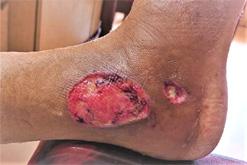
CONCLUSION
Middle aged women present with evidence of arterial, venous blockage or repeated miscarriage and occasionally present with non-healing ulcer then Antiphospholipid antibody (APLA) Syndrome should be suspected.
REFERENCES
1Levine JS, Branch DW, Rauch J — The antiphospholipid syndrome. N Engl J Med 2002; 346(10): 752-63.
2Islam, Md Asiful — Genetic risk factors in thrombotic primary antiphospholipid syndrome: A systematic review with bioinformatic analyses. Autoimmunity Reviews 2018; 17(3): 226-43. doi:10.1016/j.autrev.2017.10.014. PMID 29355608 –via Science Direct.
3Tong M, Viall CA, Chamley LW — Antiphospholipid antibodies and the placenta: a systematic review of their in vitro effects and modulation by treatment. Human Reproduction Update 2014; 21 (1): 97-118. doi:10.1093/humupd/ dmu049. PMID 25228006.
4Horton JD, Bushwick BM — Warfarin therapy: evolving strategies in anticoagulation. American Family Physician 1999; 59(3): 635-46. PMID 10029789.
5Venous thromboembolic diseases: diagnosis, management and thrombophilia testing”. www.nice.org.uk. National Institute for Health and Care Excellence. 2020. Retrieved 2020-0831.
Vol 121, No 2, February 2023Journal of the Indian Medical Association
Fig 2 — Non healing ulcer after skin grafting
Fig 1 — Non healing ulcer over Right foot.
72
Case Report
Hydrophilic Intraocular Lens Opacification — A Case Report
Shivcharan Lal Chandravanshi1, Shashi Jain2, Divya Tripathi3, Divya Ramraika4
Purpose : To report a case of late opacification of the hydrophilic acrylic Intraocular Lens (IOL) after uneventful Cataract Surgery.
Methods : A 60-year-old male presented with chief complaint of gradual diminution of vision in right eye over the past one year. He was Normotensive, Non-diabetic and had a normal Lipid Profile. History revealed that he underwent uneventful phacoemulsification with posterior chamber hydrophilic intraocular in the bag implantation for pre-senile cataract in his Right Eye ten years ago. He had the best corrected visual acuity of 6/6 in his Right Eye for nine years
Post Cataract Surgery. Slit-lamp examination confirmed Intraocular Lens Opacification.
Results : Intraocular Lens exchange was performed in his Right Eye. The hydrophilic IOL was replaced with poly methyl methacrylate intraocular lens. The explanted IOL showed uniform grayish-white opacification. Post operative period was uneventful. Intraocular pressure by applanation tonometry was 16.4 mm Hg in both the eyes. Patient’s best corrected visual acuity was 6/6 with -1 D Cyl. at 90 degree, Postoperatively. Over a follow up period of one year, the patient did not develop complications like posterior capsular IOL opacification.
Conclusion: Intraocular Lens opacification is an extremely rare late postoperative complication of Phacoemulsification which can be managed effectively by IOL exchange procedure in cases of opacified IOL optics causing visual morbidity. [J Indian Med Assoc 2023; 121(2): 73-4]
Key words :Calcification, Hydrophilic intraocular lens, Intraocular lens, Posterior capsule opacification, Intraocular lens opacification, Tertiary cataract.
Intraocular Lens (IOL) opacification is an extremely rare unilateral or sometimes bilateral IOL related complication of Cataract Surgery which may affect the surfaces (anterior, posterior or both) or the material of the optic, haptics, or whole lens. Intraocular Lens opacification is postulated to be caused mainly by calcification. Calcification of IOL may be of primary or secondary types. Primary calcification results from the problems of IOL itself in the absence of other significant causes. Secondary calcification of IOL occurs in the presence of pre-disposing factors such as Diabetes, Uveitis and following Vitreoretinal or Keratorefractive surgeries. Patients usually present to the Ophthalmologist with complaints of diminution of vision. Intraocular Lens exchange is universally accepted and safe procedure to restore vision in these cases. This case study aims to report a rare case of Hydrophilic Intraocular Lens opacification in the absence of any risk factors.
CASE REPORT
A 60-year-old male presented with chief complaints of right eye gradual painless diminution of vision since one year. He gave history of right Eye Phacoemulsification
1MS, Professor, Department of Ophthalmology, Atal Bihari
Vajpayee Government Medical College, Vidisha, Madhya Pradesh
464001 and Corresponding Author
2MS, Professor, Department of Ophthalmology, S S Medical College, Rewa 486001
3 MS, Senior Resident, Department of Ophthalmology, Government Medical College, Shahdol, Madhya Pradesh 484001
4DO, DNB, Senior Resident, Department of Ophthalmology, Hindu Rao Hospital, New Delhi 110007
Received on : 10/01/2022
Accepted on : 14/02/2022
Editor's Comment :
Intraocular Lens opacification is a rare complication. It is more common in hydrophilic intraocular lenses. The ophthalmologists must be aware of this complication and patients to be well informed about Postoperative IOL opacification to avoid litigation.
with Hydrophilic Intraocular Lens implantation 10 years back, elsewhere. There was a negative history of Diabetes, Uveitis or any Keratorefractive or Intraocular Retinal Surgeries. On initial pen light examination, there was Leucocoria in his Right Eye. His best corrected visual acuity was 6/24 in the right eye and 6/9 in the Left Eye. Slit lamp examination disclosed pseudophakia with opacified intraocular lens in the Right Eye while nuclear sclerosis grade II in the Left Eye. Dilated Slit-lamp examination confirmed opacification of both the optic and haptics sparing posterior capsule. No deposits were seen on Intraocular Lens surface on 40X magnification. The patient was posted for exchange of opacified IOL with Polymethyl Methacrylate Intraocular Lens. Postoperative visual acuity was 6/6 with -1 cylinder at 90º. The explanted IOL was sent for light microscopy examination with special stains for detection of Calcium coupled with scanning electron microscopy which did not reveal any deposits over IOL surface. However, special stains, Alizarin red S was positive while Von Kossa was negative for calcium. Postoperative period was uneventful. Postoperative visual acuity in the Right Eye at one month follow-up was 6/6 with -1D cylinder at 90º (Figs 1&2).
DISCUSSION
The Intraocular Lens opacification is an extremely rare
Vol 121, No 2, February 2023Journal of the Indian Medical Association
73
late Postoperative complication after Cataract Surgery with IOL implantation which usually occurs in the late postoperative period in Hydrophilic IOLs. Intraocular Lens opacification in Hydroview IOLs after Cataract Surgery was first reported by Chang et al in 19991. The incidence of IOL opacification ranges from 1.1% to 14.5% depending on the presence of risk factors in the patient2. Duration of IOL opacification ranges from one year to seven years or more. Silicone, Acrylic and Poly Methyl Methacrylate (PMMA) IOLs have been reported to undergo opacification. Hydrophilic acrylic IOLs have greater tendency of opacification in comparison with hydrophobic acrylic IOLs3. Patients with IOL opacification usually present with complaints of gradual painless loss of vision after Cataract extraction with IOL Implantation Surgery, decreased contrast and glare 4 . Sometimes patient may also present with Leucocoria, poor vision in dim light and hazy or foggy vision. Various risk factors for IOL opacification have been described in the literature such as Diabetes, Uveitis, Asteroid Hyalosis, Breakdown of blood aqueous barrier intraoperatively in procedures such as Parsplana Vitrectomy (PPV) with intraocular gas or air injection, penetrating keratoplasty, Descemet Membrane Endothelial Keratoplasty (DMEK) and descemet stripping (automated) Endothelial Keratoplasty (DSEK/DSAEK)5-6 In present case study, no such risk factors are present for IOL opacification.
Mechanism of IOL opacification is not well established. Various mechanisms have been proposed by numerous researchers. Different patterns of IOL opacification have been noted in different IOL substances such as snowflake opacification in PMMA IOLs, discoloration/clouding in silicone IOLs, calcification (hydroxyapatite, dicalcium phosphate, octacalcium phosphate, or hydroxyapatite deposition) in hydrophilic acrylic IOLs and glistening or subsurface nanoglistenings in Hydrophobic acrylic IOLs7
Diagnosis of IOL opacification is easily made by Slitlamp Biomicroscopy. Sometimes, it may mimic a lamellar cataract. Anterior Segment Optical Coherence Tomography offers help in detection of IOL-capsular bag adhesions. Scanning laser electron microscopy and Xray deffraction examination of an explanted IOL may be helpful in understanding the mechanism of IOL
opacification. Both the techniques show Calcium deposition over IOL surface. Von Kossa stain is useful in IOL surface calcification while Alizarin red is used for entire IOL material calcification 8
Explanation of an opacified IOL and reimplantation of new IOL made up of different material is the procedure of choice for the treatment of opacified IOL at present9. However, IOL exchange procedure may become risky in patient who has had Nd-YAG laser capsulotomy in the past. In such cases, capsular bag damage, complete dehiscence of bag, vitreous prolapse, zonular dehiscence, IOL drop, IOL decentration may be the common complications. Scleral fixated IOL, anterior chamber IOL, sutured iris fixated IOL, iris claw IOLs and retro pupillary iris claw IOL are also other viable options in case of capsular bag damage/dehiscence10. Majority of IOL exchange procedures have excellent Postoperative visual outcome if posterior segment is healthy.
CONCLUSION
Incidence of IOL opacification is extremely low; the patient should be warned in advance of the remote possibility of IOL opacification in long term. This aspect of IOL related complication and the likelihood of repeat surgery for the same, should be mentioned in the informed consent in order to avoid litigation in the future.
REFERENCES
1Chang BYP, Davey KG, Gupta M, Hutchison C — Late clouding of an acrylic lens following routine phacoemulsification. Eye 1999; 13: 807-8.
2Jain P, Pattnaik A — Intraocular lens opacification: A rare enigma. J Clin Ophthalmol Res 2021; 9: 51-4.
3Grzybowski A, Zemaitiene R, Markeviciute A, Tuuminen R — Should we abandon hydrophilic intraocular lenses? Am J Ophthalmol 2021; Nov 26: S0002-9394(21)00618-8. doi: 10.1016/j.ajo.2021.11.021.
4Grzybowski A, Markeviciute A, Zemaitiene R — A narrative review of intraocular lens opacifications: update 2020. Ann Transl Med 2020; 8: 1547.
5Fernández J, Sanchez Garcia A, Rodriguez Vallejo M, Piñero DP — Systematic review of potential causes of intraocular lens opacification. Clin Exp Ophthalmol 2020; 48: 89-97.
6Neuhann IM, Neuhann TF, Rohrbach JM — Intraocular lens calcification after keratoplasty. Cornea 2013; 32(4): e6-10.
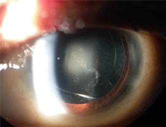
7Neuhann IM, Kleinmann G, Apple DJ — A new classification of calcification of intraocular lenses. Ophthalmology 2008; 115: 73-9.
8McGee Russell SM — Histochemical methods for calcium. J Histochem Cytochem 1958; 6: 22-42.
9YuAK, NgAS — Complications and clinical outcomes of intraocular lens exchange in patients with calcified hydrogel lenses. J Cataract Refract Surg 2002; 28: 1217-22.
10Gashau AG, Anand A, Chawdhary S — Hydrophilic acrylic intraocular lens exchange: Five year experience. J Cataract Refract Surg 2006; 32: 1340-4.
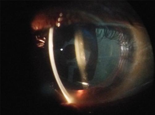
Vol 121, No 2, February 2023Journal of the Indian Medical Association
Fig 1 — Slit-lamp photograph showing grayish white opacification of intraocular lens
74
Fig 2 — Slit-lamp optical section photograph showing grayish white opacification of intraocular lens
Quiz 1
Image in Medicine
Bhoomi Angirish1, Bhavin Jankharia2
A 13-year-old female presented with painless midline neck swelling. On clinical examination, the swelling elevates on protrusion of tongue.
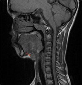
Questions :
(1)What is the diagnosis ?
(2)What is the locations of this lesion ?
(3)What are the other differential diagnosis of midline neck swelling ?
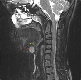
Answers :
(1)A well defined cystic lesion is seen in midline at the base of tongue ( red arrow). A thin linear track (yellow arrow) is seen which represents failure of normal developmental obliteration of the thyroglossal duct. These findings are suggestive of suprahyoid thyroglossal duct cyst.
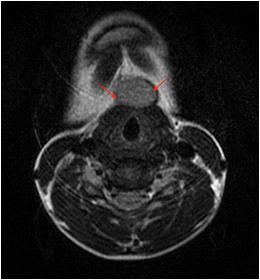
(2)The thyroglossal duct cysts can occur anywhere along the course of the thyroglossal duct from the foramen cecum to the thyroid gland. The common locations are : (A) Suprahyoid; (B) At the level of hyoid bone; (C) Infrahyoid How to differentiate this condition from its mimics?
They are typically located in the midline, while those off-midline are characteristically adjacent to the thyroid cartilage.
(3)The common differential diagnosis of midline neck swelling are – brachial cleft cyst, delphian lymph node, epidermoid cyst, thyroid lesions, laryngocele, ranula, parathyroid adenoma, ectopic thyroid.
Quiz 2
Barium swallow images of a 20 year old male who presented with dysphagia and regurgitation.
Questions :
(1)What is the diagnosis?
(2)How to differentiate this condition from its mimics?
Answers :
(1)There is significant dilatation of esophagus with smooth tapering seen in the lower esophagus (bird beak / rat’s tail sign) suggestive of achalasia.
(2)Achalasia (primaary achalasia) results from failure of organized esophageal peristalsis causing impaired relaxation of the lower esophageal sphincter. This is due
Department of Radiology, Picture This by Jankharia, Mumbai, Maharashtra 400004
1MD, DNB (Radiology)
2MD, DMRD (Radiology)
to loss/destruction of neurons in the myenteric plexus. Obstruction of the distal esophagus from other nonfunctional etiologies, may have a similar presentation and is termed “secondary achalasia” or “pseudoachalasia”. Some of the causes of pseudoachalasia are malignancy, scleroderma, esophageal stricture, chagas disease.
Vol 121, No 2, February 2023Journal
of the Indian Medical Association
75
Letters to the Editor
[The Editor is not responsible for the views expressed by the correspondents]
Manuscript Writer : Are they Eligible for Authorship in Scientific Article ?
If yes then which place ?
SIR, — Reading, writing and speaking are the skill which can be developed by personal interest and training. Everyone doesn’t have skill and all person cannot have all skills. Good writing with appropriate words makes excellent effect on the readers and reviewers too. Previously writing was not much important but in current busy word, writing gets more weightage compare to past decades. Many people who have that good writing skill, can convert it into their profession which is a good use of the skill1
Research works are being done for benefits to human and other lives, it will be a success when it reaches to other people, not from same institute or same city but whole globe, so we need to publish the data. With the help of internet, that work has become rather easy as compared to past decades2. Though it has their own drawbacks like copy the idea, plagiarism, fake data etc. Various journals are rejecting the manuscript in first screening, if it has plagiarism more than their set limits or not written in proper way. All clinical researcher and scientist don’t have the skill of good writing and presentation. To publish the data and findings, they are taking the help of the medical or research writer. Those writers use their skill with available data and present it with good writing and nice way which can be accepted in the journal easily3
Everyone has rights to use their skill for their self-use. Professional Manuscript writers are doing writing work either for some economic purpose or for some ethical purpose. There are many professionals who are available who are writing on behalf of the authors and submit it to appropriate journals. Data owner just have to give the data and have to explain the concept to the writer. This type of professional asks only for money and they do not want any authorship. Many journals asked about the medical writer details during the manuscript submission. Those journals are taking consent for same from all/ corresponding authors. We are also recommended that professionals are not eligible for authorship in any type of manuscript4
Another non-professional medical writer who writes for that friends/relatives/departments or else. Those are eligible for authorship as and when all investigators agreed to give authorship. We know that writer is not a part of investigator team but to write they have to search and read the many related articles. They give their efforts without any economic benefits. So, to write it they are fitted in to international journal authorship criteria. However, there are some types of articles doesn’t require medical writer. Original article, case repots, case series or brief repots are based on the departmental data where medical writer doesn’t have to allowed to front authorship. Their name can be added after giving departmental authors their due. Letter to editor or concept discussion doesn’t require medical writer, it is mostly by avoid writers5
So, we suggest that writers cannot be eligible for the first authorship because they are using someone else’s concepts. If
writer is not a part of data collection. Sample selection, concept and study designing, analysis or investigator, not eligible for first place authorship. As per the guidelines, innovative concept and idea given authors are most appropriate for the first author. Only searching review of literature and writing from data (Supplied by principal investigator) doesn’t make sense to appoint as first author. If all cases/ patient’s investigation and data collection done before the involvement of writers, he/she are eligible for back side place not for first authors. Person can be a corresponding author between the journal and investigator team. We hope everyone will agree with us.
REFERENCES
1Liumbruno GM, Velati C, Pasqualetti P, Franchini M — How to write a scientific manuscript for publication. Blood Transfus 2013; 11(2): 217-26.
2Resnik DB — Social Benefits of Human Subjects Research. J ClinResBestPract2008; 4(11): 1-7. PMID: 24526930; PMCID: PMC3920587.
3Singhal S, Kalra BS — Publication ethics: Role and responsibility of authors. Indian J Gastroenterol 2021; 40: 65-71. https://doi.org/10.1007/s12664-020-01129-5
4Defining the Role of Authors and Contributors issued by International Committee of Medical Journal Editors https:// www.icmje.org/recommendations/browse/roles-andresponsibilities/defining-the-role-of-authors-andcontributors.htmlLast accessed on 30 December 2022.
5Bhattacharya S — Authorship issue explained. Indian J Plast Surg 2010; 43(2): 233-4.
Department of Microbiology, Sahjid Mukhida1, Dr D Y Patil Medical College, Hospital and Sameena Khan2, Research Centre, Pune Nikunjakumar Das3, 1MBBS, Postgraduate Resident Rajashri Patil3, 2MD, Assistant Professor, Lekshmi R1 3MD, Associate Professor
What is Essential to know in Heart Failure Patients
SIR, — As heart failure prevalence increases globally, there is a growing need for new innovative solutions. Nearly 64 million people worldwide are living with Heart Failure (HF). It is estimated that half of patients living with HF will die within five years of diagnosis. It creates divesting impact on healthcare systems and the global economy.
Heart failure is a complex syndrome with current treatments helping to slow HF risks and progression in some, but not all patients. These unmet medical needs underscore the urgency to increase awareness, early detection and development of life-saving medicines to address these complexities with straightforward solutions. It is crucial to develop and deliver innovative solutions to
Vol 121, No 2, February 2023Journal of the Indian Medical Association
76
address the gaps that currently exist in the treatment of heart failure. So it is imperative to be aware of what is essential to know in heart failure patients for proper care.
In many clinical trials it has been reviled that Torsemide was not superior to furosemide in improving survival among patients treated for decompensated HF. For diuretics it has shown there are no differences in cardiovascular outcomes between chlorthalidone and HCTZ among elderly veterans with HTN. Patients suffering from bad lipid profile Pemafibrate did not improve cardiovascular outcomes among patients with diabetes and hypertriglyceridemia.
Iron deficiency is a very common symptom among HF patients for them, Iron (ferric derisomaltose) infusion is not superior to usual care. Empagliflozin has salutary effects on renal function and CV mortality among patients with CKD, with or without DM, who are already on appropriate doses of ACEi/ARB. Highly purified eicosapentaenoic acid showed borderline statistical sig in reducing the risk of adverse CV events in Japanese w/ c/c CAD who were being treated with statins, whereas Olpasiran significantly reduced lipoprotein (a) in established ASCVD. Rosuvastatin 5 mg daily lowered LDL-C significantly >placebo, fish oil, cinnamon, garlic, turmeric, plant sterols in those with increased 10-year risk for ASCVD.
For invasive cardiac procedures, Radial artery bypass graft improves adverse CV outcomes v/s R internal thoracic artery BG. Hypertension management protocol of 4 drugs, quarter-dose BPlowering combination of candesartan, amlodipine, indapamide and bisoprolol led to a greater reduction in change in BP from baseline to 12 weeks compared with standard-dose ARB immunotherapy in patients with mild to moderate HTN. Adults with HTN who participated in a mindfulness behaviour program for 8 weeks had significantly lower BP levels & greatly reduced sedentary time, at 6 months follow-up V/S those who received enhanced usual care (home BP monitor/BP edu/facilitated access to a physician).
Systematic use of a hospital-based POC tool to support clinical decision-making, followed by rapid follow-up in an outpatient clinic, led to a lower risk of death or hospitalization for CV causes within 30 days among patients with acute HF seeking emergency care. A single IV infusion of NTLA-2001, a novel gene-editing therapy based on CRISPR/Cas9, significantly reduced abnormal levels of the TTR protein by > 90% in patients with ATTR amyloid CM after 28 days. Routine collection of patient-reported health status using KCCQ-12 in the HF clinic improved accuracy of clinician assessments of patients’ health status.
Prophylactic methylprednisolone in infants undergoing cardiopulmonary bypass heart surgery did not improve postoperative outcomes compared with placebo.
Bivalirudin w/ a median 3hr post-PCI high-dose infusion significantly reduced the 30d composite rate of all-cause mortality/ BARC types 3-5 major bleeding compared with heparin monotherapy in Chinese patients with STEMI undergoing primary PCI w/ radial artery access.
DAPT with indobufen plus clopidogrel significantly reduced the risk of 1 year net clinical outcomes in Chinese patients with negative cardiac troponin undergoing DES implantation, compared to conventional DAPT of aspirin plus clopidogrel. A personalized “precision” testing approach led to more efficient evaluations for cardiovascular disease risk and improved diagnosis and treatment
of CAD when compared to usual care in more than 2,000 adults with stable chest pain. Early initiation of rivaroxaban, prescribed for 35 days in non-hospitalized patients with symptomatic COVID19 at-risk for thrombosis, was not found to reduce a composite endpoint of venous and arterial thrombotic events, hospitalization, and death. A universal EHR integrated CDS tool using a validated VTE risk model significantly increased rates of in-hospital appropriate thromboprophylaxis & significantly reduced major thromboembolic events w/o an increase in major bleeding at 30d post-discharge VS usual care.
First-line catheter ablation was associated with a significantly lower progression to persistent Afib, when compared to initial antiarrhythmic drug therapy. No significant differences in the rate of postoperative Afib in cardiac surgery patients who received either 125U or 250U doses of botulinum toxin type A (AGN-151607) compared with placebo. Implementation of a novel shared decisionmaking toolkit designed for low health literacy achieved significantly lower decisional conflict and improved preparation for decisionmaking compared to usual care in patients with AFib. Among patients with resistant HTN, Aprocitentan resulted in short-term and sustained BP-lowering effects. Among patients with treatment-resistant HTN aldosterone synthase inhibition with Baxdrostat led to dosedependent reductions in SBP.
In patients with CLTI surgical revascularization with a great saphenous venous conduit was superior to endovascular intervention in reducing major adverse limb events or death. Greater QOL improve in those undergoing endovascular interventions versus surgery. Etripamil nasal spray was effective in termination of spontaneous PSVT in patients experiencing an episode in an athome setting. Catheter ablation reduces the incidence of persistent AFib/recurrent atrial tachyarrhythmia v/s antiarrhythmic therapy.
An intensive treatment strategy of rapid up-titration of GDMT and close follow-up after an acute HF admission reduced symptoms, improved QOL and reduced the risk of 180-day allcause death or HF readmission compared with usual care. Among patients with stable ischemic heart disease and moderate to severe ischemia on non-invasive stress testing, routine invasive therapy failed to reduce major adverse cardiac events compared with optimal medical therapy.
1MBBS, MS, MCh (Plastic Surgery), Sudhir Singh1, Hon'ble IMA Professor, Manoj Kumar Srivastava2
Department of Plastic Surgery, Getwell Hospital, Varanasi 221005
2MBBS, MD, Professor, Department of Medicine, Narayan Medical College, Sasaram, Bihar 821305
The Road not Taken : A Perspective of a Medical Professional’s Inability to Choose between Clinical Practice or Postgraduation Degree after Internship
SIR, — A Postgraduation (PG) degree has almost become a requisite to pursue medical profession in India. A major part of such degrees rely on the clinical mastery of the doctors but to our dismay, the clinical exposure we get during our MBBS courses and
Vol 121, No 2, February 2023Journal of the Indian Medical Association 77
internship does not adequately prepare us for such bigger roles. Patients’ outlook has gradually changed over time and it has been often seen that a doctor with a MD or DM degree is preferred over a “suboptimal” MBBS degree, as stated by a few patients . This has subjected the young doctors into a fierce academic competition to crack entrance examinations like NEET, INICET etc. The race is getting tougher day by day in a constant crescendo so much so that it has almost become a norm to dedicate a year or two only for “PG preparation”. A vast majority of the aspirants are getting enrolled in various online courses completely sacrificing the clinical exposure of working in a hospital. The internship courses showcase a mere orientation programme with scanty clinical exposure. In most of the hospitals internees are made to fill up charts, perform phlebotomy, insert catheter, write requisitions etc and are almost never a part of the clinical decision making process. After completing the internship, if we are again detaching ourselves from clinical exposure and devoting years to post graduation entrance, is it compromising our clinical acumen further? Having said that, now if we take into account a budding doctor’s point of view, the need for postgraduation in India has outweighed the requirement of clinical practice in a doctor’s career. The question pattern keeps changing every now and then, leaving them with meagre time to balance between their yearning for clinical experience and simultaneously preparing for the mutating Multiple Choice Question (MCQ) pattern. For example, just a few years ago image based questions were rarely asked but with the commencement of computer based tests, questions with CT scan, Xray, clinical images are commonly asked and without a rigorous practice it is not possible to get success in these entrance tests. After getting into a MD course it is very difficult to have the necessary experience of the other specialities, which is a sine-qua-non to become a successful physician of any speciality. After joining a PG course in a clinical branch the doctors with sparse clinical experience are suddenly given the huge responsibility of treating patients which ultimately results in compromised patient-care at times. Moreover, there is heterogeneity in the work culture or duties of internees in different states. So, the degree of clinical exposure keeps varying from one Post Graduate trainee to another. The regulatory authority should take this issue into account to ensure optimum patient care. A mandatory and uniform clinical orientation course for the post graduate trainees all over India can probably bridge the gap of clinical experience and align them better into the clinical practice.
1MBBS, Junior Resident, Sumit Roy Chowdhury1
Department of Anesthesiology, Asmita Chakrabarti2
AIIMS, Delhi
2MBBS, Previously worked as non-academic Junior Resident, Department of Medicine, Calcutta National Medical College and Hospital, Kolkata
Monkeypox as a Global Health Emergency — A Threat after COVID-19 Pandemic
SIR, — Monkeypox is a rare zoonotic disease caused by the monkeypox virus that belongs to the Poxviridae family (1). While the source of infection is primarily zoonotic, and the disease
condition is usually seen in Central and West Africa since the 1970s. Recently, there is a rapid spread of Monkeypox all over the world due to climatic change, widespread global travel, and waning herd immunity due to the cessation of smallpox vaccination (2). Reemergence of monkeypox across nations had made World Health Organization declare it a public health emergency of international concern (PHEIC) in July 2022 (3). As the disease is mild and not fatal, there are debates on declaring it as a PHEIC as it creates panic among the public but considering the reservoir of infection,pandemic potential and susceptible population declaring monkeypox as PHEIC is the need of the hour. India has reported nine confirmed cases of Monkeypox, including one death (4th August 2022) (4). In India, the recent COVID-19 pandemic has equipped us to battle any outbreaks in the future. As we expect more emerging and re-emerging infections in the future, strengthening molecular laboratories will help in the early detection of the disease and containment. Currently, around 70% of the human population issusceptible to Monkeypox infection (2). During the COVID-19 pandemic, a significant gamechanger in controlling the outbreak was a quick roll-out of mass vaccination campaigns.As per the CDC recommendations, two FDA-approved vaccines namely JYNNEOS (Imvamune or Imvanex) and ACAM2000 may be used for the prevention of Monkeypox infection (5). But the data regarding the effectiveness of these two vaccines in the current outbreak is not available. Hence it is imperative that budget allocation for conducting vaccination effectiveness studies should be implemented in endemic countries where we have an increased incidence of Monkeypox infection.
REFERENCES
1Cho CT, Wenner HA — Monkeypox virus. Bacteriol Rev 1973; 37(1): 1-18.
2Simpson K, Heymann D, Brown CS, Edmunds WJ, Elsgaard J, Fine P, et al — Human monkeypox – After 40 years, an unintended consequence of smallpox eradication. Vaccine 2020; 38(33): 5077-81.
3Monkeypox outbreak 2022 [Internet]. [cited 2022 Aug 4]. Available from: https://www.who.int/emergencies/situations/ monkeypox-oubreak-2022
4India Monkeypox Cases: Govt to hold experts’ meeting after India reports 9 monkeypox cases | India News - Times of India [Internet]. [cited 2022 Aug 4]. Available from: https:// timesofindia.indiatimes.com/india/monkeypox-govt-to-holdmeeting-of-experts-today/articleshow/93333192.cms
5CDC. Monkeypox in the U.S. [Internet]. Centers for Disease Control and Prevention. 2022 [cited 2022 Aug 4]. Available from: http s://www.cdc.gov/poxvirus/monkeypox/ considerations-for-monkeypox-vaccination.html
1MD, DTMH, Associate Consultant, Malathi Murugesan1 Department of Infectious Diseases, Srinath Ramamurthy2 Meenakshi Mission Hospital and Research Centre, Madurai, Tamil Nadu 2MD, Consultant, Department of India Epidemic Intelligence Service, NCD, CMR, National Institute of Epidemiology, Chennai, Tamil Nadu
Vol 121, No 2, February 2023Journal of the Indian Medical Association 78