On a chilly winter night, a camel requested his master to allow him to pokeitsnoseinsidethetenttokeepitselfwarm.Thegenerousmasterallowed it. On repeated requests and approvals, the camel began entering the tent slowly but surely. Finally, the camel managed to enter the tent totally and drove the master out. Well, you may ask me why I am narrating a fable to my august readers. My request to my readers is not to concentrate on the story alone but note the subtle and sly yet completely planned movement of the camel to enter the tent to oust its owner. We will find a similarity in action between the camel and our topic of discussion. Dear readers this circumlocution is because I want to discuss in this editorial a topic that apparently may seem farfetched from medicine and health but on second thought it would be clear that these to topics are mutually closely related. I will be speaking on Farm Bills in this editorial and how they can affect the general health of the denizen of India.
The Indian agriculture acts of 2020, often referred to as the Farm Bills, are three acts initiated by the Parliament of India in September 2020. The Lok Sabha approved the bills on 17 September 2020 and the Rajya Sabha on 20 September 2020. The President of Indiagave his assent on 27 September 2020. They inspired the protests against the new acts, which gained momentum in September 2020. Let us understand the Act in detail.
According to The Gazette Of India THE FARMERS’ PRODUCE TRADE AND COMMERCE (PROMOTION AND FACILITATION) ACT, 2020 (1) is an Act to provide for the creation of an ecosystem where the farmers and traders enjoy the freedom of choice relating to sale and purchase of farmers’ produce which facilitates remunerative prices through competitive alternative trading channels; to promote efficient, transparent and barrier-free inter-State and intra-State trade and commerce of farmers’ produce outside the physical premises of markets or deemed markets notified under various State agricultural produce market legislations; to provide a facilitative framework for electronic trading and for matters connected therewith or
incidental thereto. This act seems quite philanthropic at the first glance until you get through the skin of it. Three ordinances farmers are protesting against are:
1. Farmer’s produce trade and commerce ordinance 20201
2. The farmers’ agreement on price assurance and farm services ordinance 20202.
3. Essential commodities ordinance 20203. Now let me place before you the Government stance and the farmers’ stance for and against the bill.
Government stance:
1.The farmers can market and sell their produce outside their notified agricultural produce and market community (APMC mandis)
2.The state government cannot collect the cess outside these APMC mandis
3. Greatest fear – the government will reduce the minimum support price for all their crops.
4.Farmers believe that the middlemen or agents do have credibility as their financial credibility is thoroughly checked during their license approval process.
5. The commission agents are also protesting because they think that the new law will render them jobless.
6. The state government is also protesting since their revenues will dry up that comes from these mandis.
7. The farmers believe that this bill will give monopolistic power to the private entities giving them a free hand to exploit farmers.
If we spare a little thought on the purpose of introduction of Farm Bill we can easily identify that it
3. Remove the interstate barrier by introducing electronic trading.
Farmers’ stance
1.These reforms will entirely make them dependent on traders.
2. The farmers of Punjab believe that the Food Corporation of India and other central agencies might shut down annual rice and wheat purchases from the state which will eventually make them dependent on traders leading to harassment.
has been made especially keeping in mind the few companies. The prime motive of these industries is to capture the whole 1.1 trillion dollar retail industry in India. They can skyrocket their turnover to a stupendous amount if they can capture the huge potential retail market in India which is growing at a staggering rate of 35% per annum. But where is the end-user or the ultimate consumer in this whole process? They are the ultimate sufferers as they will have the least say under the towering giant companies
controlling the retail market. It has been a time-tested process that the commission agents buy the products directly from the farmers from the mandis and retail their purchases to their respective channels. On any dispute the Block Officer intervenes to settle the discord. Now instead of the mandis which were physical spaces where the farmers and the agents could bargain on the prices of their produces the government intends to introduce online trading platform. Like a God sent command the trading price of the product would be displayed and the poor farmer with his feeble strength could do nothing but agree to that price even if it means disaster to them.
These big tycoons will enter into contracts with the farmers per se for a period of five years against a lump sum and these poor souls would have no other options but to succumb to the malicious oppressions. As if that is not enough, these giants would have the right to stock unlimited agro products with The state having no power to control them. An ideal field would be set for hoarding and black marketing and prices of products for the average man could skyrocket beyond means due to artificial scarcity. Are we paving the way to another artificial famine?
The million dollar question however still remains unanswered. Why are we discussing these in a medical journal?
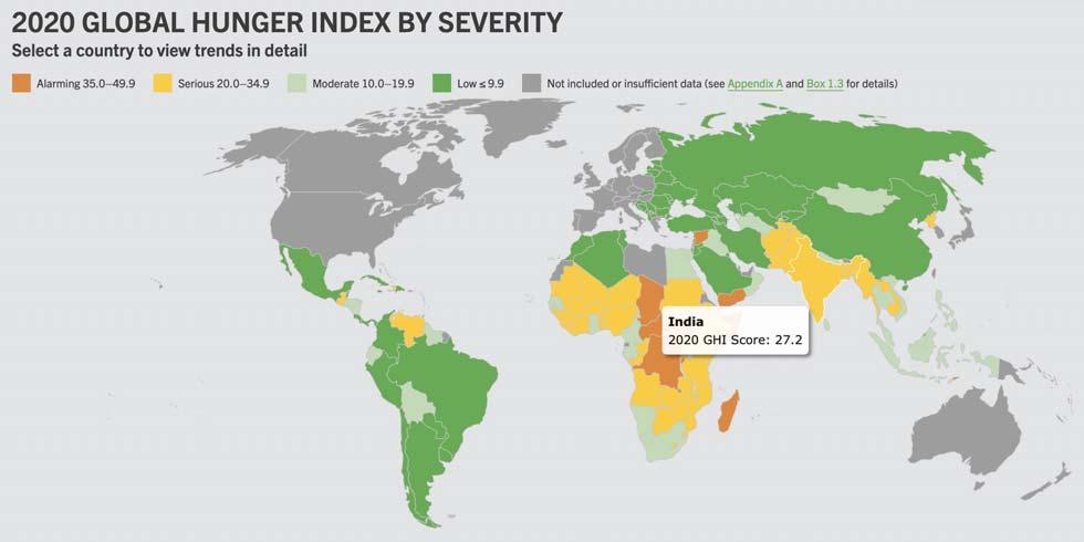
Now let us have a look upon the Global Hunger Index (GHI). In the 2020 Global Hunger Index, India ranks 94th out of the 107 countries India’s score is 27.2, which means it is in a very serious state4
While India is trying to prioritize health for all in the future how can this be achieved if consecutive manipulations lead to massive inflation and successive abysmal fall in the real income of the common man? Can we, the medical workers, prefer to remain aloof by saying this is not our field to poke into. The Indian poverty lines are based exclusively on estimates of the normative nutritional requirement of the average person in the rural and urban sectors. The national norms are 2400 kilocalories per day for the rural and 2100 kilocalories for the urban respectively. And how could an average Indian plan his intake of minimum calories when he is robbed of his very means to provide himself with the minimum support of energy to carry on his struggle for existence? What can we, as doctors, do to make India more healthy and wealthy uniformly? Should we raise our voice or should we stay out of it?
Dear readers I have posted a series of questions before you but believe me, they are more of my soliloquies than questions aimed at you. I would like to invite herewith feedbacks from you regarding what we all can do to make life more utopian for the average Indian.
Background : Lead, an important industrial heavy metal, is known to cause toxicity to human beings. Being indispensable in modern industry based civilization, lead continues to cause toxicity to persons handling it. It can damage major organ systems in human body resulting in permanent dysfunction. With proper screening program this disease can be prevented or treated at an early stage. Presentation of chronic lead toxicity varies depending on the predominant organ involvement. There is scarcity of data in recent literature on clinical presentations of lead toxicity in this part of the world. Our study aims to fill this gap.
Objective : To describe various clinical presentations of chronic lead toxicity in adult patients so that they can be detected early in patients by serum lead level estimation and other relevant testing and timely intervention including prevention of further exposure and appropriate treatment may be instituted.
Methods : Adult patients (>18 years) presented to OPD or admitted in IPD of our tertiary care level hospital with clinical feature compatible with chronic lead toxicity and elevated serum lead level (> 10 mcg/dl) were recruited in this study. Patients having co morbidity like Diabetes were excluded from study.Thorough history taking and clinical examination were performed on each patient. Laboratory tests like Complete hemogram, Renal function tests, urine routine tests were performed in all patients and Nerve conduction studies, Imaging were performed whenever indicated. Data were recorded in a pre-specified Case record form (CRF).
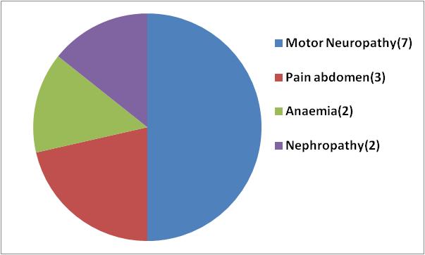
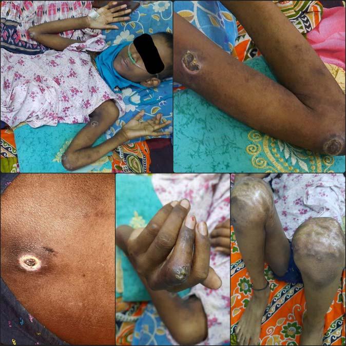
Result : 14 patients were recruited in our study. Mean age at presentation of participants in our study was 39.6 years. Main source of lead exposure was battery industry. Most common presenting symptom was motor neuropathy (50%), while most commonly involved system was hematopoietic (78.5%). Renal involvement was found in 28.5% patients.Blood lead level was in higher range in participants with considerable interpersonal variation. So called hallmark features of chronic lead toxicity like blue line in gum or basophilic stippling of RBCs in peripheral blood film were seen much less frequently.
Conclusion : In our study lead toxicity is shown to affect middle aged industrial workers. A lower threshold of clinical suspicion is required to diagnose the condition in patients from appropriate occupational background so that early diagnosis and appropriate interventional measures can be taken to halt the progression of this silent killer. The most common system involved in this study was hematopoietic, most common presenting complaint was neurological. Blue lines of gum and basophilic stippling did not appear to be very sensitive clinical features. More studies with larger population are required to achieve deeper insights into this preventable disease.
Key words :Lead, motor neuropathy, lead line, nephropathy.
Lead is one of commonest heavy metals known to cause toxicity in human beings. Lead is widely used in certain industries like mining, battery manufacturing and painting. In literature Ayurvedic products have also been reported to be source of lead1. Prevalence of Lead exposure and toxicity in developing countries is not very well documented, more so in recent years.
Department of General Medicine, ESI-PGIMSR & ESIC Medical College, Kolkata 700104
1MD (Medicine), Associate Professor
2MD (Medicine), DM (Nephrology), Assistant Professor, Department of General Medicine, KPC Medical College, Kolkata
3MD (Medicine), Assistant Professor and Corresponding Author
4MD (Medicine), Assistant Professor
Received on : 22/07/2021
Accepted on : 04/08/2021
[J Indian Med Assoc 2021; 119(8): 13-7]
Editor's Comment :
High degree of suspicion is required to diagnose chronic lead toxicity as manifestations can be as subtle as anaemia or mild nephropathy.
Thorough occupational history is important as there is consistent history of exposure.
Peripheral neuropathy is the most common complaint that brings patient to health care system.
Lead line in gum or basophilic stippling in peripheral blood smear are less commonly found, hence should not be relied upon as important clinical diagnostic criteria.
Lead is absorbed in human body through GI tract and Lungs. Rate of absorption depends on various factors including particle size, route, state of feeding, age of subject etc2. Organic lead is absorbed more readily. Once it enters blood stream it is distributed to all tissues including mineralized tissues like bones and teeth where it is stored in vast amount and ultimately contribute to largest lead store burden of
119, No 8, August 2021Journal
the body. It is also deposited in soft tissues like lung, liver, heart. Half life of lead in blood is 28-36 days. Lead is excreted through kidney and bile.
Toxic effects of lead accumulation are manifested in different organ systems. Most commonly affected ones are kidneys, hematopoietic and peripheral nervous system. Anaemia, one of the commonest manifestations, is caused by inhibition of the enzymes delta-Aminolaevulinic Acid Dehydratase (ALAD) and Ferro chelatase which are involved in heme synthesis and results in formation of zinc protoporphyrin, which is a laboratory marker of lead toxicity. Renal involvement ranges from Acute Kidney Injury to Chronic Kidney Disease and is due to injury to Proximal Convoluted Tubule which may result in fanconi syndrome. Lead is deposited in motor nerves and in brain resulting in pure motor neuropathy and myriads of higher function abnormalities respectively. Children are more prone to CNS involvement. Lead exposure is alsoshown to affect production of spermatozoa3
MATERIALS AND METHODS
This observational study was performed over 1 year period (October 2018 to September 2019) at General Medicine OPD and IPD of our Institution. The study protocol followed the principles expressed in thedeclaration of Helsinki. The study population included patients of either sex aged above 18 years attending General Medicine OPD or admitted in IPD in our Institution and being diagnosed with chronic lead toxicity by demonstration of elevated blood lead level (>10 mcg/dl). We excluded all patients having co morbidity like diabetes.
We used non probability convenient sampling method for this study. All patients satisfying inclusion and exclusion criteria were recruited. We could recruit 14 patients during the study period.
Thorough history taking and clinical examination were performed with special focus on occupation, extent and duration of exposure, symptomatology, functional status and special clinical features like looking for Anaemia, blue line in gum. Involvement of different organ systems susceptible to lead toxicity was evaluated.Involvement of kidney was evaluated by Renal function test, urine routine test,anaemia by Hb level & basophilic stippling, peripheral neuropathy by NCS. Data were recorded in a case report form (CRF) specifically designed for this purpose. The data from CRF was transcribed into an excel database. Data was summarized with routine descriptive statistics.
RESULTS
The study population included 14 patients recruited
from Medicine OPD and IPD of ESI-PGIMSR & ESIC Medical College, Joka, West Bengal, India.
Demographic Characteristics :
Analysis of Demographic Characteristics shows (Table 1) Mean age at presentation is around 40years (39.6 +/- 12.0). 11 of 14 patients were male. All were from lower socio economic class (monthly family income <Rs 21,000 as per ESI norm). Table 2 shows majority of patients worked at Battery industry. Average weekly working time is around 45 hours.
Table 1 — Demographic parameters
Parameter
Mean (standard deviation) N=13
Age (years)39.6 (12.0)
Duration of exposure (yrs)6 (2.7)
Weekly working hours44.8 (4.1)
Clinical
Characteristics :
Table 2 — Possible source of exposure
Source of LeadNo of patients
Battery Factory7 Painting3 Smelting2
Wire Factory1
Chemical Factory1
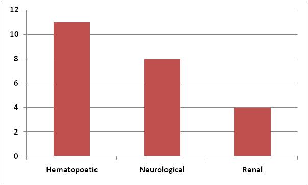
Most common symptom which prompted medical consultation in our participants was motor neuropathy followed by pain abdomen. Fig 1 shows presenting complaints. Most common organ system involvement in this study is hematopoetic system in the form of anaemia. Renal involvement is scarcest among 3 commonly involved systems. Fig 2 shows frequency of organ system involvement. It also depicts multiple organ system involvement in single individual (total frequency of organ system involvement is 23 in 14 participants).
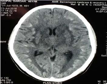
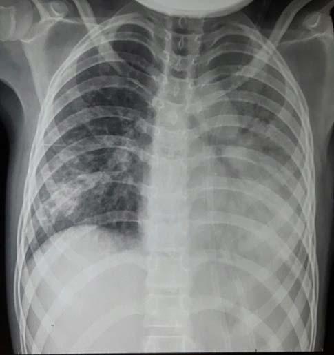
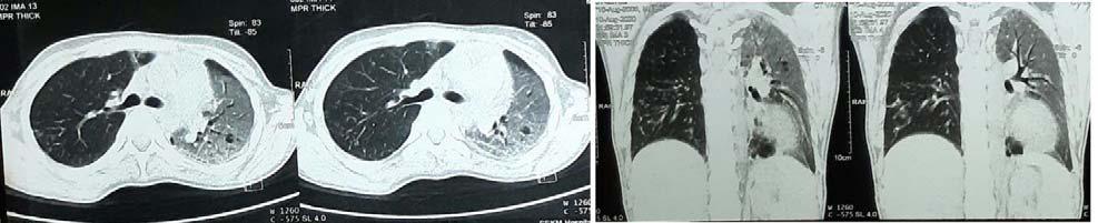
Blue line in gums, the so called lead line, was present in only 3 out of 14 patients. 1 patient complained of hematuria. Another patient presented with convulsion. NCCT brain revealed cortical calcification (Figs 3 & 4).
Laboratory Characteristics :
There was wide variation in blood lead level at
Fig 1 — Frequency of presenting symptom
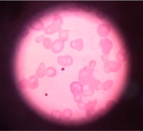
presentation, highest being 130 mcg/dl and lowest being 50.11 mcg/dl. Mean Hb level in patients having anaemia was 9.1 gm/dl. Basophilic stippling was observed in 2 out of 11 patients suffering from anaemia (Fig 5).
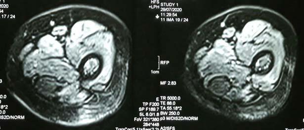
Mean Creatinine value in patients suffering from nephropathy was 1.8 mg/dl. NCS of all patients suffering from neuropathy showed predominantly motor neuropathy with degree of reduction in CMAP being more in upper limbs than in lower limbs. High level of Urinary porphobilinogen was detected in 1 patient. The classic metaphyseal line in X-ray of knee joint were found in 3 patients who presented with bone pain and arthalgia. Non contrast CT scan of a patient revealed Cortical calcification (Fig 6).
DISCUSSION
Chronic lead toxicity is one of the commonest occupational diseases encountered now a days in industrialized countries. It accounts for 0.6% of global burden of all toxic environmental diseases. Lead toxicity has resulted in chronic ill health, decreased economic output, lower life expectancy. In India, Institute for Health Metrics and Evaluation (IHME) found 4.6 million
lead-attributable DALYs and nearly 165,000 deaths (The 2016 Global Burden of Disease, Injuries and Risk Factors Study).
Occupational exposure is the main route in adults-battery workers, plumbers, paint and construction workers, lead mining, smelters, firing range instructors, rubber industry workers are most vulnerable population. Deteriorating lead paints and lead containing household dust are the main causes of chronic lead poisoning4. In our study battery factory workers constituted 50% of patient population followed by paint industry workers. Mean age of patients in our study was 40 years and mean duration of lead exposure before becoming symptomatic was 6 years.
GI tract (10-15% of adults), inhalational routes, skin are the routes of entry in adults. 99% of ingested or inhaled lead remains in the blood stream with a half life of 40 days and redistribute in brain and long bones. Kidney and liver take part in metabolism.
Lead acts by tilting the oxidant-antioxidant balance by decreasing antioxidant level. (Reduced level of glutathione, ALAD, Glutathione peroxidase and increase aminolaevulinic acid and reactive oxygen species in cell).
Serum level varies considerably among exposed individuals. It is determined by degree of absorption, rate of absorption and redistribution from bones and brain. In our study serum lead level varied considerably within participants. However even the lowest value in our study (50.11 mcg/dL) was much higher above safety level (5 mcg/dL).
Most of the clinical feature of chronic lead poisoning in adults are non specific. Chronic lead poisoning can present with number of signs and symptoms4,5 including abdominal pain (lead colic), constipation, anorexia, headache, irritability, decreased libido, difficulty concentrating and deficits in short term memory, nephropathy (Fanconi type syndrome), a lead line (bluish pigmentation seen at the gum
Fig 3 — Wrist drop in a patient of chronic lead poisoning
Fig 2 — Frequency of Organ systems involvement
Fig 4 — Blue line in gum in a patient of chronic lead poisoning
tooth junction), anaemia, basophilic stippling on blood smear, a peripheral neuropathy manifesting as extensor weakness due to wrist/ankle drop due to axonal degeneration affecting motor nerves6 The blue line, wrist drop, basophilic stippling of RBCs in peripheral blood film are classic findings but they are not always present. Hypertension, coronary artery disease are the main cardiovascular manifestation. In our study anaemia (78.57%) was most common clinical manifestation while most common presenting complaint was motor neuropathy (50%) followed by lead colic (21.4%).
Anaemia is a common manifestation of chronic lead toxicity. In our study anaemia was the most common clinical feature. Although this finding is partially offset by high prevalence of anaemia in Indian population, especially in females. Chronic lead exposure has been shown to induce dysplastic changes in erythroid precursors17. Basophilic stippling is not so common in lead poisoning as is usually described. In our study only 2 out of 11 patients with anaemia had basophilic stippling.
Chronic lead exposure has been associated with neuropsychiatric effects in the form of decline in neurocognitive function5, distal sensory and motor neuropathies6, conduction delay in ECG6. One study found that cumulative lead exposure may increase the risk Parkinson’s disease 7 . In our study neurological involvement was quite common (57.14%). 7 out of 14 patients suffered from motor neuropathy and presented with either wrist drop or weakness in handgrip while 1 patient presented with convulsion and had cerebral cortical calcification in NCCT brain.
Effect on reproductive health includes increased incidence of miscarriages, stillbirths8, low birth of weight9, cognitive impairments10,11 in babies with
high maternal bone lead level. Higher maternal blood pressure and 3rd trimester hypertension has also been associated12. We had only 3 females in our study, who did not encounter any of the above. Lead nephropathy is a potential complication of prolonged high level lead exposure. In our study 4 out of 14 (28.5%) patients suffered from nephropathy. Of them, 50% had nephropathy as main presenting symptom. Nephropathy was detected in these patients through routine screening of Serum Creatinine, emphasizing role of routine screening process in detection of this potential life threatening condition in high risk professional group. Although in our study participants blood lead level was in higher range (lowest being 50 mcg/dL) and thus explaining this high percentage of nephropathy. Rokho Kim et al demonstrated even low blood level of lead is a strong predisposing factor for developing nephropathy13.
Prolonged low level lead exposure appears to be associated with increased risk of cataract14, hearing loss15, carcinogenicity in animals-particularly renal tumours4. The National Toxicology program of the US Department of Health and Human services determined that lead has a carcinogenic role in human16
CONCLUSION
The clinical feature of chronic lead poisoning in adults are mainly nonspecific. The classical clinical presentation like blue line in the gum, wrist drop, basophilic stippling of RBCs in peripheral blood film etc are not always found. A proper history taking including occupational history and narrow threshold of clinical suspicion is important to diagnose and prevent progression this clinical condition which is a silent killer
Fig 5 — Basophilic stippling in chronic lead poisoning (Arrow)
Fig 6 — Cortical calcification in a patient with chronic lead poisoning
Vol 119, No 8, August 2021Journal of the Indian Medical Association
Our study showed main clinical presentations of lead toxicity being neurological, abdominal colic and renal involvement. Most frequently involved system in lead toxicity are hematopoietic, neurological and renal. Most common possible sources of lead exposure were found to be battery industry, followed by painting, smelting and wire factory. Our study was limited by small sample size. More studies are required in this area to further investigate into sources and distribution of symptomatology of chronic lead toxicity.
Limitation : Main limitation in this study is small sample size.
Funding : None
Conflict of Interest : None
REFERENCES
1Keen RW, Deacon AC, Delves HT, Moreton JA, Frost PG — Indian herbal remedies fordiabetes as a cause of lead poisoning. Postgraduatemedicaljournal 1994; 70 (820): 1134.
2Gaber BT, Wei E — Influence of dietary factors on gastrointestinal absorption of lead. Toxicology and applied pharmacology 1974; 27(3): 685-91
3Naha N, Chowdhury AR — Toxic effect of lead on human spermatozoa: A study among pigment factory workers. Indian Journal of Occupational and Environmental Medicine 2005; 9(3): 118.
4ATSDR — Toxicological profile for lead. US Department of Health & Human Services, Public Health Service, Agency for Toxic Substances and Disease Registry, Atlanta, GA 2007.
5Cullen MR, Robins JN, Eskenazi B — Adult inorganic lead intoxication : presentation of 31 new cases and a review of recent advances in the literature. Medicine (Baltimore) 1983; 62: 221.
6Thomson RM, Parry GT — Neuropathies associated with excessive exposure to lead. Muscle nerve 206; 33: 732.
7Weisskopt Mr, Weave J, Nie H — Association of cumulative lead exposure Parkinsons diseases. Environ Health perspect 2010; 118: 1609.
8Fischbein A, HV H — Occupational and environmental exposure to lead. In Environmental and Occupational Medicine, Rom, Con and Markowit SB (Eds) Philadephia, wolterskluwer/lippiricott Williams & Williams 2007. 958.
9Gonzalez-cossio T, Paterson KE, Sanin Ltl — Decrease in birth weight in relation to maternal bone-lead burden. Paediatires 1997; 100: 556.
10Bellingers D, Leviton A, water nayx C — Longitudinal analysis cognitive development. N Eng J Med 1957; 316: 1037
11Groma A, Hu H, Bellinger D — Maternal bone lead as an independent risk factors for fetal neurotoxicity : A prospective study. Pediatrics 2002; 110: 110.
12Rothenberg SJ, Kondrashov V, Manalo M — increases in hypertension and blood pressure during pregnancy with increased bone lead levels. Am J Epidemiol 2002; 156: 1079.
13Kim Rokho, Rotnitzky A, Sparrow D — A longitudinal study of low-level lead exposure and impairment of renal function. JAMA 1996; 275(15): 1177-81.
14Schaumbery DA, Mendes F, Balaram M — Cumulative lead exposure and risk of age-related cataract in men 2004; 292: 2750.
15Park SK, Elmarsfawy S, Mukherjee B — Cumulative lead exposure and age related hearing loss: the VA Normative Aging study. Hear Res 2010; 269: 48.
16National Toxicology Program, US Dept of Health and Human Services. 12th Report on carcinogens, 2011. Lead and lead compounds (CAS No. 7439-92-1 (lead).
17Lv C, Xu Y, Wang J — Dysplastic changes in erythroid precursors as a manifestation of lead poisoning: report of a case and review of literature. Int J Clin Exp Pathol; 8: 81823.
To Evaluate
Original Article
the Efficacy of Microplan for Emergency Department of Medical Colleges laid by the Uttar Pradesh Government of India in Reference to the COVID-19 Pandemic
Anjana Pandey1, Madhu Singh2, Prabhat Agrawal3, P K Maheshwari4, Ashish Gautam5, Nikhil Pursnani6
Objective : To evaluate the efficacy of MICROPLAN for screening and segregation of patients coming to Emergency Department (ED) of Medical Colleges of state, laid down by Uttar Pradesh Government of India, in reference to the COVID-19 pandemic.
Materials and Methods : This is a retrospective, observational case series. Data were collected from May 01, 2020, to May 31, 2020, from Emergency Department, SN Medical College, Agra, Uttar Pradesh, India.
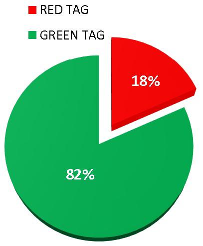
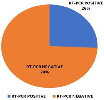
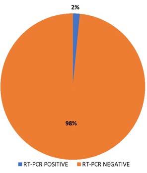
Results : Out of 1856 patients, 1516 patients were tagged green or non-suspects (81.68 %) and 340 were tagged red/ suspects (18.31%). Out of 340 red tagged patients, 87 came to be positive for 2019- nCoV by RT-PCR (25.58 %) and out of 1516 green tagged patients, 24 patients tested positive for 2019- nCoV by RT-PCR (1.58 %).
Conclusion : MICROPLAN laid down by Uttar Pradesh Government of India, in reference to the COVID-19 pandemic has certainly avoided mixing covid and non-covid patients, helped us to break the chain of infection, and above all prevented our medics, paramedics, and patients from getting an infection from asymptomatic corona patients. We recommend this plan to be implemented at every emergency department during covid pandemic in India.
[J Indian Med Assoc 2021; 119(8): 18-20]
Key words :Microplan, Emergency Department, Screening, COVID-19, Green Tag, Red Tag, RT-PCR 2019-nCoV.
Coronavirus disease 2019 (COVID-19) is a rapidly evolving global pandemic that has already caused profound effects on public health and medical infrastructure globally1 including India. In the present COVID outbreak, there was a serious need to start emergency services to cater patients who were suffering from different other ailments, but seeing the massive spread of novel coronavirus disease every patient whether showing typical clinical features of corona or not, must be considered as a suspect until proven otherwise. This article focuses on the success of MICROPLAN for Management of Patients in Emergency Department (ED) of Medical Colleges laid down by Uttar Pradesh Government, with special reference to the COVID-19 pandemic.
During the COVID-19 Pandemic, the focus of the whole medical fraternity is on novel corona virus management, which in turn severely compromised the
Department of Medicine, Sarojini Naidu Medical College, Agra, Uttar Pradesh 282003
1MD, Associate Professor and Corresponding Author
2MBBS, MD, Associate Professor, Department of Paediatrics
3MBBS, MD, Professor
4DM, Professor
5MBBS, MD, Associate Professor
6MBBS, MD, Assistant Professor
Received on : 17/03/2021
Accepted on : 28/07/2021
Editor's Comment :
MICROPLAN for Management of Patients in Emergency Department (ED) of Medical Colleges, with special reference to COVID-19 pandemic laid down by Uttar Pradesh Government, has definitely avoided mixing of COVID and non-COVID patients thus helped in curtailing the infection among patients, paramedical and medical personals. We recommend implementation of this plan to every Emergency department during covid pandemic.
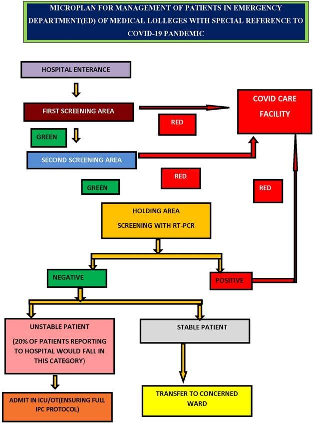
routine outdoor and indoor functioning leading to increased morbidity and mortality of non-corona patients. So, to combat this Uttar Pradesh Government came up with a MICROPLAN for Management of Patients in Emergency Department (ED) of Medical Colleges of Uttar Pradesh, for the management of Medical, Surgical Emergencies and trauma for NonCOVID patients. Every hospital, therefore, had two different care areas for the management of patients:
(A) COVID Care Facility for management of COVID19 patients.
(B) Emergency Department for the management of Medical & Surgical Emergencies for Non-COVID patients.
These two areas A and B should be separate to avoid cross infection and accidental admission of the suspect or confirmed COVID-19 patient in the NonCOVID area.
MATERIALS AND METHODS
This was a retrospective, observational case series. Data were collected from May 01, 2020, to May 31, 2020, from the Emergency Department, S.N. Medical College, Agra, Uttar Pradesh, India. The study included 1856 consecutive patients who came to the screening area of the emergency department for various complaints. Based on the protocol as laid in micro plan for emergency, we started our Emergency Department (ED). Before entry into the ED, the patient first entered the reception area which is the First Screening Area at the Non-COVID Hospital entrance. All patients were screened based on the pre-designed questionnaire2. This questionnaire changed depending on the stage of the epidemic. Temperature monitoring via infrared thermometers was performed for all patients. Any corona suspect patient was immediately tagged as RED and others as GREEN. Based on this categorization, RED tagged patient was referred to the COVID-19 triage in the dedicated COVID facility and GREEN tagged patient not suspected to be COVID-19 were referred to the Second Screening Area in the NonCOVID facility/Hospital. In case a suspect COVID-19 patient is identified then immediately the tag was changed from GREEN to RED and the patient was referred to the COVID-19 triage in the dedicated COVID-19 Facility/Hospital. Patients not suspected to be COVID-19 were continued to wear the GREEN tag and subjected to investigation for confirmation of COVID-19 status. RTPCR is the Gold Standard investigation3 All GREEN tagged suspect cases were admitted in the HOLDING Area ward till their report came. Patients admitted in this area must be categorized as STABLE or UNSTABLE depending on the ABCDE approach of the internationally accepted Emergency Severity Index. If the test results were negative and the patient did not require admission, he/she was sent home with instructions for Home Quarantine for 14 days. However, if the patient needed admission, he/she was admitted to the respective DESTINATION ward. If the test results were positive, the tag was immediately changed from GREEN to RED and the patient was referred to Isolation in the dedicated
COVID Care Facility/Hospital. All patients were screened based on a pre-designed questionnaire and segregated into a red tag (COVID suspect) and green tag (non covid). Patients were admitted in their respective wards, red tags patients were admitted into dedicated COVID facility, and green tags into holding area of the emergency department. RT-PCR (2019- nCoV) of every patient was done (Fig 1).
RESULTS
Out of 1856 patients, 1516 patients were tagged green or non-suspects (81.68 %) and 340 were tagged red/ suspects (18.31%) (Fig 2). Out of 340 red tagged patients 87 came to be positive for 2019- nCoV by RTPCR (25.58 %) (Fig 3) and out of 1516 green tag patients 24 patients tested positive for 2019- nCoV by RT-PCR (1.58 %) (Fig 4)(Table 1).
Fig 1 — Flow chart for Micro plan for Emergency Department
Table 1 — Percentage distribution of Red Tag and Green Tag Patients
Total PatientsTotal Green TagTotal Red TagPositive PatientsPositive Patients% Positivity% Positivity Admitted in EDPatients among Patients amongamong Green Tagamong Red Tagamong Green among Red Admitted Patients(%)Admitted Patients(%)(RT PCR Positive)(RT- PCR Positive)Tag PatientsTag Patients 18561516(81.68%)340(18.31%)2487 1.58 %
CONCLUSIONS
AND RELEVANCE
Screening is recommended in every patient who comes to the emergency department as it can segregate suspects from non-suspect with the help of very simple questionnaire. The green tag patients were those who did not have any signs of corona and can be considered as clean patients at first glance and can be shifted directly to the concerned department without testing but this MICROPLAN for Management of Patients in Emergency Department (ED) of Medical Colleges, with special reference to COVID-19 pandemic laid down by Uttar Pradesh Government, was designed in such a way that it considered every patient as suspect whether green or red tag. If we analyse the observations even few green tag patients incidentally turned out to be positive, probably these patients were asymptomatic carriers4, and if we had not adopted above protocol than we would have surely considered them as clean cases and would have
4 — Percentage of RT-PCR Positive patients among Green Tag patients
transferred them to concerned speciality wards. Inwards they would have mixed up with other patients and transmitted infections to them. So, the above protocol has certainly avoided mixing infectious and non-infectious patients, helped us to break the chain of infection and above all prevented our medics, paramedics and patients from getting infected from asymptomatic corona patients.
REFERENCES
1Cucinotta D, Vanelli M — WHO declares COVID-19 a pandemic. Acta Biomed 2020; 91(1): 157-60,
2Revised Strategy for COVID19 testing in India (Version 5), ICMR Dated 18/05/2020.
3Corman VM, Landt O, Kaiser M — Detection of 2019 novel coronavirus (2019-nCoV) by real-time RT-PCR. Eur Surveill 2020; 25: 2000045
4Kimball A, Hatfield KM, Arons M — Asymptomatic and presymptomatic SARS-CoV-2 infections in residents of a long-term care skilled nursing facility — King County, Washington, March 2020. MMWR Morb Mortal Wkly Rep 2020; 69: 377.
Fig 2 — Distribution of Red Tag and Green Tag among total patients
Fig 3 — Percentage of RT-PCR Positive patients among Red Tag patients
Fig
Original Article
Study on Perinatal Outcome in Relation to Maternal Vitamin D Deficiency
Dipankar Sarkar1, Babita Saha2,
Sajal Datta3
Objective : To assess the incidence of vitamin D deficiency in primigravida and to correlate perinatal outcome after substitution of Vitamin D among those deficient women.
Methodology : This observational and prospective study was conducted at VIMS, Kolkata for a period of one year. A total of 100 primigravida women whose vitamin D was less than 20ng/ml (deficient mother as per our study) were randomly selected based on inclusion criteria. These 100 women were subgrouped into two groups.
Group A : 50 women who were deficient of vitamin D on booking (less than 20 ng/ml).
Group B : 50 women who were deficient of vitamin D (<20ng/ml) and received vitamin D 2000IU/day during the course of pregnancy.
Serum vitamin D level was estimated by Chemiluminesence Immuno Assay (CLIA) method.
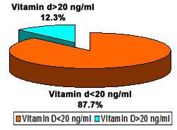
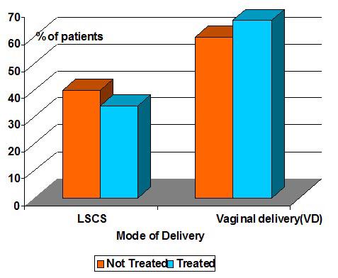
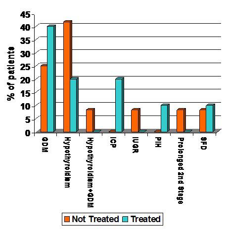
Results : Incidence of vitamin D deficiency in our study population was 87.7%. Deficient vitamin D and its associations with risk factors eg, Gestational Diabetes Mellitus (GDM), Hypothyroidism, Intrahepatic Cholestasis In Pregnancy (ICP), Pregnancy Induced Hypertension (PIH) were more or less same in both groups. Incidence of preterm delivery in non treated group (8%) was found to be higher than treated group (4%). But this difference was not statistically significant. Similar finding was noted in case of low birth weight babies between the two groups though it was 1.19 times higher among the mothers with no treatment. Caesarean section rate was higher in non treated group (p>0.05).
Conclusion : In this study no statistically significant association of adverse maternal and perinatal outcome was noted between mothers who were deficient and non deficient of vitamin D is found in many literature.
Key words :Vitamin D deficiency, Perinatal outcome.
Recent evidences suggest that women belonging to high-risk groups like vegetarians, having limited sun exposure and ethnic minorities, especially those with darker skin are Vitamin D deficient1-3. In India and its surrounding countries contrary to our old belief a huge population were found to have Vitamin D deficiency despite having a less dark skin and adequate exposure to sun rays. This is because sunrays falling on the skin of upper and lower extremities between 11am and 3 pm is mostly responsible for stimulating Vitamin D synthesis and people of these subcontinent usually stay in house during this time.
Department of Obstaetrics & Gynaecology, Raiganj Government Medical College and Hospital, Raiganj, Uttar Dinajpur 733134
1MBBS, MS (Obstat & Gynaecol), RMO cum Clinical Tutor
2MBBS, DGO, MS (Obstat & Gynaecol), Assistant Professor Department of Obstaetrics & Gynaecology, Vivekananda Institute of Medical Sciences, Kolkata 700026 and Corresponding Author
3MBBS, MD (Obstat & Gynaecol), DNB, Professor, Department of Obstaetrics & Gynaecology, Vivekananda Institute of Medical Sciences, Kolkata 700026
Received on : 05/01/2021
Accepted on : 25/02/2021
[J Indian Med Assoc 2021; 119(8): 21-4]
Editor's Comment :
Adverse maternal and perinatal outcome have been reported with vitamin D deficiency in many literature.
Assesment of micro and macronutrient deficiency is of utmost importance during pregnancy
Newborn mostly depends on mother for their Vitamin D. If mothers are already Vitamin D deficient then the newborns will also be vitamin D deficient4
As per 2010 Institute of Medicine (IOM) Report, 12ng/ml (30nmol/L) of 25(OH)D is the limit below which “persons are at risk for bone deficiency”. However,as per the 2011 ACOG Practice Bulletin “Vitamin D: Screening and Supplementation” defines a value less than 20ng/ml (50nmol/L) as Vitamin D deficient.
Mothers who are vitamin D deficient are found to suffer more from GDM, preeclampsia, small baby and operative interventions.Considering cut off as 20ng/ml, higher incidence of Vitamin D deficiency among pregnant mothers has been reported in studies throughout the world. Same has been reported in India, Pakistan, Japan, China, UK as well as in Sweden5.
119, No 8, August 2021Journal
Most of the literature shows that 1000-2000 IU of Vitamin D per day in pregnancy is safe. Although there is no adequate data of higher safer dose but consensus is mostly upto 4000 IU per day during pregnancy and lactation6.
Physiologically Vitamin D is called calciferol ie, D2 and D3. Plant source of vitamin D is known as Vitamin D2 where as human source (Vitamin D3, cholecalciferol) is produced below the skin following UV light radiation from sun7. Vitamin D3 is three times more stronger in efficacy than Vitamin D2 and more protein bound in plasma8. Vitamin D is short lived and thus needs adequate dosing to maintain its effective concentration in blood.
In this study we had measured Vitamin D level in pregnant mothers and correlated that with adverse perinatal outcome.
MATERIALS AND METHODS
This is an observational and prospective study among 100 uncomplicated primigravidas in Obstat & Gynaecol department at VIMS, Kolkata. After obtaining necessary approval from institutional ethical committee and based on inclusion criteria they were enrolled after obtaining consent provided they are all Vitamin D deficient (below 20 ng/ml) at first visit as measured by CLIA method.
A total of 100 women were enrolled and divided into two groups.
Group A : (50 patients) – who were deficient of vitamin D and did not receive any treatment.
Group B : (50 patients) –who were found to be Vitamin D deficient and substituted with 2000 IU of Vitamin D per day during their antenatal periods. These 100 pregnant women were followed up till delivery and their neonates till discharge from hospital. Finally these two groups were compared on pregnancy outcome, method of delivery and neonatal outcome.
Inclusion Criteria :
(1)Primigravida with vitamin D level less than 20ng/ ml at first visit
(2)Booked in the OPD within 16 wks of POG
(3)No history of antenatal or medical/surgical complication
Exclusion Criteria :
(1)History of treatment with vitamin D before (2) Vitamin D is contraindicated or Hypersensitivity to Vitamin D
RESULTS AND DISCUSSIONS
Statistical Analysis was performed with help of Epi Info (TM) 3.5.3 which is a trademark of CDC (Centers for Disease Control and Prevention).
Using this software, basic cross-tabulation and frequency distributions were prepared. χ2 test was applied to see the association between different study variables under study. Z-test was applied to assess the significant difference between two proportions. ttest was also used in this study to compare the means. Odds ratio (OR) with 95% Confidence Interval (CI) was calculated to measure the different risk factors. p<0.05 was considered statistically significant (Fig 1).
Incidence of Vitamin D deficiency in our study population was 87.7%. The incidence of Vitamin D deficiency is quite high in our study and corroborates with the incidence stated in different earlier studies9-11.
No significant difference was noted applying the ttest to compare the mean age of the patients,
Fig 1 — Incidence of Vitamin D deficiency in study population
Fig 2 — Mode of delivery of the patients of two groups
gestational age at their first visit and mean vitamin D level between these two groups (t98=0.04; p=0.54). Thus two groups were matched (Fig 2).
No significant association was noted when mode of delivery was considered between two groups using Chi square test (p=0.53). Proportion of LSCS was higher in Group-A (40%) than that of group-B (34%) but it was not significant (p>0.05). Similar study by Anne-Louise P et al12 found that there were four times increased incidence of caesarean section in non treated expectant mothers. Fernandez-Alonso AM et al13 in their study found no increase risk of caesarean section in pregnant women with 25-OHD insufficiency, whereas Scholl TO et al14in their study showed an higher risk of c-section among vitamin D deficient group of women (Fig 3).
Statistically significant association was not noted when risk factors were compared between the two groups using Chi-square test (p=0.39). All the associated risk factors were more or less evenly distributed over the two groups.
Prevalence of pre-term delivery was higher in group-A (8%) than that of groupB (4%) but it was not significant (p>0.05).
Gille O et al15 in their study reported that after supplementaion of Vitamin D there was a reduction of pretemlabour and small for date babies.
When weight of the babies at birth and neonatal outcome were compared between the two groups using Chi-square (χ2 ) test showed no significant association (p=0.37).
CONCLUSION AND LIMITATION
In our study, in contrary to many literature we have not found any statistical difference between the two groups when maternal and perinatal outcomes were compared.
This could be due to small sample size in our study. We were also unable to estimate other factors which can affect vitamin D level. In our population we have also noticed variation in the maternal built and nutrition which can affect birth weight of babies. In future we need to perform a better study keeping in mind different important confounding factors like maternal weight, lifestyle, nutritional status, duration and time of sun exposure etc. Quantification of Vitamin D in serum
also needs to be standardized among different centers to avoid any discrepancy in serum Vitamin D level.
REFERENCES
1Hollis BW, Wagner CL — Assessment of dietary vitamin D requirements during pregnancy and lactation. Am J Clin Nutr 2004; 79: 717-26.
2Lee JM, Smith JR, Philipp BL — Vitamin D deficiency in a healthy group of mothers and newborn infants. Clin Pediatr (Phila) 2007; 46: 42-4.
3Bodnar LM, Simhan HN, Powers RW — High prevalence of vitamin D insufficiency in black and white pregnant women residing in the northern United States and their neonates. J Nutr 2007; 137: 447-52.
4Dijkstra SH, van Beek A, Janssen JW — High prevalence of vitamin D deficiency in newborn infants of high-risk mothers [published erratum appears in Arch Dis Child 2007; 92: 1049]. Arch Dis Child 2007; 92: 750-3.
5Marwaha RK, Tandon N, Chopra S — Vitamin D status in pregnant Indian women across trimesters and different seasons and its correlation with neonatal serum 25hydroxyvitamin D levels. Br J Nutr 2011: 1-7. [PubMed]
Fig 3 — Associated risk factors among the patients of two groups
6Mazhar SB — Vitamin D supplementation for women during pregnancy: RHL commentary (last revised: 1 July 2012). The WHO Reproductive Health Library; Geneva: World Health Organization.
7DeLuca HF — Overview of general physiologic features and functions of vitamin D. American Journal of Clinical Nutrition 2004; 80(6 Suppl): 1689S-96S.
8Armas LA, Hollis BW, Heaney RP — Vitamin D2 is much less effective than vitamin D3 in humans. Journal of Clinical Endocrinology and Metabolism 2004; 89(11): 5387-91.
9Arya V, Bhambri R, Godbole MM, Mithal A — Vitamin D status and its relationship with bone mineral density in healthy Asian Indians. Osteoporos Int 2004; 15: 56-61.
10Harinarayan CV — Prevalence of vitamin D deficiency in postmenopausal south Indian women. Osteoporos Int 2005; 16: 397-402.
11Marwaha RK, Tandon N, Reddy DRHK — Prevalence and Significance of low 25-hydroxyvitamin D concentrations in healthy subjects in Delhi. Am J CliNutr 2005; 82: 477-82.
12Anne-Louise Ponsonby, Robyn M — Lucas, Sharon Lewis Vitamin D status during Pregnancy and Aspects of Offspring Health. Nutrients 2010; 2: 389-407.
13Fernandez-Alonso AM, Dionis-Sanchez EC, Chedraui P, Gonzalez-Salmeron MD, Perez-Lopez FR — First-trimester maternal serum 25-hydroxyvitamin D(3) status and pregnancy outcome. Int J Gynaecol Obstet 2011; 116: 6-9.
14Scholl TO, Chen X, Stein P — Maternal vitamin D status and delivery by cesarean. Nutrients 2012; 4: 319-30.
15Gillie O — Vitamin D ‘may cut premature birth risk and protect newborn babies’, The Times Oct 10, 2009, Available online: http://www.timesonline.co.uk/tol/news/uk/scotland/ article6868729.ece (accessed on 2 February 2010).
Disclaimer
The information and opinions presented in the Journal reflect the views of the authors and not of the Journal or its Editorial Board or the Publisher. Publication does not constitute endorsement by the journal. JIMA assumes no responsibility for the authenticity or reliability of any product, equipment, gadget or any claim by medical establishments/institutions/manufacturers or any training programme in the form of advertisements appearing in JIMA and also does not endorse or give any guarantee to such products or training programme or promote any such thing or claims made so after. — HonyEditor
Ifyouwanttosendyourqueriesandreceivethe responseonanysubjectfromJIMA,pleaseuse the E-mail or Mobile facility.
Website:https://onlinejima.com For Reception:Mobile : +919477493033 For Editorial:jima1930@rediffmail.com Mobile : +919477493027 For Circulation:jimacir@gmail.com Mobile : +919477493037 For Marketing:jimamkt@gmail.com Mobile : +919477493036
For Accounts: journalaccts@gmail.com Mobile : +919432211112
For Guideline:https://onlinejima.com
Original Article
A Drug Utilization Study of Antidepressants in the Psychiatry Unit of a
Tertiary Care Hospital
Sagar
Kumar1
This study aims at analyzing the drug utilization pattern of the different classes and individual antidepressant drugs used in the therapy of Major Depressive Disorder. Major Depressive Disorder (MDD) is an extremely common psychiatric condition. Antidepressant class of drugs are commonly used to treat this condition. In my study, analysis of prescription patterns of antidepressants was carried out for patients suffering from MDD.The prescribing patterns of antidepressants have changed globally over the last few years and therefore we wanted to observe the prescribing pattern of antidepressant drugs in our hospital.The drug utilization study was an observational cross-sectional study carried out in the Department of Pharmacology and Psychiatry, Hi-tech Medical College & Hospital, Bhubaneswar, India from 1st November 2015 to 30th October 2017 with a sample size of 262 patients suffering from Major Depressive Disorder and being treated with antidepressant class of drugs. Socio-demographic details of the patients, clinical features of each case were analysed in the study. Several quality indicators of drug use and standard parameters were observed. In my study it was seen that antidepressants were prescribed more in women (67.2%) as they comprised a majority of the patients.The mainstay of this study was to analyse the current prescription pattern of antidepressants at our hospital. A total of 492 antidepressant drugs were prescribed to the 262 patients enrolled in this study. It was seen that the average number of antidepressants per prescription was 1.87. It was seen that in this study 14.63% of the antidepressant drugs prescribed were from the Essential Medicines List (EML). This study revealed that that 77.24% of the antidepressants used were prescribed in their generic name. In this study it was seen that the SSRI class of antidepressant drugs was used most times comprising 45.7% of the total drugs used. This was followed by use of TCA class of drugs and SNRI class of drugs accounting for 23.8% and 17.9% of the total drugs used respectively. Escitalopram (SSRI) was used most times (36.99%) followed by Amitriptyline (TCA) (14.63%) and Mirtazapine (Atypical) (11.59%). The patients at the time of presentation were graded by HDRS score according to severity of illness. Choice of antidepressant drug prescribed and from which group is based on individual patient aspects, clinician’s judgement and previous response to treatment. The newer drug classes such as SSRI, SNRI and Atypical class of drugs are more preferred now because other things being equal these are usually better tolerated and less dangerous in overdose. However, it must be kept in mind that even now TCA class of drugs is an effective and proven alternative and may be preferred in some cases of MDD.
Key words :Antidepressant drugs, Depression.
Depressive disorders have plagued mankind since the earliest documentation of human experience. The earliest references, from ancient Greek descriptions of depression, refer to the Syndrome of Melancholia1.
The World Health Organisation (WHO) defines drug utilization as the marketing, distribution, prescription and the use of drugs in a society, with special emphasis on the resulting medical, social and economic consequences19
The prevalence of antidepressant usage in the community is rising in Western populations, with Iceland, Australia and Sweden having the highest consumption9. This trend has been replicated and documented in developing nations too like our own.
1MBBS, MD (Pharmacology), Tutor, Department of Pharmacology, MGM Medical College, Jamshedpur, Jharkhand 831020
Received on : 20/05/2021
Accepted on : 05/08/2021
[J Indian Med Assoc 2021; 119(8): 25-31]
Editor's Comment :
Major Depressive Disorder (MDD) is an extremely common Phychiatric condition.
Most common anti depression are prescribed in generic name.
Escitalogram is one of the most preferred drug among the SSRIs.
Depressive disorders afflict one out of five women and one out of ten men at some point in their lives2. The depressive disorders are characterized by a lifelong vulnerability to episodes of the disease.
It was estimated that by the year 2020 if current trends for demographic and epidemiological transition continue, the burden of depression will increase to 5.7% of total burden of disease3
Depression is the most common type of mental illness. An estimated 7-10% of India’s population
119, No 8, August 2021Journal
suffers from depressive illness4
This study will focus on the pharmacotherapy of unipolar major depression.
Depression is a clinical syndrome characterized by persistent sad mood, profound despair which persists 2 weeks or more and is associated with a change in previous functioning1
Many different antidepressants have established track records of efficacy for treating major depression. However, they all suffer some limitations in efficacy, since at least 20% of all depressed patients are refractory to multiple different antidepressants at adequate doses7. Therefore, a clinician’s experience, insight and particular features of the case may have an effect on the drug(s) prescribed.
All drugs commonly used to treat depression share at some level, primary effects on serotonergic or noradrenergic neurotransmitter systems8. In general antidepressants enhance serotonergic and noradrenergic transmission, although the nature of this effect may change with chronic treatment.
The optimistic expectations of a drug, based on the results of clinical trials, may not materialize when they are used outside controlled settings5.
Long term effects of antidepressant drugs evoke adaptive or regulatory mechanisms that enhance effectiveness of therapy9.
Depression often requires long term therapy as well as multi drug treatment in many cases. This becomes important because antidepressant use is often associated with a wide spectrum of adverse effects.
The recent proliferation of new drugs, the increasing recognition of delayed adverse effects and the focus on pharmacoeconomic considerations have stimulated interest in the antidepressant prescribing patterns of physicians6
Rational drug prescribing is the use of the least number of drugs to obtain the best possible effect in the shortest period and at a reasonable cost18. Goal of treatment should revolve around achieving a sustained and consolidated improvement in the mood of the patient, guarding against relapse and with minimal possible adverse effects.
This paper is a qualitative and quantitative critical analysis of the treatment of depression and the use of anti-depressants in Hi-Tech Medical College and Hospital, Odisha over the stipulated time frame.
MATERIALS AND METHODS
Study Design :
The drug utilization study was an observational cross- sectional study.
Sampling Technique :
The sampling technique was ‘purposive’ in nature.
Inclusion criterion :
All consenting patients with clinical diagnosis of Major Depressive Disorder (MDD) attending Psychiatry OPD of Hi-Tech Medical College and Hospital, Bhubaneswar, Odisha during the stipulated time frame.
3.Monotherapy or Combination therapy of different drug classes
4.Grading of patients according to severity of illness (i.e. depression; HDRS)
5.Average number of drugs per prescription
6.Percentage of drugs in generic name
7.Percentage of drugs from essential medicine list
Instruments :
Age, Sex, Marital status, Rural/Urban, Caste, Family Type, Socio Economic status
Study Technique :
Descriptive statistics was used to calculate mean and percentages.
Chi-square test was used to compare categorical variables and t-test (ANOVA) was used to compare continuous variables.
Features of this study :
Patient data for this study was collected in a predesigned proforma. The first part of which was for collecting socio-demographic details of the patient. The next part dealt with recording details about the name of drugs prescribed, duration of therapy, number of drugs in generic name, number of drugs from essential medicine list and total number of anti-depressants drugs used for each patient. The Hamilton Depression Rating Scale (HDRS) (reference) was used to grade the severity of depression. The difference in HDRS score for each patient before and after therapy gave us an idea of the clinical efficacy of the different therapy options used and a measure of improvement they provided for each patient.
The data collected in the above format was then analysed using both qualitative and quantitative statistical techniques. The results and analysis have been discussed.
Ethical clearance :
The study was approved by the Institutional Ethics committee of Hi-Tech Medical College, Bhubaneswar
(Utkal University)
Study duration :
The data was collected for a period of 2 years from 1st November 2015 to 30th October 2017.
Study location :
The drug utilization study was carried out in the Department of Pharmacology and Psychiatry, Hi-Tech Medical College and Hospital, Bhubaneswar, India.
Sample size :
The sample size was of 262 patients suffering from Major Depressive Disorder (MDD) and being treated with anti-depressant class of drugs.
RESULTS
Data was collected for 262 patients as per the protocol of the study and was analysed through IBM SPSS 24.0 software. The analysis along with interpretations is presented in three sections.
Section 1 deals with the demographic and socioeconomic profile of the patients, Section 2 analyses the prescription patterns of anti-depressants prescribed in the OPD of Psychiatry department, Section 3 deals with the clinical effectiveness of different therapy options
Demographic and Socio-economic profile
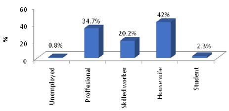
Age and Gender distribution
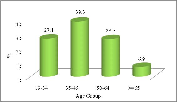
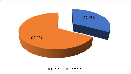
Figs 1&2 present age and gender distribution of patients suffering from MDD. It was found that maximum patients were in the age group 35-49 years (39.3%). The age group 19-34 years and 50-64 years shared nearly a quarter of the cases. The distribution was significantly concentrated in the age group 35-49 years (p=0.000). The mean age was 44.22± 12.54 years. It was found that females constituted a majority of the cases with a share of little above 2/3rd of the total cases (p=0.000).
Distribution by place of residence :
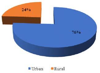
The major chunk of the cases was from urban areas with a share of 76% (Fig 3).
Education :
Graduate and above qualified individuals comprised 44.3% cases while matric pass was 38.2%, under matric and illiterate together constituted 17.6% cases. The proportion of cases was found significantly higher in graduate and above category (p=0.000)
Marital Status and Family Type (Figs 4&5) :
Prescription pattern of anti-depressants :
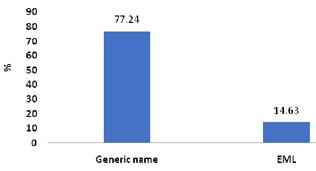
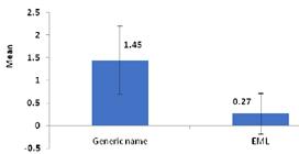
Drugs used in generic name and from EML : Figs 6&7 present distribution of anti-depressants used in generic name and from essential medicine list (EML). 262 patients were prescribed 492 drugs. Of these 492 drugs 380 (77.24%) were prescribed in their generic name. The remaining 112 drugs (22.76%) were
prescribed using their trade name. The study also showed that of the 492 drugs used only 72 (14.63%) were from the essential medicine list (EML) while the remaining 420 (85.36%) were not from the EML.
Class of drugs used :
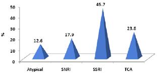
The total of 492 drugs used were distributed among four class of anti-depressant drugs namely Atypical Antidepressants, SNRI, SSRI and TCA. The majority class of drugs was SSRI with a share of 45.7% followed by TCA (23.8%). The other two categories, Atypical and SNRI, comprised of 12.6% and 17.9% respectively (Fig 8).
Fig 1 — Distribution of Patients
Fig 3 — Distribution of Patients by place of residence
Fig 2 — Gender Distribution of Patients
Fig 4 — Gender Distribution of Patients
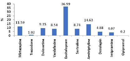
Fig 9 show the distribution of cases by the individual drugs used from different classes. Out of 225 drugs prescribed from SSRI class 182 were Escitalopram while the remaining 43 were Sertraline. These two drugs had a share of 36.99% and 8.74% respectively of the total drugs used. Under TCA class 117 drugs were prescribed of which Amitriptyline was 72, Dosulepin 24, Imipramine 20 and Opipramol 1. The respective proportions were 14.63%, 4.88%, 4.07% and 0.2% of the total drugs prescribed. In the SNRI class 88 drugs were prescribed of which 46 were Duloxetine while Venlafaxine was used 42 times with a share of 9.35% and 8.54% of the total drugs prescribed respectively. In the Atypical class 62 drugs were prescribed of which 57 were Mirtazapine and the remaining 5 were Trazodone with a share of 11.59% and 1.02% of the total drugs respectively. Escitalopram was prescribed most times followed by Amitriptyline and Mirtazapine. These three drugs together constituted 63.21% of the total drugs prescribed.
Duration of Therapy : Distribution of cases by duration of therapy is presented in Fig 10. Duration of therapy was in the
range of 1 month to 120 months with a mean ± SD of 11.53 ± 12.05 months. Maximum proportion of patients were under therapy for 6-11 months (42%).
Efficacy of Treatment
Severity of presenting illness
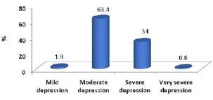
The patients at the time of presentation were graded by HDRS score according to severity of illness. HDRS score within 8-13 were classified as mild depression; HDRS score within 14-18 as moderate depression while HDRS score within 19-22 was classified as severe depression; HDRS score >23 was classified as very severe depression. The results are presented on Fig 11. Nearly 2/3rd of cases (63.4%) had moderate level of depression while 34% presented with severe level of depression. Mild depression was of the order 1.9% while 0.8% cases presented with very severe depression.
HDRS score before and after therapy (clinical improvement) :
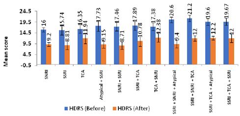
The comparison of HDRS score before and after therapy for the different treatment options used is presented on Fig 12. It was found that 262 patients were administered 11 types of therapy options. Single therapy with SNRI, SSRI and TCA was given to 5, 31 and 17 patients respectively. Combination therapies given were Atypical + SSRI, SNRI + SSRI, SSRI + TCA, TCA + SNRI, SSRI + SNRI + Atypical, SSRI + SNRI + TCA, SSRI + TCA + Atypical and SSRI + TCA + SSRI. In each of these therapies there was significant
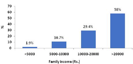
Fig 5 — Distribution of Patients by Family Income
Fig 6 — Distribution of cases by no of anti depressants used in generic name and from EML
Fig 7 — Mean no of anti depressants used in generic name and from EML per prescription
Fig 8 — Distribution of cases by class of drugs
Fig 9 — Distribution of cases by individual drug used in different classes
reduction in HDRS score after therapy (p<0.001).
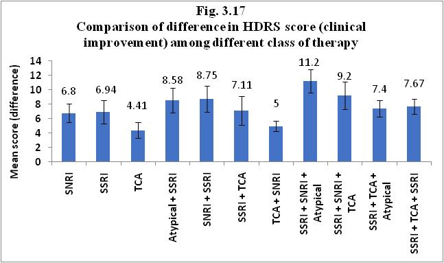
Comparison of difference in HDRS score (clinical improvement) among different class of therapies may be seen in Fig 13. ANOVA suggested mean reduction in HDRS score differed significantly among different therapy options used (p=0.000). Highest mean reduction (11.2±1.64) was observed in the three combination therapy “SSRI + SNRI + Atypical” followed by “SSRI + TCA + Atypical” (9.2±1.92). Two combination therapy like Atypical + SSRI, SNRI + SSRI, SSRI + TCA resulted in mean reduction of HDRS score in the range of 7.11±2.01 to 8.75±1.8. Single drug therapy with SNRI, SSRI and TCA had a mean reduction in HDRS score in the range of 4.41 to 6.94. The two combination therapy TCA + SNRI had a low mean reduction of 5±0.71.
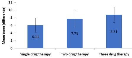
Comparison of single, two drug and three drug therapy is presented in Fig 14. Significant difference was found in the reduction of HDRS score in the therapy options used (p=0.000). The mean reduction in single drug therapy was 6.11±1.85 with a range of 2-10. Two drug therapy resulted in mean decrease of 7.73±2.14 in HDRS score with a range of 3-12. Three drug therapy resulted in a mean reduction of 8.81±2.04 with a range of 6-13. Two drug therapy was found to be significantly more effective than single drug therapy (p<0.05). Three drug therapy also caused greater reduction of HDRS (p=0.075). Use of greater number of anti-depressants
was generally consistent with a greater reduction in HDRS score however this also came with increased incidence of adverse effects. It was seen that milder cases of depression could be adequately treated with fewer drugs while the more severe or resistant cases may require use of multiple drugs from different classes. These findings need to be verified with further experiment.
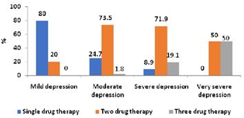
Comparison of therapy according to severity of illness is presented in Fig 15. It is observed that mild depression was commonly treated with single drug therapy (80%). Moderate depression was commonly treated with two drug therapy (73.5%). Severe depression cases had 71.9% two drug therapy and 19.1% three drug therapy with anti-depressants. This implied that as the severity of depression increases combination therapy is the preferred option (p=0.000).
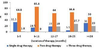
Duration of therapy :
Comparison of therapy according to duration is furnished in Fig 16. It was revealed that when the duration is 1-5 months single drug therapy was 33.3% and two drug therapy was 64.8%. When the duration becomes 6-11 months single drug therapy was 14.5% and two drug therapy was 85.4%. When the duration increased to 12-17 months single drug therapy was 18%, two drug therapy was 64% while three drug therapy was 18%. When the duration becomes 18-23
Fig 10 — Distribution of cases by duration of therapy
Fig 11 — Grading of patients by severity of presenting illness (HRDS)
Fig 12 — Comparison of HDRS score (before/after) by class of therapy
Fig 13 — Comparison of difference in HDRS score (clinical improvement) among different by class of therapy
months single drug therapy was 5.5%, two drug therapy was 50% and three drug therapy was 27.7%. This clearly indicated a higher trend of multidrug therapy with increased duration of therapy (p=0.000).
DU90 % :
DU90% presented in Table 1 presents the list of drugs in descending order of frequency and which cumulatively make up 90% of the prescriptions; these are Escitalopram which was used most times (36.99%) followed by Amitriptyline (14.63%), Mirtazapine (11.59%), Duloxetine (9.35%), Sertraline (8.74%) and Venlafaxine (8.54%). Table 1 — DU 90%
DISCUSSION
Usually, the term antidepressants refer to mainstream prescribed antidepressants; the qualitative and quantitative assessment of which in the Psychiatry department at Hi-Tech Medical College and Hospital, Bhubaneswar was the focus of this study.
Antidepressants were prescribed more in females than in males. This was consistent with the findings of other studies10,11. Antidepressants were prescribed more in women as they comprised a majority of the patients. This greater incidence of depression in women is the most consistent finding inepidemiology studies conducted on depression all over the world. In my study of the 262 patients (n=262) enrolled 67.2% were females. Similar result on gender distribution was found in a study on ‘Drug use pattern of antidepressant agents in psychiatric patients- a prospective study’ conducted in Ahmedabad15 where almost 60% of the patients were females. Similar findings of higher incidence of depression in females compared to males have been stated in community based epidemiological study according to National Institute of Mental Health (2007).
In this study it was seen that the greatest proportion of patients were in the age group 35-49 years and comprised 39.3% of the total cases. The age distribution in the retrospective drug utilization study of antidepressants in Pondicherry showed that the majority of patients who received the antidepressants belonged to the 21-40 years age group16, in contrast to the results of a study on antidepressant use in East Asia, wherein the mean age of the patients who received antidepressant prescriptions was more than 40 years17
Rational prescribing was followed as per the principles of prescription order writing12. Considering the definitions of polypharmacy which are most commonly cited, there was no polypharmacy, because there was no prescription of antidepressant medication which did not match the diagnosis and there was no prescription with more than 5 drugs13
One of the strengths of this study was the detailed analysis of socio-demographic profile of each of the patients enrolled. Few studies in India have taken such a close analytical look in this aspect.
The mainstay of this study was to analyse the current prescription pattern of antidepressants at our hospital. As per WHO prescribing indicators, we observed:
A total of 492 antidepressant drugs were prescribed to the 262 patients enrolled in this study. It was seen that the average number of antidepressants per prescription was 1.87. It must be mentioned here that records were kept only of the drugs strictly classified as antidepressants in the prescription. The average number of drugs per prescription which was 1.87
Fig 14 — Comparison of difference in HDRS score (clinical improvement) among different by class of therapy
Fig 15 — Comparison of therapy according to severity
Fig 16 — Comparison of therapy according to duration of therapy
119, No 8, August 2021Journal
reflected only the antidepressant medication used per prescription as we wanted to study their particular effect on efficacy caused to patient. A ‘Retrospective Drug Utilization Study of Antidepressants in the Psychiatry Unit of a Tertiary Care Hospital’ in Pondicherry showed that the average number of drugs per prescription according to their data was 2.3216. This was slightly higher in this study compared to our study as they also included other psychiatric medication (eg, sedative-hypnotics, antipsychotics) which was in some cases prescribed to the patients in addition to antidepressants. As per the inclusion and exclusion criteria of this study patients with co-morbid diagnosis like psychosis, bipolar disorder etc. were not included in the study; only the patients with a frank diagnosis of Major Depressive Disorder (MDD) and receiving antidepressant therapy were included in the study. Another study which was conducted on drug use patterns of antidepressant agents in psychiatric patients in Ahmedabad showed a still higher average number of drugs per prescription of 2.7215. Again comorbid psychiatric diagnosis and concomitant medication were a part of this study while our study focussed solely on depression and the use of antidepressants.
This study revealed that a total of 380 (77.24%) of the 492 antidepressants used were prescribed in their generic name. The retrospective study conducted in Pondicherry had shown that 88.54% of the antidepressants used in that study were prescribed in their generic name. Our results in this aspect was comparable to the above mentioned study.
In this study it was observed that Escitalopram from the SSRI class of antidepressants was the most highly used antidepressant 36.99%. Amitriptyline from the class TCA was second accounting for 14.63% of the antidepressants used. Mirtazapine from the Atypical class 11.59% and Duloxetine from the SNRI class 9.35% were also used in a significant number of patients.In accordance with this study many other studies have reported that selective serotonin reuptake inhibitors (SSRIs) account for the bulk of the prescribed antidepressants, with high prescribing rates.
Among the SSRIs, Escitalopram was the preferred drug. Again, this was in contrast to findings of the east Asian study on antidepressant use, wherein Fluoxetine and Sertraline were prescribed more frequently than Escitalopram and the use of Escitalopram was lower than that of Trazodone, Mirtazapine, Imipramine and Amitriptyline17
There is need for more research on this topic to improve efficacy of therapy and find ways to consolidate improvement and guard against relapse.
To conclude newer class of anti depressant drugs
are currently being used more in pharmacotherapy of depression. However older antidepressant class of drugs has also significant role to play in the treatment of this condition.
REFERENCES
1Wiley – Psychiatry 2nd edition; 1206.
2Kaplan and Sadock’s Comprehensive Textbook of Psychiatry; 1572.
3Indian Journal of Psychiatry; An overview of Indian Research in Depression; 2010 Jan; PubMed
4Indian Journal of Psychological Medicine; Depression: The Disorder and the Burden- M.S Reddy; 2010 Jan-Jun; PubMed
5Tognoni G, Laporte JR — From Clinical trials to Drug utilization studies. In: Dukes, M.N.G., ED. Drug utilization studies –Methods and uses. Denmark: WHO Regional publications, European Series 1993; 45: 28-30.
6Stolley PD, Lasagna L — Prescribing patterns of physicians. Journal of chronic diseases 1969; 22: 395-405.
7Rush J — Limitations in efficacy of antidepressant monotherapy. 2006.
8Avanthi E, Somashekar HS, Pradeep Kumar L, Sushma HK, Sudarshan CY, Raja B — Prescribing pattern of antidepressants in psychiatric unit of a tertiary care hospital; Shelton and Lester, 2006.
9Organisation for economic cooperation and development. OECD Policy brief – Mental health in OECD countries. Paris: OECD, 2008 Nov . Accessed February 7, 2011.
10Morabia A, Fabre J, Dunand JP — The influence of patient and physician gender on the prescription of psychotropic drugs. J Clin Epidemiol 1992; 45: 111-6.
11The ESEMeD/MHEDEA 2000 investigators. Psychotropic drug utilization in Europe: results from the European Study of the Epidemiology of Mental Disorders (ESEMeD) project. Acta Psychiatr Scand 2004; 109 (Suppl. 420): 55-64.
12Buxton ILO — Principles of Prescription Order Writing and Patient Compliance. In: Brunton LL, Lazo JS, Parker KL, editors. Goodman and Gilman’s The Pharmacological basis of Therapeutics. 11th edition. USA: McGraw Hill, 2006: 694.
13Morabia A, Fabre J, Dunand JP — The influence of patient and physician gender on the prescription of psychotropic drugs. J Clin Epidemiol 1992; 45: 111-6.
14The ESEMeD/MHEDEA 2000 investigators. Psychotropic drug utilization in Europe: results from the European Study of the Epidemiology of Mental Disorders (ESEMeD) project. Acta Psychiatr Scand 2004; 109 (Suppl. 420): 55-64.
15Drug use pattern of antidepressant agents in psychiatric patients – A prospective study AkshaMemon* , Kamlesh Patel** Resident Doctor* , Associate Professor**, Dept. of Pharmacology, Smt. N.H.L Medical College, Ahmedabad 16Kingshuk L, Shetty SM, Paramel A, Sharma G — A Retrospective Drug Utilization Study of Antidepressants in the Psychiatric Unit of a Tertiary Care Hospital in Pondicherry; 17Newer antidepressant drug use in East Asian psychiatric treatment settings: REAP (Research on East Asia Psychotropic Prescriptions) Study Kang Sim, N B Lee, Hong C Chua, Rathi Mahendran, Senta Fujii,1 Shu-yu Yang,2 Mian-Yoon Chong,2 Tianmei Si,3 Yan L He,4 Min S Lee,5 Kil M Sung,6 Eun K Chung,7 Yiong H Chan,8 Naotaka Shinfuku,1 Chay H Tan,8 Norman Sartorius,9 and Ross J Baldessarini 10.
18Gross F — Drug utilization therapy and practice. The present situation in the Federal Republic of Germany. Eur J Clin Pharmacol 1981; 19: 387-94.
19World Health Organisation. Introduction to drug utilisation research. Oslo: World Health Organisation, 2003.
20Jureidini J, Tonkin A — Overuse of antidepressant drugs for the treatment of depression. CNS Drugs: 2006: 20; 8: 623-32. 31
Original Article
Role of Laparoscopy in Management of Non-palpable testes : Our Experience
Ketan D Mehta1, Harshit
Rewari2
BBackground and Objective: In 2003 we have published our series on the same subject. The subject is revisited again in present study. We would like to share our experience and changes which have taken place in these 15 years with literature support.
Patients and Methods: Between March 2017 and April 2018, 26 patients with 31 non-palpable testes underwent laparoscopy. Based on the intraoperative findings they were divided into absent, intra-abdominal or inguinal/scrotal testes. In cases of absent testes, the procedure was terminated. In cases of Intra-abdominal testes laparoscopic orchidopexy or orchidectomy were performed and in cases of inguinal/scrotal testes, inguinal canal were explored by small incision and laparoscopy assisted orchidopexy or orchidectomy were carried out.
Results: 4 testes (12.90%) were absent. Extensive inguinal exploration and/or laparotomy were avoided in these cases. 19 testes (61.30%) were intra-abdominal. Out of these, 4 testes (12.90%) were atrophic and were removed laparoscopically. The remaining 15 (48.40%) underwent single stage laparoscopic orchidopexy. 8 testes (25.90%) were found in the inguinal/scrotal region. 3 (9.67%) were removed and orchidopexy performed in remaining 5(16.31%). All the laparoscopic procedure concluded successfully, especially orchidopexy, which were tension free.
Conclusion: In management of non-palpable testes, laparoscopy is an excellent dual purpose diagnostic and therapeutic tool. Diagnostically, it can replace all imaging study. Therapeutically, it helps to do tension free orchidopexy or orchidectomy without large incision.
The demand for doing laparoscopy from patient’s parents/guardian can be an important factor in selecting its use in management of non-palpable testis.
[J Indian Med Assoc 2021; 119(8): 32-6]
Key words :Laparoscopy, non-palpable testis, Orchidopexy, Orchidectomy.
Undescended testis or Cryptorchidism, is one of the most common pediatric disorder of the male endocrinal gland and the most common genital disorder found at the birth1. Its incidence varies from 1% to 4.6% in full-term baby and in preterm neonates its incidence may be as high as 45%2. Correction of cryptorchidism is required to optimize testicular function, potentially reduce and/or facilitate diagnosis of testicular malignancy, provide cosmetic benefits and prevent complication such as clinical hernia or torsion3
Out of all Undescended testes, nearly 20-25% are non-palpable4-6. Various reasons for failure to locate the Non-Palpable Testis (NPT) could be due to vanishing testis syndrome, intra-abdominal position, examination obscured due to obesity or scar tissue, and rarely due to testicular agenesis7.
NPT pose diagnostic and therapeutic challenge to the surgeon. Its presence or absence must be
Department of Surgery, M P Shah Medical College, Jamnagar, Gujarat 361008
1Department of Surgery, M P Shah Medical College, Jamnagar, Gujarat 361008
2MS (General Surgery), Ex Resident
Received on : 21/07/2021
Accepted on : 12/08/2021
Editor's Comment :
Laparoscopy is very useful & effective method in management of NPT & can replace all other diagnostic & therapeutic modalities.
established and appropriate therapy should be applied to either make it palpable or to remove it. Laparoscopy is the tool which can be used to locate and treat this disease.
First diagnostic laparoscopy to locate NPT was performed by Cortesi et al8 in 1976 and first laparoscopic single stage orchidopexy was performed by Jordan et al9 in 1991. With dissemination of use of laparoscopy in treating other diseases and refinement in technique and instrumentation, laparoscopy is considered as preferred method to locate and treat NPT. In the present study we have tried to evaluate its role in NPT.
MATERIALS AND METHODS
This study was carried out between March 2017 and April 2018 in a single surgical unit of 1200 bedded state government funded multispecialty teaching hospital. Before starting this study approval was
119, No 8, August 2021Journal
obtained from the Institutional Ethics Committee. The study was carried out on 26 boys with 31 NPT. Parents/guardian of the patients was explained regarding nature of disease and application of laparoscopy procedure in the disease and written informed consent obtained.
All the procedures were performed under general anesthesia. Abdomen was insufflated with carbon dioxide through veress needle and pressure kept at 810 mm Hg. We use 5mm zero degree telescope which was passed through the port placed at superior edge of umbilicus. Two additional 3mm/5mm working ports were put in mid-clavicular line on both sides at the level of umbilicus. Patient was given trendelenburg position to deflect bowel upward from lower abdominal cavity. Then both the inguinal region, pelvic cavity and paracolic gutter were visualized and the status of spermatic vessels, vas deferens, internal inguinal ring and testes were noted. Based on these findings the testes were classified into 3 groups:
Group 1 : Absent or vanishing testes
Here the testes were not visualized. Spermatic vessels were ending blindly inside the abdomen with closed internal inguinal ring (Fig 1). In these cases the procedure were terminated.
Group 2 : Intra-abdominal testes
In this group of patients, the testes were found lying within the abdominal cavity (Fig 2). ‘Peeping testes’, where part of testis was projected in abdominal cavity through internal inguinal ring, were also included in this group. Here the decision to perform orchidopexy or orchidectomy was taken based on the age of the patient, size of the testis and whether the condition was unilateral or bilateral. Laparoscopic orchidectomy was accomplished by ligating the spermatic vessels and vas deferens and removal of testis through one of the working port.
Single stage laparoscopic orchidopexy was carried out by first mobilizing the testicular vessels and vas deferens by incising the peritoneum covering them. The testis was freed up by dividing gubernaculum from its distal attachment. Extra care was taken to prevent injury to looping of vas around gubernaculum. The adequacy of mobilization of the testis was tested intraabdominally by ensuring that it reached the opposite internal inguinal ring. Then a scroto-peritoneal port was created as follows:
A grasper was passed from the internal inguinal ring and made to traverse through inguinal canal into the upper part of the scrotum, and its tip was palpated. A small transverse skin incision was made over the tip of the grasper and then the tip of the grasper was pushed and made to emerge through the incision. A cannula was then passed in a retrograde manner over this grasper into the abdominal cavity under direct vision. This scroto-peritoneal port has one end lying outside and other end projecting inside peritoneal cavity through the internal inguinal ring. A grasper was next introduced into the peritoneal cavity from the scrotal side of the port. The dissected testis was grasped from its bottom and drawn into the scrotum by pulling grasper out along with cannula. The testis was then put in subdartos pouch and anchored there with two intermittent 3-0 polyglactin 910 stitches. The internal ring was closed from inside with 1 or 2 stitches of same suture material.
Group
3 : Inguinal/Scrotal testes
In this group of patients, the spermatic vessels and vas deferens were seen entering the internal inguinal ring (Fig 3). This finding indicates inguinal testis or scrotal nubbin. Here the inguinal canal was explored by a small groin incision. Laparoscopic assisted Orchidopexy or orchidectomy was done based on the same criteria applied for intra-abdominal testis.
Fig 1 — Absent Testis. Blindly ended vas (A) and spermatic vessels (B)
Fig 2 — Intraabdominal Testis
RESULTS
The study was carried out on 26 boys (age group 9 month to 12 year) with 31 NPT. 21 boys had unilateral (11 left-sided, 10 right-sided) and 5 had bilateral NPT. Out of 31 NPT 4 testes (12.90%) were absent, 19 testes (61.30%) were intra-abdominal and 8 testes (25.80%) were inguinal/scrotal.
In cases of absent testis, as the procedure was terminated, these patients were spared from extensive inguinal exploration and/or laparotomy.
In case of 19 intra-abdominal testes, 4 (12.90%) had atrophic or dysmorphic testes which underwent laparoscopic orchidectomy and 15 (48.40%) had healthy looking testes which underwent laparoscopic orchidopexy. All the procedures were concluded successfully, without any undue tension over the cord structures and testes. Laparoscopic procedure allowed us the advantage of wide mobilization of cord structures under a magnified view.
8 NPT (25.90%) were found in the inguinal canal/ scrotum. 3 (1 inguinal and 2 scrotal) atrophic testes (9.67%) were removed and laparoscopic assisted orchidopexy performed in remaining 5 (16.31%). Use of laparoscopic in mobilization of spermatic vessels and vas has allowed us to do tension free orchidopexy.
There were no mortality and all patients were discharged on the second day of operation.
DISCUSSION
The two important issue involved in overall management of NPT are locating the testis and its surgical correction.
Locating NPT : Various imaging modalities and laparoscopy are used to locate NPT.
Ultrasonography (US) remains the most commonly used radiological investigation to locate NPT. Advantages of US include easy availability, non-invasive
nature, no risk of radiation exposure and no need of anesthesia in young children. US found to have a sensitivity of 45-88% and specificity of 78-100% in the diagnosis of NPT in various study10,11,12. The wide variation in accuracy of US is likely due to its operator dependency and obstruction of view of intra abdominal testes by intestinal gas. And that is its main drawback. In recent years routine use of US in evaluation of NPT is questioned. US not only drain resources, but its findings do not obviate the need of surgical exploration. Tasian GE and Copp HL in recent metaanalysis conclude that eliminating the use of ultrasound will not change management of NPT but will decrease health care expenditures10. Pekkafali et al stated that US does not exclude the necessity for laparoscopy, and it is not superior to physical examination in detection of the inguinal atrophic testes or testicular nubbin13. Elder JS found US to be unnecessary in boys with a NPT, because it rarely if ever localizes a true NPT, and it does not alter the surgical approach in these patients14. Shah and Shah showed the overall diagnostic agreement of US with laparoscopy in only 19% of cases15
CT scan may be useful in documenting the location of the NPT but it is expensive, exposes child to radiation and, sometimes is difficult to perform in a young child. Wolverson et al reported that sensitivity of CT and USG are same in the evaluation of NPT12
MRI may be more helpful in locating NPT than CT, but like CT scan it may be difficult to perform in a young child. It is very expensive and is not widely available.
European Association of Urology and European Society for Pediatric Urology guidelines16 state that physical examination is the only way of differentiating between palpable or non-palpable testes. There is no benefit in performing ultrasound, computed tomography (CT), magnetic resonance imaging (MRI) or angiography.
It seems that there is paradigm shift taking place in recent time regarding common use of imaging modality to its avoidance in locating NPT. And this seems to be happening due to widespread use of laparoscopy in management of NPT.
Laparoscopy is a powerful diagnostic tool and it can replace all imaging modality for locating NPT. The real diagnostic superiority that exists with laparoscopy for NPT is its ability to accurately characterize the location and quality of testicle, testicular vessels and vas deference. This is a valuable asset that laparoscopy brings to the diagnosis of the NPT17
Surgical correction : It includes orchidopexy or
Fig 3 — Inguinal/Scrotal Testis. Vas and spermatic vessels are seen entering the internal inguinal ring
119, No 8, August 2021Journal
orchidectomy. There are two way to accomplish it; by open surgery and laparoscopy.
The traditional open surgery involves an exploration of the inguinal canal by inguinal incision followed by retroperitoneal or peritoneal exploration, if no testis or cord structures could be found in the inguinal canal. This approach often involves extensive dissection and bigger incision.
Therapeutic use of laparoscopy allowed a minimally invasive approach for correcting NPT. Laparoscopic findings helps surgeon to precisely define further operative plan.
In patients where blind ending cord structures are visualized with closed inguinal ring, no further action is required. The most likely cause of absent testis is prenatal or perinatal vascular accident.
Intra-abdominal testis can be managed by either laparoscopic orchidopexy or orchiectomy, though the decision regarding salvage or removal of a testis is a difficult one. A small hypoplastic testis, a testis with significant ductal system abnormality or unilateral abdominal cryptorchidism in a post pubertal patient is a poor candidate for salvage.
Most intra abdominal testes are usually found lying close to internal inguinal ring. They can be mobilized with laparoscopic dissection of the spermatic vessels and vas deferens and delivered to a scrotal position without dividing the spermatic vessels as a single-stage procedure. High intra-abdominal testis can be managed either by laparoscopy assisted one or two-stage Fowler Stephen orchidopexy or laparoscopy-assisted testicular microsurgical auto transplantation. The choice of procedure depends on individual preference and availability of expertise.
If spermatic vessels and vas traverse the internal inguinal ring, there may be blind ending cord structures in the canal, a hypoplastic or dysplastic testis in the canal, a normal sized testis in canal that was not palpable preoperatively because of obesity, operative scar etc. or testicular nubbin in scrotum. It is probable that some of these inguinal gonads that were non palpable preoperatively may have indeed been intraabdominal. However with insufflation and increased intra-abdominal pressure, they may have been forced through an open internal inguinal ring into the inguinal canal. These situation warrants inguinal exploration which can be expeditiously performed through a small inguinal incision. When orchidopexy is required, laparoscopy can help surgeon by intraabdominal dissection of spermatic vessels and vas (laparoscopy assisted orchidopexy), thus avoiding bigger incision. This is particularly important in
performing tension free orchidopexy in older children in which inguinal canal is little longer than the younger ones.
Inguinal exploration or laparoscopy ?
Choice between inguinal exploration and laparoscopy as an initial surgical approach has been a matter of debate with proponents of each claiming the superiority of one over the other. However, laparoscopy has emerged as the modality of choice and is currently regarded as the gold standard for the diagnosis of NPT18,19,20. The morbidity of an abdominal exploration in the era of laparoscopy is seen as unacceptable from standpoints of both recovery and cosmetic outcome by many surgeons17. Numerous groups have reported cases of missed intra-abdominal testes on inguinal exploration alone21. Laparoscopy has the advantage of 1) high magnification and improved visualization 2) capability of extensive vascular dissection up to the origin of gonadal vessels, 3) minimal morbidity, and 4) the ability of creating a new internal ring medial to inferior epigastric vessels to achieve the straight vascular course to the scrotum19. Lorenzo et al21 has found cost saving advantage of initial laparoscopic evaluation of clinically NPT over initial inguinal-scrotal exploration.
One interesting thing emerged in our present study is demand from patient’s parents/guardian for doing laparoscopy. Out of 26 patient’s parents/gurdian, 15 (57.69%) had good knowledge of use of laparoscopy in the disease and were came to us for laparoscopy management only. This is a significant change occurred since we published our first series23 on the same subject in 2003. We believe that this can be one of the major driving forces in future, in selecting laparoscopy for treating this disease.
CONCLUSION
In our experience, we found laparoscopy to be an excellent diagnostic and therapeutic tool in management of NPT. From diagnostic point of view it is very reliable and accurate in locating NPT. It gives a clear picture of anatomy of spermatic vessels, vas, internal inguinal ring and testis. It has potential to replace all diagnostic imaging modality. Therapeutically, it is very safe and effective tool. Tension free orchidopexy or orchidectomy can be carried out without large scar.
As new generation of surgeons and people are more inclined towards minimal access surgery and profound benefits offered by laparoscopy, we are not far away from the era where laparoscopy will be considered as the single and preferred, diagnostic and therapeutic
Vol 119, No 8, August 2021Journal of the Indian Medical Association
2Radmayr C, Dogan HS, Hoebeke P, Kocvara R, Nijman R; Stein R, et al — Management of undescended testes: European Association of Urology/European Society for Paediatric Urology Guidelines. J Pediatr Urol 2016; 12(6): 335-43.
3Julia Spencer Barthold. Abnormality of the testis and scrotum and their surgical management. In Alan J. Wein editor. Campbell- Walsh Urology. 10th ed. Philadelphia; Elsevier Saunders; 2012. pp. 3557-96
4Baker LA, Docimo SG, Surer I, Peters C, Cisek L, Diamond DA, et al — A multi institutional analysis of laparoscopic orchidopexy. BJU International 2002; 87(6): 484-9.
5Kirsch AJ, Escala J, Duckett JW, Smith GH, Zderic SA, Canning DA, et al — Surgical management of the nonpalpable testis: The Children’s Hospital of Philadelphia experience. J Urol 1998; 159(4): 1340-3.
6Craig AP, Louis RK — Laparoscopy in children and adults. In: Patrick CW, Alan BR, Duracott E, Allan JW, editors. Campbell’s urology. 7th edn. Philadelphia; WB Saunders company; 1998. pp. 2875-911
7Van Savage JG — Avoidance of inguinal incision in laparoscopically confirmed vanishing testis syndrome. J Urol 2001; 166(4): 1421-4.
8Cortesi N, Ferrari P, Zambarda E, Manenti A, Baldini A, Morano FP — Diagnosis of bilateral abdominal cryptorchidism by laparoscopy. Endoscopy 1976; 8(1): 33-4.
9Jordan GH, Winslow BH — Laparoscopic single stage and staged orchiopexy. J Urol 1994; 152(4): 1249-52.
10Tasian GE, Copp HL — Diagnostic performance of ultrasound in nonpalpable cryptorchidism: a systematic review and metaanalysis. Pediatrics 2011; 127(1): 119-28.
11Kanemoto K, Hayashi Y, Kojima Y, Maruyama T, Ito M, Kohri K — Accuracy of ultrasonography and magnetic resonance imaging in the diagnosis of nonpalpable testis. Int J Urol 2005; 12(7): 668-72.
12Wolverson MK, Houttuin E, Heiberg E, Sundaram M, Shields JB — Comparison of computed tomography with highresolution real-time ultrasound in the localisation of the impalpable undescended testis. Radiology 1983; 146(1): 1336.
13Pekkafali MZ, Sahin C, Ilbey YO, Albayrak S, Yildirim S, Basekim CC — Comparison of ultrasonographic and Laparoscopic Findings in Adult NonpalpableTestes Cases. Eur Urol 2003; 44(1): 124-7.
14Elder JS — Ultrasonography is unnecessary in evaluating boys with a nonpalpable testis. Pediatrics 2002; 110(4): 74851.
15Shah A, Shah A — Impalpable testes–is imaging really helpful? Indian Pediatr 2006; 43(8): 720-3.
16Tekgul S, Dogan HS, Erdem E, Hoebeke P, Kocvara R, Nijman JM, et al — Guidelines on Paediatric Urology. European Association of Urology Guidelines 2015 Mar: 9-11.
17Gatti JM, Ostlie DJ — The use of laparoscopy in the management of nonpalpable undescended testes. Curr Opin Pediatr 2007; 19(3): 349-53.
18Pradhan MR, Ansari MS — Imaging studies for non-palpable testis: Are they at all required? : Indian J Urol 2012; 28(2): 227-9.
19Erdogan C, Bahadýr B, Taskýnlar H, Naycý A — Laparoscopic management and its outcomes in cases with nonpalpable testis. Turk J Urol 2017; 43(2): 196-201.
20Shahid SA — Diagnostic and therapeutic Management Of Impalpable testis in the Era of Laparoscopy. World Journal of Laparoscopic Surgery 2010; 3(2): 75-9.
21Lorenzo AJ, Samuelson ML, Docimo SG, Baker LA, Lotan Y — Cost analysis of laparoscopic versus open orchiopexy in the management of unilateral nonpalpable testicles. J Urol 2004; 172(2): 712-6.
22Kwanjin Park and Hwang Choi. An Evolution of Orchiopexy: Historical Aspect. Korean J Urol 2010; 51(3): 155-60.
Chronic liver diseases can occur due to various etiologies and each type is associated with specific cardiovascular manifestations. Hyperdynamic syndrome and cirrhotic cardiomyopathy are commonly associated with liver cirrhosis. These are manifested by systolic, diastolic and electrophysiological changes in the heart. However recent studies have revealed cardiovascular abnormalities in NAFLD, Chronic hepatitis C infection, Primary Biliary Cirrhosis and Hepatocellular Carcinoma. Recipients of orthotopic liver transplantation have also reported cardiac dysfunction which necessitates a proper evaluation of their cardiac status.This review discusses cardiac complications in chronic liver diseases, bringing in light over the pathophysiologic and clinical implications.
Ancient Germans had termed the liver as “lifere” (meaning life), underscoring the relation of this organ to heath and existence. The importance of liver lie in its functional diversity.It is related to other organs in health and disease. A systemic disease can involve both liver and heart. However a cardiac disease can affect the liver and also vice versa can happen. Each category of these mutually manifest diseased conditions have their own clinical implications therapeutic strategies. Cardiac complications in a patient with chronic liver disease have brought in increased mortality and morbidity1. Hepatic dysfunction can lead to various haematological, metabolic and multiple other systemic anomalies which in turn can cause cardiac dysfunction, even in the absence of previous cardiac anomalies. But specific cardiovascular pathology in these patients is now well established. Chronic Liver Disease (CLD)comprises of hepatic injury, inflammation and/ or fibrosis occurring in the liver for more than 6 months2. The etiology of CLD is diverse and each of them is now acknowledged to be associated with specific cardiac manifestations. A vasodilatatorystate, hyperdynamic circulation, cirrhotic cardiomyopathy and electrophysiological abnormalities in heart are common and characteristic cardiovascular manifestations of cirrhosis of liver3. Hepatopulmonary syndrome and porto-pulmonary hypertension are other elements of cirrhosis of liver. Non Alcoholic Fatty Liver Disease (NAFLD), due to its association with other metabolic disorders such as diabetes, dyslipidemia,
Department of Medicine, Assam Medical College & Hospital, Dibrugarh, Assam 786002
1MBBS, MD, Senior Resident and Corresponding Author
2MD, Professor
Received on : 05/01/2021
Accepted on : 10/02/2021
Editor's Comment :
Chronic liver diseases have profound effects on the cardiovascular system. This substantiates the importance of an elaborate cardiovascular evaluation in these patients. Newer therapeutic measures to reverse these changes can increase the longetivity and improve the quality of life of these patients.
hypertension and obesity is a known risk factor for cardiovascular disease. Moreover, cirrhotics are prone to especially nocturnal hypoglycaemia for a variety of reasons (poor appetite, low hepatic glycogen reserve) that can lead to sudden cardiac death too. Most recent studies have shown echocardiographic features of early LV diastolic dysfunction and impaired LV energy metabolism even in patients of NAFLD without ailments like hypertension or diabetes mellitus. Infections like chronic Hepatitis C can lead to chronic inflammation of the myocardium which can later cause dilated cardiomyopathy. Moreover, approximately 30% of liver transplant recipients have a CVD complication (myocardial infarction, heart failure, cardiac arrest, atrial fibrillation, pulmonary embolism, or stroke) within 1 year of Liver Transplantation which is a major cause of mortality5,6. It also causes perioperative mortality and graft loss even in donor liver transplant recipients.7 For these reasons, research is being focussed on these pathologies, to study the potential reversibility of these cardiovascular alterations, early diagnosis and effective treatment.
Cirrhosis of Liver :
Cirrhosis of liver leads to Hyperdynamic Syndrome which is a major cardiovascular manifestation. There is high heart rate and cardiac output on one hand ,and reduced systemic vascular resistance and arterial blood pressure on the other hand which result in this
No 8, August
syndrome. Apart from this, these patients have cardiac contractile impairment to stress and exhibit diastolic dysfunction. In addition, they have ECG abnormalities in the absence of known cardiac disease8,9. It has been termed as Cirrhotic cardiomyopathy. Some studies have suggested the Association of Cardiac Dysfunction with Hepatorenal Syndrome 10,11. QT prolongation is the commonest ECG abnormality seen in about 50% of the cirrhotic patients12
Hyperdynamic Syndrome :
Kowalski and Abelmann were the first to describe this syndrome about 60 years back13. Increased heart rate and cardiac output and reduced systemic vascular resistance , as mentioned above are the chief features of this syndrome. The resultant hypotension leads to peripheral and splanchnic vasodilatation and ultimately portal hypertension in cirrhosis. However, sepsis and hypovolemia are common etiologies of new onset or worsening hypotension in these patients and thus should be promptly recognized and treated. In this situation, a mean arterial blood pressure of 60-65 mm Hg will ensure organ perfusion.Important clinical signs owing to systemic vasodilatation are palmar erythema, bounding pulse, reddish skin etc14
Pathogenesis :
Chemicals such as Nitric Oxide (NO) is depleted in intrahepatic circulation while over produced in splanchnic circulation. This leads to splanchnic vasodilation. There is an imbalance between mediators of vasodilatation like adrenomedullin, cytokines, endothelins, natriuretic peptides and vasoconstriction like angiotensinII in peripheral circulation. Vasoconstrictors are predominantly present in hepatic microcirculation. This ultimately results in resistance in hepatic circulation and peripheral vasodilatation including splanchnic vasodilatation 16. Moreover, cirrhotics are prone to infections. Endotoxins and cytokines such as tumor necrosis factor-á further lead to increased NO production17. Endocannabinoid are lipid like substances and are also stimulated by bacterial endotoxins. Apart from these chemical mediators, renin angiotensin system plays a role in mesenteric vasodilatation. Here angiotensin 17activate the G-protein coupled Mas receptor (MasR) resulting in vascular hypocontractility and hence might serve as a potential therapeutic target for portal hypertension18. Also, studies have found a marker protein in brain (CNS Fos), which has been found to be associated with hyperdynamic circulation in rat models19 The reduction in peripheral resistance is compensated by increased cardiac output which leads to hyperdynamic syndrome. The evolution of cardiomyopathy in cirrhosis is a complex interaction
among cellular, neuronal and humoral signaling pathways in the systemic scenario rather than happening locally in the heart.
Cirrhotic Cardiomyopathy :
Cirrhotic cardiomyopathy was defined by World Congress of Gastroenterology as “cardiac dysfunction in patients with cirrhosis characterized by impaired contractile responsiveness to stress, diastolic dysfunction and electrophysiological abnormalities in the absence of known cardiac disease” 20 . The Diagnosticcriteria were
(a)Systolic dysfunction:
•Blunted increase in cardiac output with exercise, volume challenge or pharmacological stimuli
•Resting Ejection Fraction < 55%
(b)Diastolic dysfunction:
•E/A ratio (early diastolic/atrial filling) < 1.0 (agecorrected)
•Prolonged deceleration time (> 200 ms)
•Prolonged isovolumetric relaxation time (> 80 ms)
(c)Supportive criteria :
•Electrophysiological abnormalities
•Abnormal chronotropic response
•Electromechanical uncoupling/dyssynchrony
•Prolonged QTc interval
•Enlarged left atrium
•Increased myocardial mass
•Increased Brain Natriuretic Peptide (BNP) and pro-BNP
•Increased troponin level
The classical features of even established cardiomyopathy may be difficult to recognized, unless precipitated by stress factors.
Systolic Dysfunction :
Inability of the heart to generate adequate blood pressure and cardiac output signifies systolic dysfunction. Cirrhotics have a normal Left Ventricular Ejection Fraction (LVEF) at rest. However systolic dysfunction occurs with exercise21,22. This dysfunction gets unmasked due a blunted heart rate response to stress, reduced myocardial reserve and impaired ability to extract oxygen by the cardiac muscles. Moreover ,administration of vasoconstrictors like terlipressin increases the Systemic Vascular Resistance and thereby the left ventricular afterload unveiling a latent left ventricular dysfunction in cirrhosis. Hence these agents should be cautiously used.
Diastolic Dysfunction :
Increased stiffness of the myocardial wall owing to myocardial hypertrophy, fibrosis, and subendothelialedema in cirrhotics leads to diastolic dysfunction. It results in complications and impairs the outcomes of 38
119, No 8, August 2021Journal
manoeuvres such as Transjugular Intrahepatic Portosystemic Shunt (TIPS) insertion which could lead to an increased preload20
Electrophysiological Abnormalities :
QT interval prolongation, chronotropic incompetence and electromechanical uncoupling are common in advanced cirrhosis. QT prolongation has a prevalence of over 60% in these patients. Hence drugs prolonging QT should be avoided12
Almost all cardiovascular abnormalities have been found to reverse after a few months of liver transplantation14,20. The changes can be summed up in Fig 1.
Primary Biliary Cirrhosis :
Hypercholesterolemia occurs in most patients of primary biliary cirrhosis (PBC) which is a cardiovascular risk factor. Ursodeoxycholic acid lowers the circulating levels of cholesterol, thereby decreasing cholestasis. Besides hypercholesterolemia , autonomic dysfunction has been seen in PBC which can also affect the heart and its functions23
Hepatopulmonary Syndrome (HPS) :
Advanced liver disease causes increased levels of nitric oxide and vascular endothelial growth factor leading to intrapulmonary vascular dilation. This causes an oxygenation defect24 . These patients mostly remain asymptomatic during the early stages. However later develop dyspnea. Dyspnea upon standing (platypnea) is an important feature of HPS which is seen in 25% of these patients apart from digital clubbing and cyanosis. Though no established medical therapy has been formulated , it is advisable to prevent chronic hypoxemia by administration of supplemental oxygen. Definitive treatment is liver transplantation.
Non Alcoholic Fatty Liver Disease (NAFLD) :
Main cause of mortality in patients of NAFLD is cardiac complications. NAFLD and NASH are characterised by increased inflammatory biomarkers which may explain the increased association of coronary artery disease in these patients. Dyslipidemia in NAFLD manifested by increased serum triglyceride and low-density lipoprotein cholesterol levels and decreased high-density lipoprotein cholesterol levels, are all key risk factors for cardiovascular disease (CVD)25. Moreover CVD risk factors, such as metabolic syndrome, a high Framingham risk score, carotid intima media thickness, and high-sensitivity C-reactive protein (hsCRP) level as well as accelerated atherosclerosis, endothelial dysfunction of the pericardial fat pads, and coronary artery calcification, are significantly elevated in NAFLD patients. Asymptomatic obese children with NAFLD have been
Fig 1 — Proposal of changes in cardiac output during the course of the liver disease
shown to have features of early LV diastolic and systolic dysfunction which is more severe in those with Non Alcoholic Steato-Hepatitis (NASH)26. Management includes therapeutic lifestyle changes and pharmacotherapy (Statins, fibrates, ezetimibe, omega 3 fatty acids and insulin sensitizing agents).
Chronic Hepatitis C Infection :
Hepatitis C is an infection which causes increased activation of the immune system. It causes chronic inflammation of the myocardium leading to necrosis and loss of myocytes. This later leads to dilated cardiomyopathy. Sometimes it may lead to hypertrophic cardiomyopathy due to virus induced myocyte hypertrophy27. Apart from these ,congestive heart failure and presence of metabolic conditions such as type 2 diabetes mellitus and hypertension have also been reported28
Hepatocellular Carcinoma :
A significant proportion of cirrhosis complication end up in primary liver cancer, especially in Hepatitis B related cirrhotics. Involvement of the heart in Hepatocellular Carcinoma (HCC) is rare. However right atrial invasion in few cases of HCC have been reported.29
Liver Transplantation :
The fact that cardiac manifestations subside or reverse after transplantation of liver shows the association of liver dysfunction with cardiac abnormalities. These features reverse after 6 to 12 months of the procedure. However the immediate post operative period is of importance as it may lead to aggravation of complications. Arrhythmia, acute heart
Vol 119, No 8, August 2021Journal
failure (HF), and myocardial infarctioncan also occur due to reperfusion injury and hence one should be cautious during this period30. However, cardiac cirrhosis improves after heart transplant.
CONCLUSION
Chronic Liver Diseases can affect the heart and blood circulation in many ways. Most commonly LC leads to hyperdynamic syndrome and cirrhotic cardiomyopathy which in turn may facilitate the development of several complications like hepatorenal syndrome and myocyte loss. Liver transplantation causes reversal of almost all cardiovascular abnormalities. However this procedure itself can lead to cardiac death due to causes which have been stated above. Moreover recent studies have shown cardiac complications in patients with NAFLD, hepatitis C, primary biliary cirrhosis as well as hepatocellular carcinoma. Hence a rigorous assessment of cardiac status should be sought for in these patients. Further studies are required to illustrate the pathogenesis and identify therapeutic targets to reverse these changes, as well as to gain insights into the contribution of cardiac dysfunction in the natural history of cirrhosis of liver.
REFERENCES
1Fleming KM, Aithal GP, Card TR, West J — All-cause mortality in people with cirrhosis compared with the general population: a population-based cohort study. Liver Int 2012; 32(1): 79-84.
2Anstee QM, Jones DEJ — Hepatology. In: Ralston HS, Penman ID, Strachan MWJ, Hobson RP. Davidson’s Principles and Practice of Medicine. 23rd Ed. Elsevier; 2018. p856
3Møller S, Henriksen JH — Cardiopulmonary complications in chronic liver disease. World journal of gastroenterology: WJG 2006; 12(4): 526.
4Targher G, Day CP, Bonora E — Risk of cardiovascular disease in patients with nonalcoholic fatty liver disease. New England Journal of Medicine 2010; 363(14): 1341-50.
5Albeldawi M, Aggarwal A, Madhwal S, Cywinski J, Lopez R, Eghtesad B, et al — Cumulative risk of cardiovascular events after orthotopic liver transplantation. Liver Transpl 2012; 18: 370-5.
6Fussner LA, Heimbach JK, Fan C, Dierkhising R, Coss E, Leise MD, et al — Cardiovascular disease after liver transplantation: when, what, and who is at risk. Liver Transpl 2015; 21: 889-96.
7Raval Z, Harinstein ME, Skaro AI, Erdogan A, DeWolf AM, Shah SJ, et al — Cardiovascular risk assessment of the liver transplant candidate.Journal of the American College of Cardiology 2011; 58(3): 223-31.
8Caramelo C, Fernandez-Munoz D, Santos JC — Effect of volume expansion on hemodynamics, capillary permeability and renal function in conscious, cirrhotic rats. Hepatology 1986; 6: 129-34.
10Krag A, Bendtsen F, Burroughs AK, Moller S — The cardiorenal link in advanced cirrhosis. Med Hypotheses 2012; 79: 53-5.
11Ruiz-del-Arbol L, Monescillo A, Arocena C — Circulatory function and hepatorenal syndrome in cirrhosis. Hepatology 2005; 42: 439-47.
12BernardiM,Maggioli C, Dibra V, Zaccherini G — QT interval prolongation in liver cirrhosis: innocent bystander or serious threat? Expert Rev Gastroenterol Hepatol 2012; 6: 57-66.
13Kowalski H, Abelmann WH. The cardiac output at rest in Laennec’s cirrhosis. J Clin Invest 1953; 32: 1025-33.
14Moller S, Henriksen JH — Cardiovascular complications of cirrhosis. Gut 2008; 57: 268-78.
15Moller S, Krag A, Bendtsen F — Kidney injury in cirrhosis: pathophysiological and therapeutic aspects of hepatorenal syndromes. Liver Int 2014; 34: 1153-63.
16Al-Hamoudi WK — Cardiovascular changes in cirrhosis: pathogenesis and clinical implications. Saudi J Gastroenterol 2010; 16: 145-53.
18Dharmani M, Mustafa MR, Achike FI, Sim MK — Effects of angiotensin 1-7 on the actions of angiotensin II in the renal and mesenteric vasculature of hypertensive and streptozotocininduced diabetic rats. Eur J Pharmacol 2007; 561: 144-50.
19Liu H, Schuelert N, McDougall JJ, Lee SS — Central neural activation of hyperdynamic circulation in portal hypertensive rats depends on vagal afferent nerves. Gut 2008; 57: 966-73.
20Møller S, Bernardi M — Interactions of the heart and the liver. Eur Heart J 2013; 12: 2804-11
21Kelbaek H, Rabol A, Brynjolf I — Haemodynamic response to exercise in patients with alcoholic liver cirrhosis. Clin Physiol 1987; 7: 35-41.
22Wong F, Liu P, Lilly L, Bomzon A, Blendis L — Role of cardiac structural and functional abnormalities in the pathogenesis of hyperdynamic circulation and renal sodium retention in cirrhosis. Clin Sci (Lond) 1999; 97: 259-67.
23Schwartz PJ — The autonomic nervous system and sudden death. Eur Heart J 1998; 19 Suppl F: F72-F80
25Zhang QQ, Lu LG — Nonalcoholic fatty liver disease: dyslipidemia, risk for cardiovascular complications, and treatment strategy. Journal of clinical and translational hepatology 2015; 3(1): 78.
26Pacifico L, Di Martino M, De Merulis A, Bezzi M, Osborn JF, Catalano C, et al — Left ventricular dysfunction in obese . Left ventricular dysfunction in obese children and adolescents with nonalcoholic fatty liver disease. Hepatology 2013; Epub ahead of print [PMID: 23843206 DOI: 10.1002/hep.26610]
27Omura T, Yoshiyama M, Hayashi T, Nishiguchi S, Kaito M, Horiike S, et al — Core protein of hepatitis C virus induces cardiomyopathy. Circ Res 2005; 96: 148-50 [PMID: 15618537 DOI: 10.1161/01. RES.0000154263.70223.13]
28Younossi ZM, Stepanova M, Nader F, Younossi Z, Elsheikh E — Associations of chronic hepatitis C with metabolic and cardiac outcomes. Aliment Pharmacol Ther 2013; 37: 647-52.
29Agelopoulou P, Kapatais A, Varounis C, Grassos C, Kalkandi E, Kouris N, et al — Hepatocellular carcinoma with invasion into the right atrium. Report of two cases and review of the literature. Hepatogastroenterology 2007; 54: 2106-8 [PMID: 18251169]
30Raval Z, Harinstein ME, Skaro AI, Erdogan A, DeWolf AM, Shah SJ, et al — Cardiovascular risk assessment of the liver transplant candidate. J Am Coll Cardiol 2011; 58: 223-31. [PMID: 21737011 DOI: 10.1016/j.jacc.2011.03.026]
Drug Safety Issues in Cardio-oncology Practice
Madhuchanda Kar1, Subhrojyoti Bhowmick2, S K Ashik Ikbal3
Background : Patients suffering from cancer and heart disease are palliated by cardio-oncologist. The field cardio-oncology is a multi-disciplinary approach between cardiologists and oncologists. Recovery rates of cancer patients have increased over the past few years due to advent of potential drugs and targeted therapies. Combination of new targeted therapies with older chemotherapeutic regimens like anthracyclines are considered to be cardiotoxic.
Methods : This study aims to analyze published studies and guidelines to provide a systematic review of cardiotoxicity associated with chemotherapy and targeted therapy and its management.
Results : We present the algorithm of cardiovascular risk assessment to be done in cancer patients who are on cardio-toxic anticancer drugs.
Conclusion : Cardiovascular risk assessment in cancer survivors is critical because cardiovascular disease has become the leading cause of mortality among cancer survivors who have been exposed to cardio-toxic substances. The goal is to prevent, detect and manage cardio-toxicity in patients undergoing chemotherapy and targeted therapy by assessing cardiovascular risk prior to starting therapy, optimizing modifiable risk variables, and providing surveillance and treatment for an early sign of cardio-toxicity. Interdisciplinary approach between cardiologists and oncologists will certainly reduce vascular toxicity and thereby managing long term adverse effects.
Key words :Cardiology, oncology, drug safety, medication safety
Cardio-oncology aims to minimize toxins in heart muscle cells, reduce illness and death counts by continuously enhancing the standard of living in patients suffering from cancer disease. Cardio-oncology is a recently evolved field in cardiology which comprises oncology study to treat patients. The introduction of new, more potent and focused medicines has resulted in steady increase in cancer survival rates. Among the modern therapies used along with few older chemotherapeutic regimens such as anthracyclines, being cardio toxic in nature. The first example of cardio-toxicity from cancer treatment was reported in relation to anthracyclines chemotherapy more than 50 years agoin the beginning of 19701 Considering then, an expanded consciousness connection has been found between poor outcome and survival rate. Cardio-toxicity may be the price of cancer eradication. The fundamental goal of this integrated field of oncology and cardiology is to cure and prevent circulatory issues in cancer patients caused by
1MD, PhD, Clinical Director, Department of Oncology, Peerless Hospital and B K Roy Research Centre, Kolkata 700094
2MD, Associate Professor, Department of Pharmacology, KPC Medical College, Kolkata 700094 and Corresponding Author
3MBBS, Postgraduate Trainee, Department of Pharmacology, R G Kar Medical college, Kolkata 700004
Received on : 31/05/2021
Accepted on : 12/08/2021
[J Indian Med Assoc 2021; 119(8): 41-8]
Editor's Comment :
Cardio-oncology is an interdisciplinary approach between cardiologists and oncologists in the treatment of patients with cancer and heart disease.
Cardiovascular risk assessment among cancer survivors is important because cardiovascular disease becomes the most common cause of death among cancer survivors treated with cardiotoxic agents.
A Multidisciplinary approach between cardiologist and oncologist will minimize vascular toxicity and manage longterm adverse effects.
anticancer drugs. There has been an exponential growth in survival rates due to new advancements in targeted therapies. With the help of research, it is very important to maintain an equilibrium between oncologic efficiency and cardio-toxicity. Most detrimental adverse effects of anticancer therapy are cardio-toxicity. Following anticancer drugs has measured percentage of cardio-toxicity, Doxorubicin has 3-26%, trastuzumab has 2-28% and sunitinib has 2.7-11%. Breast tumours and hematological malignancies have the highest rates of cardio-toxicity. Nearly about 17-23% of this harmful effect is noticed among patients having pediatric hematological cancer 2 . In a recent study, it was reported 6.6% of patients suffering from breast and hematological cancer further developed heart failure after receiving chemotherapy3. The fatal effects are not limited to cardio-toxicity or heart failure but also
Vol 119, No 8, August 2021Journal of
includes diseases associated with arteries of heart, heart rhythms irregularity, thromboembolism4. Illness like cancer and problems in arteries of heart share known pathways at molecular and cellular level. The main goal of this new integrated oncology and cardiology specialty is to treat and prevent circulatory issues in cancer patients as a result of these anticancer medicines.
This review aims to elaborate on cardio-toxicities related to chemotherapy and targeted therapy. What is Cardio Oncology?
It is a multidisciplinary specialty of medicine that studies the molecular and clinical changes in the cardiovascular (CV) system caused by various cancer treatments (chemotherapy, targeted therapy, immunotherapy and radiotherapy).
Baseline risk factors for cardio-toxicity :
A.Advanced age, hypertension, and diabetes are examples of demographic risk factors and comorbidities.
B.Smoking, drinking, and obesity are all risk factors associated with our lifestyle.
C.Previous CV disease history, such as heart failure and sudden myocardial infarction.
D.Previous treatment history, such as the use of cardio- toxic medications or radiation.
Risk factors can be broadly classified into patientsrelated and therapy-related. The already established risk aspects for problems in arteries of heart like coronary artery disease (CAD), old age, high blood pressure, increased blood sugar level, tobacco smoking and postmenopausal status, are among the patientrelated risk factors5. At lower anthracycline doses, genetic polymorphisms may contribute to an increased risk of cardiomyopathy, meaning that differences in an individual’s DNA pattern could be very helpful to predetermine risks associated with heart. Patients of these kind may respond to anthracyclines irrespective of the dose administered6. Patients suffering from cancer may show higher margins of heart peptides such as high-sensitive troponin T (hsTnT) and Nterminal pro-BNP at the start of their treatment (NTproBNP)7. Elevations of these peptides were highly connected to all-cause mortality, implying that disease development is linked to subclinical myocardial injury. Increased amount of chemotherapy dose whether coupled with different cancer therapies, previous to anthracycline use, cardiotoxicity mediastinal radiation, and the use of specific agents linked to an elevated case of heart muscle damage due to use of anthracyclines, trastuzumab and cyclophosphamide, are all therapy-related risk factors8
Pathomechanisms of cardiotoxicity with anticancer drugs
a. Apoptosis of cardiomyocytes.
b.Calcium ion channel alterations in cardiomyocytes.
c. Endothelial damage
Topoisomerase 2B aids doxorubicin in building a complex with DNA, resulting in the generation of reactive oxygen species that break down dsDNA strands, affecting calcium ion channel functioning in cardiomyocytes and eventually leading to mitochondrial failure.
This raises the question of whether the absence of Topo 2B can protect individuals from cardio-toxicity caused by Doxorubicin.
Trastuzumab, on the other hand, binds to HER2 with a high affinity, preventing it from dimerizing with other HER receptors. Cardiomyocytes are unable to activate the cell survival pathways associated with high ROS when HER2 signaling is blocked. As a result, blocking HER2 allows for the accumulation of reactive oxygen species (ROS) within cardiomyocytes, which leads to cardiac dysfunction linked with cellular apoptosis. Furthermore, with antibody-dependent cellular cytotoxicity, trastuzumab is able to recruit immune cells to tumour areas that overexpress HER2. Chemotherapy Related Cardiac Dysfunction (CTRCD)?8
According to the European Society of Cardiology (ESC), CTRCD is defined as a decline in Left Ventricular Ejection Fraction (LVEF) >/5% in symptomatic patients or a drop in LVEF >/ 10% to an LVEF /< 50% in asymptomatic individuals.
Types of CTRCD?
After the introduction of trastuzumab for breast cancer in 1998, CTRCD classifications were created. As the drug’s use grew 9 , so did the number of documented cardio-toxic episodes, which were previously thought to happen equivalent in medical scenario as anthracyclines heart toxicity. Furthermore, it was described as two separate units, anthracyclines as Type 1 connected with heart toxicity and on the other hand Type 2 cardio-toxicity for trastuzumab10. These are pathophysiologic derived explanations, however there is no evidence with clinical implication for permanent cardiac dysfunction seen either of the types.It’s also worth noting that treatment involving chemotherapy and chemotherapeutic drugs have a passive impression on cardiac muscle cells by interfering with body’s circulatory system causing high blood pressure, arteries wall damage and irregular heartbeat.
Vol 119, No 8, August 2021Journal
Due to the cumulative prescribed dose, type 1 cardio-toxicity causes irreparable cardiac damage. The formation of reactive oxygen species, the buildup of anthracycline metabolites that impair sarcomere shape and function, and mitochondrial biogenesis have all been hypothesized as causes for their dose-dependent cardiac failure11. Anthracyclines are thought to have a higher risk of long-term heart impairment.
Trastuzumab is the best example of type 2 cardiotoxicity, which is defined by dose-independent reversible cardiac injury. Trastuzumab is a drug that has the binding capacity with a receptor called human epidermal growth factor receptor 2 and also hinder downregulated signaling processes and cellular functions. It is used to treat breast cancer.
There are two different types of CTRCD causing myocardial dysfunction and heart failure as illustrated in Fig 1.
Types of anthracycline related cardio-toxicities (Type 1 CTRCD) ?
A.Acute onset: This is a less common complication of post chemotherapy, and it usually manifests as LV systolic failure (may be reversible).
B.Early onset: Within a year of finishing anthracycline treatment, this becomes chronic and progressive (reversibility is debatable).
C.Late onset: This is a chronic, progressive condition that develops more than a year after treatment is finished. It is permanent and can take up to two decades to manifest.
Patients at higher risk of anthracycline induced cardio-toxicities?
Cancer patients who have already received high dose or has been administered anthracyclines (epirubicin 600mg/m2 doxorubicin 450mg/m2) or any patients diagnosed with LVEF 50% and having >/ 2 existing cardio vascular risk parameters are at greater risk of cardio-toxicity12. Even low dosages (300 mg/ m2) are associated with a significant risk of cardiotoxicity 1.6%13. Liposomal anthracyclines, such as pegylated liposomal doxorubicin, are much less cardio-
toxic than traditional anthracyclines and have better efficacy, making them a suitable alternative for patients at high risk of cardio-toxicity.
Type 2 CTRCD with anti Her2 agents?
Human epidermal growth factor receptor 2-targeted medicines are the gold standard approach of diagnosis for HER2 expressed breast cancer considering primary and secondary tumors14. Described by the upregulated expression of the HER2 gene in malignant cells, in both early and metastatic stages. Myocytes express the human epidermal growth factor receptor 2, which may protect against cardiac stress15. As a result, the proposed mechanism of cardio-toxicity involves HER2targeted drugs binding to this cardioprotective pathway being disrupted.
Trastuzumab is the most frequently used anti-Her2 drug, which causes cardio-toxicity by raising oxidative stress and activating proteases via calcium ion channel channels via the EGFR downstream signaling pathway. This concludes in dilated cardiomyopathy and if amalgamate with anthracycline the risk is even higher14 Trastuzumab cardio-toxicity is not dose dependent (random effect) and appears to be extremely reversible as well16. The announced data rate of CTRCD without concomitant anthracycline use is nearly 3% and the same threat rises up to 5% when anthracycline is administered before trastuzumab. A study conducted by de Azambuja et al after 12 months of completion of truastuzumab therapy found that 74.5% patients had a chance of recovery any cardiac issues while only 20.5% patients did not have recovery, and it took 6.6 months as a median recovery time.
Interestingly, remedy with these medicinesmay be reinitiated if LVEF numbers returns to baseline or to acceptable values.
Toxicities and Indirect Cardiovascular Effects
Associated with Cancer Therapies :
1.Vascular toxicity: therapy based on platinum, use of 5-FU (spasm), radiation (CAD), capecitabine, use of immunomodulator drugs that treat cancer and pathways associated with it.
2. Ventricular abnormalities: inhibitors that slow down cell proliferations and hinder receptor functions like HER2 and proteasome, TKI’s, drug like anthracyclines and immune therapy for upregulating or downregulating immune cells.
3. Arrhythmias: therapy based involving platinum, inhibitors for ALK, ibrutinib, irregular heartbeats and diterpenes.
4.Hypertension: inhibitors that hamper functions of proteasome, therapy based on platinum, vascular
119, No 8, August 2021Journal
endothelial growth factor and ibrutinib.
5. Takotsubo’s cardiomyopathy: 5-FU
6. Myocarditis: TKIs/ICPis
Diagnostic tests of CTRCD :
Prior to receiving potentially cardio-toxic chemotherapy, a baseline work-up should be performed, which includes a previous clinical information’s, undergoing medical scrutiny, molecular testing, few procedures like echocardiography and an electrocardiogram. Predictive and descriptive models can be used to intervene the risk of cardio-toxicity in the accordance of the patient’s attribute. A history of structural cardiac abnormalities, presence of risk in arteries either prior or future vulnerability to heart toxins treatments and advanced age are all factors that may predispose to cardiac adverse events.
Treatment :
A.The most extensively used technique is echocardiography because of its expanded availability, reproducibility, and lack of radiation exposure though this technique is very much operator dependent. When compared to Simpson’s approach, which showed a 10% variation in LVEF calculation, 3D echo is substantially better than 2D echo and has demonstrated to be more accurate with a 5% inaccuracy17. In a 2016 study, 3D echocardiography was found to be similar to cardiac magnetic resonance imaging (MRI) in recognizing low clinical detection of less cardiac output. Advance study has demonstrated that it is possible to detect very minimum changes in cardiac functions.
Left ventricular longitudinal global strain (GLS) can be measured using speckle tracking echo, which is the longitudinal shortening in relation to baseline length (normal 18%, pathological 16%, grey zone) (16-18 %). When it comes to diagnosing chemotherapy-related heart failure, GLS outperforms LVEF testing.
B.In the past, MUGA scan had good availability and high reliability, in patients with cancer disease multiple gated acquisition scan (MUGA) was used for diagnosing heart performance18,19. The main worry about this scan is its vulnerability to radiations. Patients undergoing regular treatment like MUGA scan prior to trastuzumab treatment in every three months as followed by Dutch Guidelines for breast cancer which states the amount of total radiation dose [20]. Sadly, MUGA can only come up with LVEF, which is insufficient for early CTRCD identification.
C.Cardiac MRI, it stands for cardiac magnetic resonance imaging (CMR) which is the gold standard to estimate overall cardiac function. Cardiac MRI
portrays few advantages which incorporates absence of ionizing radiation, showing proper 3D space view, understanding LV/RV functions and also provide excellent discrimination of the endocardial/epicardial borders21. In individuals with anthracycline-induced cardiomyopathy, the unrelated predictor of significant unfavorable cardiac happenings is LV mass index22. It is extremely precise and repeatable, and the degree of late gadolinium enhancement can help identify patients with a poor prognosis, whereas diffuse fibrosis in T1 mapping can help predict late anthracycline cardio-toxicity.
D.Molecular biomarkers of heart such as Troponin, pro-BNP are used to predict any incoming cardiac problems. Troponin and brain natriuretic peptides have shown promising outcome in detecting subclinical cardio-toxicity during cancer treatment. They have the ability to forecast patients who are at risk of developing cardio-toxicity before to the start of cancer therapy, in addition to identifying late problems among cancer survivors. However, there are discrepancies in the statistics due to different study designs.To inform optimal clinical practice, more research with well conducted prospective trials is required.
E.A noninvasive nuclear imaging technique for measuring cardiomyocyte apoptosis is the 18 F labelled tetrapeptide caspase PET-CT scan.
What are the different varieties of Chemotherapy Related Cardiac Disorders?
Vol 119, No 8, August 2021Journal
Atherosclerosis, irregular heartbeat, diseases linked with coronary arteries, systolic and diastolic abnormalities, and hypertensionare all linked to anticancer therapy.
The major cardiovascular unfavorable events of anticancer drugs are as follows:
1.Myocardial Dysfunction: When the following heart echocardiographic criteria are met, significant cardio- toxicity after chemotherapy is considered. Any drop in Left Ventricular Ejection Fraction (LVEF) >/ 5% in symptomatic patients or drop in LVEF >/ 10% to an LVEF /< 50% in asymptomatic patients. Furthermore, global longitudinal strains (GLA) for left ventricular has been recommended as a biomarker of recently occurring cardio-toxicity due to certain percentage reduction in GLA during treatment like chemotherapy which is linked to a greater worse effect of substantial for left ventricular systolic abnormalities12. Chemotherapy-induced cardio-toxicity has been linked to two different pathophysiological pathways. The first is direct toxicity and killing of cardiac cells, which results in myocardial dysfunction that is permanent and probably irreversible (type I cardio-toxicity). The succeeding one is the suppression of cardiac cell’s normal functions which follows a knock out in myocardium but changeable myocardial abnormalities. Duplet processes like these commonly interact with one another. Anthracycline cardio-toxicity is a classic example of type I cardiotoxicity, which is usually dose dependent23. Cardiotoxicity caused by trastuzumab is an example of type II cardio-toxicity that is not dose-dependent.
2.Coronary artery disease: Chance of accruing coronary atherosclerosis and acute coronary syndromes increases with the use of chemotherapeutic drugs like 5-fluorouracil and gemcitabine. Between the second and fifth days of treatment, continuous intravenous infusion of 5-fluorouracil can cause myocardial ischaemia, which presents as chest discomfort and ischemic ECG abnormalities. This happening is not dependent on dose24. Vasculitis, spasm, and thrombosis are the pathophysiological mechanisms involved. Bevacizumab and cisplatin are drugs that inhibits VEGF. They show linkage with reduced blood flow to the heart through endothelial defects, blood clots in arteries or veins13. Drug cisplatin is linked with cardiovascular disease is nearly about 2%12. Coronary artery disease also happens due to radiation therapy at the coronary ostia site, as well as an increased incidence of severe coronary syndromes. Succeeding vulnerability to Hodgkin lymphoma radiation, is substantial, even aftermany years later25
As a result, it’s realistic to expect long-term follow-up and close monitoring after radiotherapy.
3.QT Prolongation: Arsenic trioxide, a highly successful treatment for recurrent acute promyelocytic leukaemia, can cause life-threatening torsades de pointes by prolonging the QT interval. Thus, in patients taking arsenic trioxide, the QT interval should be closely monitored and coexisting and precipitating variables such as electrolyte abnormalities, concurrent medicines, and other predisposing factors must be checked out before new individual cycle of therapy. Prolong QT interval might be due to inhibitors for proteasome and tyrosine kinase26.
4.Arrhythmias: Irregular heart beat in both supraventricular and ventricular during chemotherapy are commonly noticed. Patients suffering from chronic lymphocytic and were undergoing treatment with ibrutinib developed AF27. Greater risk of developing bradyarrhythmias has been linked with the use of thalidomid. To avoid such happenings beta and calcium blockers are prescribed.
5.Systemic hypertension: Patients on VEGF inhibitors are more likely to develop systemic hypertension (11–45%). Risk of gaining hypertension or already having high blood pressure level gets elevated with the use of drugs like sunitinib and bevacizumab [28]. Sunitinib has also been connected to microcirculatory dysfunction due to its blockage of beta-type platelet-derived growth factor receptor. In these circumstances, inhibitors like angiotensinconverting-enzyme (ACE)and blockers like calcium channel pathway blockers are commonly administered29.
6.Thrombotic diseases (arterial/venous): Cancer has been related to a prothrombotic environment, which can be aggravated by chemotherapy. Immuno-modulatory imide medicines including thalidomide, lenalidomide, and pomalidomide, which are routinely used to treat multiple myeloma, are linked to a 10 to 40% risk of thromboembolism. Variability has been associated to both patient and drug-related factors30. Anticoagulation with either low molecular weight heparin or warfarin is generally indicated for low-risk patients and prophylactic use of aspirin for highly vulnerable patients31. An increased risk of thrombotic events is connected with the use of bevacizumab, erlotinib, and cisplatin, but there is no specific thrombosis prevention advice.
What are the cardio-toxicities associated with newer molecules?
It was thought that with the introduction of novel
Vol 119, No 8, August 2021Journal of the Indian Medical Association
compounds such as targeted treatments or monoclonal antibodies, side effects such as cardiotoxicity would be significantly reduced. The introduction of targeted cancer medicines has greatly expanded the therapy options available to cancer patients. These medicines have resulted in the introduction of precision medicine into the clinic and improved patient outcomes by targeting specific signaling pathways hijacked by cancer cells32. In many cases, kinases and their downstream pathways that are hijacked by cancer cells are also important for normal cell vascular and metabolic homeostasis. Depending on the drug and the specific kinase target, inhibitors of these kinases may cause cardiovascular as a side effect. As an example, inhibition of the vascular endothelial growth factor (VEGF) signaling pathway end-up in hypertension, proteinuria, cardiomyopathy, and vascular disease in a subgroup of patients 33,34 Therapeutics like Dasatinib, nilotinib, and ponatinib, new-generation ABL1 kinase inhibitors which are used for the treatment of CML, are connected with pulmonary hypertension (dasatinib), hyperglycemia and atherosclerosis (nilotinib), and hypertension and vascular disease (ponatinib). Arterial is-chemic events, such as MI, stroke, and limb ischemia, as well as venous thromboembolic (VTE) events, are the most significant vascular toxicities that could arise with the use of novel medicines. However, these substances can cause cardio-toxicities eg, Tyrosine kinase inhibitors (TKI) induced QT prolongation or even sudden death. Anti-Vascular Endothelial Growth Factor (VEGF) inspire hypertension, thromboembolism and even Immunotherapy induced myocarditis or pericarditis.
What is the medical treatment of CTRCD?
Patients with symptomatic heart failure or even asymptomatic cardiac dysfunction are advised Angiotensin Converting Enzyme Inhibitors (ACEIs) or Angiotensin Receptor Blockers (ARBs) and beta blockers.
What is the cardio-protectants against anticancer drug related cardio-toxicities?
Cardio-protectants include ACEIs, ARBs, and beta blockers. Several experiments are being conducted to strategize the avoidance of cardiotoxicity caused by chemotherapies. The PRADA research looked at how Candesartan (ARB) reduced LVEF in
patients receiving adjuvant anthracycline-based chemotherapy when compared to metoprolol or placebo. The role of carvedilol and enalapril in preventing left ventricular dysfunction in hematological malignancies was investigated in the OVERCOME research. Other cardio-protectants, such as statins, N-acetyl cysteine, Coenzyme Q 10, amifostine, and calcium channel blockers, have been explored, but there isn’t enough evidence to recommend their use in ordinary clinical practice. STOP CA and prevent trials are two ongoing global trials evaluating the role of statins in reducing cardio-toxicity.
What are the vascular side effects of anticancer chemotherapies?
Previously followed treatment pattern cytotoxic chemotherapies and radiation therapy (RT) still remains in practice, despite of having targeted cancer medicines and immunotherapies. Vascular diseases include coronary artery disease (CAD) and peripheral vascular disease (PAD). Nolotiniband ponatinib are classified as tyrosine kinase inhibitors. They may promote undesirable incidents on peripheral arterial circulations and furthermore elevate the chances of acquiring PAD. Neck irradiation in the past has been linked to an increased risk of carotid artery atherosclerosis and ischemic stroke.
Type 1: Persistent risks of vascular events continue even after drug discontinuation e.g. Nilotinib.
Type 2: Risks of toxicity is only with ongoing drug therapy but it resolves after completion of treatment eg, 5 Flurouracil.
119, No 8, August 2021Journal
Components of Cardio-Oncology Service:
Active collaboration and partnership between cardio-vascular and cancer professionals is a key theme of the cardio-oncology service. From comprehensive National Cancer Institute–designated facilities and tertiary referral centers to communitybased oncology centers, various models have been proposed, most reflecting distinctions in particular cancer programs35,36. Multidisciplinary collaboration involving medical and radiation oncologists, hematologists, surgeons, palliative care specialists, pharmacists, and cardiologists is a prevalent theme in these programs (including cardiovascular imaging, HF, interventional cardiology, electrophysiology, and more recently, vascular medicine subspecialties).
Future Research Directions in Cardio-Oncology:
CTRCD risk assessment, detection, and prevention are critical. The goal of cardio-oncology is to prevent, detect, and treat cardio-toxicity as early as possible. To determine the exact pathways involved in chemotherapy-induced cardio-toxicity, more research and study is needed.
1.Detailed monitoring of vascular and metabolic side effects during clinical trials and after drug approval in the general population.
2.Once a medicine is licensed, multi-institutional registries are used to identify vascular and metabolic side effects.
3.Pharmaceutical companies providing opensource data on cancer medicines’ having cardiovascular side effects.
4.Biomarkers and imaging are used to do a comprehensive and systematic vascular phenotyping. Cardio-oncology personalized/precision medicine.
5.Identifying patients/individuals for having cardiovascular toxicities while undergoing cancer treatment.
6.Single integrated registry with researchers, patients, providers, and clinical diagnostic laboratories entering family history, clinical and research data, and accompanying bio-specimens (including DNA) in a deidentified manner.
7.Inquiries into genetics to see whether there’s a danger of toxicity.
8.Improved vascular imaging and its application in the cardio-oncology population. In academic cardiooncology, fundamental, translational, and clinical research initiatives are all integrated.
9.For preclinical testing of new drugs, more robust model cell systems (e.g., induced pluripotent stem cells) and animal models are being developed.
10.More research into the processes of shared risk factors (such as genetic risk factors) in cancer and cardiovascular disease.
CONCLUSION
MD Anderson Cancer Center’s groundbreaking developments and hyper-drive in July 2000 grew into a complete cardiovascular discipline merged with oncology. This was a trial initiative to help cancer patients and survivors identify, manage, and prevent cardiovascular diseases. Cardio-oncology has grown naturally as a new field, and it is progressively becoming a part of everyday clinical practice. It necessitates the participation of numerous medical disciplines as well as a multidisciplinary approach in addition to oncologists and cardiologists.
REFERENCES
1Koutsoukis A, Ntalianis A, Repasos E, Kastritis E, Dimopoulos MA, Paraskevaidis I — Cardio-oncology: a focus on cardiotoxicity. European Cardiology Review 2018; 13(1): 64.
2Chang HM, Moudgil R, Scarabelli T, Okwuosa TM, Yeh ET — Cardiovascular complications of cancer therapy: best practices in diagnosis, prevention, and management: part 1. Journal of the American College of Cardiology 2017; 70(20): 2536-51.
3Abdel Qadir H, Bobrowski D, Zhou L, Austin PC, Calvillo Argüelles O, et al — Thavendiranathan P. Statin Exposure and Risk of Heart Failure After Anthracycline or Trastuzumab Based Chemotherapy for Early Breast Cancer: A Propensity Score Matched Cohort Study. Journal of the American Heart Association 2021; 10(2): e018393.
4Perez IE, Taveras Alam S, Hernandez GA, Sancassani R — Cancer therapy-related cardiac dysfunction: an overview for the clinician. Clinical Medicine Insights: Cardiology 2019; 13: 1179546819866445.
5Albini A, Pennesi G, Donatelli F, Cammarota R, De Flora S, Noonan DM — Cardiotoxicity of anticancer drugs: the need for cardio-oncology and cardio-oncological prevention. Journal of the National Cancer Institute 2010; 102(1): 14-25.
6Blanco JG, Sun CL, Landier W, Chen L, Esparza-Duran D, Leisenring W, et al — Anthracycline-related cardiomyopathy after childhood cancer: role of polymorphisms in carbonyl reductase genes—a report from the Children’s Oncology Group. Journal of Clinical Oncology 2012; 30(13): 1415.
7Pavo N, Raderer M, Hülsmann M, Neuhold S, Adlbrecht C, Strunk G, et al — Cardiovascular biomarkers in patients with cancer and their association with all-cause mortality. Heart 2015; 101(23): 1874-80.
8Curigliano G, Mayer EL, Burstein HJ, Winer EP, Goldhirsch A — Cardiac toxicity from systemic cancer therapy: a comprehensive review. Progress in cardiovascular diseases. 2010; 53(2): 94-104.
9Campia U, Moslehi JJ, Amiri-Kordestani L, Barac A, Beckman JA, Chism DD, et al — Cardio-oncology: vascular and metabolic perspectives: a scientific statement from the American Heart Association. Circulation 2019; 139(13): e579602.
10Ewer S, Lippman SM — Type II chemotherapy-related cardiac dysfunction: Time to recognize a new entity by Michael. Journal of Clinical Oncology 2005; 23: 2900-2.
11Teske AJ, Linschoten M, Kamphuis JA, Naaktgeboren WR, 47
Vol 119, No 8, August 2021Journal of the Indian Medical Association
Leiner T, van der Wall E, Kuball J, van Rhenen A, et al — Cardio-oncology: an overview on outpatient management and future developments. Netherlands Heart Journal2018; 26(11): 521-32.
12Bair SM, Choueiri TK, Moslehi J — Cardiovascular complications associated with novel angiogenesis inhibitors: emerging evidence and evolving perspectives. Trends in cardiovascular medicine 2013; 23(4): 104-13.
13Di Lisi D, Manno G, Novo G — Subclinical Cardiotoxicity: The Emerging Role of Myocardial Work and Other Imaging Techniques. Current Problems in Cardiology 2021; 16: 100818.
14Shakir DK, Rasul KI — Chemotherapy induced cardiomyopathy: pathogenesis, monitoring and management. Journal of clinical medicine research 2009; 1(1): 8.
15Zamorano JL, Lancellotti P, Rodriguez Muñoz D, Aboyans V, Asteggiano R, Galderisi M, et al — ESC Position Paper on cancer treatments and cardiovascular toxicity developed under the auspices of the ESC Committee for Practice Guidelines: The Task Force for cancer treatments and cardiovascular toxicity of the European Society of Cardiology (ESC). European heart journal. 2016 Sep 21;37(36):2768801.
16.Tocchetti CG, Cadeddu C, Di Lisi D, Femmino S, Madonna R, Mele D, Monte I, Novo G, Penna C, Pepe A, Spallarossa P. From molecular mechanisms to clinical management of antineoplastic drug-induced cardiovascular toxicity: a translational overview. Antioxidants & redox signaling. 2019 Jun 20;30(18):2110-53.
17.Okwuosa TM, Akhter N, Williams KA, DeCara JM. Building a cardio-oncology program in a small-to medium-sized, nonprimary cancer center, academic hospital in the USA: challenges and pitfalls. Future cardiology 2015; 11(4): 41320.
18Halazun JF, Wagner HR, Gaeta JF, Sinks LF — Daunorubicin cardiac toxicity in children with acute lymphocytic leukemia. Cancer 1974; 33(2): 545-54.
19Tan LL, Lyon AR — Role of biomarkers in prediction of cardiotoxicity during cancer treatment. Current treatment options in cardiovascular medicine 2018; 20(7): 1-4.
20Odak M, Kayani WT — MUGA Scan. StatPearls [Internet]. 2020.
21Manolis AA, Manolis TA, Mikhailidis DP, Manolis AS — Cardiovascular safety of oncologic agents: a double-edged sword even in the era of targeted therapies–Part 2. Expert opinion on drug safety 2018; 17(9): 893-915.
22Neilan TG, Coelho-Filho OR, Pena-Herrera D, Shah RV, Jerosch-Herold M, Francis SA, et al — Left ventricular mass in patients with a cardiomyopathy after treatment with anthracyclines. The American journal of cardiology 2012; 110(11): 1679-86.
23Monsuez JJ, Charniot JC, Vignat N, Artigou JY — Cardiac side-effects of cancer chemotherapy. International journal of cardiology 2010; 144(1): 3-15.
24Semeraro GC, Lamantia G, Cipolla CM, Cardinale D — How to identify anthracycline-induced cardiotoxicity early and reduce its clinical impact in everyday practice. Kardiologia Polska (Polish Heart Journal) 2021; 79(2): 114-22.
25De Keulenaer GW, Doggen K, Lemmens K — The vulnerability of the heart as a pluricellular paracrine organ: lessons from unexpected triggers of heart failure in targeted ErbB2 anticancer therapy. Circulation research 2010; 106(1): 3546.
26O’Hare M, Sharma A, Murphy K, Mookadam F, Lee H — Cardiooncology Part I: chemotherapy and cardiovascular toxicity. Expert review of cardiovascular therapy 2015; 13(5): 511-8.
27Kim H, Chung WB, Im Cho K, Kim BJ, Seo JS, Park SM, etal — Diagnosis, treatment, and prevention of cardiovascular toxicity related to anti-cancer treatment in clinical practice: an opinion paper from the working group on cardio-oncology of the Korean Society of Echocardiography. Journal of cardiovascular ultrasound 2018; 26(1): 1-25.
28Porêba MA — Journal of Clinical Oncology and Cancer Therapy. J Clin Oncol 2020; Jul 29.
29Chu TF, Rupnick MA, Kerkela R, Dallabrida SM, Zurakowski D, Nguyen L, et al — Cardiotoxicity associated with tyrosine kinase inhibitor sunitinib. The Lancet 2007; 370(9604): 20119.
30Manolis AA, Manolis TA, Mikhailidis DP, Manolis AS — Cardiovascular safety of oncologic agents: A double-edged sword even in the era of targeted therapies–part 1. Expert opinion on drug safety 2018; 17(9): 875-92.
31Andersen MM, Ayala-Peacock D, Bowers J, Kooken BW, D’Agostino Jr RB, Jordan JH, et al — Effect at One Year of Adjuvant Trastuzumab for HER2+ Breast Cancer Combined with Radiation or an Anthracycline on Left Ventricular Ejection Fraction. The American journal of cardiology 2020; 25(12): 1906-12.
32Walls GM, Lyon AR, Harbinson MT, Hanna GG — Cardiotoxicity following cancer treatment. The Ulster medical journal 2017; 86(1): 3.
33Kushwah AS — Cardiotoxicity associated with chemotherapy: Need for treatment and prophylaxis role. Heart failure 2013; 11: 13.
34Molinaro M, Ameri P, Marone G, Petretta M, Abete P, Di Lisa F, et al — Recent advances on pathophysiology, diagnostic and therapeutic insights in cardiac dysfunction induced by antineoplastic drugs. BioMed research international 2015; 25: 2015.
35Barac A, Murtagh G, Carver JR, Chen MH, Freeman AM, Herrmann J, et al — Cardiovascular health of patients with cancer and cancer survivors: a roadmap to the next level. Journal of the American College of Cardiology 2015; 65(25): 2739-46.
36Matos E, Zakotnik B, Kuhar CG — Effectiveness of adjuvant trastuzumab in daily clinical practice. Radiologyandoncology 2014; 48(4): 403.
Case Report
A Rare Case of Idiopathic Pulmonary Fibrosis with Parvovirus B-19 Infection
Ankit D Patel1, Sapna Dixit1, R A S Kushwaha2, Durshan Kumar Bajaj3, Jyoti Bajpai4, Surya Kant5
Idiopathic Pulmonary Fibrosis (IPF) is a chronic interstitial disease of unknown cause occurring in old age. These patients present to the Emergency Department with frequent exacerbation. Acute worsening of respiratory symptoms in IPF are primarily contributed by pulmonary or nonpulmonaryinfections, pulmonary embolism, heart failure, bronchogenic carcinoma, ischemic heart disease and stroke. However, viral infection are the rare contributing factor in exacerbation of IPF. Here we report a case of acute exacerbation of IPF with cytopenia due to parvo virus B-19 infection.
[J Indian Med Assoc 2021; 119(8): 49-51]
Key words :Dyspnea, Myelosuppression, IPF, CT thorax, Azathioprine, Viral infection.
Idiopathic Pulmonary Fibrosis (IPF) is defined as a specific form of chronic, progressive Interstitial lung disease of unknown cause occurring primarily in older adults and limited to the lungs. IPF is a diagnosis of exclusion. Median survival of IPF patients is about 2 to 3 years. Usual Interstitial Pneumonia (UIP) is the most characteristics radiological and histopathological pattern of Idiopathic Pulmonary Fibrosis. Acute exacerbation of IPF is characterized by progressive worsening of dyspnea and lung function and is associated with a poor prognosis1,2 The incidence of IPF is higher in the sixth to seven decade of life. More men have been reported with IPF. Initial presentation as exacerbation of IPF is rare. Acute worsening of dyspnoea over a few weeks and new ground glass opacities on High Resolution Computer tomography (HRCT) scan thorax with a background of lower lobe fibrotic changes are suggestive of exacerbation. The patients with idiopathic pulmonary fibrosis can require emergency hospitalization during exacerbations. Smoking is the most common risk factor connected to IPF other risk factors are gastroesophageal reflux, chronic viral infections such as Epstein- Barr virus, hepatitis C and a family history of ILD.
Bacterial and viral infections frequently are risk for exacerbations of idiopathic Pulmonary Fibrosis 3 Parvovirus (PV) is the single stranded DNA virus and one of the smallest virus. The term parvovirus came from the Latin word parvum meaning small. Parvovirus most commonly affects children. PV can cause flue like illness in adults. In rare cases of PV infection cause fulminant pulmonary failure and patients may present with a sudden
Department of Respiratory Medicine, King George's Medical University, Lucknow, Uttar Pradesh 226003
1MBBS, Junior Resident 2MD, Professor 3MD, 4MD, Senior Resident 5MBBS, MD, Professor & Head and Corresponding Author
Received on : 31/12/2020
Accepted on : 23/01/2021
Editor's Comment :
Fungal pneumonia is commonly seen in the immunocompromised host.
Pulmonary mucormycosis has a high mortality rate because of its angioinvasion nature and associated risk factors as diabetes mellitus and immunosuppression. However, pulmonary mucormycosis can be managed by controlling diabetes, judicious use of steroids,early identification of warning signs, early start of antifungal therapy, and extensive surgical debridement of necrotic material.
onset of respiratory distress4
Here we report a case of exacerbation of Idiopathic Pulmonary fibrosis presenting as bicytopenia due to addetive effect of Parvo virus B-19 infection and azathioprine induced toxicity.
CASE REPORT
A 60-year-old male non-smoker presented to us with complaints of dry cough for the past 1.5 years followed by breathlessness for 1 year, and now fever for past 2 weeks. During this course of illness he consulted to primary physician and was treated with pirfenidone for 10 months, long term oxygen therapy for 6 months and with tablet azathioprine for the last 15 days. On presentation to us patient was in severe distress with respiratory rate of 28/ min, pulse rate 116/min, Blood pressure 100/70mmHg and Spo2 91% on moist oxygen at 6lit/min via nasal prongs. The patient was thin built, had clubbing of grade 3 and was febrile with temperature of 100.4°F. On auscultation Bilateral Fine, bibasilar, end inspiratory crepts (velcrocrepts) were heard.Arterial Blood Gas analysis showed pH 7.40, PCO2 40mmHg, Po2 58 mmHg, HCO3 24.8mmol/L, lactate 1.7mmol/L suggestive of type 1 respiratory failure. Laboratory investigations showed hemoglobin 3g/dl, total leucocyte count 276cells/ mm3, differential count neutrophil 10%,lymphocytes 87%, platelet count 0.15 cells/ mm 3 , serum urea 42mg/dl, serum creatinine 1.06mg/dl,PT/INR - 17.3sec/1.34. Serum total bilirubin 1.84 mg/dl, direct bilirubin 1.09mg/
dl, indirect 0.75 mg/dl. Liver enzymes were in normal range. Serology of different viral markers as Epstein Barr virus IgM Antibody was negative but for Parvo Virus IgM Antibody was positive. Serum PCR for parvo virus B-19 was also Positive. Serum Electrolytes were in normal range value. Viral markers for human immunodeficiency virus (HIV), hepatitis B virus (HBV) and hepatitis C(HCV) were negative. Sputum smear for Acid fast bacilli was negative. Sputum for Gram stain andculture sensitivity showed no growth of microorganisms. 2D echocardiography suggestive ofmild tricuspid regurgitation, dilated Right Atrium and Dilated Right ventricle in favor of pulmonary artery hypertension. Malaria and dengue were. Chest X-ray showed bilateral middle and lower zones reticular opacities (Fig 1). HRCT thorax (Figs 2a & 2b) suggestive of bilateral basal and subpleural honeycombing, with reticulations and tractional Bronchiectasis suggestive of typical usual interstitial pneumonia pattern. On the basis of history, clinical evaluation and HRCT findings a diagnosis of idiopathic pulmonary fibrosis was made. Further comprehensive evaluation of laboratory parameters we found severe leukopenia and thrombocytopenia and positive for parvo virus B-19 infection. Thus we concluded that bicytopenia due to parvovirus B-19 infection and azathioprine induced myelosuppression as the cause for exacerbation. Patient was on moist oxygen therapy @ 15lit/min vianon rebreathing mask. Intravenous broad spectrum antibiotics piperacillin – tazobactam, amikacin and moxifloxacin, antiviral acyclovir, anti fungalvoriconazole, oral cotrimoxazole prophylaxis was given. Intravenous steroids and other supportive therapy was also given. Intravenous granulocyte- macrophage colony stimulating factor (GM-CSF) 300ug subcutaneous was given for three days. Platelet transfusion was given due to persistent thrombocytopenia for three days. The patient did not respond to the treatment and three days after hospitalization the total leucocyte count and platelet counts did not improve. Eventually the patient remained hypoxemic and was on vasopressor support on third day he was intubated. The patient had been suffered to cardiac arrest and could not be revived.
DISCUSSION
Exacerbation of idiopathic pulmonary fibrosis may be due to variety of factors such as infections of viral, bacterial and mycobacterial origin, pulmonary thromboembolism, congestive cardiac failure, atrial fibrillation etc3. In this case azathioprine along with Parvo virus B-19 Infection resulted in severe myelosuppression. which became the contributing factor for exacerbation. The Most infections with parvovirus B19 are asymptomatic but erythema
infectiosum is the most common clinical presentation seen in children. The classic slapped-cheek rash is followed by an erythematous maculopapular exanthem on the trunk and limbs. In children, B19 is usually mild and of short duration. Adults tend to be more severely ill than are children, and up to 80% of adults have been reported to have arthralgias or arthritis. Parvo virus B-19 infection usually causes self limiting illness in patients of chronic respiratory disease. Usually Myelosuppression induced by Parvovirus B-19 infection is a rare risk factor for exacerbation of idiopathic pulmonary fibrosis6 But in Immunosuppressant patients Parvo virus B-19 infection can cause severe myelosuppression. Low dose azathioprine very rarely causes myelosuppression,but in some individuals due to enzyme deficiency Thiopurine
Fig 1 — showing chest x ray – bilateral middle and lower zone reticular shadows. (orange arrows)
Methyl Transferase cause by a common genetic polymorphism it can result in severe myelosuppression4. The prime pathogenesis of pulmonary complications by PV is not defined but may be is not understood but may be affiliated to cytotoxic effect of the virus directly or indirectlyon the interstitial endothelial cells7. An aberrant host immune response triggered by PV may also contribute to the pulmonary pathologybut our case presented with severe hypoxemic respiratory failure with IPF which is a rare manifestation. Till date two drugs Nintedanib and pirfenidone has been approved for the management of IPF 8 Nintedanib has shown some improvement in the patients of IPF with exacerbation. High cost value of Nintedanib and affordability issues leads to restrict use in India.
The majority of IPF patients are male, greater than 60 years old and smokers. Recent guidelines proposed that IPF patients with exacerbation should be treated with corticosteroids 9 . Some studies reported that acute exacerbation accounts for 40% of IPF deaths. Acute exacerbation of IPF is usually associated with a poor prognosis.
CONCLUSION
Diffuse parenchymal lung disease is still being mismanaged by prescribing immunosuppressant which has no role in Idiopathic Pulmonary fibrosis. Even though we got drug induced cause of myelosuppression by Patient’s history and clinical evaluation itself, we also do comprehensive evaluation of other etiologies which can co-exists together. In our case both Azathioprine and Parvovirus B-19 had additive effect of myelosuppression. To conclude we can say that Parvo virus B -19 Pneumonia can present with severe hypoxemic respiratory failure in patients of chronic respiratory disease.
Limitation : Bone marrow study could not be done as the thrombocytopenia was severe.
Conflict of Interest : None
Funding : None
Informed and written consent was taken.
REFERENCES
1Singh S, Sharma BB, Bairwa M, Gothi D, Desai U, Kant S, et al — Management of interstitial lung diseases: A consensus statement of the Indian Chest Society (ICS) and National College of Chest Physicians (NCCP). Lung India: Official Organ of Indian Chest Society 2020; 37(4): 359.
2Singh S, Collins BF, Sharma BB, Joshi JM, Talwar D, Kant S, et al — Interstitial lung disease in India. Results of a prospective registry. American Journal of Respiratory And Critical Care Medicine 2017; 195(6): 801-13.
3Song JW, Hong SB, Lim CM, Koh Y, Kim DS — Acute exacerbation of idiopathic pulmonary fibrosis: incidence, risk factors and outcome. European Respiratory Journal 2011; 37(2): 356-63.
4Ferraz C, Cunha F, Mota TC — Acute respiratory distress syndrome in a child with human parvovirus B19 infection. Pediatr Infect Dis J 2005; 24: 1009-10.
5Anstey A, Lennard L, Mayou SC, Kirby JD — Pancytopenia related to azathioprine—an enzyme deficiency caused by a common genetic polymorphism: a review. JournaloftheRoyal Society of Medicine 1992; 85(12): 752.
6Tavakoli A, Monavari SH, Mollaei H, Bokharaei-Salim F, Esghaei M, Keyvani H, et al — Frequency of human Parvovirus B19 among patients with respiratory infection in Iran. Medical journal of the Islamic Republic of Iran 2018; 32: 38.
7Wardeh A, Marik P — Acute lung injury due to parvovirus pneumonia. Journal of Internal Medicine 1998; 244(3): 25760.
8Raghu G, Rochwerg B, Zhang Y, Garcia CA, Azuma A, Behr J, et al — An official ATS/ERS/JRS/ALAT clinical practice guideline: treatment of idiopathic pulmonary fibrosis. An update of the 2011 clinical practice guideline. Am J Respir Crit Care Med 2015; 192: e3–19.
9Arai T, Tachibana K, Sugimoto C, Inoue Y, Tokura S, Okuma T, et al — High-dose prednisolone after intravenous methylprednisolone improves prognosis of acute exacerbation in idiopathic interstitial pneumonias. Respirology 2017; 22: 1363-70.
Case Report
MDA5 Positive Juvenile Dermatomyositis with Interstinal Lung Disease
Juvenile Dermatomyositis (DM) is a form of dermatomyositis which occurs in two peaks at age group 6 years and 11 years characterised mainly by calcinosis, cutaeous ulceration, lipodystrophy more prominent than adult population along with Interstinal Lung Disease (ILD). A rare variant of this population have Melanoma Differentiation Associated protein 5 (MDA-5) autoantibodies which is particular for rapidly progressive interstinal lung disease. Here we are reporting such rare variant of juvenile dermatomyositis which is MDA-5 autoantibodies positive.
[J Indian Med Assoc 2021; 119(8): 52-5]
Key words :Juvenile Dermatomyositis, Interstinal Lung Disease, Melanoma Differentiation Associated protein 5 autoantibodies.
Juvenile Dermatomyositis is a rare disease with a incidence of 1.5 to 3 per million of children . The disease has two peaks at age 6 years and 11 years and occurs more commonly in girls. Juvenile-onset DM presents an increased risk of multiorgan vasculopathy, fasciitis and soft tissue calcinosis but a decreased risk of associated internal malignancy compared to adult population1. The most common clinical manifestations at disease onset are muscle weakness, easy fatigability, skin rash, malaise and in some cases, fever. Melanoma differentiationassociated protein 5 autoantibodies against MDA5 are detected in 14.7% and 22.7% of DM patients and clinically amyopathic dermatomyositis respectively2. MDA-5 is highly specific for clinically amyopathic DM (95% of these patients are anti-MDA5-positive) or dermatomyositis combined with interstitial lung disease3.
We are presenting a 12 year old female with MDA5 positive juvenile dermatomyositis with intestinal lung disease.
CASE REPORT
Our patient is a 12-year-old non-diabetic, nonhypertensive, non-hypothyroid female patient (Fig 1) who presented to us with a history of moderate grade fever without any chills and rigor for a week along with chest pain predominantly on the left side of the chest along with shortness of breath for last three days prior to admission without any history of cough or
Department of General Medicine, IPGME&R and SSKM Hospital, Kolkata 700020
1MBBS, MD trainee, Junior Resident and Corresponding Author
2MD (General Medicine ), Assistant Professor
3MBBS, MD Trainee, Junior Resident
Received on : 04/09/2020
Accepted on : 12/10/2020
Editor's Comment :
MDA-5 autoantibodies positive juvenile dermatomyositis typically present with a symmetric inflammatory polyarthropathy, which was frequently clinically indistinguishable from rheumatoid arthritis.
A majority of MDA5 antibody-positive patients had a clinical myopathy and ILD, when present.
Interstinal lung disease is rapidly progressive in this rare variety of juvenile dermatomyosits.
expectoration of sputum.
Prior to this episode she had a history of multiple joint pain which started 6 months back in the hip joint symetrically and then gradually involved the knee (Fig 1), elbow, wrist and also small joints including the carpometacarpal and interphalyngeal joints. During this period she developed low grade fever which was present for a period of 2 months without any diurnal variation, chills or rigor. Deformity of joints started 4 months back specially in the knee, elbow, wrist and inter phalyngeal joints and was present at the time of admission. Following this she developed oral ulcer along with multiple skin rash which started as a small papule and then gradually increased in size until it become ulcerated predominantly in the back of the elbow joint, lower back of the patient and also over the dorsal aspect of the interphalyngeal joint (Fig 1).
After admission, on examination, she was found to be tachypneic with respiratory rate of 40/min, SpO2 of 70% without O2 and 92% following nebulisation with bronchodilators and oxygen therapy. Respiratory system examination shows decreased movement of the left side of the chest wall, percussion shows dull note from the left 5th intercostal space along the midclavicular line and on auscultation crepitation was found bilaterally but predominantly in the left lower region of the lung along with increased vocal
reasonance in the left inframammary, infra-axillary, interscapular and infrascapular region. Cardiovascular system showed tachycardia with pulse rate of 177 bpm. Severe muscular weakness was noted particulary of the truncal muscles and neck extensors.
Chest X-Ray shows left lower lobe consolidation (Fig 2 ) with blood investigation showing neutrophilic leucocytosis. Intravenous antibiotics were started and HRCT Thorax was performed which suggested cryptogenic organising pneumonia, a form of interstinal lung disease (Fig 3). Blood report also shows elevated AST, LDH, aldolase level but CPK level was normal. Due to multisystem involvement rheumatological workup was initiated for lupus, rheumatoid arthritis and myositis. Lupus workup showed albuminuria (770mg/ 24 hr), DCT positive, echocardigraphy showing chink of pleural effusion but ANA was negative with normal C3, C4, anti ds-DNA levels . Anti CCP, RA factor, anti GBM , anti synthetase antibody was also negative. MRI bilateral thigh showed left sided hip effusion, joint capsular thickening and high signal intensity seen in fat supressed T2 weighted image suggestive of
presence of muscle oedema (Fig 4). EMG study shows proximal myopathy and myositis profile (16 antigen) study revealed MDA5 positive along with weak positivity for MI-2beta. As serum markers for myositis were elevated, MRI findigs were suggestive of myositis and myositis profile was positive for MDA5 along with weak positive for MI-2beta invasive procedure such as muscle biopsy was not performed in this patient.
After proper discussion with the respective departments it was concluded that the patient is suffering from a rare form of MDA5 positive form of juvenile dermatomyositis and was started with oral prednisolone along with she received 500mg of intravenous cyclophosphamide for intestinal lung disease and presently discharged for follow-up for further cyclophosphamide threapy or rituximab therapy (Table 1).
DISCUSSION
MDA5 is a RIG-I-like receptor dsRNA helicase enzyme that is encoded in humans by the IFIH1
Fig 1 — upper left : The patient ; upper right : ulcerated papule on the back of elbow and near the axillary region ; lower left : Ulcerated papule in the back ; lower middle : Ulcerated lesion in the inter phalyngeal joint ; lower right : Knee joint involvement
Fig 2 — Chest X Ray - PA View
Table 1 — Laboratory reports of the patient
Parameters Observed Value Reference Range
Hemoglobin 10.1 gm/dl12 - 16 gm/dl
Total leucocyte count25000 / mm3 4000 - 11000 / mm3
Platelet count150000 /mm3 150000-400000 / mm3
Total Bilirubin0.3 mg/dl<0.8 mg/dl
ALP47 U/L<240 U/L
AST (SGPT)20 U/L<40 U/L
AST (SGOT)136 U/L<40 U/L
LDH1200 U/L<150 U/L
Serum aldolase23 U/L1.2-8.8 U/L
CPK70 U/L30-135 U/L
Urea25 mg/dl15-40 mg/dl
Creatinine0.5 mg/dl0.5 - 1.1 mg/dl
Urinary Albumin(+)Nil
24 hr urinary protein770 mg<150 mg
C3116 mg/dl90-180 mg/dl
C435.3mg/dl 10-40 mg/dl
Anti CCP<1 RU/ml<5 RU/ml
Anti dsDNA 17.62 IU/ml<100 IU/ml
RF9.5 IU/ml<10.4 IU/ml
CRP2 mg/dl<0.6 mg/dl
ANANegative (1:160)Negative
ANCA Negative (1:10) Negative
Myositis ProfileMDA5 positive Negative
MI-2 beta weak positive Negative
Ferritin729 ng/ml 12-150 ng/ml
gene5,6. Autoantibodies against it are found in DM patients presenting with a symmetric polyarthritis, clinically similar to rheumatoid arthritis. These patients
often have features of the antisynthetase syndrome, but in the absence of antisynthetase autoantibodies.
The first symptom in majority of the patient with MDA5 positive patients is appearance of characteristic dermatomyositic rash. Most anti-MDA5 positive patients had overt clinical myopathy and ILD12 with poor prognosis due to rapidly progressive interstinal lung disease4 (RP-ILD). According to the International Consensus Statement on Idiopathic Pulmonary Fibrosis of the American Thoracic Society and the European Respiratory Society RPILD, including acute/subacute interstitial pneumonia, is a progressive deterioration associated with ILD within 3 months13. The frequency of ILD and Rapidly Progressive-ILD was higher in patients with anti-MDA5 Ab than those without (ILD: 100% versus 74%; p < 0.01, Rapidly Progressive-ILD: 71% versus 6%; p < 0.01)11. Sei-ichiro MOTEGI et al found that the cutaneous manifestations are more prominent in patient with RP-ILD compared to ILD (Gottron’s papules/signs 96.4% versus 74.4%, Palmar violaceous macules 82.1% versus 25%, Antihelix/helix violaceous macules 40.7% versus 18.6% , skin ulcers 25% versus 8.6%) 12 and such finding was also observed in our patient however cutaneous manifestations might not be associated with the prognosis of RP-ILD in DM patients with anti-MDA5 Ab.
According to study by J Tomasova et al invasive procedure such as muscle biopsy may not be required in case of MRI findings suggesting obvious feature of myositis14 and hence muscle biopsy was not done in our patient as along with MRI findings there was elevated blood markers for myositis with positive myositis profile study. ILD which is often severe, includes treatment with cyclophosphamide therapy and in refractory cases CD 20 antagonists such as rituximab7
A recent meta-analysis by Li L, Wang Q, Yang F et al has shown anti MDA5 autoantibodies have a good
Fig 4 — MRI Thigh suggestive of myositis
Fig 3 — HRCT Thorax
Vol 119, No 8, August 2021Journal of the Indian Medical Association
sensitivity (83%) and specificity (86%) for identifying the risk of Rapidly progressive-ILD in this subset of patients8 and hence anti-MDA5 antibodies have been proposed as a useful surrogate marker of disease activity9. Gono et al also found that a serum ferritin cut-off value of 1600 ng/mL was the best indicator of survival among 14 anti-MDA5 antibody-associated ILD patients. In their study, no death was reported during a 60-month follow-up among patients with ferritin levels <500 ng/mL10. In another report by Allenbach and coworkers, there is the possibility that variant inducible NOS expression might be a biomarker for a milder pattern of myositis associated with anti-MDA5 production and observed that the presence of NOS2 expression on anti-MDA positive patients has association with markers of muscle regeneration as it has been suggested that inducible NOS expression might play a role in healing healthy muscle tissue that has been damaged15
REFERENCES
1Sontheimer RD — MDA-5 autoantibody-another indicator of clinical diversity in dermatomyositis. Ann Transl Med 2017; 5: 1602.
2Isabela Bruna Pires Borges — An Bras Dermatol 2018.
3Siamak Moghadam-Kia — Arthritis Care Res (Hoboken) 2016.
6Euwer RL, Sontheimer RD — Amyopathic dermatomyositis (dermatomyositis siné myositis). Presentation of six new cases and review of the literature. J Am Acad Dermatol 1991; 24: 959-66.
7Peter S — Rituximab versus cyclophosphamide for the treatment of connective tissue disease-associated interstitial lung disease (RECITAL): study protocol for a randomised controlled trial. Trials 2017.
8Li L, Wang Q, Yang F — Anti-MDA5 antibody as a potential diagnostic and prognostic biomarker in patients with dermatomyositis. Oncotarget 2017; 8: 26552-64.
9Matsushita T, Mizumaki K, Kano M — Antimelanoma differentiation-associated protein 5 antibody level is a novel tool for monitoring disease activity in rapidly progressive interstitial lung disease with dermatomyositis. Br J Dermatol 2017; 176: 395-402.
10Gono T, Kawaguchi Y, Satoh T — Clinical manifestation and prognostic factor in anti-melanoma differentiation-associated gene 5antibody-associated interstitial lung disease as a complication of dermatomyositis. Rheumatology 2010; 49: 1713-9.
11Abe Y, Matsushita M, Tada K, Yamaji K, Takasaki Y, Tamura N — Clinical characteristics and change in the antibody titres of patients with anti-MDA5 antibody-positive inflammatory myositis. Rheumatology (Oxford) 2017; 56: 1492-7.
12Sei-ichiro M, Akiko S, Sayaka T, Chikako K, Yukie E, Masahito Y, et al — Clinical features and poor prognostic factors of anti-melanoma differentiation-associated gene 5 antibodypositive dermatomyositis with rapid progressive interstitial lung disease 2020 John Libbey Eurotext. Téléchargé par BIB INTER DE SANTE POLE le 07/01/2020
13American Thoracic Society — Idiopathic pulmonary fibrosis: diagnosis and treatment. International consensus statement. American Thoracic Society (ATS), and the European Respiratory Society (ERS). Am J Respir Crit Care Med 2000; 161: 646-64.
14J Tomasova´ Study´nkova´, F Charva´t, K Jaros¡ova´, J Vencovsky´ — The role of MRI in the assessment of polymyositis and dermatomyositis. Rheumatology 2007; 46: 1174-9.
15Rigamonti E, Touvier T, Clementi E — Requirement of inducible nitric oxide synthase for skeletal muscle regleneration after acute damage. J Immunol 2013; 190: 1767-77.
National President Dr. J. A. Jayalal & Hon. Secretary General Dr. Jayesh Lele met Hon. Minister of State in the Ministry of Health and Family Welfare Dr. Bharati Pawar and discussed about IMA demands. She patiently heard all the issues and assured to follow up.
Pictorial CME
Role of Platelet Rich Fibrinin Non-healing Ulcers
Amitabha Bhattacharya1, Anwesh Ghosh2
Blood is composed of both solid and liquid components. Liquid content is known as plasma and small solid components are red cells, white cells and platelets etc. Among all these solid components, platelets are very much important for clotting the blood. It contains hundreds of protein called as growth factors.
Platelet rich plasma contains concentric protein, which is also known as autologous conditioned plasma. It is derived from whole blood and red blood cells should be removed by centrifugation. Platelet rich plasma are very much important to heal the injuries like musculoskeletal problems. Itreleases growth factors which is 5-10 times more than humanblood.
Platelet rich fibrin is another type of PRP which is also called as second generation PRP. It is the blood product which is produced by centrifugation at a comparatively lower speed than in PRF with distinct layers. Difference between PRP and PRF :
•PRP is produced by centrifuging the blood at 2400 rpm for 10 minutes then second spin 3600 rpm for 15 minutes, but in the case of PRF centrifuge the blood at 2700 rpm for 15 minutes.
•PRP is collected in a tube containing anticoagulant but in PRF, no anticoagulant is used.
Platelet Rich Fibrin (PRF) :
Platelet rich Fibrin contains concentric protein, platelets and leucocytes. It is derived from whole blood and present in a complex fibrin matrix which helps to accelerate wound healing, tissue regeneration, increases stimulation of growth factors which is 5-10 times more than human blood. It also helps to form new blood vessels.
Non healing ulcers :
Non healing wounds are developed by the failed progression of repair and regeneration process through time with zero anatomical and functional improvement which called as ulcers as well.
Causes of non-healingulcer :
There are many causes of non-healing (chronic) ulcers and they include :
•Problems with blood supply ordrainage
•Nervedamage
•Excesspressure
1MBBS, Past Associate Editor, JIMA
2MBBS,
•Cancer
•Infection.
•Diabetes
•Leprosy etc.
When determining the cause of a non-healing ulcer, it is always important to assess the blood supply and nerve function to the area. If cancer or unusual infection is suspected, as kinbiopsy may be required. It is important to seek medical attention early for nonhealingulcers, so that appropriate diagnostic testing can be done and treatment commenced at an earlystage.
Autologous versus Allogenic:
Autologous — The patient’s own stem cells are used. Allogenic- The stem cells come from a donor.
Syngeneic — The stem cells come from all identical twin.
Role of PRF in Non-healing Ulcer :
Platelet-rich-fibrin enhances wound healing by promoting the healing process secondary to its Growth factors. These include platelet-derived Growth factors ( αα , ββ , and αβ ), fibroblast Growth factor, vascular endothelial Growth factor, epidermal Growth factor, insulin-like Growth factor, and transforming Growth factor. These Growth factors stimulate mesenchymal cell recruitment, proliferation, extracellular matrix degeneration, and cell differentiation for tissue regeneration. These factors are released from á granules in response to platelet activation by inducers of plateletaggregation.
Stages of healing :
•Haemostasis (Blood clotting)
•Inflammation
•Proliferation (Growth of new tissue)
•Maturation (Remodeling)
Stages of Healing and Few Examples :
Case 1 :
Vol 119, No 8, August 2021Journal
(A)After applying the 1st PRF in the ulcer region of the left hand
prepared
(B)After applying the 2nd PRF in the same region of the left hand (Between 1-2 weeks)
(C)Formation of granulation tissue
Fig 1 — Shows the stages of healing (PRF)
Case 2 :
Diabetic foot, (II)After one week of 1st PRF, (III) After the 2nd PRF, formation of granulation tissue
Fig 2b — Stages of healing (PRF)
CONCLUSION
Platelet rich fibrin is the new and very much promising technique in the field of Regenerative medicine to regrowth and helps to heal the damaged tissues of the body through activating body’s own mechanism of healing via haemostasis, inflammation, proliferation and maturation. In current scenario non healing ulcers are very common in diabetic patient. Controlling diabetes through other methods are also effective but in modern medicine application of PRF improves the granulation tissue formation that’s why cell therapy is very much active method to get rid of these non-healing ulcers.
Fig 1 — Freshly
Autologous PRF
Fig 2a — Freshly prepared Autologous PRF
(I)
Image in Medicine
Bhoomi Angirish1, Bhavin Jankharia2
Quiz 1
CT scan images of the chest of a 46-year old man with cough and fever since 1 month.
Questions :
(1)What is the sign ?
(2)What are the common differential diagnosis?
Answers :
(1)Reversed halo or atoll sign , is defined as central ground- glass opacities surrounded by denser consolidation.
(2)Atoll sign is classically seen in organising pneumonia. However it can also be seen in invasive fungal infections, COVID, sarcoidosis and rarely in other conditions such as pulmonary infarction, granulomatosis with polyangitis and radiation pneumonitis.
Quiz 2
A CT scan images of a 32 year old man who presented with swelling in left upper thigh following an interventional drainage procedure.
Questions :
(1)What is the diagnosis ?
(2)What is the advantage of antibiotic impregnated beads in osteomyelitis ?
(3)What are the common indications of antibiotic impregnated cement ?
Answers :
(1)Osteolytic area with cortical erosion is seen involving proximal 1/3rd shaft of femur with inflammation of surrounding muscles. Findings are in favour of osteomyelitis. Multiple antibiotic beads ( seen as multiple well defined hyperdense foci) are placed within proximal shaft of femur.
(2)Antibiotic-impregnated cement delivers a higher concentration of antibiotic locally than can be achieved with systemic theray while avoiding the drug toxicity that is often associated with high parenteral doses. The delivery of antibiotic is facilitated by local diffusion into
Department of Radiology, Picture This by Jankharia, Mumbai, Maharashtra 400004
1MD, DNB (Radiology)
2MD, DMRD (Radiology)
the tissues and allows the drug to reach avascular areas that are otherwise inaccessible. Polymethylmethacrylate (PMMA) cement is prepared by mixing powdered PMMA particles, containing barium sulfate with liquid methyl-methacrylate and an activator. The antibiotics are mixed in powedered form with this PMMA powder and are then molded into beads.
(3)Antibiotic impregnated cement beads are commonly used in revision arthroplasty, open fractures, infected internal fixation hardware and osteomyelitis. Other uses are infected thoracic and abbominal aortic grafts.
Durg Corner
Effectiveness and Safety of Nefopam in Indian Patients with Acute Traumatic Pain
Purpose : Nefopam is a non-narcotic, centrally acting analgesic agent commonly used as an adjuvant for postoperative pain. Considering the paucity of clinical evidence for nefopam in traumatic pain in the Indian setting, this study was conducted to assess the effectiveness and safety of nefopam hydrochloride in Indian patients presenting with acute traumatic pain.
Methods : This open-label, multicenter, single-arm study was conducted at 7 centers across India. Patients with acute traumatic pain (visual analog scale [VAS] score >6cm), receiving nefopam 30mg tablets, thrice a day for 5 days, were enrolled. Medical records were collected on Day 1 (baseline). Effectiveness (VAS score, physician’s global assessment [PGA] of pain) and safety were assessed at follow-up (Day 2), Day 4, and Day 6.
Findings : A total of 113 patients were enrolled (55 males and 58 females). The mean standard deviation (SD) age of the enrolled population was 44.7 (13.01) years. A significant (P<0.001) reduction in pain intensity (as measured by VAS) at 24 hours. By the end of the treatment, 94 (83.2%) patients reported significant pain relief. PGA scale scores revealed 42 patients with moderately better and a slight but noticeable change in the pain and 38 patients with definite improvement in the pain. Three (2.7%) patients reported Adverse Drug Reactions (ADRs) which included anorectal swelling, dyschezia, hyperchlorhydria, proctalgia, dizziness, headache, dysuria and blisters.
Implications : Nefopam was well tolerated and provided effective analgesia in Indian patients with acute traumatic pain.
P1MBBS, MS (Ortho), Head, Dr Suresh Uikey Clinic, Bhopal 462002 and Corresponding Author
Editor's Comment :
Despite its available in the Indian market for several years, limited clinical evidence is available on the effectiveness and safety of nefopam in Indian patients with acute traumatic pain
In this multicenter, open-label, single-arm study conducted across 7 centers in India, patients with acute traumatic pain were administered with 30 mg nefopam thrice daily and followed up for 5 days
Significant reduction in pain intensity as measured by the visual analog scale was observed at 24 hours after treatment
Nefopam was generally well tolerated with a low incidence of adverse drug reactions
ain is defined as ‘an unpleasant sensory and emotional experience relating to actual or potential tissue damage’ as per the International Association for the Study of Pain1. There are two subtypes of pain: (1) ‘nociceptive pain,’ which is caused by injury to tissues other than nerves and may be somatic or visceral, and (2) ‘neuropathic pain,’ which is caused by damage to sensory nerves either peripheral or central. Both these pain types often coexist, particularly during traumatic injuries. It is increasingly recognized that acute and chronic pain, rather than being separate entities, are part of a continuum. Corroborative evidences reported that approximately 58% of poly- trauma victims complain of persistent pain up to 2 years post-injury2 . This is often an outcome of a surgical procedure performed following a major trauma.
2MBBS, MS (Ortho), Chief Doctor, REX Ortho Hospital, Coimbatore 641002
4MBBS, MS (Ortho), Orthopedic Surgeon, Prime Orthopedic Clinic, Mumbai 400058
5MBBS, MS (Ortho), Consultant Orthopedist, Vatsalya Nursing Home, Thane 421301
6MBBS, MS (Ortho), Orthopedist, Nahar Nursing Home, Bhopal 462011
7MBBS, D (Ortho), Orthopedic Surgeon, Bone and Joint Clinic, Vidisha 464001
Received on : 20/07/2021
Accepted on : 27/07/2021
The primary goals of acute pain management include effective analgesia while promoting the resolution of the underlying causes of pain3. In general, most commonly used drugs for acute pain control are Non-steroidal Anti-inflammatory Drugs (NSAIDs) and opiates. However, NSAIDs possess side effects like allergic reactions, gastrointestinal bleeding, perforation, renal dysfunction, and platelet aggregation, while opiates induce dizziness, nausea, vomiting, constipation, pruritis, and respiratory depression4. In addition to the side effects, monotherapy alone may
Vol 119, No 8, August 2021Journal
have limited analgesic potency. Therefore, it has been suggested that analgesic drug combination may be useful to improve analgesia and limit side effects. For a multimodal approach, very few non-narcotic analgesics are available5.
Nefopam is a non-narcotic analgesic, which is chemically distinct and pharmacologically unrelated to any presently known analgesic6. It acts by inhibiting 5-hydroxytryptamine and noradrenaline uptake and reduces the presynaptic release of glutamate associated with pain. Nefopam also interferes with postsynaptic N-methyl-D- aspartate (NMDA) receptors7,8. It has the advantage of not affecting platelet aggregation without acausing a depressive effecton the central nervous system9,10
Despite its availability for many years, clinical data on the effectiveness and safety of nefopam for acute traumatic pain are scarce. In most previous studies evaluating the analgesic effects of nefopam,it was found to be an effective adjuvant in relieving postoperative pain in patients who underwent orthopedic surgery, cardiac surgery, or experienced other traumatic conditions11-14 Other safety studies reported nefopam to be well tolerated, without causing respiratory depression or having an effect on platelet function10,15. More recent evidence highlighted significant morphine-sparing effect and additive or synergistic potential of nefopam when used along with other NSAIDs5,16,17
Nevertheless, there is paucity of clinical evidence on the use of nefopam in traumatic pain in the Indian setting. Therefore, this study was conducted to assess the effectiveness and safety of nefopam hydrochloride in Indian patients presenting with acute traumatic pain.
MATERIALS AND METHODS
This open label, multicentric, observational study was conducted from July 2019 to September 2019 across 7 centers in India (Mumbai [2 centers], Coimbatore, Alibag, Bhopal [2 centers] and Vidisha).
A total of 113 in- and out-patients with acute traumatic pain of moderate severity (Visual Analog Scale [VAS] score >6cm) as assessed by the investigator were enrolled. The study cohort were prescribed with nefopam tablets, 30 mg TID (NefosarTM, Abbott India Ltd) for 5 days. Patients prescribed with other analgesics (up to 7 days prior to baseline), pregnant or lactating women, or patients with cognitive impairment, alcohol abuse, or psychiatric illness were excluded from the study. The total study duration was 6 days, wherein the demographic, baseline and safety data were collected at Visit 1 (Day 1), via telephonic/ in-patient follow-up Day 2 and on Days 4 and 6.
The primary study endpoint was the mean change in pain intensity at 24 hours from baseline using theVAS score. VAS is a self-assessment scale eliciting
pain severity on a 10-cm horizontal scale (where 0 = no pain and 10 = worst possible pain). The secondary endpoints comprised of proportion of patients exhibiting clinically significant pain relief (10-cm VAS) at Day 4 and Day 6 of the study. In addition, Physician’s Global Assessment (PGA) pain scale (a standardized 7-point scale) was employed to assess the effectiveness to relieve pain and tolerability of the study drug at Day 6. Furthermore, patients with adverse drug reactions (ADRs) and those requiring rescue medications during study period were recorded.
This study was performed in conformity with the principles of the Declaration of Helsinki, International Council for Harmonization-Good Clinical Practices (ICH-GCP) guidelines, Indian Council of Medical Research, Indian GCP guidelines. The study protocol was approved by the independent ethics committees of all participating centers and informed consent was obtained from all patients before data collection.
Statistical analysis : Continuous variables were summarized descriptively as mean (standard deviation [SD]). Categorical data was summarized as numbers and percentages. A pair-wise t-test was performed at 5% level of significance and the corresponding p-value was obtained to determine significant change in VAS score from baseline. Statistical analysis was done using SAS software (version 9.4,SAS Institute, Cary, NC, US).
OBESERVATIONS
Demographic and baseline characteristics :
The study cohort of 113 patients comprised of 55 male and 58 female patients. The mean (SD)age of study cohort was 44.7 (13.01) years. Most patients were educated and belonged to the upper middle class per the Kuppuswamy classification (Table 1).
Vital signs (body temperature, pulse rate, respiration rate, systolic and diastolic blood pressure) recorded throughout the study were within the normal range. However, physical examination reported clinically significant abnormality in cardiovascular and musculoskeletal body systems in 10 (12.8%) and 20 (25.6%) patients, respectively, on Day 2 in 9 (25.7%) and 12 (34.3%) patients, respectively, on Day 4, and in 8 (23.5%) and 6 (17.6%) patients, respectively, on Day 6.
Effectiveness of nefopam :
At baseline, the mean (SD) pain intensity of the study cohort was 8.2 (0.95), which significantly (P<0.0001) reduced to 6.7 (1.49), 5.4 (1.90), and 4.6 (2.32) after 24 hours, 3 days, and 5 days of nefopam treatment, respectively (Table 2). As assessed by VAS score, more than 70% patients reported significant pain relief on Day 4 (83 [73.5%]) and Day 6 (94 [83.2%]; Table 3).
Table 1 — Demographics and baseline characteristics
middle class
Pain severity by PGA scale indicated 42 (37.2%) patients with moderately better and a slight but noticeable change in pain, 38 (33.6%) patients with definite improvement in pain, and 18 (15.9%) patients with considerable improvement in pain (Fig 1).
Of total 113 patients, 7 patients were given rescue medications, which included analgesics in 6 patients) and anti-inflammatory/antirheumatic products in 1 patient).
Safety of nefopam :
Three (2.7%) patients reported ADRs, which included anorectal swelling, dyschezia, hyperchlorhydria, proctalgia, dizziness, headache, dysuria and blisters. All the events were of Grade 1 with mild intensity and were resolved during the study period.
DISCUSSION
Nefopam is a racemic mixture of its two enantiomers and is a centrally acting non-narcotic analgesic. Having completed more than 60 years in the Indian market and in the absence of data among the Indian milieu, it was imperative to analyze the accumulating evidences on outcomes of nefopam for acute traumatic pain in the real-world setting.
In this observational study, 113 patients with acute traumatic pain arising from different tissue injuries prescribed with nefopam were enrolled. Employing the
Table 2 — Visual analog scale scores
Visit/Follow up, (N=113)Change from Baseline P value
Visit 1 (Day 1) :
Mean (SD)8.2 (0.95)-
Median (95% CI)8.0(7.98, 8.34)-
Follow-up (Day 2) :
Mean (SD)6.7 (1.49) -1.5 (1.54)<0.0001
Median (95% CI)7.0(6.42, 6.98) -1.0(-1.75,-1.17)
Visit 2 (Day 4) :
Mean (SD)5.4 (1.90) -2.8 (2.13)<0.0001
Median (95% CI) 6.0(5.03,5.74)-2.00(-3.17,-2.38)
Visit 3 (Day 6 + 1day) :
Mean (SD)4.6 (2.32) -3.6 (2.57)<0.0001
Median (95% CI)6.0(4.12, 4.99) -3.00(-4.09,-3.13)
CI, confidence interval; SD, standard deviation
Table 3 — Summary of pain relief
Patients, n (%)Overall (N=113)CI*
Visit 1 (Day 1) :
Has the patient discontinued the treatment? Yes0 No113 (100.0)
Visit 2 (Day 4) :
Significant pain relief based on VAS Yes83 (73.5) 64.32-81.32
Has the patient discontinued the treatment? Yes3 (2.7) No110 (97.3)
Visit 3 (Day 6) :
Significant pain relief based on Visual analog scale scores (cm) Yes94 (83.2) 74.99-89.56
Has the patient discontinued the treatment? Yes1 (0.9) No112 (99.1)
‘Significant pain relief’ is defined as at least 1.4 cm decrease in pain intensity from reported on VAS *95% CI of the percentage value was calculated by Clopper–Pearson method.
CI, confidence interval; VAS, visual analog scale
standard and validated 10 cm VAS, a significant reduction in pain was observed after 24 hours of nefopam treatment (30 mg, TID) in patients with moderately severe acute traumatic pain. Continuing treatment with nefopam up to 5 days resulted in about 83% patients reporting significant pain reduction. These findings are corroborated by the observations of the PGA scale, wherein ~87% of patients experienced change in pain intensity and felt moderately better. A retrospective chart review among trauma patients who received nefopam at the emergency department of Korea University Medical Center had also reported significant pain reduction on a numerical rating scale after 30 min from baseline 18 . Evidence thus supports the effectiveness of nefopam as a potent analgesic in patients with acute traumatic pain.
Furthermore, 3 out of 113 enrolled patients reported ADRs with nefopam administration, a few of which are known effects of nefopam19,20. Interestingly all the 8
119, No 8, August 2021Journal
events such as anorectal swelling, dyschezia, hyperchlorhydria, proctalgia, dizziness, headache, dysuria and blisters were of mild intensity and resolved within the study period.
A study limitation is that our observations are largely based on the reduction of VAS score. Considering that the severity of pain is highly subjective and variable among patients, differences with the accuracy of results are likely to occur. However, the conclusions are based on cumulative assessment of observations reported by patients (VAS score) as well as the physicians (PGA score). Therefore, likelihood of a larger deviation in the study observations is minimal.
Fig 1 — Physician’s global assessment of pain
Score 1 = no change; 2 = almost the same, hardly any change at all; 3 = a little better, but no noticeable change; 4 = somewhat better, but the change has not made any real difference; 5 = moderately better and a slight but noticeable change; 6 = better and a definite improvement that has made a real and worthwhile difference; 7 = great deal better and a considerable improvement that has made all the difference.
In conclusion, the results indicate that nefopam provides effective analgesia by virtue of its ability to reduce pain significantly after 24 hours,and up to 5 days of treatment. Nefopam was also found to be tolerable in these patients with acute traumatic pain. Further studies are warranted on the use of nefopam in patients with trauma and in larger population cohorts.
ACKNOWLEDGMENT
The authors would like to thank Ravisha Wadhwa from JSS Medical Research Pvt Ltd. for providing manuscript writing support. The authors also thank the study participants, without whom this study would not have been accomplished.
Disclosure : This study was funded by Abbott India Ltd. All authors were the investigators in the study.
CTRI Number : CTRI/2019/06/019801
REFERENCES
1Merskey H, Bogduk N — Classification of chronic pain, IASP Task Force on Taxonomy. Seattle, WA: International Association for the Study of Pain Press. 1994.https://s3.amazonaws.com/ rdcms-iasp/files/production/public/Content/ContentFolders/ Publications2/FreeBooks/Classification-of-Chronic-Pain.pdf (accessed Mar 10, 2021): 210
2Ulvik A, Kvåle R, Wentzel Larsen T, Flaatten H — Quality of life 2–7 years after major trauma. Acta Anaesthesiol Scand 2008; 52: 195-201.
3Keene DD, Rea WE, Aldington D—Acute pain management in trauma. Trauma 2011; 13: 167-79.
4Slater D, Kunnathil S, McBride J, Koppala R — Pharmacology of nonsteroidal anti-inflammatory drugs and opioids. In: Seminars in Interventional Radiology 2010; 27: 400-11.
5Girard P, Chauvin M, Verleye M — Nefopam analgesia and its role in multimodal analgesia: a review of preclinical and clinical studies. Clin Exp Pharmacol Physiol 2016; 43: 3-12.
6Heel RC, Brogden RN, Pakes GE, Speight TM, Avery GS — Nefopam: a review of its pharmacological properties and therapeutic efficacy. Drugs 1980; 19: 249-67.
7Mather GG, Labroo R, Le Guern ME, Lepage F, Gillardin JM, Levy RH — Nefopam enantiomers: preclinical pharmacology/ toxicology and pharmacokinetic characteristics in healthy
subjects after intravenous administration. Chirality 2000; 12: 153-9.
8Rosland JH, Hole K — The effect of nefopam and its enantiomers on the uptake of 5 hydroxytryptamine, noradrenaline and dopamine in crude rat brain synaptosomal preparations. J Pharm Pharmacol 1990; 42: 437-8.
9Bhatt AM, Pleuvry BJ, Maddison SE — Respiratory and metabolic effects of oral nefopam in human volunteers. Br J Clin Pharmacol 1981; 11: 209.
10Dordoni PL, Ventura MD, Stefanelli A, Iannace E, Paparella P, Rocca B, et al — Effect of ketorolac, ketoprofen and nefopam on platelet function. Anaesthesia 1994; 49: 1046-9.
11Du Manoir B, Aubrun F, Langlois M, Le Guern ME, Alquier C, Chauvin M, et al — Randomized prospective study of the analgesic effect of nefopam after orthopaedic surgery. Br J Anaesth 2003; 91: 836-41.
12Kakkar M, Derry S, Moore RA, McQuay HJ— Single dose oral nefopam for acute postoperative pain in adults. Cochrane Database Syst Rev 2009; 3: CD007442.
13Kim K, Kim WJ, Choi DK, Lee YK, Choi IC, Sim JY — The analgesic efficacy and safety of nefopam in patient-controlled analgesia after cardiac surgery: A randomized, double-blind, prospective study. JInt Med Res 2014; 42: 684-92.
14Mimoz O, Incagnoli P, Josse C, Gillon MC, Kuhlman L, Mirand A, et al. Analgesic efficacy and safety of nefopam versus propacetamol following hepatic resection. Anaesthesia 2001; 56: 520-5.
17Balandin VV, Gorobets ES — Postoperative analgesia with nefopam and non-steroidal anti-inflammatory drugs in patients after surgery for tumors of head and neck. Anesteziol Reanimatol 2014; 1: 40-3.
18Lim TY, Kim JY, Choi SH, Yoon YH — Efficacy of nefopam analgesia for trauma patients in the emergency department. J Trauma Injury 2017; 30: 1-5.
19Choi E, Karm MH, So E, Choi YJ, Park S, Oh Y, et al — Effects on postoperative nausea and vomiting of nefopam versus fentanyl following bimaxillary orthognathic surgery: a prospective double-blind randomized controlled trial. J Dent Anesth Pain Med 2019; 19: 55-66.
203M Healthcare limited. Nefopam hydrochloride 30mg tablets. Available from: http s://www.drugs.com/uk/nefopamhydrochloride-30-mg-tablets-leaflet.html (accessed Feb 07, 2020).
Durg Corner
Effectiveness of Regular Monitoring on Adherence to Urate –Lowering Therapy and Its Effect on Serum Uric Acid Levels in Indian Subjects — A Retrospective Analysis
Ramesh
Dargad1
Purpose : To evaluate the effect of continuous monitoring on treatment compliance and Serum Uric Acid (SUA)levels in Indian subjects enrolled in a patient support program.
Methods : SUA level data of subjects aged >18 years attending the program, collected between July 2019 and October 2019, were considered for this retrospective analysis. Primary study variables were mean changes in SUA levels after 60 and 90 days of monitoring.The secondary study variables includedthe proportion of subjects on urateLowering Therapy (ULT) on Days 30, 60 and 90.
Results : Of 2108 subjects with hyperuricemia, SUA level data up to 90-day follow-up pointwere available for 1573 subjects. Compared to the Day 0 mean levels of 7.8 mg/dL, SUA levels declined significantly (P<0.0001) by 1.1 and 2.0 mg/dL at Days 60 and 90, respectively. In the >18—<30 years age group,this decline was by 1.4 and 2.1mg/ dL(P<0.0001) at Days 60 and 90, respectively. Similarly, in the >30—<40, >50—<65 and >65 years age groups, the decline was by 1.1 and 2.0 mg/dL (P<0.0001) on Days 60 and 90, respectively. In the >40—<50 years age group, the SUA values declined by 1.1 and 1.9 mg/dL(P<0.0001) on Days 60 and 90, respectively. Treatment compliance was 100% at Day 30 and 89.0% and 74.6% at Days 60 and 90, respectively, with 83.9% of subjects achieving target SUA levels at Day 90.
Conclusion : Clinician-guided intervention led to significant improvements in adherence to ULT and achievement of SUA goals in Indian subjects.
[J Indian Med Assoc 2021; 119(8): 63-8]
Key words :Hyperuricemia, Monitoring, Serum uric acid, Treatment compliance.
Hyperuricemia is a metabolic condition characterized by elevated Serum Uric Acid (SUA) levels1,2. Evidence suggests that hyperuricemia is the predecessor of cardiovascular diseases and closely related vascular diseases such as vascular dementia, preeclampsia, cerebrovascular disease, and renal disease1,3,4 In India, the overall prevalence rate of hyperuricemia is reported to be between 24.66%5 25.8%6, with higher preponderance in males and patients with other metabolic comorbidities like hypertension and/or type 2 diabetes5,6
Subjects with hyperuricemia typically have SUA levels >6.0 mg/dL in women and >7.0 mg/dL in men1,2. There has been a growing acknowledgment that hyperuricemia may be a strong independent predictor of hypertension and may actually be causative7 The Pressioni Arteriose Monitorate E Loro Associazioni (PAMELA) study showed that for every 1 mg/dL increase in SUA level, the risk of new-onset home and ambulatory hypertension increased by 34% and 29%, respectively8. Elevated SUA concentration has been
1MBBS, Department of Cardiology, Lilavati Hospital, Bandra (West), Mumbai 400050, Maharashtra
Received on : 20/07/2021
Accepted on : 27/07/2021
Editor's Comment :
In a retrospective analysis of 2108 adult Indian subjects on Urate-lowering Therapy (ULT) attending a patient support program,effect of hyperuricemia monitoring on treatment compliance and Serum Uric Acid (SUA) levels was analyzed
A decline in SUA levels was observed in the overall population and in subjects of all age groups following 60 and 90 days of treatment with ULT While mean SUA levels at baseline were lower in female subjects relative to male subjects, the magnitude of decline at 60 and 90 days was also lower in female subjects
Clinician-guided intervention via regular monitoring helped achieve high rates of treatment compliance and attainment of target SUA levels
associated with a significantly increased risk of heart failure (HF) when compared to adults with normal SUA9. A meta-analysis by Kim et al found a 12% increase in mortality with every 1 mg/dL increase in SUA in a person with Coronary Heart Disease (CHD)10. Moreover, elevated SUA levels increase the risk of HF and cardiovascular-related hospitalization and contribute to poor long-term survival and adverse outcomes in patients with HF11-13. Hyperuricemia also causes slow decline in kidney function 14 Hyperuricemia is both a predictor of onset and a modulator of progression for both acute kidney injury
Vol 119, No 8, August 2021Journal
and chronic kidney disease (CKD)14. Large scale trials including the German Chronic Kidney Disease (GCKD) study 15 and NHANES 16 showed that the agestandardized prevalence of hyperuricemia and gout increases with the decline in Glomerular Filtration Rate (GFR).
The disease burden associated with hyperuricemia and its associated comorbidities continues to increase17. Despite the availability of several drugs as urate-lowering therapy (ULT) with or without cardiac and renal benefits,adherence to therapy remains poor18,19. Therefore, implementing potentially effective interventions is crucial20. Healthcare provider-led continuous monitoring of patients on ULT could facilitate improved adherence to treatment and optimal control of SUA levels. Here, we present retrospective analysis of data collected from Indian subjects attending a patient support program for SUA monitoring and ULT adherence conducted across different cities in India between July and October 2019.
MATERIALS AND METHODS
Study design and population :
For this retrospective analysis, SUA level data of Indian subjects aged >18 years on ULT who attended a patient support program were collected between July 2019 and October 2019. Subject records with incomplete information were excluded. The data were collated from central laboratory information management systems of RxPONT India Private Limited, Bangalore, India. Subjects’ demographic details and results of the SUA tests performed on Day 0, 30, 60, and 90 were analyzed. Hyperuricemia was defined as SUA concentration of >6.0 mg/dL for women and >7.0 mg/dL for men1,2.
The study was conducted in conformity with the principles of the Declaration of Helsinki, International Council for Harmonization-Good Clinical Practices (ICH-GCP) guidelines, Indian Council of Medical Research, Indian GCP guidelines, and as per the approved protocol. Data analysis was initiated after approval of the study protocol by the independent ethics committee. Given the retrospective nature of data collection, informed consent was not required. Subject confidentiality was maintained during the data entry and analysis process.
Study variables :
The primary variable was mean change in SUA levels after 60 and 90days of monitoring.The secondary variables were proportion of subjects on ULT therapy on Days 30, 60, and 90, overalland by age groups >18-<30, >30-<40, >40-<50, >50-<65, and >65 years;
overall mean change in SUA levels after 60 and 90 days of monitoring; and mean change by sex and age groups >18-<30, >30 -<40, >40-<50, >50-<65, and >65 years.
Statistical analysis :
All the subjects with SUA level data up to the 90day follow-up point (per protocol [PP] set) were included in this retrospective analysis. Treatment compliance was assessed in the intention-to-treat (ITT) population consisting of all subjects with >1 SUA level reading. Qualitative and quantitative variables are presented using descriptive statistics. Quantitative variables were evaluated using a paired ttest at the 5% level of significance.
RESULTS
Disposition and baseline characteristics :
A total of 2950 subjects (1756 males and 1194 females) were enrolled in the patient support program, out of which 2108 (71.5%) had hyperuricemia and were considered for analysis. After excluding subjects with missing data, 1573 subjects (668 males and 905 females) with SUA level data up to 90 days were included in the analysis. Demographic and baseline characteristics of subjects are summarized in Table 1. Change in SUA levels
Mean overall change in SUA levels and change by age groups are shown in Table 2.
Compared to the Day 0 mean (SD) levels of 7.8 (1.1) mg/dL,SUA levels declined significantly (P<0.0001) by 14.1% at Day 60, and further by 25.6% at Day 90. The trend in the decline of SUA levels was evident across all age groups. In the >18—<30 years group, mean (SD)SUA levels on Days 60 and 90reduced significantly (P<0.0001) by 17.7% and 26.6%, respectively, compared to Day 0 levels of 7.9 (1.1) mg/dL. Likewise, the mean (SD) SUA levels at Day 0 were 7.8 (1.2) mg/dL, in the >30—<40 years group, which declined significantly (P<0.0001) by 14.1% on Day 60, and further by 25.6% on Day 90. Among the subjects in the >40- <50 years group, compared to Day 0 levels of 7.7 (1.1) mg/dL, SUA levels declined significantly (P<0.0001) by 14.3% and 24.7% on Days 60 and 90, respectively. A significant decline in SUA levels also resonated in older subjects (>50 years). In these subjects, mean SUA levels declined significantly (P<0.0001) by 14.1% and 25.6% on Days 60 and 90, respectively.
The significant reduction in SUA levels echoed in male and female subjects across all age groups (Table 3). In male subjects,SUA levels on Days 60and 90
Sex, n (%) :
Table 1 — Baseline characteristics
Males935 (44.4)668 (42.5)
Females1173 (55.6)905 (57.5)
Age (years) :
Mean (SD)43.6 (12.4)43.8 (12.4) Median (Range)42.0 (21.0-84.0)42.0 (21.0-84.0)
Age Group n (%) :
>18-<30 years 339 (16.1)241 (15.3)
>30-<40 years 604 (28.7)459 (29.2)
>40-<50 years 570 (27.0)430 (27.3)
>50-<65 years 504 (23.9)371 (23.6)
>65 years91 (4.3)72 (4.6)
#Male subjects with SUA levels >7 mg/dL and female subjects with SUA levels >6 mg/dL; SD= standard deviation
reduced significantly (P<0.0001) by 18.3% and 28.0%, respectively. Similarly, in female subjects, SUA levels declined significantly (P<0.0001) by 12.0% and 22.7% on Days 60 and 90, respectively.
Male subjects in the >18—<30 years group showed a significant decline (P<0.0001) of 21.7% and 30.1% on Days 60 and 90, respectively. Female subjects in the same age groups showed a relatively lower, yet significant (P<0.0001), decline of 12.0% and 24.0% on Days 60 and 90, respectively. In male subjects aged >30— <40 years, mean SUA levels reduced
Groups (n)Day
>30—<40 years (459)
significantly (P<0.0001) by 18.1% on Day 60 and by 28.9% on Day 90. Likewise, in female subjects of the same age group, mean SUA levels declined significantly (P<0.0001) by 12.0% and 22.7% on Days 60 and 90, respectively. In the >40-<50 years age group, significant (P<0.0001) decreases of 18.3% and 29.3% were observed on Days 60 and 90, respectively. For the same age groups, female subjectsshowed a lower, yet significant decline (P<0.0001) of 10.8% and 21.6% on Days 60 and 90, respectively.
A similar trend was observed in the >50—<65 years age group,with significant (P<0.0001) decreases of 16.0% and 27.2% for malesand decreases of 10.7% and 22.7% for females, on Days 60 and 90, respectively. In the >65 years age group, as well, male subjects showed a significant (P<0.0001) decline of 18.7% on Day 60 and 27.2% on Day 90, whereas female subjects showed a comparatively lower decline of 10.5% (P = 0018) on Day 60 and of 25.0% on Day 90.
Treatment compliance and attainment of SUA target levels :
Adherence to ULT was 100% on Day 30 and 89.0% and 74.6% on Days 60 and 90, respectively (Fig 1). Compliance on Days 60 and 90 was highest in the >65 years age group (91.2% and 79.1%, respectively), followed by the >40-<50 years group (89.8% and
Table 2 — Mean change in SUA levels overall and by age
P values by paired t test; CI= confidence interval; SD = standard deviation; SUA = serum uric acid
75.4%, respectively) and the >30-<40 years group (89.6% and 76.0%, respectively). Overall, 83.9% of subjects achieved target SUA levels (<7mg/dL for males and <6mg/dL for females)by Day 90 (94.0% male subjects and 76.5% female subjects (Fig 2).
DISCUSSION
Despite the well-known detrimental effects of elevated SUA levels, measuring of SUA levels is not a routine clinical practice21. Clinical and laboratory evaluations are generally conducted on the presentation of musculoskeletal pains or evident gout flare-ups21
The present retrospective, analysis evaluated the
impact of continuous monitoring and follow-up on adherence to ULT and its effect on SUA levels in Indian subjects. The study findings indicate that elevated SUA levels are highest in the >30- <40 years age group (28.7%), followed by >40- <50 years (27.0%), and >50<65 years (23.9%) age groups; values are also higher in females than in males.
Baseline mean SUA level of 7.8 (1.1) mg/dLin our study is comparatively higher than that reported in other Indian studies. In a study of hypertensive and normotensive volunteers (N=50 each, SUA >6.8 mg/ dL), Raina et al reported mean SUA levels of 5.5(1.7) mg/dL and 4.9(1.1) mg/dL, respectively22. A study involving healthy Assamese participants and a rural population-based study from West Bengal reported levels of 5.5 (1.4)23 mg/dLand 4.2 (1.3) mg/dL24, respectively.In contrast, a recent study evaluating SUA levels between rural and urban populations found mean SUA levels of 8.1 (0.6) mg/dL and 9.3 (1.1) mg/dL, respectively25
Non adherence to recommendations of physicians or healthcare providers is a crucial barrier to effective medical treatment, and when preventive or treatment regimens are complex and/or require changes in current habits and lifestyle, non-compliance can be as high as 70%26 Moreover, adherence to ULT is also complicated by the often asymptomatic nature of hyperuricemia. Despite multiple guidelines for the management of hyperuricemia, therapy is rarely monitored and treatment targets are
Fig 1 — Proportion of subjects on urate-lowering therapy on Days 30, 60 and 90 (ITT population) ITT = intention-to-treat
Fig 2 — Attainment of Target SUA Levels on Day 90 (PPpopulation) PP = per protocol; SUA = serum uric acid
often not achieved20. Although the use of ULT is necessary for lowering and maintaining SUA levels within the target thresholds, little is known about how patients with elevated SUA levels manage their ULT27
Despite limited studies from India about adherence to ULT and the impact of intervention, insights are available from other parts of the world. Prospectively followed-up patients show high adherence rates and close to 90% of patients reach the SUA therapeutic target of <6.0 mg/dL38. In a site-randomized trial comparing a 1-year pharmacist-led intervention via automated telephone technology versus usual care for patients with gout initiating allopurinol, patients who underwent intervention were more likely to be adherent (50% versus 37%) and reach SUA goals (30% versus 15%) as compared to patients receiving usual care29 In another 1-year, single-center study employing intensive intervention administered by a specialty nurse and rheumatologist and including patient education with an individualized gout management plan, out of 106 patients with gout who were administered ULT, 96 (91%) completed the 1-year follow-up with the vast majority (92%) achieving the urate goal30
Regular monitoring can help in improving patient outcomes by keeping the SUA levels within the desired range. The results of this retrospective analysis suggest that the patient support initiative undertaken to aid patients and clinicians in SUA level monitoring was effective in improving ULT adherence and lowering elevated mean SUA levels in both male and female subjects and across all age groups. Of note, decline in SUA levels was lower among female subjects as compared with male subjects probably because of lower baseline levels. Regular monitoring had its impact on overall compliance to ULT, with 74.6% of patients continuing to be on ULT at the end of 90days. and 83.9% of patients achieving target SUA levels. Of note, despite the baseline mean SUA levels being >8.2 mg/dL in subjects aged <30 years, via high adherence rates during the monitoring period, a large proportion of these subjects could achieve SUA target levels by end of the study. It can be hypothesized that patients with higher baseline SUA levels (ie, poor hyperuricemic control) may be more attentive toward adherence to prescribed therapy.
To the best of our knowledge, this is the first Indian study, evaluating the impact of clinician-led regular monitoring on adherence to ULT and follow-ups in the Indian population. This retrospective analysis has attempted to present the effect of facilitating treatment compliance, regular monitoring, and patient education on SUA levels in Indian subjects on ULT. The collation
of SUA level data for each patient on a real-time basis may have helped clinicians understand the level of SUA control needed over a period of time and the choice and dose of ULT to achieve therapeutic goals. Thus, the results of this retrospective data analysis present a well-defined effect of clinician-led intervention on SUA control.
However, our study has certain limitations that need to be acknowledged. Retrospective design and sample size not statistically powered can limit the inferencedrawing ability of this study. Additionally, due to retrospective design, the scope of finding the association between the SUA levels and different patient characteristics was limited. However, we have analyzed data using standard definitions of conditions and outcomes. Moreover, we feel the results of this retrospective analysis will be useful in providing preliminary data to guide the design of future prospective studies.
CONCLUSIONS
In conclusion, results of this large, retrospective, multicenter study in Indian subjects support the need for regular monitoring of SUA levels to identify patients at risk of hyperuricemia and facilitate clinician-guided intervention to ensure adherence to ULT and achievement of SUA goals.
ACKNOWLEDGMENTS
The author would like to thank medONE Pharma Solutions for providing support in manuscript writing and editorial support for the development of this manuscript.
Funding : This study was funded by Abbott Health Care Private Limited.
Conflict of Interest : The author has no conflict of interest to declare.
REFERENCES
1Jin M, Yang F, Yang I, Yin Y, Luo JJ, Wang H, et al — Uric Acid, Hyperuricemia and Vascular Diseases. Front Biosci (Landmark Ed) 2012; 17: 656-69.
2George C, Minter DA — Hyperuricemia. [Updated 2020 Aug 24]. In: StatPearls [Internet]. Treasure Island (FL): StatPearls Publishing; 2021 Jan-. [cited February 25, 2021], Available from: https://www.ncbi.nlm.nih.gov/books/NBK459218/.
3Li Q, Li X, Wang J, Liu H, Kwong JS, Chen H, etal— Diagnosis and treatment for hyperuricemia and gout: a systematic review of clinical practice guidelines and consensus statements. BMJ Open 2019; 9: e026677. doi: 10.1136/ bmjopen-2018-026677.
4Song P, Wang H, Xia W, Chang X, Wang M, An L — Prevalence and correlates of hyperuricemia in the middle-aged and older adults in China. Sci Rep 2018; 8: 4314.
Vol 119, No 8, August 2021Journal of the Indian Medical Association
5Patel H, Shah D — Hyperuricemia prevalence in Indian subjects with underlying comorbidities of hypertension and/or type 2 diabetes: a retrospective study from subjects attending hyperuricemia screening camps. Int J Res Med Sci 2020; 8: 794-800.
6Billa G, Dargad R, Mehta A — Prevalence of Hyperuricemia in Indian Subjects attending Hyperuricemia Screening ProgramsA Retrospective Study. J Assoc Physicians India 2018; 66: 43-6.
7Stewart DJ, Langlois V, Noone D — Hyperuricemia and Hypertension: Links and Risks. Integr Blood Press Control 2019; 12: 43-62.
8Bombelli M, Ronchi I, Volpe M, Facchetti R, Carugo S, Dell’oro R, et al — Prognostic value of serum uric acid: new-onset in and out-of-office hypertension and long-term mortality. J Hypertens 2014; 32: 1237-44.
9Stone ML, Richardson MR, Guevara L, Rand BG, Churilla JR — Elevated Serum Uric Acid and Self-Reported Heart Failure in US Adults: 2007-2016 National Health and Nutrition Examination Survey. Cardiorenal Med 2019; 9: 344-53.
10Kim SY, Guevara JP, Kim KM, Choi HK, Heitjan DF, Albert DA — Hyperuricemia and coronary heart disease: a systematic review and meta-analysis. Arthritis care & research 2010; 62: 170-80.
11Marchioli R, Tognoni G, Latini R, Cosmi F, Tavazzi L, Maggioni AP — GISSI-HF Investigators. Prognostic impact of elevated serum uric acid levels on long-term outcomes in patients with chronic heart failure: A post-hoc analysis of the GISSI-HF (Gruppo Italiano per lo Studio della Sopravvivenza nella Insufficienza Cardiaca-Heart Failure) trial. Metabolism 2018; 83: 205-15.
12Huang H, Huang B, Li Y, Huang Y, Li J, Yao H, et al — Uric acid and risk of heart failure: a systematic review and metaanalysis. Eur J Heart Fail 2014; 16: 15-24.
13Guo W, Yang D, Wu D, Liu H, Chen S, Liu J, et al — Hyperuricemia and long-term mortality in patients with acute myocardial infarction undergoing percutaneous coronary intervention. Ann Transl Med 2019; 7: 636.
14Benn CL, Dua P, Gurrell R, Loudon P, Pike A, Storer RI, et al — Physiology of Hyperuricemia and Urate-Lowering Treatments. Front Med (Lausanne) 2018; 5: 160. doi: 10.3389/ fmed.2018.00160.
15Jing J, Kielstein JT, Schultheiss UT, Sitter T, Titze SI, Schaeffner ES, et al — Prevalence and correlates of gout in a large cohort of patients with chronic kidney disease: the German Chronic Kidney Disease (GCKD) study. Nephrol Dial Transplant 2015; 30: 613-21.
16Juraschek SP, Kovell LC, Miller ER, Gelber AC — Association of kidney disease with prevalent gout in the United States in 1988–1994 and 2007–2010. Semin Arthritis Rheum 2013; 42: 551-61.
17Bomalaski JS, Clark MA — Serum uric acid-lowering therapies: Where are we heading in management of hyperuricemia and the potential role of uricase. Curr Rheumatol Rep 2004; 6: 240-7.
18Richette P, Latourte A, Bardin T — Cardiac and renal protective effects of urate-lowering therapy. Rheumatology (Oxford) 2018; 57: i47-i50.
19Hill-McManus D, Soto E, Marshall S, Lane S, Hughes D — Impact of non adherence on the safety and efficacy of uric acid lowering therapies in the treatment of gout. Br J Clin Pharmacol 2018; 84: 142-52.
20Aung T, Myung G, FitzGerald JD — Treatment approaches and adherence to urate-lowering therapy for patients with gout. Patient Prefer Adherence 2017; 11: 795-800.
21Mazza A, Lenti S, Schiavon L, Monte AD, Townsend DM, Ramazzina E, et al —Asymptomatic hyperuricemia is a strong risk factor for resistant hypertension in elderly subjects from general population. Biomed Pharmacother 2017; 86: 590-94.
22Raina S, Agarwal VK, Kapoor D, Sharma KN, Yadav RS — Hypertension as Determinant of Hyperuricemia: A Case Control Study from the Sub-Himalayan Region in North India. JAssoc Physicians India 2018; 66: 14-8.
23Das M, Borah NC, Ghose M, Choudhury N, et al — Reference ranges for serum uric acid among healthy Assamese people. Biochemistry Research International 2014; 1-7.
24Mukhopadhyay P, Ghosh S, Pandit K, Chatterjee P, Majhi B, Chowdhury S — Uric acid and its correlation with various metabolic parameters: A population-based study. Indian J Endocr Metab 2019; 23: 134-9.
25Sivasubramanian MJR, Kajalakshmy M, Baskar H, et al — Comparative study of uric acid levels between rural and urban populations. J Evolution Med Dent Sci 2020; 9: 86974.
26Martin LR, Williams SL, Haskard KB, Dimatteo MR — The challenge of patient adherence. Ther Clin Risk Manag 2005; 1: 189-99.
27Harrold LR, Andrade SE, Briesacher BA, Raebel MA, Fouayzi H, Yood RA, et al —Adherence with urate-lowering therapies for the treatment of gout. Arthritis Res Ther 2009; 11: R46.
28Perez-Ruiz F, Perez-Herrero N, Richette P, Stack AG — High Rate of Adherence to Urate-Lowering Treatment in Patients with Gout: Who’s to Blame? Rheumatol Ther 2020; 7: 10119.
29Mikuls TR, Cheetham TC, Levy GD, Rashid N, Kerimian A, Low KJ, et al — Adherence and Outcomes with UrateLowering Therapy: A Site-Randomized Trial. Am J Med 2019; 132: 354-61.
30Rees F, Jenkins W, Doherty M — Patients with gout adhere to curative treatment if informed appropriately: proof-of-concept observational study. Ann Rheum Dis 2013; 72: 826-30.
Durg Corner
Lincomycin : A review and meta-analysis of its efficacy and tolerance in common infections encountered in clinical practice
Anish Desai1, Varsha Narayanan2, Sunaina S Anand3
Lincomycin, the first antibiotic of the Lincosamide class, has been studied and used in several common outpatient and hospital-based infections, in both its oral and injectable forms. The main ones among these are Ear Nose Throat (ENT) and Respiratory Tract Infections (RTI), skin and Soft Tissue Infections (SSTI) including surgical wound infections, bone and joint Infections (osteomyelitis and septic arthritis), and oro-dental infections. Its spectrum of action covers Gram-positive bacteria mainly Staphylococcus, Streptococcus (pyogenes, viridans, pneumoniae), C diphtheriae, and Anaerobic bacteria including Clostridium Propionibacterium. Though there are several clinical and microbiological studies which have evaluated the efficacy and tolerance of Lincomycin in various common infections seen in clinical practice, the evidence present has not been widely reviewed, or propagated in the last few decades. Studies and data associated with the bacteriological sensitivity, clinical usage and benefit, adverse effects and place in infectious disease therapy has been reviewed and analyzed in detail here. Lincomycin can be a useful part of the currently available antibiotic armamentarium. More real-world and clinical studies, as well as study of microbiological sensitivity patterns should be further initiated for improving insights on the place of antibiotic.
[J Indian Med Assoc 2021; 119(8): 69-75]
Key words :Lincomycin, antibiotic, infections, Gram-positive, Anaerobes.
Lincomycin is the first antibiotic from the Lincosamide class, isolated from the actinomycete Streptomyces lincolnensis in 1964. It acts by inhibiting protein synthesis in susceptible bacteria by binding to the 50 S subunits of bacterial ribosomes and preventing formation of the peptide bond during transcription1-3. Though considered bacteriostatic, it is bactericidal against susceptible bacteria and also when used in high concentrations. Its spectrum of action covers Gram-positive bacteria mainly Staphylococcus, Streptococcus (pyogenes, viridans, pneumoniae), C diphtheriae, and Anaerobic bacteria including Clostridium (tetani and perfringens) and Propionibacterium.
Lincomycin has been used in bacterial infections of the respiratory system, skin and soft tissue including wounds, bone and joint, and oro-dental infections and is especially a useful option against Penicillinase producing and Erythromycin resistant strains. Lincomycin has limited activity against Enterococcus faecalis and no activity against Gram-negative bacteria like Enterobacteriaceae group, Neisseria and
1MD, FCP, PGDHEP, Director, Medical Affairs, Intellimed Healthcare Solutions, Mumbai400070
2MBBS, MS (Oph), Fellowship (Family Medicine), Health and Pharmaceutical Consultant, Dr Varsha's Health Solutions, Mumbai 400053
3Pharm D, Medical Affairs Executive, Intellimed Healthcare Solutions, Mumbai 400070
Received on : 27/07/2021
Accepted on : 27/07/2021
Hemophilus. Lincomycin is to be used in cases proven or strongly suspected to be caused by susceptible bacteria based on information from culture-sensitivity or local epidemiology and susceptibility patterns1-3.
Oral bioavailability of Lincomycin is 25-50% in fasting state, and is significantly reduced by meals. Peak plasma concentrations of 2-5ug/ml is achieved in 2-4 hours and maintained for 6-8 hours. Intramuscular administration of a single dose of 600 mg of Lincomycin produces average peak plasma levels within an hour (usually 15-20 minutes) in the range of 11-12μg/mL with therapeutic levels maintained for 17 to 20 hours for most susceptible Gram-positive organisms. If given as an IV infusion, Lincomycin attains up to 15-16ug/ml plasma concentrations maintained over 14 hours.
Peak bone concentrations are usually attained in about 2-3 hours at a level of 2-2.5ug/ml. Excretion is mostly through bile, with 10-15% excretion through urine2,4,5.
Microbiological spectrum and effectiveness :
One of the recognized ways of reducing resistance is not using a broad-spectrum antibiotic, when a narrow spectrum antibiotic effective against the causative organism is present. Lincomycin is one of the most sensitive and effective options for Group A Streptococci infections (MICs 0.12-1ug/ml) which show high resistance to Penicillin 6 . Lincomycin is effective against Staphylococcus aureus (and S albus) at
Vol 119, No 8, August 2021Journal
minimum inhibitory concentrations (MICs) of 0.7-1.55 ug/ml and compares favorably to other antibiotics at an MIC of 2ug/ml7. Studies have shown concentrations of 2.5-5.0 ug/ml and 0.67-3.9 ug/ml after the 1000mg and 500mg Lincomycin oral dosages, and 3.5-10 ug/ ml after 600mg intramuscular dose (IM), with a >95% sensitivity to Staphylococcus aureus (30ug discs) as compared to <20% for Penicillin8
In a study, Lincomycin showed 98.7% sensitivity to hospital based Staphylococcal strains (with the resistant strains being phage typed as Atypical Group III). Lincomycin also showed 100% sensitivity to hospital isolated strains of Streptococcus pyogenes, Streptococcus viridans, Pneumococcus, other hemolytic Streptococci and Clostridium perfringens, with 98% sensitivity to Enterococcus strains9. A study from Uganda showed that 90%, 49%, 10% and 18% Staphylococcal resistance was seen to Penicillin, Streptomycin, Oxytetracycline and Cloxacillin respectively with none seen for Lincomycin10 Penicillin G and Erythromycin resistant S aureus is frequently not resistant to Lincomycin 5. Against Clostridium species and C diphtheriae, Lincomycin has shown MICs in the range of 0.3-1 ug/ml3. Apart from being effective against Penicillinase resistant Staphylococci, Lincomycin is also effective against Hemophilus vaginalis11
Lincomycin shows good penetration into pleural and cerebrospinal fluid12,13. Lincomycin has very good bone penetration with 75% of serum concentration in spongy bone and 15% in compact bone14
The plasma, bone, hip capsule, synovial and drain fluid concentrations of Lincomycin were maintained above MIC of Penicillinase producing Staphylococcus15 Lincomycin is comparable to Clindamycin in attaining MIC in synovial fluid within an hour in patients of Rheumatoid Arthritis16. Lincomycin has shown good clinical response in treating acute and chronic osteomyelitis, and septic arthritis and would appear to be one of the drugs of choice for acute or chronic Staphylococcal bone or joint disease, as well as an effective option in post hip replacement surgery17. It can be given for prolonged periods due to its low toxicity and high bone penetration (achieving consistent MICs of 0.25-2 ug/ml).18
Lincomycin and Clindamycin :
Clindamycin was developed from Lincomycin 2 years later in 1966 by inversion of chirality and replacing 7 hydroxy group with a chlorine atom19 Clindamycin was ascertained to have better oral absorption (which can lower gastrointestinal side effects like diarrhea) and higher in vitro sensitivity to
susceptible organisms20. However, both antibiotics are equally potent in blocking their ribosomal target site, and show similar MICs and clinical effectiveness against susceptible organisms21-23
Lincomycin was nonetheless widely substituted in clinical use for Gram-positive and Anaerobic infections, by Clindamycin, till the association of Clindamycin with Pseudomembranous colitis due to C difficile in 1973. Thereafter the usage of Clindamycin declined but emerged again once the etiology and management of Pseudomembranous colitis had been understood and advocated24
Lincomycin showed less disturbance of faecal flora and Enterobacteriaceae counts as compared to Clindamycin. (48-50% versus 60-75%)25. Though both drugs may be associated with neuromuscular blocking actions in high doses, Lincomycin does not increase acetylcholine release, does not have an anesthetic action and has a 5 times less neuromuscular blocking effect due to an effect on the muscle, than Clindamycin26.
Due to the quick switch to Clindamycin within 2 years of the availability of Lincomycin, the robust clinical data of Lincomycin in various common clinical outpatient and hospital infections has not been widely reviewed, analyzed or propagated in the last few decades. Since Lincomycin represents an important member of the Lincosamide group with potential to effectively treat Gram-positive and Anaerobic infections, it is important to review its clinical efficacy and tolerance, for its befitting place in the currently available antibiotic armamentarium.
Lincomycin Clinical Efficacy and Usage :
Lincomycin has been studied in several common outpatient and hospital-based infections. The main ones among these are Ear Nose Throat (ENT) and Respiratory Tract Infections (RTI), Skin And Soft Tissue Infections (SSTI) including surgical wound infections, bone and joint Infections (osteomyelitis and septic arthritis), and oro-dental infections. Databases were searched for clinical studies of Lincomycin and 56 studies were reviewed and analyzed. Individual case studies, studies with ill- defined outcome parameters and improper methodology were not taken into consideration, and in all 21 studies were included for meta-analysis (Fig 1).
In all these studies, Lincomycin has been dosed in accordance to its recommendation2-4. The oral dose (available as 250/500 mg capsules) was given as 1-2 g/day in divided doses 2 hours before or after meals, for out-patient treatment or step-down therapy. For infections requiring hospitalization or related to surgery,
119, No 8, August 2021Journal
the injectable form was used, as 600mg Intramuscularly (IM) or by Intravenous (IV) infusion given 12-24 hourly depending on severity of infection. Doses through IV infusions maybe stepped up to 8 g/day for life threatening infections. Dosage in children is 30- 60mg/ kg/day and 10-20mg/kg/day for oral and injectable forms. Serum drug levels should be monitored (especially if high doses are being used) in liver dysfunction, and dose reduced by 25% or frequency decreased in patients with renal dysfunction. Duration of treatment is usually 3-7 days extending to 10 days for group A, beta-hemolytic Streptococci (GABS) infections in children. Long term treatment over a few months maybe required for bone and joint infections.
Respiratory Tract Infections :
ENT and URTI :
Lincomycin has shown efficacy in the management of ENT infections including acute upper respiratory tract infections (URTI- tonsillitis, pharyngitis, sinusitis), acute otitis media (AOM) along with pneumonia (lobar and bronchopneumonia). It has also shown efficacy in treating group A Streptococcal infection in children.
In a recent Indian study of 40 adult patients with tonsillitis or sinusitis, oral Lincomycin 500mg and Cefpodoxime 200mg dosed twice daily for 5 days, were studied. At the end of the study, 67.9% and 52.3% achieved complete symptomatic relief with Lincomycin and Cefpodoxime respectively27. Complete relief from
Fig 1 — Clinical Response Rates for Lincomycin in Infections caused by Susceptible bacteria (Meta- analysis)
Nose Throat; GABS- Group A Beta-hemolytic Streptococci, SSTISkin and Soft Tissue Infection
*SSTIs include bacterial/pustular dermatosis, pyodermas, folliculitis, furuncles and impetigo
#URTI/ENT 27-31, Pneumonia 33-36 , GABS 37-40, SSTI 41,43,44 , SSTI (wounds)41,44, 45, Bone and Joint46-48, Oro-dental50
fever and pharyngeal congestion was achieved in 93.7% and 87.5%, and 100% and 66.7% in the Lincomycin and Cefpodoxime groups respectively. In another study of 22 out- patients with predominantly Gram-positive ENT infections, there was a 100% good response28
In another study on ENT infections in 88 patients, 53/58 (91.3%) and 21/30 (70%) of acute and chronic infections showed symptomatic relief in a week. (25/ 25 acute sinusitis, 12/14 chronic sinusitis, 9/12 AOM, 6/12 chronic otitis media, 10/10 in tonsillopharyngitis)29
Resistant Gram- negative strains were seen in 50% of treatment failures, while longer treatment was recommended in chronic cases. Transient diarrhea was seen in 5 patients with no treatment cessation needed. In another similar study of 75 patients with acute URTI and AOM, clinical cure in 68/75 (90.6%) was achieved30
In a study of 60 patients (including Diphtheria 24, Scarlet fever 16, Pneumococcal pneumonia 13 and bacterial pharyngitis 7), 96.7% showed a good to excellent outcome, with no significant side effects.31
In a study on Asthma patients with Upper Respiratory Tract Infections (URTI), excellent or good outcome was seen in 36/51 (70.6%) who mainly had Gram-positive infections with Staphylococci, Streptococci or Pneumococci32
Pneumonia :
In 2 different studies of 43 and 42 evaluable patients with Pneumococcal pneumonia, 42/43 (97.6%) and 39/42 (92.8%) patients treated with Lincomycin showed good to excellent response33,34. 1 mortality occurred due to Klabsiella superinfection in the first study, and no toxicity or impaired tolerance to Lincomycin was seen in both these studies. In a multi-organism pneumonia study with 30 patients (28 hospitalized, 2 OPD; 1 lung abscess, 18 lobar and 11 bronchopneumonia), 90% were cured, 6.6% showed improvement, 29 showed radiological improvement or cure, and the lung abscess resolved completely with Lincomycin35.
In a study from Mumbai with patients of multiorganism lobar pneumonia given Lincomycin, clinical response was good in 22/25 (88%) patients, with normal temperature attained in 2-3 days and disappearance of cough and chest pain in 5-7 days36 Radiological improvement occurred in about 10 days. The organisms cultured included haemolytic Streptococci in 9, Streptococcus pneumoniae in 5, Staphylococcus albus in 5, Klebsiella pneumoniae in 4, Proteus vulgaris in 2 and Escherichia coli in 2 patients with more than one pathogen isolated in 6
119, No 8, August 2021Journal
patients. Transient diarrhea not needing stoppage of treatment was seen in only 1 patient.
Pediatric GABS infections :
There are 4 large clinical studies in children with Group A Streptococci pharyngitis and tonsillitis. In a study of 870 children, negative cultures were seen in one week 93% versus 89% for Lincomycin and Penicillin respectively37. Transient and inconsequential diarrhea was seen in 9%. In the second study of 525 patients, cure rate of 92.1% was seen with Lincomycin versus 86.1% for Penicillin38. Significant improvement in 12-24 hours along with negative throat cultures on the 3rd day was seen in all but 1 and 5 in Lincomycin and Penicillin group respectively. Majority returned to full activity by 3rd day. Clinical recurrence was 4.8% versus 7.5% for Lincomycin versus Penicillin.
In another study with 303 children, comparing Lincomycin with Ampicillin and Penicillin, the cure rates were 82% versus 71.3% versus 70.6% (Lincomycin versus Ampicillin versus Penicillin) with Lincomycin showing lowest relapse rate and 0% carrier rate at 4 weeks (versus 7% and 12.7%)39. Improvement within 24 hours and fever below 100 deg F by day 2 was seen in 95% of the patients. A study comparing with Clarithromycin showed clinical cure at 12-14 days in 88, 80, 82%, microbiological eradication in 98, 91, 96% and 3-month recurrence in 0, 3, 0% with Lincomycin, Penicillin and Clarithromycin respectively40
Skin and Soft Tissue Infections :
Skin infections are commonly caused by Grampositive and anaerobic bacteria like Staphylococcus, Streptococcus and Propionibacterium, therefore Lincomycin can have a valuable place in SSTI management.
In a recent Indian study, 30 patients with SSTIs were evaluated for response to Lincomycin 500mg oral capsules given twice/thrice daily. Complete relief of clinical signs and symptoms by day 14 was overall around 80% as follows: cellulitis 60%, folliculitis 85.7%, furuncles 66.7%, carbuncles 50%, oozing wounds 90.9%, and open wounds/surgical site infections 100%41. A patient each reported urticaria and diarrhea as adverse effect which subsided spontaneously. Another smaller study with 14 patients showed improvements within 24 hours and average healing time in eczematous dermatitis and folliculitis of 3-5 days, furuncles and carbuncles 7-13 days, cellulitis, lymphangitis, and lymphadenitis 3-7 days with Lincomycin42. One patient of cystic acne achieved first time clearance in 15 years which was maintained for 9
months on Lincomycin 500mg OD.
A large study in bacterial dermatosis with 315 patients was done with excellent or satisfactory response in 271/315 (86%)43. The study included Impetigo, furunculosis, pustular dermatitis, pustular psoriasis, cystic acne and pyodermas. High rates of clearance of cystic acne were seen 140/171 (82%) with a 100% response in Impetigo and furunculosis, and >95% response in pustular dermatosis. Transient diarrhea not needing discontinuation, was seen when high dose, or prolonged therapy was used. Another study of 30 patients with Staphylococcal (2/3rds) and Streptococcal soft tissue infections (19 abscesses, 5 cellulitis, 5 infected wounds, and 1 phebitis) showed a satisfactory clinical response in all cases44. A small study of surgical site/wound infections showed marked improvement with excellent response in 25/27 (92.6%) patients45. Majority of isolates were of Staphylococcal and the 3 cases being Streptococcal on microbiological testing. A study of 150 patients with Staphylococcal acute abscesses, similar rates of healing were seen with Lincomycin and Clindamycin when given for 4 days post incision and drainage23
Bone and Joint Infections :
Lincomycin achieves good levels in Bone and Joint, and can be an effective option in cases of Osteomyelitis due to the ability to give it for prolonged periods with low toxicity, high efficacy and low recurrent rates18. In a study of 25 Osteomyelitis cases treated with Lincomycin, no recurrence was seen in 24/25 (96%) for 2 years (recurred case was given inadequate dosage) 46. In 50 cases of Chronic Osteomyelitis, Lincomycin along with removal of dead and ischemic cells improved healing rates.47 Of the 50 patients, 47 healed (94%) and 41 remained healed for an observation period lasting from nine months to three years, and ten months. In a study conducted over a 5year period, 121 patients with Acute Hematogenous Osteomyelitis or Chronic Osteomyelitis were evaluated.48 Lincomycin produced cure in 113/121 (93.4%).
In a study of 62 patients with Post-operative Osteomyelitis (89% lower limb fractures with 54% closed fractures), Staphylococcal strains were isolated in 80% cases with 68% being Penicillin resistant. Results with Lincomycin were good in 74%, and fair in 8%, with Lincomycin resistance seen in 3 patients, and an amputation rate of 13%49
Oro-dental infections :
Lincomycin has been seen to be effective in orodental and circumoral infections caused by
119, No 8, August 2021Journal
Staphylococcus and Anaerobes50. A recent Indian clinical study evaluated 42 patients with oro- dental infections by administering oral Lincomycin 500 mg for 5 days. At end of treatment 100% of gingivitis patients and 96.8% of periodontitis patients achieved complete relief from signs and symptoms of pain/ tenderness, bleeding, halitosis, sensitivity to heat/cold, tooth mobility, redness, presence of exudates or evidence of bone destruction. Relief by day 2 was seen in 85.7% and 88.4% patients in the 2 groups. No adverse events were seen in the study.
Common infections in Clinical Practice :
Clinical studies with Lincomycin have been performed to see its efficacy, tolerance and use in general and common out-patient and in-patient clinical practice.
In a study of 18 patients (50% osteomyelitis, enteritis, arthritis and SSTI), where the cultured organism was predominantly Staphylococcus aureus, culture negativity was achieved in an average of 10 days with Lincomycin in most patients7. In another study of 70 hospitalized patients with Staphylococcal and Streptococcal infections treated with Lincomycin, total recovery rate of 78.5% (55/70) with complete recovery in 16/22 patients with Staphylococcal infections, 9/14 with pneumonia, 15/17 with acute exacerbations of bronchitis and 2 patients with other bacterial infections was seen52. The study concluded that the place of Lincomycin in therapeutics seems to be principally in the treatment of chronic osteomyelitis, in patients allergic to the Penicillins, and in the treatment of staphylococcal respiratory and other infections for which Penicillin is usually employed. Only 4 patients had mild-transient diarrhea not requiring therapy cessation.
In a group of infections comprising of osteomyelitis, septic arthritis, bronchopneumonia and SSTI, in 22 patients, 19/24 (79.2%) showed clinical cure and 14/ 24 showed bacteriological cure53. Only one patient had mild-transient diarrhea not requiring therapy cessation. Another study with 65 patients of osteomyelitis, septic arthritis, pneumococcal meningitis, endocarditis, and septicemia, cure rates were as follows: Bone Joint Infections 31/52 cured, 12 satisfactorily responded, 8 failures (all chronic infections), 1 relapsed; Pneumococcal meningitis 3/3 responded; Septicemia including endocarditis 8/10 responded well.54 Overall response rate was satisfactory/good 54/65 (83%) with 8 cases of mild-transient diarrhea not requiring therapy cessation.
In a general practice study, good clinical response with Lincomycin was seen in 83/96 (86.5%) patients
(Pneumonia 36/42, Pharyngotonsillitis 12/13, Osteomyelitis 17/17 and Wound-Soft tissue infections 12/13).55 Adverse effects included 6 cases of diarrhea and 1 mildly pruritic drug eruption, all showing spontaneous resolution. Similar good to excellent response with Lincomycin was seen in 43/56 or 76.8% patients (pharyngotonsillitis 12/13, sinusitis 7/10, carbuncle/furuncle 12/14, otitis media 5/5, pustular acne 3/4, miscellaneous infections 5/6) in another study56. Though troublesome diarrhea was seen in 5 patients, it did not interrupt treatment. A third study also showed overall 47/55 (85.4%) showed good therapeutic result in 47/55 patients (16/18 pneumonia, 9/10 tonsillopharyngitis, 1/1 lung abscess, osteomyelitis 9/9)57. A study done in 37 patients with pharyngotonsillitis, bronchitis, sinusitis, osteomyelitis, SSTI and wound Infections showed good response in 36 (97.3%) patients with rapid response <24 hours seen in some cases58
A pediatric clinical practice study performed, showed cure achieved in all 295 children with mean duration of therapy of 15 days with injectable Lincomycin59. The infections included mainly skin and soft tissue, and bone and joint infections. No significant adverse effects were seen.
DISCUSSION
Lincomycin has been seen to be of benefit in several clinical studies where infections were due to susceptible bacteria like Gram-positive and Anaerobic organisms. It has been found efficacious in acute upper respiratory tract infections and ENT infections including tonsillitis, pharyngitis, sinusitis and AOM, as well as Pneumococcal pneumonia and pediatric Streptococcal infections27-40. Lincomycin can be an effective option in RTIs when the susceptibility local trends and epidemiology are known, and when culture data is available. Among SSTI, wound infections, including surgical ones, display a high response to Lincomycin41-45. The response has been effective in bacterial dermatosis like folliculitis, furunculosis, impetigo and pyodermas, however deeper infections like cellulitis, and carbuncles may need more prolonged or injectable treatment. Lincomycin along with incisiondrainage gives effective results in abscesses 23 . Lincomycin, given as prolonged maintenance oral therapy can be an asset for nodulo-cystic acne43. For mild-moderate acne, now Lincomycin in also available in a 2% topical form60.
In acute bone and joint infections, like osteomyelitis and septic arthritis, Lincomycin has shown good results when given in appropriate prolonged regimens to maintain minimal relapse rates18,46-49. One dental
119, No 8, August 2021Journal
study has shown high response rates in gingivitis and periodontitis which are predominantly Staphylococcal and Anaerobic infections. The study also showed effectiveness of Lincomycin in managing procedure related infections50. Lincomycin has shown to have a valuable place in several common out-patient and inpatient infections encountered in general clinical practice. However often such infections may display mixed microbiology including Gram-positive, negative and Anaerobic bacteria. Combining Lincomycin with Aminoglycoside antibiotics can be an effective strategy in managing such infections in hospitalized patients61 Lincomycin has shown acceptable tolerance in the reviewed study. While diarrhea is the predominant side effect seen in up to 10% cases, it was transient, seen more with higher doses, and also did not necessitate cessation of therapy. Though cases with Lincosamides of Pseudomembranous colitis have been reported in literature, none were seen in the studies of Lincomycin reviewed62. No other significant adverse effects have been seen with Lincomycin.
CONCLUSION
Lincomycin when appropriately used can be a valuable part of the current antibiotic armamentarium. Based on the data of efficacy and tolerance reviewed, more recent clinical and real-world studies are warranted for Lincomycin (oral/injectable) used both as monotherapy in known susceptible infections, and as combination empirical therapy in common infections. Recently Lincomycin has shown efficacy and become available as a 2% topical formulation for acne and bacterial skin infections62. Also, there should be more research in to inducible and cross resistance patterns of Lincomycin along with other antibiotics. Conflict of Interest : The authors are medical consultants to Wallace Pharmaceuticals, India.
2Lincomycin FDA — Gov data and prescribing-label information accessed Aug 2020.
3Lincomycin MIMS India accessed Aug 2020.
4Lincomycin Medscape reference 342555#10 accessed Aug 2020.
5Clapper WE, Meade GH, Stewart DB — The Susceptibility Of Certain Bacteria To Lincomycin As Related To Attainable Serum Levels In Human Adults. Am J Med Sci 1964; 247: 274-77.
6Holloway BW, Asche LV— Mechanical and Clinical Implications of Antibiotic Resistance in Bacteria. Drugs 1977; 14: 283-90.
7Grondin C, St Martin M, Potvin A — Lincomycin and Staphylococcal Infections-A clinical study of 18 cases. Can Med Ass J 1964; 92: 1062-65.
8Macloed AJ, Ross HB, Osere RL, Digout G, van Rooyen GE —
Lincomycin: A new antibiotic active against Staphylococcus and other Gram positive cocci: Clinical and Laboratory studies. Can Med Ass J 1964; 91: 1056-60.
9Duncan IBR, Jeans B — Lincomycin in Hospital Practice. Can Med Assoc J 1965; 93(13): 685-91.
10Buttner DW, Westhoff H — Antibiotic sensitivity of Staphylococcus Aureus in Uganda with special reference to pyomyositis and osteomyelitis. East African Medical Journal 1973; 50: 2.
11Mickal A, Dildy GA, Miller HJ — Lincomycin in the treatment of cervicitis and vaginitis in pregnancy. SouthMedJ1966; 59(5): 567-70.
12Thomas PA, Jolly PC — Lincomycin diffusion into pleural drainage fluid of post- thoracotomy patients. Am Rev Respir Dis 1967; 96(5): 1044-8. doi:10.1164/arrd.1967.96.5.1044
13Vacek V, Hejzlar M, Skalova M — Penetration of antibiotics into the cerebrospinal fluid in inflammatory conditions. 2. Lincomycin. Int J Clin Pharmacol Ther Toxicol 1968; 1(6): 501-3.
14Lykkegaard NM, Hansen I, Nielsen BJ — The Penetration Of Lincomycin Into Normal Human Bone Determinations of Penetration into Compact Bone. Spongy Bone and Bone Marrow Acta Orthop Scand 1976; 47: 267-70.
15Parsons RL, Beavis JP, Hossak GA, Paddock GM— Plasma, Bone, Hip Capsule, Synovial and Drain Fluid concentrations of Lincomycin during Total Hip Replacement. Br J Pharmacol 1977: 433-7.
16Deodhar SD, Russel F, Dick WC, Nuki G, Buchanan WW — Penetration of Lincomycin and Clindamycin into the synovial cavity in Rheumatoid Arthritis. Current Medical Research Opinion 1972; 1: 2.
17McMillan NL, McRae RK, McDougall A — Lincomycin in the treatment of osteomyelitis. Practitioner 1967; 198(185): 3905.
18Murdoch J, Geddes AM, Munroe JF — Recent advances in Chemotherapy. SA Medical Journal 1965: 54-7.
19Birkenmeyer RD, Kagan F — Lincomycin. XI. Synthesis and structure of clindamycin. A potent antibacterial agent. J Med Chem 1970; 13(4): 616-9.
20Spízek J, Rezanka T — Lincomycin, clindamycin and their applications. Appl Microbiol Biotechnol 2004; 64(4): 455-64.
21Douthwaite S — Interaction of the antibiotics clindamycin and Lincomycin with Escherichia coli 23S ribosomal RNA. Nucleic Acids Res 1992; 20(18): 4717-20.
22Lincomycin FDA — Gov data and prescribing-label information accessed Aug 2020.
23Jones NAG, Wilson DH — The treatment of acute abscesses by incision, curettage, and primary suture under antibiotic. Br J Surg 1976; 63: 499-501
24Philips I — Past and current use of clindamycin and Lincomycin. Journal of Antimicrobial Chemotherapy 1981; 7(suppl A): 11-8.
25Leigh DA, Simmons K — Effect of clindamycin and Lincomycin therapy on faecal flora. Journal of Clinical Pathology 1978; 31: 439-43.
26Wright JM, Collier B — Characterization of the neuromuscular block produced by Clindamycin and Lincomycin. Can J Physiol Pharmacol 1976; 54(6): 937-44.
27Kothadia A — A multicentric, open labeled, randomized, postmarketing efficacy study comparing multidose Lincomycin hydrochloride 500mg capsule with multidose Cefpodoxime axetil 200mg tablet in tonsillitis, sinusitis. J Ind Med Ass 2012; 110: 580-5.
28Injectable Lincomycin use in the more common ENT infections, a case series. Maffezoni E, Maffezoni A. Gazzetta Medica
Vol 119, No 8, August 2021Journal of the Indian Medical Association
Italiana 1980; 139: 1-4.
29Trakas JC, Lind HE — Lincomycin, a new antibiotic, in otolaryngological infections. Eye Ear Nose Throat Mon 1965; 44(12): 46-50.
30Trainor GM — Lincomycin in acute upper respiratory tract infections and otitis media. Clinical Medicine 1969; 2: 20-1.
31Harnecker J, Contreras J, Gilabert B, Ubilla V — Bacteriological And Clinical Studies Of Lincomycin Hydrochloride. Antimicrob Agents Chemother (Bethesda) 1963; 161: 204-9.
32Green MA — Lincomycin, A New Antibiotic, In Respiratory Allergy Associated With Infection. J Asthma Res 1964; 2(1): 33-8.
33Andersen R, Bauman M, Austrian R — Lincomycin and Penicilling in the Treatment of Mild and Moderately Severe Pneumococcal Pneumonia: A Comparative Study. American Review of Respiratory Diseases 1968; 97(5): 914-8.
34Donohoe RF, Swift JP — Lincomycin therapy of pneumonia: clinical experience with 50 patients. South Med J 1967; 60(2): 203-8.
35Severo V — Lincomycin in acute infectious pneumopathy. Rev Bras Clin Therap 1981; 10(9): 681-2.
36Manghani KK — Evaluation of Lincomycin in lobar pneumonia. Indian Journal of Medical Sciences 1967; 21(9): 603-10.
37Jackson H, Cooper J, Mellinger WJ, Olsen AR — Group A beta-hemolytic streptococcal pharyngitis—results of treatment with Lincomycin. JAMA 1965; 194(11): 1189-92.
38Randolph MF, DeHaan RM — A comparison of Lincomycin and Penicillin in the treatment of group A streptococcal infections: speculation on the “L” form as a mechanism of recurrence. Del Med J 1969; 41(2): 51-62.
39Breese BB, Disney FA, Talpey WB — Beta-Hemolytic Streptococcal Illness: Comparison of Lincomycin, Ampicillin, and Potassium Penicillin G in Treatment. Am J Dis Child 1966; 112(1): 21-7.
40Angeli G, Fakuda A, Gallegos B, Ladue L, Miniti A, Suarez S, et al — Efficacy of lincomycin versus penicillin and clarithromycin in patients with acute pharyngitis/ tonsillitis caused by group a beta-hemolytic streptococci and a clinical history of recurrence. Current Therapeutic Research 1997; 58(12): 917-29.
41Sarkar S— Clinical efficacy of Lincomycin hydrochloride in the treatment of skin infections: Result of a pilot study in Indian adult patients. Indian Medical Gazette March 2017; 83-7.
42Kanee B — Lincomycin in Dermatological Practice. Can Med Ass J 1965; 93: 220-2.
43Murray NM — Lincomycin in management of bacterial dermatosis. Clin Med 1966; 73: 79-80.
44Noreiga ER, Casillas MAI, Ahumada SE — Infections in soft tissues, a new revision of their bacteriology and treatment. Med Mex 5th ed 1979; 96(7): 163-8.
45Koven IH — The efficacy of Lincomycin in the management of minor surgical infections. College of General Practice of Canada Journal 1968; 3: 38-40.
46McMillan NL, McRae RK, McDougall A— Lincomycin in the treatment of Osteomyelitis. The Practitioner 1967; 198: 3905.
47Paus B — Chronic Osteomyelitis, a report of 50 cases treated with Lincomycin. Acta Orthop Scandinavia 1971; 42: 320-7.
48Herrell WE — Lincomycin in the treatment of Staphylococcal Osteomyelitis. Clinical medicine 1968: 17-9.
49Hagen R — Osteomyelitis after operative fracture treatment. Acta Orthop Scand 1978; 49: 542-8.
50Davis WM Jr, Balcom JH III — Lincomycin studies of drug absorption and efficacy: An evaluation by double-blind technique in treatment of odontogenic infections. OralSurgery, Oral Medicine, Oral Pathology 1969; 27(5): 688-96.
51Katwe S — Dental Infections related to Gingivitis, Periodontitis, and Pre/Post surgical dental procedures in patients. J Ind Dent Ass 2014; 8(7): 37-42.
52Duncan IBR, Jeans B — Lincomycin in Hospital Practice. Can Med Assoc J 1965; 93(13): 685-91.
53Geddes AM, Sleet RA, Murdoch JM — Lincomycin Hydrochloride: Clinical And Laboratory Studies. Br Med J 1964; 2(5410): 670-2.
54Geddes AM, Munroe JF, Murdoch J, Begg KJ, Burns BA — 4 years Hospital Experience with Lincomycin Hydrochloride. International Congress of Chemotherapy Vienna 1967.
55Halloway WJ, Scott EG — Clinical Experience with Lincomycin. Amer J Med Sci 1965; 249: 691-5.
56Grainger GJ — Lincomycin Hydrochloride in General Practice. The Practitioner 1966; 197: 1177.
57Halloway WJ, Kallbaugh RA, Scott EG — Lincomycin: A clinical study. Antimicrobial agents and Chemotherapy 1963; 2003.
58Guslits SS — Lincomycin in Commonly Encountered Infections. Can Fam Phy 1968; 65: 67-9.
59Berry DD, Ben H. Brouhard H, Box QT — Adverse Reactions to Parenteral Lincomycin. Pediatrics March 1981; 67(3): 38991.
60Craig WA — Optimizing Aminoglycosides. Crit Care Clin 2011; 27: 107-21.
61Smart RF, Ramsden DA, Gear MW, Nicol A, Lennox WM — Severe pseudomembranous colitis after lincomycin and clindamycin. Br J Surg 1976; 63(1): 25-9.
62Sharma AD, Gupte PD, Sundaram M — Topical lincomycin gel in acne vulgaris: a multicentric placebo-controlled study. Indian J Dermatol Venereol Leprol 2003; 69(4): 271-3.
Mediquiz - 08 / 2021
Psychiatry
Dr Sujata Bhattacharya, MBBS, MD (Psy), Kolkata
(1) A patient sees a rope and gets afraid that it a snake. This sign is known as:
a)Illusion
b) Hallucination
c) Delusion
d)Depersonalization
(2) Schizophrenia of early onset which has bad prognosis is:
a)Catatonic Schizophrenia
b)Hebephrenic schizophrenia
c)Paranoid schizophrenia
d)Undifferentiated schizophrenia
(3) According to ICD for establishing a diagnosis of mania, the number of weeks the symptoms should persist for are:
a)One
b)Two
c)Three
d)Four
(4) The following drugs have abuse liability except:
a)Burenorphine
b)Alprazolam
c)Fluoxetine
d)Dextropropoxyphene
(5) Which of the following is not suicide predictor?
a)Adolescence
b)Substance abuse
c)Suicide note written
d)Previous suicide attempt
(6) Treatment of choice of OCD is:
a)Behaviour therapy
b)Drug therapy
c)Psychotherapy
d)Combination of Behaviour and Drug therapy
(7) All are true about Conversion Disorder except:
a)Presence of secondary gain
b)Onset is late age
c)Patient does not consciously produce symptoms
d)Relation with stress
(8) AUDIT test is used for:
a)Alcohol abuse disorder
b)Opioid abuse disorder
c)Sexual abuse disorder
d)Cannabis use
(9) Which of the following the drug of choice for opioid overdose:
a)Naloxone
b)Clonidine
c)Buprenorphine
d)Flumazenil
(10) Reversible cause of dementia is:
a)Hypothyroidism
b)Alzheimer disease
c)Vascular dementia
d)Vitamin A deficiency
(11) Which of the following disorder is more common in female:
a)Eating disorder
b)ADHD
c)Autism
d)Conduct disorder
(12) Appetite for non-nutritive food is:
a)Pica
b)Anorexia
c)Bulimia
d)Binge
(13) Which of the following has least risk of causing movement disorder:
a) Clozapine
b)Olanzapine
c)Quetiapine
d)Paliperidone
(14) Which of the following hypnotic agents is least likely to be addictive:
a)Ramelton
b)Zolpidem
c)Temazepam
d)Chlordiazepoxide
(15) How many days before surgery should Lithium be stopped:
a)One
b)Two
c)Three
d)Four
(Answer Page 77)
Book Review
“Principles of Medical Education by Tejinder Singh, Piyush Gupta and Daljit Singh by Jaypee Brothers Medical Publishers, 4838/24, Ansari Road, Daryaganj, New Delhi 110002,Pages xii + 1- 248, Kindle edition Rs. 470 , Paperback Rs. 550.
Introduction of competency-based curriculum for Indian Medical Graduate has brought a paradigm change in the way the teachers teach and the students learn. Competency is not about learning procedural skills but involves an overall development of the graduate to inculcate knowledge, skills, attitudes, values and responsiveness – which have been collectively envisaged as the characteristics of the Indian Medical Graduate.
The book, Principles of Medical Education has been a very useful companion for medical teachers since 1997. The review of 4th edition of this book by JIMA had stated that this book ‘has shown the way to groom the faculty to reach its utmost perfection’. It is heartening to note that the authors have continued with this purpose even in the fifth edition. One can notice many changes to orient the teachers towards competency-based medical education and its assessment. With an addition of about 50 pages to the previous edition, the readers are taken through the core educational principles related to setting educational objectives, teaching and assessment. Many newer teaching methods like prelecture assignments and flipped classroom have been added. The role of internal assessment in shaping learning has been re-emphasized with addition of a Quarter model to make it less biased and fair to the students. Competencybased education and its companion, Self-directed learning has been described in detail to enable the teachers to guide their students. For professional development of teachers, version 3.0 of a very useful tool, Microteaching, makes a very interesting reading.
Reflections are an integral part of learning, more so for competency-based curricula. The authors have included a chapter on Reflective Writing, which takes the reader through the process with very illustrative examples. The references have been updated and it is interesting to find papers and books published in 2020 being included.
The key attraction of the book – like its predecessors – remains its simplicity, conversational and interactive writing style, and plenty of examples to illustrate the concepts. Each chapter begins with a relevant quote, setting the stage to prepare the readers for what is to come. There could not possibly have been a better way to start the chapter on, for example, Integrated teaching by quoting that ‘if you want to produce music, you have to play the black and white notes together’!
Two color printing on glossy paper with plenty of graphics, tables and boxes make the book impressive and understanding effortless. A more detailed discussion of the new curriculum would have helped many readers.
Overall a highly recommendable resource for not only medical teachers but also for those of other health professions.
Professor, Prof (Dr) Jyotirmoy Pal RG Kar Medical College, Kolkata 700004
Letter to the Editor
[The Editor is not responsible for the views expressed by the correspondents]
Study on Loss of Protection Sense in Type 2 Diabetes Mellitus with Special Reference to TSH Value within Normal Range
JIMA, Vol 119, March 2021
S IR — I would like to appreciate the authors for enlightening us regarding the Loss of Protection Sense in patients suffering from Type 2 Diabetes with TSH values within normal range. Multiple studies had shown previously that overt hypothyroidism and diabetes mellitus whether Type 1 or 2, both leads to Loss of Protective Sense (LOPS) separately. This study has specifically brought out that even with normal TSH values, any value more than 3 mIU/ml could increase the risk of developing LOPS by 14.82 times. Though, higher values HbA1C has been shown to be associated with increased risk of LOPS but, as HbA1C levels in this study were lower, increased risk of LOPS was not seen in this study.
Thus, a larger longitudinal prospective case control study is required tell us that whether thyroid hormone replacement to keep the TSH value within3 mIU/ml will be more effective to preserve protective sense in the foot of persons living with Type 2 Diabetes Mellitus. Moreover, the corelation between the T3,T4 and TSH is integral in the pathophysiology of peripheral neuropathy in hypothyroidism. Thus, inclusion and association of T 3 ,T 4 levels in relation to LOPS would have further improved the vision of association of diabetes with hypothyroidism as cumulative factors in increasing the risk of LOPS.
Senior Resident Tanuka Mandal Department of General Medicine R G Kar Medical College and Hospital, Kolkata 700004