


Rising from the Ashes — The Health Phoenix
— Nandini Chatterjee MD, FRCP (Glasgow), FICP Professor, Department of Medicine, IPGME&R and SSKM Hospital, Kolkata 700020 and Hony Editor, JIMA
The world is trying to recover from the havoc wreaked by the COVID-19 pandemic on Physical and Mental Health, Livelihood and wellbeing of Humanity. In 2020 and 2021, 14.9 million people were estimated to have died due to COVID-19 including deaths of 1,15,500 Health Care Workers worldwide. Its impact on health systems and society is enormous.
Essential health services were interrupted in 92 per cent of 129 countries surveyed at the end of 2021. This resulted in increase in deaths due to Tuberculosis and Malaria and coverage of Infant Immunization slipped from 86 per cent to 83 per cent. There was a marked increase in anxiety and depression and global life expectancy had also dipped lower by one to two years .
Added to this is the burden of Non-communicable Diseases (NCDs) that lead to mortality and cumulative economic losses in low- and middle-income countries. Cardiovascular Diseases, Cancers, Chronic Respiratory Diseases and Diabetes currently contribute to 63% of deaths.
It is largely evident that a health emergency can overwhelm the infastructure of even a developed country and push any economy towards bankruptcy.
So preparedness to combat such catastrophes requires us to set well designated goals and deep commitments to strengthen health for the population at large and ensure access to affordable treatment and vaccines for all.
The result of this thought are the The Sustainable Development Goals which encompasses every aspect of life and livelihood, of which the third goal is Health and Wellbeing for all by 2030.
The main targets for improvement by 2030 are —
Reduction of Maternal mortality, Neonatal mortality and Under-5 mortality.
Vol 121, No 4, April 2023Journal of the Indian Medical Association 12
To end the epidemics of AIDS, Tuberculosis, Malaria and neglected Tropical Diseases and limit Hepatitis, Waterborne Diseases and other Communicable Diseases.
To reduce one third of premature deaths from Noncommunicable Diseases through Prevention and Treatment and Promotion of Mental Health and wellbeing.
Promotion of healthy lifestyle and preventive therapy are to be prioritized along with access to affordable essential medicines.
Nurturing research and development of Vaccines and Medicines for the communicable and non communicable diseases should be encouraged.
The health and well-being of all communities is essential to a developing a prosperous society. Health of the vulnerable population groups like the elderly, migrants and people with disabilities are to be promoted
Well-being is a comprehensive concept with physical, mental, and social health dimensions and to achieving that we require to eliminate health disparities.
A shared responsibility distributed across the National, State and Community levels, with decisionmaking and policy formulation across all sectors will be required.
Health for all was the theme of the World Health Day this year which marks the 75th anniversary of the founding of WHO on April 7, 1948. It was established with a motive to attain the highest level of health and well-being for everyone, everywhere. Seven decades of public health achievements, in collaboration across countries and cultures had been achieved and COVID19 has threatened to halt that progress in global health.
Thus our objective should be to get ahead of the crisis. Timely responses to health emergencies by anticipating future needs and formulating urgent actions needed to realize the 2030 Agenda for a healthy world is the need of the hour.
FURTHER READING
1Diener E, Scollon CN, Lucas RE — The evolving concept of subjective well-being: the multifaceted nature of happiness. In: E Diener (ed.) Assessing well-being: the collected works of Ed Diener. New York: Springer; 2009: 67-100.
2Pressman SD, Cohen S — Does positive affect influence health? Psychol Bull 2005; 131: 925-71.
Vol 121, No 4, April 2023Journal of the Indian Medical Association 13
Original Article
MR Imaging in Eclampsia
Ainun Rishadha1, Syamala Onimi2, Usha Rani3, Anupama Chandrasekharan4, Rajeswaran R5
Background : The aim of this study is to determine the distribution and nature of Cranial MRI findings in eclamptic patients, and to correlate them with clinical and laboratory data.
Materials and Methods : This study was conducted in the Department of Obstetrics and Gynecology in Sri Ramachandra Institute of Higher Education and Research. A total number of 35 Eclamptic patients were included in this study and they were analyzed retrospectively. Laboratory parameters, Blood Pressure and Cranial MRI was performed for all and the same were analyzed statistically.
Results : Out of 35 Eclamptic patients, MR Imaging was normal in 6 patients. Among the 29 patients with abnormal MRI, Cortical-subcortical Lesion, appeared iso/hypo-intense in T-1 weighted images and hyper intense in T-2 weighted images. In most of the patients, occipital lobe was involved followed by involvement of other lobes such as Parietal, Frontal, Temporal, Basal Ganglia and Cerebellum. When patients with and without positive MRI findings were compared regarding clinical features such as Headache, Blurred Vision, Nausea and Vomiting, Epigastric Pain, Loss of Consciousness, Reduced Urine Output there was no statistically significant difference between the two groups. Similarly, there was no statistical difference in mean arterial pressures between MRI positive and MRI negative patients (p=0.218) however, it was found that those with MR imaging positive features had a higher Blood Pressure than those with MRI negative findings. Among the laboratory parameters, in the patients with abnormal MRI findings Fibrinogen was found to be significantly low than those with normal MRI findings (p=0.0002).
[J Indian Med Assoc 2023; 121(4): 14-8]
Key words :Eclampsia, MR Imaging, Vasogenic Edema, Posterior Reversible Encephalopathy Syndrome (PRES).
The convulsive manifestation of the Hypertensive Disorders of pregnancy is Eclampsia which belongs to the more severe manifestations of the disease. Eclampsia is defined by new-onset tonicclonic, focal, or multifocal Seizures within the absence of other causative conditions like Epilepsy, Cerebral Arterial Ischemia and Infarction, Intracranial Haemorrhage or Drug use1.
Eclampsia could be a significant cause of maternal death, especially in low-resource settings. Seizures may result in severe maternal Hypoxia, Trauma and Bronchopneumonia. Uncorrected severe hypertension results in Cytotoxic Edema or Infarction2. Permanent nervous tissue loss has been documented on Magnetic Resonance Imaging (MRI) after Eclampsia in up to one fourth of women, however, this doesn’t relate to significant neurologic deficits 2. Eclampsia in 78-83% of the cases is often preceded by premonitory signs of Cerebral irritation such as Severe and Chronic
Department of Obstetrics and Gynaecology, Sri Ramachandra
Institute of Higher Education and Research, Tamil Nadu 600116
1MBBS, Junior Resident and Corresponding Author
2MD (Obstetrics and Gynaecology), Professor
3MD (Obstetrics and Gynaecology), Professor and Chief
4MD, DNB (Radiology), Professor, Department of Radiology
5MD (Radiology), Professor and Head, Department of Radiology
Received on : 18/03/2022
Accepted on : 14/04/2022
Editor's Comment :
The nature and distribution of Posterior Reversible Encephalopathy Syndrome (PRES) following eclampsia can be understood better through MR Imaging. The distribution of lesions were more in the parietal and occipital lobes. There is more involvement of white matter than grey matter which is consistent with Vasogenic Edema. Loss of cerebral auto regulation is the key mechanism through which eclamptic manifestations present.
Occipital or Frontal Headaches, Blurred Vision, Photophobia and Altered Mental Status. Eclampsia can occur before, during, or after labor. Headaches are due to the development of elevated Cerebral Perfusion Pressure, Cerebral Edema and Hypertensive Encephalopathy3. There was a significant amount of patients who had abrupt-onset Eclampsia without warning signs or symptoms 4 . Nervous System manifestations frequently encountered in preeclampsia include Headache, Blurred Vision, Scotomata and Hyperreflexia. Although uncommon, temporary blindness (lasting a few hours to as long as a week) may be the prdominant feature in severe pre-eclampsia and eclampsia5. Posterior Reversible Encephalopathy Syndrome (PRES) includes a range of clinical neurologic signs and symptoms such as vision loss or deficit, Seizure, Headache and Altered Sensorium or Confusion6
Vol 121, No 4, April 2023Journal of
the Indian Medical Association
14
AIMS AND OBJECTIVES
The aim of this study is to determine the distribution of and nature of Cranial MRI findings in Eclamptic patients and to correlate them with clinical and laboratory data
MATERIALS AND METHODS
This study was conducted in the Department of Obstetrics and Gynaecology in Sri Ramachandra Institute of Higher Education and Research. A total number of 35 Eclamptic patients between January 2018- October, 2021 were included in this study and they were analysed. Demographic details such as Age, Parity, Locality, Booking Status, Type of Eclampsia, Number of Convulsions at the time of presentation, treatment with Magnesium Sulphate coverage were analysed. Laboratory parameters such as Haemoglobin, WBC Count, Platelet, Prothrombin Time (PT), Partial Thromboplastin Time (PTT), International Normalized Ratio (INR), Lactate Dehydrogenase (LDH), Uric acid, Fibrinogen, Serum Glutamic Oxaloacetic Transaminase (SGOT), Serum Glutamic Pyruvic Transaminase (SGPT), Total Bilirubin, Direct Bilirubin, Alkaline Phosphatase (ALP), Total Protein, Albumin, Globulin, Blood Urea Nitrogen (BUN), Creatinine, Sodium, Potassium, Chloride, Bicarbonate was recorded for all patients and same were analysed in both the positive MRI findings and negative MRI findings patients. Mean arterial pressure was calculated using the formula 1/3 x (Systolic BP + 2 x Diastolic BP). MR Imaging were performed for all women between day 1 to day 7 of the onset of convulsions. MRI sequences performed included T1 weighted images, T2 weighted images, Gradient Recalled Echo (GRE) and Diffusion Weighted Images (DWI). Clinical data of patients with positive and negative MRI findings were also analysed statistically. All statistical analysis were performed using Microsoft Excel Program. For statistical evaluation, Mann Whitney 'U' test and Chi square tests were used and p<0.005 was accepted to be statistically significant and P<0.005 was accepted to be statistically insignificant (Table 1).
OBSERVATIONS
In our study among the 35 Eclamptic women, it was found that 51.43% were at the age of 20-25 years, 57.14% were primi and 42.86% were multi. 82.86% of the 35 Eclamptic women were from the Urban area and 57.14% were booked at an outside hospital. Among the types of Eclampsia, 62.86% were
Postpartum Eclampsia and 37.14% were Antepartum Eclampsia. Most of them which included 65.71% presented with one episode of convulsion to the hospital whereas 34.29% presented with 2-4 Convulsions. All of them required Magnesium Sulphate coverage as primary anti-convulsant therapy. The regimen followed for Magnesium Sulphate coverage in our institution is Zuspan regimen which is given as a 4-gram IV loading dose followed by continuous IV infusion of 1 gram/hour. Out of 35 patients in our study, 34 (97.14%) required complete MgSO4 coverage for 24 hours. 1 (2.86%) did not have MgSO4 coverage as it was stopped due to Magnesium Sulphate toxicity (Table 2).
The findings at MRI included T2/ T2 FLAIR hyper intensities representing Vasogenic Edema involving the cortex/ white matter of the affected Lobes, Basal Ganglia, Pons and Cerebellum. For patients with coexistent Cytotoxic Edema, DWI revealed restricted diffusion. Out of the 35 patients included in this study, 15 patients (42.85%) presented with areas of hyperintensities with foci of restricted diffusion in T2 weighted images suggestive of Vasogenic and Cytotoxic Edema (atypical PRES syndrome), 14 patients (40%) presented with features of hyperintensities in T2 weighted images without foci of restricted diffusion suggestive of pure vasogenic edema (typical PRES syndrome) and 6 patients (17.15%) had no significant abnormality in MRI (Table 3).
From the above Table 3, it is clearly seen that the distribution of lesion is more in the Occipital and Parietal Lobe (75.86%) each, followed by Frontal Lobe
Vol 121, No 4, April 2023Journal of the Indian Medical Association
Demographic DistributionParametersNo of Cases Percentage Age20-25 Years18 51.43 % 26-30 Years10 28.57 % 31-35 Years6 17.14 % >35 Years1 2.86 % Parity Primi20 57.14 % Multi15 42.86 % LocalityRural6 17.14 % Urban29 82.86 % Booking StatusBooked 1542.86 % Booked Outside2057.14 % Types of EclampsiaAntepartum13 37.14 % Postpartum2262.86 % No of Convulsions at the12365.71 % time of Presentation2-41234.29 % Magnesium Sulphate CoverageYes3497.14 % No12.86 %
Table 1 — Demographic Distribution
Vasogenic EdemaVasogenic + NormalTotal No (Typical Pres)Cytotoxic EdemaMRIof (Atypical Pres) FindingsPatients 1415635 15
Table 2 — MRI Features of Eclamptic Patients Included in the Study
(58.62%), Basal Ganglia (37.93%), Temporal Lobe (24.13%), Cerebellum (27.58%) and Pons (20.69%)(Table 4).
Out of 35 patients, 28 (80%) had Headache, 16 (45.71%) had Blurred Vision, 7 (20%) had Loss of Consciousness, 7 (20%) had reduced urine output, 6 (17.14%) had Nausea and Vomiting and 5 (14.28%) had Epigastric pain as seen clearly from Table 2. MRI Brain was done in all 35 patients out of which 6 (17.14%) patients had normal MRI and the remaining 29 patients (82.86%) had abnormal MRI findings indicative of PRES which appeared hyper intense in T1 and T2 weighted images. Clinical features such as Headache, Blurred vision, Nausea and Vomiting, Epigastric pain, Loss of Consciousness and reduced urine output in patients with and without positive MRI findings were observed and they were compared statistically and ‘p’ values were obtained (Table 4) using Chi square test and it was found that there was no statistical significance between the two groups (p=0.822, p=0.503, p=0.972, p=0.854, p=0.369, p=0.822)(Table 5).
Laboratory parameters such as haemoglobin, WBC Count, Platelet Count, Sodium, Potassium, Chloride, Bicarbonate, BUN, Creatinine, Serum Uric Acid, LDH, Total Bilirubin, Direct Bilirubin, Total Protein, Albumin, Globulin, ALP, SGPT, SGOT, Fibrinogen, PT, PTT, INR in patients with and without positive MRI findings were observed and they were compared statistically and ‘p’ values were obtained using Mann Whitney 'U' test and it was found that there was no statistical significance between the two groups in all the parameters except Serum Fibrinogen where p=0.0002 which was strongly significant (Table 4). Similarly there was no statistical significance in the mean arterial pressures between MRI positive and MRI negative groups (p=0.218), however patients with MRI positive findings had a higher Blood Pressures than those with MRI negative findings.
DISCUSSION
In our study among the 35 Eclamptic women, it was found that 51.43% were at the age of 20-
25 years, similarly to a study by Kaur, et al7 50% of the patients were between 20-25 years age group. 57.14% were primi and 42.86% were multi which was similar to the study by Haque, et al8 where Eclampsia was reported in 73% of the primigravida. Swain, et al9 and Prabhakar Gawandi, et al10 also reported similar
Vol 121, No 4, April 2023Journal of the Indian Medical Association
Lobe involvement in MRINo of casesPercentage Occipital22 75.86% Parietal22 75.86% Frontal17 58.62% Temporal7 24.13% Basal ganglia 1137.93% Pons620.69% Cerebellum8 27.58%
Table 3 — Distribution of Lesions in Patients with Positive MRI Findings (n=29)
Clinical FeaturesMRI PositiveMRI NegativeP (n= 29)(n= 6) Headache2350.822 Blurred Vision1420.503 Nausea and Vomiting510.972 Epigastric Pain410.854 Loss of Consciousness520.369 Reduced Urine Output610.822
Table 4 — Clinical Findings in Patients with and without Positive MRI Findings and their Statistical Comparison
Lab ParametersMRI PositiveMRI NegativeP MinMaxMeanMinMaxMean Hemoglobin8.1 14.111.9311.51312.20.984 WBC Count 490029720 14837.24 830030400 16366.660.944 Platelet0.8 3.842.9341.282.692.1310.674 Na+130145136.68129137134.330.131 K+2.54.94.0063.94.84.20.417 Cl-99121106102107104.160.230 HCO3-82617.3482316.830.984 BUN41910.415139.160.631 Creatinine0.21.4 0.6550.50.80.60.617 Uric Acid3.9 12.47.465.59.76.630.218 LDH1482471 476.55262 1986650.50.417 Total Bilirubin 0.191.140.5470.271.520.5830.726 Direct Bilirubin 0.010.60.1190.040.490.1360.582 Total Protein3.77 5.5725.17.26.0160.312 Albumin1.83.9 2.7372.53.32.8660.459 Globulin1.94 2.8482.63.93.1330.378 ALP95548138.75110347193.1660.944 SGPT6285 42.55727566.660.944 SGOT14331 53.82723557120.8330.471 Fibrinogen 143.9596 404.182176653406.4160.0002 PT9.312.911.165 10.813.711.750.262 PTT19.335.4 26.272 24.630.626.630.368 INR0.843.451.0560.931.131.0050.496 Mean Arterial Pressure103.33156.67123.21 106.67126.67117.600.218 PT- Prothrombin TimePTT- Partial Thromboplastin Time INR- International Normalized Ratio LDH- Lactate Dehydrogenase ALP- Alkaline PhosphataseBUN- Blood Urea Nitrogen SGOT- Serum Glutamic Oxaloacetic Transaminase SGPT- Serum Glutamic Pyruvic Transaminase 16
Table 5 — Comparison of Biochemical Data in Patients with and without Positive MRI Findings
findings in their studies. 82.86% of the 35 Eclamptic women were from the Urban area and 57.14% were booked at an outside hospital. Among the types of Eclampsia, 62.86% were postpartum Eclampsia and 37.14% were antepartum Eclampsia which was found to be in contrast to the study by Haque, et al8 were 91.3% were antepartum eclampsia. Most of them which included 65.71% presented with one episode of convulsion to the hospital whereas 34.29% presented with 2-4 convulsions. All of them required Magnesium Sulphate coverage as primary anti-convulsant therapy. The regimen followed for Magnesium Sulphate coverage in our institution is Zuspan regimen which is given as a 4-gram IV loading dose followed by continuous IV infusion of 1 gram/hour. Out of 35 patients in our study, 34 (97.14%) required complete MgSO4 coverage for 24 hours. 1 (2.86%) did not have MgSO4 coverage as it was stopped due to Magnesium Sulphate toxicity.
Eclampsia is one of the many causes of PRES. MR Imaging of the brain in PRES is characterized by areas of altered signal intensity predominantly involving the occipital and parietal regions. There is more involvement of white matter than grey matter, which is consistent with vasogenic edema. Lesions are mostly hemispheric and bilaterally symmetric11. Lesions with similar characteristics were detected in our MR imaging–positive cases. Out of the 35 patients included in this study, 15 patients (42.85%) presented with areas of hyperintensities with foci of restricted diffusion in T2 weighted images suggestive of Vasogenic and Cytotoxic Edema (Atypical PRES Syndrome), 14 patients (40%) presented with features of hyperintensities in T2 weighted images without foci of


restricted diffusion suggestive of pure Vasogenic Edema (typical PRES syndrome) and 6 patients (17.15%) had no significant abnormality in MRI. This was found to be in contrast to the study by Mubarak, et al12 were all of the eclamptic patients had hyperintensities and none of the lesions showed diffusion restriction.

With more severe disease, extensive involvement of the brain occurs along with involvement of atypical areas like the Frontal lobes, Temporal lobes, Corpus callosum, Cerebellum, Brain stem, Basal ganglia and Thalami11. The occipital lobe is the most frequently affected region in pre-eclampsia/eclampsia; followed by Parietal, Frontal, Temporal lobe and basal ganglion involvement. The cerebellum and brain stem may be involved in more severe cases13,14. Similarly in our study it was seen that the distribution of lesion is more in the occipital and Parietal lobe (75.86%) each, followed by Frontal lobe (58.62%), Basal ganglia (37.93%), Temporal lobe (24.13%), Cerebellum (27.58%) and Pons (20.69%). On comparing with the study by Junewar, et al15 similar findings were observed with 100% involvement of parietal and occipital lobes, 88.89% frontal lobe involvement, 44.44% had temporal lobe involvement, 22.22% cerebellar involvement and 14.81% with brain stem involvement (Fig 1).
Out of 35 patients in our study, 80% had headache, 45.71% had Blurred vision, 20% had Loss of Consciousness, 20% had reduced urine output, 17.14% had nausea and vomiting and 14.28% had epigastric pain as seen clearly from Table 2. Clinical features such as headache, blurred vision, nausea and vomiting, epigastric pain, loss of consciousness and reduced urine output in patients with and without
Vol 121, No 4, April 2023Journal
of the Indian Medical Association
Fig 1 (a)Fig 1 (b)Fig 1 (c)
17
Fig 1 a, b, c — Eclampsia. A 32 years old, primigravida, 28 weeks+ 5 days presented after 2 episodes of seizures in a post ictal state with elevated BP of 160/100 mmHg and complaints of decreased urine output for 1 week, Headache and Nausea for 2 days, Delirium for 1 day. Fetal Heart Sound was absent and diagnosed as intrauterine death. Fundus examination revealed papilledema. Large confluent areas of T2/ T2 FLAIR hyperintensities involving the cortical and subcortical deep white matter of (a) Frontal and Parietal Lobes, (b) Frontal and Occipital Lobes (c) Bilateral Cerebellar Hemisphere.
positive MRI findings were observed and they were compared statistically and ‘p’ values were obtained (Table 4) using Chi square test and it was found that there was no statistical significance between the two groups (p=0.822, p=0.503, p=0.972, p=0.854, p=0.369, p=0.822). Similarly in the study by Junewar, et al15 among the patients with MRI positive findings and MRI negative findings, it was seen that there was no statistical difference between the two groups in frequency of Headaches, Focal Neurological Deficit and Edema whereas there was statistical significance for altered Sensorium (p=0.006) and Visual disturbance (p=0.018). In contrast to this in a study by Dahiya, et al16 on comparison between the patients with positive MRI findings and those with negative MRI findings it was seen that clinical findings such as Unconsciousness, Altered sensorium, Headache, Blurring of Vision and Seizures was statistically significant (p= 0.000, p=0.027, p=0.001, p=0.007, p=0.005, p=0.000).
Laboratory parameters such as haemoglobin, WBC Count, Platelet Count, Sodium, Potassium, Chloride, Bicarbonate, Bun, Creatinine, Serum Uric Acid, LDH, Total Bilirubin, Direct Bilirubin, Total Protein, Albumin, Globulin, ALP, SGPT, SGOT, Fibrinogen, PT, PTT, INR in patients with and without positive MRI findings were observed and they were compared statistically and ‘p’ values were obtained using Mann Whitney 'U' Test and it was found that there was no statistical significance between the two groups in all the parameters except Serum Fibrinogen where p=0.0002 which was strongly significant (Table 5). Similarly there was no statistical significance in the mean arterial pressures between MRI positive and MRI negative groups (p=0.218), however, patients with MRI positive findings had a higher Blood Pressures than those with MRI negative findings. In the study by Junewar, et al15 it was observed that the mean Serum Creatinine, Serum Uric Acid, Serum LDH were significantly higher in MRI positive cases (p=0.019, p=0.003, p=0.001 respectively) however, the difference between Systolic, Diastolic and mean Blood Pressure were statistically insignificant. Similarly in a study by Dahiya, et al16 it was seen that the mean Uric Acid and Serum Creatinine levels were higher in the MRI positive cases and was statistically significant. Among the 35 eclamptic patients, only one maternal death was reported due to Cardiac Arrest.
CONCLUSION
Cerebral autoregulation is the important mechanism by which Eclampsia is prevented. Loss of cerebral
autoregulation leads to the onset of Seizures by means of increased permeability of the Blood Brain Barrier due to endothelial injury. Thus, acute fluctuations in the Blood Pressures leads to loss of cerebral autoregulation which in turn leads to distribution of cerebral lesions in the posterior watershed zones which is sparsely innervated by sympathetic Nerves leading to various presentations in Eclampsia. These changes are seen in MR Imaging through various tools which aids in the diagnosis and effective management.
REFERENCES
1Gestational Hypertension and Preeclampsia, Obstetrics & Gynecology: June 2020 - Volume 135 Issue 6 - p e237-e260 doi: 10.1097/AOG.0000000000003891
2Zeeman GG — Neurologic complications of pre-eclampsia. Semin Perinatol 2009; 33: 166-72. (Level III)
3Belfort MA, Saade GR, Grunewald C, Dildy GA, Abedejos P, Herd JA, et al — Association of cerebral perfusion pressure with headache in women with pre-eclampsia. Br J Obstet Gynaecol 1999; 106: 814-21. (Level II-3)
4Sibai BM — Diagnosis, prevention, and management of eclampsia. Obstet Gynecol 2005; 105: 402-10. (Level III)
5Cunningham FG, Fernandez CO, Hernandez C — Blindness associated with preeclampsia and eclampsia. Am J Obstet Gynecol 1995; 172: 1291-8. (Level III)
6Hinchey J, Chaves C, Appignani B, Breen J, Pao L,Wang A, et al —A reversible posterior leukoencephalopathy syn- drome. N Engl J Med 1996; 334: 494-500. (Level III)
7Kaur K, Shrivastav RD, Rahatgaonkar V, Bhosale UT — Study of fetal and maternal outcome in eclampsia. Int J Recent Trends Sci Technol 2014; 11: 42-4.
8Haque, Husneyara & Thapa, Kalpana — Maternal and Fetal Outcome in Eclampsia: A Study From Tertiary Care Hospital. Journal of Nepalgunj Medical College 2017; 15: 6-9.10.3126/ jngmc.v15i2.22816.
9Swain S, Singh S, Das L, Sahoo B — Maternal and perinatal outcome of eclampsia in a tertiary care centre. Int J Reprod Contracept Obstet Gynecol 2016; 5: 384-90.
10Gawandi P, Shinde MA, Jadhav CA — Clinical study of eclampsia patients at Dr. V. M. Government medical college Solapur, India. IOSR J Dental Med Sci 2014; 13(7): 10-6.
11Feske SK — Posterior reversible encephalopathy syndrome: a review. Semin Neurol 2011; 31: 202–15CrossRef PubMedGoogle Scholar
12Mubarak F, Idrees M, Hadi Q — Features of magnetic resonance imaging brain in eclampsia: clinicoradiologic correlation. Reports in Medical Imaging 2012; 5: 51-5. https:/ /doi.org/10.2147/RMI.S15838
13Keswani SC, Wityk R — Don’t throw in the towel! A case of reversible coma. J Neurol Neurosurg Psychiatry 2002; 73: 83-4.
14Sengar AR, Gupta RK, Dhanuka AK, Roy R, Das K — MR imaging, MR angiography, and MR spectroscopy of the brain in eclamp- sia. AJNR Am J Neuroradiol 1997; 18: 1485-90.
15Junewar V, Verma R, Sankhwar PL, Garg RK, Singh MK, Malhotra HS, et al — Parihar American Journal of Neuroradiology 2014, 35(9): 1728-34; DOI: 10.3174/ ajnr.A3923
16Dahiya K, Rohilla S, Agarwal K, Rathod M, Dahiya A — MRI Brain Lesions in Eclampsia: A Series of 50 Cases Admitted to HDU of a Tertiary Care Hospital. J Family Reprod Health 2018; 12(1): 51-6. PMID: 30647759; PMCID: PMC6329993
Vol 121, No 4, April 2023Journal of the
Medical Association
Indian
18
Original Article
Prolonged Stay in Intensive Care Unit and Its Predictors in a Tertiary Care Centre of West Bengal, India
Nairita Mayur1, Shatanik Mondal2
Background : Intensive Care Unit (ICU) is considered as one of the most expensive and complex medical resources of any hospital. Research on ICUs may provide valuable inputs in developing an improved model of patient-care and hospital management and a better utilisation of the scarce resources especially in this ongoing pandemic crisis ICU Length of Stay has long been used as a surrogate marker for resource utilisation. The following study was conducted in a Tertiary Care Hospital of West Bengal to find out the prevalence of prolonged ICU stay and their related factors.
Methodology : This was an Observational, descriptive type study conducted in an intensive care unit of a teaching hospital of West Bengal during April-September 2021. Potential predictors were analysed along with various clinicodemographic profiles of the study subjects for possible association with prolonged ICU-Length of Stay (LOS >14days). Results and Discussion : Out of total 287 patients almost 19% patients had a Length of Stay (LOS) of more than 14 days. The patients admitted in the ICU due to surgical trauma, respiratory or neurological cases were more likely to have a prolonged LOS. Patient who had Coagulopathy, Infection, Oliguria or needed Mechanical ventilation or Vasopressor therapy in the first 24-hour following admission had higher ICU stay. The patients having LOS of >14 days had a higher mean APACHE II score.
Conclusion : The predictors identified in this study can be used in targeting this particular group to improve resource utilization and efficiency of ICU. [J Indian Med Assoc 2023; 121(4): 19-22]
Key words :Intensive Care Unit, Length of Stay, Predictors, Resource Utilisation.
Intensive Care Unit (ICU) is a key component in the hospital management which provides a speciality care to patients who are critically ill and require special attention. Their care involves the use of highly complex technological equipment and the work of the large number of specialized staffs employed in these units1. ICUs care for the most severely ill hospitalized patients and in doing so are one of the most resource demanding and stressful areas of the hospital2 The concept of ICU originated during the Poliomyelitis epidemic in 1953 where many patients requiring constant ventilation and monitoring were managed in a specific part of the hospital and were provided one-to-one nursing care. From then on, there was a gradual development in this concept and nowadays ICUs have become a recognisable component of most of the general hospitals even in the developing countries like India3 Patients requiring prolonged resuscitation and treatment in ICUs may develop metabolic, immunological or neuromuscular disturbances or
1MD, Assistant Professor, Department of Anaesthesiology, College of Medicine and Sagore Dutta Hospital, Kolkata 700058
2MD, Assistant Professor, Department of Community Medicine, Malda Medical College and Hospital, Malda 732101 and Corresponding Author
Received on : 16/02/2022
Accepted on : 31/07/2022
Editor's Comment : Study on clinical profile and other possible predictors of higher Intensive care unit Length-of-stay can give insight into better utilisation of scarce resources and thus may help to improve efficiency of an intensive care unit. Prolonged stay in Intensive Care Unit and its predictors in a tertiary care centre of West Bengal, India.
become dependent on intensive care therapies and may even require prolonged organ support. These patients are sometimes described as being ‘chronically critically ill’4. The development of these type of patients in the modern healthcare poses a novel challenge for resource utilization in the ICU as well as post-discharge from the hospital5
Since the ICU is one of the most complex and expensive medical resources of a hospital, research on ICUs may provide valuable inputs in developing an improved model of patient-care and hospital management and a better utilisation of the scarce resources. Prolonged ICU stay can adversely affect the outcome by increasing not only the cost of therapy but also the risk of hospital acquired infections, complications and thus mortality. From the operational point of view, it affects the ICU bed availability which may lead to longer waiting time for other critically ill patients. These issues have become all more relevant
Vol 121, No 4, April 2023Journal of the Indian Medical Association
19
in the context of global shortages of trained critical care staffs especially during this ongoing COVID-19 crisis6
Study on utilisation of ICU beds can help plan a better management of patients and facilitate covering more number of patients requiring intensive care. It has been found in many studies that ICU cost per day per patient is remarkably consistent across most of the diagnoses and so ICU Length of Stay has long been used as a surrogate marker for resource utilisation. Another measure is the duration of mechanical ventilation as this is one of the most commonly performed procedures in an ICU set-up7
In this background, the following study was conducted in a Tertiary Medical College of West Bengal to find out the prevalence of prolonged ICU stay and their related factors.
MATERIALS AND METHODS
This was an Observational, descriptive study of longitudinal design conducted in an Intensive Care Unit of a multi-speciality teaching hospital of West Bengal, India. After getting approval from the Scientific Review Committee and Institutional Ethics Committee, this study was conducted which included all the consecutive admission cases in the non-COVID ICU over a six-month period from 1st April to 30th September 2021. As this hospital has no Cardiac Care Unit so patients admitted due to Cardiovascular System abnormalities could not be included in this study. Only the patients staying for more than 24 hours in ICU were considered for inclusion in this study. Data analysed included all the demographic and clinical profile of each new admission. Acute Physiology and Chronic Health Evaluation score (APACHE II)8 was used to assess the severity of illness.
Statistical Analysis :
The data were analyzed using SPSS version 20 for windows. Continuous variables were expressed as Mean ± Standard Deviation (SD). Categorical variables were expressed in absolute and relative frequency and analysed using the Chi-square test. For identification of the significant predictors of prolonged ICU stay, univariate analysis was used and results were expressed as Odds-Ratio (OR) and 95% Confidence Interval (CI). The p-value was considered significant if <0.05.
RESULTS
Profile of the study group (Table 1): Age and Sex : Over the 6-month
period, a total of 321 patients were admitted in ICU and as per the inclusion criteria 287 patients were considered to be included in this study having a mean age of 51.7(±9.1) years. The majority of the patients (58.8%) were male and in between the age group of 45-64 years.
Nature of admission : Most of the patients (94.5%) were admitted due to non-elective reasons, either having emergency surgical or medical indications.
Severity of illnesses : The mean APACHE II score of the study group was 19±3.
Outcome of the admitted patients : The ICU mortality rate was found to be almost 29% and majority (83%) of the deaths occured within 14 days of admission. The difference between the mortality rate of patients having a LOS of less than 14 days and patients having a LOS of more than 14 days was not significant.
Utilisation of Resources :
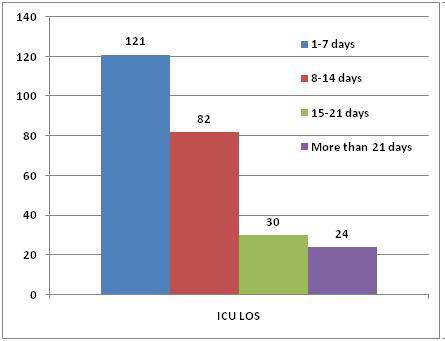
ICU LOS : Out of total 287 patients 54(18.8%) patients had a Length of Stay of more than 14 days. Overall mean ICU LOS was 4.8 days and group of patients having LOS of >14 days had a mean LOS of 19.7 days.
ICU Ventilation Days : Though majority (46%) of the patients had mechanical ventilation between 1-7 days, almost 6% of the patients were put in Ventilation for more than 21 days. Correlation matrix showed a high correlation between ICU Length of Stay and mechanical ventilation days (Fig 3, pearson correlation coefficient 0.95, p<0.05)
Predictors of prolonged stay (Table 2):
Age : There was no significant differences of LOS between the various age groups.
Sex : The gender differences in the two groups also did not show any statistical significance.
CategoryNo (%) ICU Length of StayP-value <14 Days>14 Days (n1=233)(n2=54)
AgeLess than 19 years 19(6.6)14(6.1)5(9.2)NS
19-44 years67(23.3) 55(23.6)12(22.2)
45-64 years122(42.5)101(43.3)21(38.8)
More than 65 years 79(27.5)63(27.0)16(29.6)
SexMale 169(58.8)136(58.3)33(61.1)NS
Female118(41.2)97(41.7) 21(39.9)
Type ofElective 16(5.5)15(6.4)1(1.8)< 0.05
Admission Non-Elective 271(94.5)218(93.6)53(98.2)
SeverityAPACHE II Score of Illness (Mean±SD) 19±318±923±5<0.05
Tracheostomy 49(17.1) 12(5.1)37(68.5)NS
ICU Mortality 83(28.9)69(29.6)14(25.9)NS
NS=Not significant (p >0.05)
Vol 121, No 4, April 2023Journal of
the Indian Medical Association
Table 1 — Clinico-demographic profile of the study subjects (n=287)
20
Type of admission : The mean ICU LOS of elective and non-elective patients was respectively 4.2±5.3 and 9.6±5.7. This difference was stastistically significant.
Readmission : The patients who were readmitted in the ICU had a higher LOS than those who were admitted for the first time OR 2.78 (1.17-4.97)

Main reason for admission : The patients admitted in the ICU due to surgical trauma, respiratory or neurological cases were more likely to have a prolonged LOS.
First 24-hour data : Patient who had Coagulopathy, Infection, Oliguria or needed Mechanical ventilation or Vasopressor therapy in the first 24-hour following admission were more likely to have a prolonged ICU stay.

Severity : The patients having LOS of >14 days had a higher mean APACHE II score and the difference between the two groups was significant.
DISCUSSION
Patients having a prolonged ICU stay form a smaller proportion of total ICU patients, yet they consume a disproportionate percentage of Healthcare Resources. The outcome of patients having a prolonged ICU stay was comparable with those having a shorter stay. The various characteristics of patients having prolonged ICU stay were different from those having a shorter stay, in terms of reasons for admission or physiological abnormalities during admission. These findings helped to determine some of the possible predictors for prolonged stay in ICUs. Patients’ severity score (APACHE II) was significantly higher in the group having a prolonged stay. This finding was in accordance to many similar studies done
Vol 121, No 4, April 2023Journal of the Indian Medical Association
Fig 3 — Distribution of study subjects according to ICU Length of Stay and ventilation days
Fig 1 — Distribution of study subjects according to ICU Length of Stay
Fig 2 — Distribution of study subjects according to Ventilation day
PredictorNumber ofOR for p-value patients prolonged stay OR95% C/I Non-elective admission 271(94.5)4.151.81-5.51< 0.05 Readmission 17(5.9)2.781.17-4.97< 0.05 Main reason for admission Surgical : Trauma 79(27.5)1.9 1.3-3.6<0.05 Non-trauma Surgical 98(34.1)0.5 0.2-0.7<0.05 Medical : Respiratory53(18.4)2.561.23-4.64<0.05 Neurological 29(10.1)3.9 2.1-5.3<0.05 Others28(9.8)0.7 0.3-1.2NS First 24-hour data Coagulopathy 99(34.4)0.620.27-1.42<0.05 Coma 35(12.2)1.41 0.33-5.9NS Infection 78(27.1)1.200.37-3.89<0.05 Oliguria 54(18.8)1.46 .58-3.68<0.05 Mechanical ventilation 201(70.1)1.301.13-1.69<0.05 Vasopressor therapy 49(17.1)1.241.18-2.41<0.05 NS=Not significant (p >0.05) 21
Table 2 — Possible predictors of Prolonged ICU stay (n=287)
elsewhere in this topic9.
Overall, prevalence of prolonged ICU stay was revealed to be 18.8% in our study. It was higher than most of the studies done on similar topic. Non-elective admissions or the re-admission cases were significantly associated with a prolonged Length of Stay . A strong correlation was found between the duration of mechanical ventilation and ICU Length of Stay. This finding was congruent with many similar studies6,9.
A significantly increased Length of Stay was observed in patients with Respiratory System Diseases and Neurological Diseases. In another prospective study done by Wong, et al10 for patients in ICU, the most common reasons for admission were Neuromuscular Weakness, Pneumonia, Multiple Traumas and Septic Shock. Similarly, Toptas, et al11 in a study done in Turkey found that Cardiovascular System Diseases, Nervous System Diseases and Cerebrovascular Diseases to be the most common precursors for increased ICU stay.
Patients having a prolonged ICU stay were more likely to undergo Tracheostomy.
CONCLUSION
A smaller number of Intensive unit admissions consume a great proportion of overall ICU bed-days. The predictors identiûed in this study can be used in targeting this particular group to improve resource utilization and efficiency of ICU.
Conflict of Interest : None declared.
Source of Funding : Self
REFERENCES
1Halpern NA, Pastores SM, Greenstein RJ. Critical care medicine in the United States 1985-2000: an analysis of bed numbers, use, and costs. Crit Care Med 2004; 32: 1254-9.
2Weled BJ, Adzhigirey LA, Hodgman TM, Brilli RJ, Spevetz A, Kline AM, et al — Critical care delivery: The importance of process of care and ICU structure to improved outcomes 2015; 43: 1520-25.
3Nelson JE, Cox CE, Hope AA, Carson SS — Chronic critical illness. Am J Respir Crit Care Med 2010; 182: 446-54. [PubMed: 20448093]
4Estenssoro E, Reina R, Canales HS, Saenz MG, Gonzalez FE, Aprea MM, et al — The distinct clinical profile of chronically critically ill patients: A cohort study. Crit Care 2006; 10: R89. [PubMed: 16784546]
5Higgins TL, McGee WT, Steingrub JS, Rapoport J, Lemeshow S, Teres D — Early indicators of prolonged intensive care unit stay: Impact of illness severity, physician staffing, and preintensive care unit length of stay,” Critical Care Medicine 2003; 31(1): 45-51, 2003.
6Gruenberg DA, Shelton S, Rose SL, Rutter AE, Socaris S, McGee G — Factors influencing length of stay in the intensive care unit. American Journal of Critical Care 2006; 15(5): 502-9.
7Ryan TA, Rady MY, Bashour CA, Leventhal M, Lytle B, Starr NJ — Predictors of outcome in cardiac surgical patients with prolonged intensive care stay. Chest 1997; 112: 1035-42.
8Kanus WA, Dropper EA, Wagner DR, Zimmerman JE — APACHE severity of disease classification system. Crit Care Med 1985; 13: 818-29
9Arabi Y, Venkatesh S, Haddad S, Al Shimemeri A, Al Malik S — A prospective study of prolonged stay in the intensive care unit: predictors and impact on resource utilization. Int J Qual Health Care 2002; 14(5): 403-10. doi: 10.1093/intqhc/ 14.5.403. PMID: 12389806.
10Wong DT, Gomez M, McGuire GP, Kavanagh B — Utilization of intensive care unit days in a Canadian medical-surgical intensive care unit. Crit Care Med 1999; 27(7): 1319-24. doi: 10.1097/00003246-199907000-00020. PMID: 10446826
11Toptas M, Sengul Samanci N, Akkoc Ý, Yucetas E, Cebeci E, Sen O, et al — Factors Affecting the Length of Stay in the Intensive Care Unit: Our Clinical Experience. Biomed Res Int 2018; 2018: 9438046. doi: 10.1155/2018/9438046. PMID: 29750174; PMCID: PMC5884409.
Vol 121, No 4, April 2023Journal of
the Indian Medical Association
22
Original Article
Postoperative Comfort Score after Septoplasty among Patients
Undergoing Nasal Packing versus Suturing of the Septal Flap by Modified Technique without Packing : A Randomized Controlled Trial
Abhinav Srivastava1, Chander Mohan2
Background : Septal surgery is one of the most common surgical procedures performed by an Otorhinolaryngeal surgeon since ancient times. Various modifications in the approach, changing concept of conserving septal cartilage, use of an endoscope and good antibiotics to control postoperative infection have played a key role in controlling the complication rates but still, one thing which is mostly practiced worldwide is nasal packing in the postoperative period which is a nightmare for many patients, as the pain threshold varies from patient to patient. It also causes dryness of mouth, throat irritation, facial heaviness, headache, excessive watering from eyes, aural fullness. There is a lack of proper evidence to prove whether nasal packing really decreases postoperative hemorrhage as the incision is properly approximated and sutured. The main reason for nasal packing was an approximation of nasal septal flap thereby reducing the chances of septal Haematoma and stabilization of septal flap in the midline. The present study has been taken to study and compare postoperative nasal packing and modified quilting suture of the septal flap without the nasal pack.
Material and Methods : This one-year prospective comparative study was conducted on 149 patients who underwent septoplasty with 3 months follow-up. One group had Postoperative nasal packing and the other had only modified septal flap suturing without the nasal pack.
Results : Out of the total of 149 patients, 88 underwent nasal packing in the postoperative period and 61 patients had undergone suture of the nasal septal flap without nasal packing. A statistically significant value of VAS score was found in the non-packing group of 61 patients, where the average postoperative VAS score was 1.46 against 3.7 among the packing group of 88 patients. An unpaired t-test was applied and a value of 15.431 was obtained with a pvalue less than 0.001. No cases presented with septal perforation in the postoperative period in patients without a nasal pack and there were 2 cases (2.2%) of septal perforation in the nasal packing group. There was no significant bleeding in the postoperative period in both groups of patients.
Conclusion : Stabilization of the nasal septal flap by modified quilting technique is better option after septoplasty with good comfort score.
[J Indian Med Assoc 2023; 121(4): 23-7]
Key words :Deviated Nasal Septum (DNS) ,Septoplasty, Visual Analogue Scale (VAS).
Septal surgery is one of the most common surgical procedures performed by an Otorhinolaryngeal surgeon since ancient times. Nasal obstruction is one of the most common indications for the surgery for ages but with a better understanding of the nose and paranasal sinus anatomy and physiology, other indications have evolved like correction of septal spur as a cause for recurrent headache and epistaxis, as a part of Septorhinoplasty. Deviated Nasal Septum correction may be required as an adjunct to other surgeries if it is interfering with the access to the target sites like patients undergoing Endoscopic Sinus
Department of ENT, Rohilkhand Medical College and Hospital, Bareilly Uttar Pradesh 243006
1MS (ENT), Professor and Corresponding Author
2MS (ENT), Professor
Received on : 15/03/2022
Accepted on : 02/06/2022
Editor's Comment :
The discomfort caused by postoperative nasal packing after septoplasty can be avoided by stabilizing the nasal septal flap with modified suturing technique as described. The technique also prevents postoperative nasal synechiae with no incidence of septal haematoma and nasal bleeding.
Surgery (ESS), Endoscopic Dacryocystorhinostomy. In the modern era of minimum invasive intracranial surgery, Septum acts as a gateway to ventral skull base surgery.
For ages, Septal surgery is not without complications like Septal perforation, Septal haematoma, Septal deformity and rarely Toxic Shock Syndrome (TSS). Various modifications in the approach, changing concept of conserving septal cartilage, use of an endoscope, and good antibiotics to control postoperative infection have played a key
Vol 121, No 4, April 2023Journal of the Indian Medical Association
23
role in controlling the complication rates but still, one thing which is mostly practiced worldwide is nasal packing in the postoperative period which is a nightmare for many patients, as the pain threshold varies from patient to patient. It also causes dryness of mouth, throat irritation, facial heaviness, headache, excessive watering from eyes and aural fullness. There is a lack of proper evidence to prove whether nasal packing really decreases postoperative hemorrhage, as proper decongestion and local infiltration of lignocaine with adrenaline is given before incision, avascular sub perichondrium plane is made for the surgery and lastly, an incision is properly approximated and sutured. The main reason for nasal packing is an approximation of nasal Septal flap thereby reducing the chances of Septal Haematoma and stabilization of Septal flap in the midline1.
The present study has been taken to put light on the various aspects of Septal surgery and an emphasis has been given on the replacement of the postoperative nasal packing with modified quilting suture of the Septal flap to give nasal breathing and a pain-free postoperative period.
MATERIALS AND METHODS
This Prospective Randomized Control study was conducted in the Department of Otorhinolaryngology, Rohilkhand Medical College and Hospital, Bareilly after obtaining approval from the institutional ethical committee.
A detailed entry of all the patients who underwent Septal surgery for various reasons was made in a proforma generated by Epi info version 7.0 from 1st October, 2018 to 30th September, 2019 and have completed at least 3 months follow up. The result was statistically analyzed using the software provided with Epi info.
A total of 149 patients of either sex above the age of 18 years, who underwent only Septal surgery after obtaining written informed consent for various indications during this period of one year and have completed at least 3 months postoperative follow were included in the study and relevant data were obtained and entered in the form generated in the software. The objective of the study was to find the comfort score in the postoperative period and also to analyze complications in the two groups. The patients who had a history of Diabetes Mellitus, Hypertension and bleeding disorder were excluded from the study as well as any patient who underwent Sinus surgery, Turbinate surgery or Dacryocystorhinostomy along with Septal correction were also excluded from the study. Once selected for the Septal surgery, all the
patients underwent routine blood tests along with diagnostic nasal endoscopy. All the patients in the study underwent Septoplasty under Sedation with Pentazocine 15 mg and Promethazine 25 mg Intramuscular (IM) 30 minutes prior to surgery. In preoperative preparation, the nose was topically decongested with Cotton pledget soaked in 4% lignocaine with adrenaline in the concentration of 1:30000 for at least 15 minutes. During the procedure and in the postoperative period thorough monitoring of vitals was made.
Just before the start of the Operation randomization was done as per the random number table by a Computer system software, one group underwent nasal packing with Medicated Ribbon gauze and the other group of patients underwent modified suturing of the nasal Septal flap with 3-0 vicryl, ½ circle round-bodied without nasal packing. The needle chosen was 3-0 because of its appropriate length, neither too long nor too short and this much length is good enough to easily catch with forceps. A long needle causes much trauma to the lateral nasal wall whereas a smaller size is sometimes lost within the flap. A round body needle is relatively less traumatic when compared with a cutting needle. Vicryl was preferred as it usually takes 60 to 90 days for complete absorption and by that time septal flaps heal completely. Septal splints were not used in any of the cases in this study.
Postoperative pain was assessed using the Visual Analogue Scale from 0 to 10 where 0 was no pain to 10 was unbearable pain.
Our technique of modified suturing of the nasal Septal flap (Fig 1-6)
Step 1 : Using 3-0 Vicryl mounted on a cutting needle, first it is inserted along the caudal end of the septum on the opposite side of deviation, marked as point “A” in the figure.
Step 2 : Suture is passed in the upper half of the Septum around the mid-point of the septum in an anterior-posterior direction, marked as point “B” in the diagram.
Step 3 : Around the mid-point of the distance between “A” and “B” the needle is passed on the other side in an oblique direction, marked as point “C” in the diagram.
Step 4 : A single knot is made around point “C” to make the stability of the suture along with obliterating the space between the septal flaps.
Step 5 : The needle is passed near the caudal end at point “D”, which lies near to point “A”. A final double knot is made and the final suture is finally stabilized on the same side where suturing started.
Vol 121, No 4, April 2023Journal of the Indian Medical Association
24
Figs 1 to 6 — (1) An Orientation of the Nasal septum as seen from above. (2) Needle inserted near the caudal end at point A, (3) On the opposite side, the suture is passed in an oblique direction at point B which is around the middle of the septum in the Anterio-Posterior direction, (4) The suture is again passed to the other side at point C, which is at the midpoint of AB distance, (5) At point C, a single knot is made to make the suture stable, (6) At point D, which lies near the caudal end close to point A level, the suture is passed to the other side and a double knot is made at point A.
Note : If there is caudal dislocation then, points “A” and “D” are made near the caudal end close to anterior nasal spine for extra stability.
Statistical Analysis :
Statistical Analysis was done by software Epi info version 7.0. A datasheet was formed and all the patients’ data were entered into the software along with all the follow-up records. Statistics were applied by the same software. Mean value and Standard Deviation (SD), percentage and unpaired student t-test was used to compare two groups in quantitative data and Chi-square to compare two independent qualitative variables.
OBSERVATIONS AND RESULTS
Out of the total 149 patients, 64 were females and 85 were males with 21–30 years as the most common age group who underwent Septal surgery (Table 1). Nasal obstruction (89.26%) was the main indication for the surgery followed by Headache (66.44%) and Recurrent Epistaxis (20.81%) (Table 2).
The cartilaginous part of the Nasal Septum was the most common Deviated part of the nasal septum in 138 (92.6%) patients followed by the Bony Part in
Table 1 — Table showing percentage of patients in different age groups
Table 2 — Table showing percentage of patients undergoing surgery for various clinical presentations
Presenting ComplaintsPercentage with SD
Nasal Obstruction89.26±0.31
Headache66.44±0.47
Recurrent Epistaxis20.81±0.41
Hyposmia7.38±0.26
Snoring6.71±0.25
As a part of other operation8.72±0.28
116 (77.8%) Patients with associated Maxillary crest prominence was seen in 58.48% of patients and 14.1% were with Caudal Dislocation.
Out of the total of 149 patients, 88 underwent nasal packing in the postoperative period and 61 patients had no nasal packing, and suturing of the nasal Septal flap was done as described in the methodology (Fig A).

Vol 121, No 4, April 2023Journal of the Indian Medical Association
Age GroupPercentage 11-2028.19 21-3046.98 31-4020.13 41-50 4.03 51-60 0.67
25
There was a statistically significant result in terms of postoperative pain score (VAS) in the two groups of Nasal packing and others with no packing. Here in this study, VAS score up to 2 was considered as low pain and above 2 was considered a more pain score. In the non- packing group of 61 patients, the average postoperative VAS score was 1.46 against 3.7 among the packing group of 88 patients. An unpaired t-test was applied and a value of 15.431 was obtained with a p-value less than 0.001 (Table 3 and Fig B).

In the follow-up period of 3 months, there were 5 (5.6%) patients who were not relieved of nasal obstruction and were attributed due to synechia as a result of nasal packing whereas, there were no cases of synechia in the non-packing group though the result was not statistically significant. No cases presented with Septal perforation in the postoperative period in patients without a nasal pack and there were 2 cases (2.2%) of Septal perforation in the nasal packing group.

There was no significant bleeding in the postoperative period in both groups of patients.
In the present study, none of the patients presented with Septal hematoma or abscess, postoperative nasal bleeding and external nasal deformity.
DISCUSSION
Septoplasty is one of the most common Otorhinolaryngeal procedures with nasal obstruction as one of the most common indications for surgery. In the postoperative period, various types of nasal packing and splints are used for the control of nasal bleeding and flap apposition like medicated ribbon gauze, Polyvinyl alcohol sponge (Merocel) with and without
airway, silicon splints, etc. These nasal packs act as a foreign body and cause discomfort to the patient and especially pain in the postoperative period. They also act as a source of infection in the nose and paranasal sinus as they affect the mucociliary activity of the sinus mucosa and lead to stasis of the mucus secretions.
In our study, seventy-five percent of Septoplasty were done in the younger age group that too within 30 years of age. The reason for this can be attributed to the cessation of development of the nasal septum around 18 years of age and so patients with nasal Septal deviation usually become symptomatic after this age and seek medical advice and treatment at an early stage. So, a symptomatic deviation is manifested in the second or third decade of life. Alotaibi AD, et al2 also found a similar result with 74.8% of the study population presenting within the third decade of life.
A study done by Peric A, et al3 found a significant association between headache and spur and also found statistically significant improvement in headache following surgery. In our study, we have also found a significant association between headaches and spurs, and the same showed a statistically significant improvement in VAS score.
In our study, 5 cases (5.6%) of nasal synechiae were found in the nasal packing group as against no case in the non-packing group, a similar finding was observed in the study done by Awan MS, et al4 who found nasal synechiae in 18.2% of cases among patients undergoing nasal packing with no case of synechia in non- packing group. The probable reason can be attributed to injury to the nasal mucosa of the lateral and medial wall of the nasal cavity at the same level either during surgery or caused or aggravated by tight packing of the nasal cavity, as there are no standard guidelines on how much tight one should pack. However, the reason for not being statistically significant can be due to less sample size. These
Vol 121, No 4, April 2023Journal of the Indian Medical Association
Fig A — Pie chart showing the number of patients in the two groups
Fig B— Graph showing average VAS in the two groups
VAS Group Mean±SD t-valueP value With nasal packing3.74±0.903 15.431<0.001 Without nasal packing1.28±1.030(highly significant) 26
Table 3 — Table showing comparison in the two groups in terms of Visual Analogue Scale (VAS) for pain
patients presented with persistent nasal obstruction at their subsequent visit and needed release of nasal synechiae at 3 months postoperative visit.
Septal perforation is caused by injury of both nasal septal flaps at the same level accompanied by loss of bone or cartilage. It was very common when Submucous Resection (SMR) was mostly done for septal deviation but with Septoplasty, the rate has gone to a very lower level and if the injury is found intra-operatively they are repaired simultaneously. Our study has got no significant results in terms of incidence of Septal perforation in the two groups and it goes in hand with the study done by Walikar B N, et al5 and Eski E, et al6. Thus, our study concludes that nasal packing has no significant influence on causing Septal perforation.
In our study, “p” value of VAS score in two groups of less than 0.001 was obtained which was highly significant statistically thereby concluding significant lower postoperative pain in the non-packing group in comparison to the group with nasal packing. The studies done by Naghibzadeh B, et al7, Walikar BN, et al5 and Mane RS, et al8, have also observed a statistically significant less postoperative pain score in patients who did not undergo nasal packing postoperatively.
CONCLUSION
Nasal Packing after Septal surgery can be easily avoided in the postoperative period as it may be replaced by stabilization of the nasal septal flap by modified quilting technique which obliterates the dead
space, thereby preventing septal Haematoma. It also may maintain the septum in midline position with no post-operative discomfort without any undue complication.
REFERENCES
1Salem ASF, Idrees ZAH, Saad YA — The Incidence of PostSeptoplasty Bleeding in Patients without Nasal Packing. Bahrain Medical Bulletin 2015; 37(4): 243-5.
2Alotaibi AD, Almutlaq BA, Alshammari FN, Gadelkarim Ahmed H — The Common Clinical Presentation of Patients Selected for Septoplasty in Northern Saudi Arabia. Int J Otolaryngol 2018; 2018: 8536387.
3Peric A, Rasic D, Grgurevic U — Surgical treatment of rhinogenic contact point headache: An experience from a tertiary care hospital. IntArchOtorhinolaryngol 2016; 20: 16671.
4Awan MS, Iqbal M — nasal packing after septoplasty: A randomized comparison of packing versus no packing in 88 patients. Ear Nose Throat 2008; 87(11): 624-7.
5Walikar BN, Rashinkar SM, Watwe MV, Fatima A, Kakkeri A — A Comparative study of septoplasty with or without nasal packing. Indian J Otolaryngol Head Neck Surg 2011; 63(3): 247-8.
6Eski E, Yilmaz I — Septoplasty without nasal packing: Functional outcomes and Complications A Prospective Clinical Study. J Otolaryngol ENT Res 2015; 3(2): 00062.
7Naghibzadeh B, Peyvandi AA, Naghibzadeh G — Does Post Septoplasty Nasal Packing Reduce Complications? Acta Medica Iranica 2011; 49(1): 9-12.
8Mane RS, Patil B, Mohite A — Comparison of Septoplasty with and without Nasal Packing and Review of Literature. Indian J Otolaryngol Head Neck Surg 2013; 65(2): 406-8.
Vol 121, No 4, April 2023Journal
of the Indian Medical Association
27 JIMA
https://onlinejima.com
is now fully ONLINE and Publishes only ONLINE submitted Articles through
Original Article
Study of the Prevalence of Type 2 Diabetes Mellitus in Patients with Heart Failure in a Tertiary Care Hospital in Eastern India
Torsha Chatterjee1, Indira Maisnam2, Prabir Kumar Kundu3 , Sudipta Bandyopadhyay4, Aniruddha Ray5, Apurba Kumar Mukherjee6
Heart Failure and Type 2 Diabetes Mellitus are closely related. Diabetic patients have an increased risk of developing Heart Failure and those with Heart Failure are at higher risk of developing diabetes. The objective of the study was to estimate the prevalence of Type 2 Diabetes Mellitus in patients with heart failure. This analytical observational type of epidemiological study with case control design was conducted at in patient department of General Medicine of RG Kar Medical College & Hospital, Kolkata, West Bengal, India from July, 2019 to June, 2020. 100 study subjects by purposive sampling method were taken as per inclusion and exclusion criteria. Data were collected based on History, Clinical examination, relevant investigations and review of records. In this study proportion of Diabetes was much higher among cases with Heart Failure (30%) than controls (10%), among cases with NYHA class IV (56.3%) and among cases with reduced Ejection Fraction (100%). Thus pre-existing or newly development of Type 2 Diabetes Mellitus should be kept in mind in all hospitalized Heart Failure Patients.
[J Indian Med Assoc 2023; 121(4): 28-31]
Key words :Heart Failure, Type 2 Diabetes Mellitus.
Heart Failure (HF) occurs due to structural and functional defects in myocardium associated with impairment of ventricular filling or the ejection of blood. It growing to a modern epidemic and despite advances in therapy, it still carries an ominous prognosis and a significant socio-economic burden1 Diabetes Mellitus is highly prevalent especially amongst patients with Heart Failure with preserved Ejection Fraction (HFpEF), and patients with co-existent two conditions have a higher risk of mortality compared with patients without these two condition2 Patients with Heart Failure demonstrate impaired glucose metabolism and insulin resistance3 . The altered glucose metabolism places them at increased risk for developing diabetes, 29% versus 18%, compared to the general population4 Nearly one quarter of all HF patients have concomitant diabetes and this number rises drastically to 40% in patients admitted with Acute
Department of General Medicine, RG Kar Medical College & Hospital, Kolkata 700004
1MD (General Medicine) Ex -Senior Resident & DNB Resident, Department of Endocrinology, Sir Gangaram Hospital, New Delhi 110060
2 MD (General Medicine); DM(Endocrinology) Assistant Professor, Department of Endocrinology, IPGME&R and SSKM Hospital, Kolkata 700020
3 MD (General Medicine); Assistant Professor and Corresponding Author
4MD (General Medicine), Associate Professor
5MD (General Medicine), Professor & Head
6MD (General Medicine), FICP, Ex- Professor and Head
Received on : 09/03/2022
Accepted on : 23/09/2022
Editor's Comment : Heart Failure patients have increased risk of developing Diabetes. Therefore, Diabetes should be routinely screened in all patients of Heart Failure. Early detection with appropriate management can reduce both morbidity and mortality.
Decompensated Heart Failure. In the SOLVD (Studies of Left Ventricular Dysfunction) trial 6% of patients developed DM within three years of enrollment5. The overall prevalence of DM in Heart Failure is significantly higher (25%) in comparison to general population (9%) and patients with Heart Failure with preserved Ejection Fraction (HFpEF) have a slightly higher prevalence of DM (40%)6 Many population studies and clinical trials have demonstrated that DM significantly increases the risk of repeated hospitalisations due to Heart Failure, the duration of hospital stay and associated with a significantly higher mortality rate in comparison to those Heart Failure patients without Diabetes5
There is possibility that the increased incidence of DM during the course of HF may be an epiphenomenon of the lenient monitoring for impaired glucose metabolism with HbA1C and with Oral Glucose Tolerance Tests in the early stages of HF7. Decreased physical activity in HF patients may lead to decreased insulin sensitivity and to compensatory insulin requirements and hyperglycaemia. Increased catecholamines levels and sympathetic over activity stimulate gluconeogenesis and glycogenolysis 8
Vol 121, No 4, April 2023Journal of the Indian Medical Association
28
Insulin sensitivity is decreased with declining of New York Heart Association (NYHA) functional status of HF patients 3 Haemodynamic consequences accompanying HF (decreased forward blood flow and increased central venous pressure) lead to hypoperfusion and congestion of the Pancreas and Liver, which may impair their ability to regulate metabolic homeostasis. Confirmatory data are provided by a recent study, which concludes that left ventricular assist devices improve blood glucose control in DM patients9
This study aims not only to provide the correlation between Heart Failure and Type 2 Diabetes Mellitus but also to provide information regarding prevalence of Diabetes Mellitus in low and preserved Ejection Fraction Heart Failure patients separately.
MATERIALS AND METHODS
This analytical observational type of epidemiological study with case control design was done in the in patient Department of General Medicine of RG Kar Medical College & Hospital, Kolkata, West Bengal from 1st July 2019 – 30th June 2020. A total of 50 patients aged above 18 years admitted with clinical features of Heart Failure and fulfilling Framingham criteria for Heart Failure was included as cases in the study. Equal number of controls admitted with other types of Cardiac Disease without Heart Failure like Acute Myocardial Infarction, Arrythmia, Valvular Heart Disease etc were included after age and sex matching. Patients who did not give consent, those with features of Chronic Liver Disease, Chronic Kidney Disease, Severely Anaemic, Pregnancy, Type 1 DM were excluded from the study. Purposive sampling method was followed to select those 100 study subjects. There is no formula for setting the sample size for purposive sampling. As a rule of thumb based on empirical experience 100 study subjects are included for purposive sampling in our study.
Framingham Criteria for Congestive Heart Failure10
Diagnosis of CHF requires the simultaneous presence of at least 2 major criteria or 1 major criterion in conjunction with 2 minor criteria.
Major Criteria :
•Paroxysmal nocturnal dyspnea
•Neck vein distention
•Rales
•Radiographic cardiomegaly (increasing heart size on chest radiography)
•Acute pulmonary edema
•S3 gallop
•Increased central venous pressure (>16 cm H2O at right atrium)
•Hepatojugular reflux
•Weight loss >4.5 kg in 5 days in response to treatment
Minor Criteria :
•Bilateral ankle edema
•Nocturnal cough
•Dyspnea on ordinary exertion
•Hepatomegaly
•Pleural effusion
•Decrease in vital capacity by one third from maximum recorded
•Tachycardia (Heart rate>120 beats/min.)
Type 2 Diabetes ADA Diagnostic Criteria 11:
•A Fasting Plasma Glucose (FPG) level of 126 mg/dL (7.0 mmol/L) or higher, or
•A 2-hour plasma glucose level of 200 mg/dL (11.1 mmol/L) or higher during a 75-g Oral Glucose Tolerance Test (OGTT), or
•A random Plasma glucose of 200 mg/dL (11.1 mmol/L) or higher in a patient with classic symptoms of hyperglycemia or hyperglycemic crisis.
As per above two criteria data were collected in predesigned pretested schedule with the help of interview of study subjects and/or their accompanying persons, clinical examination, review of records and relevant investigations . Collected data was checked for consistency and completeness. Data was entered in Microsoft Excel data sheet for analysis. Data was analysed by IBM Statistical Package for Social Sciences (SPSS) version 22. Data were organised and presented applying the principles of descriptive statistics in the form of frequency and percentage and also in tables. Chi-square test was applied as test of significance for categorical variables and significance level was set at p value <0.05.
RESULTS
There was no statistically significant difference of age groups and gender between cases and controls, means there was proper age matching between this two groups. Among 50% of the cases Blood Pressure was elevated than normal and 16% had Hypotension, rest others were Normotensive. Tachycardia was found among 46% of the cases. History of nocturnal cough was present among 46% of the cases. Ankle edema, engorged neck veins were observed among 84% and 68% of the cases. Almost all cases had history of dyspnea on exertion. Hepatojugular reflux, rales,
Vol 121, No 4, April 2023Journal
of the Indian Medical Association
29
hepatomegaly, cardiomegaly, S3 gallop, PND were seen among 50% , 92%, 56% , 78% , 64% and 76% of the cases respectively. Bilateral pleural effusion was present in 26% cases. Acute pulmonary edema was present among 60% cases. Reduced ejection fraction was observed among 10% cases and 30% had ejection fraction in mid range. Among all cases 32% belong to NYHA class IV and 54 belong to NYHA class III.
Prevalence of Type 2 Diabetes among heart failure patients in the present study was 30% (Table 1). Whereas, Prevalence of Type 2 Diabetes among controls in the present study was 10%. There was absence of ketonuria or acidosis among the diabetic patients.
In the present study 62% of the Heart Failure patients included was under treatment with diuretics, 66% of them were under treatment with beta blocker and 54% of them were under treatment with statins.
Table 1 shows that, prevalence of Diabetes among Heart Failure patients in the present study was 30% Table 2 shows that, proportion of Diabetes was much higher among cases reduced Ejection Fraction (100%). This finding was statistically significant.
DISCUSSION
In the present study, prevalence of Diabetes was 30 percent (Table 1) among the Heart Failure patients; whereas prevalence of Diabetes was 10% among the control group. Data from Melle, et al1 suggest that more than one-third of patients who are hospitalized for Heart Failure without Diabetes exhibit impaired Fasting Glucose or Impaired Glucose Tolerance. Recent data from various registries show that prevalence of Diabetes in patients with Heart Failure
ranges from approximately 25% to 40%, depending on the study population12,13. So, findings of present study showed almost similar proportion of diabetes among heart failure patients. Heart Failure per se is involved in the pathogenesis of Diabetes due to numerous factors like heightened insulin resistance, pro-inflammatory markers and diabetogenic drugs used in heart failure. There is also increased risk of atherosclerotic cardiovascular disease in diabetes mellitus; which is a risk factor for Heart Failure. Moreover; pre-existing Diabetes Mellitus could have been missed in the Heart Failure patients in our study due to lack of awareness or lack of facility for screening for Diabetes Mellitus.
The possible adverse effects of established HF treatments, such as beta (β )-blockers and high dose thiazide diuretics may also hamper blood glucose control14 Previous studies reported that, patients with HF have a four-fold higher prevalence of T2DM (20%) than patients without HF (4-6%)15 and this rises to 40% in T2DM patients hospitalized for HF16. T2DM worsens prognosis for patients with HF with reduced Ejection Fraction (HFrEF) (Table 2), but even more with HFpEF, by increasing the risk of death and hospitalization17. Reduced and mid-range Ejection Fraction was observed among two/fifth of the Heart Failure patients. Among all cases one-third of them belong to NYHA class IV and one-half belonged to NYHA class III.
Statistically insignificant higher prevalence of Diabetes was observed among cases with age less than 40 years and among males.This could be because of many reasons like a graver outcome in young onset Type 2 Diabetes compared to onset at older age; more severe degree of Heart Failure thereby increasing the diabetogenic potential of Heart Failure; smoking; alcohol and other factors. But there was statistically significant increased proportion of Diabetes cases those were on regular treatment with diuretics. This may be attributed to the fact that high loop diuretic dosages determine more severe HF. Thiazide diuretics in high dosage are postulated to be involved in worsening of insulin resistance, inhibition of glucose uptake and decreased insulin release. Diabetes was more in proportion in cases who used beta-blockers. This may be attributed to the fact that beta blockers block the release of insulin by interacting with nerve signals to the pancreas. Higher incidence of statin use was seen in patients diagnosed to have Diabetes. Though it has been postulated that statins could be diabetogenic; the benefits of statin use outweigh the risks. The association between statin use and
Vol 121, No 4, April 2023Journal of the
Medical
Indian
Association
Diabetes Status Frequency Percent Nondiabetic28 56.0 Pre-Diabetic7 14.0 Diabetic1530 Total50100
Table 1 — Distribution of the cases according to status of Diabetes (n=50)
Ejection Diabetes StatusTotal Fraction (Category) Non diabeticPre diabetic Diabetic Preserved212730 70.0%6.7%23.3% 100.0% Mid range75315 46.7%33.3%20% 100.0% Reduced0055 0.0%0.0% 100.0%100.0% 28 (56%) 7(14%)15(30%)50(100%) Chi square value= 19.052, df=4, P=0.001(p< 0.05) 30
Table 2 — Association of Ejection fraction of cases with diabetic status
Diabetes in our study could be due to the presence of dyslipidemia which is an earlier presentation of insulin resistance in the metabolic progression to Diabetes Mellitus. Diabetes was also statistically increased in cases that belonged to NYHA class IV. This may be attributed to decreased exercise tolerance in these patients, increased usage of drugs like beta blockers, statin, thiazides etc. Diabetes was also found to be statistically increased in cases with reduced ejection fraction.
In our study Diabetes Mellitus was highly prevalent among the patients with Heart Failure, especially those with mid-range to reduced Ejection Fraction and patients with these two conditions have a higher risk of mortality. Thus in our study, we found that not only is there increased prevalence of Heart Failure in Diabetes; but the presence of diabetes and preDiabetes is also associated with a worse prognosis in patients with Heart Failure.
CONCLUSION
Diabetes should be routinely screened for in all patients with all forms of heart failure. Early detection with timely and appropriate management will improve both morbidity and mortality outcomes.
REFERENCES
1van Melle JP, Bot M, De Jonge P, De Boer RA, van Veldhuisen DJ, Whooley MA — Diabetes, glycemic control, and newonset heart failure in patients with stable coronary artery disease: data from the heart and soul study. Diabetes Care 2010; 33(9): 2084-9.
2Stratton IM, Adler AI, Neil HA, Matthews DR, Manley SE, Cull CA, et al — Association of glycaemia with macrovascular and microvascular complications of type 2 diabetes (UKPDS 35): prospective observational study. BMJ 2000; 321(7258): 405-12.
3Doehner W, Rauchhaus M, Ponikowski P, Godsland IF, Von Haehling S, Okonko DO, et al — Impaired insulin sensitivity as an independent risk factor for mortality in patients with stable chronic heart failure. Journal of the American College of Cardiology 2005; 46(6): 1019-26.
4Vermes E, Ducharme A, Bourassa MG, Lessard M, White M, Tardif JC — Enalapril reduces the incidence of diabetes in patients with chronic heart failure: insight from the Studies Of Left Ventricular Dysfunction (SOLVD). Circulation 2003; 107(9): 1291-6.
5SOLVD Investigators. Effect of enalapril on survival in patients with reduced left ventricular ejection fraction and congestive heart failure. N Engl J Med 1991; 325: 293-302.
6Shindler DM, Kostis JB, Yusuf S, Quinones MA, Pitt B, Stewart D, et al — SOLVD investigators. Diabetes mellitus, a predictor of morbidity and mortality in the Studies of Left Ventricular Dysfunction (SOLVD) Trials and Registry. The American Journal of Cardiology 1996; 77(11): 1017-20.
7Andersson C, Norgaard ML, Hansen PR, Fosbøl EL, Schmiegelow M, Weeke P, et al — Heart failure severity, as determined by loop diuretic dosages, predicts the risk of developing diabetes after myocardial infarction: a nationwide cohort study. EuropeanJournalofHeartFailure 2010; 12(12): 1333-8.
8Heck PM, Dutka DP — Insulin resistance and heart failure. Current Heart Failure Reports 2009; 6(2): 89.
9Guglin M, Maguire K, Missimer T, Faber C, Caldeira C— Improvement in blood glucose control in patients with diabetes after implantation of left ventricular assist devices. ASAIO Journal 2014; 60(3): 290-3.
10Mahmood SS, Wang TJ — The epidemiology of congestive heart failure: the Framingham Heart Study perspective. Global Heart 2013; 8(1): 77-82
11American Diabetes Association. Diagnosis and classification of diabetes mellitus. Diabetes Care 2010; 33(Supplement 1): S62-69.
12Rosano Giuseppe MC, Vitale Cristiana, Seferovic P — Heart Failure in Patients with Diabetes Mellitus. Card Fail Rev 2017; 3(1): 52-5.
13Dei Cas A, Fonarow GC, Gheorghiade M, Butler J, Concomitant diabetes mellitus and heart failure. Curr Probl Cardiol 2015; 40: 7-43
14Elliott WJ, Meyer PM — Incident diabetes in clinical trials of antihypertensive drugs: a network meta-analysis. The Lancet 2007; 369(9557): 201-7.
15Bertoni AG, Hundley WG, Massing MW, Bonds DE, Burke GL, Goff DC — Heart failure prevalence, incidence, and mortality in the elderly with diabetes. Diabetes Care 2004; 27(3): 699703.
16Greenberg BH, Abraham WT, Albert NM, Chiswell K, Clare R, Stough WG, et al — Influence of diabetes on characteristics and outcomes in patients hospitalized with heart failure: a report from the Organized Program to Initiate Lifesaving Treatment in Hospitalized Patients with Heart Failure (OPTIMIZE-HF). American Heart Journal 2007; 154(2): 277e1-8
17Sarma S, Mentz RJ, Kwasny MJ, Fought AJ, Huffman M, Subacius H, et al — Association between diabetes mellitus and post discharge outcomes in patients hospitalized with heart failure: findings from the EVEREST trial. European Journal of Heart Failure 2013; 15(2): 194-202.
Vol 121, No 4, April 2023Journal
of the Indian Medical Association
31
Original Article
Biometry of the Submandibular Salivary Glands in Indian Population Using Computed Tomography : A Retrospective Study
Nitin Ratanrao Rathod1, Deepali Prashant Onkar2, Prashant Onkar3, Avinash Parashuram Dhok4, Shubham Baburao Bodhankar5, Tushar Suresh Yadav5
Objectives : To study the biometric measurements of the submandibular salivary gland in the Indian population using Computed Tomography.
Materials and Methods : From the available database, 300 patients who have undergone Computed Tomography without any salivary gland disease, the CT scans were retrospectively evaluated. The CT scans were obtained with TOSHIBA Activion 16 slice CT machine. The antero-posterior, transverse and cranio-caudal dimensions of bilateral submandibular salivary glands were measured in the axial, coronal and sagittal planes.
Results : The mean CT measurements of the patients included was 2.0 ± 0.38 cm in Antero-posterior, 2.1 ± 0.31 cm in Transverse and 2.90 ± 0.27 cm in Cranio-caudal dimensions. The average volume of the submandibular gland was 6.68 ± 0.21 cc.
[J Indian Med Assoc 2023; 121(4): 32-5]
Key words :Submandibular gland, Salivary glands, Computed Tomography.
The submandibular salivary glands are related to the extrinsic muscles of the base of the tongue and are an integral part of the floor of the mouth. The submandibular gland Is about half the size of the parotid gland. It is a hockey-stick-shaped structure with platysma covering the bigger surface section of the gland and located inferior to the mylohyoid muscle, which supports the floor of the mouth1.
The radiographic evaluation of the tongue and the floor of the mouth, as well as the salivary glands using Computed Tomography (CT) has proven to be an effective method2. Artifacts caused by swallowingrelated movement are one of the drawbacks of MR Imaging. Imaging of the oral cavity and pharynx using multi-detector CT can be done in a single short breathhold or even while the patient is breathing normally. Patient movement artifact is almost non-existent. Image reconstruction in any required plane is possible with the help of data volume acquisition3. A CT scan of the submandibular salivary gland can provide clinically useful information that can aid in treatment selection4
There is very scarce data available regarding biometric measurements of the submandibular salivary
Department of Radiodiagnosis, NKPSIMS, Lata Mangeshkar Hospital, Nagpur, Maharashtra 440019
1MBBS, Junior Resident and Corresponding Author
2MD, Professor and Head, Department of Anatomy
3MD, Associate Professor
4MD, Professor and Head
5MBBS, Junior Resident
Received on : 09/03/2022
Accepted on : 01/06/2022
Editor's Comment :
There have been very few studies in the Indian population on the biometry of submandibular salivary glands using computed tomography. Therefore, knowing the biometry of the submandibular gland is critical. Clinical and imaging data will aid in the diagnosis of various submandibular gland disorders.
The size of the submandibular salivary gland does not change significantly with age.
They can be used as a reference values in various disorders and pathologies related to the submandibular salivary glands as well as aid clinicians in the management of those pathologies.
gland by using CT in the Indian population. This study was undertaken to outline the normal biometry of the submandibular salivary glands as demonstrated by CT.
MATERIALS AND METHODS
Patient Selection :
The study was performed in the Department of Radiodiagnosis in a Tertiary Care Hospital. Institutional Ethics Committee had approved this retrospective study. The study period is from January, 2020 to December, 2021. Inclusion criteria include all patients who had undergone CT neck and CT face in the Department of Radiodiagnosis during the study period. Exclusion criteria include patients with salivary glands pathology and patients with oral cavity and neck pathology.
Image Acquisition and Viewing :
The TOSHIBA helical 16-MDCT scanner was used to obtain Computed Tomography scans. Patients were
Vol 121, No 4, April 2023Journal of the Indian Medical Association
32
positioned in a supine position in the CT scanner machine. The scans covered the submandibular salivary glands and were obtained in a cranio-caudal direction.
All scans were carried out without the use of any contrast agents. Vitrea Advanced Visualization (version 7.11) software was used to analyze all CT images on a dedicated workstation. The dataset was evaluated, and reconstructions on three standard planes—axial, coronal and sagittal planes—were created from each dataset.

Data Collection :
300 patients were included. The following information was collected: Age, Gender, Anteroposterior, Transverse and Cranio-caudal measurements of submandibular salivary gland bilaterally. All data were kept confidential and entered into an excel sheet and used for further analysis.
RESULTS
We had included patients between the age group 20 to 70 years. The mean antero-posterior length of the submandibular salivary gland was found to be 2.0 ± 0.38 cm. The lateral-medial width (transverse) was 2.1 ± 0.31 cm, whereas the cranio caudal vertical depth was 2.90 ± 0.27 cm. The submandibular gland has an average volume of 6.68 ± 0.21 cc.
In different age groups, the dimensions varied within standard deviation. There was no significant change in normal dimensions of the submandibular glands as age advanced. The gender of the subjects had no statistically significant correlation with the size of the submandibular gland. Fig 1 shows measurements of the bilateral submandibular salivary gland in axial, transverse and cranio-caudal dimensions using Computed Tomography.
Table 1 shows age-wise normal dimensions of the submandibular salivary gland measured using Computed Tomography.
Table 2 shows age-wise volume of Submandibular salivary gland by using Computed Tomography.
DISCUSSION
The second-largest salivary gland is the
submandibular gland. It is placed beneath the mylohyoid in the floor of the mouth. The deep lobe of the submandibular gland wraps around the posterior free border of the mylohyoid and rests on the superior surface3
The weight of the Submandibular Gland (SMG) is about half that of the parotid gland. The anterior lobe of the SMG is smaller, whereas the posterior lobe is larger. The primary excretory duct, also known as Wharton’s duct, enters the oral cavity and opens into the frenulum linguae at the sublingual caruncle from the deep lobe inferior to the mucosa of the floor of the mouth5. Fig 2 shows anatomical location and relations of submandibular salivary gland.
The submandibular salivary gland is frequently evaluated using Computed Tomography. Crosssectional imaging techniques like Computed Tomography (CT) and Magnetic Resonance Imaging (MRI) have transformed salivary gland imaging6. A CT scan is a useful tool for determining the typical size of the submandibular glands since it allows us to better identify the gland’s anatomical characteristics. Because of the low-density fascial planes that delineate the extrinsic muscles, lingual arteries and hypoglossal nerves, the anatomy of the floor of the mouth is easily observable on CT 7 . On
Vol 121, No 4, April 2023Journal of the
Medical Association
Indian
Fig 1 — Measurements of the bilateral Submandibular Salivary Gland in Axial, Transverse and Cranio-caudal Dimensions using Computed Tomography
Age (years)Volume (cc) 20-306 ± 0.30 30-40 7.8 ± 0.22 40-50 7.7 ± 0.23 50-60 7.0 ± 0.20 60-70 5.1 ± 0.21
Table 2 — Age-wise volume of Submandibular Salivary Gland by using Computed Tomography
Age (years) Antero-posterior Transverse Cranio-caudal (cm) (cm) (cm) 20-30 2.0 ± 0.202.1 ± 0.263.0 ± 0.30 30-40 2.2 ± 0.302.3 ± 0.273.1 ± 0.29 40-50 2.4 ± 0.282.2 ± 0.292.9 ± 0.27 50-60 2.3 ± 0.242.2 ± 0.232.8 ± 0.26 60-70 2.0 ± 0.271.9 ± 0.282.7 ± 0.27 33
Table 1 — Age-wise measurements of Submandibular Salivary Gland by using Computed Tomography
clinical examination, a normal-sized gland is barely perceptible. Hence, enlargement is usually indicated by an easily palpable gland.
When the lymph nodes surrounding the gland enlarge, it might be difficult to tell the difference between these enlarged lymph nodes and an enlarged salivary gland. The submandibular glands, as well as any swollen lymph nodes, can be easily outlined using CT. Knowledge of the normal biometry of the gland will aid in the diagnosis of any submandibular swelling.
The majority of patients with tongue base cancer exhibit glandular growth and platysma muscle bulging2
Mumps, sialadenitis, Sjögren syndrome, cysts, and infections can all induce nonmalignant swelling. Salivary gland calculi and/or strictures commonly cause acute unifocal submandibular salivary gland swelling due to obstructive sialadenitis. The submandibular swelling may occur due to a mechanical restriction of salivary flow within the duct. Acute multifocal submandibular salivary gland swelling is caused by viral sialadenitis (eg, mumps) 8 Sialadenitis and sialadenosis can cause the submandibular gland to swell unilaterally or bilaterally.
Tumours of the submandibular gland, the tail of the parotid gland, Hodgkin’s disease and Non- Hodgkin’s Lymphomas can cause submandibular swelling. There is also an outward displacement of normal-size submandibular glands in patients with primary amyloidosis and macroglossia9. Metastasis is a major cause of the asymmetric submandibular enlargement

in adults10. Carcinoma of the tongue base can cause unilateral enlargement.
Congenital Submandibular Gland (SMG) absence is rare. Hypertrophy of the contralateral SMG may be due to the absence of the SMG11.
Following ptyalolithiasis, sialadenitis, salivary gland tumours, Sjogren’s disease or salivary gland surgery, the salivary gland frequently atrophies12. Chronic obstruction due to calculi can also cause submandibular gland atrophy.
Pain and swelling are the most common symptoms for which a patient is advised salivary gland imaging. Imaging helps find salivary gland masses and distinguish them from masses/pathologies in nearby cervical areas, as well as mandibular Lesions. Clinically, enlarged lymph nodes, peripheral nerve sheath tumors and hypertrophy of masseter muscle can all be mistaken for salivary gland tumors. Imaging aids in differentiation of these lesions and invasion into the adjacent spaces in cases of salivary gland carcinomas6. A CT scan is a useful tool for detecting and staging oropharyngeal cancer13
There have been very few studies in the Indian population on the biometry of Submandibular Salivary glands using Computed Tomography. Therefore, knowing the biometry of the submandibular gland is critical. Clinical and imaging data will aid in the diagnosis of various submandibular gland disorders.
Our findings revealed that the size of the submandibular salivary gland does not change significantly with age. However, knowing the normal submandibular gland measurements is critical in assisting the clinician in the treatment of various submandibular gland diseases.
Larsson SG, et al2 observed in a study that the submandibular salivary glands have a wide range of normal sizes. When these glands become swollen or misplaced, they produce bulging and displacement of the underlying platysma muscle. Unilateral glandular enlargement and platysma bulging should prompt a more thorough search for a gland-interfering extrinsic lesion.
CONCLUSION
The size of the submandibular salivary glands does not change significantly with the patient’s age.
The mean antero-posterior length of the submandibular gland was found to be 2.0 ± 0.38 cm. The lateral-medial width (transverse) was 2.1 ± 0.31 cm, whereas the cranio caudal vertical depth was 2.90 ± 0.27 cm. The submandibular gland has an average volume of 6.68 ± 0.21 cc.
Vol 121, No 4, April 2023Journal of the Indian Medical Association
34
Fig 2 — Anatomical location and relations of submandibular salivary gland.
They can be used as a reference in various disorders and pathologies related to the Submandibular salivary glands as well as aid clinicians in the management of those pathologies.
ACKNOWLEDGEMENT
We are indebted to the participants for making this research possible and to all Physicians, Faculty and Junior Residents of Radiology Department and staff of NKP Salve Institute of Medical Sciences and Research center, Digdoh Hills, Nagpur 440019, Maharashtra, India.
Author Contributions : All authors made substantial contributions to conception and design, acquisition of data or analysis and interpretation of data; took part in drafting the article or revising it critically for important intellectual content; gave final approval of the version to be published and agree to be accountable for all aspects of the work.
Ethical considerations : Ethics approval for the study was obtained from the Institutional Ethics Committee. Data were kept anonymous and confidential during all stages of the study.
Funding : This study has not received any external funding.
Conflict of Interest : The authors declare that there are no conflicts of interests.
REFERENCES
1Bryan RN, Miller RH, Ferreyro RI, Sessions RB — Computed tomography of the major salivary glands. American Journal of Roentgenology 1982; 139(3): 547-54.
2Larsson SG, Lufkin RB, Hoover LA — Computed tomography of the submandibular salivary glands. Acta Radiologica 1987; 28(6): 693-6.
3Tibrewala S, Roplekar S, Varma R — Computed Tomography Evaluation of Oral Cavity and Oropharyngeal Cancers. Int J Otorhinolarygol Clinic 2013; 5: 51-62.
4Rabinov K, Kell Jr T, Gordon PH — CT of the salivary glands. Radiologic Clinics of North America 1984; 22(1): 145-59.
5Ghannam MG, Singh P. Anatomy, head and neck, salivary glands.
6Rastogi R, Bhargava S, Mallarajapatna GJ, Singh SK. Pictorial essay: Salivary gland imaging. The Indian Journal of Radiology & Imaging 2012; 22(4): 325.
7Larsson SG, Mancuso A, Hanafee W — Computed tomography of the tongue and floor of the mouth. Radiology 1982; 143(2): 493-500. doi: 10.1148/radiology.143.2.7071353.
8Adhikari R, Soni A — Submandibular Sialadenitis And Sialadenosis. StatPearls [Internet]. 2020 Aug 26.
9Larsson SG, Benson L, Westermark P — Computed tomography of the tongue in primary amyloidosis. J Comput Assist Tomogr 1986; 10(5): 836-40. doi: 10.1097/00004728198609000-00025.
10Eleftheriadis I, Papadimitriou P, Tzelepi H — [Submandibular Swelling and Its Differential Diagnosis]. Hell Period Stomat Gnathopathoprosopike Cheir 1990; 5(2): 59-68. PubMed PMID: 2130058.
11Kara M, Güçlü O, Dereköy FS, Resorlu M, Adam G — Agenesis of submandibular glands: a report of two cases with review of literature. Case Reports in Otolaryngology 2014; 2014.
12Hishida S, Ozaki N, Honda T, Shigetomi T, Ueda M, Hibi H, etal Atrophy of submandibular gland by the duct ligation and a blockade of SP receptor in rats. Nagoya Journal of Medical Science 2016; 78(2): 215.
13Muraki AS, Mancuso AA, Harnsberger HR, Johnson LP, Meads GB — CT of the oropharynx, tongue base, and floor of the mouth: normal anatomy and range of variations, and applications in staging carcinoma. Radiology 1983; 148(3): 725-31. doi: 10.1148/radiology.148.3.6878693.
Vol 121, No 4, April 2023Journal
of the Indian Medical Association
35 JIMA is now fully ONLINE and Publishes only ONLINE submitted Articles through https://onlinejima.com
Original Article
Analysis of Set Up Margin Required during Delivery of Conformal Radiotherapy in Head and Neck Malignancy Using On Board Imaging: Experience from Tertiary Care Center
Anjan Bera1, Shatarupa
Dutta2, Chandrima Banerjee3, Saptarshi Banerjee4, Srikrishna Mandal5
Background : Adequate dose to Clinical Target Volume is needed to control tumour and to deliver adequate dose without missing the target, this Clinical Target Volume must be encompassed by two margins for uncertainties; first, Internal margin uncertainties and second, set up margin uncertainty will form Planning Target Volume. Three mm setup error of couch location resulted in 38% decrease of minimum target radiation dose and 42 % increase of minimal Spinal Cord and Parotid Gland radiation dose.
Aims and Objectives : Objectives of this retrospective study are, before implementation of high precession radiotherapy technique for Head and Neck Malignancy, we want determine optimal 3-dimensional Clinical Target Volume to planning target volume margin and to assess our setup accuracy in our institute, NRS Medical College & Hospital, Kolkata.
Material and Methods : We analyzed retrospectively set up error from 691 set Cone Beam CT images of 94 patients. According to Standard Guidelines Target Volume delineated and for creation Clinical Target Volume to Planning target volume margin, we have used 5-7 mm margin around Clinical Target Volume.
Results : In 99% patients’ setup deviation were within 0.5 cm. The population systematic error (Σ) in in Super Inferior; mediolateral; and anterior posterior direction were 0.13 cm, 0.12 cm and 0.14 cm respectively. The population random error in Super Inferior; mediolateral; and anterior posterior direction were 0.021 cm, 0.022 cm and 0.173 cm respectively. Using van Herk formula Clinical Target Volume to Planning Target Volume margin in Super Inferior; mediolateral; and anterior posterior direction were 0.34, 0.47 and 0.32 cm respectively. Corresponding values with Stroom formula 0.28, 0.40 and 0.26 cm respectively.
Conclusions : In our study Set up margin of 5mm all around the CTV to create PTV is found to be safe and adequete. [J Indian Med Assoc 2023; 121(4): 36-40]
Key words :Clinical Target Volume (CTV), Planning Target Volume (PTV).
Adequate dose to Clinical Target Volume (CTV) is needed to control tumour and to deliver adequate dose without missing the target, this CTV must be encompassed by two margins for uncertainties; first, internal margin uncertainties and second, set up margin uncertainty will form Planning Target Volume (PTV)1. Intenal margin uncertainties are due to physiologic, anatomic variations during treatment course for example tumour shrinkage, weight loss, filling of rectum, respiratory movements and from practical view point it is almost difficult to control. Set up margin
Department of Radiotherapy, NRS Medical College and Hospital, Kolkata 700014
1MD (Radiotherapy), Associate Professor
2MD, Assistant Professor, Department of Radiotherapy, RG Kar Medical College and Hospital, Kolkata 700004
3MD, RMO cum Clinical Tutor
4MD, Assistant Professor, Department of Radiotherapy, Medical College and Hospital, Kolkata 700073 and Corresponding Author
5MD, Professor
Received on : 11/03/2022
Accepted on : 05/04/2022
Editor's Comment :
The Optimal 3-dimensional Clinical Target Volume (CTV) to Planning Target Volume (PTV) margin should not be adopted blindly from published literature. Every institution should generate and follow its own set up accuracy data.
uncertainty are related largely to technical factor that can be dealt with more accurate setup; immobilization of patients and improved mechanical stability of machine. However, exactly how these margins should be combined is still not clear.Large PTV margin results in unnecessary irradiation of large amount of normal tissue. Small amount of PTV margin may results geographical miss.The effect this set up uncertainty is more important an in case of IMRT and IGRT because high dose gradient. Only three mm setup error of couch location resulted in 38% decrease of minimum target dose and 42% increase of minimal Spinal Cord dose2 In another study 3 mm setup error along superior and lateral direction resulting in alter dose to Spinal Cord and long anterior and lateral
Vol 121, No 4, April 2023Journal of the Indian Medical Association
36
displacements affect dose bilateral Parotid3. Therefore set up errors need to be evaluated carefully and regularly. Availability of Electronic Portal Imaging Devices (EPID) few decades ago and On Board Imaging (OBI) and other in room imaging technology, at present time,help delivery of high precession and high dose of radiotherapy without missing the target and unnecessary radiation of adjacent normal tissue4,5 Above mentioned imaging technique that allows online and offline correction protocol, a reduction of PTV margin is possible. Systematic and random errors can be reduced by an online correction protocol, but it will increase treatment time. Offline protocols that correct systematic errors, is more efficient strategy for routine treatment in a department with patient burden by minimizing image acquisition and analysis6-8 Most of published literature on set up accuracy and errors based EPID images. Our LINAC is equipped with On Board Imaging (OBI) technology. In this context we want to generate setup accuracy data for LINAC (Varian–Truebeam and vital beam) of our institution, NRS Medical College and Hospital, Kolkata.
AIMS AND OBJECTIVES
Objectives of this retrospective study are, before implementation of high precession radiotherapy technique (IMRT, IGRT) for head and neck malignancy, we want determine optimal 3-dimensional CTV to PTV margin and to assess our setup accuracy in our institute.

MATERIALS AND METHODS
Total 94 patients with Head and Neck Cancer receiving radiotherapy on LINAC (Varian medical system Inc. Palo Alto, CA USA.) equipped with on board imaging [Cone Beam CT Scan (CBCT) and KV X-ray], included in the study. Only patients with at least 4 set of orthogonal On Board Portal Imaging were included for retrospective analysis. All patients are immobilized supine position, hand by the side of body; on All In One board (AIO; Orfit Industries Belgium) with4
clampsthermoplastic mold. Appropriate neck rest chosen on the basis of comfort of the individual patient and ensuring normal neck position. Planning Contrast Enhanced CT scan (CECT) with 3 mm slice thickness done in CT simulator in our own Department,in order to delineate GTV, CTV, PTV and OAR. For CTV to PTV creation we have used 5- 7 mm margin around CTV. After contouring and treatment planning, treatment plan along with Digitally Reconstructed Radiograph (DRR) of the planning CT scan transferred to treatment consol. This DRR serve as reference image for comparison with Portal Images (PI) acquired before Radiotherapy Treatment and this is done verification of the accuracy of treatment delivery. At first 3 consecutive days of radiation, pre-treatment kV CBCT images were obtained and subsequently kV CBCT repeated once weekly for whole duration.Our Linac and TPS equipped with auto, manual image registration and fusion technology, translational set up errors evaluated by matching of bony land marks in orthogonal Portal Images with DRR of planning CT scan. DRR with PI were initially super imposed, auto matched and fine tuned manually if needed for best possible matching by position bony land marks for example vertebral body,bony orbit, mandible, base of skull and clavicle. If translational set errors were >3mm in any direction, then set up error correction was made before treatment. We analyzed set up error from 691 set of kV CBCT portal images (range 4-14 Portal Images per patients with median of 5) of 94 patients.MS office 2007 and SPSS version 20 used statistical analyses (Systematic and random error, Standard Deviation (SD) The population systematic error (Σ). Fig 1 depicts online correction, treatment execution protocol in our Department.
RESULTS
In ninety four patients (Total Portal Image - 691) Superior-Inferior (SI), Medial-Lateral (ML) and AnteroPosterior (AP) translational displacements measured. The mean displacement in SI; ML; and AP direction
Vol 121, No 4, April 2023Journal
of the Indian Medical Association
37
Fig 1 — Radiotherapy Treatment execution protocol. (CBCT-Cone Beam CT)
were .004 cm (range -0.50 to 0.27 cm); 0.0142 cm (range - 0.30 to 0.42 cm ) and 0.0651cm (range -0.33 to 0.47 cm ) respectively (Figs 2,3,4). In 99% patients’setup deviation were within 0.5 cm.
Systematic and Random Error :
The displacement that was present throughout treatment period is systematic component. Mean value of all displacement for an individual patient represents systematic displacement. The Standard Deviation from values of mean displacement of all individual patients represents systematic error for whole population. Day to day set up variation of a patient represents random error. The root mean squares of Standard Deviation (SD) of all patients represent random displacement.


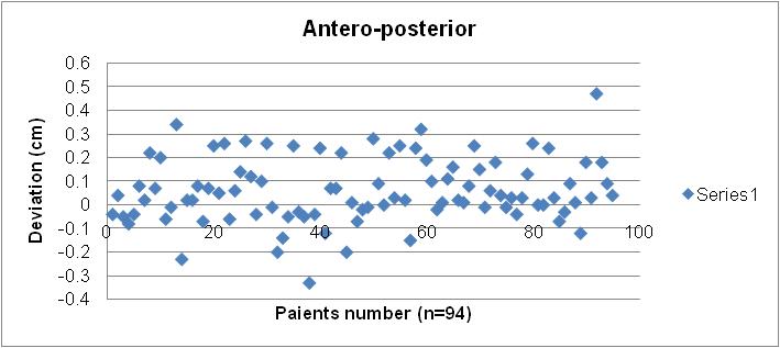

The population systematic error (Σ) in SI; ML and AP direction were 0.13 cm, 0.12 cm and 0.14 cm respectively. The population random error in SI; ML and AP direction were 0.021 cm, 0.022 cm and 0.173 cm respectively.
Setup Margin Calculation : Using van Herk formula, ICRU report 62;Stroms formula CTV to PTV margin calculated. Using van Hark formula CTV to PTV marginin SI; AP and ML direction were 0.34, 0.47 and 0.32 cm respectively. Corresponding values with ICRU report 62 were 0.15, 0.31 and 0.14 cm and with Stroom formula 0.28, 0.40 and 0.26 cmrespectively (Table 1).
Vol 121, No 4, April 2023Journal
of the Indian Medical Association
Fig 2 — Scatter plot of translational deviation in all three directions
38
Fig 3 — Translational displacements in all three directions
Direction Systematic
and AP direction were 0.021 cm, 0.022 cm and 0.173 cm respectively.
In one study by Tejpal, et al9 from ACTREC, Mumbai, India, systematic error was 0.96-1.2 and random error was 1.94-2.48 , 93 % displacements with in 5 mm; <5 mm in all direction CTV to PTV.In another study from Japan, by Suzuki, et al10, systematic error was0.7 -1.3and random error was 0.7-1.6 and they recommend 5 mm margin for PTV, 3 mm margin for PRV. Similar finding were reportedby Durim Delishaj, et al where360 portal images of 60 patients were analyzed and systematic error (Σ) was 0.7 -1.3 and random error ( σ) was 1.1-1.6. Author recommends adding 3-5 mm margin in all direction around CTV to create PTV is adequate 15 Set-up errors and translational displacements, in our study, were comparable to the results of other trial reported in literature (Table 2).
Major strength of our study is we analyzed highest number of portal images (691 portal images of 94 patients) so far reported in literature. Another important strength is our TPS and Linac equipped automatic image overlying auto fusion technology (ARIA) which resulted in the reducing human reading error and more accurate measurement of displacement. Only one drawback of our study is we did not report rotational error. We as Radiation Oncologist for optimal 3dimensional CTV to PTV margin should not adopt blindly published margin formula. Every institution should generate its own set accuracy data. Our study includes large number of patient so we could use these data for calculation from CTV to PTV margin in Head and Neck Malignancy in future treatment in our Department.


CONCLUSIONS
PTV margin(cm)
Table
DISCUSSION
Σσ Displacements or errors
Tejpal et al[7] 0 .96 – 1.21.94-2.4893 % displacements within 5 mm. < 5 mm in all direction CTV to PTV.
To conclude, in our study Set up margin <0.48 cm in all three dimension. Translational displacement in all direction with in 0.5 cm in 99% patients. During course of radiotherapy, set-up accuracy evaluation could be done by taking kV CBCT portal images. Five mm margin all around to the CTV to create PTVis safe, adequate and able to overcome the setup error problems.
Suzuki, et al8 0.7 -1.3 0.7-1.65 mm margin for PTV 3 mm margin for PRV.
Zhang, et al9 1.5- 3.2 1.1-2.95.5 mm margin for 90 % probability for target coverage.
Gilbeu, et al10 1- 2.20.7-2.3 4.5-5.5 mm margin for 90 % probability for target coverage De Boer, et al11 1.5 -21.5- 2probability value not specified.
Humphrey, et al12 0.02-.090 .04-.073 mm for 90% errors, 5 mm for 99 % errors. Our study0.12-0.140.021-0.173 99% displacements within 0.5 cm. < 0.48 cm CTV to PTV margin in all direction.

In our study, the mean displacement in SI; ML; and AP direction were .004 cm (range -0.50 to 0.27 cm); 0.0142 cm (range - 0.30 to 0.42 cm) and 0.0651cm (range -0.33 to 0.47 cm) respectively. In 99% patients’ setup deviation were within 0.5 cm. The population systematic error ( Σ) in SI; ML and AP direction were 0.13 cm, 0.12 cm and 0.14 cm respectively. The population random error (ó) in SI; ML
Conflict of Interest : Nil
Source of Support : Nil
Vol 121, No 4, April 2023Journal of the Indian Medical Association
Fig 4 — Histogram of translational displacements in all three directions including mean and Standard Deviations
2 — Population systemic and random errors selected series Series
Table 1 — Population Systematic and random errors and CTV to PTV margin Population set-up errorsCTV to
Random van Herk ICRUStroom ( Σ )errors (σ)(2.5Σ+ 0.7σ)62(Σ+ σ)(2Σ +0.7σ) SI0.130.0210.340.150.28 AP0.140.1730 .470.310.40 ML0.120.0220.320.140.26 SI –Supero-Inferior, AP-Antero-Posterior; ML –Medio- Lateral
39
of the Indian Medical Association
ACKNOWLEDGEMENTS
The authors express their appreciation to Prof Indira De, MSVP, NRS Medical College & Hospital, for reviewing our statistical analysis. We likewise acknowledge the contributions to their work by Chief Radiation Physicist Mr Anshuman Roy and all radiation technologists of our Radiotherapy Department.
REFERENCES
1International Commision on Radiation Units and Measurements: Prescribing, recording and reporting photon beam therapy (Supplement to ICRU report 50). In ICRU Report, 62 Bethesda, MD: ICRU Publications; 2000.
2Xing L, Lin Z, Donaldson SS, Le QT, Tate D, Goffinet DR, et al — Dosimetric effects of patient displacement and collimatorand gantry angle misalignment on intensity modulated radiation therapy. Radiother Oncol 2000; 56: 97-108.
3Prabhakar R, Laviraj MA, Haresh KP, Julka PK, Rath GK — Impact of patient setup error in the treatment of head and neck cancer with intensity modulated radiation therapy. Phys Med 2010; 26: 26-33.
4Jaffray DA, Drake DG, Moreau M — A radiographic and tomographic imaging system integrated into a medical linear accelerator for localization of bone and soft-tissue targets. Int J Radiat Oncol Biol Phys 1999; 45: 773-89.
5Jaffray DA, Siewerdsen JH, Wong JW — Flat-panel conebeam computed tomography for image-guided radiation therapy. Int J Radiat Oncol Biol Phys 2002; 53(5): 1337-49.
6Ludbrook JJS Correction of systematic setup errors in prostate radiation therapy: how many images to perform? Med Dosim 2005; 30: 76-84.
7Yan D An off-line strategy for constructing a patient’s pecific planning target volume in adaptive treatment process for prostate cancer. Int J Radiat Oncol Biol Phys 2000; 48: 299-302.
8De Boer JCJ A new approach to off-line setup corrections: combining safety with minimum workload. Med Phys 2002; 29: 1998-2012.
9Tejpal Assessment of three-dimensional set-up errors in conventional head and neck radiotherapy using electronic portal imaging device. Radiation Oncology 2007; 2: 44-8.
10Suzuki M, Nishimura Y, Nakamatsu K, Okumura M, Hashiba H, Koike R, Kanamori S, et al — Analysis of inter-fractional set up errors and intra fractional organ motion during IMRT for head and neck tumours to define an appropriate planning target volume (PTV) and planning organ at risk volume (PRV) margins. Radiother Oncol 2006, 78: 283-90.
11Zhang L, Garden AS, Lo J, Ang KK — Multiple regions of interest analysis of set up uncertainties for head and neck cancer radiotherapy. Int J Radiat Oncol Biol Phys 2006, 64: 1559-69.
12Gilbeau L, Octave-Prignot M, Loncol T, Renard L — Comparison of set up accuracy of three different thermoplastic masks for the treatment of brain and head and neck tumors. Radiother Oncol 2001; 58: 155-62.
13De Boer HC, van Sornsen de Koste JR, Creutzberg CL, Visser AG — Electronic portal image assisted reduction of systematic set up errors in head and neck irradiation. Radiother Oncol 2001; 61: 299-308.
14Humphreys M, Guerrero Urbano MT, Mubata C — Assessment of customized immobilization system for head and neck IMRT using electronic portal imaging. Radiother Oncol 2005; 77: 39-44.
15Delishaj D — Set-up errors in head and neck cancer treated with IMRT technique assessed by cone-beam computed tomography: a feasible protocol. Radiat Oncol J 2018; 36(1): 5-62.
Vol 121, No 4, April
2023Journal
40 Submit Article in JIMA — Online See website : https: // onlinejima.com Any queries : (033) 2237-8092, +919477493027; +919477493033
Original Article
Antimicrobial Utilization Study in a Neurology Setting
Namita Vilas Nasare1, Ankit Bhardwaj2, Vineeta Bablani3, Renu Gupta4, Suman Kushwaha5, Sangeeta Sharma6
Background : Measuring antimicrobial consumption is necessary to understand the volume and patterns of use, to design appropriate interventions to reduce and rationalize its use.
Materials and Methods : The antimicrobial consumption in Neurology ICU and IPD were measured over a 5-year period using WHO Defined Daily Dose (DDD) methodology.
Results : There was an increasing trend in Antimicrobial Consumption (AMC) from 125.7 to 155.5 DDDs/100 days over 5 years with highest consumption in 2017-2018 (190.7 DDDs). The consumption of Watch group of antibiotics was higher than access group antibiotics both in ICU and IPD and constituted more than 75% of total antibiotic consumption.
Conclusions : The initiation of empiric therapy though may be necessary depending on the patients condition, however, duration of antibiotic therapy and reducing usage of prophylactic antibiotics for aspiration pneumonia and reducing consumption of Watch group of antibiotics were identified as stewardship opportunities. Feedback on AMC data and persuasive educational interventions to rationalize and reduce antimicrobial use are required.
[J Indian Med Assoc 2023; 121(4): 41-4]
Key words :Defined Daily Dose, Antibiotic Resistance, Antimicrobial Resistance, Antimicrobial Use, Antimicrobial Consumption.
Antimicrobial Resistance (AMR) has been recognized as a major public health problem with catastrophic consequences on human health, economy and food security1. Antimicrobial Use (AMU) is the key driver for AMR and has increased substantially globally, over past decades with nearly 30% of all hospital in-patients receiving antibiotics and out of it around 30-50% of all antibiotics prescribed are reported to be unnecessary, inappropriate or misused2.
Antimicrobial Stewardship (AMS) is urgently required to rationalize antimicrobial use ie, reduce unnecessary use and promote appropriate usage when indicated in order to preserve these precious resources1,2. It is imperative that before initiation of any AMS activities, the magnitude of antibiotic consumption is measured to understand the extent and understand the causes of
Institute of Human Behaviour and Allied Sciences, Delhi 110095
1MSc (Pharmacology), Former Senior Resident, Department of Neuropsychopharmacology
2 MBBS, MD, Former Senior Resident, Department of Neuropsycho Pharmacology
3 MSc (Microbiology), Lab Assistant, Department of Neuropsycho Pharmacology
4MBBS, MD, Assistant Professor, Department of Microbiology and Corresponding Author
5MBBS, MD, DM (Neurology), Professor and Head, Department of Neurology
6MBBS, MD (Pharmacology), MBA, Professor and Head, Department of Neuropsycho Pharmacology
Received on : 23/03/2022
Accepted on : 28/03/2022
Editor's Comment :
Antimicrobial usage is the key driver for antimicrobial resistance; measuring antimicrobial consumption is necessary to reduce its use.
The overall antimicrobial usage and initiation of antibiotics for empirical management may be necessary in Neurology settings but duration of antibiotics should be reduced.
The reduced usage of Watch antibiotics, prophylactic therapy and timely de-escalation were identified as possible interventions towards further reducing antibiotic usage.
irrational prescribing so that interventions for reducing AMU can be designed accordingly1. The consumption data allows monitoring of trends and comparison across countries, regions, healthcare facilities as well as to inform policies, regulations and interventions for optimization of AMU. The Antimicrobial Consumption (AMC) data can also serve as benchmark for riskadjusted inter and intra-facility use and to establish epidemiological association between antibiotic use and resistance over time1
World Health Organization (WHO), Defined Daily Dose (DDD) methodology is an aggregate data collection method. The DDD is the assumed average maintenance dose per day for an antibiotic used for its main indication in adults. It allows identification of broad problem areas with drug use and data can be captured relatively easily3,4
Access, Watch, Reserve (AWaRe) categorization is a WHO tool to classify antibiotics into Access, Watch, Reserve category to target use of narrow-
Vol 121, No 4, April 2023Journal of the Indian Medical Association
41
spectrum antibiotics while reserving broad-spectrum antibiotics for “hardest to treat infections”5. ‘Access’ group are narrow-spectrum antibiotics which should be easily accessible as these are life-saving and have less potential for development of resistance. Access group antibiotics should be preferred over Watch and Reserve group. The ‘Watch’ group comprises broader spectrum antibiotics with higher potential for development of resistance and ‘Reserve’ group consists of last-resort antibiotics for targeted use in multidrugresistant infections 5 . The DDD when linked with ‘AWaRe’ categorization reflects the pattern or quality of antimicrobial use.
The present study was conducted to measure antimicrobial consumption in Intensive Care Unit (ICU) and In-patient Department (IPD) of neurology over a 5-year period using DDD methodology, to understand the quality of prescribing using AWaRe categorization and make recommendations for appropriate AMS strategy(ies).
MATERIALS AND METHODS
The data on antimicrobial consumption was collected retrospectively from Neurology Department (10 bedded Intensive Care Unit (ICU) and 34 bedded In-patient Department (IPD) of a Super-specialty Public Hospital from the indent records. The study was conducted after approval of Institutional Ethics Committee.
Defined Daily Dose (DDD) : The aggregated antimicrobial consumption (except for antitubercular drugs) was calculated year wise using the Anatomical Therapeutic Chemical (ATC) for (J01) DDD Index 2019, as per WHO Collaborating Centre for Drug Statistics Methodology, Oslo (Norway)using AMC tool version 19.1.13,4. Bed occupancy data was collected from Medical Record Department.
Drug Utilization 90% (DU 90) : The average year wise DDD/bed days was arranged in descending order and cumulative DDD/1000 days (DID) calculated and expressed as Drug Utilization 90% (DU 90%).
Access, Watch, Reserve (AWaRe) : The quality of antimicrobial used was assessed by WHO AWaRe categorization method 5 . The antibiotics consumption by AWaRe category was calculated as the percentage consumption in each category.
Statistical analysis :
Data analysis was done using MS Excel and statistical measures like range, frequency, average, percentage, DU90% were calculated.
Trend analysis was done using linear regression.
RESULTS
The trends of total AMC in neuro ICU & IPD is shown in Fig 1. There was increasing trend in AMC over the years from 125.7 to 155.5 DDDs (R² = 0.317) with highest consumption in 2017-2018 (190.7 DDDs). The AMC was higher in ICU compared to IPD in all the years. Figs 2a and 2b depict the year wise antimicrobial consumption of antimicrobials (DU90) in neuro ICU and IPD respectively. The most consumed antimicrobial agents in ICU were ceftriaxone followed by metronidazole, piperacillin-tazobactam, meropenem whereas ceftriaxone, amoxycillinclavulanic acid, metronidazole were most commonly consumed in IPD. Fig 3 depicts percentage consumption of antibiotics based on Access, Watch and Reserve categorization.

DISCUSSION
This study analyzed the trends and pattern of antibiotic consumption in neurology setting. Although there are many reports of antibiotic use in ICU setting but this is the first study reporting AMC in neuro ICU and IPD, to the best of our knowledge. Intensive care settings are an appropriate focus area for AMS as a large proportion of antibiotics use, especially broadspectrum and parenteral antibiotics occurs in these areas. It is important to study extent of use of antimicrobials in neuro settings as the profile of patients admitted here differs from other critical care setting.
We observed a rising trends in AMC over 5 years. The average DDDs of 5 years in Neurology ICU was quite high (116.3) but is comparable to total antibiotic consumption reported from several medical, surgical, and neurosurgical ICUs across India. A large variation has been reported in DDDs, globally, varying from 118.2
Vol 121, No 4, April 2023Journal
of the Indian Medical Association
42
Fig 1 — Trends in total DDD of antimicrobials in neurology ICU and IPD over 5 years
to 560.11 DDDs, however a neurosurgical ICU in Gujarat, India reported DDDs as less as 52.86-10. A large antibiotic surveillance study involving 35 ICUs in Germany reported DDD of 133 which is also more than our ICU11. These differences in the reported DDDs could be due to differences in the criticality and patient profile of the patients admitted in different settings. In the present study, the overall AMC consumption was three-fold higher in ICU compared to IPD with average DDD of 116.3 in ICU and 38.2 in IPD. Higher antimicrobial consumption in ICU can be justified in patients presenting with life-threatening infections requiring empirical broad-spectrum antibiotics within 1 hour of the suspicion of the infection.


The most consumed antimicrobial was ceftriaxone (a Watch group) in both ICU and IPD. In our setting, tubercular meningitis followed by acute meningoencephalitis, seizure disorders (status epilepticus), cerebrovascular accidents (stroke) are the most common clinical presentations. Ceftriaxone is commonly used empirically in critically ill patients on clinical suspicion of an infection to reduce morbidity, mortality and improve functional outcome 12 . The high consumption of ceftriaxone in IPD may also be justified as 14-28 days of therapy with antibiotics is required for completion of a course in a case of bacterial meningitis.
Metronidazole, piperacillin-tazobactam, meropenem and
amoxycillinclavulanic acid were the next commonly prescribed broad spectrum empirical antimicrobials, in ICU similar to the pattern reported in previous studies4-8. These antimicrobials including ceftriaxone were prescribed prophylactically for suspected aspiration/acute aspiration pneumonitis in patients with status epilepticus, CVA (strokes, hemiparesis), multiple sclerosis affecting bulbar functions. Aspiration pneumonitis is most often a chemical injury requiring only supportive care without need to initiate antibiotics, but antibiotics are often prescribed in most patients in practice13,14 Our study reports high use of metronidazole in ICU. Metronidazole was used probably to provide a cover for anaerobes, however, there is limited evidence supporting role of anaerobes in aspiration pneumonia13 There is now sufficient evidence that neither prophylactic antimicrobial therapy nor metronidazole offers any clinical benefit for suspected aspiration/ acute aspiration pneumonitis. In fact, it may result in selection of antibiotic resistant strains requiring escalation of antibiotic therapy and adverse events such as Clostridioides difficile infection 13,14 . Empirical antibiotics with anaerobic coverage should be considered only in the presence of clinical risk factors for aspiration or presence of lung abscess, empyema, or necrotizing pneumonia15.
Vol 121, No 4, April 2023Journal of the Indian Medical Association
Fig 2 — Trends in DDD of antimicrobials (DU 90) in neurology ICU (2a) and IPD (2b) over 5 years (A: Access; W: Watch; R: Reserve)
43
Fig 3 — Trends in percent antibiotic consumption according to Access, Watch and Reserve categorisation in ICU (3a) and IPD (3b)
WHO advocates that Access group of antibiotics should be preferred over Watch and Reserve group to improve access and should comprise 60% of the total antibiotic consumption5. However, in the present study, overall use of Access group antibiotics was ~20% in both ICU and IPD, whereas, Watch group antibiotics accounted for major proportion (>75%). Higher consumption of Watch group antibiotics in ICU could be because of its empirical use in life threatening infections as seen in Neuro ICU. In the present study though, AMC was lower in IPD but Watch group antibiotics comprised the major proportion. One of the reasons for high proportion could be failure to deescalate the antibiotic started in ICU. Further, in ICU over the years, a gradual decline in consumption of Watch group antibiotics was observed without any change in consumption of Watch group in IPD.
The overall antimicrobial consumption and increasing trends require active intervention by providing feedback to the prescribers with targeted persuasive education programs to rationalize antimicrobial use and decrease overall consumption.
LIMITATIONS
DDD of anti-tubercular drugs has not been calculated as medicines for these patients are obtained directly from the DOTS centre. Further, DDD data should also be collated with the antimicrobial resistance data to guide antibiotic therapy in accordance with local antibiogram.
CONCLUSION
The overall extent and quality of antimicrobial use, initiation of empiric antibiotic therapy was appropriate and comparable to other published studies but there are opportunities to further reduce antimicrobial use particularly of Watch group antibiotics. AMS interventions such as feedback on AMC data and persuasive educational interventions, guidelines for management of common neuro-infections based on antibiogram, regular review and documentation of start and stop dates of antibiotics should be implemented. Regular audit of AMS actions such as de-escalation to narrower spectrum antibiotic or IV to oral switch after 48-72 hours can further help in timely deescalation. To reduce the prophylactic antibiotics in aspiration pneumonia guidelines on prophylactic use of antibiotics for aspiration Pneumonia need to be developed. Cohort nursing training can reduce the incidence of aspiration reduction and thus reducing the antibiotic usage.
Source(s) of Support : Nil
Conflicting Interest : None
REFERENCES
1World Health Organization — Antimicrobial stewardship programmes in health-care facilities in low- and middle-income countries: a WHO practical toolkit 2019.https:// academic.oup.com/jacamr/article/doi/10.1093/jacamr/dlz072/ 5623027[accessed 16Jul2021]
2Indian Council of Medical Research (2018) — Antimicrobial Stewardship Program Guidelines. Retrieved fromhttps:// main.icmr.nic.in/sites/default/files/guidelines/AMSP_0.pdf [accessed January 30, 2021].
3AMC Tool: the antimicrobial consumption tool. Available from: https://amu-tools.org/amctool/amctool.html[accessed 20 Jul2021].
4World Health Organization — WHO Collaborating Centre for Drug Statistics Methodology, ATC classification index with DDDs, Oslo, Norway. https://www.whocc.no/atc_ddd_index/ [accessed 20 Jul2021].
5World Health Organization — WHO AWaRe Classification Database of Antibiotics for evaluation and monitoring of use.http s://www .who.int/publications-detail-redirect/ WHOEMPIAU2019.11 [accessed 20 Jul2021].
6Kavar KS, Jha RK, Gaikwad NR, Jarande SS, Ranpura AM — Antibiotic Use Density in Medicine ICU in a Tertiary Care Rural Hospital of Central India. Res J Pharm Biol Chem Sci 2012; 3(1): 133-42.
7Sarin K, Vadivelan M, Bammigatti C — Antimicrobial Therapy in the Intensive Care Unit. Indian J Clin Pract 2013; 23(10): 6019.
8Anand N, Nagendra Nayak IM, Advaitha MV, Thaikattil NJ, Kantanavar KA, Anand S — Antimicrobial agents’ utilization and cost pattern in an Intensive Care Unit of a Teaching Hospital in South India. Indian J Crit Care Med 2016; 20(5): 274-9.http s://www .ncbi.nlm.nih.gov/pmc/articles/ PMC4876648/
9Saxena S, Priyadarshi M, Saxena A, Singh R — Antimicrobial consumption and bacterial resistance pattern in patients admitted in I.C.U at a tertiary care center. J Infect Public Health 2019; 12(5): 695-9.https://www.sciencedirect.com/ science/article/pii/S1876034119301236
10Shelat PR, Gandhi AM, Patel PP — A Study of Drug Utilization Pattern According to Daily Define Dose in Intensive Care Unit (ICU)s at Tertiary Care Teaching Hospital, India. JYoungPharm 2015; 7(4): 349-58. http://www.jyoungpharm.org/article/788
11Meyer E, Schwab F, Gastmeier P, Rueden H, Daschner FD — Surveillance of Antimicrobial Use and Antimicrobial Resistance in German Intensive Care Units (SARI): A Summary of the Data from 2001 through 2004. Infection 2006; 34(6): 303-9. https://www.ncbi.nlm.nih.gov/pmc/articles/PMC2778699/
12Singh S, Rattan A, Goel N, Nangia V, Manchanda V, Ghosh S, et al — Convergence of Minds: For Better Patient Outcome in Intensive Care Unit Infections. Indian J Crit Care Med 2017; 21(3): 154-9. http s://www.ijccm.org/doi/10.4103/ ijccm.IJCCM_365_16
13Dragan V, Wei Y, Elligsen M, Kiss A, Walker SAN, Leis JA — Prophylactic Antimicrobial Therapy for Acute Aspiration Pneumonitis. Clin Infect Dis Off Publ Infect Dis Soc Am 2018; 67(4): 513-8.
14Kwong JC, Howden BP, Charles PGP — New aspirations: the debate on aspiration pneumonia treatment guidelines. Med J Aust 2011; 195(7): 380-1. https://onlinelibrary.wiley.com/ doi/10.5694/mja11.10298
15Medha — Antibiotic Prescription in ICU: Indian Critical Care Medicine Society Releases Guidelines 2019. https:// speciality.medicaldialogues.in/first-ever-india-centricguideline-for-antibiotic-use-in-icu
Vol 121, No 4, April 2023Journal of the
Medical
Indian
Association
44
Original Article
An Effective Management for Thalassaemic Patients during Pregnancy
Arpita Das1, Sangeeta Ghosh2, Sandip Ghosh3, Tapas Kumar Sur4
Background : Presence of thalassaemia is considered high risk in pregnancy. The present study find out the role of antenatal and intrapartum care to improve the outcome of pregnancy in thalassaemia.
Materials and Method : The prospective longitudinal study was carried out on 25 patients. CBC, LFT, Ferritin, Hb electrophoresis, serial USG was done. Chorionic villous sampling was offered to thalassaemic women to decrease the number of babies born with Thalassaemia. Furthermore, management and treatment options provided to mother to overcome the incidence of maternal and neonatal complications during current pregnancy.
Results : 44% of thalassaemic pregnant women had HbE β thalassaemia, 8% β thalassaemia major and others had thalassaemia carriers, while only 20% of their partners showed β thalassaemia trait. All patients were anemic and during delivery mean Hb% were 8.42 g/dl, MCH 22.09 pg, MCV 73.56 fl, MCHC 29.86 g/dl and ferritin 241.51 ng/ ml. Third trimester USG exhibited 32% developed IUGR (Intrauterine growth restriction). 40% patients received blood transfusion after delivery. β thalassaemia trait was identified in 20% babies at 6 month.
Conclusion : Postpartum haemorrhage is a major complication of thalassaemia in pregnancy. After delivery a thorough neonatal check-up and haematological work-up is important for prevention of neonatal mortality and early detection of thalassaemia. [J Indian Med Assoc 2023; 121(4): 45-8]
Key words : β β-thalassaemia, Primigravida, Trimester, Chorionic Villous Sampling, IUGR.
Thalassaemia syndromes are autosomal recessive disorders and the most commonly inherited haemoglobinopathies in the world1. It is of two types: α-thalassaemia and β-thalassaemia. In β-thalassaemia, there is an impaired synthesis of β chains of globin part resulting in low concentration of normal HbA with a compensatory increase in HbF and/or HbA22-3. In βthalassaemia major (homozygous form), both β chains are missing and there is severe anaemia and secondary organ damage4. Regular transfusions are necessary to maintain adequate RBC levels, which further aggravates the iron accumulation due to shorter half-lives of transfused RBC5. In thalassaemia minor (heterozygous state), only one locus of the globin chain is lacking which results in microcytosis without significant anaemia6. Women with β-thalassaemia minor are mildly anaemic but generally otherwise healthy. Hb E βthalassaemia is a double heterozygous state of Hb E and β-thalassaemia and it is the most common type of thalassaemia noted in Eastern India7. Approximately, 15 million people are globally affected in thalassemia8 Thalassaemia mandates close maternal and foetal
1MBBS, MD, Medical Officer, ESI Hospital, Maniktala, Kolkata 700067
2MBBS, MD, Assistant Professor, Department of Microbiology, RG Kar Medical College and Hospital, Kolkata 700004 and Corresponding Author
3MBBS, MS, Principal, RG Kar Medical College and Hospital, Kolkata 700004
4MSc, PhD, Research Scientist II, Multidisciplinary Research Unit (ICMR), RG Kar Medical College and Hospital, Kolkata 700004
Received on : 22/03/2022
Accepted on : 12/08/2022
Editor's Comment :
Thalassaemia in pregnancy is a high risk. Proper management of pregnant thalassaemic mother by monitoring during the antenatal, intrapartum and postpartum periods only can facilitate to decrease the associated maternal and neonatal mortality and morbidity.
surveillance during pregnancy. Antenatal counselling is required for all antenatal mothers with thalassaemia. In case of minor thalassaemia, partner should be tested for thalassaemia trait9 . If the partner is also trait, antenatal diagnosis should be offered. Chorionic Villous Sampling should be done between 10-13 weeks to determine if the foetus is affected or not. If thalassaemia major is diagnosed, termination of pregnancy is offered10. Thalassaemia in pregnancy is a high risk and therefore, hospital delivery is mandatory. Several guidelines are set up for the proper management of such a case during both antenatal and postnatal period11-13. The objectives of this study were to find out the treatment options offered to pregnant women with thalassaemia to reduce the morbidity and the mortality during antepartum, intrapartum and postpartum period and to study neonatal outcome in babies born to mothers with thalassaemia along with the thalassaemia workup of the baby.
MATERIALS AND METHODS
This longitudinal study was carried out in the Department of Obstetrics and Gynaecology, Eden Hospital, Medical College, Kolkata after receiving approval from Institutional Ethics Committee. Among
Vol 121, No 4, April 2023Journal of the Indian Medical Association
45
all antenatal mothers attending Obstetrics Out-patient Department with suspected and diagnosed thalassaemia in pregnancy, 25 pregnant women were randomly selected. Guideline of management of βthalassaemia in pregnancy was strictly followed11-13. The inclusion criteria were antenatal mothers in the age group of 18-45 years diagnosed to have β thalassaemia major, E β thalassaemia and thalassaemia trait. Exclusion criteria were antenatal mothers with associated serious medical disorders like Heart disease, Endocrinological disorders, Infective disease etc.
Total 25 antenatal mothers conforming to the inclusion and exclusion criteria were recruited after receiving their informed consent. The following investigations were conducted: (i) Routine investigations: Complete Haemogram, Liver Function Test and Serial Ultrasound Review; and (ii) Special Haematological Investigations: Hb Electrophoresis, Serum Ferritin, Reticulocytes C ount, MCV, MCH, MCHC. Furthermore, Management and Treatment options provided to mother to overcome the incidence of maternal and neonatal complications during current pregnancy.
Data were analysed and expressed as descriptive pattern. Statistical analysis was carried out using the Chi square test, independent Sample t-test, MannWhitney U test and paired t-test wherever appropriate. P value less than 0.05 was considered statistical significance. For the data analysis, statistical software SPSS were used.
RESULTS
Table 1 exhibited the demographic details of selected patients. In this study, 25 number of β-thalassaemic pregnant women at mean age of 23.36 years (range 19-30yrs) were selected. Proportion of primi gravid patients were significantly higher (n=11; 44%) than other parity groups. HPLC reports confirmed 44% of patients had HbE β-thalassaemia, 24% each of HbE trait and βthalassaemia trait; only 5% of their husbands were βthalassaemia carrier. 32% of women having history of Abortion, 20% Infertility and 40% Jaundice. Blood examination reports depicted that mean Hb was 7.54 g/dl in the time of booking and 8.42 g/dl during delivery. Moreover, Serum Ferritin was 241.51ng/ml, Reticulocyte was 4.9%, MCH 22.09 pg, MCV 73.56 fl, MCHC 29.86 g/dl and Bilirubin was 1.45 mg/dl (Table 2).
USG reports of β-thalassaemic pregnant mother in third trimester showed 24% IUGR, 12% Oligohydramnios and 8% IUGR with Oligohydramnios, while 56% was normal (Table 3). Frequency of blood transfusion during pregnancy was 6.84 times
*Results
(maximum 15 times) and highest frequency of blood transfusion was after delivery (40.0%) followed by blood transfusion every 15 days (32.0%). 40% mothers showed Splenomegaly, 12% mothers developed gestational Diabetes Mellitus and 16% pregnancy induced hypertension. Percentage of Caesarean section was significantly higher than vaginal delivery (Z=2.5456, p= 0.01078).
The analysis of indications for Caesarean section showed a wide variability. 20% patients had post partum haemorrhage, 12% patients developed wound infection.
Table 4 showed the physical indicators of neonates of β-thalassaemia Trait-thalassaemic mothers. Birth weight of neonates was 2.51 kg (range 1.40-3.25 kg), 24% neonates developed hypoglycaemia and 20% βthalassaemia trait was found in babies at 6 m.
Vol 121, No 4, April 2023Journal of the Indian Medical Association
Number of subjects25 Mean age (years)23.36 ± 2.87* <2016% 21-2560% 26-3024% History of abortion Yes32% No68% History of infertility Yes20% No80% Mother’s HPLC report Homozygous β-thalassaemia 8.0% β-thalassaemia trait24.0% HbE Trait 24.0% HbE β-thalassaemia 44.0% Husband’s HPLC report β-thalassaemia trait20.0% Normal 80.0% CVS report of husbands β-thalassaemia Carrier12.0% Normal 8.0% Not done 80.0% Jaundice Yes40% No60%
Table 1 — Demographic data of β-thalassaemia pregnant women
are mean ± SD
Mean ± SDRange (min – max) Haemoglobin (g/dl) at booking7.54 ± 1.23 5.4-9.20 at delivery 8.42 ± 0.806.8-10.10
(ng/ml)241.51 ± 92.30 123.4-576.5
(%) 4.92 ± 1.87 2.0-8.0 MCH
19.30-23.60
Table 2 — Blood examination of β-thalassaemia pregnant women
Ferritin
Reticulocytes
(pg)22.09 ± 1.27
MCV (fl)73.56 ± 3.9167.0-80.0
29.86
MCHC (g/dl)
± 2.2616.0-34.40
Serum bilirubin (g/dl)1.45 ± 0.970.50-3.40
46
N=25; results are mean ± SD
DISCUSSION
In 1936, George Whipple was first who coined thalassaemia from the Greek word “thalassa” for sea and “hema” for blood14. Pregnancy in women with βthalassaemia Trait-thalassaemia major is rare until recently due to their short life span, multiple organ damage and reduced fertility. Moreover, hypogonadotropic hypogonadism is the most frequent in β -thalassaemic major with marked sexual dysfunction, infertility and short stature15. Presence of thalassaemia is considered as high risk at pregnancy and the pregnant mothers should be monitored properly during antenatal period and cared enthusiastically during intrapartum and postpartum periods to decrease maternal morbidity and mortality13
Pre-marriage counselling of couples and prenatal screening of partners of thalassaemic mothers should
be considered to decrease the thalassaemic burden of society. Screening is able to identify couples having a 25% risk or more having a pregnancy with a significant haemoglobinopathy. In the present study, HPLC report of all thalassaemic pregnant women showed frequency of HbE β -thalassaemia was maximum (44%), while 5% of their husbands were βthalassaemic carrier. In India, HbE β -thalassaemia is frequent for individuals to inherit alleles for both Hb E and β -thalassaemia16. The estimated number of βthalassaemia carriers in India is about 29.7 million, while births of β-thalassaemia patients are 7000 per year17. Over the last 20 years more than 200 different varieties of molecular defects causing β-thalassaemia were characterized affecting RNA transcription and processing or translation of the β-globin gene18
Anaemia has deleterious effects both on mother as well as on growing foetus. WHO defined anaemia in pregnancy as Hb% is less than 11g and CDC as Hb% is less than 11g during the first and third trimesters and less than 10.5g during the second trimester 19-20 . Anaemia in thalassaemic mothers is to be corrected with adequate blood transfusion, to decrease maternal morbidity and mortality associated with anaemia in pregnancy. In the present study, Hb was 7.54 g% during pregnancy and 8.42 g% after birth. Blood transfusion frequency was 32% in every 15 days and 40% after delivery to treat anaemia in β-thalassaemic mother. The rate of blood transfusion in this time period was 6.84 times. In other report, it has mentioned that transfusion therapy has given during pregnancy to maintain Haemoglobin >10 g/dl to ensure appropriate foetal growth21. Furthermore, serum ferritin level of was 241.51 ng/ml. No β-thalassaemic mother had iron overload due to iron chelation therapy. Unfortunately Splenomegaly (40%), Jaundice (40%), Pregnancy induced hypertension (16%) and Gestational diabetes (12%) were observed in β-thalassaemic mothers.
Thalassaemia in combination with anaemia reported for different complications of the thalassaemic pregnancy, like fatal Intrauterine Growth Restriction (IUGR) and preterm labor12. According to guideline, first Ultrasound scan has performed at 7th-9th week of gestation, followed by first-trimester (11th-14th weeks), second trimester (18th-21st weeks) and third trimester (24th week), focusing on possible IUGR11,21. For safety, Chorionic Villous Sampling (CVS) has been done when both mother and husband were affected by thalassaemia to decrease the number of babies born with thalassaemia. USG at third trimester showed 32% IUGR and 56% normal. Moreover, post partum haemorrhage is a major complication of thalassaemia in pregnancy. Hence, serial USG with colour Doppler
Vol 121, No 4, April 2023Journal of the Indian Medical Association
Birth weight (kg)2.51 ± 0.53 Neonatal jaundice Absent68% Present32% Neonatal hypoglycemia Absent76% Present24% At 6 month blood test β-thalassaemia trait20% Normal80%
Table 4 — Physical indicators of neonates of βthalassaemic mother
Third trimester USG : IUGR24% IUGR, Oligohydamnios8% Oligohydamnios12% WNL56% Mean blood transfusion 6.84 ± 5.39 Blood transfusion frequency : After Delivery40% Every 60 days8% Every 45 days4% Every 30 days16% Every 15 days32% Organomegaly : Absent 60% Splenomegaly40% Gestational diabetes : Absent88% Present12% Pregnancy induced hypertension : Absent84% Present16% Mode of delivery : Caesarean section68% Vaginal32% Postpartum haemorrhage : Absent80% Present20% Postpartum wound infection : Absent88% Present12% 47
Table 3 — Clinical examination of β-thalassaemia mother
is suggested to diagnose IUGR and other complications like oligohydramnios.
Thalassaemia is not an indicator for Caesarean section. If vaginal delivery is decided, active management of the third stage of delivery is recommended, as this intervention is supposed to reduce blood loss 23 . In this study, 68% patients underwent caesarean section. 17.6% caesarean section was done due to IUGR, 11.8% for oligohydramnios and 5.9% for IUGR with oligohydramnios and rests were due to obstetric and medical indications. Aessopos et al (1999) performed caesarean delivery in all patient24. Mean birth weight of 25 babies was 2.51 kg, with a range from 1.40 to 3.25 kg. Neonatal complications were mainly Jaundice (32%) and Hypoglycaemia (24%). Furthermore, β thalassaemia trait was detected only in 20% babies at 6 month. The incidence of complications observed in our study was less than other recent reports25-26
CONCLUSION
Pregnancy in thalassemia should be considered a high risk and need special medical attention, monitoring and treatment throughout the antepartum, intrapartum and postpartum periods to overcome the pregnancy associated complications and mortality of both mother and baby.
ACKNOWLEDGEMENT
The authors appreciate the support and cooperation of personnel of Department of Obstetrics and Gynaecology, Medical College, Kolkata for helping collection of data.
Funding : The funding source of all authors was the Department of Obstetrics and Gynaecology, Eden Hospital, Medical College, Kolkata.
Conflict of Interest : None
Limitations : The study was conducted in a single centre and small number of population; hence multi centric study is suggested.
REFERENCES
1Galanello R, Origa R — Beta-thalassaemia. Orphanet J Rare Dis 2010; 21; 5:11 https://doi.org/10.1186/1750-1172-5-11
2Weatherall DJ — The inherited diseases of hemoglobin are an emerging global health burden. Blood 2010; 115: 43314336.
3Rund D, Rachmilewitz E — Beta-thalassaemia. N Engl J Med 2005; 353: 1135-46.
4Cohen AR, Galanello R, Pennell DJ, Cunningham MJ, Vichinsky E — Thalassemia. Hematology Am Soc Hematol Educ Program 2004; 2004: 14-34.
5Borgna-Pignatti C, Rugolotto S, De Stefano P, Zhao H, Cappellini MD, Vecchio CD, etal— Survival and complications in patients with thalassemia major treated with transfusion and deferoxamine. Haematologica 2004; 89: 1187-93.
6Cao A, Galanello R — Beta-thalassaemia. Genet Med 2010; 12: 61-76.
7Soliman A, Yasin M, El-Awwa A, Osman M, De Sanctis V — Acute effects of blood transfusion on pituitary gonadal axis and sperm parameters in adolescents and young men with thalassaemia major: a pilot study. Fertil Steril 2012; 98: 63843.
8Global distribution of Thalassemia. Available from: www.lawesome12.blogspot.in/2012/03/global-distributionof-thalassamia.html
9Origa R, Comitini F — Pregnancy in thalassaemia. Mediterr J Hematol Infect Dis 2019; 11(1): e2019019, DOI: http:// dx.doi.org/10.4084/MJHID.2019.019
10Saxena R, Banerjee T, Aniyery RB — Thalassemia and its management during pregnancy. World J Anemia 2017; 1: 517.
11Guidelines of Management of Beta Thalassaemia in Pregnancy. Green-top guideline No. 66, Royal College of Obstetricians and Gynaecologists, London, 2014.
12National Institute for Health and Clinical Excellence. Intrapartum care: Care of healthy women and their babies during childbirth. NICE clinical guideline 55. Manchester: NICE; 2007.
13Petrakos G, Andriopoulos P, Tsironi M — Pregnancy in women with thalassaemia: challenges and solutions. Int J Women’s Health 2016; 8: 441-51.http://dx.doi.org/10.2147/ IJWH.S89308
14Whipple GH, Bradford WI — Mediterranean disease: thalassaemia (erythroblastic anemia of Cooley). J Pediatr 1936; 9: 279-311.
15De Sanctis V, Soliman AT, Elsedfy H, Skordis N, Kattamis C, Angastiniotis M, et al — Growth and endocrine disorders in thalassemia: the international network on endocrine complications in thalassaemia (I-CET) position statement and guidelines. Indian J Endocrinol Metab 2013; 17: 8-18.
16De M, Das SK, Bhattacharya DK, Talukder G — The occurrence of â-thalassaemia mutation and its interaction with haemoglobin E in the Eastern India. Int J Haematol 1997; 66: 31-4.
17Verma IC, Saxena R, Thomas E, Jain PK — Regional distribution of â-thalassaemia mutations in India. Hum Genet 1997; 100: 109-13.
18Rosatelli MC, Saba L — Prenatal diagnosis of â-thalassemia and haemoglobinopathies. MediterrJHematolInfectDis2009; 15; 1(1): e2009011. https://doi.org/10.4084/MJHID.2009.011 PMid:21415992 PMCid:PMC3033155
19World Health Organization. Nutritional anaemia. Tech Rep Ser 1972; 503.
20CDC criteria for anaemia in children and child-bearing-aged women. MMWR Morb Mortal Wkly Rep 1989; 38: 400-4.
21Origa R, Piga A, Quarta G, Forni GL, Longo F, Melpignano A, et al — Pregnancy and beta-thalassaemia: an Italian multicenter experience. Haematologica 2010; 95: 376-81.
22Levy A, Fraser D, Katz M, Mazor M, Sheiner E — Maternal anemia during pregnancy is an independent risk factor for low birth weight and preterm delivery. Eur J Obstet Gynecol Reprod Biol 2005; 122: 182-6.
23Begley CM, Gyte GM, Devane D, McGuire W, Weeks A — Active versus expectant management for women in the third stage of labour. Cochrane Database Syst Rev 2011: CD007412.
24Aessopos A, Karabatsos F, Farmakis D, Katsantoni A, Hatziliami A, Youssef J, et al — Pregnancy in patients with well-treated beta-thalassaemia: outcome for mothers and newborn infants. Am J Obstet Gynecol 1999; 180: 360-5.
25Sorrentino F, Maffei L, Caprari P, Cassetta R, Dell’Anna D, Materazzi S, et al — Pregnancy in thalassemia and sickle cell disease: the experience of an Italian thalassaemia center. Front Mol Biosci 2020; 7: 16. doi: 10.3389/fmolb.2020.00016
26Lao TT — Obstetric care for women with thalassaemia. Best Pract Res Clin Obstet Gynaecol 2017; 39: 89-100. doi: 10.1016/j.bpobgyn.2016.09.002
Vol 121, No 4, April 2023Journal
of the Indian Medical Association
48
Original Article
Reproductive Tract Infection : Challenges to Health Policy and Education, Research and Impact on the Socio-economic Situation of Indians
Dilip Kumar Dutta1, Indranil Dutta2, Ipsita Dutta3
Background : Reproductive Tract Infection (RTI) due to poor hygiene caused by faecal Bacteria or viruses which goes into the vaginal canal during and after defecation is not only damaging to the reproductive organ, but also causes indirect or direct impact on Socio-economic status of the patient and their family as well.
Objective : This study highlights the importance of faecal hygiene in preventing damage to the reproductive tract and curbing the impact on social and economic problems in patients and their families.
Materials and Methods : This study was conducted at GICE Nursing Home, Kalyani, Nadia, WB, between April 2016 and March, 2022. During this period, 11,872 cases were reported at GICE Nursing Home of which 10,000 cases were selected.
Discussion : It was interesting to observe that those who wipe from back to front or vice versa are more likely suffering from Vaginitis (85%), Cervicitis (92%), Endometritis (55%), PID (60%), UTI (70%) and Tubal Block (17%) as compared to those who used Hand shower (10%) or Tissue Paper (9%). This is an indication that proper faecal hygiene can prevent damage to the reproductive system. Further analysis revealed that the inability to pay for medical expenses created a Socio-economic crisis among family members.
Conclusion : It is concluded from this study that maintenance of proper faecal hygiene by women in Rural, Urban and Industrial areas, can not only help with prolonged illnesses but also prevent financial losses. As a result, Guardians, Teachers, Health Care Providers and Local Government should conduct outreach programs to teach the importance of faecal hygiene at home, in Schools, Colleges, Public health institutions and so on, etc.
[J Indian Med Assoc 2023; 121(4): 49-51]
Key words :Faecal Hygiene, Reproductive Tract Infection, Socio-economic Problems.
Reproductive Tract Infections (RTIs) such as Gonorrhoea, Syphilis, Chlamydia, Bacterial Vaginosis (BV), Lymphogranuloma Venereum (LGV), Chancroid and Trichomoniasis are regarded as the second most common infectious disease in the world9, and a silent epidemic in women’s lives. RTis may present a wide variety of clinical symptoms, including Acute, Chronic, Complicated, Simple, Symptomatic and Asymptomatic.
Women with untreated RTIs often experience complications such as infertility, chronic and pelvic pain and other conditions that may have a lifetime impact 8 . In underserved areas like Rural and Industrialised areas, RTIs can cause peripartum maternal mobility and mortality5.
In this study, we concentrate on the effects of RTIs
1MD (OBG), FRCOG (London) PhD, Director, GICE Nursing Home & Diagnostic Centre, Kalyani, West Bengal 741235 and
Corresponding Author
2MBBS, MS (OBG), DIPALS (Germany), Professor, IQ City Medical College Hospital, Durgapur, West Bengal 713206
3MA, International Media Business, UK
Received on : 28/10/2022
Accepted on : 17/11/2022
Editor's Comment :
Reproductive tract infection due to poor hygiene caused by faecal Bacteria or viruses which goes into the vaginal canal during and after defecation is not only damaging to the reproductive organ, but also causes indirect or direct impact on socio-economic status of the patient and their family as well.
Awareness programs must be conducted in rural/urban & industrial areas to teach the importance of maintaining faecal hygiene such as the importance of using hand showers or tissue papers during bowel movements can deter such medical cases in the future.
amongst patients in different societal areas and how the proper faecal hygiene can curb the negative social and economic impact.
MATERIALS AND METHODS
This study was conducted at GICE Nursing Home, Kalyani, Nadia, WB, between April, 2016 and March, 2022. During this period, 11,872 cases were reported at GICE Nursing Home of which 10,000 cases were selected.
Theprimaryobjectivesofthatstudywere— (a)Be aware of the types of wipes used during bowel movements.
Vol 121, No 4, April 2023Journal of the Indian Medical Association
49
(b)To uncover any pathological changes to the reproductive tract/ urinary bladder.
(c)Assess the socio-economic impact on the patient and family.
OBSERVATIONS
Between April, 2016 and March, 2022, 11,872 cases were observed at a GICE Nursing Home - 10,000 cases were selected for this study (Table 1).
Out of 10,000 cases, 62% of women were Hindu and 30% were Muslim, which outnumbered the other minorities (Table 2).
The majority of women resided in rural (47%) and industrial (40%) regions, compared to urban (13%) (Table 3).
It was mentioned that the Educational Qualifications amongst women comprised of <10 school pass (32%), Higher Secondary pass (26%), UG degree holder (28%) & PG degree holder (14%). It meant that qualification has no effect on reproductive hygiene (Table 4).
The results showed that 58% of patients came from lowincome areas, while 32% came from middle-income households, compared with 10% who had high economic status (Table 5).
Upon detailed inquiry, it was discovered that 15% of women spent more than Rs 4000/year in medical expenses as compared to 58% of women who spent Rs 2000-Rs 4000/year. In addition, the other 27% of respondents spent between 1000 and 2000 rupees/year, respectively. Consequently, the estimated financial burden of 10,000 cases amounted to nearly 1 to 4
crores per year (Table 6).
Concerning the use of various methods adopted after the passage of bowel movements, it was observed that 40% had the habit of washing from back to front and 35% used front to back method using water as compared to either used Hand shower (10%) or used Tissue paper (9%) (Table 7).
It was noted that those who used to wash front to back or vice versa, they are mostly suffering from Vaginitis (85%), Cervicitis (92%) and Endometritis (55%), PID (65%), UTI (70%) and Tubal block (17%) as compared to those who used hand shower and tissue paper (Table 8).
In culture and sensibility (N-1000), Trichomoniasis (47.5%) predominated over E Coli (21.7%), Bacterial vaginitis (11.1%), Streptococci (group B) - 7.6%, etc, respectively (Table 9).
After initial treatment by proper counselling, use of antibiotics, Metrogyl tab, Antiseptic, Betadine lotion (hip bath) – 50% (N-5000), each group were counselled to use tissue paper/ and or hand shower, during wiping for 6 months to 1 year (Table 10).
Out of 2000 cases, the incidence of Reproductive Tract Infections was found to be reduced by 9.4% (Vaginitis), 4.7% (Cervicitis) and 2.6% (Endometritis),
Vol 121, No 4, April 2023Journal of the Indian Medical Association
YearCases Seen April 2016 to March 2017782 April 2017 to March 2018 2983 April 2018 to March 2019 2716 April 2019 to March 2020 2000 April 2020 to March 2021 1519 April 2021 to March 2022 1872
Table 1— Total Cases Seen (N =11,872)
Religion PercentagesTotal No Hindu62% (6200) Muslim30% (3000) Christian8% (800)
Table 2 — Religion (N= 10,000)
Place of Origin PercentagesTotal No Rural47% (4700) Urban13%(1300) Industrial40% (4000)
Table 3 — Place of Origin (N= 10,000)
Education PercentagesTotal No <10 Class Pass32% (3200) 10-1226% (2600) Degree28% (2800) Post Degree14% (1400)
Table 4 — Education (N= 10,000)
Income PercentagesTotal No Low58%(5800) Medium32% (3200) High10% (1000)
Table 5 — Income (N= 10,000)
Rupee/yr (Rs) PercentagesTotal No <10005% (500) 1001-200022%(2200) 2001-300038%(3800) 3001-400020%(2000) >400015%(1500)
Table 6 — Financial Liability (N= 10,000)
Types PercentagesTotal No Back to Front46% (4600) Front to Back35% (3500) Hand Shower 10%(1000) Tissue Paper9% (900)
Table 7 — Types of Wiping (N= 10,000)
Types Percentages Number Trichomoniasis 47.5%475 Bacteria Vaginitis11.1%111 E.Coli (Echerichiacoli ) 21.7%217 Streptococci (Group-B) 7.6%76 Staphylococci Aureus 3.9%39 Neisseria Gonorrhea 4.1%41 Klebsiella Preumoriae 2.5%25 Listeria Mmocytogene… 1.6%16
Table 9 — Type of Organism (N=1000)
Findings PercentagesTotal No Vaginitis 85%(8500) Cervicitis92% (9200) Endometritis55% (5500) PID65%(6500) UTI70%(7000) Tubal Blockone Side (10%), Both Side ( 7%)17% (1700) 50
Table 8 — Clinical Findings (N= 10,000)
indicating the importance of maintaining hygiene. It also aided in overcoming mental agony, social issues and financial liabilities caused by prolonged illnesses (Table 11).
DISCUSSION
Reproductive Tract Infection due to Faecal Bacteria or virus that gets into the vaginal canal during and after passing stool, is not only damaging to the reproductive tract leading to PID, Infertility, Cervical Cancer, etc, but also found to cause direct or indirect impact on the socio-economic burden on patients and their families1-4,6,7.
In 82% of adult females who attended GICE Outpatient Department (OPD) were from Rural & Industrial areas in and around Kalyani, Nadia, WB, with education 8-12 class pass (52%) and low Socioeconomic status (50%) respectively. All present at the clinic had a history of abdominal pain, white discharge, irregular menstruations, dysuria and infertility, etc. Following appropriate consultation, various cases of contamination have been discovered by Faecal Hygiene, historical bathing in the polluted pond, inadequate Menstrual Hygiene, etc. Additional studies were performed to validate the acquired data, using clinical tests, S/E smears, colposcopy and USG
It was observed that those who used just used water to wash from the back to the front or vice versa suffered from Vaginitis (85%), Cervicitis (92%), Endometritis (55%), PID (60%), UTI (70%), and Tubal block (17%) as compared to those who used hand shower (10%) or Tissue paper (9%). Significantly highlighting that proper faecial hygiene has a wider impact of preventing harm to the reproductive tract. Additionally, responders were advised to use hand showers or tissue papers apart from receiving treatment through counselling, antibiotics, metrogyl and vaginal wash. A follow up checkup was also advised after 6 months - 1year of treatment .
As regards the Socio-economic impact, 15% of women stated that they spent more than Rs 4000 per year as compared to 58% of women who spend Rs 2000-4000/year and 27% had spent Rs 1000-2000/ year respectively, costing roughly 1-4 crores for 10,000 responders.
A follow-up study conducted after 6 months of the original study, saw 2,000 returnees who attended the clinic for infertility/ other treatments. Most of them had a positive response to treatments with infections having significantly decreased after usage of appropriate hygiene methods. Of these, only 9.4% were still suffering from Vaginitis, 4.7% from Cervicitis and 2.6% from Endometritis.
CONCLUSION
Damage to the Reproductive System caused by a Bacterial, Viral or Parasitic infection that enters the vaginal canal may affect the reproductive organ and cause socio-economic/mental problems in addition to being potentially harmful.
Awareness programs must be conducted in rural/ Urban & Industrial areas to teach the importance of maintaining Faecal Hygiene such as the importance of using Hand Showers or Tissue Papers during bowel movements can deter such medical cases in the future.
Hence, active action must be taken to educate the masses by Tutors, Teachers, Government and Healthcare Providers by organising campaigns in Homes, Schools, Colleges, PHC, Hospitals and Private institutions.
REFERENCES
1Wasserheit JN, Holmes KK — Reproductive tract infections: challenges for international health policy, programs, and research. In: Germain A, Holmes KK, Piot P, Wasserheit JN, editors. Reproductive tract infections: global impact and priorities for women’s reproductive health. New York: Plenum Press; 1992. 7-33.
2CDC — Family planning methods and practice: Africa. 2nd ed. Atlanta (GA): United States Department of Health and Human Services, CDC; 1999.
3Aral SO — Sexual behavior as a risk factor for sexually transmitted disease. In: Germain A, Holmes KK, Piot P, Wasserheit JN, editors. Reproductive tract infections: global impact and priorities for women’s reproductive health. New York: Plenum Press; 1992. 185-98.
4Cates W Jr — Reproductive tract infections. In: Hatcher RA, Trussell J, Stewart F, et al., editors. Contraceptive technology. New York: Ardent Media, Inc.; 1998. 179-210.
5Schulz KF, Schulte JM, Berman SM — Maternal health and child survival: opportunities to protect both women and children from the adverse consequences of reproductive tract infections. In: Germain A, Holmes KK, Piot P, Wasserheit JN, editors. Reproductive tract infections: global impact and priorities for women’s reproductive health. New York: Plenum Press; 1992. 145-82.
6USAID — Integration of family planning/MCH with HIV/STD prevention. Washington: USAID; December 1998.
7World Health Organization — Integrating STI management into family planning services: what are the benefits? Geneva, Switzerland: World Health Organization; 1999.
8Rietmeijer CA — Report from the national academies of sciences, engineering and medicine–STI: adopting a sexual health paradigm–a synopsis for sti practitioners, clinicians, and researchers. Sex. Transm. Dis. https://doi.org/10.1097/ olq.0000000000001552 (2021).
9Rowley J — Chlamydia, gonorrhoea, trichomoniasis and syphilis: global prevalence and incidence estimates, 2016. Bull. World Health Organ 2019; 97: 548-62.
Vol 121, No 4, April 2023Journal of the Indian Medical Association
PercentagesTotal
Vaginitis 9.4%(187) Cervitis 4.7%
Endometritis 2.6%
Table 11 — Clinical Findings (N= 2,000) Findings
No
(99)
(51)
Method PercentagesTotal
Hand
Table 10 — Types of Methods (N=10,000)
No Tissue Paper50% (5000)
Shower50%(5000)
51
Review Article
Patient Safety Attitude among Indian Medical Students — A Comparative Cross-sectional Questionnaire Study
Kavita Sreekumar1, Nikhil Bhoomkar2, Ridhima Gaunkar3, Mohnish Sardessai4, Megan Rodrigues5, Esha Satarkar5
Introduction : Medical errors in Healthcare is now a global concern and patient safety is become a significant priority. Despite the escalating need for patient safety curriculum in Medical Education, few Medical Schools have a structured training in place. We did this study to evaluate the attitudes of Indian Medical Students towards patient safety.
Methods : This was a descriptive, cross-sectional, questionnaire based study done over 6 months among cohorts of 2nd and 3rd year Medical Students. The Attitudes to Patient Safety Questionnaire-III was used. The responses were noted using a 7-point Likert scale. Mean scores were analysed for 9 domains consisting of 26 items and compared for the two groups using Students 't' test. The sample was set at 95% confidence interval. p < 0.05 was considered statistically significant.
Results : Total of 190 students participated, 70 second year and 120 third year students with response rate of 46.6% and 80% respectively. Both groups showed positive response for domains of working factors as an error cause, team functioning and error inevitability. Lowest score was received for professional incompetence as an error and disclosure responsibility. There was a significant difference between the two groups in one domain and 6 individual items.
Discussion : The results of the study reveal that there is a need to include structured patient safety curriculum in Undergraduate Medical Education which was strongly acknowledged by the participants. Non-technical skill training would help student develop positive attitudes towards teamwork, error reporting and disclosure responsibility.
[J Indian Med Assoc 2023; 121(4): 52-8]
Key words :Patient safety, Medical students, APSQ-III, India.
Health care outcomes have significantly improved with the discoveries of modern medicine 1 However, studies from a host of countries show that with these benefits come substantial risks to patient safety. In 1999, the Institute of Medicine of the National Academy of Sciences, United States, released a report titled ‘To Err is Human: Building a Safer Health System’ 2 . According to this report, up to 98,000 preventable deaths had reportedly occurred annually due to medical errors in hospitals, with 7,000 preventable deaths being related to medication errors alone2. Jha, etalin 2013 reported that around 5.2 million
1 MD (Paediatrics), Associate Professor, Department of Paediatrics, Goa Medical College, Panaji, Goa 403202 and Corresponding Author
2MD, Consultant, Department of Anesthesiology, Healthway Hospital, Goa 403402
3MD, Associate Professor, Department of Public Health Dentistry, Goa Dental College and Hospital, Goa 403202
4MBBS, Assistant Lecturer, Department of Cardiology, Goa Medical College, Panaji, Goa 403202
5MBBS, Medical Officer, Department of Medicine, Directorate of Health Services, Madgaon, Goa 403001
Received on : 11/02/2022
Accepted on : 10/09/2022
Editor's Comment :
Medical errors and patient safety are major concerns in healthcare globally, and structured patient safety training is lacking in many medical schools.
Students are willing to learn and participate in a patient safety culture-oriented curriculum.
Patient safety needs to be introduced early during Undergraduate medical education and reinforced throughout Postgraduate education as it involves fundamental cultural changes and concepts outside traditional medical training.
medical errors take place in India annually3. Reducing harm caused by health care is a Global priority4 Medical students are the future Health Care providers, hence they should be able to identify unsafe conditions, systematically report errors, investigate and improve systems with an understanding of human shortcoming and be able to disclose errors to patients1
Traditionally, Medical Education has focused on pure clinical skills: diagnosis, treatment of disease, aftercare and follow-up. However, skills essential to patient safety like team work, quality improvement, communication and risk management have been ignored4. Continued development and implementation of patient safety curriculum will make Medical
Vol 121, No 4, April 2023Journal of the
Indian Medical Association
52
Students, as future doctors, prepared to offer safer health care services 5 . The WHO Patient Safety Curriculum Guide for Medical Schools was released in February 20094. This guide was tested by introducing in the medical curriculum in 11 Medical Schools across the world including one from India. Medical Teachers who participated in the study expressed that the Curriculum Guide was an important resource and helped them implement patient safety teaching. Students reported positive attitudes with regard to patient safety and that their knowledge increased after the teaching and were supportive of inclusion of patient safety in the undergraduate curriculum6. Even after the release of the WHO report, many countries in Asia, have not included Patient Safety as part of the Medical School Curriculum7-11. The Medical Education in Indian schools consists of 36 months of pre-dominantly preclinical subjects with short limited exposure to actual patient care in the 2 nd year (1 st year: Anatomy, Physiology, Biochemistry; 2 nd year-part 1: Microbiology, Pharmacology, Pathology and Forensic Medicine). It is only in latter half of 2nd year and 3rd year that Medical students have clinical postings where in they have to examine patients and get exposed to clinical care (2nd year- part 2: Ophthalmology, Otolaryngology, and Preventive Medicine; 3rd year: Internal Medicine, Surgery, Obstaetrics and Paediatrics). In order to introduce Patient Safety in the Curriculum, we need to understand the baseline knowledge and attitudes of student towards this aspect of healthcare12. Questionnaires like Madigosky, et al questionnaire 13 and Attitudes to Patient Safety Questionnaire (APSQ-III)14, can be used to provide baseline data and information regarding patient safety which can guide instructors to plan, develop and implement suitable educational programs for the curriculum. APSQ-III has good and constant factor structure and criterion validity; it can also distinguish between different student subgroups14. At the same time, it must be highlighted that the APSQ-III reports students’ self-assessment of training received rather than the actual knowledge given.
We planned this study to measure the level of knowledge towards patient safety among Undergraduate Medical Students in a Medical College in South-west India and to examine if they differed among the 2nd year and 3rd year students. To the best of our knowledge, this is the first study that will evaluate Indian Undergraduate Medical Students’ attitudes to patient safety.
Objective : To assess the knowledge of patient safety among undergraduate medical students and to compare the 2nd year and 3rd year students with
respect to awareness of patient safety.
MATERIALS AND METHODS
Survey Design :
This was a descriptive, cross-sectional, questionnaire-based online survey carried out from June, 2019 to November, 2019 at Tertiary Care Medical College in a coastal state of South-west India. The study was approved by the Ethical Review Board of the institution.
Participants :
Cohorts of 2nd year (150) and 3rd year (150) Medical Students from the institute were invited to participate in the study. None of the students had received any prior formal teaching on patient safety, but according to the medical curriculum, the number of clinical postings and exposure to actual patient care was more among the 3rd year students compared to the 2nd year students. The Attitudes to Patient Safety Questionnaire-III (APSQ-III) was administered to all the students via Google Forms application. Text messages with a link to the questionnaire were sent twice to the participants at an interval of 7 days. The participants were informed of the survey objectives and rationale prior to the start of the survey and each one of them signed a computer-generated consent form declaring their voluntary participation. Survey was anonymous and students were given an option to decline to participate.
Questionnaire :
The questionnaire consists of 26 items covering nine key patient safety factors: (a) patient safety training received (items 1-3); (b) error reporting confidence (items 4-6); (c) working hours as an error cause (items 7-9); (d) error inevitability (items 10-12); (e) professional incompetence as an error cause (items 13-16); (f) disclosure responsibility (items 17-19); (g) team functioning (items 20 and 21); (h) patient involvement in reducing error (items 22 and 23); and (i) importance of patient safety in the curriculum (items 24-26). Seven-point Likert scale was used for the responses on items with 7 being “Strongly Agree”, 4 “Neutral”, 1 being “Strongly Disagree”. For items (11, 14-17, 25), the scoring was reversed ranging from 1 (strongly agreed) to 7 (strongly disagreed) according to the instructions of the original creators of the instrument14. The scoring of individual response was classified as a “positive” response if the response was “Strongly Agree, Agree or Somewhat Agree” (score of 5-7), “Neutral” response if the response was neutral (score of 4-4.99) and “Negative” response if the response was “Strongly Disagree, Disagree or
Vol 121, No 4, April 2023Journal of the
Indian Medical Association
53
Somewhat Disagree” (score of 1-3.9). Each participant’s responses were summed up into nine sub-scores that corresponded to the nine key factors or domains.
Statistical Analysis :
The data were entered into the MS Excel (Microsoft Office Version 2007 developed by Microsoft, Redmond, WA, USA). The results were tallied and quantitative analysis was performed to obtain the descriptive statistics (frequency and percentage) using Statistical Package for the Social Sciences (SPSS) version 20 (IBM Corporation). Normality of the data was assessed using Kolmogorov Smirnov test. The mean scores of the nine key factors and each item were compared for the two groups using Students t test. The sample was set at 95% confidence interval. p <0.05 was considered statistically significant.
RESULTS
Of the 190 students who participated in the study, 70 (36.84%) were from 2nd year and 120 (63.16%) were from 3rd year. The response rate for second- and third-year students was 46.67% and 80% respectively. The distribution of the study participants according to gender and age is given in Table 1. Overall there was no statistically significant difference (p=0.06) between mean patient safety score of 2nd year (5.21 ± 0.498) and 3rd year students (5.33 ± 0.381).
Results showed that both groups of participants showed strong acknowledgement for the key factors of “working hours as an error cause” (6.01 ± 1.021), “team functioning” (6.12 ± 0.8129) and “error inevitability” (5.735 ± 0.972). Statistically significant difference (p=0.026) was found amongst the responses of the two groups for key factor “working hours as an error cause”. Lowest score of agreement was received for key factors “Professional incompetence as an error” (4.034 ± 1.00) and “disclosure responsibility” (4.74 ± 0.772) (Table 2).
When each item response score was compared between the 2nd and 3rd years, there was significant difference in six items (7, 9, 18, 21, 24 and 26) in the APSQ-III. For all these items, students from 3rd year expressed a more positive agreement. No significant
differences were found for the remaining 21 items (Table 2). Study cohort agreed that by taking regular breaks during shifts (6.06 ± 1.114), teaching students about patient safety (6.34 ± 0.881) and learning the same before they qualify (6.26 ± 0.874), developing multidisciplinary teamwork skills (6.15 ± 0.856) will help in reducing medical errors. Students did not agree with the key factor “Patient safety issues cannot be taught, they can only be learned through clinical experience, which is gained when one is qualified (3.87 ± 1.633)” and “if people paid more attention at work, medical errors would be avoided (2.77 ± 1.207)”.
DISCUSSION
Stefan Lindgren, President of the World Federation for Medical Education said, “Patient safety is a core attitude and thus needs to be introduced early and then reinforced throughout Postgraduate Education and continuing professional development”15 . The implementation of Patient Safety Programmes involves fundamental cultural changes and the introduction of concepts that are outside traditional medical training10. The formation of a Patient Safety Culture among students may be influenced by the number years completed in the medical school. The knowledge and attitude of 1st and 2nd year Medical School Students may differ from the students who are undergoing the final years of training. Therefore, understanding students’ baseline attitudes and perceptions is critical for the design of effective programmes10
The present study uses the APSQ-III in identifying differences between students from two different semesters in medical school. Both the groups had no formal training in patient safety, however, the 3rd year students spend more number of hours in the clinical setting, having to take case histories, doing examinations and understanding the course of treatment of patients admitted in hospitals. This was a voluntary survey and the response rate for 2nd and 3rd year students was 46.67% and 80% respectively. Because both groups had no formal Patient Safety Training, their perceptions may have been affected by information received from the clinical environment. Due to the lesser clinical exposure, the 2nd year students may not be confident in taking the survey while the 3rd years may feel more equipped to take a survey related to patient safety.
The overall score indicating attitude towards Patient Safety was positive for both groups which was a similar finding in previously conducted studies in other countries7,16. This shows that students are willing to learn and participate in a culture that is Patient Safety oriented. When the individual domains were compared,
Vol 121, No 4, April 2023Journal of the Indian Medical Association
year MBBS3rd year MBBSp value (n=70)(n=120) Gender : Male (%) 28(40%)41(34.1%) 0.419* Female (%) 42(60%)79(65.8%) Age20.03±0.9 years21.68±0.622 years 0.420# Total score5.21 ± 0.4985.33 ± 0.381 0.060# *Chi square test #Independent 't' test 54
Table 1 — Distribution of participants with mean total score 2nd
*p<0.05 is shows statistical significance Independent 't' test

Vol 121, No 4, April 2023Journal of the
Medical Association
Indian
Table 2 — Comparison of key factor and item-wise mean scores among 2nd year and 3rd year medical students
55
both cohorts had a neutral response for “PS training received”. This response is obvious and it reflects the lack of Patient Safety training in the current Medical Education Curriculum. Studies reported in the past have shown a similar outcome. The students either gave a negative or neutral response8,11,17 to this domain. Interestingly though, some studies9,10,16 reported a positive response to this domain in spite of lack of formal training. The rationale given by the authors was a fear among the students of telling the truth and inability to understand the difference between structured training versus what they hear from social media9,10. The neutral response obtained in the “Patient Safety (PS) training received” domain in our study goes hand in hand with the positive response given by students in the last domain of “Importance of PS in the Curriculum”. This was similar to findings from other studies7,8,11,16. The 3rd year students gave a significantly higher score in item 24 and 26 (“Teaching students about PS should be an important priority in medical students training” and “Learning about PS issues before I qualify will enable me to become a more effective doctor”) compared to the 2nd year students. Having worked in clinical settings and getting hands on experience in patient care makes the 3rd year student understand the importance and implications of patient safety and realise the need for having a structured subject related to patient safety to be taught along with the other medical subjects. The 2nd year students gave a neutral response and 3rd year students gave a negative response for the item 25 “PS issues cannot be taught, they can only be learned through clinical experience, which is gained when one is qualified” again reflecting the insight gained by the 3rd years while working in wards and dealing with patients. The study reported from China9, however, had a positive response for this domain and authors mention that students are pressured with the existing number of subjects and examinations and do not want another subject to be added to the curriculum.
In the current study, both groups of students showed an overall positive response in the domain of “Error reporting confidence”. All the respondents agreed that they feel comfortable to report errors made by them and they would openly talk to supervisors. However, in item 5 (“I would feel comfortable reporting any errors other people had made, no matter how serious the outcome had been for the patient”), students gave a neutral response. All other studies have uniformly shown a neutral or negative response to this particular item7,8,10,11. This finding is of serious concern and should be addressed while planning a
Patient Safety Curriculum. The students may be worried about the repercussions of reporting errors, especially of their senior colleagues and teachers. In a survey done by Schwappach, et al17 among German Medical Students to assess ‘Speaking Up about Patient Safety Questionnaire’, it was noted that although students perceived patient safety issues, they remained silent, which was more noticeable in students of higher terms. The reasons cited were “reaction of the person causing concern is not possible to predict”, “lack of clarity about the risks of a situation”, “fear of negative consequences”, “presence of patients or relatives” and “ineffectiveness of speaking up (it does not make a difference)”17. Hence, students, in particular, must be made to understand that: (a) errors are not the same as negligence, (b) errors can be reported without consequences, and (c) faculty, despite hierarchy, respect the roles and contributions of all team members16
Another concerning response outcome was noted in the domain, “Disclosure responsibility”. Although both groups were positive that all medical errors should be reported, they did not feel it necessary to report errors which do not result in adverse outcomes for patient. The 2nd year students gave a negative response to this item compared to a neutral response given by 3rd year students. This finding is different from studies done in Germany, Malaysia and Saudi Arabia where in the junior students were more willing to disclose errors compared to the senior students8,11,17. The unwillingness to disclose all errors irrespective of whether they cause patient harm or not, might be caused by a fear of speaking-up within a dominant clinical hierarchy. Students might have also seen senior health personnel not reporting every error happening in practice. To support the culture of error disclosure, healthcare providers should avoid blaming each other and instead promote an interdisciplinary clinical culture that understands human error inevitability and prioritises patient care and safety through good communication between team members17. The management of a medical error is now believed to consist of open disclosure, a systematic analysis of root causes and the implementation of systemic measures to address underlying contributory factors at different levels. This contrasts with previous approaches that focused mainly on human and individual factors10. Disclosure of errors needs to be addressed while planning to induct patient safety training curriculum in the medical schools.
“Working hours as an error cause”, in this domain the students gave an overall positive response and the
Vol 121, No 4, April 2023Journal of the Indian Medical Association
56
3rd year students’ responses were significantly higher than then 2nd year students. Once students get clinical exposure, they also see the resident doctors and nurses’ working for long hours without taking breaks. In a systematic review done by Chao, et al, it was found that weekly working hours and shift-length less than 80 and 16 hours respectively, are likely to the reduce medical error rate and optimize the patient outcomes in the division of Internal Medicine18. Another review done by Bernstrom, et al showed that some working time arrangements, particularly night work and increased total number of working hours per week, are related to adverse health consequences like low quantity and quality of sleep and burnout which are related to an increased risk of error in patient care19 Both groups had positive reply for the domain of “Error inevitability” and they felt that human errors are inevitable and all professionals are capable of making errors, however, they disagreed that professional incompetence or carelessness was a reason for medical errors. In fact, “Professional incompetence as an error cause” had the lowest score among all the domains. These findings are consistent with studies done all over the world7,9,11,16,17, except, a study done among Malaysian students8, wherein they believed that individual carelessness causes medical errors. The authors in this study felt that either “blame-culture” is still overriding or the students are not sure of what constitutes medical errors. “Team functioning” had highest positive score among all domains. In the item 20, “Teaching student’s teamwork skills will reduce medical errors”, there was significant difference among the 2nd year and 3rd year students although, both mean scores were positive. This finding emphasises the fact that as medical schooling advances, students become more aware of the need for team work for optimal patient care in hospitals. In a study conducted by Dynamics Research Corporation, weaknesses and error patterns in teamwork of Emergency Department were assessed and a prospective assessment of a formal teamwork training intervention was conducted. Improvements were obtained in five key teamwork measures, and more importantly, clinical errors were significantly reduced20. The students also felt positively regarding patient involvement in reducing errors. This is encouraging as it shows that students are willing to work “with patients” for their health and safety. This attitude was reported in other studies as well7,8 Even during medical schooling, students are often exposed to medical errors. In a study done by Wetzel, etal16, 78% of student cohort reported having observed an error during clinical years of medical school. Lack
of education or training on how to respond to medical errors that compromise patients’ safety could lead to moral distress among medical students21. Our study describes medical students’ patient safety attitudes and behaviors within the current medical curriculum. This research adds to the already existing data from different countries and reinforces the immediate need to plan and introduce patient safety related knowledge and training in the curriculum. There are studies done by Patey, et al, Myung, et al and Roh, et al which show that even short well-designed patient safety courses, that include lectures, case discussions, simulations and debriefing ranging from 3-7 days can significantly improve the attitudes of students towards patient safety5,22,23. Another good example is the program initiated and studied by Ryder, et al24. The authors combined a 90-minute preclinical patient safety curriculum in the 1st year with an interactive patient safety reporting curriculum during internal medicine clerkship. During the interactive session, students learn the basic tenets of patient safety and they collaborate to analyse a case study, to identify systemlevel errors and vulnerabilities, and to create an action plan. Students who experienced the pre-clinical patient safety curriculum reported more positive attitudes toward patient safety and practicing medicine in a complex environment. Students reported improved attitudes toward medical error and increased comfort with analysing and disclosing them. Several students were successful in improving patient safety at the medical centres in which they practiced 24 . This approach not only trains the students to identify and deal with a medical error but also addresses the emotional stress a student undergoes on having observed a medical error. Along with knowledge-based approach, patient safety curriculum should incorporate training in non-technical skills as well. Undergraduate education in non-technical skills would provide students with a basic understanding of the psychological and physiological factors influencing human performance. This would facilitate discussion of unsafe behaviours, improve team communication, and help to develop solutions for reducing risks to patients25. In a randomised trial done by Hagemann, et al26, 4th year students exposed to simulated Nontechnical skills training showed an improvement in situation awareness, decision making, teamwork, and perceived stress. Hence, safer healthcare requires that undergraduate medical programmes include a spiral patient safety curriculum with definitive competencies and innovative assessment to reflect the realities of practice27
Vol 121, No 4, April 2023Journal of the Indian Medical Association
57
CONCLUSION
Medical Students in our study had an overall positive attitude towards Patient Safety and introduction of a training program in the Medical Curriculum. There was a low level of confidence in “Error reporting” and “Disclosure responsibility” and tendency to blame individual factors as error causes.
Introduction of Patient Safety training which includes knowledge based as well non-technical skills during the formative years of a Medical Student will help them in identifying, reporting, and handling a medical error competently.
Funding : None
Acknowledgments : We thank all the Medical Students who participated in the survey.
Declaration of Interest : None to be declared
REFERENCES
1Nie Y, Li L, Duan Y, Chen P, Barraclough BH, Zhang M, et al — Patient safety education for undergraduate medical students: A systematic review. BMC Med Educ Internet 2011; 11(1): 33. Available from: http://www.biomedcentral.com/14726920/11/33
2Kohn LT, Corrigan JM, Molla S. Rapporteur’s Report Session I: Origin of the problem: Malcolm Ross. Vol. 52, Regulatory Toxicology and Pharmacology. 2008.
3Jha AK, Larizgoitia I, Audera-Lopez C, Prasopa-Plaizier N, Waters H, Bates DW — The global burden of unsafe medical care: Analytic modelling of observational studies. BMJ Qual Saf 2013; 22(10): 809-15.
4WHO Patient Safety Curriculum Guide for Medical Schools. 2009.
5Myung SJ, Shin JS, Kim JH, Roh H, Kim Y, Kim J, et al — The patient safety curriculum for undergraduate medical students as a first step toward improving patient safety. J Surg Educ Internet 2012; 69(5): 659-64. Available from: http://dx.doi.org/ 10.1016/j.jsurg.2012.04.012
6Patey R, Flin R, Ross S, Parker S, Cleland J, Jackson J, et al WHO Patient Safety Curriculum Guide for Medical Schools Evaluation Study Report to WHO Patient Safety Programme August 2011. 2011; (August): 1-51.
7Kamran R, Bari A, Khan RA, Al-Eraky M — Patient safety awareness among undergraduate medical students in Pakistani medical school. Pakistan J Med Sci 2018; 34(2): 305–9.
8Nadarajan SP, Karuthan SR, Rajasingam J, Chinna K — Attitudes toward patient safety among medical students in malaysia. Int J Environ Res Public Health Internet. 2020 Nov 1 cited 2021; 17(21): 1-9. Available from: https:// pubmed.ncbi.nlm.nih.gov/33105745/
9Liu H, Li Y, Zhao S, Jiao M, Lu Y, Liu J, et al — Perceptions of patient safety culture among medical students: a crosssectional investigation in Heilongjiang Province, China. BMJ Open 2018; 8(7): e020200.
10Leung GKK, Ang SBL, Lau TC, Neo HJ, Patil NG, Ti LK — Patient safety culture among medical students in Singapore and Hong Kong. Singapore Med J 2013; 54(9): 501-5.
11Alshahrani S, Alswaidan A, Alkharaan A, Alfawzan A, Alshahrani AA, Masuadi E, et al — Medical students’ insights towards patient safety. Sultan Qaboos Univ Med J Internet 2021; Jun 21 cited 2021 Nov 8;21(2):e253–9. Available from:
/pmc/articles/PMC8219338/
12Leung GKK, Patil NG, Ip MSM — Introducing patient safety to undergraduate medical students A pilot program delivered by health care administrators. Med Teach 2010; 32(12).
13Madigosky WS, Headrick LA, Nelson K, Cox KR, Anderson T — <Artikel - Madigosky - Changing and sustaing med stud competencies about patient safety.pdf>. 2006;81(1):94–101.
14Carruthers S, Lawton R, Sandars J, Howe A, Perry M — Attitudes to patient safety amongst medical students and tutors: Developing a reliable and valid measure. Med Teach Internet 2009 cited 2021 Nov 8;31(8). Available from: https:/ /www.t andfonline.com/doi/abs/10.1080/ 01421590802650142
15Wu AW, Busch IM — Patient safety: a new basic science for professional education.
16Wetzel AP, Dow AW, Mazmanian PE — Patient Safety Attitudes and Behaviors of Graduating Medical Students. Eval Heal Prof 2012; 35(2): 221-38.
17Kiesewetter J, Kager M, Lux R, Zwissler B, Fischer MR, Dietz I — German undergraduate medical students’ attitudes and needs regarding medical errors and patient safety-A national survey in Germany. Med Teach 2014; 36(6): 505-10.
18Chao W-Y, Chen ; Shyr-Chyr, Geerdes E, Hsiung J-C, Li ; Cheng-Han, Lee ; Chien-Chang, et al — Long Working Hour Related Medical Errors and Patient Outcomes among Physicians: A Systematic Review Article information Systematic Review J. 2019; 4(1): 25-35.
19Bernstrøm VH, Alves DE, Ellingsen D, Ingelsrud MH — Healthy working time arrangements for healthcare personnel and patients: A systematic literature review. BMC Health Serv Res Internet 2019 Mar 27 cited 2021 Nov 9; 19(1): 1-13. Available from: https://bmchealthservres.biomedcentral.com/ articles/10.1186/s12913-019-3993-5
20Barrett BJ, Gifford C, Morey J, Risser D, Salisbury M — Through Teamwork Training. J Healthc Risk Manag 2001; 619.
21Martinez W, Lo B — Medical students’ experiences with medical errors: An analysis of medical student essays. Med Educ 2008; 42(7): 733-41.
22Roh H, Park SJ, Kim T — Patient safety education to change medical students’ attitudes and sense of responsibility. Med Teach Internet. 2015 Jan 1 cited 2021 Nov 8; 37(10): 908-14. Available from: https://pubmed.ncbi.nlm.nih.gov/25336257/
23Patey R, Flin R, Cuthbertson BH, MacDonald L, Mearns K, Cleland J, et al— Patient safety: Helping medical students understand error in healthcare. Qual Saf Heal Care 2007; 16(4): 256-9.
24Ryder HF, Huntington JT, West A, Ogrinc G — What Do I Do When Something Goes Wrong? Teaching Medical Students to Identify, Understand, and Engage in Reporting Medical Errors. Acad Med 2019; 94(12): 1910-5.
25Gordon M, Darbyshire D, Baker P— Non-technical skills training to enhance patient safety: a systematic review. Med Educ Internet. 2012 Nov cited 2021 Nov 10; 46(11): 1042-54. Available from: https://pubmed.ncbi.nlm.nih.gov/23078681/
26Hagemann V, Herbstreit F, Kehren C, Chittamadathil J, Wolfertz S, Dirkmann D, et al — Does teaching non-technical skills to medical students improve those skills and simulated patient outcome? Int J Med Educ Internet. 2017 Mar 29 cited 2021 Nov 10; 8: 101-13. Available from: https://www.ijme.net/ archive/8/teaching-non-technical-skills-to-medical-students/
27Armitage G, Cracknell A, Forrest K, Sandars J — Twelve tips for implementing a patient safety curriculum in an undergraduate programme in medicine. Med Teach 2011; 33(7): 535-40.
Vol 121, No 4, April
2023Journal of the Indian Medical Association
58
Review Article
COVID-19 Pandemic and the Role of Control Room in the Tertiary Care Hospital
Preeti Chauhan1, Sanjukta Naik2, Neeta Wardhan3, Ekta Debnath4, H R Singh5, Priyanka Kundra6, Sameeksha Jain6, Dharmendra Kumar Basu7, P K Das8
COVID-19 has been labeled a serious public health emergency globally. Due to its high infectivity it has led to an increased burden to the Medical Fraternity as well as the Government. Therefore, the need for preparedness as well as conventional intervention strategies became integral during such health emergencies. To monitor the implementation of proper activities and to manage the patient load with adequate safety of the staff, COVID control rooms were established in the hospital premises. The main objective of the Control Room is risk mitigation, planning strategies, alleviating concerns and addressing issues associated with the health emergency across the hospital and further delegating the information to the higher authorities and the Ministry. Hereby in this review we have tried to summarize the administrative structure, functions and limitations of the 24x7 Control Room established in Tertiary Care Hospital, New Delhi.
[J Indian Med Assoc 2023; 121(4): 59-62]
Key words :Control Room, COVID-19, Role of COVID Control Room, Tertiary Care Hospital.
SARS-CoV-2, the Novel Corona Virus has recently caused a serious public health emergency across the world. COVID-19 was declared as a global pandemic by the World Health Organization (WHO) in March, 20201. Ever since till date it has affected almost around 17 crore people globally and has led to over 35.4 lakh deaths across the world2. India has recorded the second highest number of cases worldwide after USA with a total number of almost 2.8 crore cases3 India’s first confirmed case of COVID-19 was reported in the state of Kerala on 30th June 2020. Ever since the cases kept rising to a State of national emergency. This not only led to an increased health burden to the Medical Fraternity including Hospitals as well as the Country but has made management difficult with limited resources at all levels. This is where the Control Room, was established as an important organizing and management body, to deal with all the issues encountered during this pandemic, which became
Lady Hardinge Medical College, New Delhi 110001
1MD, Associate Professor, Department of Biochemistry and Corresponding Author
2MD, Senior Resident, Department of Biochemistry
3MD, Professor, Department of Pharmacology
4MD (Biochemistry), Professor, Department of Biochemistry, Maulana Azad Medical College, New Delhi 110002
5MBBS, Additional MS, Department of Emergency Medicine
6MBBS, Medical Officer, Department of Emergency Medicine
7MD, Associate Professor, Department of Physiology
8MD, Professor, Department of Anatomy
Received on : 09/03/2022
Accepted on : 18/08/2022
Editor's Comment :
COVID-19 control room played an important role in establishing coordination with in the hospital among various health care workers and optimisation of available resources to tackle COVID-19 patient care load. Not only it provided data/information to higher authorities at ministry level and help them to formulate various protocol / directions for management of COVID surge.
crucial for the Hospital Administration. LHMC and Sucheta Kriplani Hospital (SSKH) established COVID Control Room as on March, 2020, around the time positive cases of COVID-19 were yet to be admitted in hospital. It was a wise decision on part of the higher authorities to set up Control Room for COVID-19 before start of pandemic, as it proved beneficial not only to patients but also to the Hospital especially during the second wave of COVID-19 in March, 2021 .
Hospitals comprise of all clinical, diagnostic as well as Administrative Service Departments. Control Room, an intricate part of the Hospital Administration Department, is usually headed by the Medical Superintendent with medical background. It comprises of all the communication channels including all information system, duty rosters, important telephone numbers, hotline, directory and TV news channels that help in taking quick decisions according to the emerging situations/crisis 4 . COVID-19 was a challenging situation as the Ministry of Health nor were the hospitals equipped to deal with a pandemic. It not only required proper segregation of the patients for isolation and treatment but also availability of Beds,
Vol 121, No 4, April 2023Journal of the Indian Medical Association
59
ICUs and Ventilators. Further it required to ensure safety of the staffs with proper duty distribution and training programs starting from registration to admission of patients with proper documentation of the data and further delegated to the concerned authorities for review and decision making.
Concept of COVID Control Room :
In our Hospital, COVID Control Room was established in early March, 2020 under the supervision of the Additional Medical Superintendent (AMS) with the approval from the Director, LHMC. Later on, Control Room became backbone of the Hospital Administration in addressing various issues encountered during hospital operation at time of the pandemic. It assured 24x7 availability of a Central Health Service (CHS) Officer with a medical background, so that Operations in the hospital continue to function ceaselessly. CHS officer is the pillar of the COVID Control Room and is responsible for all the compilation and reporting of the data to the AMS (hospital) and in turn reviewed by the Director. Before assigning duties, all Officers were sensitized with details about the COVID-19 epidemiology, transmission, risks, prevention and control and various do’s and don’ts etc. Further Officers were provided with updated contact lists of State and District Surveillance Officers, Public Health Officers, State Helpline Numbers, Ministry of Health and Family Welfare Numbers.
Administrative Structure (Fig 1) :

Government authorities and allowed discussions to mitigate strategies during COVID-19 pandemic5
• Bed Management
In Hospitals, major challenges are proper isolation and segregation of patients according to severity of disease. Along with this, making Emergency Beds available to other patients also remained crucial in today’s set up. COVID patients were classified according to their severity and further divided into different zones that included Red, Green and Orange zone. All positive cases were treated in Red zone, while Orange Zone included all suspected cases; all the COVID negative patients were transferred to Green zone. The CHS Officer has the authority to allot any Hospital Bed to serious patients on life saving basis.
• Availability of Ventilators, Oxygen beds
The Control Room Officer also maintains details of availability of Ventilators, Oxygen supply and ICU beds. This information becomes crucial at time of surge of cases or any emergency conditions. During second wave of COVID-19 when there was scarcity of oxygen beds due to increased demand, the Control Room coordinated with stakeholders making arrangements for availability of Oxygen as well as necessary referrals according to the need of patients.
• Data Collection from Various Centers
From OPD Registration Counter all details of the patients including basic details like Name, Age, Sex, Mobile number, Date of visit with proper history including symptoms and signs are sent to the COVID Control Room (Fig 2). According to new guidelines of Ministry of Health and Family Welfare all patients have to be considered as a COVID suspect. Accordingly National Informatics Center (NIC) made a new format of OPD slip of such patient. It carried the desired information in Yes/No format which helped Data Entry Operators at OPD Registration Counter to segregate and send the patients either to Flu tent or the normal OPD.
Screening Center and the Control Room
Functions of the Control Room : COVID Control Room had the following functions:
• Centralized Repository
Engage with relevant State, local, and/or Hospital authorities to develop appropriate solutions for any challenging situation arising due to the pandemic. All important decisions and policies for proper functioning of the hospital are easily rolled out through the Control Room. It played a crucial role in coordinating among the Head of the Hospital, State and the Central
Screening Center is a place where a COVID suspect patient is sent for sample collection. All information regarding number of tests collected after screening was documented in every shift by the Control Room. Further total number of samples collected in each shift, total number of patients tested positive till date including total number of samples collected till date all archived by the Administrators and details were conveyed further to the Ministry of Health and Family Welfare.
Chief Minister (CM) Office and the Control room
• Real time data of COVID Positive patients
Vol 121, No 4, April 2023Journal of
the Indian Medical Association
60
Fig 1
admitted in Red zone of the Hospital is sent to CM Office on daily basis. CHS Officer collects all data regarding patient and conveys it further to CM Office. This is the most difficult data to compile as it requires not only the basic information but also needs details on patient health status, co-morbid conditions like Diabetes Mellitus, Hypertension, Chronic Kidney Disease (CKD), Tuberculosis (TB) and Interstitial Lung Disease (ILD) etc. This also include information regarding how many times sample were sent and on which date. Daily in the morning, information regarding the number of patients on ICU Beds, Oxygen, Ventilators detailing along with the patient name and co-morbid condition along with how many patients were admitted in last 24 hours, total number of discharges, transfers and home isolations are forwarded to CM Office (Tables 1&2). Further, patients moved to ICU, on Ventilators including any deaths in last 24 hours are all reported every morning to the CM Office. Data Collection from Other Places :

•Data of newborn whose mother are COVID positive or suspected to have COVID.
•Data of patients waiting for elective surgery and are suspected of COVID.
•And data of all other patients admitted
in Orange zone of the Hospital are also recorded and reported by the CHS Officer.
Coordination with Quarantine Center :
•Control Room Officer also coordinates with Nodal Officer of different Quarantine Center mapped to our Hospital. In Delhi three main Quarantine centers are AIIMS Jhajjar, Najafgarh and Sultanpuri. They also coordinate with other Hospitals including LNJP Hospital, Ram Manohar Lohia Hospital and Safdarjung hospital for management of COVID patients. All Hospitals work hand in hand for segregating and isolating patients according to their needs. Criterion for distribution of patients is based on the following eg: Few Quarantine Centers accept only mild symptomatic patients. Few accept totally asymptomatic patients. Few accept after due permission from their area District Surveillance Officer. Few accept those requiring repeated Dialysis while few accept who will be going into Labor in few days. Accordingly the Control Room Officer coordinates with different centers and ensures quality of life to all patients.
Testing and Management of COVID Reports : Control Room also helped in prioritizing RT-PCR testing for patients. All samples were analyzed by RTPCR technique in VRDL laboratory in Microbiology Department and the positive and negative reports were sent via e-mail to Control Room. It is decided by a
Vol 121, No 4, April 2023Journal of the Indian Medical Association
Table 2
COVID-19 Patient Data Night Shift DateTotalIn UseName of the (9pm – 9am) (23/5/20) Patient Ventilator in Use5Nil——ICU Bed in Use541) ABC, 2) XYZ, 3) LMN, 4) EFG Oxygen Bed in Use531) ABC, 2) XYZ, 3)MNO Table 1 — Details filled according to the date SlDateNewTotal Case PatientTotal Patient Patient Patient MovedPatient Moved PatientTotal Death noCOVID-19 Case Postive Dicharge in Discharge Moved in in Ventilatorin Oxygen Died in In Hospital in Last 24 h Till Date Last 24h Till Date ICU Last 24h Last 24hLast 24hLast 24h Till Date 117.5.202211(XYZ )--None-Nil7 Till Date 218.5.20224Na- -None-Nil7 319.5.20324Nil-- None2 (ABC) (XYZ)Nil7 420.5.20Nil253----1(ABC)8 61
Fig 2 — COVID-19 Screening Form
—
competent authority that COVID reports will be printed and issuing authority will be CHS Officer. Control Room Officer is hence responsible for carefully examining reports, printing and sending them to the screening centre for further distribution. All details are counted, recorded and reported to the Ministry of Health and Family Welfare.
Telemedicine :
The pandemic has created a new normal which has led to importance of digitization in all phases of life ranging from daily life activities to even Healthcare. Telemedicine is a modern technology which has not only helped Health Workers to create situational awareness in the remote areas but has also enabled patients to get Doctor consultation whenever in distress through means of Videoconferencing. This has benefits to both Health Workers as well as the patients as it helps break the chain of transmission. Control Room also receives phone calls of patients in distress so they advice them to make use of Telemedicine facility available in Hospital for consultation.
IDSP (Integrated Disease Surveillance Programme)
•Use of Surveillance Programme in a pandemic is to assess the magnitude of the problem, monitor and implement Health programmes. To understand local epidemiology of the problems, to assess and monitor changes in the trend of magnitude /distribution of the problem, to identify the groups at risk and plan accordingly and enable predictions about patterns of disease occurrence. This basically involves training of State Surveillance Officer, District Surveillance Officer and Rapid Response Team. Control Room sends the COVID Positive reports including all the details of the patient to the IDSP team. The CHS Officer sends details about those patients who are COVID Positive and are asymptomatic and wish to stay at home. IDSP official are contacted through phone/ mail (idspdelhi5) and they make visit and monitor the house for suitability for Quarantine facility. IDSP Officer also put up stickers outside of the house of the COVID Positive patient with details of period of Quarantine. For Home Isolation details about Patient’s name, Age, Guardian name, Positive report, CR no, Address and Phone number are must to send information to IDSP. The patients are also instructed that if any worsening of symptoms occurs, they must report to COVID Emergency, in nearby hospital.
Challenges Faced by Control Room : Control Room has to deal with innumerous number of challenges each day as it is Heart of Operation and Administration in the hospital. Key challenge is
responding to needs of patients as well as Health Care Workers appropriately, especially when hospital is providing Health Care Services to both COVID and non COVID patients. Repeated efforts need to be made for reports and admission of patients and making bed available for them in other Health facilities. The Officer even needs to make arrangement of staffs and MTS for transportation of the patients. The Duty Officer also has to deal with behavior issues of the Staff and Faculty. He sometimes needs to resolve conflicts among employees or with patients or the attendants. Another key challenge for Control Room is to collect and compile patient data from various wards and centers of hospital for onward transmission to Chief Minister Office, IDSP and MOHFW Control Room Officer is responsible for authenticity and correctness of data sent to various higher agencies of Government.
Conclusion :
Comprehensive preparedness at times of such health crisis requires setting up of a Control Room for risk mitigation, alert generation and Call Centers for prompt patient care and management. Therefore Control Rooms are crucial in rolling out various administrative strategies and policy decisions for controlling and prevention of this pandemic. It has further helped in prioritizing testing strategies at all levels and provided ways for communication with Ministry of Health for contact tracing and surveillance activities. The important role was in setting up guidelines for referral, isolation and treatment. Answering queries of patient and providing direction to Health Care Worker for tackling any situation arising due to COVID-19 crisis. Thus a Control Room set up is one of the fundamental administrative structures that complements with strengthening the Health Care System and the Government in keeping the disease spread under control.
REFERENCES
1WHO Director-General’s opening remarks at the media briefing on COVID-19-11 March 2020. [Internet]. Available from: https:/ /www.who.int/dg/speeches/detail/who-director-general-sopening-remarks-at the-media-briefing-on-covid-19—11march-2020.
2WHO. COVID-19 Dashboard. [Internet]. Available at: https:// covid19.who.int/ . Accessed May 31, 2021
3Ministry of Health and Family Welfare. [Internet]. Available at: https://www.mohfw.gov.in/. Accessed May 31, 2021
4Horita FEA, de Albuquerque JP, Marchezini V — Understanding the decision-making process in disaster risk monitoring and early-warning: A case study within a control room in Brazil. Int J Disaster Risk Reduct [Internet] 2018; 28: 22-31.
5Noori NS — Modeling the escalation/de-escalation of response operation levels in disaster response networks using hierarchical Colored Petri Nets (CPN) approach. In: 2018 Annual IEEE International Systems Conference (SysCon) [Internet]. IEEE; 2018. p. 1-8. Available from: https:// ieeexplore.ieee. org/document/8369593/
Vol 121, No 4, April 2023Journal of the
Medical Association
Indian
62
Case Report
Burkholderia Pseudomallei Infection in a Diabetic Patient Presenting as Multiple Splenic Abscesses — A Case Report
P B Meenakumari1, Rekha R2, Neena PS3, Suresh R4
Background : Meliodosis is an infection by Burkholderia Pseudomallei, is now endemic in India. It can have varied clinical manifestations. We report a case of Meliodosis in a diabetic patient presented as multiple Splenic Abcesses.
Case Report : A 56-year-old male patient, manual labour, diabetic with uncontrolled glycemic levels presented with prolonged Fever and abdominal pain which on evaluation revealed multiple Splenic Abscesses. Culture of aspirate from abscess grew Burkholderia Pseudomallei. He improved with appropriate antibiotic therapy and Splenectomy.
Conclusion : The case is presented to highlight the importance of making early clinical and microbiological diagnosis for a better outcome.
Key words :Meliodosis, Splenic abcess.
Meloidosis is an infection caused by Burkholderia Pseudomallei. It is endemic in South East Asia and Northern Australia. India is now considered endemic for Meliodosis and has highest predicted rate of mortality due to Meliodosis. In India more cases are reported from Western coast, Tamil Nadu, Andhra Pradesh, West Bengal, Jharkhand, Bihar and North Eastern states1
Occupational exposures to contaminated soil like farming, manual labour are important risk factors. Males are more affected. Up to 80% of cases are reported in immuno compromised individual. People with Diabetes Mellitus, Alcoholism, Chronic Kidney Disease, Chronic Obstructive Pulmonary Disease and those who on immunosuppressive treatment are at increased risk 2 . Transmission of infection is mainly by inhalation and inoculation. Clinical spectrum ranges from asymptomatic seroconversion to Chronic Infection like Pneumonia, Visceral abscesses, Fulminant sepsis and Death3
CASE REPORT
The patient 52-year-old male, manual labour admitted with Fever, Abdominal Pain, Anorexia and Weight Loss of one month duration.There was no Vomiting or Urinary Symptoms. He is diabetic for the past 10 years on oral anti diabetic drugs with poorly controlled Blood Sugar levels. He is a Chronic Smoker and Alcoholic. Examinations — On examination his vitals were stable.He was poorly built and nourished. Chest
Department of Medicine, General Hospital, Thiruvananthapuram, Kerala 695035
1MBBS, MD (General Medicine), Chief Consultant and Head and Corresponding Author
2MBBS, DNB, Junior Resident
3MBBS, DTCD, DNB (Pulmonary Medicine), Junior Consultant Pulmonologist, Chest Diseases Hospital, Pulayanarkotta, Thiuvananthapuram, Kerala 695033
3MBBS, MS (General Surgery), Chief Consultant Surgeon
Received on : 10/07/2022
Accepted on : 02/11/2022
[J Indian Med Assoc 2023; 121(4): 63-5]
Editor's Comment : Always have the suspicion of Meliodosis in an immunocompromised patient who presents with Fever, Multiple Abscesses and Pulmonary Symptoms, so that right treatment can be ensured.
examination showed Bilateral rhonchi. Abdominal examination revealed Palpable Liver of 2 cm,which was non tender . Spleen was enlarged, 4 cm in size and tender, free fluid was also present. Cardiovascular and Central Nervous system examinations were within normal limits.
Investigation Reports (Table 1) :
ECG and CxR were normal, USG abdomen showed Hypoechoic Lesion of 2-3 cm and mild ascites. Ascitic fluid study was done which showed exudate with high Adenosine deaminase. Sputum AFB was negative, HBsAg was positive, Anti HCV and HIV were negative. In view of high ADA, raised ESR and clinical features possibility of Abdominal Tuberculosis was considered. Category I DOTS started and patient was discharged. After one month, patient reported with persistant Fever, increase in Abdominal pain, Anorexia and Weight loss. On examination he was found to have increase in Ascites and tender splenomegaly. CT scan of abdomen was taken which revealed multiple Splenic Abscesses. CT guided aspiration of the Abscess was done and the material send for Culture and Sensitivity which revealed Burkholderia pseudomallei. Blood Culture was sterile. Patient was started on Injection Ceftazidime 2 gram intravenously 8th hourly for two weeks. Surgery team was consulted and Splenectomy was done. Postoperative period was uneventful 4 . Patient became afebrile and general condition improved. DOTS stopped. He was discharged on Trimethoprim Sulfamethoxazole combination for three months. Patient doing well on follow up.
Vol 121, No 4, April 2023Journal of the Indian Medical Association
63
Haemoglobin 8.5% Total Leucocyte Count11,000 cmm Polymorphs55%
Eosinopils10%
Platelet count1.4 lakhs/cmm
ESR85mm/hr
RBS500 mg%
Urine Acetone NEGATIVE
SGOT25
SGPT20 IU/L
ALP112
S Bil1.5
S Protein8.7 g%
S Albumin3.5 g%
S Globulin4.5 g%
PT INR1.2
B Urea25 mg%
S Creatinine0.5 mg%
Ascitic Fluid
– Adenosine Deaminase104 U/L
DISCUSSION
Meliodosis is now endemic in India, but often the condition goes unrecognised. High index of suspicion clinically and in microbiological evaluation needed for early diagnosis. The causative organism Burkhelderia Pseudomallei is a small gram negative motile aerobic bacillus .It is easily grown in standard culture media. It is a saprophytic organism seen in soil and surface waters. Occupational exposure to wet soil is the most important cause. Males are more affected. Inhalation and inoculation remains the important modes of transmission.
The incubation period may vary from few days to 3-4 weeks depending on the size of inoculum, virulence of the strain and host factors. This patient is diabetic, which is commonest risk factor. He presented with prolonged Fever, Vomiting, Abdominal Distention. These symptoms and raised ADA in ascitic fluid led to the diagnosis of Tuberculosis which is considered as immediate differential diagnosis of Meliodosis.
Though he was put on anti-tubercular treatment DOTS for one month he remained symptomatic with Fever, Anorexia, Weight loss and Abdominal Distention. There was increase in ascites and spleen size. CT abdomen revealed multiple Splenic Abscesses (Fig 1). Aspirate from the abcess revealed gram negative bacilli with typical safety pin appearance..5 Culture from the aspirate grew dry wrinkled colonies on Blood agar and pinkish rugose colonies on Mac Conkey’s agar (Fig 2).

Treatment includes intensive phase of Injection Ceftazidime 2 g I/V Q8H for two weeks and eradication phase of co trimoxazole alone or with doxycycline for three months. Drainage of abcesses and procedures like Splenectomy in selected cases6 (Fig 3).
CONCLUSION
High index of suspicion of Meliodosis should be there when an immunocompromised patient presents with fever, Pneumonia, Sepsis and Multiple abscesses.
Because of the diverse clinical presentations, Meliodosis is considered as a great imitator.It is often misdiagnosed as tuberculosis since both infections are endemic in India.
The distinct appearance in culture pattern helps in identifying Meliodosis and differentiating it from other Burkholderia species and Pseudomonas.

Completing the course of antibiotic in intensive phase and eradication phase is important for better outcome.7
ACKNOWLEDGEMENT
Dr Ponni Krishnan-Gastroenterologist, GH Thiruvananthapuram, Dr Lali Alex Senior Consultant Radiologist, Dr M Renuka Microbiologist, State P H Lab Thiruvananthapuram, Dr Arun Sabu, DNB Trainee Surgery,
Vol 121, No 4, April 2023Journal of the Indian Medical
Association
Fig 1 — CT Abdomen
Table 1 — Investigation Reports
Lymphocytes35%
64
Fig 2 — Colonies of Burkhelderia pseudomallei
G H Thiruvananthapuram, State Public Health Laboratary, Thiruvananthapuram.
REFERENCES
1Mendel, Douglas and Bennett Principles and practice of Infectious Diseases. 2869972, .
2White NJ — Meliodosis, Lancet 2003; 361(9370): May 17. 1715-22
3Jesudason M V, Anbarasu A, John TJ — Septicemic meliodosis in a tertiary care hospital in south India. Indian JournalofMedRes2003; 11721.
4Muralidharan J, Ralph R, Mathuram A, Prakash V, Nayak S, Zachariah A — Role of Diagnostic Splenectomy in Patients Presenting with Pyrexia of Unknown Origin with Splenomegaly and Non-Contributory Pre-surgical Evaluation. JAssocPhysiciansIndia 2019; 67(8): 42-5.
5Cheng AC, Currie BJ — Melioidosis: Epidemiology, Pathophysiology, and Management. Clin Microbiol Rev 2005; 18(2): 383-416. doi: 10.1128/CMR.18.2.383-416.2005
6Dhodapkar R, Sujatha S, Sivasangeetha S, Prasanth G, Parija SC — Burkholderia pseudomallei infection in a patient with diabetes presenting with multiple splenic abscesses and abscess in the foot: a case report. Cases J 2008; 1(1): 224. doi: 10.1186/1757-1626-1-224.

7Proceedings of Kerala Institute of Medical Sciences net. 2edIssue 7-27-29-Dr.Elsa George and Dr.Rajaleksmi
Ifyouwanttosendyourqueriesandreceivethe responseonanysubjectfromJIMA,pleaseuse the E-mail or Mobile facility.
Know Your JIMA
Website:https://onlinejima.com
For Reception:Mobile : +919477493033
For Editorial:jima1930@rediffmail.com
Mobile : +919477493027
For Circulation:jimacir@gmail.com
Mobile : +919477493037
For Marketing: jimamkt@gmail.com
Mobile : +919477493036
For Accounts: journalaccts@gmail.com
Mobile : +919432211112
For Guideline:https://onlinejima.com
Vol 121, No 4, April 2023Journal of the Indian Medical Association
Fig 3 — Histopathology picture of Splenic abscess (H&E X 48)
65
Case Report
Left Basilar Pneumonia Simulating Perforated Appendicitis with Peritonitis in a Child
Ramnik V Patel1, Favour Mfonobong Anthony2, Rohan Ashit Chhaniara3, Rajvi Anilkumar Trambadia4
We present an 8-year-old boy who presented with predominant abdominal symptoms initially to the referring hospital and the initial Chest radiograph being reported as normal. Inflammatory markers being sky high with abdominal symptoms and minimal Chest signs, he was referred to as an atypical case of Perforated Appendicitis. By the time patient was seen in the Tertiary University Teaching Hospital, he had developed classic clinical features of Left Basilar Pneumonia which was clearly evident on good quality repeat Chest radiograph and Ultrasound confirmed it and ruled out any abdominal pathology. The patient responded well to intravenous antibiotics followed by oral one with excellent recovery. Our case is a usual reminder of the fact that a patient is more likely to have a rare presentation of a common disease, than a common presentation of a rare one. Take a careful history and examine the child; consult the appropriate specialist resources and then, if necessary, extend your history and examination with review of available investigations especially being good at reading plain radiographs.
[J Indian Med Assoc 2023; 121(4): 66-7]
Key words :Acute abdomen, Appendicitis, Pneumonia, Peritonism, Referred Pain.
Basilar Pneumonia and Pleurisy are some of the common and Universal Medical Diseases in children which commonly mimic acute abdomen1-2. Referred pain due to a disease in one organ of the body may call forth pain in another part of the body. While in Pleurisy and Pneumonia, the Pain is usually referred to the chest and to the side affected, it may be referred to points remote from the site of the disease, as to the opposite side or to the abdomen.
CASE REPORT
An 8-year-old boy who had a history of foreign body ingestion at 1 year of age (Fig 1A) developed 2 weeks of Cold, Harsh Cough, High Fever, Central Abdominal Pain with Radiation to Left Shoulder Tip, Anorexia, Nausea, One Episode of Vomiting and Constipation for the past 5 days and presented to district hospital. On examination by junior paediatric consultant at the district hospital; the patient was Pale, Listless, with pulse rate 108/minute, Respiratory Rate 24/minute, Blood Pressure 94/50 mm Hg, oxygen saturation 97% on room air. There was reduced air entry to left base with few crepitations and bronchial breathing and periumbilical tenderness with mild guarding and peritonism.
Complete Blood Count showed white cell count of 26X 109/L with 83% neutrophils, platelets 223 X 109/L
1MB, MD (New York), MS, MCh, LLM, MNAMS. DNBS, DNBPS, FRCS Ed, FRCS Ped Surg, FEBPS, FACS, FAAP, DA, DCH, DRCOG, Consultant, Department of Ped Surgery, PGICHR, Rajkot, Gujarat 360001 and Corresponding Author
2ECFMG, Cambridge Exam, TOEFL, MD Candidate, All Saints University School of Medicine, Dominica
3MBBS, Resident, Smt NHL Municipal Medical College and Sardar Vallabhbhai Patel Institute of Medical Sciences, Ahmedabad, Gujarat 380006
4MBBS, Resident Medical Officer, Saurashtra Children Hospital, Nana Mava Road, Rajkot Gujarat 360005
Received on : 21/04/2022
Accepted on : 29/04/2022
Editor's Comment :
Basilar pneumonia is far more common than Appendicitis in children which may present with referred pain, minimal subtle clinical signs, high inflammatory markers and poor-quality emergency chest radiograph. Input of senior staff, repeat chest radiograph and use of ultrasound by pediatric radiologist may avoid computed tomogram. This clinico-patho-radiological correlation approach allow early accurate diagnosis, appropriate prompt treatment and reduce morbidity.
and C-reactive protein of 328 mg/L. Chest radiograph showed scoliosis convex to right and streaky appearance of Left Lower Lobe which was interpreted as normal (Fig 1B). The team was in dilemma as the Chest X-ray showed minimal signs and the abdominal features and inflammatory markers suggested possibility for perforated appendicitis or dual pathology and wanted a paediatric surgical opinion. He was referred and examination by senior paediatric surgeon showed signs of ileus, mesenteric adenitis and peritonism, and signs of left lower lobe collapse and consolidation were very marked.
A repeat chest radiograph confirmed left lower segmental pneumonia (Fig 1C). An abdominal Ultrasound Scan (USS) showed normal bladder without any pelvic collection, no distended appendix or any free fluid in right iliac fossa. Chest USS confirmed consolidation of left lower segment without any pleural effusion.
He was admitted and received intravenous coamoxiclav to which he responded well overnight. The abdominal symptoms and the ileus subsided soon after the antibiotic therapy. He was transferred back on the following day for one more day of intravenous coamoxiclav and he was afebrile and inflammatory markers showed a downward trend. He was switched over to oral route and discharged home on 5 days of antibiotics and recovered completely. He was well and asymptomatic at 2 years follow up.
Vol 121, No 4, April 2023Journal of the Indian Medical Association
66
DISCUSSION
Appendicitis is the commonest abdominal emergency in the age group between first and second decade and all patients do not present with typical manner. The pain of Pleurisy is sometimes shifting its site with referred pain to shoulder tip and having predominant abdominal symptoms instead of the chest symptoms especially in children.
Basilar Pneumonia may lead to added morbidity if unrecognized3-5. Our case highlights the importance of the need for considering variations in the mode of onset and symptoms that may be exhibited by paediatric acute pneumonia. The usual associated syndrome of Abdominal Pain, Constipation, Vomiting, Guarding, Rigidity and Tenderness With Appendicitis must be kept in the mind. Even the segmental basilar pneumonia with limited radiological involvement may give rise to increase in inflammatory markers and poor-quality initial radiographs may complicate the matter further especially if the paediatric surgical services are not available on site and general surgical colleagues will be reluctant to see them.Contrary to common belief, it was observed that left-sided Pneumonia is capable of mimicking appendicitis almost as frequently as right-sided Pneumonia. Twelve children with acute abdominal pain, which was suspected of being Acute Appendicitis, were subsequently found to have Lower Lobe Pneumonia4
Since the possibility of Acute Appendicitis associated with pneumonia is very small, diagnostic laparoscopy or operative exploration is rarely indicated and only after careful clinical examination, laboratory investigations and imaging 6 . In infants presenting with predominant abdominal symptoms and raised inflammatory markers, it could be secondary to acute pericarditis and chest ultrasound scan should look at this possibility 7 Conversely, perforated appendicitis with subphrenic abscess may present as Pneumonia and additional precautions should be taken to obtain further evidence by appropriate investigations8
Recently, congenital Secreto-motility Disorders of the hindgut have been implicated as predisposing factor in the development of Appendicitis 9. A literature review assesses the current knowledge about the immunological aspects of the vermiform appendix in health and disease indicate its essential part in the
immunological role in Gut Associated Lymphoid Tissue (GALT) and intestinal flora 10 . They reviewed the immunological and microbiotical changes in the appendix during acute and chronic inflammation of the appendix and suggested that this association becomes increasingly plausible. We, therefore, tend to preserve the appendix and avoid negative Appendicectomies or even the Laparoscopic exploration diagnostically especially in view of chest infection in our case.
In Pneumonia, the Fever is higher, Breathing is faster, Cough is harsh and the systemic symptoms more marked with signs of lower lobe consolidation. The abdominal signs are less prominent than in acute abdomen. Pneumonia may cause diaphragmatic irritation and referred pain to shoulder tip should not be considered as sign of subphrenic collection and Peritonitis secondary to Appendicitis with perforation which is being stressed in the surgical text books.
CONCLUSION
In conclusion, our case is a usual reminder of the fact that a patient is more likely to have a rare presentation of a common disease, than a common presentation of a rare one. Clinical findings should be primary and the skills of recognize the soft film of chest radiograph with clinico-radiological correlation and requesting a repeat film should be considered.

REFERENCES
1Vendargon S, Wong PS, Tan KK — Pneumonia Presenting as Acute Abdomen in Children: A Report” of Three Cases. Med J Malaysia 2000; 55(4): 520-23.
2Ravichandran D, Burge DM— Pneumonia presenting with acute abdominal pain in children. Br J Surg 1996; 83: 1707-708.
3Lee TWR, Brownlee KG, Chetcuti PAJ — Pneumonia. In: Parikh DH, Crabbe DCG, Auldist AW, et al, eds. Pediatric Thoracic Surgery, London, Springer-Verlag, 2009, 95-108.
4Jona JZ, Belin RP— Basilar pneumonia simulating acute appendicitis in children. Arch Surg 1976; 111: 552-3. doi: 10.1001/archsurg.1976.01360230052009 PMID: 1267602
5Patel RV, Kumar H, More B — Primary group A streptococcal septic shock syndrome simulating perforated appendicitis in a previously healthy girl. BMJ Case Rep 2013; 2013. pii: bcr2013009502. doi: 10.1136/bcr-2013-009502.
6Samir A — Acute appendicitis and COVID 19 pneumonia. Case study, Radiopaedia.org. (Accessed on 06 Jan 2022) https://doi.org/10.53347/rID-76604
7Govani DR, Scott V, Kumar H, Patel RR, Patel RV, Vaghela SV — “Pus Somewhere, Pus Nowhere Else, Pus above the Diaphragm”-Pediatric Pericarditis Puzzling Professionals. Austin J Clin Case Rep 2014; 1(6): 4. (July 08, 2014).
8Sasegbon A — When is pneumonia not pneumonia? BMJ Case Rep. 2015; 2015: bcr2014207588.Published online 2015 Jun 8. doi: 10.1136/bcr-2014-207588 PMCID: PMC4460389 PMID: 26055585
9Anthony FM, Govani D, Patel RR, Patel RV— Spontaneous regression of clinical inguinal hernias in preterm female infantsrole of congenital secreto-motility disorders. Ped Surg Intl (in press).
10Kooij IA, Sahami S, Meijer SL, Buskens CJ, Te Velde AA— The immunology of the vermiform appendix: a review of the literature. Clin Exp Immunol 2016; 186(1): 1-9. doi: 10.1111/ cei.12821. Epub 2016 Jul 19. PMID: 27271818; PMCID: PMC5011360.
Vol 121, No 4, April 2023Journal of the Indian Medical Association
Fig 1A — Controlled babygram at 1 year for ingested foreign body (arrow) Fig 1B — Initial chest radiograph showing scoliosis concave to left and streaky appearance of left lower segment
67
Fig 1C — Repeat chest radiograph confirming left basilar segmental pneumonia
Case Report
AL Amyloidosis with Polycythemia, Leucocytosis and Thrombocytosis : A Case Report

Urmila Anandh1, Bharat Vaswani2, Syeda Hurmath3
A young lady presented to us with clinical and biochemical evidence of Nephrotic Syndrome. Her laboratory investigations also revealed Erythrocytosis, Leucocytosis and Thrombocytosis. A renal biopsy revealed a diagnosis of Amyloidosis which was further characterized as AL amyloidosis with further investigations (kappa chain monoclonal gammopathy). She was started with appropriate therapy and she showed significant decline in her monoclonal Proteins on follow up. Her Erythrocytosis, Leucocytosis and Thrombocytosis also normalized with the decline in the levels of monoclonal light chains. We postulate a link between the monoclonal protein associated growth factors and inflammatory markers which were responsible for this unique association between AL Amyloidosis and tri-lineage hematopoietic cell proliferation.
[J Indian Med Assoc 2023; 121(4): 68-70]
Key words :AL Amyloidosis, Nephrotic Syndrome, Polycythemia, Leucocytosis, Thrombocytosis.
AL Amyloidosis is a rare clinical presentation in young adults. Kidney is the site of amyloid deposition in 50% of cases. They often present with Nephrotic Syndrome2. With the advent of newer therapies the overall prognosis has improved over the last few years. A simultaneous presentation of Myeloproliferative disorder with monoclonal gammopathies is extremely rare with very few cases reported in literature. We report a case of a young women who presented with Renal insufficiency, Nephrotic syndrome, Polycythemia, Leucocytosis and Thrombocytosis. Her hematological manifestations improved with therapy of her underlying Renal Disease.
CASE REPORT
A 36-year-old lady presented to our Out-patient Department with progressive loss of appetite, generalized weakness, progressive leg swelling and frothy urine for the last two weeks. She was evaluated and found to have facial puffiness, swelling of the legs and a blanching skin rash. She was advised routine investigations which revealed the presence of Polycythemia, Leucocytosis, Thrombocytosis, Normal Serum Creatinine, Hypoalbuminemia and Nephrotic Proteinuria. She underwent a kidney biopsy which was diagnostic of Amyloidosis (Figs 1a & 1b). The
Department of Nephrology, Yashoda Hospitals, Secunderabad 500082
1DM, Senior Consultant and Head and Corresponding Author
2MD, Senior Consultant, Department of Medical Oncology, Yashoda Hospitals, Secunderabad 500003
3MBBS, Registrar
Received on : 17/02/2022
Accepted on : 21/03/2022
Editor's Comment :
Co-existent myelo-proliferation with AL Amyloidosis is very rare. The reason for this association is the high levels of erythropoietin and IL-6 which is responsible for the Polycythemia, Leucocytosis and Thrombocytosis. Improvement in amyloidosis also leads to the resolution of myelo-proliferation in such cases.
IF study showed a differential staining with smudgy κ deposits (2+) and 1+λ deposits.
As the immunofluorescence revealed differential light chain staining, investigations for AL Amyloidosis were done. The Serum light chain assay and urine immunofixation showed increased ê chain secretion. The Bone Marrow showed 26% plasma cells, however, other investigations did not confirm the diagnosis of multiple myeloma. Interestingly the Bone Biopsy also stained for amyloid (Fig 2). The skeletal survey and PET-CT were normal. There was no hypoxia in the arterial blood gas evaluation.
For the evaluation of coexistent myeloproliferation, the Bone Marrow showed increased tri-lineage proliferation.
Vol 121, No 4, April 2023Journal of the Indian Medical Association
68
Fig 1 — Renal biopsy showing mesangial widening and weakly PAS positive deposits in the glomeruli
A next generation sequencing to look for mutations in Primary Myeloproliferative Disorders did not reveal any significant mutations in our patient. The Erythropoietin levels were on the upper limit of normal (high relative to the polycythemia). Serum IL-6 and ferritin levels were elevated. All investigations are summarized in Table 1.
With the diagnosis of AL Amyloidosis she was started on steroids, bortezomib and lenalidomide. She responded to the therapy with decline in urinary κ levels (61.68 mg/L). With the improvement of her primary disease, levels of Haemoglobin (10.8 g/dl), Total Leucocyte Count (7860 /µl) and Platelets (234 thousand/µL) also normalized. The repeat IL6 levels declined to 10.2 pg/ml.
She is currently on regular treatment and on follow up in our Outpatient Department. Her Serum Creatinine is 1.2 mg/dl.
DISCUSSION
This case illustrates an unusual combination of AL Amyloidosis and Myeloproliferative Disorder manifesting as Polycythemia, Leucocytosis and Thrombocytosis. Both Clinical Syndromes are Clonal Disorders of hematopoetic stem cells. Early case reports have shown a simultaneous occurrence of Polycythemia and multiple myeloma 3 . In some cases primary Polycythemia has preceded the development of multiple myeloma suggesting the Clonal Disorder carries a higher risk of secondary hematological malignancies 4 . Besides cases reporting association between Polycythemia and multiple myeloma, there are rare instances of association of Polycythemia with Lymphomas5 and Monoclonal Gammopathy of Unknown Significance (MGUS)6. Most of these cases, the Polycythemia is of primary
in nature and have JAK 2 mutations7. A combination of secondary Polycythemia and clonal proliferation of plasma cells is also seen in POEMS and TEMPI Syndromes. POEMS is associated with significant elevation of proinflammatory cytokines including IL-6. The increased pro-inflammatory cytokines are considered to be responsible for Thrombocytosis and Polycythemia 8 TEMPI syndrome is another rare syndrome where there is MGUS and Erythrocytosis. In this syndrome there is elevated Erythropoietin levels responsible for the increased haemoglobin level 9 . Secondary Polycythemia in association with myeloma is also reported in literature10,11, but to our knowledge, there is no reported case of coexistent AL Amyloidosis with
Urine Analysis4+ Protein, Inactive Urine Sediment
Creatinine (0.52-1.04 mg/dl)1.6
Serum Albumin(3.5-5.1 g/dl)2.1g/dl
AG Ratio 0.80
Serum Calcium6.86 mg/dl
Interleukin -6 levels Normal <6.4 pg/ml61.36 pg/ml
Erythropoeitin Levels (4.3-29 mIU/mL) 14.4
Urine Protein Creatinine Ratio (0-0.2)6.4
Serum Protein ElectrophoresisNo M spike in the γ region
Urine Immunofixation ElectrophoresisFree κ - 231.82 mg/LIgG +IgM+IgA and Free and bound λ absent B2 Microglobulin (670-2143 ng/L )>20000
Serum Ferritin (6.24-137)1560 ng/ml
Antinuclear Antibody
Negative
SARS-CoV-2 Antibodies IgM and IgG <1.0 -NegativeIgM-0.48, IgG 18.84
Skeletal SurveyNo lytic lesions noted.
Ultrasound AbdomenHepatomegaly, Enlarged kidneys and mild ascites
Cardiac MRIOn inversion time (T1) scout myocardium is nulling before the blood pool -suggestive of cardiac amyloidosis.
PET-CTNormal except for hepatomegaly
Bone Marrow Biopsy
Bone marrow shows erythroid hyperplasia, lymphocytosis (Plasma cells 15-20%), adequate megakaryocytosis, and amyloid deposits.

NGS for specific mutations
JAK 2 MutationsNone noted
Vol 121, No 4, April 2023Journal of the Indian Medical Association
Parameter Value Hemoglobin18 g/dl
Table 1 — Laboratory investigations of the Patient
Count 12050/µL
Total Leucocyte
784000
Platelet Count
/µL
69
Fig 2 — Bone Marrow Biopsy showing amyloid deposits (a) which has apple green birefringence on congo
Vol 121, No 4, April 2023Journal of the Indian Medical Association
Polycythemia, Leucocytosis and Thrombocytosis. Our case is unique in that it is a combination of Secondary Myeloproliferative Disorder in association with AL amyloidosis. We postulate that tubular deposition of the Monoclonal Proteins lead to hypoxia stimulated Erythropoietin release and is responsible for the Erythrocytosis. The Leucocytosis and Thrombocytosis are similarly attributed to the elevated proinflammatory cytokines secondary to the Amyloid Protein modified Bone marrow microenvironment. The hypothesis of Monoclonal Protein driven hematopoietic cell lineage proliferation is further strengthened by the fact that Erythrocytosis, Leucocytosis and Thrombocytosis improved with the therapy induced decline in ê monoclonal proteins in our case. Similar improvement in polycythemia with treatment of the monoclonal neoplasm has been reported in literature12
CONCLUSION
Our case highlights this rare association of Polycythemia, Thrombocytosis and Leucocytosis with AL Amyloidosis. Most cases of Monoclonal Gammopathy of Renal Significance often have anemia. Absence of Anaemia should not exclude the presence such disorders.
REFERENCES
1Falk RH,Comenzo RL,Skinner M — The systemic amyloidoses. N Engl J Med 1997; 337: 898-909.
2Dember LM — Amyloidosis -associated kidney disease. J Am Soc Nephrol 2006; 17: 3458-3471.doi:10.1681/
ASN.2006050460
3Fink L, Bauer F, Perry JJ — Coincidental polycythemia vera and multiple myeloma. Am J of Hematology 1993; 44: 196200
4Maeda K, Abraham J— Polycythemia associated with myeloma. Am J Clin Pathol 1984; 82: 501-5.
5Omellas EP, LeBeau MM, Venkataraman M — Coexistent double gammopathy, myeloproliferative disorder, and malignant lymphoma. Am J Clin Pathol 1990; 93: 132-7.
6Kyrtsonis MC, Kokoris SI,Kontopidou FN — Development of myeloproliferative disorder in a patient with monoclonal gammopathy of undetermined significance secreting immunoglobulin of the m class and treated with thalidomide and anti-CD 20 monoclonal antibody. Blood 2001; 97: 25278.
7Malhotra J, Kremyanskaya M, Schorr E, Hoffman R, Mascarenhas J— Coexistence of myeloproliferative neoplasm and plasma-cell dyscrasia. Clinical Lymphoma, Myeloma and Leukemia 2014; 14: 31-6.
8Gherardi RK,Belec L, Soubier M — Overproduction of proinflammatory cytokines imbalanced by their antagonists in POEMS syndrome. Blood 1996; 87: 1458-65.
9Rosado FG, Olivera JL, Sohani AR — Bone marrow findings of the newly described TEMPI syndrome: when erythrocytosis and plasma cell dyscrasia coexist. Modern Pathology 2015; 28: 367-72.
10Concurrent polycythemia of undetermined etiology and smouldering plasma cell myeloma. Case Reports in Pathology2018.https://doi.org/10.1155/2018/8781721.
11Lee SG, Lim G, Cho SY — JAK 2 mutation -negative secondary erythrocytosis in smoldering myeloma: A case study and review of the literature. Acta Haematologica 2011; 169-71.
12Hutchison EJ, Taverna JA, Yu AM Yeager AM — Polycythemia: an unusual presentation of multiple myeloma. BMJ Case Reports 2016, https;//dx.doi.org:10.1136/bcr-2016-216686.
Disclaimer
The information and opinions presented in the Journal reflect the views of the authors and not of the Journal or its Editorial Board or the Publisher. Publication does not constitute endorsement by the journal.
JIMA assumes no responsibility for the authenticity or reliability of any product, equipment, gadget or any claim by medical establishments/institutions/manufacturers or any training programme in the form of advertisements appearing in JIMA and also does not endorse or give any guarantee to such products or training programme or promote any such thing or claims made so after.
— HonyEditor
70
Quiz 1
Image in Medicine
Bhoomi Angirish1, Bhavin Jankharia2
CT Scan Images of a 60-year-old Female who Presented with Acute Abdominal Pain and Vomiting.


Questions :
(1)What is the Diagnosis?
(2)What is the Etiology?
(3)What are the common locations?
Answers :
(1)There is invagination of a segment of bowel loop along with its mesentery into its adjacent segment giving bowel-within-bowel configuration. These findings are suggestive of ileocecal intussusception. The prolapsing part of the bowel is described as the intussusceptum (red arrow), while the distal segment of bowel receiving the intussusceptum is described as the intussuscipiens (yellow arrow).
(2)In children, a lead point is not identified in 90% of cases, and this is most frequently related to hypertrophic lymphoid tissue. In infants and adults, a lead point is more frequently identified. The common lead points are Gastrointestinal Malignancy (Colorectal Carcinoma, Metastases, Lymphoma), benign neoplasms (GIST, Polyps, Lipoma), Congenital (meckel’s diverticulum, duplication cyst).
(3)Ileocolic is the most common site followed by ileoileocolic. Ileoileal and colocolic are uncommon.
Quiz 2
Incidental Pick up on CT Scan Images of a 34-year-old Male, who was Scanned to Follow up Frontal Lobe Gliosis.
Questions :
(1)What is the diagnosis ?
(2)What is the pathology ?
(3)What are the differential diagnosis ?
Answers :
(1)Well defined rounded , sharply demarcated hyperdense lesion is seen at the foramen of Monro ( arrow), suggestive of colloid cyst.
(2)Colloid cysts are lined by a single layer of columnar epithelium which produces mucin. They originate from the abnormal folding of the primitive neuroepithelium. They are seen at roof of the third ventricle, immediately adjacent to the foramen of Monro and can cause obstructive hydrocephalus.
Department of Radiology, Picture This by Jankharia, Mumbai, Maharashtra 400004
1MD, DNB (Radiology)
2MD, DMRD (Radiology)
(3)Other masses which arise in the region of foramen of Monro are subependymal giant cell astrocytoma and meningioma.

Vol 121, No 4, April 2023Journal
of the Indian Medical Association
71
Letter to the Editor
Truth Unveiled : ICMR Guidelines Debunks Common Misconceptions about Umbilical Cord Blood Banking
SIR, — The Indian Council of Medical Research (ICMR) has recently published “Guidelines for Umbilical Cord Blood Banking, (Collection, Processing, Testing, Storage, and release for Clinical Application) 20231.Umbilical cord blood contains hematopoietic stem cells which can self-renew and differentiate into myeloid and lymphoid lineages. In addition to that it has self-renewing mesenchymal cells which are less immunogenic with the potential to be of great rescue in the management of Graft Versus Host Disease (GVHD).
In India, the Drugs and Cosmetics Act 1940, Rules 1945 (Amendments 2016) currently governs the regulatory framework of umbilical cord blood banking, and new guidelines have been issued to ensure the quality and ethical regulation of the cord blood banking process.There has been a huge unscientific persuasive market-driven approach towards umbilical cord blood banking in India so far. In a nutshell, the purpose of the guidelines is to dismantle the pseudoscience behind the propaganda that cord blood banking is ‘biological insurance for a lifetime’ without undermining the actual benefits of utilizing cord blood cells in clinical practice and regenerative medicine research.
The document released by the ICMR emphasizes that the only accepted standard use of cord blood cells is allogeneichematopoietic cell transplantation.Intriguingly, the availability of HLA haploidentical stem cell transplantation from a family member makes the scientific rationale for cord blood banking questionable.The guidelines also contended that consensus emerged in the scientific community that autologous transplant of cord blood cells for treating one’s genetic condition is flawed sincethe cells would still harbour the same genetic abnormality that causes the primary disease. Furthermore, it has been put forward that the data from the registry of the Indian Society of Blood and Marrow Transplantation shows that only sixty unrelated cord blood transplants were undertaken from 2012 to 2020 which makes it clear that there is a meagreutilization of stored cord blood for therapeutic purposes. The guidelines have also brought forward that the chance of using stored blood for a Haematopoietic Stem Cell Transplant (HSCT) is as low as 0.005% to 0.04% in the first 20 years of life. This data is collated from consensus statements of experts in the fields of haematology, clinical genetics, obstetrics, and paediatrics.In the present scenario, private banking is suggested only if there is a relative or sibling in the family suffering from a malignant or genetic disorder requiring HSCT.With the recent progress in utilizing induced pluripotent stem cells (iPSC) for regenerative medicine, the significance of umbilical cord blood in certain applications is expected to decline2
The guidelines exhorted for quality and ethics in cord blood banking in India considering the prevalent unethical advertisement of the issue coupled with poor quality compliance of existing cord blood banks.The principles laid down by the ICMR concerningdonor management, collection procedure, sample processing, cryopreservation, storage, and release are worth practicing for all the existing umbilical cord blood banks in India. As regards misleading advertisements, one should not hesitate to seek legal remedies as stated in the document which includes prosecuting the culprits under various laws like the Drugs and Magic Remedies Act and Consumer Protection Act. The practical point that all of us should bear in mind is the operational quality of private cord blood bankscontemplatingthe lack of a third-party quality control/ assurance mechanism. Even these ICMR guidelines are not legally binding on the market players except for compliance under the Drugs and Cosmetics Act 1940.
So far, the cord blood banking companies successfully misled several gullible expectant parents by promoting cord blood banking as a ‘once in a lifetime opportunity’.One point worth mentioning here is that the actual therapeutic use of stored cord blood for autologous purposes has been very minimal in practice and the yield of viable cells that remain utilizable is always a question. As practitioners of allopathic medicine, all of ushave a responsibility to dispel the myths behind such practices by using the ICMR guidelines as a vision document. In any case, there is a strong need to set up a national network of just a handful of umbilical cord blood banks by the government for therapeutic and research purposes and the role of private players in this sector should be slowly phased out.
REFERENCES
1Mehra NK, Jotwani G, Kjarlwa G, Dalal V, Hemlata — Guidelines for Umbilical Cord Blood Banking, Collection, Processing, Testing, Storage, Banking and Release for Clinical Application (2023) [Internet], [Cited 2023 12 March] Indian Council of Medical Research, New Delhi, India, Jan 2023. Available from: Microsoft Word - Cover (icmr.nic.in). Last Accessed 2023 12 March 11:45 IST.
2Glicksman MA — Induced Pluripotent Stem Cells: The Most Versatile Source for Stem Cell Therapy. Clin Ther 2018; 40(7): 1060-5.
MD, Assistant Professor Ananth Rupesh Kattamreddy
Department of Forensic Medicine and Toxicology
ACSR Government Medical College, Nellore, Andhra Pradesh
Vol 121, No 4, April 2023Journal of the Indian Medical Association
views expressed by the correspondents] 72
[The Editor is not responsible for the


