Activate the eBook version of this title at no additional charge.
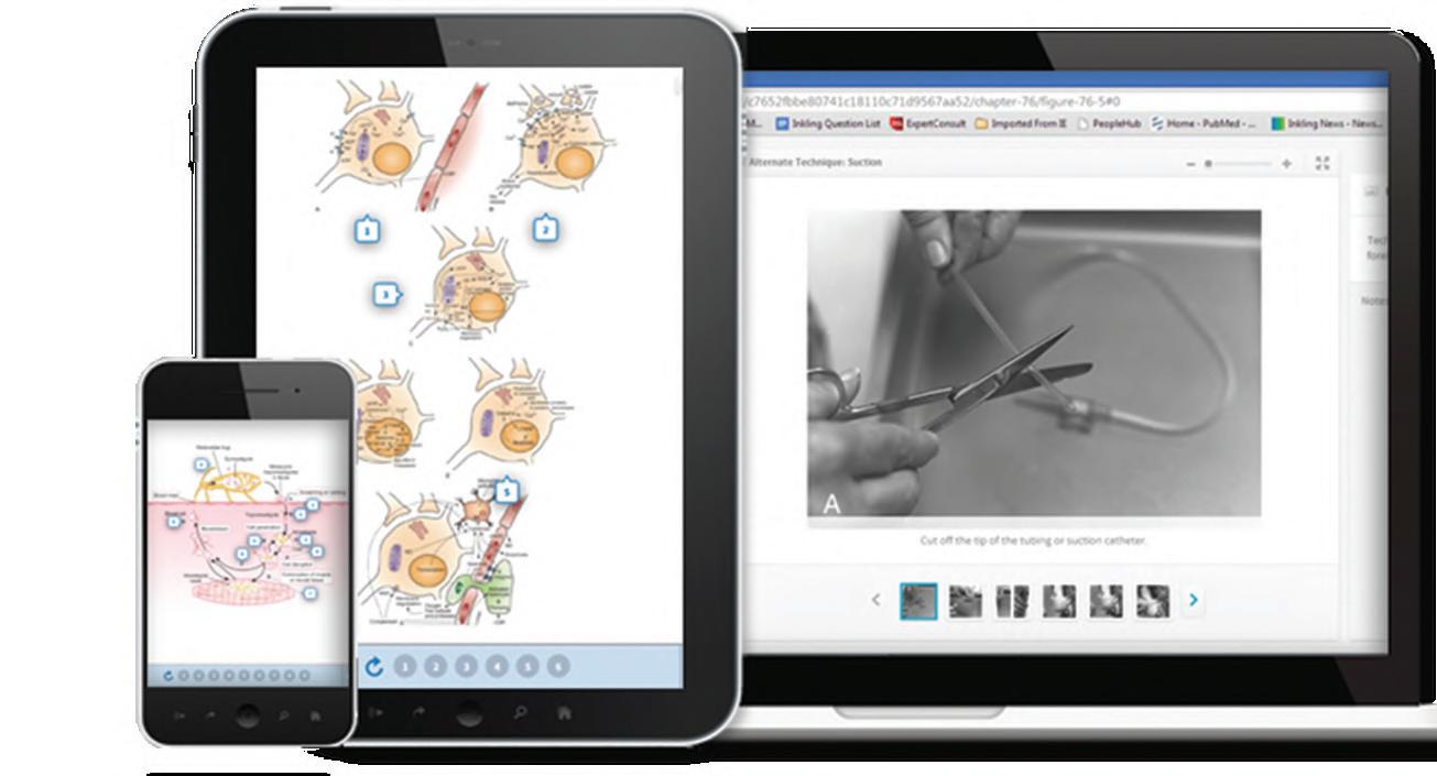
Elsevier eBooks for Practicing Clinicians gives you the power to browse and search content, view enhanced images, highlight and take notes—both online and offline.
Unlock your eBook today.
1. Visit expertconsult.inkling.com/redeem
2. Scratch box below to reveal your code
3. Type code into “Enter Code” box
4. Click “Redeem”
5. Log in or Sign up
6. Go to “My Library” It’s that easy!
Place Peel Off Sticker Here
For technical assistance: email expertconsult.help@elsevier.com call 1-800-401-9962 (inside the US) call +1-314-447-8300 (outside the US)
KAUFMAN’S CLINICAL NEUROLOGY FOR PSYCHIATRISTS
KAUFMAN’S CLINICAL NEUROLOGY FOR PSYCHIATRISTS
Ninth Edition
David Myland Kaufman, MD
Departments of Neurology and Psychiatry
Montefiore Medical Center
Albert Einstein College of Medicine Bronx, New York
Howard L. Geyer, MD, PhD
Department of Neurology
Montefiore Medical Center
Albert Einstein College of Medicine Bronx, New York
Mark J. Milstein, MD
Department of Neurology
Montefiore Medical Center
Albert Einstein College of Medicine Bronx, New York
Jillian L. Rosengard, MD
Department of Neurology
Montefiore Medical Center
Albert Einstein College of Medicine
Bronx, New York
Elsevier
1600 John F. Kennedy Blvd. Ste 1800 Philadelphia, PA 19103-2899
KAUFMAN’S CLINICAL NEUROLOGY FOR PSYCHIATRISTS, NINTH EDITION
No part of this publication may be reproduced or transmitted in any form or by any means, electronic or mechanical, including photocopying, recording, or any information storage and retrieval system, without permission in writing from the publisher. Details on how to seek permission, further information about the Publisher’s permissions policies and our arrangements with organizations such as the Copyright Clearance Center and the Copyright Licensing Agency, can be found at our website: www.elsevier.com/permissions
This book and the individual contributions contained in it are protected under copyright by the Publisher (other than as may be noted herein).
Notices
Practitioners and researchers must always rely on their own experience and knowledge in evaluating and using any information, methods, compounds or experiments described herein. Because of rapid advances in the medical sciences, in particular, independent verification of diagnoses and drug dosages should be made. To the fullest extent of the law, no responsibility is assumed by Elsevier, authors, editors or contributors for any injury and/ or damage to persons or property as a matter of products liability, negligence or otherwise, or from any use or operation of any methods, products, instructions, or ideas contained in the material herein.
Senior Acquisitions Editor: Melanie Tucker
Senior Content Development Specialist: Rishi Arora
Senior Content Development Manager: Somodatta Choudhury
Publishing Services Manager: Shereen Jameel
Senior Project Manager: Umarani Natarajan
Design Direction: Brian Salisbury Printed in India
Dedication, vii
Acknowledgments, viii
Notes About References, ix Physician-Readers, Please Note, x Preface, xi
SECTION I Classic Anatomic Neurology
1 First Encounter With a Patient: Examination and Formulation, 2
2 Signs of Central Nervous System Disorders, 6
3 Psychogenic Neurologic Deficits, 18
4 Cranial Nerve Impairments, 25
5 Peripheral Nerve Disorders, 57
6 Muscle Disorders, 84
SECTION II Major Neurologic Conditions
7 Dementia, 111
8 Aphasia and Anosognosia, 159
9 Headaches, 184
10 Epilepsy, 205
11 Transient Ischemic Attacks and Stroke, 248
12 Visual Disturbances, 271
13 Congenital Cerebral Impairments, 295
14 Neurologic Aspects of Chronic Pain, 325
15 Multiple Sclerosis, 344
16 Neurologic Aspects of Sexual Function, 365
17 Sleep Disorders, 379
18 Involuntary Movement Disorders, 412
19 Brain Tumors, Metastatic Cancer, and Paraneoplastic Syndromes, 474
20 Lumbar Puncture and Imaging Studies, 498
21 Neurotransmitters and Drug Abuse, 522
22 Traumatic Brain Injury, 552
Appendix 1 Patient and Family Support Groups, 570
Appendix 2 Costs of Various Tests, 572
Appendix 3 Diseases Transmitted by Chromosome or Mitochondria Abnormalities, 573
Appendix 4 Chemical and Biological Neurotoxins, 575
Additional Review Questions and Answers, 576
Index, 687
To Rita, my wife of 50 years, whose love has made everything possible, and to our grandchildren—Lila, Owen, Aaron, Penelope, Eliana, and Benjamin.
David Myland Kaufman
To my parents, my wife, and my daughters, with gratitude, admiration, and love.
Howard L. Geyer
To Chris, who loves me enough to occasionally let me think I’m right, and to my loving parents, David and Nancy, who provided the supportive foundation from which to build my life and career.
Mark J. Milstein
To Gabe and Tommy, I love and adore you both, and to my parents and grandmother for your endless support and encouragement.
Jillian L. Rosengard
ACKNOWLEDGMENTS
My wife and best friend, Rita, acted as my muse by originally suggesting writing this book by expanding the syllabus for my course, “Clinical Neurology for Psychiatrists,” and then giving me numerous ideas for each future edition.
David Myland Kaufman
I am privileged to have worked with and learned from many wonderful mentors, colleagues, and patients over the years, and am thankful for all they have taught me. I am especially grateful to my wife Laurence for her love, encouragement, and patience, during my work on this project and always.
Howard L. Geyer
Thanks to Dr. David Kaufman, who not only taught me invaluable lessons during my residency training but brought me into this project, and to Dr. Steven Herskovitz, Director of the Neuromuscular Division, who always pushes me to examine all angles of a problem and answer medical questions as precisely as possible. Additionally, I owe tremendous gratitude to Dr. Sheryl Haut, whose mentorship and support have helped me achieve success in student and resident teaching and personal academic endeavors.
Mark J. Milstein
Thank you to my mentors, Dr. Sheryl Haut and Dr. Nico Moshé, from whom I continue to learn a great deal. Thank you to Dr. David Kaufman for inviting me to participate in this project, which has been a rewarding and invigorating experience. Most importantly, I would like to acknowledge and thank my patients, who inspire me.
Jillian L. Rosengard
Drs. Philip Ozuah (CEO of Montefiore Medical Center), Gordon F. Tomaselli (Dean of the Albert Einstein College of Medicine), Jonathan E. Alpert (Chair of Psychiatry), Mark Mehler (Chair of Neurology), Nico Moshé (Vice Chair of Neurology), Sheryl Haut (Chief of Service), and Michael Swerdlow (partner at Neurologic Associates) have provided the framework and encouragement to pursue writing this book and undertake other academic work in the midst of our clinical responsibilities. At the same time, they have grown Montefiore – Albert Einstein College of Medicine into a vibrant, world-renowned, urban medical center dedicated to medicine as social justice.
Our housestaff and faculty colleagues at Montefiore Medical Center/Albert Einstein College of Medicine and other academic medical centers have revised or reviewed chapters and in other ways offered invaluable help with this edition: Jelena Pavlović, Gail Solomon, Renee Monderer, Jacqueline Bello, Judah Burns, Noam Lax, and Michael Kaufman. Meryl Ranzer, Barry Morden, and Ann Mannato captured the sense of neurology in wonderful illustrations.
We also thank our editors and others at Elsevier who have opened the doors and provided many improvements for this edition: Joslyn Chaiprasert-Paguio, Melanie Tucker, Nicole Congleton, Rishi Arora, Umarani Natarajan and Sara Watkins
David Myland Kaufman, MD
Howard L. Geyer, MD, PhD
Mark J. Milstein, MD
Jillian L. Rosengard, MD
NOTES ABOUT REFERENCES
Most chapters in this book provide specific references from the neurologic and general medical literature. In addition, several standard, well-written textbooks contain relevant information about many topics:
Biller J. Practical Neurology DVD Review. 2nd ed. Philadelphia: Wolters Kluwer; 2013.
Blumenfeld H. Neuroanatomy Through Clinical Cases. 2nd ed. Sunderland, MA: Sinauer; 2010.
Brazis PW, Masdeu JC, Biller J. Localization in Clinical Neurology 7th ed. Philadelphia: Wolters Kluwer; 2017.
Daroff RB, Jankovic J, Mazziotta JC, et al. Bradley’s Neurology in Clinical Practice. 7th ed. Philadelphia: Elsevier; 2016.
Ellison D, Love S, Chimelli L, et al. Neuropathology: A Reference Text of CNS Pathology. 3rd ed. Philadelphia: Elsevier; 2013.
Howard J. Neurology Video Textbook. New York: DemosMedical; 2013. Jones KL, Jones MC, del Campo M. Smith’s Recognizable Patterns of Human Malformations. 7th ed. Philadelphia: Elsevier; 2013. Kanner AM. Depression in Neurologic Disorders. Oxford, UK: Wiley-Blackwell; 2012.
Louis ED, Mayer SA, Rowland LP, eds. Merritt’s Neurology. 13th ed. Philadelphia: Wolters Kluwer; 2016.
Posner JB, Saper CB, Schiff N, et al. Plum and Posner's Diagnosis of Stupor and Coma (Contemporary Neurology Series). 5th ed. New York: Oxford University Press; 2019.
Ropper AH, Samuels MA, Klein JP, eds. Adams and Victor's Principles of Neurology. 11th ed. New York: McGraw Hill, Waltham Mass., Academic Press; 2019. 2014.
Rosenberg RN, Pascual JM. Rosenberg's Molecular and Genetic Basis of Neurological and Psychiatric Disease. 5th ed. New York: Elsevier; 2015.
Swaiman KF, Ashwal S, Ferriero DM, et al. Swaiman’s Pediatric Neurology: Principles and Practice. 6th ed. New York: Elsevier; 2017.
Web Sites That Offer Information About Several Areas
(Sites relevant to single areas are listed in each chapter’s references and in Appendix 1.)
American Academy of Neurology’s Practice Guidelines: https:// www.aan.com/policy-and-guidelines/guidelines/ Medlink Neurology (a commercial neurology textbook): http:// www.medlink.com
UpToDate (a commercial medical textbook): http://uptodate.com
National Institute of Health: http://health.nih.gov/category/BrainandNervousSystem
Online Mendelian Inheritance in Man: http://www.ncbi.nlm.nih. gov/omim
Pubmed: http://www.pubmed.ncbi.nlm.nih.gov/
Registry and results database of publicly and privately supported clinical studies of human participants conducted around the world: http://ClinicalTrials.gov
Reference to diagnose rare diseases by listing two or more symptoms or signs: http://Mendelian.co
PHYSICIAN-READERS, PLEASE NOTE
Kaufman’s Clinical Neurology for Psychiatrists discusses medications, testing, procedures, and other aspects of medical care. Despite their purported effectiveness, many are fraught with side effects and other adverse outcomes. Discussions in this book neither recommend nor offer medical advice, and they do not apply to individual patients. The physician, who should consult the package insert and the medical literature, remains responsible for medications’ indications, dosage, contraindications, precautions, side effects, adverse reactions, and
alternatives, including doing nothing. Some aspects of medical care that this book discusses are widely and successfully used for particular purposes not approved by the Food and Drug Administration (FDA). As regards these “off-label” treatments, as well as conventional ones, this book is reporting— not endorsing—their use by neurologists or other physicians. Finally, because medical practices rapidly evolve, readers should expect that sooner or later new diagnostic criteria and treatments will replace those discussed in this edition.
PURPOSE
We have written Kaufman’s Clinical Neurology for Psychiatrists—a collegial straightforward guide—from our perspective as neurologists at a major, urban academic medical center. In a format combining traditional neuroanatomic correlations with symptom-oriented discussions, the book will assist psychiatrists in learning modern neurology. It emphasizes neurologic conditions that are frequently occurring, common to psychiatry and neurology, illustrative of a scientific principle, or have prominent psychiatric manifestations. It also includes descriptions of many neurologic conditions that may underlie aberrant behavior, disturbances in mood, or cognitive impairment—symptoms that prompt patients or medical colleagues to solicit psychiatry consultations. This book is not intended to replace comprehensive neurology textbooks or convert psychiatrists into semi-professional neurologists; however, it contains essential information required of psychiatrists.
ORGANIZATION AND CONTENT
The organization and content of Kaufman’s Clinical Neurology for Psychiatrists arose from our experience as faculty at the Albert Einstein College of Medicine, attending neurologists at Montefiore Medical Center, and supervisors of numerous neurology and psychiatry residents; consultation with our colleagues, many of whom are world-renowned physicians; and feedback from many of the 22,000 psychiatrists who have attended the course, “Clinical Neurology for Psychiatrists,” since its inception, and the more than 60,000 individuals who have purchased previous editions of this book. Learning the material in this book should help readers prepare for examinations, perform effective consultations, and improve their practice and teaching.
Section I reviews classic anatomic neurology and describes how to approach patients with a suspected neurologic disorder, identify central or peripheral nervous system disease, and correlate physical signs. Section II discusses common and otherwise important clinical areas, emphasizing aspects a psychiatrist may encounter. Topics include illnesses, such as multiple sclerosis, brain tumors, strokes, autoimmune encephalitis, and traumatic brain injury; and symptoms, such as headaches, chronic pain, seizures, and tardive dyskinesias. For each topic, chapters describe the relevant symptoms including psychiatric comorbidity, easily performed office and bedside examinations, appropriate laboratory tests, differential diagnosis, and some management options.
Many chapters contain outlines for a bedside examination; reproductions of standard bedside tests, such as the MiniMental Status Examination (MMSE), Montreal Cognitive Assessment (MoCA), and Abnormal Involuntary Movement Scale (AIMS), references to recent medical literature, and
pertinent web sites. One chapter provides a compilation of computed tomography (CT), magnetic resonance imaging (MRI), and positron emission tomography (PET) images that other chapters reference. Appendices contain information pertaining to most chapters.
In addition, the book reviews neurologic conditions that have entered the public arena because, willingly or unwillingly, psychiatrists are liable to be drawn into debates involving their own patients or those in the medical community at large. Psychiatrists should be well versed in the intricacies of the following conditions that this book describes:
• Certain neurologic illness as battlegrounds of assisted suicide
• Meningomyelocele with Arnold-Chiari malformation as an indication for abortion and the value of spending limited resources on this fatal or severely debilitating condition
• Using marijuana, opioids, or heroin for chronic pain
• Parkinson disease, spinal cord injury, and other disorders amenable to research and treatment with stem cells
• Persistent vegetative state and continuing life-support technology
ADDITIONS AND OTHER CHANGES FOR THE NINTH EDITION
The first eight editions of Kaufman’s Clinical Neurology for Psychiatrists have enjoyed considerable success in the United States, Canada, and abroad. The book has been translated into Japanese, Italian, Korean, and Spanish. In the ninth edition, written 5 years after the eighth, we have clarified the presentations, discussed recent developments in many areas, and added many clinical, anatomic, and radiologic illustrations. To give the question-and-answer sections greater power, we have increased the number of questions, refined them, expanded the discussions, and added illustrations. We have increased the usage of questions formulated as clinical vignettes because they mimic clinical experience and the trend of national specialty examinations.
COMPARISON TO DSM-5
Kaufman’s Clinical Neurology for Psychiatrists refers to the diagnostic criteria for various neurologic disorders in the Diagnostic and Statistical Manual of Mental Disorders, 5th Edition (DSM-5). The book compares and contrasts DSM-5 diagnostic criteria to neurologists’ diagnostic criteria. It shows whereas, with a few exceptions, DSM-5 criteria base the patient’s diagnosis entirely on the nature and duration of symptoms, neurologists, depending on the situation, base the patient’s diagnosis on genetic testing, biopsy results, blood tests, imaging studies, and various laboratory testing as well
as the neurologic examination. Diagnostic criteria for neurologic illnesses admittedly remain for the most part uncodified and variable, but they are reliable and have little inter-rater variability. Nevertheless, neurologists sometimes must base the diagnosis and treatment plan entirely on the patient’s symptoms—as when patients have symptoms of migraine or chronic pain.
UPDATES, EXPANDED TOPICS, NEW MATERIAL
• New classification of seizures and epilepsy
• Neurology of violence
• Revised diagnostic criteria for multiple sclerosis
• New treatments for neurologic illnesses: epilepsy, Alzheimer disease, multiple sclerosis, migraine, pain, sleep disorders, Parkinson disease, and tardive dyskinesia
• Neurologic and psychiatric adverse effects of medications and other treatments
• New imaging techniques: dopamine transporter (DaT) scans and diffusion tensor imaging (DTI)
• Current guidelines for the diagnosis of concussions and their management
• Chronic traumatic encephalopathy
• COVID-19 neurologic complications, including delirium, strokes, myasthenia gravis, and multiple sclerosis
• Psychiatric comorbidity of neurologic illnesses
• Autoimmune/paraneoplastic encephalitis
• Additional clinical illustrations
• Additional short answer and clinical vignette questions-and-answers
DIDACTIC DEVICES: THE VISUAL APPROACH AND QUESTION-AND-ANSWER SECTIONS
Kaufman’s Clinical Neurology for Psychiatrists—like much of the practice of neurology—emphasizes a visual approach to diagnosis. This book provides abundant illustrations, including many sketches of “patients” that personify or reinforce clinical descriptions, correlate the basic science with clinical findings, and serve as the basis for question-and-answer
learning. The visual approach conforms to neurologists’ predilection to “diagnose by inspection.” For example, they rely on their observations for the diagnosis of gait abnormalities, psychogenic neurologic deficits, neurocutaneous disorders, strokes, and tardive dyskinesia. In addition, the book reproduces neurologic test results, which are also visual records, such as CT, MRI, and electroencephalography (EEG).
Question-and-answer (Q&A) sections at the end of most chapters and at the conclusion of the book complements the text. The Q&A sections at the end of chapters generally refer to material discussed within that chapter, whereas those questions at the book’s conclusion tend to require comparison of neurologic disorders that have appeared under different headings. In Chapter 4, before the question-and-answer review of the preceding chapters’ material, the book offers a guide to preparing for standardized tests.
Many medical schools rely on similar “problem-based interactive studying”—case-based question-and-answer problems—as the optimum meaningful and efficient learning strategy. Not merely quizzing the reader, the book’s Q&As form an integral part of the learning experience. In fact, many readers find these sections are the single most informative portion of the book and term them “high yield.” In keeping with the visual emphasis of the book, many of the questions are based on visual material, including sketches of patients and reproductions of MRIs, CTs, DaT scans, and EEGs.
ONE CAVEAT
Kaufman’s Clinical Neurology for Psychiatrists expects welleducated and thoughtful readers. It demands attention and work, and asks them to follow a rigorous course. Readers should find the book, like the practice of medicine, complex and challenging, but at the same time rich and fulfilling.
Even with the additions to the text, illustrations, and Q&A sections, the ninth edition of Kaufman’s Clinical Neurology for Psychiatrists remains manageable in size, depth, and scope, but still succinct enough for psychiatrists to read and experience from cover to cover.
David Myland Kaufman, MD
Howard L. Geyer, MD, PhD
Mark J. Milstein, MD
Jillian L. Rosengard, MD
SECTION I
Classic Anatomic Neurology
First Encounter With a Patient: Examination and Formulation 1
OUTLINE
Examination 2
Formulation 3
Despite the ready availability of sophisticated tests, the neurologic examination remains fundamental to the specialty. Beloved to neurologists, the examination provides a vivid portrayal of both function and illness.
When a patient’s history suggests a neurologic illness or dysfunction localized to a particular site in the nervous system, the neurologic examination may unequivocally demonstrate it. Even if psychiatrists themselves do not perform the examination, they should be able to appreciate neurologic signs and assess a neurologist’s conclusion.
Neurologists systematically examine the nervous system’s major components, paying particular attention to those areas of interest in an individual patient. Neurologists generally adhere to the routine while avoiding omissions and duplications. Despite obvious dysfunction in one part of the nervous system, they usually evaluate all of them. A neurologist can usually complete an initial or screening examination in 20 minutes or less and return to perform detailed or otherwise special testing of particular areas, such as the mental status.
EXAMINATION
Neurologists usually begin by noting a patient’s age, sex, and handedness before reviewing the primary symptom, present illness, medical history, family history, and social history. They explore the primary symptom, associated symptoms, and possible etiologic factors. If a patient cannot relate the history, the neurologist might interrupt the process to look for language, memory, or other cognitive deficits. Many chapters in Section II contain outlines of standard questions related to common symptoms.
After obtaining the history, the neurologist should be able to anticipate the patient’s deficits and prepare to look for disease primarily affecting the central nervous system (CNS) or the peripheral nervous system (PNS). At this point, without succumbing to rigid preconceptions, the physician should have developed some sense of the problem at hand.
Then neurologists should look for the site of involvement (i.e., “localize the lesion”). The art of localization, one of the initial goals of most neurologic examinations, is valuable in
Responding as a Neurologist to Consultations 4
Neurologic Diagnosis 4
the majority of cases. However, it is largely inapplicable in several important neurologic illnesses, such as Alzheimer disease.
The examination is not only of historical interest but irreplaceable in diagnosis. It consists of a survey of neuroanatomical function: mental status, cranial nerves, motor system, reflexes, sensation, cerebellar system, and gait (Box 1.1). This format should be followed during most examinations. Trainees still mastering this structure may find it helpful to bring a printed copy to the patient’s bedside to serve both as a reminder and as a place to record neurologic findings.
The examination usually starts with an assessment of the mental status because cognition is the most fundamental neurologic function and cognitive impairments may preclude an accurate assessment of other neurologic functions. The examiner should consider specific intellectual deficits, such as language impairment (see Aphasia, Chapter 8) and general cognitive impairment (see Dementia, Chapter 7). Tests of cranial nerves may reveal malfunction of nerves either individually or in groups, such as the ocular motility nerves (III, IV, and VI) or the cerebellopontine angle nerves (V, VII, and VIII) (see Chapter 4).
The examination of the motor system defines the anatomic pattern of weakness as well as its severity. Whether weakness is partial (paresis) or complete (plegia), the pattern offers reliable clues to localization. On a practical level, the severity of the paresis determines the patient’s functional capacity (e.g., whether a patient walks, requires a wheelchair, or stays bedridden).
Neurologists frequently speak of three patterns of paresis. If the lower face, arm, and leg on one side of the body are paretic, they call the pattern hemiparesis. They usually attribute hemiparesis to damage in the contralateral cerebral hemisphere or brainstem. They call weakness of both legs paraparesis, and weakness of all four limbs quadriparesis. In both cases, neurologists may ascribe the weakness to either PNS or CNS injury depending on any accompanying findings and test results.
Eliciting two categories of reflexes assists in determining whether paresis—or other neurologic abnormality—originates in CNS or PNS injury. Deep tendon reflexes (DTRs) are
BOX 1.1 Neurologic Examination
Mental status
Cooperation
Orientation (to month, year, place, and any physical or mental deficits)
II Visual acuity, visual fields, optic fundi, pupil size and reactivity (afferent limb), III, IV, VI Pupil size and reactivity (efferent limb), extraocular movements
V Corneal reflex and facial sensation
VII Strength of upper and lower facial muscles, taste
normally present with uniform reactivity (speed and forcefulness) in all limbs, but neurologic injury typically alters their activity or symmetry. In general, CNS injury that includes corticospinal tract damage makes DTRs hyperactive, whereas PNS injury makes DTRs hypoactive.
In contrast to DTRs, pathologic reflexes are not normally elicitable beyond infancy. If found, they are a sign of CNS damage. The most widely recognized pathologic reflex is the famous Babinski sign. After plantar stimulation, the great toe normally moves downward (i.e., it has a flexor response), but with corticospinal tract damage, whether in the brain or spinal cord, plantar stimulation typically causes the great toe to move upward (i.e., to have an extensor response). This reflex extensor movement, the Babinski sign (see Fig. 19.3), is thus a manifestation of CNS damage. Neurologists say that the Babinski sign and other pathologic reflexes are “present,” “found,” or “elicited,” but not “positive” or “negative.” The terminology is similar to describing a traffic stop sign: it may be present or absent, but not positive or negative.
Frontal release signs, which are also pathologic reflexes, reflect frontal lobe injury; when present, they reflect loss of the inhibitory capacity of the frontal lobes. Thus, they point to an “organic” or structural basis for a change in personality and sometimes correlate with cognitive impairment (see Chapter 7).
Unlike abnormal DTRs and Babinski signs, which are reproducible, objective, and difficult to mimic, the sensory examination relies almost entirely on the patient’s report. Its subjective nature has led to the practice of disregarding reports of disturbances inconsistent with the rest of the examination. Under most circumstances, the best approach is to test the major sensory modalities in a clear anatomic order and tentatively accept the patient’s report.
Depending on the nature of the suspected disorder, physicians may first test light touch sensation with their fingertips or a cotton swab, and then two sensations carried by the posterior columns of the spinal cord: position and vibration. Neurologists might test pain (pinprick) sensation, which is carried in the spinothalamic tracts, but only in a careful manner with a nonpenetrating, disposable instrument, such as a broken wood shaft of the cotton swab.
Neurologists evaluate cerebellar function by observing several standard maneuvers that include the finger-to-nose test and rapid alternating movement test (see Chapter 2). These tests may demonstrate intention tremor or incoordination. If at all possible, neurologists watch the patient walk because a normal gait requires intact and well-integrated CNS and PNS motor pathways, coordination, proprioception, and balance. Examining the gait is probably the single most valuable assessment of non-cognitive functions of the nervous system. Neurologists watch for gait abnormalities that characterize many neurologic illnesses (see Table 2.1). In addition, they expect certain gait abnormalities to be comorbid with cognitive impairment. Whatever the underlying abnormality, gait impairment is not merely a neurologic sign but a condition that routinely leads to fatal falls and permanent incapacity for numerous people each year.
FORMULATION
Although somewhat ritualistic, a succinct and cogent formulation remains the basis of neurologic problem solving. A neurologist’s classic approach consists of an appraisal of the four aspects of the examination: symptoms, signs, localization, and differential diagnosis. A neurologist might also have to support a conclusion that neurologic disease does or does not explain the patient’s symptoms and signs. For this step, neurologists at least tentatively separate psychogenic signs from neurologic (“organic”) ones.
Localization of neurologic disease requires the clinician not only to determine whether the illness affects the CNS, PNS, or muscles (see Chapter 2 through 6), but precise localization of lesions within these regions of the nervous system also is generally expected. The physician must also establish whether the illness affects the nervous system diffusely or in a focal, discrete area. The site and extent of neurologic damage
generally indicates certain diseases. A readily apparent example is that strokes and tumors usually involve a discrete area of the brain, but Alzheimer disease usually causes widespread, symmetrical changes.
Finally, neurologists create a differential diagnosis that lists the disease or diseases most consistent with the patient’s symptoms and signs. The differential diagnosis should include unlikely but potentially life-threatening conditions. In addition, many neurologists, in a flourish of intellectualism, include unlikely but fascinating explanations. However, even at tertiary care institutions, common conditions arise often. Just as “hoofbeats are usually from horses, not zebras,” patients are more likely to have hemiparesis from a stroke than a mitochondrial disorder.
A typical formulation might be as follows: “Mr. Jones, a 56-year-old right-handed bartender, has had left-sided headaches for two months and, on the day before admission, had a generalized seizure. He is lethargic. He has papilledema, a right hemiparesis with hyperactive DTRs, and a Babinski sign. The lesion is probably situated in the left cerebral hemisphere and causes increased intracranial pressure. It is most likely a tumor or stroke, but possibly an abscess.” This formulation briefly recapitulates the salient elements of the history and physical findings. Here, neurologists would tacitly assume that neurologic disease is present because of the obvious and objective physical findings. The history of seizures, the right-sided hemiparesis, and abnormal reflexes localize the lesion. Neurologists would base their differential diagnosis on the high probability that a discrete space-occupying cerebral lesion is causing these abnormalities.
A house officer presenting a case to a superior should separate the wheat from the chaff and complete the presentation within two minutes, the limit of most listeners’ attention span. The clinician should practice the presentation before rounds, bearing in mind Benjamin Franklin’s proverb, “By failing to prepare, you are preparing to fail.”
In summary, the neurologist should present a succinct, well-rehearsed formulation that answers The Four Questions of Neurology:
• What are the symptoms of neurologic disease?
• What are the signs of neurologic disease?
• Where is the lesion?
• What is the lesion?
RESPONDING AS A NEUROLOGIST TO CONSULTATIONS
During their training, psychiatry residents often rotate through a neurology service where they are required to provide neurology consultations. Consultants at all levels must work with a variation of the traditional summary-and-formulation format.
While the patient’s interests remain paramount, the consultant’s “client” is the referring physician. Both the referring physician and consultant should be clear about the reason for the consultation. Reasons for consultations typically concern a neurologic symptom, the significance of a report of
computed tomography (CT) or magnetic resonance imaging (MRI) (see Chapter 20), or a treatment recommendation. Sometimes physicians request a broad review, such as when they ask the consultant to provide a second opinion or offer a prognosis. On the other hand, the referring physicians may not care to know the diagnosis or treatment options but simply want the neurology service to assume the primary care of the patient.
Without belaboring the obvious, the consultation note must be organized, succinct, and practical; the primary physician in an acute care hospital should be able to digest it in two minutes. Lengthy notes are usually boring and inadvertently hide worthwhile information. Cutting and pasting information and conclusions in computerized medical records by a consultant is redundant, liable to repeat errors and, if a previous physician made an astute diagnosis, appears to take credit for someone else’s idea. Notes that are bad, for whatever reason, reflect poorly on the consultant and the consultant’s service. Moreover, they hamper the patient’s care. At least in an academic setting, the consultant should offer at least one teaching point about the case and provide general guidelines for future handling of similar inquires.
Finally, consultants should show an awareness of the entire situation, which often contains incomplete and conflicting elements. They should be mindful of the referring physician’s and patient’s situation. Consultants in emergency situations might help by ordering—not merely suggesting—routine tests (such as blood studies) and important treatments (such as thiamine injections). Except in unusual circumstances, consulting residents should not suggest hazardous tests or treatments without first presenting the case to their supervisor. Consultants should not divert the primary physicians’ efforts from the patient’s most important medical problems. They should not suggest embarking on elaborate, time-consuming testing for obscure, unlikely diagnoses when the patient’s illness is obvious and requires the primary medical team’s full attention. The consultant should perpetually keep in mind the question, “How can I help?”
NEUROLOGIC DIAGNOSIS
Neurologists confirm a clinical diagnosis using different frames of reference. For some diseases, such as migraine and chronic pain, neurologists rely almost entirely on a patient’s symptoms. For others, such as Parkinson disease, they base their diagnosis on physical abnormalities or constellations of findings. For many other diseases, regardless of the patient’s symptoms and signs, their diagnosis rests on an abnormal test result. For example, the diagnosis of stroke or a brain tumor typically requires imaging studies, and confirmation of seizures often necessitates an electroencephalogram. Neurologists diagnose many asymptomatic individuals as having a neurologic disease on the basis of a single test, such as genetic analysis or MRI.
The clinical formulation remains the mainstay of neurologic diagnosis, but abnormal findings on MRI or other
studies routinely trump clinical impressions. For example, the clinical examination may indicate the presence, location, and etiology of a cerebral lesion, but if an MRI indicates a different process, neurologists generally forsake their clinical formulation and defer to the MRI findings as the diagnosis.
Overall, neurologists’ and psychiatrists’ diagnoses routinely differ in several respects. Neurologists shift the basis of their diagnosis from clinical constellation to MRI, to pathologic specimen, and to genetic test—whichever is the most specific. In contrast, psychiatrists tend to base most diagnoses (except for sleep disorders and perhaps a few others) entirely
on their patients’ history and observable clinical presentation without performing a physical examination. Whereas neurologists routinely diagnose illnesses in asymptomatic individuals, such as those carrying mutations for Huntington disease or a spinocerebellar ataxia, psychiatrists almost always require symptoms. Finally, neurologists do not have a Diagnostic and Statistical Manual (DSM) of Neurologic Disorders. While the lack of a DSM prevents uniformity, neurologists remain flexible in their diagnostic criteria and free from pigeonholing patients’ symptoms and signs, which may considerably vary from patient to patient with the same illness.
Signs of Central Nervous System Disorders 2
OUTLINE
Signs of Cerebral Hemisphere Lesions 6
Signs of Damage of the Dominant, Nondominant, or Both Cerebral Hemispheres 7
Signs of Basal Ganglia Lesions 10
Signs of Brainstem Lesions 11
Disorders of the brain and the spinal cord—the two major components of the central nervous system (CNS)—typically cause readily recognizable combinations of paresis, sensory loss, visual deficits, and neuropsychologic disorders (Box 2.1). Such signs of CNS disorders differ from those of peripheral nervous system (PNS), and both differ from the signs of psychogenic disorders. Neurologists formulate their preliminary diagnosis and often initiate treatment on the basis of the patient’s history and the examination, but if results of investigations—such as laboratory testing or magnetic resonance imaging (MRI)—contradict their diagnosis, they will usually revise it.
SIGNS OF CEREBRAL HEMISPHERE LESIONS
Hemiparesis, usually accompanied by changes in reflexes and muscle tone, is one of neurology’s most prominent and reliable signs. Damage to the corticospinal tract, also called the pyramidal tract (Fig. 2.1), in the cerebrum or brainstem rostral to (above) the decussation of the pyramids, causes contralateral hemiparesis (Box 2.2) with weakness of the arm and leg—and, if the lesion is rostral enough, the lower face— opposite the side of the lesion. Damage to this tract within the spinal cord causes ipsilateral arm and leg or only leg paresis, but no facial weakness.
The division of the motor system into upper and lower motor neurons is a basic construct of clinical neurology. The corticospinal tract’s entire path from the cerebral cortex to the motor cranial nerve nuclei and the anterior horn cells of the spinal cord consists of upper motor neurons (UMNs) (Fig. 2.2). The anterior horns contain the cell bodies of the lower motor neurons (LMNs) and hence are part of the PNS.
Cerebral lesions that damage the corticospinal tract cause signs of UMN injury (see Figs. 2.2–2.5):
• Paresis with muscle spasticity
• Hyperactive deep tendon reflexes (DTRs)
• Babinski signs
Signs of Cerebellar Lesions 12
Signs of Spinal Cord Lesions 13
Spinal Cord Transection 14
Syringomyelia 16
Neurologic Illnesses 16
2.1 Signs of Common Central Nervous System Lesions
Cerebral hemispherea
Hemiparesis with hyperactive deep tendon reflexes, spasticity, and Babinski sign
Hemisensory loss
Homonymous hemianopsia
Focal (partial) seizures
Aphasia, hemi-inattention, and dementia
Pseudobulbar palsy
Basal gangliaa
Movement disorders: parkinsonism, athetosis, chorea, and hemiballismus
Postural instability
Rigidity
Brainstem
Cranial nerve palsy with contralateral hemiparesis
In contrast, PNS lesions, including motor neuron diseases (diseases of the anterior horn cells) and disorders of nerves (neuropathy), cause signs of LMN injury:
BOX
Fig. 2.1 Each corticospinal tract originates in the cerebral cortex, passes through the internal capsule, and descends into the brainstem. The tracts cross in the pyramids, which are protuberances on the inferior surface of the medulla, to descend in the spinal cord mostly as the lateral corticospinal tract. The corticospinal tracts synapse with the anterior horn cells of the spinal cord, which give rise to peripheral nerves. Neurologists often call the corticospinal tract the pyramidal tract because it crosses in the pyramids. The extrapyramidal system, which modulates the corticospinal tract, originates mostly in the basal ganglia within the brain.
• Paresis with muscle flaccidity and atrophy
• Hypoactive DTRs
• No Babinski signs
Another indication of a cerebral lesion is loss of certain sensory modalities over one half of the body, that is, hemisensory loss (Fig. 2.6). A patient with a cerebral lesion characteristically loses contralateral position sensation, two-point discrimination, and the ability to identify objects by touch (stereognosis). Neurologists often describe loss of those modalities as “cortical” sensory loss.
Pain sensation, a “primary” sense, is initially carried to the thalamus, after which it is projected rostrally to the cortex, limbic system, and elsewhere. Because the thalamus is situated above the brainstem but below the cerebral cortex, most patients with cerebral lesions still perceive painful stimuli. For example, patients with cerebral infarctions may be unable to specify a painful area of their body, but they will still feel the pain’s intensity and discomfort (see Chapter 14).
BOX 2.2 Signs of Common Cerebral Lesions
Either hemispherea
Hemiparesis with hyperactive deep tendon reflexes and a Babinski sign
Hemisensory loss
Homonymous hemianopsia
Focal seizure
Dominant hemisphere
Aphasia: fluent, nonfluent, conduction, or isolation
Gerstmann syndrome: acalculia, agraphia, finger agnosia, and left–right confusion
Alexia without agraphia
Nondominant hemisphere
Hemi-inattention
Anosognosia
Constructional apraxia
Both hemispheres
Dementia
Pseudobulbar palsy
aSigns contralateral to lesions.
Visual loss of the same half-field in each eye, homonymous hemianopsia (Fig. 2.7), is a characteristic sign of a contralateral cerebral lesion. Other equally characteristic visual field deficits are associated with lesions involving the eye, optic nerve, or optic tract (see Chapters 4 and 12). Lesions in the brainstem, cerebellum, or spinal cord, because they are situated far from the visual pathway, do not cause visual field loss.
Another sign of a cerebral hemisphere lesion is a seizure (see Chapter 10). In fact, most focal seizures that alter awareness or induce psychomotor phenomena originate in the temporal lobe.
Signs of Damage of the Dominant, Nondominant, or Both Cerebral Hemispheres
Although hemiparesis, hemisensory loss, homonymous hemianopsia, and focal seizures may result from lesions of either cerebral hemisphere, several neuropsychologic deficits are related to either the dominant or nondominant hemisphere. Neurologists usually ask a patient’s handedness when taking a history, but if this information is unavailable, because approximately 85% of people are right-handed, they assume that the left hemisphere is dominant.
Lesions of the dominant hemisphere may cause language impairment, aphasia, a prominent and frequently occurring neuropsychologic deficit (see Chapter 8). Because the corticospinal tract sits adjacent to the language centers, right hemiparesis often accompanies aphasia (see Fig. 8.1).
Lesions of the nondominant parietal lobe tend to produce one or more striking neuropsychologic disturbances (see Chapter 8). For example, patients may neglect or ignore leftsided visual and tactile stimuli (hemi-inattention). They may fail to use their left arm and leg because they neglect their
Internal capsule
Midbrain Pons
Medulla Spinal cord
Fig. 2.2 (A) Normally, when neurologists strike a patient’s quadriceps tendon with a percussion hammer, the maneuver elicits a DTR. In addition, when neurologists stroke the sole of the foot to elicit a plantar reflex, the big toe normally bends downward (flexes). (B) When brain or spinal cord lesions injure the corticospinal tract, producing upper motor neuron (UMN) damage, DTRs react briskly and forcefully, i.e., DTRs are hyperactive. As another sign of UMN damage, the plantar reflex is extensor (a Babinski sign). (C) In contrast, peripheral nerve injury causes lower motor neuron (LMN) damage, the DTR is hypoactive, and the plantar reflex is absent. DTR, Deep tendon reflex
Fig. 2.3 This patient shows right hemiparesis. The right-sided facial weakness causes the widened palpebral fissure and flat nasolabial fold; however, the forehead muscles remain normal (see Chapter 4 regarding this discrepancy). The right arm is limp, and the elbow, wrist, and fingers take on a flexed position; the right leg is externally rotated; and the hip and knee are extended.
Fig. 2.4 When the patient stands up, his weakened arm retains its flexed posture. His right leg remains externally rotated, but he can walk by swinging it in a circular path. This maneuver is effective but results in circumduction or a hemiparetic gait
Fig. 2.5 Mild hemiparesis may not be obvious. To exaggerate it, the physician has asked this patient to extend both arms with his palms held upright, as though his outstretched hand were supporting a pizza box. His weakened arm sinks (drifts) and his forearm turns inward (pronates).The imaginary pizza box would slide to his right. His arm drift and pronation represent a forme fruste of the posture observed with severe paresis (see Fig. 2.3).
Fig. 2.7 In homonymous hemianopsia, the same half of the visual field is lost in each eye. Here, damage to the left cerebral hemisphere has caused a right homonymous hemianopsia. This sketch portrays visual field loss, as is customary, from the patient’s perspective (see Figs. 4.1 and 12.8).
Lateral spinothalamic tract
Parietal cortex
Medulla
Cervical spinal cord
Fasciculus cuneatus
Temperature Pain Position Stereognosis
Fig. 2.6 Peripheral sensory nerves carry pain and temperature impulses to the spinal cord. After a synapse, these impulses cross then ascend in the contralateral lateral spinothalamic tract (blue) to terminate in the thalamus. From there, tracts relay sensation to the limbic system and reticular activating system as well as the cerebral cortex. In parallel, the peripheral nerves also carry position, vibration, and stereognosis impulses to the ipsilateral fasciculus cuneatus and fasciculus gracilis, which together constitute the spinal cord’s posterior columns (crosshatched) (see Fig. 2.15). Unlike pain and temperature sensation, these sensations ascend in the spinal cord via ipsilateral tracts (black). They cross in the decussation of the medial lemniscus, which is in the medulla, synapse in the thalamus, and terminate in the parietal cortex. (To avoid spreading blood-borne illnesses, examiners should avoid using a pin when testing pain.)
Fig. 2.8 A patient with constructional apraxia from a right parietal lobe infarction was unable to complete a circle (top figure), draw a square on request (second highest figure), or even copy one (third highest figure). She spontaneously tried to draw a circle and began to retrace it (bottom figure). Her constructional apraxia consists of rotation of the forms, perseveration of certain lines, and leaving incomplete the second and lowest figures. In addition, the figures tend toward the right-hand side of the page, which indicates she has neglect of the left-hand side of the page, i.e., left hemiinattention (see Chapter 8).
limbs rather than because of paresis. When they have left hemiparesis, patients may not appreciate it (anosognosia) Many patients with nondominant lesions lose their ability to arrange matchsticks into certain patterns or copy simple forms (constructional apraxia, Fig. 2.8).
TABLE 2.1 Gait
Abnormalities Associated With Neurologic Disorders
As opposed to signs resulting from unilateral cerebral hemisphere damage, bilateral cerebral hemisphere damage produces several important disturbances that psychiatrists are likely to encounter in their patients. One of them, pseudobulbar palsy, best known for producing emotional lability, results from bilateral corticobulbar tract damage (see Chapter 4). The corticobulbar tract, like its counterpart, the corticospinal tract, originates in the motor cortex of the posterior portion of the frontal lobe. It innervates the brainstem motor nuclei, which in turn innervate the head and neck muscles. Traumatic brain injury (TBI), multiple cerebral infarctions (strokes), multiple sclerosis (MS), and neurodegenerative conditions, including frontotemporal dementia (see Chapter 7), are apt to strike the corticobulbar tract along with the surrounding frontal lobes and thereby cause pseudobulbar palsy.
Damage of both cerebral hemispheres—from large or multiple discrete lesions, degenerative diseases, or metabolic abnormalities—also causes dementia (see Chapter 7). In addition, because CNS damage that causes dementia must be extensive and severe, it usually also produces at least subtle physical neurologic findings, such as hyperactive DTRs, Babinski signs, mild gait impairment, and frontal lobe release reflexes. However, many neurodegenerative illnesses that cause dementia, particularly Alzheimer disease, do not cause “hard” findings such as hemiparesis.
While certainly not peculiar to cerebral lesions, and even typically absent in early Alzheimer disease, gait impairment is a crucial neurologic finding. Because walking requires intact and well-integrated strength, sensation, and coordination, testing the patient’s gait is the single most reliable assessment of a patient’s noncognitive neurologic function. Gait impairment constitutes the primary physical component of the subcortical dementias, such as vascular dementia, dementia
with Lewy body disease, and Parkinson disease dementia (see Chapter 7). Several distinct gait abnormalities are clues to specific neurologic disorders, such as normal pressure hydrocephalus (see Table 2.1). As a general rule, slow gait speed, that is, 0.7 m/s, is associated with an increased risk of dementia, stroke, falls, disability, hospitalization, and death.
SIGNS OF BASAL GANGLIA LESIONS
The basal ganglia, located subcortically in the cerebrum, consist of the caudate and putamen (together constituting the striatum), globus pallidus, substantia nigra, and subthalamic nucleus (corpus of Luys) (see Fig. 18.1). They constitute the extrapyramidal motor system, which modulates the corticospinal (pyramidal) tract. It controls muscle tone, regulates motor activity, and generates postural reflexes. Its efferent fibers project to the cerebral cortex, thalamus, and other CNS structures. However, because its efferent fibers are confined to the brain, the extrapyramidal tract does not act directly on the spinal cord or LMNs.
Signs of basal ganglia dysfunction include a group of fascinating, often dramatic, involuntary movement disorders (see Chapter 18):
• Parkinsonism is the combination of resting tremor, rigidity, bradykinesia (slowness of movement) or akinesia (absence of movement), and postural instability. Parkinson disease and related neurodegenerative illnesses, exposure to dopamine receptor-blocking antipsychotic medications, or toxins are the most common causes of parkinsonism.
• Athetosis is the slow, continuous, writhing movement of the fingers, hands, face, and throat. Kernicterus or other perinatal basal ganglia injury usually causes it.
• Chorea is intermittent, randomly located, jerking of limbs and the trunk. The best-known example occurs in Huntington disease (previously called “Huntington chorea”), in which the caudate nuclei characteristically atrophy.
• Hemiballismus is the intermittent flinging of the arm and leg of one side of the body. It is classically associated with small infarctions of the contralateral subthalamic nucleus, but similar lesions in other basal ganglia nuclei may be responsible.
In general, when damage is restricted to the extrapyramidal system, patients have no significant paresis, DTR abnormalities, or Babinski signs—which are hallmarks of corticospinal (pyramidal) tract damage. More important, in many of these conditions, such as hemiballismus and athetosis, patients have no cognitive impairment or other neuropsychologic disorder. On the other hand, several conditions—such as Huntington disease, Wilson disease, and advanced Parkinson disease—affect the cerebrum as well as the basal ganglia. In those illnesses, dementia, depression, and psychosis frequently accompany involuntary movements (see Table 18.4).
With unilateral basal ganglia damage, signs develop in the contralateral limbs. For example, an infarction of a subthalamic nucleus causes contralateral hemiballismus.
SIGNS OF BRAINSTEM LESIONS
The brainstem contains, among a multitude of structures, the cranial nerve nuclei, the corticospinal tracts, other “long tracts” that travel between the cerebral hemispheres and the limbs, and cerebellar afferent (inflow) and efferent (outflow) tracts. Combinations of cranial nerve and long tract signs, and the absence of signs of cerebral injury, such as visual field cuts and neuropsychologic deficits, indicate the presence and location of a brainstem lesion. For example, brainstem injuries cause diplopia (double vision) through cranial nerve impairment, but visual acuity and visual fields remain normal because the afferent visual pathway, which passes from the optic chiasm to the cerebral hemispheres, does not travel within the brainstem (see Fig. 4.1). Similarly, a right hemiparesis associated with a left third cranial nerve palsy localizes the lesion to the brainstem (specifically the left midbrain). Moreover, that pair of findings indicates that further examination will reveal neither aphasia nor dementia.
Several brainstem syndromes illustrate critical anatomic relationships, such as the location of the cranial nerve nuclei or the course of the corticospinal tract; however, none of them involves neuropsychologic abnormalities. Although many vascular syndromes of the brainstem have eponyms, for practical purposes it is only necessary to identify the clinical findings and, if appropriate, attribute them to a lesion in one of the three major divisions of the brainstem: midbrain, pons, or medulla (Fig. 2.9). Whatever the localization, most brainstem lesions consist of an infarction due to occlusion of a small branch of the basilar or vertebral arteries.
In the midbrain, where the oculomotor (third cranial) nerve fibers pass through the descending corticospinal tract, a single small infarction can damage both pathways. Patients with oculomotor nerve paralysis and contralateral hemiparesis typically have a lesion in their midbrain ipsilateral to the paretic eye (see Fig. 4.9). In an analogous situation, patients with an abducens (sixth cranial) nerve paralysis and contralateral hemiparesis have a lesion in the pons ipsilateral to the paretic eye (see Fig. 4.11).
Lateral medullary infarctions create a classic but complex picture, the lateral medullary syndrome, commonly known by its eponym Wallenberg syndrome. Patients have dysarthria due to paralysis of the ipsilateral palate from damage to cranial nerves IX through XI; ipsilateral facial numbness to pinprick (hypalgesia) (Greek, decreased sensitivity to pain) due to damage to the nucleus of cranial nerve V, with contralateral anesthesia of the body (crossed hypalgesia) due to ascending spinothalamic tract damage; and ipsilateral ataxia due to inferior cerebellar peduncle dysfunction. They also have nystagmus and vertigo from damage to the vestibulocochlear nerve and ipsilateral Horner syndrome (ptosis, miosis, anhidrosis) due to interruption of sympathetic fibers. In other words, the most important elements of this syndrome consist of damage to three groups of nuclei (V, VIII, and IX-XI) and three white matter tracts (spinothalamic, sympathetic, and inferior cerebellar peduncle). Although the lateral medullary syndrome commonly occurs and provides an excellent example of clinical-pathologic correlation, physicians need not recall all of its pathology or clinical features; however, they should know that lower cranial nerve palsies accompanied
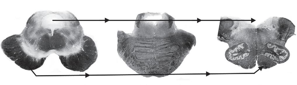
Fig. 2.9 Myelin-stains of the three main divisions of the brainstem—midbrain, pons, and medulla—show several clinically important tracts, the cerebrospinal fluid (CSF) pathway, and motor nuclei of the cranial nerves. (Midbrain) The midbrain (Greek, meso, middle) has a distinctive silhouette and gently curved (pale, unstained in this preparation) substantia nigra (S). The periaqueductal gray matter surrounds the aqueduct of Sylvius (A). Below the aqueduct, near the midline, sit the oculomotor (CN III) and trochlear (not pictured) (CN IV) cranial nerve nuclei. The nearby medial longitudinal fasciculus (MLF), which ascends from the pons, terminates in the oculomotor nuclei. The large, deeply stained cerebral peduncles, ventral to the substantia nigra, contain the corticospinal (pyramidal [Δ]) tract. Originating in the cerebral cortex, the corticospinal tract descends ipsilaterally through the midbrain, pons, and medulla until it crosses in the pyramids of the medulla to continue within the contralateral spinal cord. CSF flows downward from the lateral ventricles through the aqueduct of Sylvius into the fourth ventricle (IV), which overlies the lower pons and medulla. CSF exits from the fourth ventricle into the subarachnoid space. (Also see a functional drawing [Fig. 4.5], a computer-generated rendition [Fig. 18.2], and a sketch [Fig. 21.1]). (Pons) The pons (Latin, bridge) houses the trigeminal motor division (CN V), abducens (CN VI), facial (CN VII), and acoustic/vestibular (not shown) cranial nerve nuclei and, inferior and lateral to the fourth ventricle, the locus ceruleus (*). In addition to containing the descending corticospinal tract, the basilar portion of the pons (basis pontis) contains large criss-crossing cerebellar tracts. (Also see a functional drawing [Fig. 4.7] and an idealized sketch [Fig. 21.2].) (Medulla) The medulla (Latin, marrow), readily identifiable by the pair of unstained scallop-shaped inferior olivary nuclei, includes the cerebellar peduncles (C), which contains afferent and efferent cerebellar tracts; the corticospinal tract (Δ); and the floor of the fourth ventricle (IV). It also contains the decussation of the medial lemniscus (M), the nuclei for cranial nerves 9-11 grouped laterally and XII situated medially, and the trigeminal sensory nucleus (not pictured) that descends from the pons to the cervical-medullary junction. (Also see a functional drawing [Fig. 2.10].)
CSF Pathway
Corticospinal (pyramidal) tract
by crossed hypalgesia, without cognitive impairment or limb paresis, result from a lesion in the lower brainstem (Fig. 2.10). They should also know that vascular disease affecting the medulla is one cause of bulbar palsy (see Chapter 4).
Nystagmus, repetitive jerk-like eye movements that are usually conjugate (i.e., both eyes move equally and simultaneously) is not peculiar to the lateral medullary syndrome but may result from any type of injury to the brainstem’s large vestibular nuclei (as well as other structures, such as cerebellum). Nystagmus can be a manifestation of various disorders, including intoxication with alcohol, phenytoin (Dilantin), phencyclidine (PCP), or barbiturates; ischemia of the vertebrobasilar artery system; MS; Wernicke-Korsakoff syndrome; or labyrinthitis. Among individuals who have ingested PCP, coarse vertical and horizontal (three-directional or multidirectional) nystagmus characteristically accompanies an agitated delirium and markedly reduced sensitivity to pain and cold temperature. Unilateral nystagmus may be a component of internuclear ophthalmoplegia, which is usually a manifestation of MS or a small brainstem infarction (see Chapters 4 and 15).
SIGNS OF CEREBELLAR LESIONS
The cerebellum (Latin, diminutive of cerebrum) consists of two hemispheres and a central portion, the vermis. Each hemisphere controls coordination of the ipsilateral limbs, and the vermis controls coordination of “axial” or “midline structures”: the head, neck, and trunk. Note that the cerebellum controls coordination of the limbs on the same, ipsilateral side of the body, while in the cerebrum each hemisphere governs the opposite, contralateral side of the body.
Another unique feature of the cerebellum is when one hemisphere is damaged, the other will eventually assume the functions for both, at least in part. In other words, although loss of one cerebellar hemisphere will temporarily cause incapacitating ipsilateral incoordination, the patient’s deficit lessens as the remaining hemisphere compensates, sometimes almost entirely. For example, patients who lose one cerebellar hemisphere to a stroke typically regain their ability to walk, although they may never dance. Children who sustain such an injury are more resilient and often can learn to dance, ride a bicycle, and participate in athletic activities.
In addition to causing incoordination, cerebellar lesions cause subtle motor changes, such as muscle hypotonia and pendular DTRs. However, cerebellar lesions do not cause paresis, hyperactive DTRs, or Babinski signs.
Several sophisticated studies have shown that the cerebellum contributes to cognition and emotion. In fact, researchers have described a “cerebellar cognitive affective syndrome,” whose main feature is dysregulation of affect and includes deficits in executive function, linguistic processing, and spatial cognition. Some researchers have also described cerebellar dysfunction in behaviorally defined disorders, particularly attention-deficit/hyperactivity disorder, autism spectrum disorder, congenital intellectual disability, and schizophrenia.
In routine neurologic practice, however, the cerebellum does not play a discernible role in everyday cognition, behavior, or affect. For example, lesions restricted to the cerebellum do not lead to dementia, language impairment, or other cognitive deficits.
On the other hand, intoxication with numerous substances impairs the function of the cerebrum as well as the cerebellum. For example, toxic levels of alcohol, phenytoin, lithium, lead, and toluene typically cause prominent ataxia and cognitive impairment. Another interesting cause of ataxia and other physical signs of cerebellar malfunction is the paraneoplastic syndrome, cerebellar degeneration (see Chapter 19).
Neurologists assess cerebellar function in tests of coordinated motor function. Thus, intention tremor, demonstrable in the finger-to-nose (Fig. 2.11) and heel-to-shin tests (Fig. 2.12), characterizes cerebellar dysfunction. This tremor is evident when the patient’s body part moves to a target but is absent when the patient rests. By way of a classic contrast, Parkinson disease causes a resting tremor, which is present when the patient sits quietly but is reduced or even abolished when the patient moves (see Chapter 18). Physicians should not confuse the neurologic term “intention tremor” with “intentional tremor,” which would imply a self-induced or psychogenic tremor.
Another sign of incoordination due to a cerebellar lesion is dysdiadochokinesia, impaired rapid alternating movements of the limbs. When asked to slap the palm and then the back of the hand rapidly and alternately on his or her own knee, for example, a patient with dysdiadochokinesia will do so with uneven force and irregular rhythm and lose the alternating pattern.
Damage to either the entire cerebellum or the vermis alone causes incoordination of the trunk (truncal ataxia). This manifestation of cerebellar damage forces patients to place their feet widely apart when standing and leads to a lurching, unsteady, and wide-based pattern of walking (gait ataxia) (Table 2.1 and Fig. 2.13). A common example is the staggering and reeling of people intoxicated by alcohol. In addition, such cerebellar damage prevents people from walking heel-to-toe, i.e., performing “tandem gait.” Another common example of ataxia occurs in individuals who have inherited genetic mutations that cause combinations of cerebellar and spinal cord degeneration. In several of these disorders, patients have abnormalities beyond the nervous system (Fig. 2.14). Extensive damage to the cerebellum causes scanning speech, a variety of dysarthria. Scanning speech, which reflects incoordination of speech production, is characterized by poor modulation, irregular cadence, and inability to separate adjacent sounds. Physicians should be able to distinguish dysarthria caused by cerebellar injury, bulbar palsy, or pseudobulbar palsy. Even more important, they should be able to distinguish dysarthria from aphasia (see Chapter 8).
Before considering the illnesses that damage the cerebellum (see Section II), physicians must appreciate that the cerebellum normally undergoes age-related changes that appear between ages 50 and 65 years in the form of mildly impaired functional ability and abnormal neurologic test
Cerebellar peduncle
Sympathetic tract
Trigeminal nucleus
Spinothalamic tract
Nucleus ambiguus
Fig. 2.10 (A) An occlusion of the right posterior inferior cerebellar artery (PICA) or its parent artery, the right vertebral artery, has caused an infarction of the lateral portion of the right medulla. This infarction damages important structures: the inferior cerebellar peduncle, the spinal trigeminal nerve (V) sensory nucleus, the spinothalamic tract (which arose from the contralateral side of the body), the nucleus ambiguus (cranial nerves IX and X motor nuclei), and poorly delineated sympathetic fibers. However, this infarction spares medial structures: the corticospinal tract, medial longitudinal fasciculus (MLF), and hypoglossal nerve (XII) nucleus. (B) Because this patient has sustained an infarction of his right lateral medulla, he has a right-sided Wallenberg syndrome. He has a right-sided Horner syndrome (ptosis and miosis) due to damage to the sympathetic fibers (also see Chapter 12). He has right-sided ataxia due to damage to the ipsilateral cerebellar tracts. He has crossed hypalgesia: diminished pain sensation on the right side of his face, accompanied by loss of pain sensation on the left trunk and extremities. Finally, he has hoarseness and paresis of the right soft palate due to damage to the right nucleus ambiguus. Because of the right-sided palate weakness, the palate deviates upward toward his left on voluntary phonation (saying “ah”) or in response to the gag reflex.
results. For example, as people age beyond 50 years, they walk less rapidly and less sure-footed. They begin to lose their ability to ride a bicycle and to stand on one foot while putting on their socks. When asked to tandem walk during a neurologic examination, they are likely to topple.
SIGNS OF SPINAL CORD LESIONS
The spinal cord’s gray matter, which when viewed in the axial plane appears as a broad butterfly-shaped structure in the center of the spinal cord, consists largely of neurons that transmit nerve impulses at one horizontal level. The spinal cord’s white
Fig. 2.11 This young man has a multiple sclerosis plaque in the right cerebellar hemisphere. During the finger-to-nose test, his right index finger touches his nose and then the examiner’s finger by following a coarse, irregular path. The oscillation in his arm’s movement is an intention tremor, and the irregularity in the rhythm is dysmetria.
Fig. 2.12 In the heel-to-shin test, the patient with the rightsided cerebellar lesion in the previous sketch displays limb ataxia as his right heel wobbles when he pushes it along the crest of his left shin.
Fig. 2.13 Because this man has developed cerebellar degeneration from alcoholism, he has a typical ataxic gait. His base is broad-based. His gait is unsteady and lurching.
matter, composed of myelinated tracts that convey information in a vertical direction, surrounds the central gray matter (Fig. 2.15). This pattern—gray matter on the inside with white outside—is opposite that of the cerebrum. Interruption of the myelinated tracts causes most of the signs of spinal cord injury, which neurologists call “myelopathy.”
The major descending pathway, entirely motor, is the lateral corticospinal tract.
The major ascending pathways, entirely sensory, include the following:
• Posterior columns (or dorsal columns), which consist of the fasciculi cuneatus and gracilis, carry position and vibration sensations to the thalamus.
• Lateral spinothalamic tracts carry temperature and pain sensations to the thalamus.
• Anterior spinothalamic tracts carry light touch sensation to the thalamus.
• Spinocerebellar tracts carry joint position and movement sensations to the cerebellum.
Fig. 2.14 The pes cavus foot deformity consists of a high arch, elevation of the dorsum, and retraction of the first metatarsal. When pes cavus occurs in families with childhood-onset ataxia and posterior column sensory deficits, it is a reliable sign of Friedreich ataxia, the most common hereditary ataxia in the United States and Europe.
Spinal Cord Transection
If an injury severs the spinal cord, the level of transection— cervical, thoracic, or lumbosacral—determines the pattern of the ensuing motor and sensory deficits. Cervical spinal cord transection, for example, blocks all motor impulses from descending and sensory information from arising through the neck. This lesion causes paralysis of the arms and legs (quadriparesis) and, after 1 to 2 weeks, hyperactive DTRs, and Babinski signs. In addition, it prevents the perception of all limb, trunk, and bladder and bowel sensation. Similarly,