Aquaculture
journal homepage: www.elsevier.com/locate/aquaculture
Evaluation of Nannochloropsis gaditana raw and hydrolysed biomass at low inclusion level as dietary functional additive for gilthead seabream (Sparus aurata) juveniles
María Isabel S´ aez a , Alba Galafat a , Antonio Jesús Vizcaíno a , Elena Chaves-Pozo b , María Dolores Ayala c , Marta Arizcun b , Francisco Javier Alarc ´ on a , María Dolores Su´ arez a , Tom´ as Francisco Martínez a, *
a Departamento de Biología y Geología, Escuela Superior de Ingeniería, CEIMAR, Universidad de Almería, 04120 Almería, Spain
b Centro Oceanografico de Murcia, Instituto Espanol de Oceanografía (IEO – CSIC), Carretera de la Azohía s/n, Puerto de Mazarron 30860, Murcia, Spain
c Departamento de Anatomía y Anatomía Patol´ ogica Comparada, Facultad de Veterinaria, Universidad de Murcia, 30100 Murcia. Spain
ARTICLE INFO
Keywords:
Bioactive compounds
Cellulase
Functional additive
Lipid oxidation
Microalgae hydrolysis
1. Introduction
ABSTRACT
Abundant research is being carried out in the last years aimed at exploring microalgal biomass as nutrient source for different species of aquacultured fish. Some microalgae species, such as Nannochloropsis gaditana, have thick cell walls rich in cellulose, which might well reduce the bioavailability of intracellular active compounds. Among the alternatives aimed at overcoming this limitation, cellulase enzyme hydrolysis is proposed as a convenient and practical solution. In this regard, an in vitro assay was carried out, in which N. gaditana biomass was treated with cellulase (5% w/w basis) and the release of soluble compounds (reducing sugars, free amino acids and total phenolics) into the reaction medium was measured and compared to untreated raw biomass. The results confirmed increased yields of those compounds as a result of the enzyme pre-treatment. A 90-d feeding trial was also carried out in order to assess in vivo the influence of the inclusion of N. gaditana in feeds on juvenile gilthead seabream (Sparus aurata) growth, digestive physiology and body composition. Microalgal biomass was added at two inclusion levels (25 and 50 g kg 1 dry weight) in four experimental feeds, either crude or enzymatically pretreated. Animals (15.1 g initial body weight) were randomly assigned to five dietary treatments (two inclusion levels, 2.5 and 5%, and two microalgae formats, raw and enzymatically hydrolysed, plus a microalgae-free control), and distributed triplicate tanks per dietary treatment. Fish were withdrawn after 45 and 90 days, and proximate composition, muscle fatty acid and amino acid profiles, muscle and liver lipid oxidation, instrumental skin colour, digestive enzyme activities, as well as structural and ultrastructural changes in the intestinal mucosa were determined. No differences attributable to the dietary treatments were found with regard to fish growth or proximate composition at the end of the feeding trial. On the contrary, the inclusion of microalgal biomass, irrespectively of the cellulase pre-treatment, caused beneficial effects on some physiological parameters (namely digestive mucosa structure and functionality, oxidative status of muscle lipids, and instrumental colour). The only clear improvement found in fish attributable to the cellulase pre-treatment of the microalgal biomass was related to the prevention of muscle lipid oxidation. Overall, the results suggest that N. gaditana used as additive (at inclusion level below 5%) in feeds might represent a valuable nutritional strategy for S. aurata juveniles, even if growth was not affected.
Interest in microalgae has emerged strongly in recent years, as they present a valuable and still relatively unexploited potential to reduce dependence on unsustainable ingredients, namely fishmeal, in
* Corresponding author.
E-mail address: tomas@ual.es (T.F. Martínez).
https://doi.org/10.1016/j.aquaculture.2022.738288
aquaculture feed manufacturing (Yarnold et al., 2019).
However, they are also drawing the attention of nutritionists as a valuable source of bioactive compounds, many of which remain to be identified. Different studies have pointed out that these substances can exert positive effects on several aspects of fish physiology, even if added
Received 23 December 2021; Received in revised form 30 March 2022; Accepted 22 April 2022
Availableonline28April2022
0044-8486/©2022ElsevierB.V.Allrightsreserved.
at low inclusion level (e.g., less than 10%) in feeds (Becker, 2003; Kiron, 2012). Given their current production costs, the interest in microalgae is turning from large-scale use as main ingredient towards their use as functional additives at low inclusion levels in feeds. The search for bioactive compounds aimed at improving not only fish growth, but also the general condition of the animals is nowadays a thriving field in aquaculture research. This concept is based upon numerous reports indicating that many microalgae species are valuable sources of essential n-3 long chain polyunsaturated fatty acids (n3-PUFAs), vitamins, minerals, pigments, and polyphenols, among others (Sansone et al., 2020; Teimouri et al., 2013; Tibaldi et al., 2015; Shah et al., 2018). In this context, Nannochloropsis gaditana, by virtue of its richness in eicosapentanoic acid (EPA, C20:5n-3), pigments and other natural antioxidants and other bioactive compounds (Kilian et al., 2011; Tibbetts et al., 2017; Cer ´ on-García et al., 2018), together with their availability at industrial or semi-industrial scale (Heredia et al., 2021; Kavitha et al., 2021), is a promising candidate as a commercial additive in aquafeeds.
However, it has been reported in the literature that the theoretical nutritional potential of microalgae might not always be reflected straight on the animals, neither in terms of fish growth nor on physiological condition (Cerezuela et al., 2012; Cardinaletti et al., 2018). This could be explained by the existence of a cell wall that limits the bioavailability of inner microalgal compounds (Wu et al., 2017; Yong et al., 2020). Specifically, the existence of a cellulose-rich cell wall in certain genus, such as Nannochloropsis, together with the lack of digestive cellulase activity in fish, might well limit their further practical utilization. In fact, cellulose accounts for 75% of cell walls dry matter in N. gaditana (Scholz et al., 2014).
When it comes to improving bioavailability prior to their inclusion in aquafeeds, several strategies have been proposed in the literature aimed at weakening or disrupting microalgae cell walls, all of them with advantages and disadvantages (Gomes et al., 2020; Rojo et al., 2021; Pagels et al., 2021). Studies have reported that both enzymatic (Agboola et al., 2019; Batista et al., 2020; Galafat et al., 2020) and physical (Timira et al., 2021; Rojo et al., 2021) disruption strategies (such as bead milling, ultrasonication and high-pressure homogenization, Lee et al., 2010; Günerken et al., 2015) are able to increase the yield of antioxidant compounds (Almendinger et al., 2021), as well as the nutrient availability and digestibility of algae by fish (Teuling et al., 2019). However, species-specific factors must be taken into account when selecting the most appropriate method to disrupt microalgal cell walls (Batista et al., 2020).
Even if successful at laboratory scale, however, methods of physical disruption may not end up in scalable, and economically feasible procedures applicable to the feed processing industry, taking into account that additional costs should be added to the production prices of microalgal biomass (Batista et al., 2020). Consequently, there is still considerable scope for developing simple, economical, and costeffective cell wall disruption protocols. The use of hydrolytic enzymes capable of weakening cell walls prior to its incorporation into feeds could overcome many of the existing limitations. Fibrolytic enzymes, not least cellulases, have a wide range of industrial applications, and hence they are available at affordable prices, and generally speaking, any bioprocess including enzymes could be certainly scalable at industrial level.
In this context, we hypothesize that the use of cellulases capable of weakening N. gaditana cell walls may represent a valuable strategy to improve the bioavailability of nutrients and bioactive compounds of the biomass when added into gilthead seabream (Sparus aurata) experimental diets. To this end, either crude or enzymatically hydrolysed N. gaditana was added at low inclusion levels (2.5 and 5% w/w) to diets for gilthead seabream juvenile. This is, the microalgal biomass was assessed as a potential functional additive rather than as a main ingredient. A 90-d feeding trial was carried out, and the occurrence of potential effects of the microalgae on fish growth, muscle composition, lipid oxidative status, skin pigmentation, and digestive structure and
functionality were assessed.
2. Materials and methods
2.1. Microalgae biomass and enzyme hydrolysis
Nannochloropsis gaditana was cultured in tubular photobioreactors at the pilot plant (EU-H2020 SABANA facilities) of the Universidad de Almería (Spain) as reported by Menegol et al. (2019) The culture pH was maintained at 8 by the on-demand addition of CO2 The culture was harvested daily by centrifugation (at a dilution rate of 0.3 d 1), then the concentrated biomass was freeze-dried. Raw microalgae biomass (approx. 15% dry matter) was freeze-dried and stored at 20 ◦ C until further use. The proximal composition of N. gaditana meal yielded 44.5% crude protein, 33.3% carbohydrates, 4.5% ash, and 17.7% crude lipid on dry matter basis.
Enzymatic hydrolysis was carried out by mixing the microalgal biomass, at a final concentration of 150 g dry weight L 1 in 50 mM sodium citrate buffer solution (pH 5.5), and incubated at 45 ◦ C under continuous agitation for 5 h. Commercial cellulase (from Aspergillus oryzae, Sigma-Aldrich, Madrid, Spain) was added at an enzyme-tomicroalgae ratio of 0.05 (50 g cellulase kg 1 dry microalgae). The estimation of the microalgae hydrolysis was carried out by monitoring the amount of reducing sugars (expressed as g of glucose equivalents 100 g 1 biomass, according to Miller, 1959) and total amino acids (expressed as g L-leucine 100 g 1 protein, according to Church et al., 1983) released into the reaction vessel at different sampling times (0, 15, 30, 60, 90, 120, 180, 240, and 300 min). Total polyphenols (expressed as mg gallic acid equivalents 100 g 1 biomass, according to Singh et al., 2002) were also measured in reaction vessels at the beginning and at the end of the in vitro hydrolysis. A control assay was also carried out under the same experimental conditions, without the addition of cellulase enzyme.
Following the hydrolysis, the mixture was immediately used for manufacturing aquafeeds.
2.2. Experimental diets
Four isonitrogenous and isolipidic experimental diets containing Nannochloropsis gaditana biomass were elaborated at the CEIA3-Universidad de Almería facilities (Servicio de Piensos Experimentales, htt p://www.ual.es/stecnicos_spe). Two inclusion levels (25 and 50 g kg 1 w/w), and two microalgae formats (raw and enzymatically hydrolysed) were considered. Therefore, diets were designed as R25 and R50 for raw microalgae lots, and H25 and H50 for enzymaticallyhydrolysed biomass. A microalgae-free diet was used as control (CT). The formulation and proximal composition of the experimental diets is shown in Table 1 In addition, fatty acid and amino acid profiles of each diet are presented in Table 2 and Fig. 1, respectively. Feed ingredients were finely ground and mixed in a vertical helix mixer (Sammic 13 M11, 5-L capacity, Sammic SA, Azpeitia, Spain) for 20 min. Then the microalgae (crude or hydrolysed) were added at the specified inclusion level, and water content was adjusted to provide 400 mL per kg of the ingredient mixture, in order to obtain homogenous dough. The dough was passed through a single screw laboratory extruder (Miltenz 51SP, JS Conwell Ltd., New Zealand), provided with matrixes so as to obtain 2 and 3 mm pellets, according to the size of fish. The feeds were dried with forced-air circulation (Airfrio, Almería, Spain) at 30 ◦ C for 24 h, and then stored at 20 ◦ C until use.
2.3. Fish maintenance and experimental design
The feeding trial was carried out at the aquaculture facilities (REGA: ES300261040017) of Centro Oceanogr´ afico de Murcia (Mazarr ´ on, south-eastern Spain), Instituto Espanol de Oceanografía (IEO-CSIC).
Gilthead seabream juveniles (15.06 ± 1.40 g average initial body
Table 1
Ingredients and composition of the experimental diets. Diets
Ingredients
CT: control diet. R25 and R50: diets including 25 and 50 g kg 1 raw microalgal biomass, respectively. H25 and H50: diets including 25 and 50 g kg 1 hydrolysed microalgae, respectively.
169.4% crude protein, 12.3% crude lipid (Norsildemel, Bergen, Norway);
2Nannochloropsis gaditana (44.5% crude protein, 33.3% carbohydrates, 4.5% ash, and 17.7% crude lipid);
3, 4,5purchased from Bacarel (UK). CPSP90 is enzymatically pre-digested fishmeal.
678% crude protein (Lorca Nutrici ´ on Animal SA, Murcia, Spain).
7Soybean protein hydrolysate, 65% crude protein, 8% crude lipid (DSM, France).
8AF117DHA (Afamsa, Spain).
9P700IP (Lecico, DE).
10Local provider (Almería, Spain).
11,12, 13,14Lorca Nutrici ´ on Animal SA (Murcia, Spain).
15Lifebioencapsulation SL (Almería, Spain). Vitamins (mg kg 1): vitamin A (retinyl acetate), 2000,000 UI; vitamin D3 (DL-cholecalciferol), 200,000 UI; vitamin E (Lutavit E50), 10,000 mg; vitamin K3 (menadione sodium bisulphite), 2500 mg; vitamin B1(thiamine hydrochloride), 3000 mg; vitamin B2 (riboflavin), 3000 mg; calcium pantothenate, 10,000 mg; nicotinic acid, 20,000 mg; vitamin B6 (pyridoxine hydrochloride), 2000 mg; vitamin B9 (folic acid), 1500 mg; vitamin B12 (cyanocobalamin), 10 mg vitamin H (biotin), 300 mg; inositol, 50,000 mg; betaine (Betafin S1), 50,000 mg. Minerals (mg kg 1): Co (cobalt carbonate), 65 mg; Cu (cupric sulphate), 900 mg; Fe (iron sulphate), 600 mg; I (potassium iodide), 50 mg; Mn (manganese oxide), 960 mg; Se (sodium selenite), 1 mg; Zn (zinc sulphate) 750 mg; Ca (calcium carbonate), 18.6%; (186,000 mg); KCl, 2.41%; (24,100 mg); NaCl, 4.0% (40,000 mg).
16TECNOVIT, Spain.
17EPSA, Spain.
weight) were selected and randomly distributed in 15 tanks (triplicate tanks per dietary treatment) of 500 L capacity to reach an initial average biomass of 1200 g m 3 (40 fish tank 1).
Fish were fed with CT diet (microalgae-free) during a 15-d acclimation period prior to the beginning of the feeding trial. Afterwards, the experimental diets were offered thrice per day (9:00, 14:00 and 18:00) at 2% of the biomass, until triplicating initial body weight. The amount of feed ingested was recorded daily in each tank.
The 90-d feeding trial was carried out in an open flow circuit,
keeping seawater renewal rate (37‰ salinity) at 500 L h 1 and ammonia and nitrite values (<0.1 mg L 1) suitable for gilthead seabream culture. Animals were kept under natural photoperiod (ranging from 10 to 13 daylight hours per day) and the water temperature was kept at 18 ± 0.5 ◦ C. Light intensity ranged from 100 to 150 lx. Tanks were equipped with aerators to maintain an adequate level of oxygenation (above 6 mg L 1).
After 45 and 90 days of the feeding trial, twenty fish per tank (60 animals per dietary treatment) were withdrawn at each sampling time and killed by anaesthetic overdose (200 mg L 1 clove oil; isoeugenol) followed by spine severing. Body weight was recorded, and growing parameters were calculated as follows: feed conversion rate (FCR): (total feed being consumed/weight gain) and specific growth rate (SGR) (% d 1): 100 × {(ln final weight – ln initial weight)/days}. Immediately after slaughtering, instrumental colour parameters were determined on the right side of the anterior dorsal skin of fish. Then, sampled fish were dissected, and the digestive tract and dorsal muscle were removed. The liver and portions of muscle (5 g per fish) were stored at 80 ◦ C for lipid oxidation determinations (TBARS). The rest of individual muscle samples were freeze-dried and stored at 20 ◦ C for further analysis of proximate composition and fatty acids. Small segments (around 5 mm long) of the anterior intestine from five fish per tank were collected for examination by optical, transmission (TEM) and scanning (SEM) electron microscopy. For digestive enzyme activity determinations, intestines from 15 fish per tank were randomly pooled (5 fish per pool, 3 pools per tank and sampling time) to obtain three enzymatic extracts from each experimental tank (a total of 9 pooled extracts per dietary treatment and sampling time).
2.4. Proximate composition, fatty acid and amino acid analysis
Moisture (drying at 105 ◦ C in an oven, J.P. Selecta S.A., Barcelona, Spain), crude protein (Kjeldahl Nx6.25, Foss KT200 Kjeltecdistiller, FOSS, Denmark), and ash (mufla oven 1100 ◦ C, J.P. Selecta S.A., Barcelona, Spain) were determined in feeds and muscle samples according to AOAC (2000) procedures.
Lipids were extracted from samples following Folch (1957) methodology using chloroform/methanol (2:1 v/v) as solvent, and crude lipid content was calculated gravimetrically. Fatty acid (FA) profiles of N. gaditana, feeds and muscle samples were determined by gas chromatography (Hewlett Packard, 4890 Series II, Hewlett Packard Company, Avondale, PA) following the method described in Rodríguez-Ruiz et al. (1998), using a modification of the direct transesterification method described by Lepage and Roy (1984) that requires no prior separation of the lipid fraction. Briefly, 150 mg of sample were mixed with 1 mL of n-hexane, and an internal standard solution (pentadecanoic acid, C15:0) was added to each tube. Then, 1 mL of methylation mixture (methanol and acetyl chloride 20:1 v/v) was added to each tube and placed in a thermoblock (100 ◦ C, 30 min) with the purpose of obtaining the corresponding fatty acid methyl esters (FAMEs). Once methylated, 1 mL distilled water was added to each tube in order to remove the hexane phase, and tubes were centrifuged (3500 g, 5 min). The hexane phase that accumulated the FAMEs was removed from the tubes, and then inserted in vials for fatty acid identification and quantification by gas chromatography.
Amino acid profiles of N. gaditana and feeds were determined by ion exchange chromatography with post column derivatization with ninhydrin (Biochrom 30+ amino acid analyser, Biochrom LTD Cambridge, UK) after hydrolysis of the samples (20 mg mL 1 HCl 6 M, 110 ◦ C, 24 h, under N2 atmosphere), using norleucine as standard.
2.5. Digestive enzyme activities
For intestinal extracts, samples were homogenized in distilled water (0.5 g mL 1) at 4 ◦ C. Supernatants were obtained after centrifugation (16,000 g, 12 min, 4 ◦ C) and stored in aliquots at 20 ◦ C until further
Table 2
Fatty acid profile of N. gaditana meal and experimental diets (% of total fatty acids). Diets Fatty
1The statistical comparison was carried out among the experimental diets, excluding N. gaditana meal. Therefore, P-values illustrate the statistical significance of differences among CT, R25, R50, H25 and H50 diets. CT: control diet. R25 and R50: diets including 25 and 50 g kg 1 raw microalgal biomass, respectively. H25 and H50: diets including 25 and 50 g kg 1 hydrolysed microalgae, respectively Different lower-case superscripts indicate significant differences among diets within each row (P < 0.05). Values (n = 3) are mean ± standard deviation. n.s.: not significant.
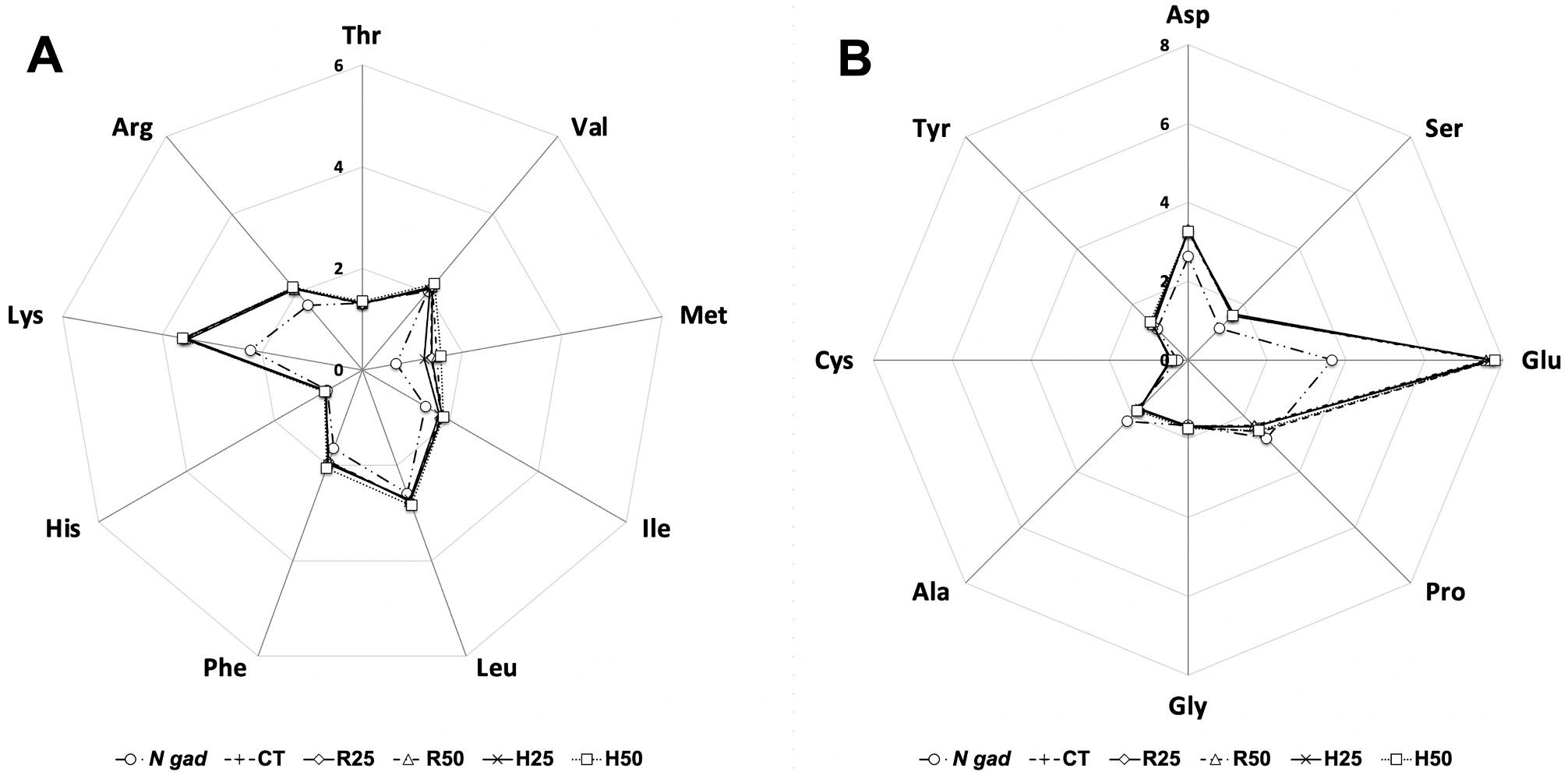
1. Amino acid profile of N. gaditana meal and the experimental diets. A: essential amino acids; B: non-essential amino acids. Results (n = 3) are expressed as % of total amino acids.
use. Total soluble protein was determined according to Bradford (1976) using bovine serum albumin as standard.
Total alkaline protease activity in intestinal extracts was measured spectrophotometrically following the procedure described by Alarcon et al. (1998), using 5 g L 1 casein in 50 mM Tris-HCl (pH 9.0) as substrate. One unit of total protease activity was defined as the amount of enzyme that released 1 μg of tyrosine per min in the reaction mixture,
considering an extinction coefficient of 0.008 μg 1 mL 1 cm 1 for tyrosine, measured at 280 nm wavelength. Trypsin and chymotrypsin activities were assayed using 0.5 mM BAPNA (N-α-benzoyl-DL-arginine4-nitroanilide) as substrate according to Erlanger et al. (1961), and 0.2 mM SAPNA (N-succinyl-(Ala)2-Pro-Phe-p-nitroanilide) according to Del Mar et al. (1979), respectively, in 50 mM Tris-HCl, 10 mM CaCl2 buffer, pH 8.5. Leucine aminopeptidase activity was determined by using 2 mM
Fig.
L-leucine-p-nitroanilide in 100 mM Tris-HCl buffer, pH 8.8, as substrate (Pfleiderer, 1970), and alkaline phosphatase was assayed using 450 mM p-nitrophenyl phosphate in 1 M diethanolamine, 1 mM MgCl2 buffer, pH 9.5 (Bergmeyer, 1974) as substrate. For trypsin, chymotrypsin, and leucine aminopeptidase activities, one enzyme activity unit (U) wasdefined as the amount of enzyme releasing 1 μmol of p-nitroanilide (pNA) per minute, considering as extinction coefficient 8800 M cm 1 , measured spectrophotometrically at 405 nm. For alkaline phosphatase, one activity unit was defined as the amount of enzyme that released 1 μg of nitrophenyl per min considering an extinction coefficient of 17,800 M cm 1 for p-nitrophenol, measured also at 405 nm. All assays were performed in triplicate, and specific enzymatic activity was expressed as units (U) g tissue 1 .
2.6. Histology of the intestinal mucosa
Intestine samples were fixed in phosphate-buffered formalin (4% v/ v, pH 7.2) for 24 h, then dehydrated and embedded in paraffin according to standard histological techniques, as described in Vizcaíno et al. (2018) Samples were cut in transversal sections (5 μm), and the slides were stained with haematoxylin-eosin (H&E). The preparations were examined under light microscope (Olympus ix51, Olympus, Barcelona, Spain) equipped with a digital camera (CC12, Olympus Soft Imaging Solutions GmbH, Muenster, Germany). Images were analysed with specific software (Image J, National Institutes of Health, USA). The length of mucosal folds and total enterocyte height were determined in intestinal samples (10 independent measurements performed at 4 different optical areas of each section from 5 fish per tank; 2 sections per fish).
2.7. Ultrastructure
of the intestinal mucosa
Intestine samples for scanning electron microscopy (SEM) were washed with 1% S-carboxymethyl-L-cysteine (Sigma Chem.) for 20 s, with the aim of removing the epithelial mucus, prior to fixation. Then, specimens were fixed in phosphate-buffered formaldehyde (4% v/v, pH 7.2) for 24 h; next excess glutaraldehyde was removed by washing samples in 0.1 mol L 1cacodylate buffer, pH 7.2, and then dehydrated with a series of increasing concentrations of ethanol (50% to 100% v/v). Samples were critical point dried in absolute ethanol as intermediate fluid, and CO2 as transition fluid (CDP 030 Critical point dryer, Leica Microsystems, Madrid, Spain). After drying, specimens were mounted on aluminium stubs, immobilized with graphite (PELCO® Colloidal Graphite, Ted Pella INC., Ca, USA), and then gold sputter coated (SCD 005 Sputter Coater, Leica Microsystems). Finally, all samples were screened with a scanning electron microscope (HITACHI S-3500, Hitachi High-Technologies Corporation, Japan).
Samples for transmission electron microscopy (TEM) were fixed (4 h, 4 ◦ C) in 25 g L 1 glutaraldehyde, 40 g L 1 formaldehyde in phosphate buffer saline (PBS), pH 7.5. Next, intestine sections were washed with PBS for 20 min and then, a post-fixation step with 20 g L 1 osmium tetroxide was carried out. Samples were dehydrated by consecutive immersion in increasing concentrations of ethanol, embedded for two hours, in 1:1 mixture of Epon resin and 100% (v/v) ethanol under continuous shaking, and then, included in pure Epon resin, and let polymerize at 60 ◦ C. Finally, ultrathin cuts were obtained from resin blocks, and placed on a 700 Å copper mesh and stained with uranyl acetate and lead citrate. The mesh observation was performed with a Zeiss 10C TEM at 100 Kv (Carl Zeiss, Barcelona, Spain).
SEM and TEM visualization fields were recorded and digital images were analysed using UTHSCSA ImageTool software (University of Texas Health Science Center, San Antonio, TX). Microvilli length (ML) and microvilli diameter (MD) were determined in TEM micrographs according to (Vizcaíno et al., 2014). SEM images were used to obtain measurements of enterocyte apical area (EAA). Finally, data obtained from TEM and SEM images were used to estimate the total absorption
surface per enterocyte (TAS) according to Vizcaíno et al. (2014)
2.8. Lipid oxidation
Lipid oxidation was estimated by thiobarbituric acid-reactive substances (TBARS) analysis in muscle and liver according to the method of Buege and Aust (1978). Briefly, samples (2 g each) were homogenized in 4 mL 50 mM NaH2PO4, 0.1% (v/v) Triton X-100 solution. The mixture was centrifuged (10,000 g, 20 min, 4 ◦ C) and supernatants were mixed in a ratio 1:5 (v/v) with 2-thiobarbituric acid (TBA) reagent (0.375% w/v TBA, 15% w/v TCA, 0.01% w/v 2,6-dibutyl hydroxytoluene (BHT) and 0.25 N HCl). The mixture was heated for 15 min and then centrifuged (3600 g, 10 min, 4 ◦ C), and the absorbance of supernatants was measured at 535 nm. The amount of TBARS was expressed as mg of malonyl dialdehyde (MDA) per kg of muscle after comparing with a MDA standard.
2.9. Instrumental colour determination
For all fish samples colour was measured on skin dorsal portion by L*, a*, and b* system (CIE, 1986), using a Minolta Chroma meter CR400 device (Minolta, Osaka, Japan). The parameter lightness (L*, on a 100point scale from black to white), redness (a*, assesses the position between red, positive values, and green, negative values), and yellowness (b*, assesses the position between yellow, positive values, and blue, negative values) were recorded.
2.10. Statistics
The effect of the categorical variables “pre-treatment” and “doses” , as well as their interactions, were determined for each numeric parameter studied by fitting a generalized lineal multifactorial statistical model (GLM analysis) that relates measured parameters to predictive factors, using specific software (SPSS 25, IBM Corporation Inc.). Least square means were tested for differences using Fisher’s least significant difference (LSD) procedure. Unless otherwise is specified, a significance level of 95% was considered to indicate statistical differences (P < 0.05). When measurements were expressed as a percentage (e.g., fatty acids profile), arcsine transformation of their square root was carried out in order to normalize data prior to the statistical analysis.
3. Results
3.1.
Microalgae hydrolysis
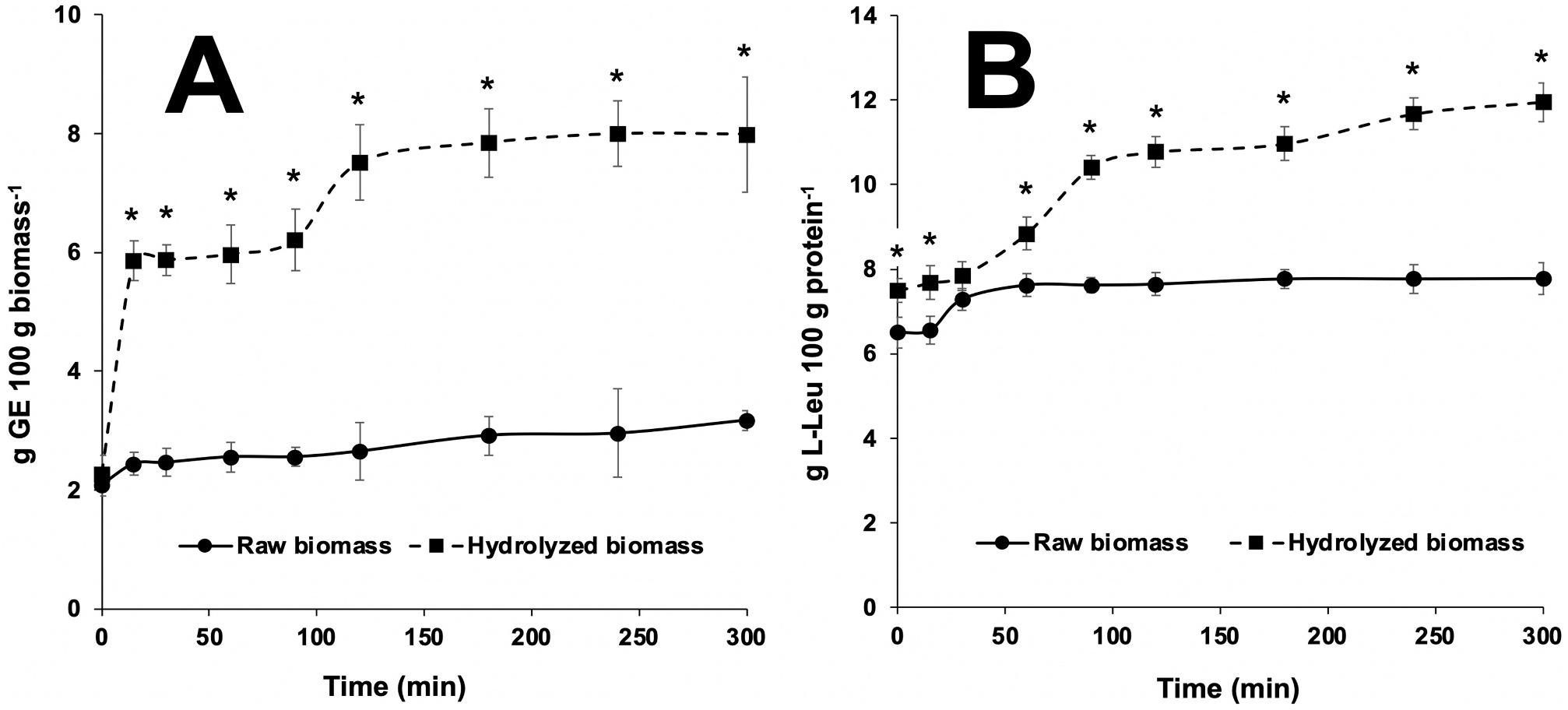
The concentration of reducing sugars in the reaction vessels increased throughout the in vitro assay owing to the addition of the commercial cellulase enzyme (Fig. 2A), being differences significant (P < 0.001) at each sampling time throughout the hydrolysis. Results indicate that enzyme-treated (5% cellulase) microalgal biomass yielded final values in the region of 8 g glucose equivalents (GE) per 100 g microalgae dry mass. This value was about 4-fold the amount of reducing sugars released from untreated raw algae (control), which accounted for stable values about 2–2.5 g GE throughout the complete assay (300 min).
Analogously, the total amount of amino acids released (Fig. 2B) during the assay indicated that cellulase hydrolysis increased significantly (P < 0.001) their concentration in the reaction vessels, compared to raw biomass. Under the assay conditions, final concentration of free amino acids in enzyme-treated batches reached 12 g 100 g protein 1 , compared to 6–7 g free amino acids 100 g protein 1 measured in controls (Fig. 3).
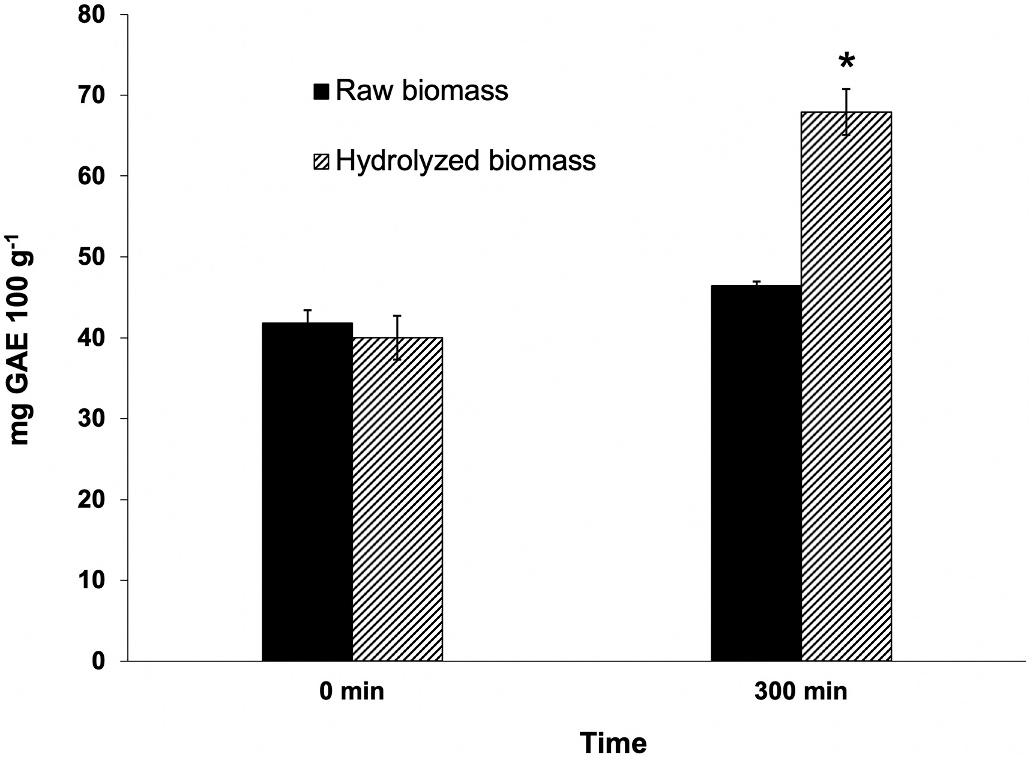
The quantification of total polyphenols at the beginning and at the end of the enzymatic assay revealed a 70% increase of these substances when the algal biomass was enzymatically treated (Fig. 3). The final concentration of total phenolics in supernatants was significantly higher
Fig. 2. Time-course of the concentration in reducing sugars (A, expressed as D-glucose equivalents, GE, 100 g dry biomass-1) and total free amino acids (B, expressed as g L-leucine, L-Leu, 100 g protein 1) measured from raw and cellulose-hydrolysed biomass of N. gaditana during the in vitro assay. Within each sampling time, asterisks indicate significant differences between values (P < 0.05). Values (n = 6) are mean ± standard deviation.
Fig. 3. Total phenolics released from raw and cellulase-hydrolysed N. gaditana biomass at the beginning and at the end of the in vitro hydrolysis. Results are represented as mg gallic acid equivalents (GAE) 100 g microalgal dry biomass 1 Within each sampling time, asterisks indicate significant differences between values (P < 0.05). Data (n = 6) are mean ± standard deviation.
(P < 0.01) in cellulase-treated N. gaditana (reaching 70 mg gallic acid equivalents (GAE) 100 g dry microalgae 1), than that measured in controls (40 mg GAE 100 g 1). In absence of cellulase, no significant differences were observed in total phenolics measured in the reaction vessel after the 5-h (300 min) incubation period.
3.2. Fish biometric parameters, muscle proximate composition and fatty acid profile
Experimental diets were well accepted by the fish, and feed intake was similar in all groups. Mortality was below 1%. During the experimental period, no differences were observed regarding growth parameters (final BW, FCR and SGR) among experimental lots at the end of the feeding trial (Table 3). Throughout this period, final body weight (approx. 50 g) at least triplicated initial values (approx. 15 g). No
Table 3
Fish biometric parameters and muscle proximate composition at the end of the feeding trial (90 d).
Diets
BW: final body weight. FCR: feed conversion rate. SGR: specific growth rate. CT: control diet. R25 and R50: diets including 25 and 50 g kg 1 raw microalgal biomass, respectively. H25 and H50: diets including 25 and 50 g kg 1 hydrolysed microalgae, respectively. Different lower-case superscripts indicate significant differences among diets within each row (P < 0.05). Values are mean ± standard deviation. For proximate composition n = 15. For biometric parameters n = 90. n.s.: not significant.
significant differences (P > 0.05) were observed for any of the parameters of muscle proximate composition. Although not significantly, microalgae-enriched diets tended to decrease slightly total muscle lipid content compared to CT diet, no matter the microalgae concentration or treatment considered.
Overall results on muscle fatty acid profile indicated that the inclusion of raw or hydrolysed microalgae yielded significant changes in FA profile (Table 4), especially with regard to MUFAs and PUFAs, which displayed opposing tendencies. Thus, microalgae-enriched diets reduced total MUFAs compared to CT, being this decrease more evident in diets including raw biomass (R25 vs. H25, and R50 vs. H50; P < 0.05). Regarding individual MUFAs, oleic acid (18:1n9) was the predominant FA, and its tendency paralleled that of total MUFA values. On the other
Table 4
Effects of the dietary inclusion of N. gaditana on fatty acid profile of gilthead seabream muscle after a 90-d feeding trial (% of total fatty acids).
Diets
14:0
22:6n3,
∑
∑n-3
±
± 0.19a
±
±
±
∑n-6 10.09 ± 0.08a 10.28 ± 0.09b 10.50 ± 0.10b
±
± 0.13b
<
±
± 0.09b 0.026
n3/n6 3.15 ± 0.02a 3.24 ± 0.01b 3.20 ± 0.01b 3.19 ± 0.02b 3.19 ± 0.03ab < 0.001
EPA/ DHA 0.21 ± 0.00a 0.24 ± 0.00c 0.24 ± 0.01c 0.22 ± 0.01 b 0.22 ± 0.00b < 0.001
CT: control diet. R25 and R50: diets including 25 and 50 g kg 1 raw microalgal biomass, respectively. H25 and H50: diets including 25 and 50 g kg 1 hydrolysed microalgae, respectively. Values with different lowercase superscript within each row indicate significant differences in muscle lipids attributed to dietary treatments (p < 0.05). SFA: saturated fatty acids; MUFA: monounsaturated fatty acids; PUFA: polyunsaturated fatty acids; EPA: eicosapentaenoic acid; DHA: docosahexaenoic acid. Values (n = 15) are expressed as average ± standard deviation. n.s.: not significant.
hand, increased total PUFAs in muscle was observed in fish fed on microalgae-containing diets compared to CT lot. As indicated for total MUFAs, although with opposite trend, the hydrolyzed biomass was responsible for significantly higher values (P < 0.05) of muscle PUFAs than raw biomass within each inclusion level. It is worth mentioning that both EPA and DHA paralleled such increase.
3.3. Digestive enzyme activities
In general, the supplementation with N. gaditana increased significantly (P < 0.05) the enzyme activities measured in intestinal extracts
compared to controls (Table 5), with the exception of leucine aminopeptidase (LAP) activity. Also considering all the activities as a whole, significant differences were attributable to sampling time, as values measured at the end of the assay (90 d) where significantly higher (P < 0.05) than those measured at day 45, irrespectively of the dietary treatment, with some exceptions for LAP activity again.
After 45 days of feeding, significant differences (P < 0.05) were observed in trypsin activity attributable to both biomass pre-treatment and inclusion level, whereas only microalgae hydrolysis influenced total alkaline protease and alkaline phosphatase activities (P < 0.05). No changes due to these factors were observed for leucine aminopeptidase. At the end of the feeding trial, the influence of both variables was significant on total alkaline protease, trypsin and chymotrypsin and activities, but only a hydrolysis-dependent effect was observed for and leucine aminopeptidase activity.
3.4. Intestinal mucosa histology
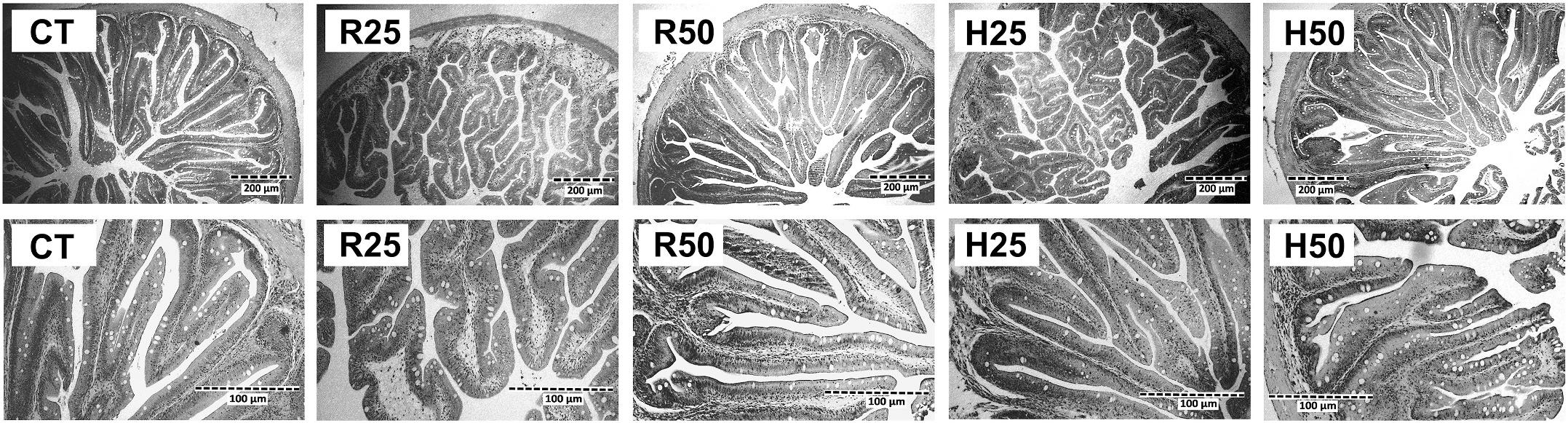
The histological characteristics of intestinal sections obtained from fish receiving the experimental dietary treatments at the end of the feeding trial are shown in Fig. 4, and results of the measurements carried out on haematoxylin-eosin stained sections are summarized in Table 6 Neither evidence of lipid droplet accumulation in the intestinal epithelium nor inflammatory changes in the lamina propria were observed. Consequently, no apparent damage attributable to any of the dietary treatments was found. Enterocytes presented aligned nucleus, homogenous supranuclear vacuolization and adequate cell shape (columnar and high). Intercellular spaces were not visible between enterocytes, and goblet cells were evenly dispersed throughout the epithelium.
Image analysis of the preparations indicated that no significant differences in fold length or enterocyte height were found among the dietary treatments. However, differences attributable to the inclusion level were observed, as the animals receiving R50 and H50 diets showing a significantly thinner lamina propria than the rest of the experimental batches, irrespectively of the enzyme pre-treatment.
3.5. Ultrastructure of the intestinal mucosa
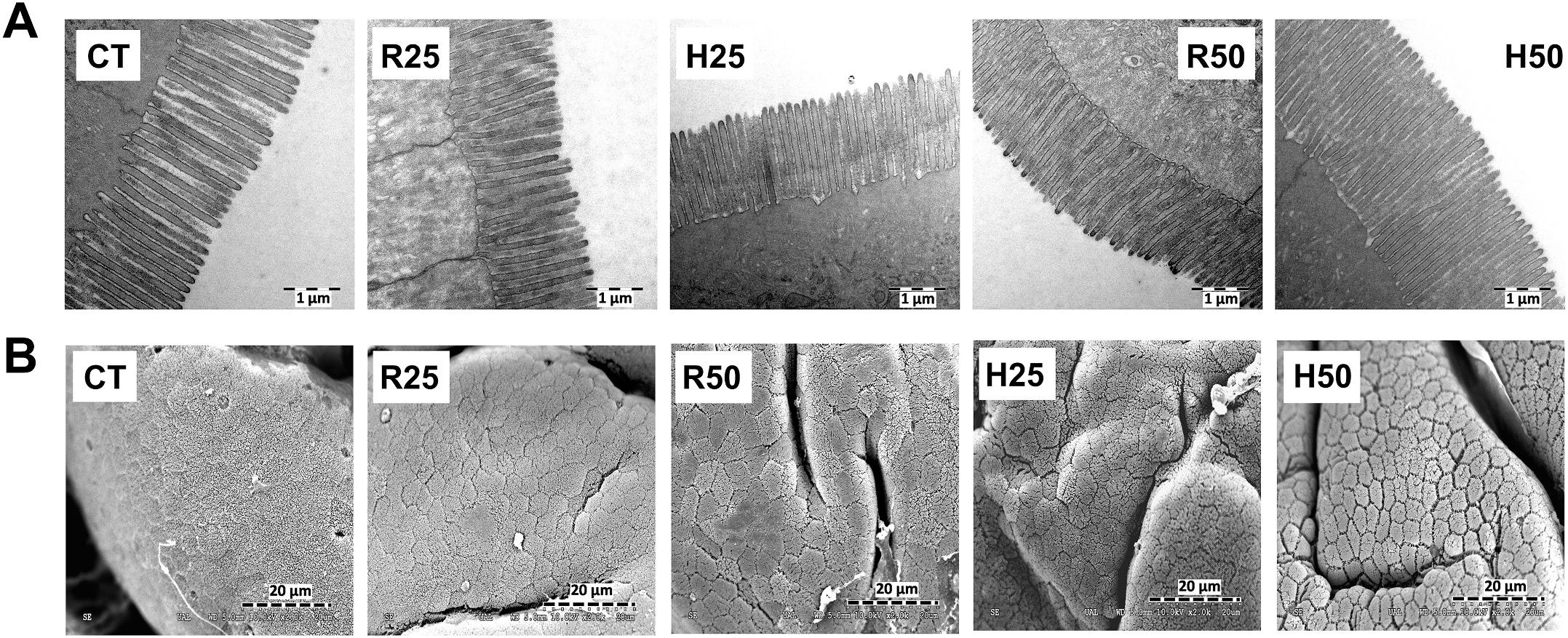
TEM and SEM observations indicated that none of the experimental diets caused any perceptible damage on the enterocyte brush border ultrastructure (Fig. 5), since specimens from animals from all the experimental groups presented a well-defined and organized intestinal brush border membrane. Moreover, no intercellular spaces were visible in the apical zone of the epithelium. Image analysis (Table 7) showed that only 5% inclusion level caused changes in all the parameters studied. R50 treatment yielded higher microvilli diameter (MD) than the rest of treatments, whereas only H50 caused significant increase in microvilli length (ML). Both R50 and H50 treatments increased significantly enterocyte apical area (EAA). The factor that accounted for most of the changes observed in total enterocyte absorption surface (TAS) was the biomass pre-treatment, given that both H25 and H50 batches showed significantly higher values for this parameter compared to CT and R25.
3.6. Muscle and liver lipid oxidation (TBARS)
The overall tendency for TBARS values (Table 8) indicates that CT batch yielded the highest values for this parameter, irrespectively of the tissue, sampling time, and dietary treatment considered (P < 0.001). Nevertheless, not all factors were responsible for significant differences in all cases.
With regard to muscle, R50, H25 and H50 treatments decreased significantly lipid oxidation compared to CT batch at both sampling times (45 and 90 d). Within each dietary treatment, only H50 showed differences attributable to sampling time.
The effects of the microalgae-enriched diets were even more evident
Table 5
Enzyme activities (U g tissue 1) measured in intestinal extracts of Sparus aurata juveniles fed with the experimental diets. Diets
CT: control diet. R25 and R50: diets including 25 and 50 g kg 1 raw microalgal biomass, respectively. H25 and H50: diets including 25 and 50 g kg 1 hydrolysed microalgae, respectively. LAP: Leucine aminopeptidase. Values (n = 9) are mean ± standard deviation. Values in the same row with different lowercase superscript indicate significant differences owing to dietary treatments (P < 0.05). Values of each enzyme activity with different superscript in Roman numerals indicate significant differences due to sampling time (P < 0.05). n.s.: not significant.
Fig. 4. Light microscopy details of intestine sections of S. aurata juveniles fed on the experimental diets for 90 days. H&E stain, magnification x100 (upper images) and ×400 (lower images). CT: control diet. R25 and R50: diets including 25 and 50 g kg 1 raw microalgal biomass, respectively. H25 and H50: diets including 25 and 50 g kg 1hydrolysed microalgae, respectively.
Table 6
Measurements in histological preparations of the intestinal mucosa of S. aurata juveniles fed with the experimental diets during 90 days.
Parameters
Diets
Values in the same row with different lowercase indicate significant differences (P < 0.05) owing to dietary treatments. CT: control diet. R25 and R50: diets including 25 and 50 g kg 1 raw microalgal biomass, respectively. H25 and H50: diets including 25 and 50 g kg 1 hydrolysed microalgae, respectively. Values are expressed as mean ± standard deviation. n.s.: not significant.
in liver, given that all treatments, irrespectively of biomass hydrolysis or inclusion level, yielded lower TBARS values than CT group. In this tissue, significant differences attributable to biomass hydrolysis (lower values for H25 and H50 in comparison with R25 and R50, respectively) and inclusion level (lower values for R25 and H25 compared to R50 and H50, respectively) were also observed. Same as found in muscle, also in liver only H50 showed differences attributable to sampling time.
3.7. Instrumental colour determinations
After 45 days of the feeding trial, skin L* values were similar in all lots (Table 9). The same lack of significant differences was found at the end of the experimental period for this parameter, although L* values were influenced for the factor sampling time (P < 0.05). Skin a* parameter presented negative values in all specimens, irrespectively of the sampling time. Figures for fish fed with any diet supplemented with
N. gaditana were significantly lower thanthose of CT group, indicating a more greenish colorationat both 45 and 90 d. Nevertheless, no differences attributable to biomass pre-treatment (R25 vs. H25; R50 vs. H50) or inclusion level (2.5% vs. 5.0%) were observed (P > 0.05). Similarly, all treatments including algae biomass tended to increase b* parameter (more yellowish pigmentation of the skin) compared to CT batch, although differences were significant only for R50 treatment at day 45, and for R25, R50 and H25 specimens at day 90.
4. Discussion
Given that cellulose accounts for the most abundant structural carbohydrate in N. gaditana, the breakage of cell walls by hydrolysis with cellulase enzymes emerges as a promising alternative aimed at improving nutrient bioavailability and digestibility. The following may be cited as advantages of this procedure: i) owing to the wide variety of
Fig. 5. Transmission (upper images, A) and scanning (lower images, B) electron microscopy micrographs from the anterior intestinal region of juvenile gilthead seabream at the end of the feeding trial (90 days). CT: control diet. R25 and R50: diets including 25 and 50 g kg 1 raw microalgal biomass, respectively. H25 and H50: diets including 25 and 50 g kg 1 hydrolysed microalgae, respectively.
Table 7
Microvilli morphological parameters obtained from transmission electron microscopy ultramicrographs of the anterior intestine of S. aurata juveniles fed with the experimental diets during 90 days.
Parameters Diets
ML (μm)
ML: microvilli length; MD: microvilli diameter; EAA: enterocyte apical area; TAS: total enterocyte absorption surface. CT: control diet. R25 and R50: diets including 25 and 50 g kg 1 raw microalgal biomass, respectively. H25 and H50: diets including 25 and 50 g kg 1 hydrolysed microalgae, respectively. Values in the same row with different lowercase superscripts indicate significant differences (P < 0.05) owing to the dietary treatment. Values (n = 15) are expressed as mean ± standard deviation.
industrial applications, cellulases are reasonably inexpensive; ii) no complex equipment is needed for hydrolysis, as incubators widely utilized in the food and feed industry can be used; iii) negative impacts on thermolabile compounds are not expected, as mild temperatures are involved in the hydrolysis process; and iv) given the specificity of the catalytic action on cellulose, the remaining released compounds would not be hydrolysed by the enzymatic pre-treatment.
Under this perspective, this study evaluatedin a 90-d feeding trial the effects of a cellulase pre-treatment on Nannochloropsis gaditana biomass prior to its incorporation into feeds for gilthead seabream juveniles. It was expected that nutrient bioavailability would increase as a result of the enzyme pre-treatment, and according to the results of the enzyme hydrolysis (Figs. 2 and 3), the increased release of reducing sugars, free amino acids and polyphenols taking place in the reaction vessels seem to confirm this hypothesis.
Even if the enzyme treatment increased in vitro bioavailability, however, no impact on fish growth was observed for any of the experimental batches throughout the 90-d feeding trial. These results are in
Table 8
Estimation of lipid oxidation (TBARS) in muscle and liver of juvenile fish fed on the different experimental diets.
Diets Time
Muscle
CT: control diet. R25 and R50: diets including 25 and 50 g kg 1 raw microalgal biomass, respectively. H25 and H50: diets including 25 and 50 g kg 1 hydrolysed microalgae, respectively. TBARS stands for thiobarbituric acid reactive substances, expressed as mg malonyldialdehyde (MDA) kg tissue 1 Values in the same row with different lowercase superscripts indicate significant differences (P < 0.05) owing to dietary treatments. Within each tissue, values with different superscript in Roman numerals indicate significant differences due to sampling time (P < 0.05). Values (n = 15) are mean ± standard deviation.
line with previous studies on this microalgae genus carried out on several species of commercial fish (Qiao et al., 2019; Sørensen et al., 2017; Walker and Berlinsky, 2011). Nevertheless, other reports pointed to improved fish growth owing to microalgae inclusion in diets, but higher inclusion levels were evaluated (up to 10% inclusion in diets for Nile tilapia, Abdel-Tawwab and Ahmad, 2009; from 10 to 39% for S. aurata, in Vizcaíno et al., 2014, 2016, 2018). It is likely that the low inclusion levels considered have accounted for this lack of effect on growth.
With regard to fish muscle proximal composition, overall, no differences attributable to the experimental diets were observed, in agreement with previous studies on N. gaditana enriched diets (Qiao et al., 2019; Vizcaíno et al., 2018; Sales et al., 2021). Only slight, but not significant differences in lipid and protein contents were measured among the experimental groups (Table 3). The study by Galafat et al.
Values in the same row with different lowercase superscripts indicate significant differences (P < 0.05) owing to dietary treatments. CT: control diet. R25 and R50: diets including 25 and 50 g kg 1 raw microalgal biomass, respectively. H25 and H50: diets including 25 and 50 g kg 1 hydrolysed microalgae, respectively.
Values (n = 30) are mean ± standard deviation. Parameters L*, a* and b* as defined in M&M section. n.s.: not significant.
(2020) reported a significant increase inmuscle protein in juvenile gilthead seabream fed enzymatically hydrolysed Arthrospira platensis added at 2% inclusion level, as well as a significant decrease in total lipids when added at 4% inclusion level. Reduced muscle lipid storage has also been reported not only for microalgae species (Hussein et al., 2013; El-Sheekh et al., 2014; Vizcaíno et al., 2014, 2016), but also for macroalgae (Ortiz et al., 2006; Yildirin et al., 2009; S´ aez et al., 2020). These findings suggest the existence of bioactive compounds in algae capable of influencing protein and lipid metabolism, although the nature of such substances or the underlying mechanisms involved in such effects have not yet been fully ascertained. Recent evidence in this regard was provided by Perera et al. (2020), although microalgaecontaining commercial products rather than pure microalgae biomass were considered in the study.
Whilst no quantitative differences in muscle lipid content were observed, however, qualitative differences were found in this analytic component. It is known that fish muscle lipids reflect dietary FA profiles (Grigorakis et al., 2002; Grigorakis, 2007; Yildiz, 2007), and this might explain the significant increase of EPA muscle content of fish fed with all the algae-containing diets (Table 4), compared to control batch.
N. gaditana is rich in EPA (33% of total FA), and consequently, all microalgae-supplemented diets, either raw or hydrolysed (Table 4), were enriched in this specific FA in a dose-dependent manner, but not influenced by the enzymatic pre-treatment of the biomass (no significant differences, between R25 and H25, or between R50 and H50, Table 2).
Although all the microalgae-enriched diets yielded muscle EPA contents higher than those found in CT batch (Table 4), this effect wasn’t dose-dependent. Interestingly, and contrary to what was expected, the enzymatic treatment of the biomass yielded lower EPA in muscle compared to the raw microalgae. A possible explanation would be that EPA released from cells could be more susceptible to structural damage than that remaining within the microalgae cells. In other words, intact cell walls might have acted as a sort of “natural microcapsule” for EPA.
All the experimental batches fed with the supplemented diets yielded significantly higher DHA muscle content than control fish. This fact can’t be explained by differences in this FA in the experimental diets (Table 2), given that no DHA was measured in N. gaditana biomass. On
the contrary, control diet showed the highest DHA figures, but yielded the lowest content in fish muscle. In this regard, previous studies have also reported certain selective retention of this structural FA owing to the addition of both macro and microalgae (Hussein et al., 2013; Vizcaíno et al., 2014, 2016; Kousoulaki et al., 2016; S´ aez et al., 2020). In short, the results suggested that N. gaditana, even at the low inclusion levels studied, was responsible for some selective retention of n-3-PUFA in muscle (mostly owing to DHA, the most abundant n3-PUFA), whilst the opposite effect was observed with regard to MUFAs (Table 4). Such decrease in total MUFAs is in agreement with the evidenced lower oleic acid content, which is the main MUFA in muscle of gilthead seabream. Nevertheless, disparate results have been reported in the literature regarding the effects of dietary microalgae on S. aurata lipid metabolism (Perera et al., 2020), and further studies aimed at fully understanding the intrinsic mechanisms underlying the results observed are needed.
On the other hand, microalgae are acknowledged as valuable source of pigments and phenolic compounds with antioxidant capacity (Koyande et al., 2019; Almendinger et al., 2021; S´ aez et al., 2021), many of which remain unidentified (Sansone et al., 2020).
Due to the interest of the pharmaceutical industry in pigments, these substances have received more attention than phenolics, but some authors suggested that both groups of substances might contribute similarly to the antioxidant activity of microalgae (Almendinger et al., 2021). Nevertheless, the relative contribution of phenolics and pigments to the antioxidant capacity of most microalgae species remains to be ascertained (Goiris et al., 2012). N. gaditana contains chlorophylls, β-carotene, violaxanthin y vaucheriaxanthin, as well as trace amounts of astaxanthin (Cer ´ on-García et al., 2018), which might explain our results pointing to higher antioxidant response in muscle and liver of fish supplemented with the microalgal biomass. Teimouri et al. (2016, 2019) also described this effectas a result of the inclusion of microalgae in feeds, and more specifically, Qiao et al. (2019) reported lower TBARS values both in liver and serum in Scophtalmus maximus juveniles fed on 5% N. gaditana diets. A recent study (Sales et al., 2021) has shown that purified extracts of the unsaponifiable fraction of N. gaditana, rich in carotenoids, included in feeds to partially replace fish oil yielded potent antioxidant effects in muscle of S. aurata juveniles.
The cellulase pre-treatment of the microalgal biomass was responsible for a trend towards increased in vivo antioxidant effects on muscle lipids (Table 8), compared to untreated N. gaditana Such increase attributable to enzyme hydrolysis reached statistical significance in the case of liver lipids. These results suggest that increased release and further bioavailability of some inner bioactive compounds contained in the cells might have occur, as was the case for total phenolics (Fig. 3). Galafat et al. (2020) also found lower TBARS values in muscle of S. aurata juveniles fed with Arthrospira sp. protease hydrolysates included at low inclusion level (2 and 4%) in diets. In agreement, and with regard to phenolics, N. gaditana contains certain amount of these substances in raw biomass, in line with previous studies (Kherraf et al., 2017; Haoujar et al., 2019), which might explain the potent antioxidant effects found on fish lipids in our study. Noticeably, as mentioned, total phenolics measured in the reaction vessels increased as a result of the cellulase treatment (Fig. 3).
Physical treatments, even if valuable when it comes to increasing the yield of microalgae main compounds (i.e. protein and lipid fractions), might jeopardize the chemical integrity of thermolabile minor compounds (Schafberg et al., 2020), and consequently, impair their functional activity.
Given the susceptibility of pigments, especially carotenoids, to different factors (temperature, oxygen, light, acidic pH, etc., Schieber and Weber, 2016), and even if the extraction procedures increase the releasing of inner compounds, it should also be born in mind that microalgal biomass, as part of the ingredient mixture, will be extruded during the elaboration of the experimental diets, a process involving high pressure and temperature. Consequently, doubts could arise related to the integrity and the subsequent in vivo bioavailability of some of the Table 9
M.I.
compounds released in vitro Previous research suggests that the resulting balance of disrupting strategies is favourable to the enrichment of aquafeeds (Schafberg et al., 2020), and our results coincide with that idea. But given the diversity and complex nature of the antioxidant substances involved, this specific issue deserves further research. Especially the balance between phenolics and carotenoids in a given microalgae species is likely that could determine the persistence of the antioxidant effects in feeds after processing procedures.
Although instrumental colour measurements at early stages of the productive cycle have no interest in practical terms of fish quality assessment, they can still provide valuable information about pigment deposition and antioxidant effects in growing fish tissues. The favourable influence of microalgae on fish colour parameters found in our study (increased a* and b*, Table 9) has been documented previously (Teimouri et al., 2013; Cardinaletti et al., 2018; Galafat et al., 2020; Kousoulaki et al., 2020; Sales et al., 2021). Although tendencies observed for skin pigmentation suggest that raw microalgae intensified the effects compared to hydrolysed biomass (Table 9), however, no significant differences attributable to the enzymatic treatment were found. This might well be explained again by the fact that some pigments contained in the hydrolysed biomass might have been damaged to a higher extent than those from raw biomass due to feed processing.
The activity of digestive enzymes is acknowledged not only as a marker of their digestive and absorptive capacity (Alarc ´ on et al., 1998), but also as a reliable indicator of the nutritional status of aquacultured fish. More specifically, the activity of some brush border membrane enzymes, such as leucine aminopeptidase (LAP) and alkaline phosphatase, reveals the integrity and the absorptive capability of the intestinal mucosa (Silva et al., 2010). Disparate results have been reported on the effects of raw microalgae on digestive enzyme activities, and thus, Qiao et al. (2019) found increased trypsin activity in juvenile turbot supplemented with N. gaditana biomass at 7.5% inclusion level after a 10-week feeding trial. On the contrary, Jorge et al. (2019) observed no effects on total alkaline protease, trypsin, α-amylase and lipase activities in response to dietary N. gaditana supplementation, although the low inclusion levels considered (0.5, 1, and 1.5%) together with the short duration of the feeding trial (37 d) might well have accounted for such lack of effects. Few studies are available assessing the physiological consequences of microalgae enzyme hydrolysates on such activities. Galafat et al. (2020) described higher trypsin and LAP activities as a result of protease hydrolysates of Arthrospira sp. at low inclusion level (2 and 4%) in S. aurata juveniles. More recently, Galafat et al. (2022) reported increased trypsin, chymotrypsin and leucine aminopeptidase activities in gilthead seabream fry as a result of supplementing starting diets with Arthrospira platensis at 5 and 10% compared to control fish. In addition, within each inclusion level, animals fed with diets that included the hydrolysed biomass yielded consistently higher digestive enzyme activities than those receiving the crude biomass. The results obtained in this study indicate that N. gaditana supplementation, even at the low inclusion levels tested, overall increased the enzyme activities assayed compared to control fish, irrespectively of sampling time (45 or 90 d), or biomass pre-treatment, with the exception of leucine aminopeptidase activity at day 45 (Table 5). It is also worth mentioning that the favourable effects of the experimental diets on intestinal digestive activities observed in this work concur, roughly speaking, with the ultrastructural determinations (Table 7) carried out on the intestinal mucosa at the end of the feeding trial, especially at the highest concentration assayed (5%).
Overall, concerning the enzyme pre-treatment considered in this work, no decisive effects were observed in terms of fish growth, muscle composition, or digestive functionality, but the remarkable influence of this treatment on the oxidative status of fishlipids could result in beneficial effects on other parameters linked to the health status of aquacultured fish, a fact that deserves further investigation as well. It should also be born in mind that any feeding trial under controlled conditions and short duration has evident limitations in terms of further
applicability on-farm, bearing in mind the numerous additional factors involved in the operation of long-term production cycles in commercial fish farms.
5. Conclusions
Although N. gaditana biomass at low inclusion level in feeds had no impact on growth and muscle proximal composition, however, it is worth mentioning that the lack of detrimental effects, together with some beneficial effects on other physiological parameters (digestive structure and functionality, oxidative status of muscle and liver lipids, and skin colour), overall indicate that might represent a valuable additive in long-term S. aurata production cycles.
The results obtained evidenced the effectiveness of the cellulase pretreatment when it comes to in vitro releasing of intracellular compounds from N. gaditana cells, which could improve not only extraction yields in industrial applications, but also increase the bioavailability of certain metabolites with potentially bioactive and functional effects. However, no conclusive evidence was found regarding the impact of this strategy on most of the physiological parameters tested, with the exception of the enhanced effects on lipid oxidation.
It would be interesting to carry out further research aimed at assessing the possible influence of N. gaditana hydrolysates on other valuable physiological aspects, such as their influence on the intestinal microbiome, the intermediary metabolism (not least lipid metabolism) and the immune response of gilthead seabream, especially at early stages of the production cycle.
Authors’ contributions
S´ aez, M.I., Alarc ´ on, F.J. and Martínez T.F. conceived and designed the experiments. Alarc ´ on, F.J. and Galafat, A. prepared the aquafeeds. Galafat, A., S´ aez, M.I., Vizcaíno,A.J. and Martínez, T.F. performed fish sampling. Arizcun, M., Chaves-Pozo, E.and Ayala, M.D. participated in sampling, fish care and maintenance, and in biometric and proximal analysis. Su´ arez, M.D. performed and interpreted fatty acid analysis. Galafat, A., S´ aez, M.I., Martínez, T.F., Su´ arez, M.D., Arizcun M., and Chaves-Pozo E. performed analytical analysis and discussed the data. S´ aez, M.I., Alarc ´ on, F.J. and Martínez, T.F. drafted the manuscript. T.F. Martinez and M. Arizcun obtained the necessary funds for conducting the research. All authors critically revised and approved the manuscript.
Funding information
This research was funded by Spanish MCIU-FEDER (grant # RTI2018-096625-B-C33 and grant # RTI2018–096625-B-C31), SABANA project (the European Union’s Horizon 2020 Research and Innovation program, grant # 727874), AquaTech4Feed (grant # PCI2020-112204) granted by MCIN/AEI/10.13039/501100011033, the EU “NextGenerationEU”/PRTR within the ERA-NET BioBlue COFUND). Servicio de piensos experimentales was granted by EQC2018-004984-P and EQC2019-006380-P.
Statement of informed consent, human/animal rights
The authors state that no conflicts, informed consent, human or animal rights are applicable. All studies involving fish were conducted in accordance with the requirements of the Directive 2010/63/EU, and the Spanish legislation (Real Decreto 53/2013, as amended by RD218/ 2021), regarding the ethical rules applicable in research involving laboratory animals. Thereby, all the procedures were authorized by the Bioethics and Animal Welfare Committee of the Instituto Espanol de Oceanografía (REGA code ES300261040017) with the approval of the Ministry of Water, Agriculture and Environment of the Autonomous Community Region of Murcia (Spain; A13200101).
Declaration of Competing Interest
The authors declare that they have no conflict ofinterest.
References
Abdel-Tawwab, M., Ahmad, M.H., 2009. Live Spirulina (Arthrospira platensis) as a growth and immunity promoter for Nile tilapia, Oreochromis niloticus (L.), challenged with pathogenic Aeromonas hydrophila Aquac. Res. 40, 1037–1046. https://doi.org/ 10.1111/j.1365-2109.2009.02195.x
Agboola, J.O., Teuling, E., Wierenga, P.A., Gruppen, H., Schrama, J.W., 2019. Cell wall disruption: an effective strategy to improve the nutritive quality of microalgae in African catfish (Clariasgariepinus). Aquac. Nutr. 25, 783–797. https://doi.org/ 10.1111/anu.12896
Alarc ´ on, F.J., Díaz, M., Moyano, F.J., Abell´ an, E., 1998. Characterization and functional properties of digestive proteases in two sparid; gilthead sea bream (Sparus aurata) and common dentex (dentex dentex). Fish Physiol. Biochem. 19, 257–267. https:// doi.org/10.1023/A:1007717708491
Almendinger, M., Saalfranka, F., Rohnab, S., Kurthc, E., Springerd, M., Pleissner, D., 2021. Characterization of selected microalgae and cyanobacteria as sources of compounds with antioxidant capacity. Algal Res. 53, 102168 https://doi.org/ 10.1016/j.algal.2020.102168
AOAC, 2000. Official methods of analysis. In: 17th Edition. The Association of Official Analytical Chemists. Gaithersburg, MD, USA. Methods 925.10, 65.17, 974.24, 992.16
Batista, S., Pintado, M., Marques, A., Abreu, H., Silva, J.L., Jessen, F., Valente, L.M.P., 2020. Use of technological processing of seaweed and microalgae as strategy to improve their apparent digestibility coefficients in European seabass (Dicentrarchus labrax) juveniles. J. Appl. Phycol. 32, 3429–3446. https://doi.org/10.1007/s10811020-02185-2
Becker, W., 2003. Microalgae in human and animal nutrition. In: Handbook of Microalgal Culture. Blackwell Publishing Ltd., Oxford, UK, pp. 312–351. https://doi. org/10.1002/9780470995280.ch18
Bergmeyer, H.V., 1974. Methods of enzymatic analysis. In: Phosphatase, vol. 2. Academic Press, New York, pp. 856–860
Bradford, M., 1976. A rapid and sensitive method for the quantitation of microgram quantities of protein utilizing the principle of protein-dyebinding. Anal.Biochem. 72, 248–254
Buege, J.A., Aust, S.D., 1978. Microsomal lipid peroxidation. In: Methods in enzymology, Vol. 52. Academic Press, pp. 302–310. Cardinaletti, G., Messina, M., Bruno, M., Tulli, F., Poli, B.M., Giorgi, G., Tibaldi, E., 2018. Effects of graded levels of a blend of Tisochrysis lutea and Tetraselmissuecica dried biomass on growth and muscle tissue composition of European sea bass (Dicentrarchuslabrax) fed diets low in fish meal and oil. Aquaculture 485, 173–182. https://doi.org/10.1016/j.aquaculture.2017.11.049
Cerezuela, R., Guardiola, F.A., Gonzalez, P., Meseguer, J., Esteban, M.A., 2012. Effects of dietary Bacillus subtilis, Tetraselmischuii, and Phaeodactylumtricornutum, singularly or in combination, on the immune response and disease resistance of sea bream (Sparus aurata L.). Fish Shell. Immunol. 33, 342–349. https://doi.org/10.1016/j. fsi.2012.05.004
Ceron-García, M.C., Gonzalez-Lopez, C., Camacho-Rodríguez, J., Lopez-Rosales, L., García-Camacho, F., Molina-Grima, E., 2018. Maximizing carotenoid extraction from microalgae used as food additives and determined by liquid chromatography (HPLC). Food Chem. 257, 316–324. https://doi.org/10.1016/j. foodchem.2018.02.154
Church, F.C., Swaisgood, H.E., Porter, D.H., Catignani, G.L., 1983. Spectrophotometric assay using o-phthaldehyde for determination of proteolysis in milk proteins. J. Dairy Sci. 66, 1219–1227
CIE, 1986. Recommendations on uniform color spaces, color difference equations, psychometric color terms, supplement 2 to CIE publication, vol. 15. Central Bureau of the Commission Internationale de l’Eclairage Vienna, Austria (E1.3.1) 1971/ (TC1.3)
Del Mar, E.G., Largman, C., Broderick, J.W., Geokas, M.C., 1979. A sensitive new substrate for chymotrypsin. Anal. Biochem. 99, 316–320. https://doi.org/10.1016/ s0003-2697(79)80013-5
El-Sheekh, M., El-Shourbagy, I., Shalaby, S., Hosny, S., 2014. Effect offeeding Arthrospira platensis (Spirulina) on growth and carcass composition of hybrid red tilapia (Oreochromis niloticus x Oreochromis mossambicus). Turk. J. Fish. Aquat. Sci. 14, 471–478. https://doi.org/10.4194/1303-2712-v14_2_18
Erlanger, B., Kokowsky, N., Cohen, W., 1961. The preparation and properties of two new chromogenic substrates of trypsin. Arch. Biochem. Biophys. 95, 271–278. https:// doi.org/10.1016/0003-9861(61)90145-X
Folch, J., 1957. A simple method for the isolation and purification of total lipids form animal tissues. J. Biol. Chem. 226, 497–509. https://doi.org/10.1016/S0021-9258 (18)64849-5
Galafat, A., Vizcaíno, A.J., Saez, M.I., Martínez, T.F., Jerez-Cepa, I., Mancera, J.M., Alarcon, F.J., 2020. Evaluation of Arthrospira sp enzyme hydrolysate as dietary additive in gilthead seabream (Sparus aurata) juveniles. J. Appl. Phycol. 32, 3089–3100. https://doi.org/10.1007/s10811-020-02141-0
Galafat, A., Vizcaíno, A.J., S´ aez, M.I., Martínez, T.F., Arizcun, M., Chaves-Pozo, E., Alarc ´ on, F.J., 2022. Assessment of dietary inclusion of crude or hydrolysed Arthrospira platensis biomass in starter diets for gilthead seabream (Sparus aurata). Aquaculture 548, 737680. https://doi.org/10.1016/j.aquaculture.2021.737680
Goiris, K., Muylaert, K., Fraeye, I., Foubert, I., De Brabanter, J., De Cooman, L., 2012. Antioxidant potential of microalgae in relation to their phenolic and carotenoid content. J.Appl.Phycol. 24, 1477–1486. https://doi.org/10.1007/s10811-012-98046:1-10
Gomes, T.A., Zanette, C.M., Spier, M.R., 2020. An overview of cell disruption methods for intracellular biomolecules recovery. Prep.Biochem.Biotech. 50, 635–654. https:// doi.org/10.1080/10826068.2020.1728696
Grigorakis, K., 2007. Compositional and organoleptic quality of farmed and wild gilthead sea bream (Sparus aurata) and sea bass (Dicentrarchus labrax) and factors affecting it: a review. Aquaculture 272, 55–75. https://doi.org/10.1016/j. aquaculture.2007.04.062
Grigorakis, K., Alexis, M.N., Taylor, K.D.A., Hole, M., 2002. Comparison of wild and cultured gilthead sea bream (Sparus aurata); composition, appearance and seasonal variations. Int. J. Food SciTechn37(5), 477–484. https://doi.org/10.1046/j.13652621.2002.00604.x.
Günerken, E., D’Hondt, E., Eppink, M.H.M., Garcia-Gonzalez, L., Elst, K., Wijffels, R., 2015. Cell disruption for microalgae biorefineries. Biotechnol. Adv. 33, 243–260. https://doi.org/10.1016/j.biotechadv.2015.01.008
Haoujar, I., Cacciola, F., Abrini, J., Mangraviti, D., Giuffrida, D., Oulad El Majdoub, Y., SkaliSenhaji, N., 2019. The contribution of carotenoids, phenolic compounds, and flavonoids to the antioxidative properties of marine microalgae isolated from Mediterranean Morocco. Molecules 24, 4037. https://doi.org/10.3390/ molecules24224037
Heredia, H., Pruvost, J., Gonçalves, O., Drouin, L., Marchal, L., 2021. Lipid recovery from Nannochloropsis gaditana using the wet pathway: investigation of the operating parameters of bead milling and centrifugal extraction. Algal Res. 56, 102318 https:// doi.org/10.1016/j.algal.2021.102318
Hussein, E.E.S., Dabrowski, K., El-Saidy, D.M., Lee, B.J., 2013. Enhancing the growth of Nile tilapia larvae/juveniles by replacing plant (gluten) protein with algae protein. Aquac. Res. 44, 937–949. https://doi.org/10.1111/j.1365-2109.2012.03100.x
Jorge, S.S., Enes, P., Serra, C.R., Castro, C., Iglesias, P., Oliva Teles, A., Couto, A., 2019. Short-term supplementation of gilthead seabream (Sparus aurata) diets with Nannochloropsisgaditana modulates intestinal microbiota without afecting intestinal morphology and function. Aquac. Nutr. 25, 1388–1398. https://doi.org/10.1111/ anu.12959
Kavitha, S., Gajendranb, T., Saranyaa, K., Selvakumarc, P., Manivasagana, V., 2021. Study on consolidated bioprocessing of pre-treated Nannochloropsisgaditana biomass into ethanol under optimal strategy. Ren.Energy 172, 440–452. https://doi.org/ 10.1016/j.renene.2021.03.015.
Kherraf, A., Tehami, W., Boufeldja, W., Yahla, I., Dra, G.A., Mansour, I.F.Z., Benali, M., 2017. Determination of the nutritional and functional metabolites of Nannochloropsisgaditana produced in Algeria and evaluation of its antioxidant activity. Der. Pharma. Chem. 9 (14), 8–13. https://doi.org/10.38150/sajeb.7(1)
Kilian, O., Benemann, C.S.E., Niyogi, K.K., Vick, B., 2011. High-efficiency homologous recombination in the oil-producing alga Nannochloropsis sp. Proc. Natl. Acad. Sci. U. S. A. 108, 21265–21269. https://doi.org/10.1073/pnas.1105861108
Kiron, V., 2012. Fish immune system and its nutritional modulation for preventive health care. Anim. Feed Sci. Technol. 173, 111–133. https://doi.org/10.1016/j. anifeedsci.2011.12.015
Kousoulaki, K., Mørkøre, T., Nengas, I., Berge, R.K., Sweetman, J., 2016. Microalgae and organic minerals enhance lipid retention efficiency and fillet quality in Atlantic salmon (Salmo salar L.). Aquaculture 451, 47–57. https://doi.org/10.1016/j. aquaculture.2015.08.027
Kousoulaki, K., Gerd Marit, B., Mørkøre, T., Krasnov, A., Baeverfjord, G., Ytrestøyl, T., Ruyter, B., 2020. Microalgal Schizochytriumlimacinum biomass improves growth and filet quality when used long-term as a replacement for fish oil, in modern salmon diets. Front. Mar. Sci. 7 https://doi.org/10.3389/fmars.2020.00057
Koyande, A.K., Chew, K.W., Rambabu, K., Tao, Y., Chu, D.T., Show, P.L., 2019. Microalgae: a potential alternative to health supplementation for humans, food Sci. Human Wellness 8, 16–24. https://doi.org/10.1016/j.fshw.2019.03.001
Lee, J.Y., Yoo, C., Jun, S.Y., Ahn, C.Y., Oh, H.M., 2010. Comparison of several methods for effective lipid extraction from microalgae. Bioresour. Technol. 101, S75–S77. https://doi.org/10.1016/j. biortech.2009.03.058.
Lepage, G., Roy, C.C., 1984. Improved recovery of fatty acid through direct transesterification without prior extraction or purification. J. Lipid Res. 25, 1391–1396
Menegol, T., Romero-Villegas, G.I., Lopez-Rodríguez, M., Navarro-Lopez, E., LopezRosales, L., Chisti, Y., Molina-Grima, E., 2019. Mixotrophic production of polyunsaturated fatty acids and carotenoids by the microalga Nannochloropsisgaditana J. Appl. Phycol. 31, 2823–2832. https://doi.org/10.1007/ s10811- 019-01828-3
Miller, G.L., 1959. Use of dinitrosalicylic acid reagent fordetermination of reducing sugar. Anal. Chem. 31, 26–428
Ortiz, J., Romero, N., Robert, P., 2006. Dietary fiber, amino acid, fatty acid and tocopherol contents of the edible seaweeds Ulva lactuca and Durvillaea Antarctica Food Chem. 99, 98–104. https://doi.org/10.1016/j.foodchem.2005.07.027
Pagels, F., Pereira, R.N., Vicente, A.A., Guedes, A.C., 2021. Extraction of pigments from microalgae and cyanobacteria - a review on current methodologies. Appl. Sci. 11, 5187. https://doi.org/10.3390/app11115187
Perera, E., S´ anchez-Ruiz, D., S´ aez, M.I., Galafat, A., Barany, A., Fern´ andez-Castro, M., Martos-Sitcha, J.A., 2020. Low dietary inclusion of nutraceuticals from microalgae improves feed efficiency and modifies intermediary metabolisms in gilthead sea bream (Sparus aurata). Sci. Rep. 10, 18676. https://doi.org/10.1038/s41598-02075693-3
Pfleiderer, G., 1970. Particle-bound aminopeptidase from pig kidney. Met.Enzymol. 19, 514–521. https://doi.org/10.1016/0076-6879(70)19038-0.
Qiao, H., Hu, D., Ma, J., Wang, X., Wu, H., Wang, J., 2019. Feeding effects of the microalga Nannochloropsis sp. on juvenile turbot (Scophthalmus maximus L.). Algal Res. 41, 101540 https://doi.org/10.1016/j.algal.2019.101540
Rodríguez-Ruiz, J., Belarbi, E.H., Sanchez, J.L.G., Alonso, D.L., 1998. Rapid simultaneous lipid extraction and transesterification for fatty acid analyses. Biotech. Techn. 12 (9), 689–691. https://doi.org/10.1023/A:1008812904017
Rojo, E.M., Piedra, I., Gonz´ alez, A.M., Vega, M., Bolado, S., 2021. Effect of process parameters on the valorization of components from microalgal and microalgalbacteria biomass by enzymatic hydrolysis. Bioresour. Technol. 335, 125256 https:// doi.org/10.1016/j.biortech.2021.125256
S´ aez, M.I., Vizcaíno, A.J., Galafat, A., Anguís, V., Fern´ andez-Díaz, C., Balebona, M.C., Martínez, T.F., 2020. Assessment of long-term effects of the macroalgae Ulva ohnoi included in diets on Senegalese sole (Solea senegalensis) fillet quality. Algal Res. 47, 101885 https://doi.org/10.1016/j.algal.2020.101885.
S´ aez, M.I., Su´ arez, M.D., Alarc ´ on, F.J., Martínez, T.F., 2021. Assessing the potential of algae extracts for extending the shelf life of rainbow trout (Oncorhynchus mykiss) fillets. Foods. 10, 910. https://doi.org/10.3390/foods10050910
Sales, R., Galafat, A., Vizcaíno, A.J., Saez, M.I., Martínez, T.F., Ceron-García, M.C., Alarcon, F.J., 2021. Effects of dietary use of two lipid extracts from the microalgae Nannochloropsisgaditana (Lubian, 1982) alone and in combination on growth and muscle composition in juvenile gilthead seabream, Sparus aurata Algal Res. 10, 2270. https://doi.org/10.1016/j.algal.2020.102162
Sansone, C., Brunet, C., Noonan, D.M., Albini, A., 2020. Marine algal antioxidants as potential vectors for controlling viral diseases. Antioxidants 9, 392. https://doi.org/ 10.3390/antiox9050392
Schafberg, M., Loest, K., Müller-Belecke, A., Rohn, S., 2020. Impact of processing on the antioxidant activity of a microorganism-enriched fish feed and subsequent quality effects on fillets of rainbow trout (Oncorhynchus mykiss). Aquaculture 518, 734633. https://doi.org/10.1016/j.aquaculture.2019.734633
Schieber, A., Weber, F., 2016. Carotenoids. In: Handbook on natural pigments in food and beverages.Industrial applications for improving food color. Woodhead Publishing Series in Food Science, Technology and Nutrition, pp. 101–123. https:// doi.org/10.1016/B978-0-08-100371-8.00005-1
Scholz, M.J., Weis, T.L., Jinkerson, R.E., Jing, J., Roth, R., Goodenough, U., 2014. Ultrastructure and composition of the Nannochloropsisgaditana cell wall. Eukaryot. Cell 13, 1450–1464. https://doi.org/10.1128/EC.00183-14
Shah, M.R., Lutzu, G.A., Alam, A., Sarker, P., Chowdhury, M.K., Parsaeimehr, A., Daroch, M., 2018. Microalgae in aquafeeds for a sustainable aquaculture industry. J. Appl. Phycol. 30 (1), 197–213. https://doi.org/10.1007/s10811-017-1234-z.
Silva, F.C.P., Nicoli, J.R., Zambonino-Infante, J.L., Le Gall, M., Kaushik, S., Gatesoupe, F. J., 2010. Influence of partial substitution of dietary fishmeal on the activity of digestive enzymes in the intestinal brush border membrane of gilthead sea bream, Sparus aurata and goldfish, Carassius auratus Aquaculture 306, 233–237. https:// doi.org/10.1016/j.aquaculture.2010.05.018
Singh, R.P., Murthy, K.N.C., Jayaprakasha, G.K., 2002. Studies on the antioxidant activity of pomegranate (Punicagranatum) peel and seed extracts using in vitro models. J. Agric. Food Chem. 50, 81–86. https://doi.org/10.1021/jf010865b
Sørensen, M., Gong, Y., Bjarnason, F., Vasanth, G.K., Dahle, D., Huntley, M., Kiron, V., 2017. Nannochloropsis oceania-derived defatted meal as an alternative to fishmeal in Atlantic salmon feeds. PLoS One 12 (7), e0179907. https://doi.org/10.1371/journal. pone.0179907
Teimouri, M., Amirkolaie, A.K., Yeganeh, S., 2013. The effects of Spirulina platensis meal as a feed supplement on growth performance and pigmentation of rainbow trout (Oncorhynchus mykiss). Aquaculture 396, 14–19. https://doi.org/10.1016/j. aquaculture.2013.02.009
Teimouri, M., Yeganeh, S., Amirkolaie, A.K., 2016. The effects of Spirulina platensis meal on proximate composition, fatty acid profile and lipid peroxidation of rainbow trout
(Oncorhynchus mykiss) muscle. Aquac.Nutr. 22, 559–566. https://doi.org/10.1111/ anu.12281
Teimouri, M., Yeganeh, S., Mianji, G.R., Najafi, M., Mahjoub, S., 2019. The effect of Spirulina platensis meal on antioxidant gene expression, total antioxidant capacity, and lipid peroxidation of rainbow trout (Oncorhynchus mykiss). Fish Physiol. Biochem. 45 (3), 977–986. https://doi.org/10.1007/s10695-019-0608-3
Teuling, E., Wierenga, P.A., Agboola, J.O., Gruppen, H., Schrama, J.W., 2019. Cell wall disruption increases bioavailability of Nannochloropsisgaditana nutrients for juvenile Nile tilapia (Oreochromis niloticus). Aquaculture 499, 269–282. https://doi.org/ 10.1016/j.aquaculture.2018.09.047
Tibaldi, E., ChiniZittelli, G., Parisi, G., Bruno, M., Giorgi, G., Tulli, F., Poli, B.M., 2015. Growth performance and quality traits of European seabass (D. labrax) fed diets including increasing levels of freeze-dried Isochrysis sp. (T-ISO) biomass as a source of protein and n-3 long chain PUFA in partial substitution of fish derivatives. Aquaculture 440, 60–68. https://doi.org/10.1016/j.aquaculture.2015.02.002.
Tibbetts, S.M., Mann, J., Dumas, A., 2017. Apparent digestibility of nutrients, energy, essential amino acids and fatty acids of juvenile Atlantic salmon (Salmo salar L.) diets containing whole-cell or cell-ruptured Chlorella vulgaris meals at five dietary inclusion levels. Aquaculture 48, 25–39. https://doi.org/10.1016/j. aquaculture.2017.08.018
Timira, V., Meki, K., Li, Z., Lin, H., Xu, M., Pramod, S.N., 2021. A comprehensive review on the application of novel disruption techniques for proteins release from microalgae. Crit. Rev. Food Sci. Nutr. 1–17, Web. https://doi.org/10.1080/ 10408398.2021.1873734
Vizcaíno, A.J., Lopez, G., Saez, M.I., Jimenez, J.A., Barros, A., Hidalgo, L., Alarcon, F.J., 2014. Effects of the microalga Scenedesmus almeriensis as fishmeal alternative in diets for gilthead sea bream, Sparus aurata, juveniles. Aquaculture 431, 34–43. https:// doi.org/10.1016/j.aquaculture.2014.05.010
Vizcaíno, A.J., Saez, M.I., Lopez, G., Arizcun, M., Abellan, E., Martínez, T.F., Alarcon, F. J., 2016. Tetraselmissuecia and Tisochrysis lutea meal as dietary ingredients for gilthead seabream (Sparus aurata L.) fry. J. Appl. Phycol. 28 (5), 2843–2855. https:// doi.org/10.1007/s10811-016-0845-0
Vizcaíno, A.J., Rodiles, A., L ´ opez, G., S´ aez, M.I., Herrera, M., Hachero, I., Alarc ´ on, F.J., 2018. Growth performance, body composition and digestive functionality of Senegalese sole (Solea senegalensis Kaup, 1858) juveniles fed diets including microalgae freeze-dried biomass. Fish Physiol.Biochem. 44, 1–17. https://doi.org/ 10.1007/s10695-018-0462-8
Walker, A.B., Berlinsky, D.L., 2011. Effects of partial replacement of fish meal protein by microalgae on growth, feed intake, and body composition of Atlantic cod. N. Am. J. Aquac. 73, 76–83. https://doi.org/10.1080/15222055.2010.549030
Wu, C., Xiao, Y., Lin, W., Li, J., Zhang, S., Zhu, J., Rong, J., 2017. Aqueousenzymatic process for cell wall degradation and lipid extraction fromNannochloropsissp. Bioresour. Technol. 223, 312–316. https://doi.org/10.1016/j.biortech.2016.10.063
Yarnold, J., Karan, H., Oey, M., Hankamer, B., 2019. Microalgal aquafeeds as part of a circular bioeconomy. Trends Plant Sci. 24, 959–970. https://doi.org/10.1016/j. tplants.2019.06.005
Yildirin, O., Ergün, S., Yaman, S., Türker, A., 2009. Effect of two seaweeds (Ulva lactuca and Enteromorpha linza) as a feed additive in diets on growth performance, feed utilization, and body composition of rainbow trout (Oncorhynchus mykiss). KafkasÜniv. Vet. FakültesiDergisi 15, 455–460
Yildiz, M., S ¸ ener, E., Timur, M., 2007. Effects of differences in diet and seasonal changes on the fatty acid composition in fillets from farmed and wild sea bream (Sparus aurata, L) and sea bass (Dicentrarchus labrax, L). Int. J. Food Sci. Technol. 43, 853–858
Yong, T.C., Chiu, P.-H., Chen, C.H., Hung, C.H., Che, C.N.N., 2020. Disruption of thinand thick-wall microalgae using high-pressure gases: effects of gas species, pressure and treatment duration on the extraction of proteins and carotenoids. J. Biosci. Bioeng. 129, 502–507. https://doi.org/10.1016/j.jbiosc.2019.10.008