No part of this publication may be reproduced or transmi�ed in any form or by any means, electronic or mechanical, including photocopying, recording, or any information storage and retrieval system, without permission in writing from the publisher. Details on how to seek permission, further information about the Publisher’s permissions policies and our arrangements with organizations such as the Copyright Clearance Center and the Copyright Licensing Agency, can be found at our website: www.elsevier.com/permissions. This book and the individual contributions contained in it are protected under copyright by the Publisher (other than as may be noted herein).
Notice Practitioners and researchers must always rely on their own experience and knowledge in evaluating and using any information, methods, compounds or experiments described herein. Because of rapid advances in the medical sciences, in particular, independent verification of diagnoses and drug dosages should be made. To the fullest extent of the law, no responsibility is assumed by Elsevier, authors, editors or contributors for any injury and/or damage to persons or property as a ma�er of products liability, negligence or otherwise, or from any use or operation of any methods, products, instructions, or ideas contained in the material herein.
Previous editions copyrighted 2017, 2011, 2001, and 1994.
Library of Congress Control Number: 2021945395
Senior Content Strategist: Lauren Willis Senior Content Development Manager: Luke Held Senior Content Development Specialist: Maria Broeker Senior Project Manager: Umarani Natarajan Publishing Services Manager: Shereen Jameel Senior Book Designer: Margaret Reid Printed in Canada Last digit is the print number: 987654321
Dedication
This book is dedicated to my beloved family for all their love and support, as well as their understanding during my endless hours of working on this edition:
To my husband Dan, who is my rock and my biggest supporter whom I could not live without.
To my three wonderful children: Jamie, Christi, and Patrick (and his wife Tiffany) who give me moral support, make me laugh, and who constantly try to keep me up to date on all the modern technologies that have helped me communicate with them, communicate with my colleagues, and write this book! They keep me young with their ideas and assistance and they constantly have a “joie de vivre”; I also need to dedicate this to my three grandchildren: Grae, Grimes, and Brooks, as well as to my future grandchildren. These wonderful children are our future. In addition, my grand dogs give me great pleasure and are part of my full life: Gus, Beans, Darcy, and Roo.
To my dogs: Bear and Ernie (and in loving memory of Sparky) who kept my feet warm while I sat for hours at the computer working on this edition but demanded daily play and provided a wonderful mental break from writing. And,
In loving memory of my parents, John and Norma Ze�ler, who kept me busy as their daughter and caregiver while they were alive and were always proud of everything I did.
And, to my brother-in-law George Hillegass, who was an inspiration to everyone he knew and met with his positive a�itude and fighting spirit that he had up until the day he died from pancreatic cancer.
In addition, I dedicate this edition:
To my colleagues who keep me informed, give me moral and intellectual support, and who keep me inspired to maintain my passion for the field of cardiovascular and pulmonary physical therapy. I especially rely on the support and inspiration of some very dear friends/colleagues including Angela Campbell, Talia Pollok, Morgan Johanson, Dianne Jewell, Andrew Ries, Claire Rice, and Joanne Watchie.
To my current and all of my former students in DPT programs and from continuing education courses I have presented, as well as my former residents. I have especially enjoyed being a mentor to many rising cardiopulmonary specialists. My former residents will be seen throughout this edition as co-authors and you should expect to see their names as they rise in the profession: Tiffany Haney, Stephen Ramsey, Jenna Floyd Hightower, Liana Geddes, Cydney Nagridge Reilly, Ben Purrington, and Erica Colclough.
And finally, I can never forget my very special friends/mentors to whom I am forever grateful and whose memories and teachings are with me always: Michael Pollock (1937–1998), Linda Crane (1951–1999), and Gary Dudley (1952–2006).
Contributors
Jennifer Adams, FNP , Emergency Medicine, Baylor Sco� & White Health, Dallas, Texas
Pamela Bartlo, PT, DPT, CCS , Clinical Associate Professor, Physical Therapy, D’Youville College, Buffalo, New York Naomi Bauer, DPT , Program Director, Pulmonary Rehab and Cardiopulmonary Therapy, WakeMed Health & Hospitals, Raleigh, North Carolina Traci Be�s, PT, DPT, CCS , Assistant Professor, School of Physical Therapy, University of Texas Southwestern, Dallas, Texas Benjamin Carrion, PT, DPT , Physical Therapist, Department of Physical Therapy and Occupational Therapy, Duke University Medical Center, Durham, North Carolina Rohini Krishnan Chandrashekar, PT, MS, CCS
Physical Therapist, Rehabilitation, Nexus Specialty Hospital, Houston, Texas Physical Therapy Consultant, Vikasid Solutions, LLC, Texas Meryl I. Cohen, DPT, MS, CCS, FAPTA , Associate Professor, Department of Physical Therapy, University of Miami Miller School of Medicine, Coral Gables, Florida Kelley Crawford, DPT, CCS
Lead Physical Therapist, Rehabilitation Medicine Department, Maine Medical Center, Portland, Maine Adjunct Faculty, Physical Therapy, University of New England, Portland, Maine Rebecca H. Crouch, PT, DPT, MS, CCS, MAACVPR
Assistant Professor, Graduate Program of Physical Therapy, Campbell University, Duke University Medical Center, Chapel Hill, North Carolina Physical Therapist, Physical Therapy, Duke University Hospital, Durham, North Carolina Nicole DeLuca, Doctor of Physical Therapy , Physical Therapist - Board Certified Cardiopulmonary Clinical Specialist, Physical Medicine and Rehabilitation, Miami VA Healthcare System, Miami, Florida Konrad J. Dias, PT, DPT, PhD, CCS , Professor, Physical Therapy, Maryville University of St. Louis, St. Louis, Missouri Jennifer Edelschick, PT, DPT , Coordinator of Acute Pediatric Physical & Occupational Therapy, Physical & Occupational Therapy, Duke Health, Durham, North Carolina Germaine Ferreira, PT, DPT, MSPT, BHMS , Assistant Professor, Doctor of Physical Therapy Program, University of St. Augustine for Health Sciences, Austin, Texas Ann Fick, PT, DPT, MS, CCS , Director of Clinical Education, Physical Therapy, Maryville University, St. Louis, Missouri Jenna Floyd, PT, DPT, CCS , Critical Care Physical Therapist, Physical Medicine and Rehabilitation, Mayo Clinic, Jacksonville, Florida Liana Geddes Pt, DPT, CCS , Physical Therapist, Critical Care Physical Therapy, Piedmont Atlanta Hospital, Atlanta, Georgia Natalie Goldberg, DPT, CCS
Physical Therapist, Department of Rehabilitation, Hartford Hospital, Hartford, Connecticut Adjunct Professor, Department of Rehabilitation Sciences, University of Hartford, West Hartford, Connecticut Kate Grimes, PT, MS, DPT, CCS, CCRP , Senior Physical Therapist, Cardiovascular Health Center/Cardiac Rehab, Newton-Wellesley Hospital, Newton, Massachuse�s Meghan Gushurst, DPT , Physical Therapist III, Rehabilitation Services, Advocate Christ Medical Center, Oak Lawn, Illinois Tiffany Haney, PT, Cardiopulmonary Certified Specialist , Physical Therapist, Rehabilitation Services, Piedmont Healthcare, Atlanta, Georgia Ellen Hillegass, PT, EdD, CCS, FAPTA , President and CEO, PT Cardiopulmonary Educators, Atlanta, Georgia
President, Continuing Education, Good Heart Education, Bellaire, Michigan Adjunct Faculty, Doctorate of Physical Therapy, University of Toledo, Toledo, Ohio Physical Therapist, Rehabilitation, Grand Traverse Pavilions, Traverse City
Kristin Lefebvre, PT, PhD , Professor, Physical Therapy, Concordia University St. Paul, St. Paul, Minnesota Ana Lotshaw, PT, PhD, CCS , Advanced Clinical Specialist, Physical Medicine and Rehabilitation, Baylor Sco� and White Institute for Rehabiliation - Baylor University Medical Center, Dallas, Texas Sean T. Lowers, PT, DPT, CCS , Physical Therapist, Cardiopulmonary Rehab, Duke Health, Durham, North Carolina Kate MacPhedran, PT, PhD (CCS)
Assistant Professor, Doctor of Physical Therapy Program, Gannon University, Erie, Pennsylvania Frailty Consultant and Cardiac Researcher, Consultants in Cardiovascular Disease, LLC., Erie, Pennsylvania Hannah F. McHugh, PT, DPT, CCS
Senior Physical Therapist, Department of Physical and Occupational Therapy, Duke University Hospital, Durham, North Carolina Adjunct Faculty, Department of Physical Therapy, Elon University, Elon, North Carolina Susan Butler McNamara, Portland, Maine
Harold Merriman, PT, PhD, CLT , Associate Professor, Department of Physical Therapy, University of Dayton, Dayton, Ohio Andrew Mills, PT, DPT, Board Certified Cardiovascular and Pulmonary Specialist , Assistant Professor, Physical Therapy, Touro University Nevada, Henderson, Nevada Cydney O. Nagridge, PT, DPT, CCS , Physical Therapist, Rehab Therapy–Acute Care, University of Wisconsin Hospitals and Clinics, Madison, Wisconsin Ashley Parish, PT, DPT, CRT, CCS , Assistant Professor, Department of Physical Therapy, UAB, Birmingham, Alabama Amy Pawlik, PT, DPT , Co-Owner/Physical Therapist, Vitality Women’s Physical Therapy & Wellness, Elmhurst, Illinois Christiane Perme, PT, CCS, FCCM
Rehabilitation Educations Specialist, Rehabilitation Services, Houston Methodist Hospital, Houston, Texas President, Perme ICU Rehab Seminars, Houston, Texas Talia Pollok, PT, DPT, CCS , Physical Therapist III, Therapy Services, University of Virginia Medical Center, Charlo�esville, Virginia Stephen Ramsey, PT, DPT, CCS
Critical Care Physical Therapist, Rehabilitation Services, Piedmont Hospital, Atlanta, Georgia Adjunct Faculty, College of Health Professions, Mercer University, Atlanta, Georgia Adjunct Faculty, College of Health & Human Sciences, Western Carolina University, Cullowhee, North Carolina Debra Seal, PT, DPT, PCS , Department Director, Rehabilitation Services, Lucile Packard Children’s Hospital Stanford, Palo Alto, California
Preface
Originally, this text was developed to meet the needs of the physical therapy community because cardiopulmonary was identified as one of the four clinical science components in a physical therapy education program, as well as in clinical practice. Those aspects of physical therapy commonly referred to as “cardiovascular and pulmonary physical therapy” are recognized as fundamental components of the knowledge base and practice base of all entry-level physical therapists. Therefore, this text was developed for entry-level physical therapists, as well as individuals in practice who need more in-depth knowledge of cardiopulmonary content. This text is also used by many clinicians studying for advanced practice board certification, in addition to those involved in residency programs. Although intended primarily for physical therapists, this text has been useful to practitioners in various disciplines who teach students or who work with patients suffering from primary and secondary cardiopulmonary dysfunction. This fifth edition can also be used by all practitioners who teach entry-level clinicians, work with residents, as well as to help in clinical practice of patients with cardiopulmonary dysfunction.
This fifth edition has gone through update and revision from the fourth edition to make the text more user friendly and provide more interactive learning. The same six sections exist: Anatomy and Physiology; Pathophysiology; Diagnostic Tests and Procedures; Surgical Interventions, Monitoring, and Support; Pharmacology; and Cardiopulmonary Assessment and Intervention. The six sections were maintained because they facilitate the progression of understanding of the material to be able to perform a thorough assessment and provide an optimal intervention, as well as provide measurable outcomes to assess change.
The revisions you should notice include both major and minor changes. Two NEW chapters were added: Chapter 4, Management of Cardiovascular Disease in Women and Chapter 8, Pulmonary Vascular Disease Both are very much needed for understanding these special topics in cardiopulmonary disease, and I celebrate their addition!
All chapters have been revised and supplemented with many updated figures and tables; there are also some videos to help the learner visualize the wri�en information. Additional figures, case studies, and resource material can also be found on the Evolve website that accompanies this text. The number of clinical notes was increased to help clinicians and students understand certain clinical findings and help them relate them to the pathophysiology of cardiovascular and pulmonary disease. All chapters were updated with new information, technology, and research.
Each chapter had specific revisions that should be highlighted. Chapters 1 and 2, which explain anatomy and physiology, increased the number of figures to help the learner relate the pathophysiology to the normal anatomy and physiology. Chapter 3, Ischemic Cardiovascular Conditions and Other Vascular Pathologies, underwent revision particularly in areas that were lacking such as venous dysfunction including deep vein thrombosis. New material was added, so that you will now find hypertension, peripheral arterial disease, cerebrovascular disease, renal disease, and aortic aneurysm in this chapter, in addition to ischemic disease. Chapter 5, Cardiac Muscle Dysfunction and Failure, was restructured and revised to improve the flow and understanding of this important pathologic condition, as well as all new figures and tables to help understand heart dysfunction and failure.
Because of the complexities and number of conditions of restrictive lung dysfunction, many more tables were created in Chapter 6 to separate the material and assist the learner to identify key information quickly. Chapter 7, Chronic Obstructive Pulmonary Diseases, was updated and revised to emphasize the importance of this disease and the fact that COPD is the third leading cause of death. Revisions in Chapter 9, Cardiopulmonary Implications of Specific Diseases, emphasize information on obesity, diabetes, and metabolic syndrome, as well as cancer and neuromuscular diseases.
New technologies and advancements in diagnostic tests and surgical procedures were added to Chapters 10, 11, 12, and 13 Chapter 13, Cardiovascular and Thoracic Interventions, underwent revision with many new figures and text. The advances in transplantation were discussed in Chapter 14 and monitoring
and life support (Chapter 15) was revised to increase the depth of information on ventilators, as well as other monitoring equipment found in intensive care units and used by Physical Therapists when mobilizing patients earlier.
As advances in healthcare and diagnostics occur, so do improvements and changes in medications, so both Cardiovascular Medications (Chapter 16) and Pulmonary Medications (Chapter 17) required updating. Chapter 18, Examination and Assessment Procedures, was revised with the addition of new tables to help organize assessments and improve the understanding of this material. Chapter 19, Interventions for Acute Cardiopulmonary Conditions, added a greater emphasis on early mobility and Chapter 20, Interventions and Prevention Measures for Individuals With Cardiovascular Disease, or Risk of Disease had major updating and revision, new clinical notes, and many new figures and tables. Chapter 21, Pulmonary Rehabilitation was revised to correspond with changes in the new pulmonary rehabilitation (PR) definition and in the changing practice since Medicare revised payment for PR. Chapter 22, Pediatric Cardiopulmonary Physical Therapy and Chapter 23, the outcomes chapter was totally revamped and provides great information for measurement of improvement in the cardiopulmonary patient population. Finally, Chapter 24, The Lymphatic System underwent update and revision.
Whenever possible, case studies are provided to exemplify the material being presented. Additional case studies are found on Evolve.
No ma�er how well you understand the material in this book, it will not make you a master clinician, skilled in the assessment and treatment of cardiovascular and pulmonary disorders. To become even a minimally competent clinician, you will have to practice physical therapy under the tutelage of an experienced clinician. Essentials of Cardiopulmonary Physical Therapy cannot provide you with everything there is to know about the assessment and treatment of cardiovascular and pulmonary disorders. It will provide the essentials as the title indicates. Learning is a continuous process, and technology and treatment are forever improving; therefore, this text provides clinicians, as well as educators, with the most current information at the time of publication.
It is my true hope that you appreciate this edition and are able to learn from all the wealth of information provided by such wonderful contributors. Without heart and breath there is no therapy!
Acknowledgments
“Change is good and change equals opportunity!” This statement explains how I have approached each edition, but most especially this edition! Hopefully, you will gain knowledge and insight from all the changes as there are many excellent contributions from my colleagues, who are THE experts in cardiovascular and pulmonary physical therapy and who poured their passion into their chapters. This edition is what I consider the “Mentoring” edition…. Many of the co-authors in the chapters are newly recognized cardiopulmonary specialists and past Residents of Cardiopulmonary Residency programs and new to writing. They were mentored along the way, and what they provided to this edition was amazing content, figures, videos, and updated material that makes this text stand out. We can all learn from these experts and you will as you dig into the material in the following pages.
This edition was wri�en and published during COVID-19, so of course there are sections throughout the text discussing COVID-19. Speaking of change… COVID-19 has certainly made our lives different and changed everything we do! But, now more than ever, we need to understand the cardiovascular and pulmonary system and evaluate our patients for risk for, or presence of, dysfunction in these important systems!
Learning does not stop with this text. Continuing education is a vital component of lifelong learning so I would also encourage all of my readers to continue their lifelong learning in cardiopulmonary physical therapy by using always updated webinars from PT Cardiopulmonary Educators from their website: www.ptcardioed.com.
During the publication phase of the first edition of the Essentials of Cardiopulmonary Physical Therapy, I was always worried about new developments in the field of cardiovascular and pulmonary diagnosis and treatment that were not going to be covered in the book. My very first editor, Margaret Biblis, kept saying “that’s what the next edition is for” and that is how I approached the second edition and again the third and fourth edition and now this edition. I have saved comments and suggestions along the way, as well as a�ended conferences regularly to stay current with new developments in the field. And, with the age of the internet, you have access to the new Evolve site that accompanies this text. Instructional material including PowerPoint presentations and a test bank is available to instructors in the course, as well as updated information.
So, I would like to thank all the amazing experts who have helped with this fifth edition, including each of the wonderful contributors, as well as all those clinicians, students, and faculty members who provided feedback on previous editions and who continue to use this book in their courses and their everyday practice. I would like to especially thank the contributors for their ability to work under my constant nagging to achieve their deadlines and for providing great material including figures, tables, and clinical notes.
Of course, my family and my dogs need to be acknowledged for all the time I spent at the computer working on this edition instead of spending time with them.
Lastly, this edition truly would not be published were it not for my wonderful editor, Maria Broeker. Thanks, Maria!
SECTION 1
Anatomy and Physiology
OUTLINE
1. Anatomy of the cardiovascular and pulmonary systems
2. Physiology of the cardiovascular and pulmonary systems
1: Anatomy of the cardiovascular and pulmonary systems
Konrad J. Dias, and Germaine Ferreira
CHAPTER OUTLINE
Thorax
Sternum Ribs
The respiratory system
Muscles of ventilation
Muscles of expiration
Pulmonary ventilation
The cardiovascular system
Mediastinum
Heart
Innervation
Cardiac and pulmonary vessels
Aorta
Right coronary artery
Left coronary artery
Pulmonary artery
Pulmonary veins
Vena cava and cardiac veins
Systemic circulation
Arteries
Endothelium
Veins
Summary
References
This chapter describes the anatomy of the cardiovascular and pulmonary systems because it is relevant to the physical therapist. Knowledge of the anatomy of these systems provides clinicians with the foundation to perform the appropriate examination and provide optimal treatment interventions for individuals with cardiopulmonary dysfunction. An effective understanding of cardiovascular and pulmonary anatomy allows for comprehension of function and an appreciation of the central components of oxygen and nutrient transport to peripheral tissue. A fundamental assumption is made; namely, that the reader already possesses some knowledge of anatomic terms and cardiopulmonary anatomy.
Thorax
The bony thorax covers and protects the major organs of the cardiopulmonary system. Within the thoracic cavity exists the heart, housed within the mediastinum centrally, and laterally are two lungs. The bony thorax provides a skeletal framework for the a�achment of the muscles of ventilation.
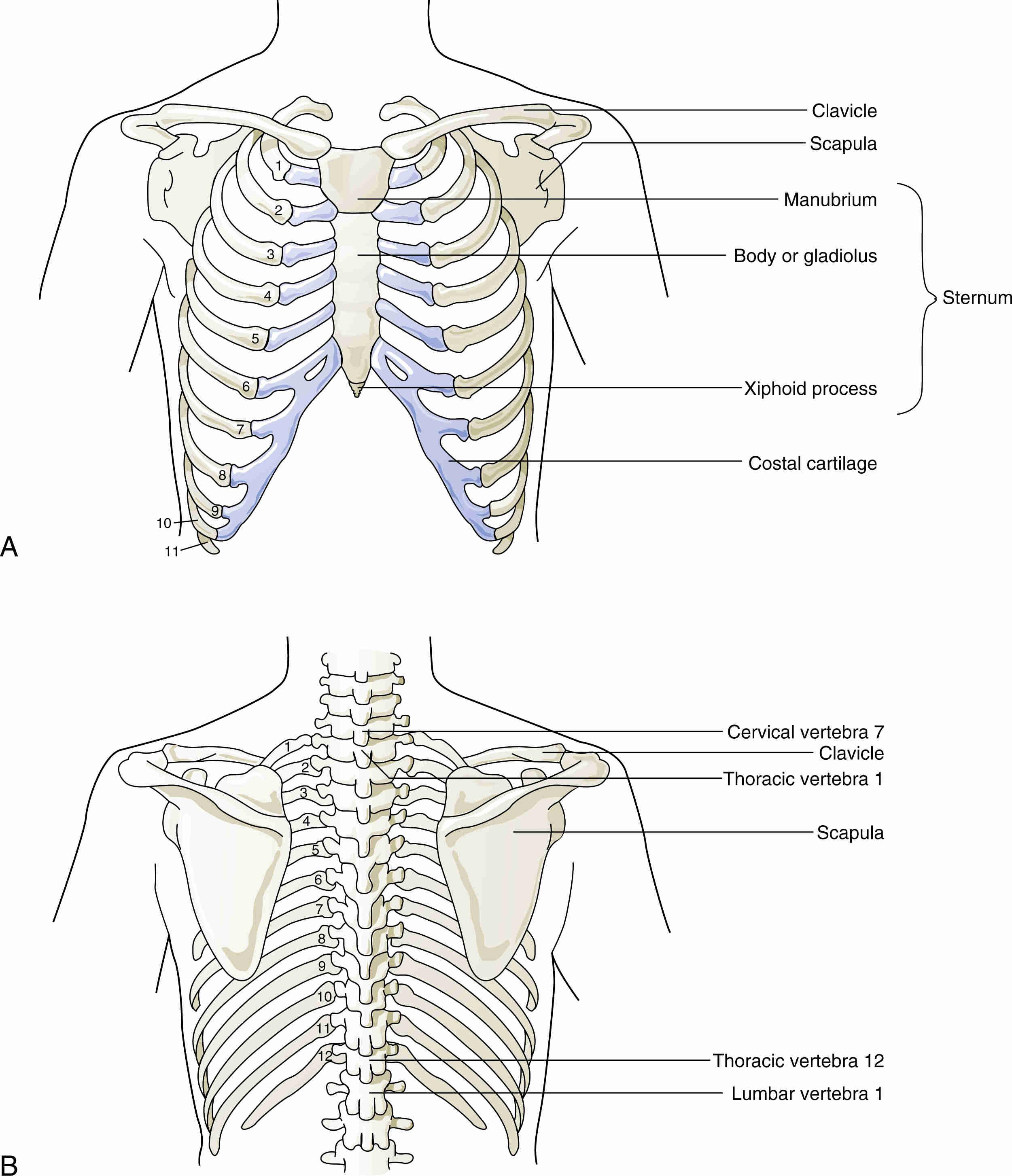
The thoracic cage (Fig. 1.1) is conical at both its superior and inferior aspects and somewhat kidney shaped in its transverse aspect. The skeletal boundaries of the thorax are the 12 thoracic vertebrae dorsally, the ribs laterally, and the sternum ventrally.
Sternum
The sternum, or breastbone, is a flat bone with three major parts: manubrium, body, and xiphoid process (see Fig. 1.1). Superiorly located within the sternum, the manubrium is the thickest component articulating with the clavicles and first and second ribs. A palpable jugular notch or suprasternal notch is found at the superior border of the manubrium of the sternum. Inferior to the manubrium lies the body of the sternum, articulating laterally with ribs three to seven. The sternal angle, or “angle of Louis,” is the anterior angle formed by the junction of the manubrium and the body of the sternum. This easily palpated structure is in level with the second costal cartilage anteriorly and thoracic vertebrae T4 and T5 posteriorly. The most caudal aspect of the sternum is the xiphoid process, a plate of hyaline cartilage that ossifies later in life.
The sternal angle marks the level of bifurcation of the trachea into the right and left main stem bronchi and provides for the pump-handle action of the sternal body during inspiration. 1
Pectus excavatum is a common congenital deformity of the anterior wall of the chest in which several ribs and the sternum grow abnormally (see Fig. 5.25). This produces a caved-in or sunken appearance of the chest. It is present at birth, but rapidly progresses during the years of bone growth in the early teenage years. These patients have several pulmonary complications, including shortness of breath caused by altered mechanics of the inspiratory muscles on the caved-in sternum and ribs, and often have cardiac complications caused by the restriction (compression) of the heart. 2
FIGURE 1.1 (A) Anterior. (B) Posterior views of the bones of the thorax.
From Hicks GH: Cardiopulmonary anatomy and physiology, Philadelphia, 2000, Saunders.
To gain access to the thoracic cavity for surgery, including coronary artery bypass grafting, the sternum is split in the median plane and retracted. This procedure is known as a median sternotomy. Flexibility of the ribs and cartilage allows for separation of the two ends of the sternum to expose the thoracic cavity. 3
Ribs
The ribs, although considered “flat” bones, curve forward and downward from their posterior vertebral a�achments toward their costal cartilages. The first seven ribs a�ach via their costal cartilages to the sternum and are called the true ribs (also known as the vertebrosternal ribs); the lower five ribs are termed the false ribs—the 8th, 9th, and 10th ribs a�ach to the rib above by their costal cartilages (the vertebrochondral ribs), and the 11th and 12th ribs end freely (the vertebral ribs; see Fig. 1.1). The true ribs increase in length from above downward, and the false ribs decrease in length from above downward.
Each rib typically has a vertebral end separated from a sternal end by the body or shaft of the rib. The head of the rib (at its vertebral end) is distinguished by a twin-faceted surface for articulation with the facets on the bodies of two adjacent thoracic vertebrae. The cranial facet is smaller than the caudal, and a crest between these permits a�achment of the interarticular ligament.
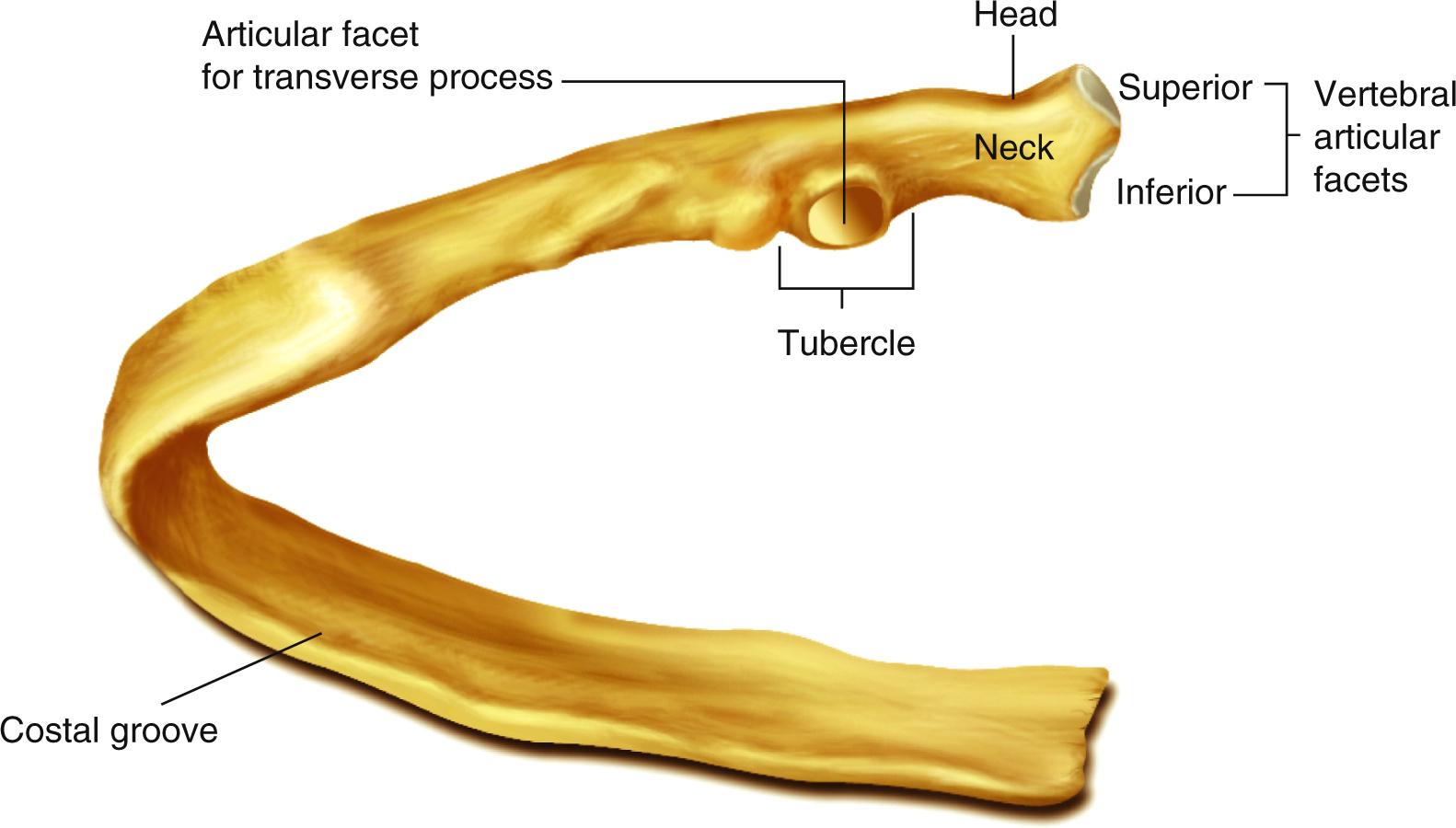
Fig. 1.2 displays the components of typical ribs 3 to 9, each with common characteristics, including a head, neck, tubercle, and body. The neck is the 1-inch long portion of the rib extending laterally from the head; it provides a�achment for the anterior costotransverse ligament along its cranial border. The tubercle at the junction of the neck and the body of the rib consists of an articular and a nonarticular portion. The articular part of the tubercle (the more medial and inferior of the two) has a facet for articulation with the transverse process of the inferior-most vertebra to which the head is connected. The nonarticular part of the tubercle provides a�achment for the ligament of the tubercle.
The shaft, or body, of the rib is simultaneously bent in two directions and twisted about its long axis, presenting two surfaces (internal and external) and two borders (superior and inferior). A costal groove for the intercostal vessels and nerve extends along the inferior border dorsally but changes to the internal surface at the angle of the rib. The sternal end of the rib terminates in an oval depression into which the costal cartilage makes its a�achment.
FIGURE 1.2 Typical middle rib as viewed from the posterior. The head end articulates with the vertebral bones, and the distal end is attached to the costal cartilage of the sternum.
From Wilkins RL: Egan’s fundamentals of respiratory care, ed 9, St. Louis, 2009, Mosby.
Although rib fractures may occur in various locations, they are more common in the weakest area where the shaft of the ribs bend—the area just anterior to its angle. The first rib does not usually fracture because it is protected posteroinferiorly by the clavicle. When it is injured, the brachial plexus of nerves and subclavian vessel injury may occur. 4 Lower rib fractures may cause trauma to the diaphragm resulting in a diaphragmatic hernia. Rib fractures are extremely painful because of their profound nerve supply. It is important for all therapists to recommend breathing, splinting, and coughing strategies for patients with rib fractures. Paradoxical breathing pa�erns and a flail chest may also need to be evaluated in light of multiple rib fractures in adjacent ribs. 3
Chest tubes are inserted above the ribs to avoid trauma to vessels and nerves found within the costal grove. A chest tube insertion involves the surgical placement of a hollow, flexible drainage tube into the chest. This tube is used to drain blood, air, or fluid around the lungs and effectively allow the lung to
expand. The tube is placed between the ribs and into the space between the inner lining and the outer lining of the lung (pleural space).
The 1st, 2nd, 10th, 11th, and 12th ribs are unlike the other, more typical ribs. The first rib is the shortest and most curved of all the ribs. Its head is small and rounded and has only one facet for articulation with the body of the first thoracic vertebra. The sternal end of the first rib is larger and thicker than it is in any of the other ribs. The second rib, although longer than the first, is similarly curved. The body is not twisted. There is a short costal groove on its internal surface posteriorly. The 10th through 12th ribs each have only one articular facet on their heads. The 11th and 12th ribs (floating ribs) have no necks or tubercles and are narrowed at their free anterior ends. The 12th rib sometimes is shorter than the first rib.
The respiratory system
The respiratory system includes the bony thorax, the muscles of ventilation, the upper and the lower airways, and the pulmonary circulation. The many functions of the respiratory system include gas exchange, fluid exchange, maintenance of a relatively low-volume blood reservoir, filtration, and metabolism, and they necessitate an intimate and exquisite interaction of these various components. Because the thorax has already been discussed, this section deals with the muscles of ventilation, the upper and lower airways, and the pulmonary circulation.
Muscles of ventilation
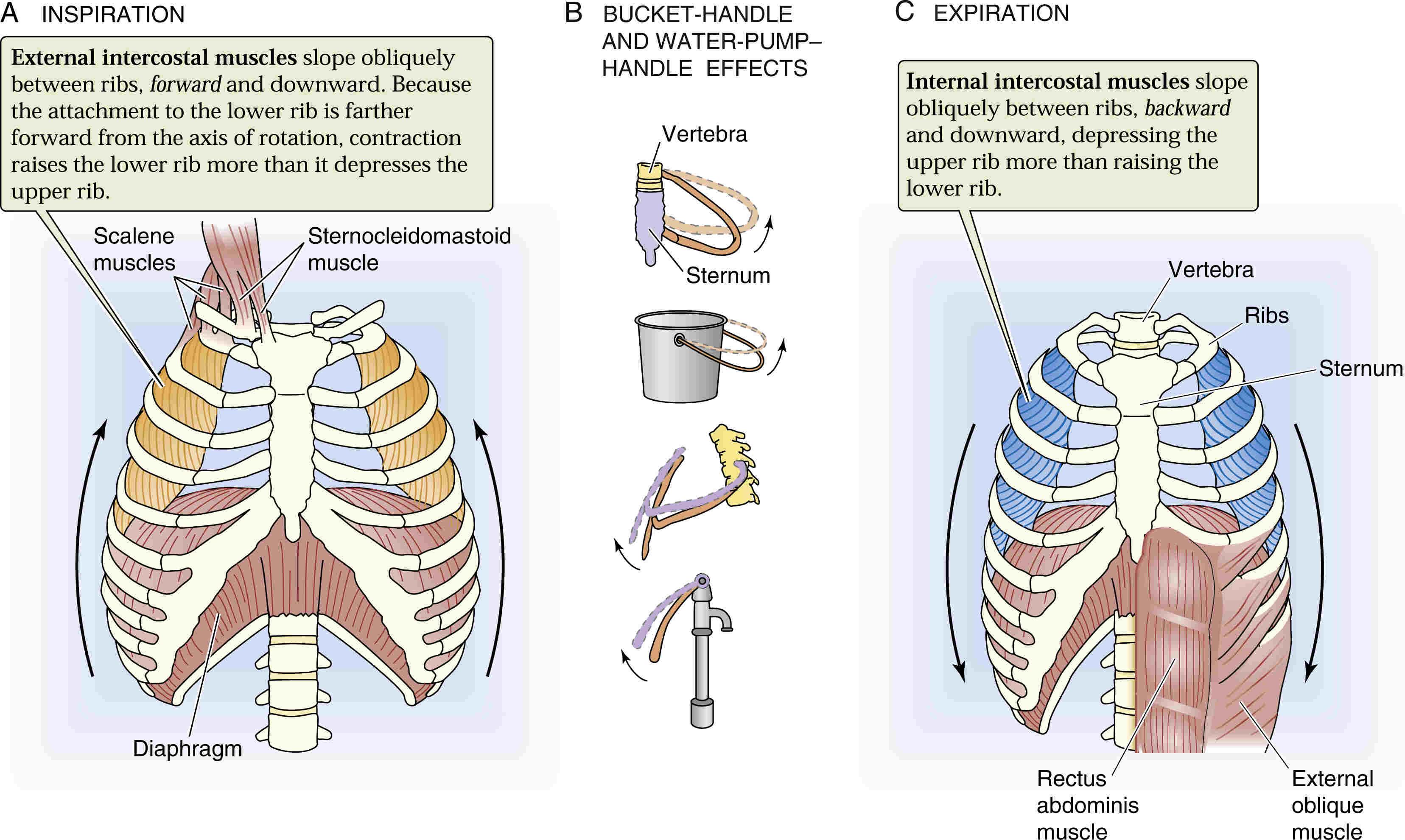
Ventilation, or breathing, involves the processes of inspiration and expiration. For air to enter the lungs during inspiration, muscles of the thoracic cage and abdomen must move the bony thorax to create changes in volume within the thorax and cause a concomitant reduction in the intrathoracic pressure. Inspiratory muscles increase the volume of the thoracic cavity by producing bucket-handle and pump-handle movements of the ribs and sternum, as depicted in Fig. 1.3. The resultant reduced intrathoracic pressure generated is below atmospheric pressure, forcing air into the lungs to help normalize pressure differences. The essential muscles to achieve the active process of inspiration at rest are the diaphragm and internal intercostals. To create a more forceful inspiration during exercise or cardiopulmonary distress, accessory muscles assist with the inspiration. The accessory muscles include the sternocleidomastoid, scalenes, serratus anterior, pectoralis major and minor, trapezius, and erector spinae muscles.
Diaphragm
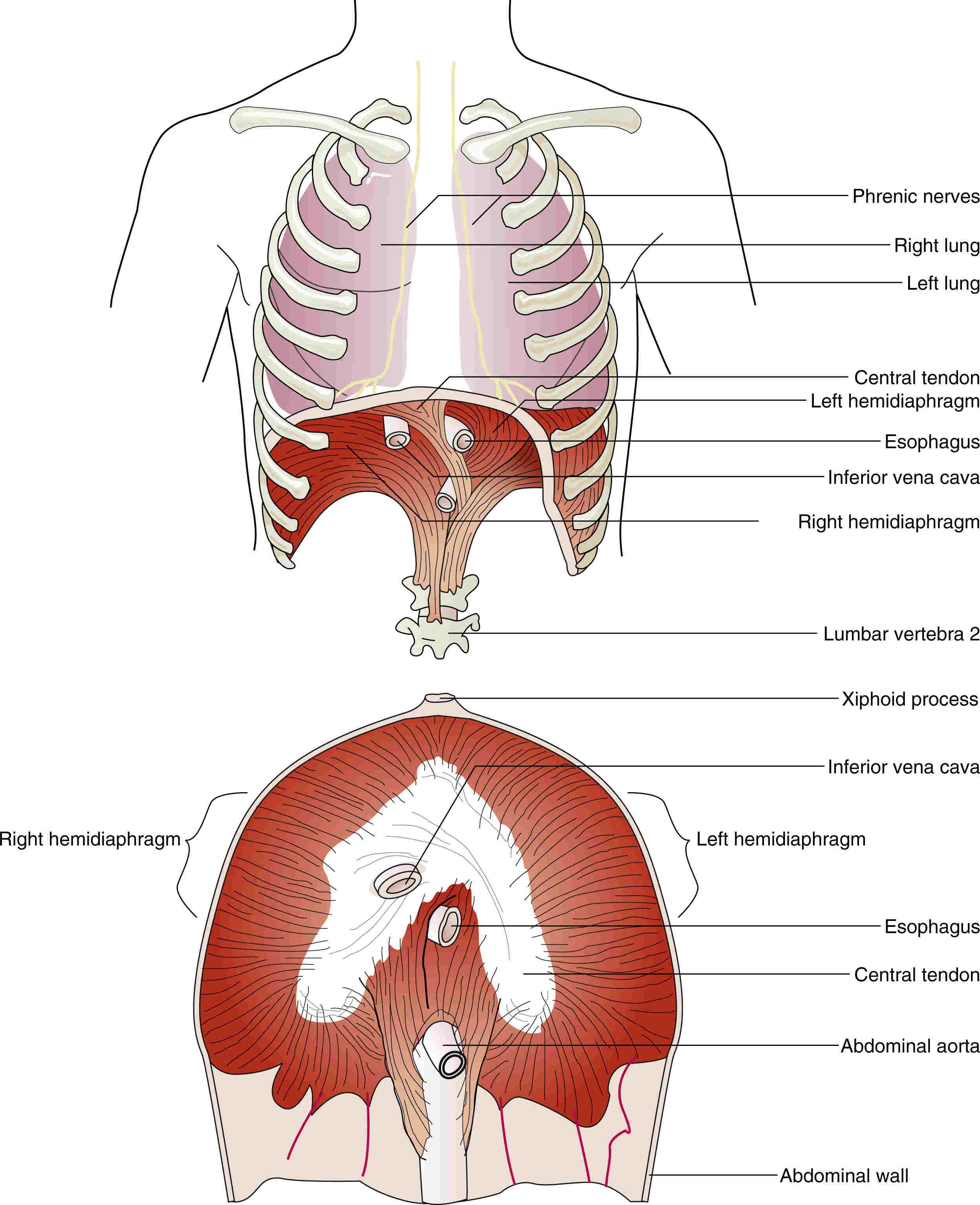
The diaphragm is the major muscle of inspiration. It is a musculotendinous dome that forms the floor of the thorax and separates the thoracic and abdominal cavities (Fig. 1.4). The diaphragm is divided into right and left hemidiaphragms. Both hemidiaphragms are visible on radiographic studies from the front or back. The right hemidiaphragm is protected by the liver and is stronger than the left. The left hemidiaphragm is more often subject to rupture and hernia, usually because of weaknesses at the points of embryologic fusion. Each hemidiaphragm is composed of three musculoskeletal components, including the sternal, costal, and lumbar portions that converge into the central tendon. The central tendon of the diaphragm is a thin but strong layer of tendons (aponeurosis) situated anteriorly and immediately below the pericardium. There are three major openings to enable various vessels to traverse the diaphragm. These include the vena caval opening for the inferior vena cava; the esophageal opening for the esophagus and gastric vessels; and the aortic opening containing the aorta, thoracic duct, and azygos veins. The phrenic nerve arises from the third, fourth, and fifth cervical spinal nerves (C3‒C5) and is involved in contraction of the diaphragm.
The resting position of the diaphragm is an arched position high in the thorax. The level of the diaphragm and the amount of movement during inspiration vary as a result of factors such as body position, obesity, and size of various gastrointestinal organs present below the diaphragm. During normal ventilation or breathing, the diaphragm contracts to pull the central tendon down and forward. In doing so, the resting dome shape of the diaphragm is reversed to a fla�ening of the diaphragm. Contraction of this muscle increases the dimensions of the thorax in a cephalocaudal, anterior posterior, and lateral direction. 1 The increase in volume decreases pressure in the thoracic cavity and simultaneously causes a decrease in volume and an increase in pressure within the abdominal cavity. The domed shape of the diaphragm is largely maintained until the abdominal muscles end their extensibility, halting the downward displacement of the abdominal viscera, essentially forming a fixed platform beneath the central tendon. The central
tendon then becomes a fixed point against which the muscular fibers of the diaphragm contract to elevate the lower ribs and thereby push the sternum and upper ribs forward. The right hemidiaphragm meets more resistance than the left during its descent, because the liver underlies the right hemidiaphragm and the stomach underlies the left; it is therefore more substantial than the left.
FIGURE 1.3 (A–C) Actions of major respiratory muscles.
From Boron WF: Medical physiology, updated ed, St. Louis, 2005, Saunders.
In patients with chronic obstructive pulmonary disease (COPD), there is compromised ability to expire. This results in a fla�ening of the diaphragm as a result of the presence of hyperinflated lungs. 1 , 5 It is essential for therapists to reverse hyperinflation and restore the normal resting arched position of the diaphragm using any exercise aimed at strengthening the diaphragm muscle. A flat and rigid diaphragm cannot be strengthened and will cause an automatic firing of the accessory muscles to trigger inspiration.
Body position in supine, upright, or side lying alters the resting position of the diaphragm, resulting in concomitant changes in lung volumes. 6 In the supine position, without the effects of gravity, the level of the diaphragm in the thoracic cavity rises. This allows for a relatively greater excursion of the diaphragm. Despite a greater range of movement of the diaphragm, lung volumes are low as a consequence of the elevated position of the abdominal organs within the thoracic cavity. In an upright position, the dome of the diaphragm is pulled down because of the effects of gravity. The respiratory excursion is less in this position; however, the lung volumes are larger. In the side-lying position, the hemidiaphragms are unequal in their positions: the uppermost side drops to a lower level and has less excursion than that in the si�ing position; the lowermost side rises higher in the thorax and has a greater excursion than in the si�ing position. In quiet breathing, the diaphragm normally moves about two-thirds of an inch; with maximal ventilatory effort, the diaphragm may move from 2.5 to 4 inches. 5
Clinical tip
Stomach fullness, obesity with presence of a large pannus, ascites with increased fluid in the peritoneal space from liver disease, and pregnancy are additional factors affecting the normal excursion of the diaphragm during inspiration.
External intercostal muscles
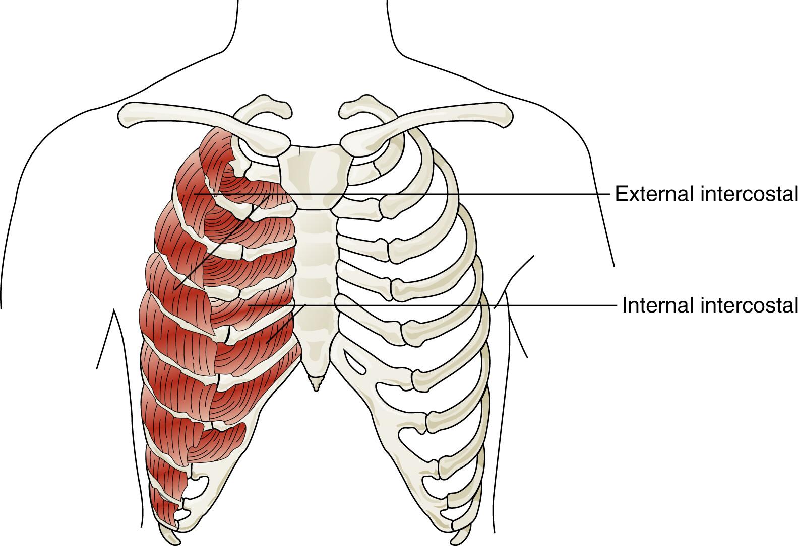
The external intercostal muscles originate from the lower borders of the ribs and a�ach to the upper border of the ribs below (Fig. 1.5). There are 11 external intercostal muscles on each side of the sternum. Contraction of these muscles pull the lower rib up and out toward the upper rib, thereby elevating the ribs and expanding the chest.
Accessory muscles
Figs. 1.6 and 1.7 present the anatomy of the accessory muscles.
Sternocleidomastoid muscle
The sternocleidomastoid arises by two heads (sternal and clavicular from the medial part of the clavicle), which unite to extend obliquely upward and laterally across the neck to the mastoid process. For this muscle to facilitate inspiration, the head and neck must be held stable by the neck flexors and extensors. This muscle is a primary accessory muscle and elevates the sternum, increasing the anteroposterior diameter of the chest.
FIGURE 1.4 The diaphragm originates from the lumbar vertebra, lower ribs, xiphoid process, and abdominal wall and converges in a central tendon. Note the locations of the phrenic nerves and openings for the inferior vena cava, esophagus, and abdominal aorta.
From Hicks GH: Cardiopulmonary anatomy and physiology, Philadelphia, 2000, Saunders.
Scalene muscle
The scalene muscles lie deep to the sternocleidomastoid, but may be palpated in the posterior triangle of the neck. These muscles function as a unit to elevate and fix the first and second ribs:
Get Complete eBook Download
1. The anterior scalene muscle passes from the anterior tubercles of the transverse processes of the third or fourth to the sixth cervical vertebrae, a�aching by tendinous insertion into the first rib.
2. The middle scalene muscle arises from the transverse processes of all the cervical vertebrae to insert onto the first rib (posteromedially to the anterior scalene, the brachial plexus, and subclavian artery pass between the anterior scalene and middle scalene).
3. The posterior scalene muscle arises from the posterior tubercles of the transverse processes of the fifth and sixth cervical vertebrae, passing between the middle scalene and levator scapulae, to a�ach onto the second or third rib.
Upper trapezius
The trapezius (upper fibers) muscle arises from the medial part of the superior nuchal line on the occiput and the ligamentum nuchae (from the vertebral spinous processes between the skull and the seventh cervical vertebra) to insert onto the distal third of the clavicle. This muscle assists with ventilation by helping to elevate the thoracic cage.
Pectoralis major and minor
The pectoralis major arises from the medial third of the clavicle, from the lateral part of the anterior surface of the manubrium and body of the sternum, and from the costal cartilages of the first six ribs to insert upon the lateral lip of the crest of the greater tubercle of the humerus. When the arms and shoulders are fixed, by leaning on the elbows or grasping onto a table, the pectoralis major can use its insertion as its origin and pull on the anterior chest wall, lifting the ribs and sternum, and facilitate an increase in the anteroposterior diameter of the thorax.
FIGURE 1.5 The external intercostal muscles lift the inferior ribs and enlarge the thoracic cavity. The internal intercostal muscles compress the thoracic cavity by pulling together the ribs.
From Hicks GH: Cardiopulmonary anatomy and physiology, Philadelphia, 2000, Saunders.
The pectoralis minor arises from the second to fifth or the third to sixth ribs upward to insert into the medial side of the coracoid process close to the tip. This muscle assists in forced inspiration by raising the ribs and increasing intrathoracic volume.
Serratus anterior and rhomboids
The serratus anterior arises from the outer surfaces of the upper eight or nine ribs to a�ach along the costal aspect of the medial border of the scapula. The primary action of the serratus is to abduct, rotate the scapula, and hold the medial border firmly over the rib cage. The serratus can only be used as an accessory muscle in ventilation, when the rhomboids stabilize the scapula in adduction. 7 The action of the rhomboids fixes the insertion, allowing the serratus to expand the rib cage by pulling the origin toward the insertion.
Latissimus dorsi
The latissimus dorsi arises from the spinous processes of the lower six thoracic, the lumbar, and the upper sacral vertebrae, from the posterior aspect of the iliac crest, and slips from the lower three or four ribs to a�ach to the intertubercular groove of the humerus. 7 The posterior fibers of this muscle assist in inspiration as they pull the trunk into extension.
Serratus posterior superior
The serratus posterior superior passes from the lower part of the ligamentum nuchae and the spinous processes of the seventh cervical and first two or three thoracic vertebrae downward into the upper borders of the second to fourth or fifth ribs. This muscle assists in inspiration by raising the ribs to which it is a�ached and expanding the chest.