Aquaculture
journal homepage: www.elsevier.com/locate/aquaculture
Comparative study of Chronic Ulcerative Dermatopathy in cultured meagre, Argyrosomus regius
M.I. Tsertou a, b , N. Papandroulakis b , K. Keklikoglou b, c , I. Kalantzi d , M. Tsapakis d , A. Tsalafouta e , M. Pavlidis e , E. Antonopoulou a , P. Katharios b, *
a Laboratory of Animal Physiology, Department of Zoology, School of Biology, Faculty of Sciences, Aristotle University of Thessaloniki, 54124 Thessaloniki, Greece
b Institute of Marine Biology, Biotechnology and Aquaculture, Hellenic Centre for Marine Research, Former American Base of Gournes, Heraklion 71003, Crete, Greece
c Biology Department, University of Crete, 70013 Heraklion, Crete, Greece
d Institute of Oceanography, Hellenic Centre for Marine Research, Former American Base of Gournes, 71500 Heraklion, Crete, Greece
e Laboratory of Fish Physiology, Department of Biology, University of Crete, Heraklion, Greece
ARTICLE INFO
Keywords:
Lateral line erosion
Ulcer
micro-CT
Histopathology
Heavy metals
Water quality
Meagre
ABSTRACT
Chronic Ulcerative Dermatopathy (CUD) is a disease that affects all cultured meagre when reared in facilities supplied with borehole water, resulting in ulceration of the skin overlying the lateral line canals. The aims of this study were (i) to describe the morphogenesis of the cephalic lateral line, (ii) to investigate the effect of the use of borehole water vs natural seawater in the development of the disease, (iii) to assess the recovery of the lesions and (iv) to evaluate the effect of CO2 and pH on the development of CUD. The development of the lateral line canals in the head was completed by day 28 post hatching when fish were 19.3 mm in total length, while the source of the water did not affect the developmental process. The characteristic lesions of CUD were induced when meagre were reared in borehole water, while the lesions were resolved when fish were transferred to natural seawater. Lesions were macroscopically visible by day 56 post hatching. Moreover, significant differences in the expression of genes regulating osteoclast’s activity were observed between healthy and CUD-affected fish, while neither pH nor CO2 were involved in the development of the disease. Finally, higher concentrations of heavy metals were found in the heads of CUD-affected meagres reared in borehole water compared to healthy fish reared in natural seawater.
1. Introduction
The lateral line is a mechanosensory system found in all fishes and in the larvae of aquatic amphibians, which is used for the detection of water movements and/or pressure fluctuations (Bleckmann and Zelick, 2009; Mogdans et al., 2004). The receptors of the lateral line that detect water flow are called neuromasts and they are distributed on the head, the trunk and the tail of the fish. Neuromasts can be either superficial on the skin or enclosed in the fluid-filled canals of the lateral line that open to the environment through a series of pores (Bleckmann and Zelick, 2009; Webb, 1989). The development and maintenance of the lateral line canals is achieved through a bone remodeling process which includes the participation of both osteoclasts (bone-resorption cells) and osteoblasts (bone-forming cells) (Wada et al., 2014; Webb, 2013).
Several conditions affecting the lateral line organ of the head and the trunk of various marine and freshwater fish have been reported under
* Corresponding author.
E-mail address: katharios@hcmr.gr (P. Katharios).
https://doi.org/10.1016/j.aquaculture.2022.738301
the terms, hole-in-the-head, Head and Lateral Line Erosion syndrome (HLLE) and Lateral Line Depigmentation (LLD) (Corrales et al., 2009; Morrison et al., 2007; Noga, 2010). Apart from these, Chronic Ulcerative Dermatopathy (CUD) is a pathological condition affecting the lateral line canals of freshwater and marine cultured fish species. It was first described in the Australian freshwater fish Murray cod (Maccullochella peelii peelii (Mitchell)), when reared in sites supplied by groundwater (Baily et al., 2005; Ingram et al., 2004; Schultz et al., 2011; Schultz et al., 2008). The clinical signs of this first report included focal erosion, ulceration and loss of epidermis around the lateral line canals of the head and the trunk and fin erosion. It has been associated with reduced growth rates, increased mortalities and significant reduction of marketability due to the severe disfigurement of the affected fish (Baily et al., 2005; Ingram et al., 2004; Schultz et al., 2008). Due to the localization of the lesions exclusively on the lateral line canals, it was hypothesized that the disease mechanism involved the binding of an
Received 29 September 2021; Received in revised form 13 April 2022; Accepted 25 April 2022
Availableonline27April2022
0044-8486/©2022ElsevierB.V.Allrightsreserved.
Table 1
Specific primers used for the expression of genes encoding for tartrate-resistant acid phosphatase (TRAP), cathepsin K (CathK,) and vATPase, as well as, for the reference gene β-actin.
Gene Forward Reverse
b-actin
TRAP
5’ TGTCCCTGTATGCCTCTGGT 3’
5’ TGCGGAAGTCACAAAGAACAA
3’
Cath K 5’ ACGCTCACTCCAAATCCAACTG
vATPase
5’ AAGTCCAGACGGAGGATGG 3’
GGAGAGGACAGTGCGATAGA
3’ 5’ CCGTGCCGCTACAATTCATCA 3’
5’ TGTATGCCTGTTATGCCATTG
3’ 5’ TCCTGAGCGATGAAGTTCTT 3’
unknown waterborne toxin to the mucus content of the sensory canals, resulting in focal hyperplasia and necrosis. Moreover, it was shown that when CUD-affected Murray cod were transferred to river water, the majority of the fish were structurally recovered after a period of 8–10 weeks. Based on these results and in the absence of viral or bacterial agents, it was suggested that some component of the groundwater was the driving force for the development of CUD. However, after analysis of basic water quality parameters as well as heavy metal and pesticide/ insecticide content of the groundwater, the exact component of the water that could have resulted in the development of the disease could not be identified. Following the association of groundwater with the disease, several water treatment methods were evaluated in order to reduce the severity of the lesions on Murray cod, including electrolyte enrichment, pre-treatment with UV irradiation and pre-conditioning of groundwater either in a vegetated earthen pond or in tanks containing artificial macrophytes (Schultz et al., 2011). Among them, preconditioning of the water for 72 h into a vegetated earthen pond or a tank containing biofilms growing on an artificial macrophyte, was found to be an effective method for the reduction of both the incidence and the severity of CUD in juvenile Murray cod (Schultz et al., 2011).
Regarding marine fish species, CUD was described in sharpsnout seabream (Diplodus puntazzo) when cultured in saline borehole water (Katharios et al., 2011). The CUD-affected fish exhibited bilateral lesions in the head canals of the lateral line and eroded fins and recovery of the lesions was also observed following transfer of the fish to natural sea water. Вy excluding an infectious agent for the onset of the disease and in an attempt to determine the causative agent of CUD in sharpsnout seabream it was hypothesized that borehole water, which was rich in CO2, as indicated also by the lower pH compared to the pH of natural seawater, increases the enzymatic activity of the osteoclasts (Katharios
et al., 2011). In this scenario, there would be an environmentally induced imbalance between osteoclasts and osteoblasts that would cause the lesions seen in the fish, located exclusively in the lateral line canals which are in direct contact with the water.
A range of marine fishes have been reported as CUD-sensitive including one of the most important marine aquaculture species, European seabass (Dicentrachus labrax). The lesions in the seabass become visible when the fish is more than 5 g. Although most of the hatcheries in the Mediterranean use borehole water, the disease was undetected until recently due to the common practice of growing the fish in inland facilities until 2-3 g and then transfer it to sea cages. Many hatcheries changed their strategy and grew the seabass in larger size before the transportation to sea cages so the disease became apparent and a bigger concern for the producers since the damaged epidermis could affect the susceptibility of the fish to a wide range of pathogens in the sea (Katharios, personal observations).
Apart from the seabass and sharpsnout seabream, meagre (Argyrosomus regius) which is an emerging species for the Mediterranean aquaculture was found also to be sensitive to CUD when reared in facilities supplied with borehole water. The disease affects 100% of the population and results in ulceration of the skin overlying the lateral line canals, however, it is not associated with mortalities (Rigos and Katharios, 2010; Soares et al., 2018). The aim of this study is to describe the morphogenesis of the lateral line organ in the head of meagre as it is the organ affected from CUD and to describe the disease using histology, scanning electron microscopy (SEM) and microcomputed tomography (μ-CT). In addition, the osteoclast activity was investigated in CUDaffected fish using molecular markers while the CO2 in the water was examined as the aetiological factor of the disease.
2. Materials and methods
2.1. Ethics
All animal experimental procedures and handling in this study were conducted in the HCMR’s licensed facility (EL91-BIOexp-04) under the protocol 255,332 (29/11/2017) approved by the regional veterinary authority, which is the competent agency according to the Directive 2010/63/EU.
2.2. Rearing trial for the description of the disease
Two parallel rearing trials of meagre in borehole and natural seawater, respectively, were conducted in order to study the
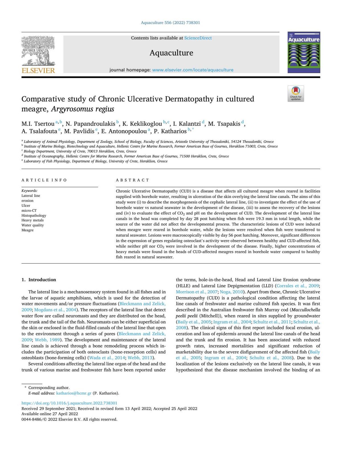
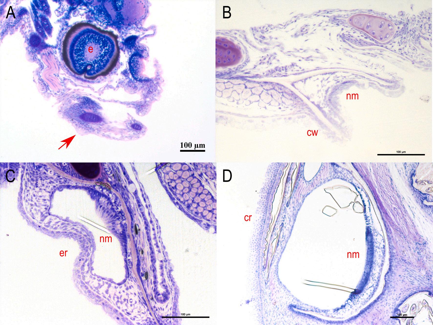
visible the sensory cells, the mantle cells and the supporting cells. Stain with methylene blue/azure II/basic fuchsin. (For interpretation of the references to colour in this figure legend, the reader is referred to the web version of this article.)
Fig. 1. A: Superficial neuromast on the head of 1 dph meagre (Argyrosomus regius). B: Superficial neuromast on the body of 6 dph meagre (TL: 4.05 ± 0.08 mm). C: Higher magnification of superficial neuromast on the body of 11 dph meagre (TL: 6.03 ± 0.39 mm) where is
M.I. Tsertou et al.
0.00
B:
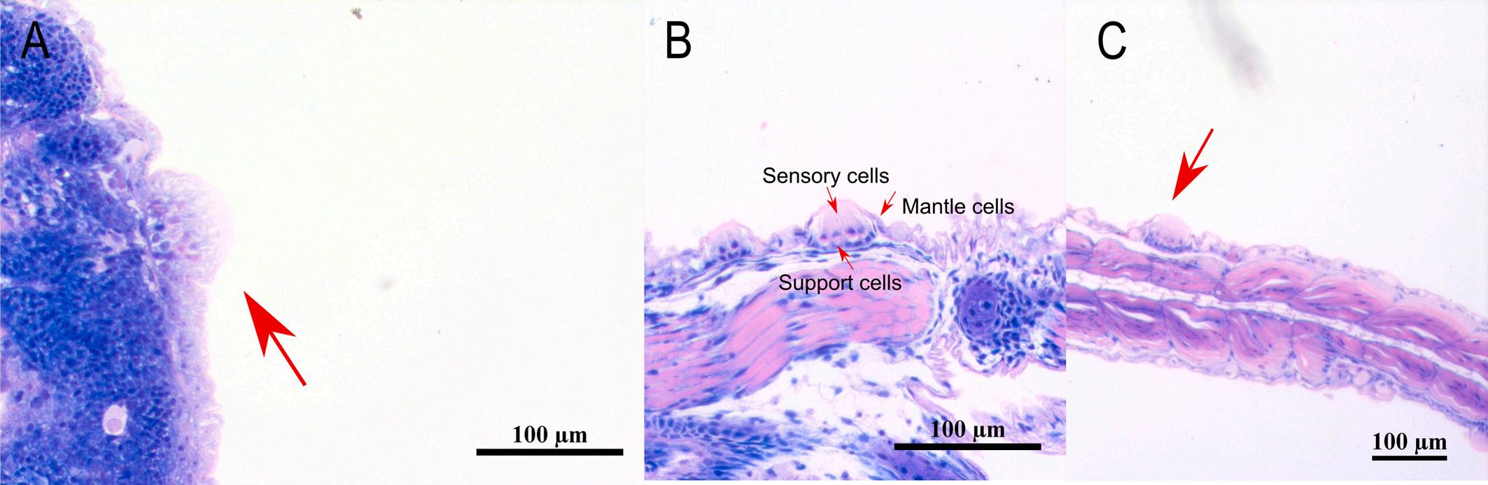
dph – TL: 9.48 ± 0.7 mm). D:
Higher magnification of picture C with the stereocilium and the kinocilium of the hair cells. Double-headed arrows show hair cell orientation. S: stereocilia, K: kinocilium.
development of CUD. The eggs used in this study were obtained from a broodstock maintained at the facilities of the Institute of Marine Biology, Biotechnology and Aquaculture, HCMR, Crete, Greece. Specifically, 100,000 eggs were placed in each of the 2 tanks (40m3), the first of which was supplied with natural seawater and the second with borehole water. Eggs were incubated under natural photoperiod, at 19.5 ◦ C and 36 and 38 ppt salinity for borehole and natural sea water, respectively. The trial lasted 56 days after hatching (dph) with the same rearing protocols applied in both tanks. The feeding protocol included the addition of the microalgae Chlorella sp. in the rearing water from 4 to 15 dph, feeding with enriched rotifers (Вrachionus sp.) from 4 to 18 dph, enriched Artemia spp. nauplii from 12 to 36 dph and artificial food (INVE SA, Belgium) from 19 dph. Measurements of pH, O2 (HQ40D Portable Multi Meter, Hach), CO2 (CO2 Portable Carbon Dioxide Analyzer, OxyGuard) and water temperature were performed daily in the two water sources. Random samples of larvae and juvenile fish from both tanks were euthanized with an overdose of tricaine (MS222) and sampled for histology, scanning electron microscopy (SEM), micro-CT analysis, Quantitative Real-Time PCR (qPCR) and SDS-PAGE and immunoblot analysis.
2.3. Histology
Three fish from each tank were sampled daily from day 1 to 7, every two days from day 9 to 21 and every five days until day 56 post hatching. The samples were preserved in buffered 4F:1G, containing 4% formaldehyde: 1% gluteraldehyde for at least 24 h (McDowell and Trump, 1976). Subsequently they were dehydrated in gradually increased ethanol solutions (70–96%) and then embedded in glycol methacrylate
resin (Technovit 7100, Heraeus Kulzer). Sections of 4 μm were obtained with a microtome (RM 2035, Leica, Germany). After drying, slides were stained with methylene blue/azure II/basic fuchsin according to Bennett et al. (1976) and examined under a light microscope.
2.4. Scanning electron microscopy (SEM)
Three fish from each tank were sampled at 1, 2, 3, 4, 5, 6, 7, 9, 11, 13, 15, 17, 19, 21, 26, 31, 36, 41, 46 and 56 dph fixed in 2.5% glutaraldehyde in 0.1 M sodium cacodylate buffer (pH 7.4) for 1 or 2 days (depending on the size of the fish) and then stored in sodium cacodylate buffer at 4 ◦ C. The samples were then dehydrated through a graded acetone series, critical point dried and sputter-coated with gold. Samples were viewed using a JEOL JSM-6390LV scanning electronic microscope at 15 kV at the Electron Microscopy Laboratory of the University of Crete.
2.5. Micro-CT
One fish from each tank was sampled at 56 dph, fixed in 10% phosphate-buffered formalin and dehydrated to 70% ethanol for 3 days before scanning. Subsequently, the samples were stained with 0.3% phosphotungstic acid (PTA) in 70% ethanol in order to enhance the contrast between the soft tissues. The micro-CT scans of the samples were performed at the Hellenic Centre for Marine Research (HCMR) using the SkyScan 1172 micro-CT scanner (SkyScan, Bruker, Belgium). This scanner uses a tungsten X-ray source with an anode voltage ranging from 20 to 100 kV, 11 MP CCD camera (4000 × 2672 pixel) and a maximal resolution of <0.8 μm/pixel. Samples were scanned at a
Fig. 2. SEM micrographs of meagre’s (Argyrosomus regius) head. A: Diamond shaped superficial neuromast on the head (3 dph – TL:3.54 ±
mm).
Round shaped superficial neuromast on the head (5 dph – TL: 3.68 ± 0.00 mm). C: Diamond shaped superficial neuromast on the head (17
M.I. Tsertou
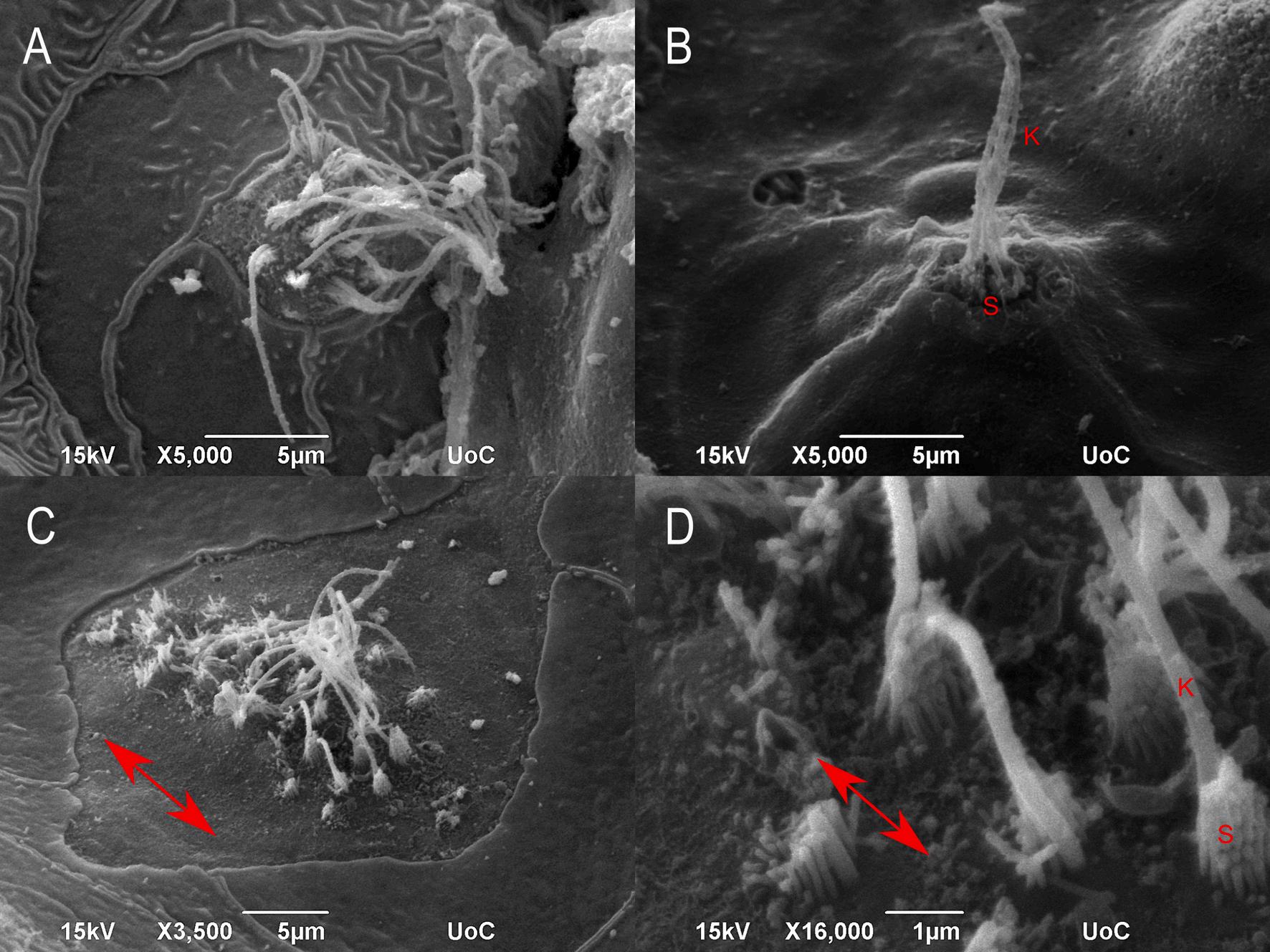
3. Development of the subraobrital canal of meagre (Argyrosomus regius). A: Longitudinal section of a presumptive canal neuromast sitting on the epithelial surface (5 dph – TL: 3.68 ± 0.00 mm) (red arrow). B: Cross section of the neuromast in canal groove (11 dph – TL: 6.03 ± 0.39 mm). C: Cross section of the development of the epithelial canal roof with the enclosed neuromast (19 dph – TL: 9.75 ± 1.21 mm). D-H: Cross sections of the fully formed supraorbital canal as it is distributed from the anterior to the posterior part of the head (46 dph – TL: 41.78 ± 0.87 mm). b: brain, cr: canal roof, cw: canal walls, e: eye, er: epithelial roof, nm: neuromast. Stain with methylene blue/azure II/basic fuchsin. (For interpretation of the references to colour in this figure legend, the reader is referred to the web version of this article.)
Fig.
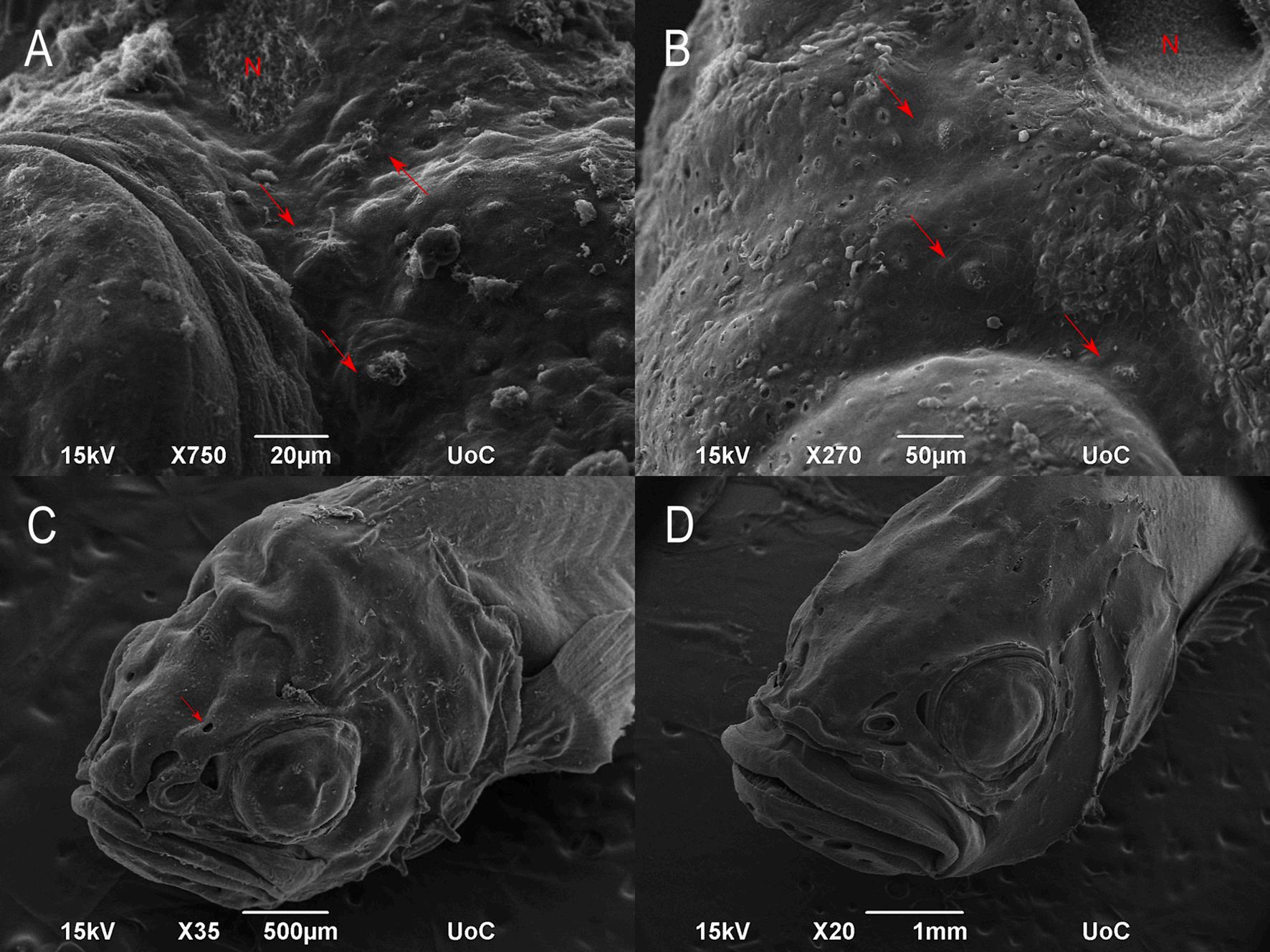
Fig. 4. SEM micrographs with the development of the supraorbital canal of meagre (Argyrosomus regius). A: Superficial neuromasts over the eye of meagre (5 dph –TL: 3.68 ± 0.00 mm) (red arrows). B: Neuromasts in epithelial depressions (red arrows) over the eye of meagre (17 dph – TL: 9.48 ± 0.7 mm). C: Grooves of partially enclosed supraorbital canals with one formed pore (red arrow) (21 dph – TL: 11.75 ± 0.83 mm). D: 31dph meagre (ΤL: 19.30 ± 1.27 mm) with enclosed canals on the head. N: nostril. (For interpretation of the references to colour in this figure legend, the reader is referred to the web version of this article.)
voltage of 80 kV and a current of 124 μA with an aluminum filter of 0.5 mm while the images were acquired at a pixel size of 13.78 μm and exposure time 1435 ms. To minimize the scanning duration, scans were performed for a half rotation of 180o. The projection images acquired during the scanning procedure were reconstructed into cross-section images using the SkyScan’s NRecon software (NRecon, Skyscan, Bruker, Belgium) which implements a modified Feldkamp’s backprojection algorithm.
2.6. Gene expression of Cathk, TRAP and vATPase
Ten fish of similar weight (1.62 ± 0.33 g and 1.69 ± 0.40 g from borehole water and natural seawater respectively) from each tank was sampled at 56 dph, frozen in liquid nitrogen and stored at 80 ◦ C until analyzed. All fish reared in borehole water had visible signs of the disease as opposed to the fish reared in natural seawater which appeared normal. Head samples were homogenized in 600 μL RLT plus buffer (RNeasy Plus Mini Kit Qiagen, Valencia, USA) using the TissueRuptor (Qiagen, Hilden, Germany). Total RNA was extracted using RNA isolation nucleospin RNA plus (Macherey-Nagel) according to the instructions of the manufacturer. In order to determine RNA yield and purity, measurement of the absorbance at 260 and 280 nm was conducted using the Nanodrop® ND-1000 UV–Vis spectrophotometer (Peqlab, Erlangen, Germany) while the integrity of RNA was tested by electrophoresis in 1% agarose gels. The cDNA was synthesized by reverse transcription of 1 μg RNA using the QuantiTect Reverse Transcription kit (Qiagen Inc., CA, USA) according to the manufacturer’s instructions. The mRNA expression of genes encoding for tartrateresistant acid phosphatase (ΤRAP), Cathepsin K (CathK) and vATPase
(primers in Table 1) was determined in healthy and CUD-affected head samples with quantitative polymerase chain reaction (qPCR) which was performed on the CFX ConnectTM Real-Time PCR Detection System (Bio-Rad) using the KAPA SYBRR FAST qPCR Kits (KAPA Biosystems, USA). Cycling parameters were as follows: 95 ◦ C for 3 min (HotStarTaq DNA Polymerase activation step) followed by 36 cycles at 95 ◦ C for 15 s (denaturation step) and 60 ◦ C for 30 s (annealing step). Dissociation curve analysis was performed at the end of the cycles to ensure that single amplifications were obtained. A standard curve was constructed for each gene, using four serial dilutions (1:5) of a pool of all cDNA samples by plotting the negative log of the dilution factor against the relative cycle threshold value. Еach primer pair was required to have a linear standard curve with an R2 value above 0.98 and primer amplification efficiency between 95 and 105% in order to be considered suitable for analysis,. Results were evaluated with the Bio-Rad CFX Manager 2.1 software while the data were calculated by the comparative method using Ct values of β-actin as the reference control.
2.7. SDS-PAGE and
immunoblot analysis
Six fish of similar weight (1.68 ± 0.31 g and 1.65 ± 0.36 g from borehole water and natural seawater respectively) from each tank were sampled at 56 dph, frozen in liquid nitrogen and stored at 80 ◦ C until analyzed. Hsps (Hsp70, Hsp90) and MAPK (p38 MAPK, ERK1/2) members were determined in homogenized head samples according to well established protocols, as described in Antonopoulou et al., 2020, Antonopoulou et al., 2014. Briefly, healthy and CUD-affected heads (4550 mg) were homogenized in 3 mL g-1 of cold lysis buffer (20 mM Hepes pH 7.5, 20 mM β-glycerophosphate, 50 mM NaF, 2 mM EDTA, 10 mM
M.I. Tsertou
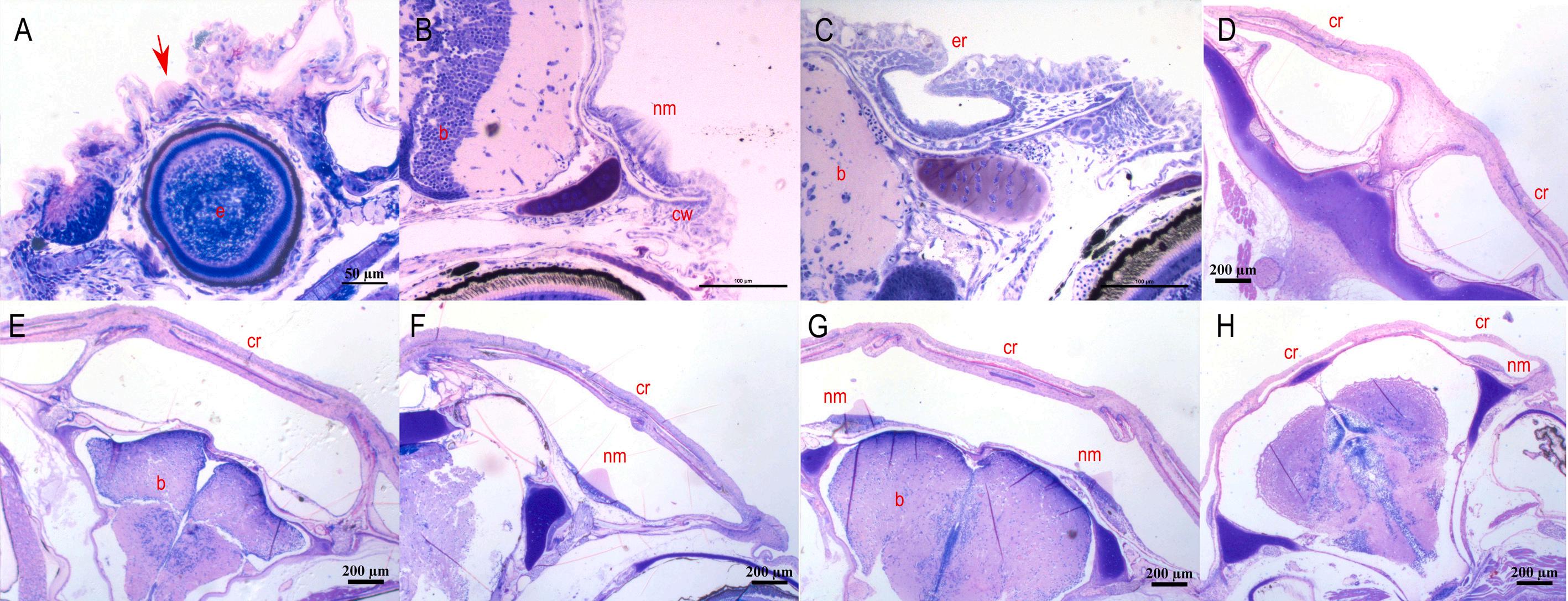
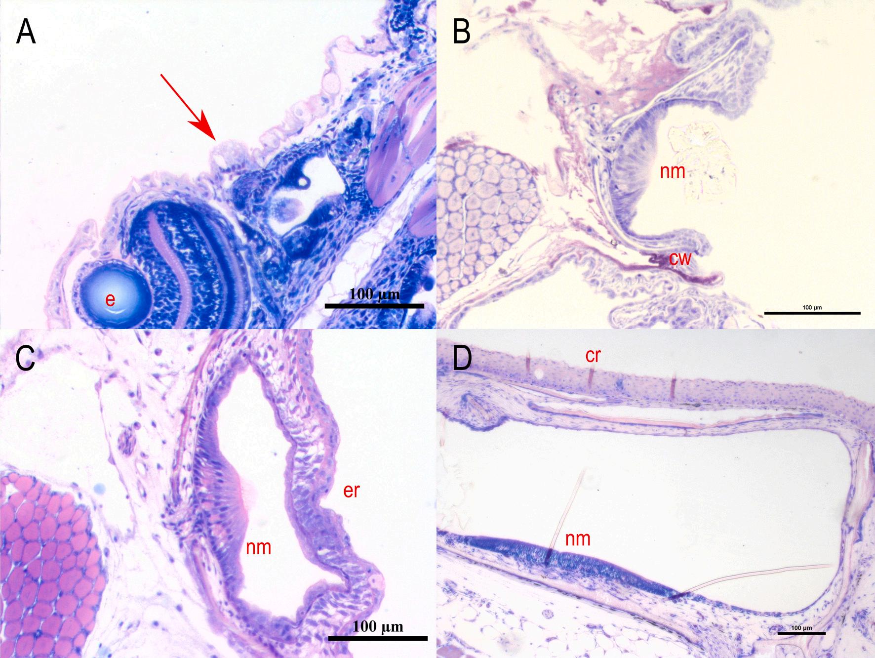
Fig. 5. Development of the mandibular canal of meagre (Argyrosomus regius). A: Longitudinal section of a presumptive canal neuromast sitting on the epithelial surface (7 dph – TL: 4.08 ± 0.03 mm) (red arrow). B: Cross section of the neuromast in canal groove (17 dph – TL: 9.48 ± 0.70 mm). C: Cross section of the epithelial canal roof with the enclosed neuromast (21 dph – TL: 11.75 ± 0.83 mm). D: Cross sections of the fully formed mandibular canal (56 dph – TL: 46.8 ± 0.18 mm). cr: canal roof, cw: canal walls, e: eye, er: epithelial roof, nm: neuromast. Stain with methylene blue/azure II/basic fuchsin. (For interpretation of the references to colour in this figure legend, the reader is referred to the web version of this article.)
benzamidine, 0.2 mM Na3VO4, in pH 7, containing 200 μM leupeptin, 300 μМ phenyl methyl sulfonyl fluoride (PMSF), 10 μМ trans-epoxy succinyl-L-leucylamido-(4- guanidino) butane, 5 mM DTT (dithiothreitol) and 1% v/v Triton X-100). Protein concentration was determined using the BioRad protein assay, a dye-binding assay based on the differential colour change of a dye (Coomansie Brilliant Blue G-250) in response to various protein concentrations. Equivalent amounts of protein (50 μg) were separated on 10% (w/v) acrylamide, 0.275% (w/v) bisacrylamide slab gels, and transferred electrophoretically onto nitrocellulose membranes (0.45 μm, Schleicher and Schuell, Keene N.H. 03431, USA). All nitrocellulose membranes were dyed with Ponceau stain to assure a good transfer quality and equal protein loading. The antibodies used were as follows: monoclonal mouse anti-heat shock protein, 70 kDa (Cat. No. H5147, Sigma, Darmstadt, Germany); monoclonal mouse anti-heat shock protein, 90 kDa (Cat. No. H1775, Sigma, Darmstadt, Germany); monoclonal rabbit anti-phospho p44/42 MAPK (Thr202/Tyr204) (Cat. No. 4376, Cell Signaling, Beverly, MA, USA); polyclonal rabbit anti-phospho-p38 MAP kinase (Thr180- Tyr182) (Cat. No. 9211, Cell Signaling, Beverly, MA, USA). Finally, bands were detected by enhanced chemiluminescence (Cell Signaling, Beverly, MA, USA) with exposure to Fuji Medical X-ray films. Films were quantified by laser- scanning densitometry (GelPro Analyzer Software, Media Cybernetics).
2.8. Metal and element analysis
Nine fish from each tank were sampled at 56 dph, the whole head was dissected from each sample, snap frozen on liquid nitrogen and
stored at 80 ◦ C until analysis. Τhe concentrations of 22 metals and elements were determined in the whole head by Inductively Coupled Plasma – Mass Spectrometer (ICP–MS NexION300, PerkinElmer, Shelton, CT, U.S.) following the protocols described in detail by Kalantzi et al. (2013).
2.9. Recovery trial
For the recovery trial, a group of 4-month-old meagre (n = 500) with visible lesions associated with CUD were transferred from the inland facilities of HCMR in Heraklion to sea cages in the Bay of Souda, Chania. For the next 5 months (once per month), 10 fish were randomly sampled, anesthetized with MS222 and visually examined for external lesions. Τhe farm is certified as an aquaculture facility from the national veterinary authority (code GR94FISH0001). A group of the same population (n = 500) was kept in the inland facilities of HCMR in Heraklion and was reared in borehole water for the same period and fish were monitored with the same procedure as with the fish transferred to sea cages.
2.10. Investigation of the effect of CO2 and pH in the development of CUD
A second rearing trial was performed, in order to investigate whether increased CO2 in borehole water is the aetiological agent responsible for the development of CUD lesions. The eggs used in this trial were obtained by a broodstock maintained at the facilities of the Institute of Marine Biology, Biotechnology and Aquaculture, HCMR, Crete, Greece.
M.I. Tsertou
Fig. 6. Development of the infraorbital canal of meagre (Argyrosomus regius). A: Horizontal section of a presumptive canal neuromast sitting on the epithelial surface (4 dph – TL: 3.53 ± 0.00 mm) (red arrow). B: Cross section of the neuromast in canal groove (21 dph – TL: 11.75 ± 0.83 mm). C: Cross section of the epithelial canal roof with the enclosed neuromast (26 dph – TL: 18.04 ± 0.69 mm). D: Cross sections of the fully formed infraorbital canal (46 dph – TL: 41.78 ± 0.87 mm). cr: canal roof, cw: canal walls, e: eye, er: epithelial roof, nm: neuromast. Stain with methylene blue/azure II/basic fuchsin. (For interpretation of the references to colour in this figure legend, the reader is referred to the web version of this article.)
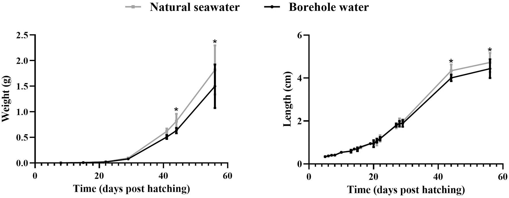
Fig. 7. Average weight (g) and length (cm) of meagre (Argyrosomus regius) reared in borehole (black) and natural seawater (grey). The values are mean ± SD and asterisk indicate the statistically significant differences between the two water sources as indicated after independent t-test analysis (p < 0.05). (For interpretation of the references to colour in this figure legend, the reader is referred to the web version of this article.)
In total, 125,000 eggs were placed in each of the 2 tanks (40m3) supplied with natural seawater. In one of the tanks, CO2 was injected into the seawater before entering the larvae tank, maintaining the pH value at a mean of 7.4, lower to the natural value of pH that had a mean of 8.0, in order to simulate the pH/CO2 conditions of the borehole water. The trial lasted 60 days dph with the same rearing protocols applied in both tanks. The feeding protocol included the addition of the microalgae
Chlorella sp in the rearing water from 3 to 15 dph, feeding with enriched rotifers Вrachionus sp from 3 to 16 dph, enriched Artemia spp. nauplii from 11 to 26 dph and artificial food (INVE SA, Belgium) from 18 dph.
Measurements of pH, O2 (HQ40D Portable Multi Meter, Hach), CO2 (CO2 Portable Carbon Dioxide Analyzer, OxyGuard) and water temperature were performed daily in the two water sources. Random samples of larvae (n = 10) from both tanks were sampled every 7 days and
M.I. Tsertou
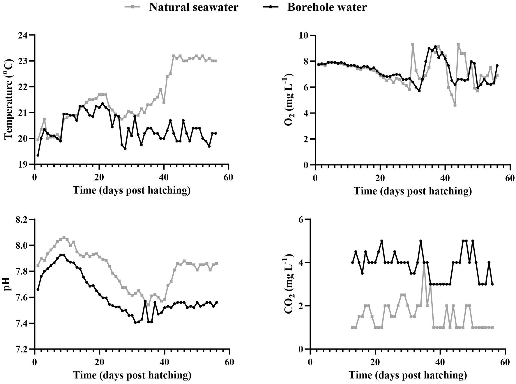
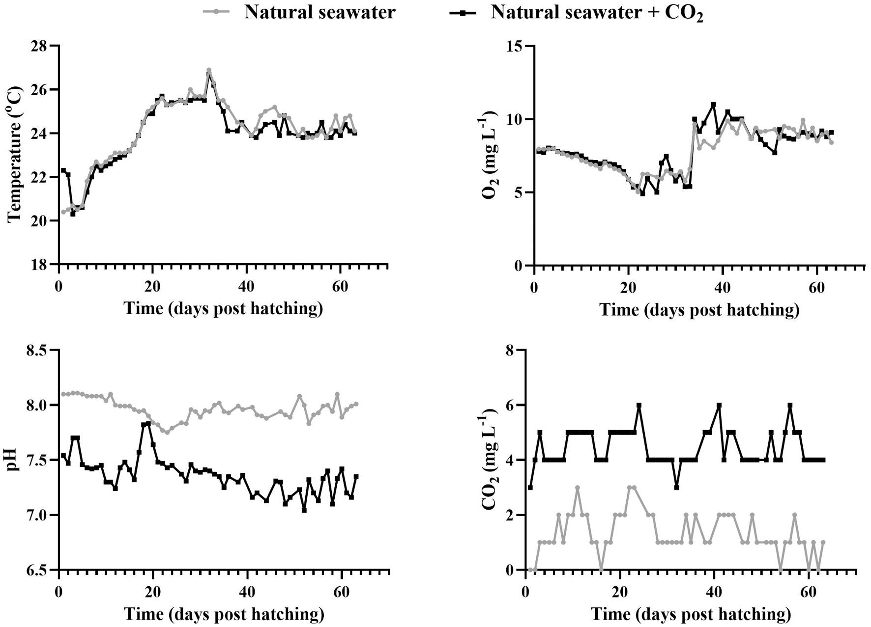
8. Physicochemical parameters of the two water sources during the rearing trial with borehole water (black) and natural seawater (grey). (For interpretation of the references to colour in this figure legend, the reader is referred to the web version of this article.)
preserved in buffered 4F:1G for histology as described above.
3. Results
3.1. Development of the lateral line canals
The first superficial neuromasts appear from the 1 dph both on the head and the trunk of meagre, consisting of the sensory hair cells, the mantle cells and the supporting cells (Fig. 1). Scanning electron microscopy revealed the presence of two morphological types of superficial
neuromasts, round or diamond-shaped, with the latter being larger in size (Fig. 2).
The morphogenesis of the lateral line canals in the head of meagre is not a synchronized process, however all the canals are completely formed by the 31st dph. The supraorbital is the first canal that begins to form as the first grooves with the submerged neuromasts appear at 9 dph (TL: 5.40 ± 0.27 mm). The ossified canal walls on either side of the neuromast are formed from the 17 dph (TL: 9.48 ± 0.7 mm) while the first neuromasts, enclosed by soft tissue canal roof are observed from the 21 dph (TL: 11.75 ± 0.83 mm) (Figs. 3 & 4).
The first grooves of the mandibular canal appear at 11 dph (TL: 6.03 ± 0.39 mm) while the walls of the canal begin to rise on each side of the neuromast at 19 dph (TL: 9.75 ± 1.21 mm) and the first epithelial canal roofs have formed at 21 dph (Fig. 5). The formation of infraorbital canal starts with the appearance of the first grooves at the 19 dph, the walls of which begin to rise at 21 dph while the first enclosed neuromasts are observed at 26 dph (TL: 18.04 ± 0.69 mm) (Fig. 6).
3.2. The effect of the borehole water in the development of CUD
The growth of fish in terms of total length and wet weight, reared with different water sources is presented in Fig. 7 The growth performance of the fish was not affected by the different source of water until 41 dph. On the 44th and 56th dph the weight and the length of the fish reared in the natural seawater was significantly higher than the fish reared in the borehole water.
The temperature of the borehole water remained relatively constant during the rearing trial with a mean value of 20.4 ± 0.5 ◦ C (range 19–21 ◦ C) while in natural seawater the temperature increased from the 35th dph onwards with a mean value of 21.6 ± 1.0 ◦ C (range 20–23 ◦ C).
The dissolved O2 in natural seawater was 7.27 ± 0.98 mgL 1 (range
Fig.
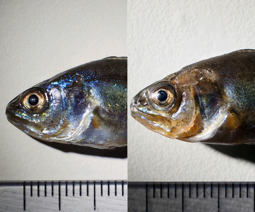
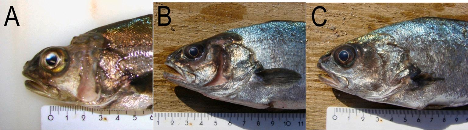
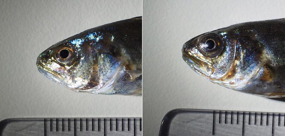
Fig. 9. Meagre (Argyrosomus regius) reared in natural seawater (left) and borehole water (right) for 56 dph. All fish reared in borehole water had visible lesions on the head associated with CUD at the end of the rearing trial. M.I.
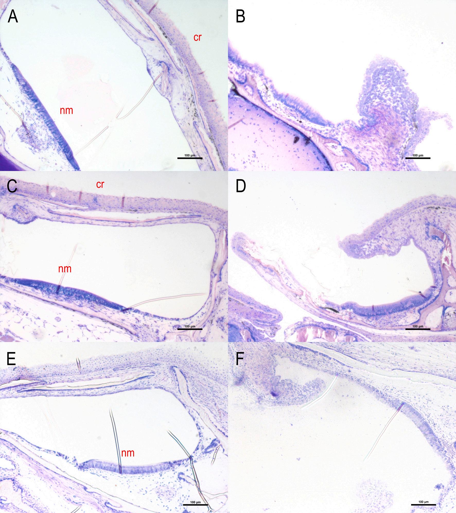
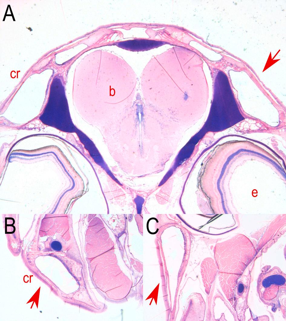
Fig. 10. Cross sections of a supraorbital, an infraorbital and a mandibular canal of healthy (A, C, E) and CUD-affected meagre (Argyrosomus regius) (B, D, F). The canal roofs of the CUD-affected meagre showed hyperplasia and loss of the basal membrane while the neuromasts were exposed to the external environment. cr: canal roof, nm: neuromast. Stain with methylene blue/azure II/basic fuchsin. (For interpretation of the references to colour in this figure legend, the reader is referred to the web version of this article.)
4.60–9.0 mgL 1) and the corresponding value in borehole water was 7.16 ± 1.04 mgL 1 (range 5.70–9.13 mgL 1).
The average pH value in natural seawater was 7.82 ± 0.14 (range 7.54–8.01) and in borehole water 7.62 ± 0.15 (range 7.40–7.92) while CO2 was consistently lower in natural seawater with a mean value of 1.56 ± 0.66 mgL 1 (range 1.00–4.00 mgL 1) compared to borehole water where the mean value was 3.88 ± 0.63 mgL 1 (range 3.00–5.00 mgL 1) (Fig. 8).
At the end of the rearing trial (56dph) all fish reared in the tanks supplied with borehole water had visible lesions associated with CUD in comparison with the fish reared in natural sea water (Fig. 9).
From the comparative histological analysis of meagre reared in borehole and natural seawater no differences were observed until 31 dph. Fig. 10 shows a supraorbital, an infraorbital and a mandibular
canal of meagre reared in natural seawater (Fig. 10 A, C, E) and in borehole water (Fig. 10 B, D, F) on 56 dph. In meagre reared in natural seawater, the canals were completely developed. Instead, in meagre from borehole water erosion, ulceration and loss of the basal membrane was observed while the neuromasts were exposed to the external environment. The lesions were initially manifested as hydropic swelling and hyperplasia of the epidermis before becoming ulcerative.
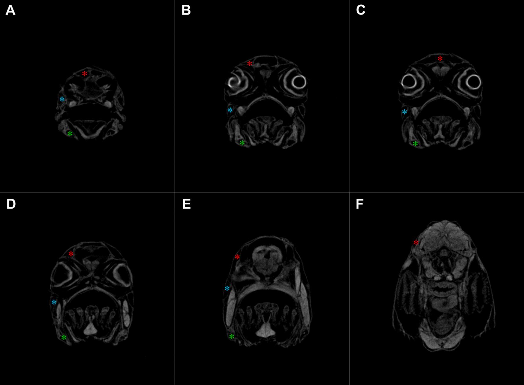
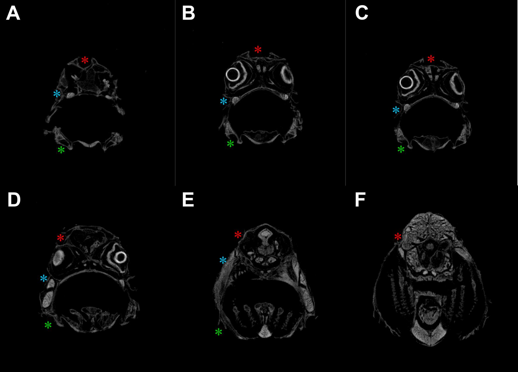
The micro-CT scans from the CUD-affected head samples of meagre that was reared in borehole water, revealed that the supraorbital and the infraorbital were the main affected canals while the mandibular had lesions in a smaller extent at least until the 56 dph (Fig. 12), whereas, in the samples from the healthy fish that was reared in natural seawater fully formed canals were observed (Fig. 11).
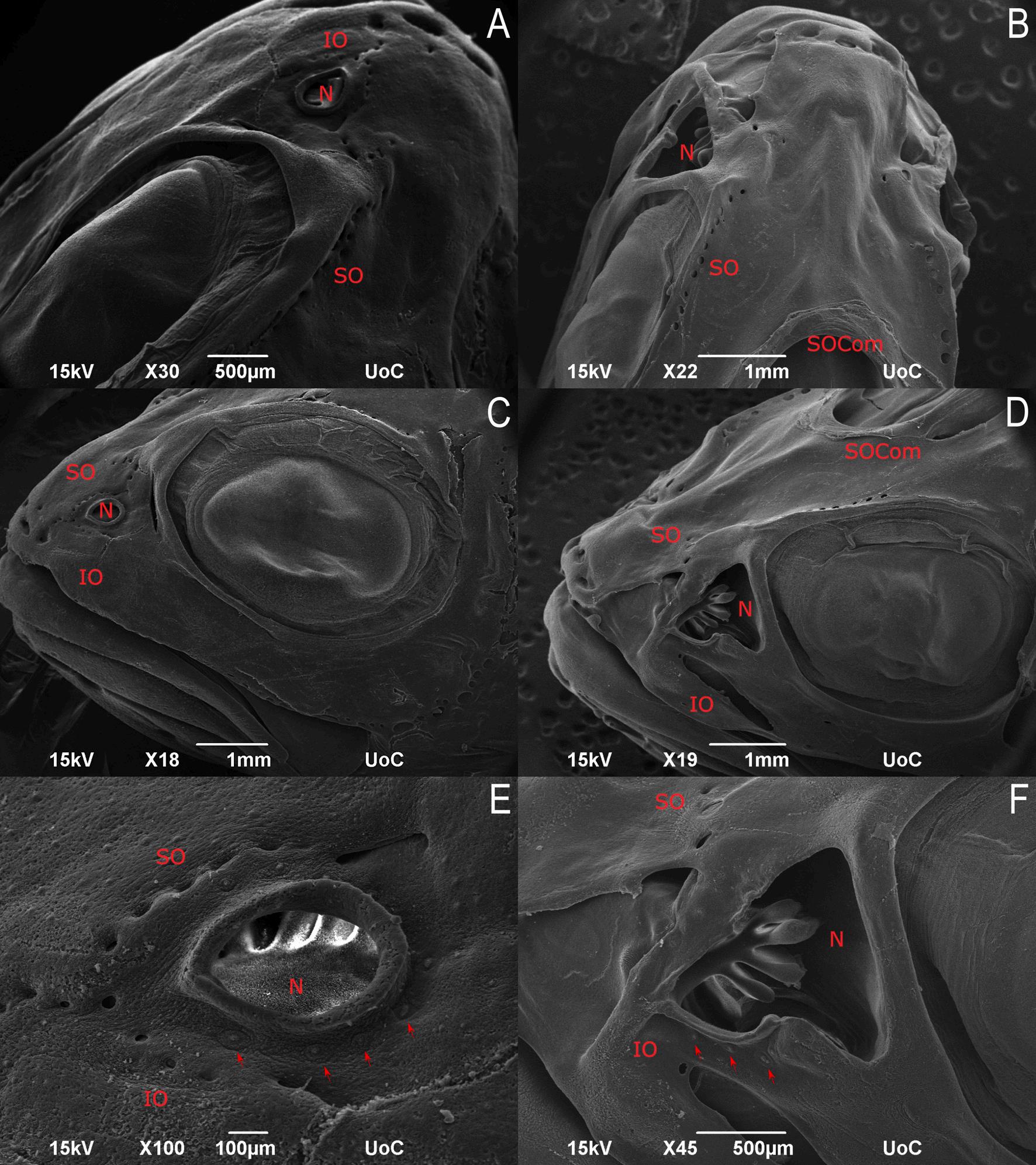
Scanning electron microscopy of CUD-affected fish revealed the
referred
presence of lesions mainly in the nasal cavity and around it where they are normally located the pores of the supraorbital and infraorbital canals while in affected individuals the pore openings have been widened, leaving the canal neuromasts exposed. Moreover, the opening of the nasal cavity in CUD-affected meagre was larger than in healthy individuals however, the olfactory rosette appeared normal. In both healthy and affected individuals, superficial neuromasts located around the opening of the nasal cavity were normal. In addition, the roof of the canal at the area of the supraorbital commissure (SOCom) where the left and right supraorbital canals join and the roof of the supraorbital canal posterior to SOCom, were absent in the CUD-affected individual, while the exposed canal neuromasts did not appear to be affected at least until 56 dph (Figs. 13, 14).
3.3. Recovery trial
The transfer of 4-month meagre with lesions associated with CUD from borehole water to natural seawater showed that CUD is a reversible condition as after 5 months in natural sea water the meagre showed almost 100% healing of the lesions as assessed by macroscopic observations. On the other hand, the group of fish that was kept in the inland facilities of HCMR and was reared in borehole water for the same period showed deterioration of the condition with severe ulceration in the head area. (Fig. 15).
3.4. Expression of genes and proteins in healthy and CUD-affected meagre
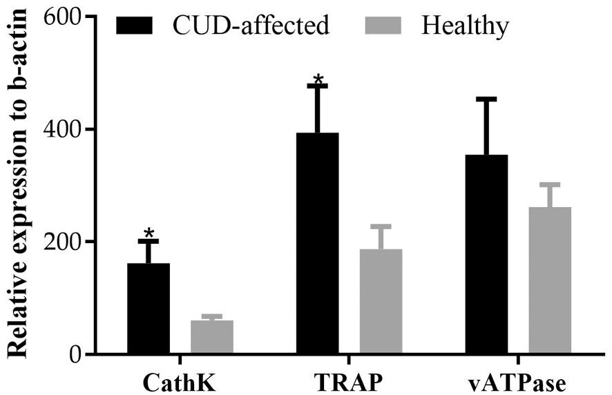
The expression profile of CathK, TRAP and vATPase in the head tissues of the fish reared in different water sources was significantly different at the end of the rearing trial (56 dph) (Fig. 16). In particular, cathepsin K and TRAP expression was 2.7 and 2.1 times higher,
respectively, in the CUD-affected fish of the borehole water group compared to the healthy fish of the natural seawater group (t(17) = 2.26, p = 0.037 for cathepsin K and t(17) = 2.41, p = 0.028 for TRAP). The expression of vATPase did not exhibit significant differences between the fish from the two water sources (t(17) = 0.219, p = 0.830).
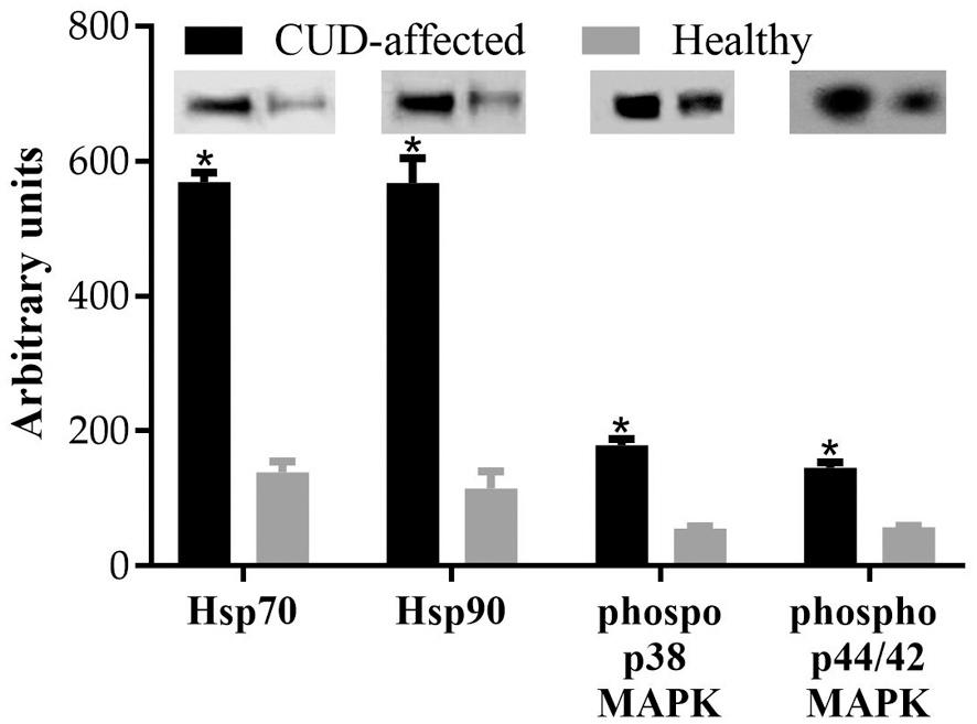
The relative protein expression of Hsp90, Hsp70, phospho p38 MAPK and phospho p44/42 MAPK in the head tissues of healthy and CUDaffected fish which were reared in natural seawater and borehole water respectively, was significantly different at the end of the rearing trial (56 dph) (Fig. 17). In particular, the Hsp70 and Hsp90 was 4 and 4.9 times higher, respectively, in the CUD-affected fish of the borehole water compared to the healthy fish of the seawater (t(10) = 51.14, p = 0.000 for Hsp70 and t(10) = 21.01, p = 0.000 for Hsp90). Moreover, CUD-affected fish exhibited increased phosphorylation of p38 MAPK (3.2 times higher, t(10) = 30.03, p = 0.000) and p44/42 MAPK (2.5 times higher, t(10) = 26.36, p = 0.000) compared to the healthy meagre.
3.5. Metal and element concentrations in healthy and CUD-affected meagre
Mean elemental concentrations in the heads of healthy and CUDaffected meagre reared in natural seawater and borehole water respectively are summarized in Table 2 The CUD-affected meagre were found to have significantly higher concentrations of Lithium (Li), Chromium (Cr), Cobalt (Co), Copper (Cu) and Barium (Ba) compared to healthy fish (p < 0.05). Moreover, Aluminum (Al), Vanadium (V), Cadmium (Cd), Caesium (Cs) and Lead (Pb) were detected only in fish reared in borehole water.
Fig. 11. μCT slices of healthy meagre’s (Argyrosomus regius) head showing the cephalic canals from anterior (A) to posterior (F). Red asterisk: supraorbital canal, blue asterisk: infraorbital canal, green asterisk: mandibular canal. (For interpretation of the references to colour in this figure legend, the reader is
to the web version of this article.)
M.I. Tsertou
3.6. The effect of the CO2 and pH in the development of CUD
At the end of the second rearing trial (60 dph) no statistically significant differences were observed on the average weight and length of the fish that were reared in natural seawater and natural seawater + CO2 (t(118) = 1.02, p = 0.309 for the weight and t(118) = 1.89, p = 0.062 for the length). Specifically, the fish reared in natural seawater had an average weight of 2.00 ± 0.65 g and an average length of 5.37 ± 0.61 cm, while those reared in natural seawater+CO2 had a mean final weight 2.10 ± 0.50 g and mean final length 5.55 ± 0.39 cm.
The temperature in both tanks showed a similar upward trend during the experiment with the mean value of 24.1 ± 1.32 ◦ C (range 20.4–26.9 ◦ C) in the tank that was supplied with natural seawater and 23.95 ± 1.39 ◦ C (range 20.3–26.7 ◦ C) in the tank that was supplied with natural seawater+CO2
The dissolved O2 in the tank with the natural seawater was 7.79 ± 1.34 mgL 1 (range 5.03–9.94 mgL 1) and the corresponding value in the natural seawater+CO2 water was 7.85 ± 1.49 mgL 1 (range 4.90–11.00 mgL 1).
The mean pH value in natural seawater was 7.98 ± 0.13 (range 7.75–8.66) and in natural seawater+CO2 7.36 ± 0.19 (range 6.72–7.83) while CO2 was consistently lower in natural seawater tank with a mean value of 1.32 ± 0.74 mgL 1 (range 0.00–3.00 mgL 1) compared to natural seawater+CO2 tank where the mean value was 4.43 ± 0.65 mgL 1 (range 3.00–6.00 mgL 1) (Fig. 18).
Although the CO2 and pH in the tank with the natural seawater + CO2 were adjusted to replicate the conditions of the borehole water, at the end of the rearing trial (60 dph) none of the fish that reared in this water had visible lesions associated with CUD as shown in Fig. 19. Histological analysis of head samples from meagre reared in natural seawater+CO2 confirmed the non-development of CUD-related lesions,
as the lateral canal were found to be fully formed and normal (Fig. 20).
4.
Discussion
The aim of this study was to investigate the development of CUD in meagre by a comparative study of two populations reared parallelly in tanks supplied with natural seawater or borehole seawater. From the specific comparative rearing trial, it was confirmed that the use of borehole water leads to the development of lesions related to CUD in the entire farmed meagre population. The ulcerative lesions in the head became visible macroscopically in fish at 56 dph (TL: 4.37 ± 0.11 cm), while CUD was found to be a reversible pathological condition as the transport of affected individuals in natural sea water led to complete healing of the lesions within a period of 5 months. These results are in accordance with other reported cases of CUD, both in freshwater and marine fish species. In the Australian freshwater fish Murray cod, M. peelii peelii the first gross signs appeared approximately three weeks after exposure to groundwater as enlargement of the pores of the head and trunk canals. Progressively, the elongated pores began to coalesce, resulting in exposure of the bed of the canal and finally all the canal beds were exposed while ulceration on the head started to extend into surrounding areas. Similar to meagre, it was shown that when CUD-affected Murray cod were transferred to river water, the majority of the fish were structurally recovered after a period of 8–10 weeks (Baily et al., 2005). Concerning marine fish species, the first lesions of CUD in sharpsnout seabream were observed at 70 dph while at 130 dph all fish that were reared in borehole water had bilateral grooves at the area of the lateral line canals. The transportation of the affected fish in natural sea water led to the recovery of the lesions over a period of about 4 months (Katharios et al., 2011).
In contrast to Murray cod, in which CUD led to reduced growth rate
Fig. 12. μCT slices of CUD-affected meagre (Argyrosomus regius) head showing the cephalic canals from anterior (A) to posterior (F). The canal roofs of the affected fish were open with the neuromasts exposed to the external environment. Red asterisk: supraorbital canal, blue asterisk: infraorbital canal, green asterisk: mandibular canal. (For interpretation of the references to colour in this figure legend, the reader is referred to the web version of this article.)
M.I. Tsertou et al.
13. SEM micrographs of healthy and CUD-affected juvenile meagre (Argyrosomus regius) (56 dph). Dorsal view showing the supraorbital canal (SO), the infraorbital canal (IO) and supraorbital commissure (SOCom) of healthy (A) and CUD-affected meagre (B). Lateral view of healthy (C) and CUD-affected meagre (D) showing the infraorbital canal, the nostril (R) and the mandibular canal (MD). Higher magnification of the nostril (N) with the supraorbital and the infraorbital canal (IO) of healthy (E) and CUD-affected meagre (F). Red arrows indicate the superficial neuromasts around the nostril. (For interpretation of the references to colour in this figure legend, the reader is referred to the web version of this article.)
and increased mortality, no mortality was observed in CUD-affected meagre. The relatively reduced growth that was observed in CUDaffected meagre is probably related to the lower temperature of the borehole water in comparison to the natural sea water (20.4 ± 0.5 ◦ C and 21.6 ± 1.0 ◦ C, respectively) as it is known that increasing the temperature up to 26 ◦ C has a beneficial effect on the growth of the meagre (Antonopoulou et al., 2020; Chatzifotis et al., 2018).
The results from histology, SEM and μ-CT confirmed that the lesions in meagre were limited to the lateral line organ in the head and in the nasal cavity which is in agreement with the conclusions of Baily et al. (2005) for Murray cod and of Katharios et al. (2011) for sharpsnout seabream.
From the histological comparative analysis, no differences were
observed up to 36 dph between the groups reared in natural seawater and in borehole water respectively, with the development of the lateral line canals in the head occurring normally in both groups. The cranial lateral line canals of meagre like most teleost bony fish (Webb, 2014; Webb, 2013) are narrow and well-ossified. The development of the lateral line canals is an asynchronous process both within the same canal and between the different canals, with the supraorbital and the mandibular canals being the first to begin forming, followed by the infraorbital (Webb, 2014). This was also confirmed in the case of meagre, as the supraorbital and the mandibular canals starts to form when the fish are 5.40 ± 0.27 mm and 6.03 ± 0.39 mm, respectively while the infraorbital canal starts to develop when the fish are 9.75 ± 1.21 mm and all canals are fully formed when the fish are 19.3 ± 1.27
Fig.
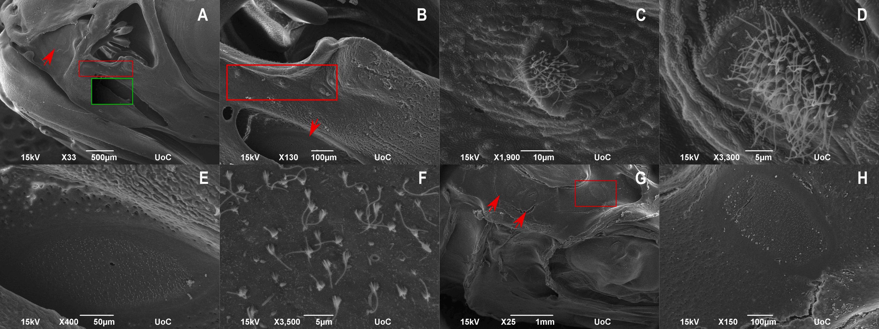
M.I. Tsertou et
14. SEM micrographs of CUD-affected meagre (Argyrosomus regius – 56 dph). A: Lateral view of the head with the ulcerative nostril, supraorbital (red arrow) and infraorbital canal (green frame). The olfactory rosette was not affected. B: Higher magnification of the nostril with the opened infraorbital canal (red arrow). Framed area indicates normal superficial neuromasts around the nostril. C-D: Higher magnification of the superficial neuromasts around the nostril. E: Higher magnification of the infraorbital canal with the exposed canal neuromast. F: Higher magnification of the exposed canal neuromast showing the normal sensory hair cells. G: Dorsal view of the opened supraorbital canal with the exposed neuromasts (red arrows, red frame). H: Higher magnification of the exposed neuromast from the framed area of picture G. (For interpretation of the references to colour in this figure legend, the reader is referred to the web version of this article.)
Fig.
Fig. 15. Nine-month-old meagre (Argyrosomus regius) reared А: exclusively in borehole seawater B: in borehole water for 4 months and transferred to natural seawater for 5 months, with partial resolution of the lesions. C: in borehole water for 4 months and transferred to natural seawater for 5 months, with complete resolution of the lesions.
Fig. 16. Relative expression of CathK, TRAP and vATPase in heads of healthy and CUD-affected meagre (Argyrosomus regius) at the end of the rearing trial (56 dph). Values are means+SD while (*) indicates statistically significant differences between the two groups (p < 0.05).
Fig. 17. Relative expression of Нsp70, Hsp90, phospho p38 MAPK και phospho p44/42 MAPK in heads of healthy and CUD-affected meagre (Argyrosomus regius) at the end of the rearing trial (56 dph). Values are means+SD while asterisk (*) indicates statistically significant differences between the two groups (p < 0.05).
mm. Τhe development of the lateral cranial canals has been studied mainly in various species of cichlids (Amatitlania nigrofasciata, Labeotropheus fuelleborni, Maylandia zebra, Aulonocara baenschi, Aulonocara stuartgranti and Tramitichromis sp) and does not differ significantly compared to meagre. In all cases the onset of canal formation is observed when the fish are 5–8 mm in length while the completion of the formation is observed when the fish are 16–22 mm (Becker et al., 2016; Tarby and Webb, 2003; Webb, 2014). In contrast to both meagre and the
various species of cichlids, the development of lateral canal canals in zebrafish (Danio rerio) appears to be delayed as the first submerged neuromasts of the supraorbital and the mandibular canals appear when the fish are 10 mm and 11.5 mm respectively, however, their closure is observed when the fish is 22 mm in length (Webb and Shirey, 2003).
Histologically, the first lesions related to CUD in meagre were observed from 36 dph onwards. The lesions were initially manifested as hydropic swelling and hyperplasia of the epidermis before becoming ulcerative leaving the canal neuromasts exposed. The neuromasts did not appear to be affected by CUD at least until the age of 56 dph. In murray cod, the first lesions of CUD were observed histologically 3 weeks after exposure of the fish to borehole water, as marked hyperplasia and necrosis. Two weeks later, the tissue above the canals was completely necrotic, however as in the case of meagre, no degeneration of the exposed neuromasts was observed (Baily et al., 2005). In CUDaffected sharpsnout seabream, the histological examination revealed the presence of ulcers in the cranial lateral line canals while the roof of the canals was always absent leaving the canal neuromasts exposed. In contrast to meagre and to murray cod the exposed neuromasts of sharpsnout seabream appeared either normal or degenerated and necrotic (Katharios et al., 2011). In the case of sharpsnout seabream the nasal cavity was affected from CUD which was also observed and in meagre. However, in the sharpsnout seabream the olfactory rosette was also damaged (Katharios et al., 2011) which was not observed in meagre.
In either cases of meagre, murray cod and sharpsnout seabream there is evidence that the development of CUD is directly linked with the use of groundwater, which is reinforced by the fact that the condition is reversed by transporting the fish in natural sea or fresh water. However, the actual causative agent of CUD is unknown in all cases. It is noteworthy that the pH was lower and CO2 higher in borehole water in comparison with natural seawater. High levels of CO2 in rearing water of fish leads to a decrease in pH of their extracellular environment. The decrease of pH in the extracellular environment is one of the factors that can lead to the activation of osteoclasts (Arnett, 2008; Yuan et al., 2016), which in collaboration with osteoblasts, participate in the remodeling of the bones that contain the lateral line canals as the fish grows in size (Wada et al., 2014; Webb, 2013). An imbalance between osteoclasts and osteoblasts could lead to the lesions seen in the fish, located exclusively in the lateral line canals. The tartate-resistant acid phosphatase (TRAP) is the most widely used marker enzyme for the identification of osteoclasts and with the cysteine proteinase cathepsin K (CathK) are being used as molecular markers of bone resorption (Azuma et al., 2007; Costa et al., 2011; Minkin, 1982). Moreover, the vacuolar-type proton pumping ATPases (V-ATPase) are important for the normal function of these enzymes as they are responsible for the acidic pH of the bone resorption lacunae which is formed between the osteoclast plasma membrane and the bone surface through secretion of protons (Futai et al., 2019). Gene expression of TRAP, cathK, and v-ATPase showed that the CUD-affected meagre had significantly higher levels of TRAP and cathK compared to healthy meagre while v-ATPase levels were
M.I. Tsertou
18. Physicochemical parameters of the two water sources during the rearing trial with natural seawater (grey) and natural seawater+CO2 (black). (For interpretation of the references to colour in this figure legend, the reader is referred to the web version of this article.)
higher but not statistically significant compared to healthy individuals. These results indicate that there is an increased osteoclastic activity in the head of the CUD-affected meagre. In mammals it has been shown that increased osteoclastic activity could lead to an imbalance in bone remodeling which promotes resorption resulting to skeletal diseases such as osteoporosis, rheumatoid arthritis, periodontal disease, multiple myeloma and metastatic cancers (Boyle et al., 2003; Rodan and Martin, 2000). Moreover, in accordance with the results from meagre, preliminary results from CUD-affected sharpsnout seabream using enzyme histochemistry has shown increased TRAP activity at the area of the lesions compared to normal samples (Katharios et al., 2011). The increased osteoclastic activity in the head area of CUD-affected meagre reared in borehole water compared to those reared in natural seawater was also confirmed by the comparative expression of phospho p38 MAPKs and phospho p44/42, since in healthy fish reared in natural
seawater their levels were significantly lower. The higher expression of MAPKs in CUD-affected fish can be explained by the fact that both p38 MAPKs and p44/42 are activated by the binding of RANKL to the RANK receptor of osteoclasts, which in turn leads to the downstream activation of transcription factors controlling the expression of the genes encoding for TRAP and CathK, in areas where bone resorption occurs (Boyle et al., 2003; Lee et al., 2018). Hsp70 and Hsp90 were also overexpressed in the CUD-affected individuals compared to the healthy fish, which enhances the increased osteoclastic activity as Hsp70 induces osteoclastic bone resorption via the RANKL / RANK pathway, while even though the role of Hsp90 in osteoclastogenesis is controversial it has been shown that it can induce the expression of osteoclast-associated genes (Hang et al., 2018).
Based on the results from the comparative rearing of meagre in natural seawater and borehole water, the second larval rearing trial was performed in order aiming to test whether increased CO2 concentration is the aetiological agent for the development of CUD lesions. This trial was designed in order to replicate the pH and CO2 conditions of the borehole water without including any other chemical or environmental factor. After macroscopic examination and histological analysis, it was revealed that none of the fish reared in the natural seawater where CO2 was added showed any lesions related to CUD. The lack of the lesions in the head of the fish suggests that neither the increased CO2 nor the consequently reduced pH are the factors affecting the development of CUD.
The results from the metal analysis of healthy meagre reared in natural seawater and CUD-affected meagre reared in borehole water showed that fish with lesions had significantly higher levels of Li, Cr, Co, Cu and Ba in relation to healthy individuals. In addition, Al, V, Cd, Cs and Pb were detected only in CUD-affected fish while in healthy individuals they were below detectable limits. Metals can be categorized as biologically essential and nonessential. The nonessential metals (e.g.,
Fig.
Fig. 19. Meagre (Argyrosomus regius) reared in natural seawater (left) and natural seawater+CO2 (right) at the end of the rearing trial (60dph). None of the fish reared in natural seawater+CO2 had visible lesions associated with CUD.
Fig. 20. Cross sections of the fully formed lateral line canals on the head of meagre (Argyrosomus regius) reared in natural seawater+CO2 (60 dph, TL: 5,65 ± 0,49 cm). A: Supraorbital canal (magnification: x1). B: Mandibular canal (magnification: x3). C: Infraorbital canal (magnification: x3). No lesions associated with CUD were observed in any of the canals. b: brain, cr: canal roof, e: eye. Stain with methylene blue/azure II/basic fuchsin. (For interpretation of the references to colour in this figure legend, the reader is referred to the web version of this article.)
Table 2
Metal and element concentrations in the head of healthy and CUD- affected meagre individuals (Argyrosomus regius). The values are mean ± SD while different superscript letters indicate statistically significant differences (p < 0.05) between the two water groups (bdl: below detection limit).
CUD-affected fish Healthy fish
Calcium (Ca) (mg/g) 92.44 ± 10.61
Phosphorus (P) (mg/g) 49.26 ± 4.95
± 2.53
± 6.83
Zinc (Zn) (mg/kg) 65.72 ± 9.67 55.69 ± 1.82
Sodium (Na) (mg/g) 16.25 ± 1.05 14.57 ± 2.48
Copper (Cu) (mg/kg) 1.70 ± 0.51a 0.85 ± 0.25b
Magnesium (Mg) (mg/kg) 1.33 ± 0.53 0.86 ± 0.03
Chromium (Cr) (mg/kg) 0.89 ± 0.20a 0.52 ± 0.09b
Potassium (K) (mg/g) 0.75 ± 0.11 0.83 ± 0.13
Selenium (Se) (mg/kg) 0.65 ± 0.02 0.61 ± 0.07
Lithium (Li) (mg/kg) 0.16 ± 0.04a 0.07 ± 0.00b
Cobalt (Co) (mg/kg) 0.15 ± 0.05a 0.08 ± 0.01b
Molybdenum (Mo) (mg/kg) 0.10 ± 0.03 0.08 ± 0.04
Manganese (Mn) (mg/g) 0.03 ± 0.01 0.02 ± 0.00
Aluminum (Al) (mg/kg) 2.41 ± 0.57 bdl
Barium (Ba) (mg/kg) 2.14 ± 0.28a 1.12 ± 0.13b
Cadmium (Cd) (mg/kg) 0.01 ± 0.00 bdl
Lead (Pb) (mg/kg) 0.62 ± 0.10 bdl
Vanadium (V) (mg/kg) 0.22 ± 0.03 bdl
Nickel (Ni) (mg/kg) 3.44 ± 0.57 2.82 ± 0.14
Mercury (Hg) (mg/kg) 0.07 ± 0.01 0.07 ± 0.01
Caesium (Cs) (mg/kg) 9.15 ± 1.46 bdl
Rubidium (Rb) (mg/kg) 0.15 ± 0.02 0.12 ± 0.03
Al, V, Cd, Pb, Ba) which have no proven biological function can become extremely toxic for organisms due to their persistence and their tendency to bioaccumulate (Amoussou et al., 2019; Carvalho et al., 2005; Tarley et al., 2001). On the other hand, essential metals (e.g., Cu, Zn, Cr, Co), have a known biological role, and they can become toxic either at
deficiencies or at high concentrations (Kennedy, 2011). In general, metals can affect multiple physiological systems of fish while toxicity depends on metal form, bioavailability, toxicokinetics and toxicodynamics (Kennedy, 2011; Sfakianakis et al., 2015). Heavy metals in the water can cause reduction of the survival and the growth of fish larvae, behavioral anomalies or structural damages (Stominska and Jezierska, 2000). More specifically, metals can reduce calcium uptake and bone calcium accumulation leading to changes in bone properties and can induce disturbances of neuro-muscular transmission leading to skeletal deformities (Hassanain et al., 2012). It was reported that exposure of freshwater fish to Hg and Cd leads to a disturbed calcium metabolism, resulting in hypocalcemia and anomaly of the bone. Both metals, first influence osteoclastic activities under acute exposure and then inhibit osteoblastic activities under long-time exposure (Suzuki et al., 2004). Moreover, carp larvae exposed to Pb, developed scoliosis, while Cu caused inhibition of skeletal ossification which might have resulted from impaired ionic regulation. In addition, both metals caused reduction of the fish survival and the growth rates (Stominska and Jezierska, 2000). Disturbance of bone ossification was also reported in Nile tilapia after exposure to Pb (Hassanain et al., 2012). In addition to fish, it was reported that exposure of chick femur culture to Zn and Cd led to decreased mineralization of bone, with or without suppression of matrix formation while exposure to Zn and Cu caused decreased mineralization and matrix formation (Toshiyuki et al., 1991). Moreover, it was found that Zn is an extremely potent inhibitor of rat osteoclasts in vitro, since significant inhibition of resorption was reported at concentrations as low as 10 14 M (Moonga and Dempster, 1995). In the case of meagre, despite the differences between healthy and CUD affected fish, the values observed are within the range of values reported for other fish species, including gilthead seabream and European seabass when lesions are absent (El-Moselhy et al., 2014; Kalantzi et al., 2016; Kalantzi et al., 2013). On the other hand, it has been reported that Cu concentrations above 1 mg L 1 can cause damage to the epithelium of the lateral canal of Fundulus hereroclitus while in some cases canal neuromasts were also affected (Eisler and Gardner, 1973). It has been shown that metals such as Cu are toxic to the peripheral sensory systems of fish and other aquatic organisms by reducing the physiological response or at higher concentrations by cell death of the olfactory and mechanosensory neurons (Linbo et al., 2009). Moreover, lateral line dysfunction was reported after exposure of zebrafish embryos to Cu (68 and 244 μg Cu L1). The copper-exposed larvae showed fewer functional neuromasts which was associated with a reduced ability of orientation in a current (Johnson et al., 2007). Neuromasts cellular damage, apoptosis, and loss of hair cell markers were also reported in zebrafish after exposure to sublethal concentrations of waterborne copper while other metals such as Zn, Fe, Ag, Mn, Co, Cd and Sn, did not show the same effects (Hern´ andez et al., 2006).The concentration of Cu in the head of CUD-affected meagre was 1.70 ± 0.51 mg/kg, while in healthy fish it was significantly lower (0.85 ± 0.25 mg/kg) suggesting that Cu could be one of the factors leading to the development of CUD. Moreover, in the study of CUD in the sharpsnout seabream the concentrations of Ni, Pb, Zn and Cu were higher in the borehole water than in the natural seawater, however these levels were within the acceptable limits for marine aquaculture and much lower than the toxic limits (Katharios et al., 2011). Nevertheless, metal toxicity as a causative factor for the development of CUD cannot be ruled out because of the lower pH of the borehole water and the longer exposure times of the fish and therefore it should be further studied in the future as environmental parameters and interactions among various metals may affect their toxicity to fish (Sfakianakis et al., 2015).
Although the disease is directly associated with the use of borehole water, the causative agent is still unknown for meagre, as well as for Murray cod and sharpsnout seabream. For all species the lesions resolve when the fish are transferred to natural freshwater or seawater (Baily et al., 2005; Katharios et al., 2011). Furthermore, for Murray cod, Schultz et al. (2011) found that the retention of groundwater into a
M.I. Tsertou
vegetated earthen pond or in a tank containing biofilms growing on an artificial macrophyte for 72 h prevents the development of CUD. Thus, it is recommended to avoid borehole seawater for the rearing of meagre if natural sea water sources are available and to pay careful attention to the water quality of the source of the water used. Alternatively, the residence time of meagre in borehole water should be reduced to the minimum necessary, and fish should be moved to natural seawater (e.g. in sea cages) as soon as possible once the nursery phase is completed, in order to allow the tissue regeneration process to complete before marketing the fish.
Declaration of Competing Interest
The authors declare that they have no known competing financial interests or personal relationships that could have appeared to influence the work reported in this paper.
Acknowledgments
Funding was provided for this project from The European Union’s Seventh Framework Programme for research, technological development and demonstration (KBBE-2013-07 single stage, GA 603121, DIVERSIFY).
References
Amoussou, N., Marengo, M., Durieux, E.D.H., Douny, C., Scippo, M.L., Gobert, S., 2019. Trace elements and fatty acid profile of Argyrosomus regius (Asso, 1801) from Mediterranean aquaculture. Biol. Trace Elem. Res. https://doi.org/10.1007/s12011019-01925-x
Antonopoulou, E., Kousidou, E., Tserga, E., Feidantsis, K., Chatzifotis, S., 2014. Dietary lipid levels in meagre (Argyrosomus regius): effects on biochemical and molecular indicators of liver. Aquaculture 428–429, 265–271. https://doi.org/10.1016/j. aquaculture.2014.03.024
Antonopoulou, E., Chatzigiannidou, I., Feidantsis, K., Kounna, C., Chatzifotis, S., 2020. Effect of water temperature on cellular stress responses in meagre (Argyrosomus regius). Fish Physiol. Biochem. 46, 1075–1091. https://doi.org/10.1007/s10695020-00773-0
Arnett, T.R., 2008. Extracellular pH Regulates Bone Cell Function. J. Nutr. 138, 415S–418S. https://doi.org/10.1093/jn/138.2.415s
Azuma, K., Kobayashi, M., Nakamura, M., Suzuki, N., Yashima, S., Iwamuro, S., Hattori, A., 2007. Two osteoclastic markers expressed in multinucleate osteoclasts of goldfish scales. Biochem. Biophys. Res. Commun. 362, 594–600. https://doi.org/ 10.1016/j.bbrc.2007.08.010
Baily, J.E., Bretherton, M.J., Gavine, F.M., Ferguson, H.W., Turnbull, J.F., 2005. The pathology of chronic erosive dermatopathy in Murray cod, Maccullochella peelii peelii (Mitchell). J. Fish Dis. 28, 3–12. https://doi.org/10.1111/j.1365-2761.2004.00586. x.
Becker, E.A., Bird, N.C., Webb, J.F., 2016. Post-embryonic development of canal and superficial neuromasts and the generation of two cranial lateral line phenotypes. J. Morphol. 277, 1273–1291. https://doi.org/10.1002/jmor.20574
Bennett, H.S., Wyrick, A.D., Lee, S.W., McNeil, J.H., 1976. Science and art in preparing tissues embedded in plastic for light microscopy, with special reference to glycol methacrylate, glass knives and simple stains. Stain. Technol. 51, 71–97. https://doi. org/10.3109/10520297609116677
Bleckmann, H., Zelick, R., 2009. Lateral line system of fish. Integr. Zool. 4, 13–25 Boyle, W.J., Simonet, W.S., Lacey, D.L., 2003. Osteoclast differentiation and activation. Nature 423, 337–342. https://doi.org/10.1038/nature01658
Carvalho, M.L., Santiago, S., Nunes, M.L., 2005. Assessment of the essential element and heavy metal content of edible fish muscle. Anal. Bioanal. Chem. 382, 426–432. https://doi.org/10.1007/s00216-004-3005-3
Chatzifotis, S., Clavero, S., Kounna, C., Soumalevris, A., Feidantsis, K., Antonopoulou, E., 2018. Effects of long-term feed deprivation on body weight loss, muscle composition, plasma metabolites, and intermediate metabolism of meagre (Argyrosomus regius) under different water temperatures. Fish Physiol. Biochem. 44, 527–542. https://doi.org/10.1007/s10695-017-0451-3
Corrales, J., Ullal, A., Noga, E.J., 2009. Lateral line depigmentation (LLD) in channel catfish, Ictalurus punctatus (Rafinesque). J. Fish Dis. 32, 705–712. https://doi.org/ 10.1111/j.1365-2761.2009.01069.x
Costa, A.G., Cusano, N.E., Silva, B.C., Cremers, S., Bilezikian, J.P., 2011. Cathepsin K: its skeletal actions and role as a therapeutic target in osteoporosis. Nat. Rev. Rheumatol. 7, 447–456. https://doi.org/10.1038/nrrheum.2011.77.
Eisler, R., Gardner, G.R., 1973. Acute toxicology to an estuarine teleost of mixtures of cadmium, copper and zinc salts. J. Fish Biol. 5, 131–142. https://doi.org/10.1111/ j.1095-8649.1973.tb04441.x
El-Moselhy, K.M., Othman, A.I., Abd El-Azem, H., El-Metwally, M.E.A., 2014. Bioaccumulation of heavy metals in some tissues of fish in the Red Sea, Egypt. Egypt. J. Basic Appl. Sci. 1, 97–105. https://doi.org/10.1016/j.ejbas.2014.06.001
Futai, M., Sun-Wada, G.-H., Wada, Y., Matsumoto, N., Nakanishi-Matsui, M., 2019. Vacuolar-type ATPase: a proton pump to lysosomal trafficking. Proc. Japan Acad. Ser. B 95, 261–277. https://doi.org/10.2183/pjab.95.018
Hang, K., Ye, C., Chen, E., Zhang, W., Xue, D., Pan, Z., 2018. Role of the heat shock protein family in bone metabolism. Cell Stress Chaperones 23, 1153–1164. https:// doi.org/10.1007/s12192-018-0932-z
Hassanain, M.A., Abbas, W.T., Ibrahim, T.B., 2012. Skeletal ossification impairment in Nile Tilapia (Oreochromis niloticus) after exposure to lead acetate. Pakistan J. Biol. Sci. 15, 729–735. https://doi.org/10.3923/pjbs.2012.729.735
Hern´ andez, P.P., Moreno, V., Olivari, F.A., Allende, M.L., 2006. Sub-lethal concentrations of waterborne copper are toxic to lateral line neuromasts in zebrafish (Danio rerio). Hear. Res. 213, 1–10. https://doi.org/10.1016/j.heares.2005.10.015
Ingram, B.A., Gavine, F., Lawson, P., 2004. Diseases and health management in intensive Murray cod aquaculture. In: Ingram, B.A., De Silva, S.S. (Eds.), Development of Intensive Commercial Aquaculture Production Technology for Murray cod. Primary Industries Research Victoria, Marine and Freshwater Systems, Department of Primary Industries, Queenscliff, Victoria, pp. 129–146
Johnson, A., Carew, E., Sloman, K.A., 2007. The effects of copper on the morphological and functional development of zebrafish embryos. Aquat. Toxicol. 84, 431–438. https://doi.org/10.1016/j.aquatox.2007.07.003
Kalantzi, I., Black, K.D., Pergantis, S.A., Shimmield, T.M., Papageorgiou, N., Sevastou, K., Karakassis, I., 2013. Metals and other elements in tissues of wild fish from fish farms and comparison with farmed species in sites with oxic and anoxic sediments. Food Chem. 141, 680–694. https://doi.org/10.1016/j.foodchem.2013.04.049
Kalantzi, I., Pergantis, S.A., Black, K.D., Shimmield, T.M., Papageorgiou, N., Tsapakis, M., Karakassis, I., 2016. Metals in tissues of seabass and seabream reared in sites with oxic and anoxic substrata and risk assessment for consumers. Food Chem. 194, 659–670. https://doi.org/10.1016/j.foodchem.2015.08.072
Katharios, P., Papadaki, M., Ternengo, S., Kantham, P.K., Zeri, C., Petraki, P.E., Divanach, P., 2011. Chronic ulcerative dermatopathy in cultured marine fishes. Comparative study in sharpsnout sea bream, Diplodus puntazzo (Walbaum). J. Fish Dis. 34, 459–474. https://doi.org/10.1111/j.1365-2761.2011.01257.x
Kennedy, C.J., 2011. TOXICOLOGY | The Toxicology of Metals in Fishes, in: Encyclopedia of Fish Physiology. Elsevier, pp. 2061–2068. https://doi.org/10.1016/ B978-0-12-374553-8.00236-7
Lee, K., Seo, I., Choi, M.H., Jeong, D., 2018. Roles of mitogen-activated protein kinases in osteoclast biology. Int. J. Mol. Sci. 19 https://doi.org/10.3390/ijms19103004
Linbo, T.L., Baldwin, D.H., McIntyre, J.K., Scholz, N.L., 2009. Effects of water hardness, alkalinity, and dissolved organic carbon on the toxicity of copper to the lateral line of developing fish. Environ. Toxicol. Chem. 28, 1455–1461. https://doi.org/10.1897/ 08-283.1
McDowell, E.M., Trump, B.F., 1976. Histologic fixatives suitable for diagnostic light and electron microscopy. Arch. Pathol. Lab. Med. 100, 405–414
Minkin, C., 1982. Bone acid phosphatase: tartrate-resistant acid phosphatase as a marker of osteoclast function. Calcif. Tissue Int. 34, 285–290. https://doi.org/10.1007/ BF02411252
Mogdans, J., Krother, S., Engelmann, J., 2004. Neurobiology of the fish lateral line: adaptations for the detection of hydrodynamic stimuli in running water. Senses Fish 265–287. https://doi.org/10.1007/978-94-007-1060-3_12
Moonga, B.S., Dempster, D.W., 1995. Zinc is a potent inhibitor of osteoclastic bone resorption in vitro. J. Bone Miner. Res. 10, 453–457. https://doi.org/10.1002/ jbmr.5650100317
Morrison, C.M., O’Neil, D., Wright, J.R., 2007. Histopathology of “hole-in-the-head” disease in the Nile Tilapia, Oreochromis niloticus Aquaculture 273, 427–433 Noga, E.J., 2010. Fish Disease: Diagnosis and Treatment, Second edition. WileyBlackwell. https://doi.org/10.1002/9781118786758
Rigos, G., Katharios, P., 2010. Pathological obstacles of newly-introduced fish species in Mediterranean mariculture: a review. Rev. Fish Biol. Fish. 20, 47–70
Rodan, G.A., Martin, T.J., 2000. Therapeutic approaches to bone diseases. Science (80-.) 289, 1508–1514. https://doi.org/10.1126/science.289.5484.1508
Schultz, A.G., Healy, J.M., Jones, P.L., Toop, T., 2008. Osmoregulatory balance in Murray cod, Maccullochella peelii peelii (Mitchell), affected with chronic ulcerative dermatopathy. Aquaculture 280, 45–52. https://doi.org/10.1016/j. aquaculture.2008.04.011
Schultz, A.G., Shigdar, S.L., Jones, P.L., Ward, A.C., Toop, T., 2011. Groundwater pretreatment prevents the onset of chronic ulcerative dermatopathy in juvenile Murray cod, Maccullochella peelii peelii (Mitchell). Aquaculture 312, 19–25. https://doi.org/ 10.1016/j.aquaculture.2010.12.013
Sfakianakis, D.G., Renieri, E., Kentouri, M., Tsatsakis, A.M., 2015. Effect of heavy metals on fish larvae deformities: a review. Environ. Res. 137, 246–255. https://doi.org/ 10.1016/j.envres.2014.12.014
Soares, F., Roque, A., Gavaia, P.J., 2018. Review of the principal diseases affecting cultured meagre (Argyrosomus regius). Aquac. Res. 49, 1373–1382. https://doi.org/ 10.1111/are.13613
Stominska, I., Jezierska, B., 2000. The effect of heavy metals on postembryonic development of common carp, Cyprinus carpio L. Arch. Polish Fish. 8, 119–128
Suzuki, N., Yamamoto, M., Watanabe, K., Kambegawa, A., Hattori, A., 2004. Both mercury and cadmium directly influence calcium homeostasis resulting from the suppression of scale bone cells: the scale is a good model for the evaluation of heavy metals in bone metabolism. J. Bone Miner. Metab. 22, 439–446. https://doi.org/ 10.1007/s00774-004-0505-3
Tarby, M.L., Webb, J.F., 2003. Development of the supraorbital and mandibular lateral line canals in the cichlid, Archocentrus nigrofasciatus J. Morphol. 255, 44–57. https://doi.org/10.1002/jmor.10045
M.I. Tsertou
Tarley, C.R.T., Coltro, W.K.T., Matsushita, M., De Souza, N.E., 2001. Characteristic levels of some heavy metals from Brazilian canned sardines (Sardinella brasiliensis). J. Food Compos. Anal. 14, 611–617. https://doi.org/10.1006/jfca.2001.1028
Toshiyuki, K., Masakazu, T., Tatsuro, M., Hiroshi, K., Fumitomo, K., 1991. Interaction between cadmium and copper on ossification of embryonic chick bone in tissue culture. Toxicol. Lett. 55, 255–262. https://doi.org/10.1016/0378-4274(91)90005Q
Wada, H., Iwasaki, M., Kawakami, K., 2014. Development of the lateral line canal system through a bone remodeling process in zebrafish. Dev. Biol. 392, 1–14. https://doi. org/10.1016/j.ydbio.2014.05.004
Webb, J.F., 1989. Gross morphology and evolution of the mechanoreceptive lateral-line system in teleost fishes. Brain Behav. Evol. https://doi.org/10.1159/000115896
Webb, J.F., 2013. Morphological diversity, development, and evolution of the mechanosensory lateral line system. In: Coombs, S., Bleckmann, H., Fay, R.,
Popper, A. (Eds.), The Lateral Line System, pp. 17–72. https://doi.org/10.1007/ 2506_2013_12
Webb, J.F., 2014. Lateral Line Morphology and Development and Implications for the Ontogeny of Flow Sensing in Fishes, in: Flow Sensing in Air and Water. Springer, Berlin Heidelberg, Berlin, Heidelberg, pp. 247–270. https://doi.org/10.1007/978-3642-41446-6_10
Webb, J.F., Shirey, J.E., 2003. Postembryonic development of the cranial lateral line canals and neuromasts in zebrafish. Dev. Dyn. 228, 370–385. https://doi.org/ 10.1002/dvdy.10385
Yuan, F.L., Xu, M.H., Li, X., Xinlong, H., Fang, W., Dong, J., 2016. The roles of acidosis in osteoclast biology. Front. Physiol. 7, 1–8. https://doi.org/10.3389/ fphys.2016.00222
M.I. Tsertou