ALLERGY ESSENTIALS
SECOND EDITION
Robyn E. O’Hehir, MD, PhD, FAHMS
Professor of Allergy, Immunology, and Respiratory Medicine
Monash University and Alfred Hospital
Melbourne Australia
Stephen T. Holgate, MD, FMedSci
Clinical Professor of Immunopharmacology
Medical Faculty in Clinical and Experimental Sciences
Southampton General Hospital
Southampton
United Kingdom
Gurjit K. Khurana Hershey, MD, PhD
Kindervelt Endowed Chair in Asthma Research
Professor of Pediatrics
Director, Division of Asthma Research Cincinnati Children’s Hospital Medical Center Director, Medical Scientist Training Program
University of Cincinnati College of Medicine
Cincinnati, Ohio
United States
Aziz Sheikh, MD, FMedSci
Professor of Primary Care Research and Development Director and Dean of Data
Usher Institute
University of Edinburgh
Edinburgh
United Kingdom
Elsevier
1600 John F. Kennedy Blvd.
Ste 1800 Philadelphia, PA 19103-2899
ALLERGY ESSENTIALS, SECOND EDITION
Copyright © 2022 by Elsevier, Inc. All rights reserved
Previous edition copyrighted 2017.
ISBN: 978-0-323-80912-2
No part of this publication may be reproduced or transmitted in any form or by any means, electronic or mechanical, including photocopying, recording, or any information storage and retrieval system, without permission in writing from the publisher. Details on how to seek permission, further information about the Publisher’s permissions policies and our arrangements with organizations such as the Copyright Clearance Center and the Copyright Licensing Agency, can be found at our website: www.elsevier.com/permissions
This book and the individual contributions contained in it are protected under copyright by the Publisher (other than as may be noted herein).
Notices
Knowledge and best practice in this field are constantly changing. As new research and experience broaden our understanding, changes in research methods, professional practices, or medical treatment may become necessary.
Practitioners and researchers must always rely on their own experience and knowledge in evaluating and using any information, methods, compounds or experiments described herein. Because of rapid advances in the medical sciences, in particular, independent verification of diagnoses and drug dosages should be made. To the fullest extent of the law, no responsibility is assumed by Elsevier, authors, editors or contributors for any injury and/or damage to persons or property as a matter of products liability, negligence or otherwise, or from any use or operation of any methods, products, instructions, or ideas contained in the material herein.
Library of Congress Control Number: 2021942482
Content Strategist: Robin Carter
Content Development Manager: Meghan Andress
Publishing Services Manager: Deepthi Unni
Project Manager: Janish Paul
Design Direction: Patrick Ferguson
Printed in the United States
Cezmi A. Akdis, MD
Professor and Director, Swiss Institute of Allergy and Asthma Research (SIAF), University of Zürich, Zürich, Switzerland; Professor, Christine Kühne Center for Allergy Research and Education (CK-CARE), Davos, Switzerland
Mübeccel Akdis, MD, PhD
Swiss Institute of Allergy and Asthma Research (SIAF), University of Zürich, Zürich, Switzerland; Christine Kühne Center for Allergy Research and Education (CK-CARE), Davos, Switzerland
Fuad M. Baroody, MD, FACS Professor of Surgery, Section of Otolaryngology-Head and Neck Surgery; Professor of Pediatrics, Director of Pediatric Otolaryngology; Director, Residency Program, Otolaryngology-Head and Neck Surgery; The University of Chicago Medicine and The Comer Children’s Hospital, Chicago, IL, United States
Mark Boguniewicz, MD Department of Pediatrics, National Jewish Health, Denver, CO, United States
Simon G.A. Brown, MBBS, DA(UK), PhD, FACEM Centre for Clinical Research in Emergency Medicine, Harry Perkins Institute of Medical Research; Royal Perth Hospital, University of Western Australia, Perth, WA, Australia
A. Wesley Burks, MD
Curnen Distinguished Professor and Chair, Pediatrics, University of North Carolina, Chapel Hill, NC, United States
Anca-Mirela Chiriac, MD, PhD
Département de Pneumologie et Addictologie, Hôpital Arnaud de Villeneuve, University Hospital of Montpellier, Institut Desbrest d’Epidémiologie et de Santé Publique UMR INSERM, Université de Montpellier, Montpellier, France
Jonathan Corren, MD
Associate Clinical Professor of Medicine, Medicine and Pediatrics, David Geffen School of Medicine at UCLA, Los Angeles, CA, United States
Adnan Custovic, MD, PhD, FMedSci Professor of Paediatric Allergy, National Heart and Lung Institute, Imperial College London, London, United Kingdom
Pascal Demoly, MD, PhD Département de Pneumologie et Addictologie,
Hôpital Arnaud de Villeneuve, University Hospital of Montpellier, Institut Desbrest d’Epidémiologie et de Santé Publique UMR INSERM, Université de Montpellier, Montpellier, France
Luz Fonacier, MD Department of Medicine, NYU Langone Health, Mineola, NY, United States
David B.K. Golden, MD Associate Professor of Medicine, Johns Hopkins University, Baltimore, MD, United States
Clive E.H. Grattan, MA, MD, FRCP St John’s Institute of Dermatology, Guy’s Hospital, London, Middlesex, United Kingdom
Oliver V. Hausmann, MD ADR-AC GmbH, Adverse Drug Reactions, Analysis and Consulting, Bern, Switzerland; Loewenpraxis und Klinik, St. Anna, Lucerne, Switzerland
Gurjit K. Khurana Hershey, MD, PhD Kindervelt Endowed Chair in Asthma Research
Professor of Pediatrics Director, Division of Asthma Research Cincinnati Children’s Hospital Medical Center Director, Medical Scientist Training Program University of Cincinnati College of Medicine Cincinnati, Ohio United States
Stephen T. Holgate, MD, FMedSci Professor, Southampton School of Medicine, Southampton, Hampshire, United Kingdom
Lukas Joerg, MD Department of Rheumatology, Immunology and Allergology, Inselspital; Bern University Hospital; University of Bern, Bern, Switzerland
Catherine Lemière, MD, MSc Professor of Medicine, Department of Chest Medicine, CIUSSS du Nord de l’île de MontréalHôpital du Sacré-Coeur de Montréal, Université de Montréal, Montreal, Canada
Donald Y.M. Leung, MD, PhD Department of Pediatrics, National Jewish Health, Denver, CO, United States
Tesfaye B. Mersha, PhD Associate Professor, Pediatrics, Cincinnati Children’s Hospital Medical Center, Cincinnati, OH, United States
Anna Nowak-Węgrzyn, MD, PhD Professor of Pediatrics, NYU Langone School of Medicine, New York, NY, United States
Robyn E. O'Hehir MD, PhD, FAHMS Professor of Allergy, Immunology, and Respiratory Medicine, Monash University and Alfred Hospital, Melbourne, Australia
Clive Robinson, BSc, PhD Professor of Respiratory Cell Science, Institute for Infection & Immunity, St George’s, University of London, London, United Kingdom
Umit Sahiner Professor, Hacettepe University, School of Medicine, Department of Pediatric Allergy, Ankara, Turkey
Sarbjit S. Saini
Professor of Medicine, Johns Hopkins University School of Medicine, Baltimore, MD, United States
Hugh A. Sampson, MD Professor of Pediatrics, Icahn School of Medicine at Mount Sinai, New York, NY, United States
Aziz Sheikh, MD, FMedSci Professor of Primary Care Research and Development Director and Dean of Data Usher Institute University of Edinburgh, Edinburgh, United Kingdom
Michael G. Sherenian, MD Assistant Professor, Department of Pediatrics, Cincinnati Children’s Hospital Medical Center, Cincinnati, OH, United States
Helen E. Smith, BMedSci BMBS, MSc DM Professor, Lee Kong Chian School of Medicine, Nanyang Technological University, Novena, Singapore
Geoffrey A. Stewart, BSc, PhD Emeritus Professor, School of Biomedical, Biomolecular and Chemical Sciences, University of Western Australia; Director, Institute for Respiratory Health, Perth, WA, Australia
Hille Suojalehto, MD, PhD Adjunct Professor, Respiratory Medicine and Allergology Department, Finnish Institute of Occupational Health, Helsinki, Helsinki, Finland
Paul J. Turner, FRACP, PhD, MRCPCH Section of Inflammation, Repair and Development, National Heart & Lung Institute, Imperial College London, London, United Kingdom
1. Introduction to Mechanisms of Allergic Diseases, 1
Umit Sahiner, Mübeccel Akdis, and Cezmi A. Akdis
2. Precision Medicine, 25
Gurjit K. Khurana Hershey, Michael G. Sherenian, and Tesfaye B. Mersha
3. Epidemiology of Allergic Diseases, 40 Adnan Custovic
4. Indoor and Outdoor Allergens and Pollutants, 56
Geoffrey A. Stewart and Clive Robinson
5. Principles of Allergy Diagnosis, 95
Anca-Mirela Chiriac and Pascal Demoly
6. Allergen-Specific Immunotherapy, 111
Helen E. Smith, Aziz Sheikh, and Robyn E. O'Hehir
7. Asthma, 123
Stephen T. Holgate
8. Allergic Rhinitis and Conjunctivitis, 170
Jonathan Corren and Fuad M. Baroody
9. Drug Allergy, 184
Oliver V. Hausmann and Lukas Joerg
10. Ur ticaria, 202
Clive E.H. Grattan and Sarbjit S. Saini
11. Atopic Dermatitis and Allergic Contact Dermatitis, 212
Mark Boguniewicz, Luz Fonacier, and Donald Y.M. Leung
12. Food Allergy and Gastrointestinal Syndromes, 240
Anna Nowak-We˛grzyn, A. Wesley Burks, and Hugh A. Sampson
13. Anaphylaxis, 271
Paul J. Turner and Simon G.A. Brown
14. Occupational Allergy, 283
Catherine Lemière and Hille Suojalehto
15. Insect Allergy, 294
David B.K. Golden Internet Resources, 306
Index, 308
This page intentionally left blank
Introduction to Mechanisms of Allergic Diseases
CHAPTER OUTLINE
Summary of Important Concepts, 1
Introduction, 2
Innate Immunity, 2
Microbial Pattern Recognition by the Innate Immune System, 2
Pattern Recognition Receptors, 2
Cellular Responses of Innate Immunity, 2
Innate Instruction of Adaptive Immune Responses, 3
Innate Immunity and Allergy, 4
Adaptive Immunity, 4
Adaptive Immune Response in Allergic Disease, 4
Main Components of the Adaptive Immune System, 4
Features of the Adaptive Immune Response, 4
Mechanisms of Diseases Involving Adaptive Immunity, 5
Immunoglobulin Structure and Function, 5
B Lymphocytes and the Humoral Immune Response, 5
Immunoglobulin Structure and Gene Rearrangement, 6
Immunoglobulin Function, 6
Immunoglobulins and Human Disease, 6
Immune Tolerance, 6
Introduction, 6
Central and Peripheral Tolerance Mechanisms, 6
Central Tolerance, 6
Peripheral Tolerance, 8
Histamine Receptors in Peripheral Tolerance, 8
Immune Effector Cells and Molecules, 8
Treg Cells and Regulatory B cells, 8
Transforming Growth Factor-β (TGF-β), 9
Interleukin-10 (IL-10), 9
Cytokines and Chemokines in Allergic Inflammation, 10
SUMMARY OF IMPORTANT CONCEPTS
• Allergic inflammation is a result of a complex interplay among structural tissue cells and inflammatory cells, including mast cells, basophils, lymphocytes, dendritic cells, eosinophils, and, sometimes, neutrophils.
• Cytokines are families of secreted proteins that mediate immune and inflammatory reactions at local or distant sites.
Cytokines in Allergic Inflammation, 10
Interleukin-4 (IL-4), 10
Interleukin-5 (IL-5), 11
Interleukin-9 (IL-9), 11
Interleukin-13 (IL-13), 11
Interleukin-25 (IL-25), 11
Interleukin-33 (IL-33), 11
Interleukin-35 (IL-35), 11
Thymic Stromal Lymphopoietin (TSLP), 12
Chemokines in Allergic Diseases, 12
Asthma, 12
Atopic Dermatitis, 13
Biology of Immune Cells, 14
T Lymphocytes, 14
B Lymphocytes, 15
Innate Lymphoid Cells, 15
Dendritic Cells, 16
Mast Cells, 16
Basophils, 17
Eosinophils, 17
Contribution of Structural Cells to Allergic Inflammation, 18
Airway Epithelial Cells, 18
Airway Smooth Muscle Cells, 19
Neuronal Control of Airway Function, 19
Cytokine Networks in Allergic Inflammation, 19
Microbiome and Immune System, 21
Food Allergy as a Model for Allergic Diseases, 21
Resolution of Allergic Inflammation and Major Pathways, 21 References, 22
• The innate immune system first responds to early infectious and inflammatory signals, activating and instructing the adaptive immune system for antigen-specific T and B lymphocyte responses and the development of immunologic memory.
• Allergen recognition and uptake, allergic sensitization, inflammation, and disease originate in the innate immune system.
• Adaptive immune responses depend on activation of naive CD4+ T cells and differentiation into effector cells. CD4+ T
Umit Sahiner, Mübeccel Akdis, and Cezmi A. Akdis
helper type 2 (Th2) cells are critical mediators of allergic inflammation.
• Production of IgE antibody is regulated mainly by Th2 cells. Activated Th2 cells trigger IgE production in B cells through a combination of signals, including secreted cytokine (interleukin [IL]-4 or IL-13) and cell surface (CD40L).
• Better understanding of the pathophysiology of allergic inflammation will enable us to identify novel therapeutic targets in the treatment of chronic allergic inflammation.
INTRODUCTION
The inflammatory process has several common characteristics shared by various different allergic diseases, including asthma, allergic rhinitis (AR) or rhinosinusitis, atopic dermatitis (AD) (eczema), and food allergy. Allergic inflammation is characterized by IgE-dependent activation of mucosal mast cells and an infiltration of eosinophils that is orchestrated by increased numbers of activated CD4+ T helper type 2 (Th2) lymphocytes. In addition to these cells, various types of inflammatory cells produce multiple inflammatory mediators, including lipids, purines, cytokines, chemokines, and reactive oxygen species. Both innate and adaptive immune mechanisms and involvement of multiple cytokines and chemokines play roles.
INNATE IMMUNITY
Innate immunity is an essential part of the immune system and is the first line of defense against microorganisms and foreign bodies such as allergens. It acts by the action of a limited number of receptors specific to microbial components. As a result, both rapid immune response and activation of the adaptive immune system occurs. Starting from body surfaces, epithelial cells, dendritic cells (DCs), natural killer (NK) cells, innate lymphoid cells (ILCs), macrophages, mast cells, eosinophils, basophils, and neutrophils are main players for the innate immune response. Epithelial barrier and microbiome as well as physicochemical factors such as mucus, antimicrobial peptides (AMPs), ciliary movement, cough, and peristaltism all play a role in innate defense mechanisms.1
Microbial Pattern Recognition by the Innate Immune System
Microbial recognition by the innate immune system is mediated by germline-encoded receptors with genetically predetermined specificities for microbial constituents. Natural selection has formed and refined the repertoire of innate immune receptors to recognize highly conserved molecular structures that distinguish large groups of microorganisms from the host. These microbespecific structures are called pathogen-associated molecular patterns (PAMPs), and the pattern recognition receptors (PRRs) of the innate immune system recognize these structures (Table 1.1).
Pattern Recognition Receptors
PRRs of the innate immune system can be divided into two groups: secreted receptors and transmembrane signal-transducing
receptors (Table 1.1). Secreted PRRs typically have multiple effects in innate immunity and host defense, including direct microbial killing, serving as helper proteins for transmembrane receptors, opsonization for phagocytosis, and chemoattraction of innate and adaptive immune effector cells. AMPs are secreted PRRs that are microbicidal and rapidly acting. When secreted onto skin and mucous membranes, they create a microbicidal shield against microbial attachment and invasion.
Transmembrane PRRs are expressed on many innate immune cell types, including macrophages, DCs, monocytes, and B lymphocytes (Fig. 1.1). These PRRs are exemplified by the Toll-like receptors (TLRs) and their associated recognition, enhancing, and signal transduction proteins (Fig. 1.1). Innate immune response at the epithelial cell–related and DC-related processes are controlled by the activation of the epithelial PRR by PAMPS found in the microorganisms as well as the host-derived damage-associated molecular patterns (DAMPs). Airway epithelial cells and DCs express a wide range of TLRs, NOD-like receptors (NLRs), RIG-I-like receptors (RLRs), AIM2-like receptors (ALRs), C-type lectin receptors (CLRs), protease-activated receptors (PARs), and others.2,3
Cellular Responses of Innate Immunity
Microbial detection by PRRs activates the cells that express or bind them. Those in frontline positions for detection are the first responders of the innate immune system, such as tissue macrophages, fibrocytes, epithelial cells, and mast cells.
Innate immune activation also leads to multifaceted antimicrobial responses by tissue infiltrating immune cells (e.g., neutrophils, NK cells, DCs, monocytes). These responses are potent antimicrobial effectors that usually are recruited by an innate immune intermediary to induce the full weight of their response, but they can respond directly to microbial stimuli through their own surface-expressed PRRs. On reaching the infected site, neutrophils phagocytose invading microorganisms that are opsonized by complement C3 fragments (e.g., C3b, iC3b) and immunoglobulin G (IgG).4 Recruited and activated NK cells mediate antimicrobial activities by induction of apoptosis of cell targets and cytokine secretion that promote innate immune functions and contribute to adaptive immune responses.
DCs are transformed into active antigen-presenting cells (APCs) by stimulation of the TLR and they initiate and mediate adaptive immune responses. Additionally, DCs together with interferon (IFN)-γ can induce macrophage polarization, which is important for phagocytosis.5 DCs in the blood can be divided into two groups as myeloid DCs (mDCs) and plasmacytoid DCs (pDCs). mDCs selectively express TLR2–6 and TLR2–8 and respond to bacterial and viral infections by producing large amounts of interleukin (IL)-12. However, pDCs express TLR7 and TLR9 associated with the endosome and produce type 1 IFNs.6 The newly described cell type native lymphoid cells have effector functions in homeostasis and inflammation. ILC1s and ILC3s are essential for defense against infection by viruses, intracellular bacteria, and parasites. However, ILC2s direct type 2 inflammation and mediate allergic inflammation, tissue repair, and anti-helminth innate immunity.5
TABLE 1.1 Innate Pattern Recognition Receptors in Humans
Pattern Recognition Receptors PAMP Structures Recognized Functions
Secreted
Antimicrobial peptides
α- and β-Defensins
Cathelicidin (LL-37)
Dermcidin
RegIIIγ
Collectins
Mannose-binding lectin
Surfactant proteins A and D
Pentraxins
C-reactive protein
Secreted and membrane bound
CD14
LPS binding protein
MD-2
Membrane bound
Toll-like receptors
C-type lectin receptors
Mannose receptor (CD206)
DECTIN-1
Microbial membranes (negatively charged)
Opsonization, microbial cell lysis, immune cell chemoattractant
Microbial mannan
Bacterial cell wall lipids; viral coat proteins
Bacterial phospholipids (phosphorylcholine)
Endotoxin
Endotoxin
Endotoxin
Microbial PAMPs
Microbial mannan
β-1,3-Glucan
DECTIN-2 Fungal mannose
DC-SIGN
Siglecs
Cytosolic NOD-like receptors
NOD-1
NOD-2
NLRP1
NLRP3 (cryopyrin)
NLRC4
Microbial mannose, fucose
Sialic acid containing glycans
Opsonization, complement activation, microbial cell lysis, chemoattraction, phagocytosis
Opsonization, killing, phagocytosis, proinflammatory and antiinflammatory mediator release
Opsonization, complement activation, microbial cell lysis, chemoattraction, phagocytosis
TLR4 signaling
TLR4 signaling
TLR4 co-receptor
Immune cell activation
Cell activation, phagocytosis, proinflammatory mediator release
Cell activation, phagocytosis, proinflammatory mediator release
Cell activation, phagocytosis, proinflammatory mediator release
Immunoregulation, IL-10 production
Cell inhibition, endocytosis
Peptidoglycans from gram-negative bacteria
Bacterial muramyl dipeptides
Anthrax lethal toxin
Microbial RNA
Bacterial flagellin
RIG-I and MDA5 Viral double-stranded RNA
Cell activation
Cell activation
PAMP recognition in inflammasome
PAMP recognition in inflammasome
PAMP recognition in inflammasome
Type 1 IFN responses
DC-SIGN, Dendritic cell–specific intracellular adhesion molecule 3 (ICAM-3)–grabbing non-integrin; DECTIN, dendritic cell–specific receptor; IFN, interferon; IL, interleukin; LPS, lipopolysaccharide; MD-2, myeloid differentiation factor 2 (also called lymphocyte antigen 96 [LY98]); MDA5, melanoma differentiation-associated 5 (also called interferon induced with helicase domain 1 [IFIH1]); NLR, NOD-like receptor; NOD, nucleotidebinding oligomerization domain protein; PAMP, pathogen-associated molecular pattern; RegIIIγ, regenerating islet-derived 3 γ (REG3G); RIG-I, retinoic acid-inducible 1 (also called DDX58); Siglecs, sialic acid–binding immunoglobulin-like lectins; TLR, Toll-like receptor.
Innate Instruction of Adaptive Immune Responses
The immediate and infiltrative responses of innate immunity activate and instruct the adaptive immune system for antigen-specific T and B lymphocyte responses and the development of immunologic memory. Because the adaptive immune system essentially has a limitless antigen receptor repertoire, instruction is necessary to guide adaptive antimicrobial immune responses toward pathogens and not self-antigens or harmless environmental antigens.
Microbial pattern recognition by innate immune cells controls the activation of adaptive immune responses by directing microbial antigens linked to TLRs and other PRRs through the cellular processes leading to antigen presentation and the expression of costimulatory molecules (e.g., CD80 with CD86). This two-step activation of the immune system, an innate immune response first and then an adaptive immune response, prevents unnecessary inflammatory responses and is highly effective.7
Macrophages
Neutrophils
Toll-like receptors
Antimicrobial peptides Collectins
Epithelial cells
C-reactive protein Mast cells
C-type lectin receptors
Dendritic cells
Fig. 1.1 Main categories of pattern recognition receptors and the innate immune cell types that express them. NOD, Nucleotidebinding oligomerization domain protein. (Adapted from Liu AH. Innate microbial sensors and their relevance to allergy. J Allergy Clin Immunol. 2008;122:846-858.)
Innate Immunity and Allergy
The innate immune system of the airways, gastrointestinal tract, and skin is continuously exposed to potential allergens. As with microbial antigens, allergens can engage innate PRRs, are processed through innate immune cells, and can lead to pathologic allergic/inflammatory immune responses. Although the circumstances leading to allergic immunity in humans are not clear, evidence suggests that allergic susceptibilities can originate in the innate immune system.8
ADAPTIVE IMMUNITY
Adaptive Immune Response in Allergic Disease
A remarkable property of the adaptive immune system is its memory. Immunologic memory is made possible by the clonal expansion of T and B lymphocytes in response to antigen (including allergen) stimulation. From the time the human immune system begins to differentiate in fetal life, lymphocytes possessing unique reactivity are created by the recombination of genes encoding antigen receptors expressed on the lymphocyte cell membrane. Through the expression of these receptors, T and B lymphocytes have the ability to bind to and become activated by a specific antigen, which may be natural or artificial. Interaction with antigen activates the lymphocytes and generates long-lived, antigen-specific memory T and B cell clones. When the same antigen enters the body, there is immediate recognition by these memory cells. Cellular and humoral responses to the antigen are produced more rapidly than in the first encounter, and more memory cells are generated. This process of expansion of clonal populations of
Fig. 1.2 Interaction of a human leukocyte antigen (HLA) class I molecule on an antigen-presenting cell (APC) with a CD8+ T cell. The antigen receptor (i.e., T cell receptor [TCR]) complex (purple) recognizes a combination of an antigen peptide (red) and an HLA molecule (brown and pink). The CD8 molecule (aqua blue) in the T cells interacts with the α3 domain of the HLA molecule. HLA class II molecules present antigen peptides to CD4+ T cells in a similar manner, interacting with the TCR and the CD4 molecules.
uniquely reacting lymphocytes first explained the B cell origin of antibody diversity and applies to cellular (T cell) immune responses.
Main Components of the Adaptive Immune System
All cells of the immune system are derived from the pluripotent hematopoietic stem cell found in the bone marrow. This pluripotential stem cell gives rise to lymphoid stem cells and myeloid stem cells. The lymphoid progenitor cell differentiates into three types of cell, T cell, B cell, and ILC and NK cell, and contributes to the development of subsets of DCs. The myeloid stem cell gives rise to DCs, mast cells, basophils, neutrophils, eosinophils, monocytes, and macrophages, as well as megakaryocytes and erythrocytes. Differentiation of these committed stem cells depends on an array of cytokine and cell–cell interactions.
Features of the Adaptive Immune Response
APCs, which include DCs, monocytes, or macrophages, process and present antigen within an antigen-binding cleft of major histocompatibility complex (MHC) molecules. These events start at the APC cell surface with the capture and endocytosis of antigens, followed by a complex sequence of enzymatic activities leading to the association of antigenic peptides with MHC molecules and expression back to the cell surface. CD4+ T cells recognize antigenic peptides when presented in the context of a class II MHC molecule (Fig. 1.2) together with the appropriate costimulatory signals and become activated in response to monocyte-derived IL-1 and other cytokines, including autocrine stimulation by IL-2.
Subsets of Th cells dictate the cytokine production involved in three types of immune responses. Th1 response, induced by IL-12 and IFN-γ, is responsible for T cell–mediated cytotoxicity. Th2 response, induced by IL-4, IL-5, and IL-13, is responsible for the development of IgE- and eosinophil-mediated allergic disease. Th17 response leads to a characteristic neutrophilic inflammation and is pathogenic in some experimental models of autoimmunity. Transforming growth factor-β (TGFβ), IL-23, and IL-6 are essential cytokines for developing the Th17 response, which is mediated by IL-17A, IL-17F, IL-21, and IL-22.
The defensive capacity of the immune system needs a mechanism to counterbalance this proinflammatory response and to minimize unnecessary tissue damage. Several processes ensure that the different immune effector cells are not activated against host tissues and innocuous substances and that they can downregulate a response after the threat is resolved. All of these processes underlie immune tolerance, which is classified as central when occurring in primary lymphoid organs, or as peripheral when occurring in other tissues. Together with central and peripheral tolerance processes, a subset of T cells characterized by high levels of CD25 expression (IL-2R α chain) have been identified as regulatory T (Treg) cells because they were found to suppress the function of other T cells when present in the same site (Fig. 1.3).9
Mechanisms of Diseases Involving Adaptive Immunity
Distinct mechanisms of immune-mediated diseases are IgE-mediated hypersensitivity, antibody-mediated cytotoxicity, immune complex reaction, delayed hypersensitivity response, antibody-mediated activation or inactivation of biologic function, cell-mediated cytotoxicity, and granulomatous reaction.
IMMUNOGLOBULIN STRUCTURE AND FUNCTION
B Lymphocytes and the Humoral Immune Response
Engagement of the B cell receptor (BCR) by antigen initiates receptor aggregation at the cell surface followed by recruitment to lipid rafts. Lipid rafts are specialized membrane microdomains that facilitate assembly and activation of downstream signaling molecules.10 This step places the complex in proximity to the LYN tyrosine kinase, which phosphorylates tyrosine residues in the Igα/Igβ ITAM motifs and triggers recruitment of spleen tyrosine kinase (SYK) and Bruton tyrosine kinase (BTK). Activated SYK phosphorylates and recruits the B cell linker (BLNK) protein, which provides binding sites for phospholipase Cγ2 (PLCγ2), BTK, and VAV proteins, which are guanine nucleotide exchange factors. PLCγ2 generates the second messengers inositol triphosphate and diacylglycerol, which are necessary for calcium release from intracellular stores and protein kinase C activation. BCR signal transduction also leads to activation of the mitogen-activated protein kinase (MAPK) pathway. B cell activation is further aided by a co-receptor complex that amplifies signals delivered by the BCR. The members of this complex include CD19, the complement receptor type 2 (CR2 or CD21), and CD81. The CR2 enables the complement pathway to synergize with BCR signal transduction, which enhances B cell activation. Collectively, these signaling events lead to the activation of the transcription factors known as nuclear factor of activated T cells (NFAT), nuclear factor-κB (NF-κB), and activator protein 1 (AP-1). Activation of the BCR on naive and memory B cells results in their activation and migration to the draining lymph node or other lymphatic tissue. B cells can respond to three types of antigens, and the type of antigenic exposure dictates the quality of the ensuing response.
Regulatory T cell
Fig. 1.3 Regulatory T cells are generated by the interaction of antigen-presenting cells and T cells, mediated by the cytokines interleukin-10 (IL-10) and transforming growth factor-β (TGF-β). These cytokines are secreted when the antigen is presented under certain conditions, such as when administering allergen immunotherapy at very low concentration. Regulatory T cells secrete IL-10 and inhibit effector T cells that share similar antigen specificity.
IL-10
Dendritic cell
CD4+ CD25+ T cell
CD4+ CD25+ FOXP3+
IL-10
Th1 or Th2 cell
Immunoglobulin Structure and Gene Rearrangement
Immunoglobulins are composed of two identical heavy chains and two identical light chains (Fig. 1.4A). Light chains lack transmembrane domains and are anchored to heavy chains by disulfide bonds. The two heavy chains are linked to each other by a distinct set of disulfide bonds. Each heavy chain or light chain has two major domains referred to as the constant region (C) and the variable region (V), with each domain responsible for a specialized function. They are denoted as CL and VL for the light chains and as CH and VH for the heavy chains.
Heavy-chain variable regions are encoded by one V gene, which encodes most V-region amino acids, as well as 1 of 23 diversity (D) and 1 of 6 joining (J) gene segments that are located 3′ of the V gene cluster. In contrast, light chain variable regions are encoded by only two types of genes: V genes and J genes. Whereas the Jκ genes are organized in a cluster 3′ to the V κ gene cluster, Jλ genes are interspersed with λ constant-region genes.
Immunoglobulin diversity has four sources: multiple V(D) J genes in the germline, random assortment of heavy chains and light chains, junctional nucleotide variability introduced during pre-B cell immunoglobulin gene rearrangement, and somatic hypermutation of immunoglobulin variable regions after encounters with antigens.
Immunoglobulin Function
The five classes of antibody molecules are designated IgM, IgD, IgG, IgA, and IgE. The IgG and IgA classes have more than one member. There are four IgG (γ) sub-classes, designated as IgG1, IgG2, IgG3, and IgG4, and their constant regions exhibit 90% homology with each other. However, because each IgG subclass constant region is encoded by a separate constant-region gene, the IgG sub-classes are closely related isotypes that exhibit a similar overall structure. The two sub-classes of IgA are similarly related to each other. There are two types of light chains: κ and λ. There are four λ sub-types but only one form of κ. The nine class and sub-classes of antibody molecules have significantly different expression levels, anatomic locations, and effector functions (Table 1.2). The five antibody classes also display characteristic structural features (Fig. 1.4B).
IMMUNOGLOBULINS AND HUMAN DISEASE
Human conditions of dysregulated immunoglobulin production include antibody deficiencies and overproduction of specific antibodies. The most serious of the three major categories of antibody deficiencies result in reduced B cell numbers and a severe decrease in all isotypes of serum immunoglobulin, as in agammaglobulinemia. This type of immunodeficiency underscores the importance of tyrosine kinases in early B cell BCR signal transduction. The second category includes selective deficiencies of IgA or IgG2 production and various genetic mutations that result in hypogammaglobulinemia, such as deficiencies in transmembrane activator and calcium-modulating cyclophilin ligand interactor (TACI). The third category includes
a number of mutations that give rise to hyper-IgM syndromes, which result from the failure of B cells to undergo class-switch recombination. These disorders highlight the critical role that CD40–CD40L interaction plays in class-switch recombination, as revealed by the lack of IgG, IgA, and IgE antibodies in these patients.
There are also some other disorders characterized by abnormalities in immunoglobulins. IgG4-related disease is a chronic inflammatory condition characterized by tissue infiltration by lymphocytes and IgG4-secreting plasma cells, varying degrees of fibrosis (scarring), and generally rapid response to oral steroids. IgG4 serum levels increased in the acute period in twothirds of the patients. IgA nephropathy, also known as Berger disease, is a kidney disease that occurs when IgA accumulates in the kidneys and causes inflammation that damages kidney tissues. Hyperimmunoglobulinemia E syndromes (HIESs) are a heterogeneous group of immune disorders characterized by recurrent “cold” staphylococcal infections (due to inadequate accumulation of neutrophils), unusual eczema-like skin rash, pneumatoceles, and severe lung infections resulting in very high serum IgE levels.
IMMUNE TOLERANCE
Introduction
The physiopathology of immune tolerance–related diseases, such as allergies, asthma, autoimmunity, organ transplantation, tumor, chronic infections, and abortions, is complex and is influenced by factors, such as genetic susceptibility, environmental factors and route, dose, or time of the antigen exposure. Many common biologic mechanisms prevent immune responsiveness to innocuous environmental allergens and to self-antigens. Although most autoreactive T cells undergo selection and clonal deletion in the thymus, a small fraction of cells escape into the periphery. Additional immunologic control mechanisms eliminate or inactivate potentially hazardous effector cells that emerge from the thymus and move into the periphery (Fig. 1.5). Allergens enter the body through the respiratory and alimentary tract or injured skin, and the result usually is induction of tolerance in healthy individuals.11
Central and Peripheral Tolerance Mechanisms
The processes that constitute immune tolerance normally ensure that immune effector cells are not activated against host tissues or innocuous agents. Immune tolerance is called central when the response occurs in primary lymphoid organs, such as thymus or peripheral when it occurs in peripheral lymph nodes, Peyer’s patches, tonsils, or other tissues.
Central Tolerance
T cells experience the first step of tolerance during their maturation in the thymus. Prethymic T cells reach the subcapsular region of the thymus, where they proliferate. Maturing cells move deeper into the cortex and adhere to cortical epithelial cells. The T cell receptors (TCRs) on thymocytes are exposed to epithelial MHC molecules through these contacts. Negative
Pepsin cleavage
Fig. 1.4 Basic structure of immunoglobulin molecules. (A) In the monomeric structure of immunoglobulin molecules, disulfide bridges link the two heavy chains and the light chains with heavy chains. Enzymatic digestion with papain cleaves the immunoglobulin molecule into three fragments: two Fab fragments, each of which can bind a single antigen epitope, and the Fc fragment, which can bind to Fc receptors. Alternatively, pepsin digestion of immunoglobulins results in a single F(ab′)2 fragment, which remains capable of cross-linking and precipitating multivalent antigen. The Fc portion usually is digested into several smaller peptides by pepsin (pFc′). (B) Schematic structures of the five classes of antibodies. IgG1 and IgA1 are shown as examples of the basic structure of the IgG and IgA classes of antibodies. The other IgG sub-classes differ primarily in the nature and length of the hinge, and the IgA2 hinge region is very short compared with IgA1. Although membrane IgM and IgA exist as monomers, secreted IgA can exist as dimers, and secreted IgM as pentamers, when linked by an extra polypeptide called the J chain. Both multimeric forms of antibodies can be transported across mucosal surfaces by binding to the polymeric immunoglobulin receptor. Dimeric IgA coupled to the J chain and secretory component, a part of the polymeric immunoglobulin (Ig) receptor remaining after transport through epithelial cells, is shown as an example of secretory Ig. (From Delves PJ, Martin SJ, Burton DR, Roitt IM. Roitt’s Essential Immunology. 12th ed. Oxford: Wiley-Blackwell; 2011:56, 62.)
selection occurs by deletion of self-reactive T cells. Autoantigens are presented by medullary thymic epithelial cells, interdigitating cells, and macrophages at the corticomedullary junction. Cells expressing CD4 or CD8 subsequently exit to the periphery. The autoimmune polyendocrine syndrome is a good example of central tolerance loss, which is caused by mutations in the AIRE gene. In this disease, self-antigens are not displayed in the
thymus, and T cells escape from deletion and negative selection and enter the peripheral circulation. T cell infiltration of the tissues and autoantibody production results in tissue destruction.12 Cells that have escaped negative selection in the thymus are still subject to control in the periphery, because some selfreactive CD4+ T cells that are not deleted by negative selection develop into central Treg cells. These central Treg cells circulate
TABLE 1.2 Selected Biologic Properties of Human Immunoglobulin Isotypes
Physical properties
Anatomic
Immediate hypersensitivity
ADCC, Antibody-dependent cellular cytotoxicity; , no effect; ±, no effect or negligible degree; +, small degree; ++, moderate degree; +++, large degree.
aPentameric IgM plus J chain. bDimer. cMonomer.
in the periphery as mature T cells and inhibit immune or inflammatory responses against self-antigens.
Peripheral Tolerance
There are multiple mechanisms of peripheral immune tolerance (Fig. 1.5). These mechanisms prevent overactivation of immune system which cause intensive tissue inflammation. The fundamental strategy of immunotherapy for allergic diseases is to correct dysregulated immune responses by inducing peripheral allergen tolerance.
During inflammation, apoptosis of immune effector cells is induced by neighbor cells’ death-inducing ligands. Immune effector cells can undergo apoptosis by expressing death receptors and ligands simultaneously. To keep tissue inflammation at low levels, effector T cells are directly tolerized by suppressive cytokines released by tissue cells. Treg cells suppress effector T cells. DCs induce tolerization of host T cells. In asthma, spatial separation of T cells and tissue cells, such as the presence of a basement membrane between the epithelium and immune cells, results in ignorance of effector mechanisms. Tissue cells in organs with immune privilege use many mechanisms to suppress or delete highly activated effector cells that could otherwise damage these tissues.
During an immune response, CD4+ T cells normally receive signals activated through engagement of the TCR, which recognizes peptides of specific antigens presented on the surface of APCs by MHC class II molecules. Costimulatory receptors, such as CD28, CD2, and inducible costimulator (ICOS) recognize ligands, such as B7 proteins, CD80, CD86, lymphocyte function–associated antigen 3 (LFA-3), and ICOS ligand (ICOSL) expressed on the surface of APCs. These costimulatory receptors contribute to activation of the T cell. When T cells receive stimulus only through the TCR without any engagement
of costimulatory receptors, they enter into a state of unresponsiveness. This state has been called T cell anergy. In addition to Treg cells, different subgroups of regulatory B cells (Breg) play important roles in peripheral tolerance to allergens as well as immune tolerance in autoimmunity, tumor and chronic infections.
Histamine Receptors in Peripheral Tolerance
One of the primary mediators released from mast cells is histamine and this acts through histamine receptors. Histamine receptor 2 (HR2) activation mediates early desensitization of basophils. The initial decrease in basophil activity is also associated with symptom scores in grass pollen immunotherapy. H2R suppresses allergen-associated FcεRI-mediated basophil activation. HR2 mainly plays a role in immune tolerance mechanisms. Its expression increases in Th2 cells and both suppress allergeninduced T cell responses and trigger the development of peripheral tolerance by increasing IL-10 production in beekeepers.13–15 Histamine acts through HR2 and induces IL-10 production by DCs and Th2 cells; it increases the suppressive effect of TGF-β on T cells and decreases the production of Th2 cytokines, IL-4, and IL-13, which are central Th2-type cytokines.14,16
Immune Effector Cells and Molecules
Treg Cells and Regulatory B cells
Although various types of cell contribute to establishing immune tolerance, CD4+FOXP3+ Treg cells play a central role in immune control in the periphery. Additionally, in peanut allergy, demethylation of FOXP3+ has been shown to be associated with tolerance development.17 Two broad categories of Treg cells have been described: naturally occurring Treg cells and antigen-induced Treg cells that secrete inhibitory cytokines,
FASFASLG
Deletion or activation-induced cell death
TGF-β T
Direct suppression by tissue cells
TGF-β IL-10 Teff Treg C
Suppression by Treg cells
TGF-β IL-10
CD80-CTLA4 PD-L1 or PD-L2-PD1 Teff
Tolerization by dendritic cells
Ignorance T E
FASLG Trail TWEAK
Privileged tissue T
TGF-β IL-10
Immune privilege
Fig. 1.5 Multiple mechanisms of immune tolerance. (A) Direct deletion of immune effector cell by expression of death-inducing ligands. (B) Direct tolerization of effector T cells by suppressive cytokines released by tissue cells. (C) Suppression of effector T cells by regulatory T cells. (D) Tolerization of host T cells by tolerizing dendritic cells. (E) Ignorance of effector mechanisms as a result of spatial separation of T cells and tissue cells, such as by basement membranes between the epithelium and immune cells in asthma. (F) Immune privilege refers to certain sites in the body that can tolerate the introduction of antigen without eliciting an inflammatory immune response. These sites include the eyes, the placenta and fetus, and the testicles. Tissue cells in these organs use many mechanisms to suppress or delete highly activated effector cells that can damage these tissues. CTLA4, Cytotoxic T lymphocyte–associated protein 4; DC, dendritic cell; FAS, member of the tumor necrosis factor receptor superfamily, member 6; FASLG, FAS ligand; IL, interleukin; PD-L1, programmed death-ligand 1; PD-L2, programmed deathligand 2; PD-1, programmed cell death 1; T, T cell; Teff, effector T cell; TGF-β, transforming growth factor β; Trail, tumor necrosis factor (ligand) superfamily, member 10; TWEAK, tumor necrosis factor (ligand) superfamily, member 12; Treg, regulatory T cell.
such as IL-10 and TGF-β. In allergic disease, the balance between allergen-specific Treg cells and disease-promoting Th2 cells appears to determine whether an allergic or healthy immune response against allergen occurs. In healthy individuals, predominant Treg cells are specific for common environmental allergens, indicating a state of natural tolerance.
IL-10–secreting allergen-specific Breg cells have been defined in bee venom–tolerant beekeepers, and patients treated with venom immunotherapy (VIT). Breg cells are CD73 CD25+CD71+ B cells, which are capable of suppressing allergen-specific CD4+ T cells and produce allergen-specific IgG4 antibodies after allergy immunotherapy (AIT). Moreover, Breg cells produce IL-35 and TGF-β. IL-10–producing NK regulatory cells suppress allergen-stimulated T cell proliferation in patients during AIT, and these cells may take place in tolerance development as other regulatory cell types.14,18
T follicular helper cells (Tfh) are a newly defined cell type identified by CXCR5+ surface receptor and function in B cell maturation and immunoglobulin class switching. A subgroup of Treg, defined as CXCR5+ FoxP3+ Treg cells, are called follicular regulatory T (TFR) cells. They act in the germinal centers of the lymph nodes and suppress T and B cell responses. TFR cells produce more IL-10 compared to TFH cells. There is plasticity between TFH and TFR cells, and this suggests that TFR cells may play essential roles in allergen-specific IgE production and suppression of Th2 responses during immune tolerance development.13,19
Transforming Growth Factor-β (TGF-β)
TGF-β is associated with the resolution of immune responses and the induction of Treg cell populations (Table 1.3). However, the effects of TGF-β in allergic disease are complex, with evidence of both disease inhibition and promotion. TGF-β can inhibit human Th2 responses in vitro. In a murine model, overexpression of TGF-β1 in OVA-specific CD4+ T cells abolished airway hyperresponsiveness and airway inflammation induced by OVA-specific Th2 cells.
On the other hand, in a mouse model exhibiting properties of chronic asthma, blockade of TGF-β significantly reduced peribronchiolar extracellular matrix (ECM) deposition, airway smooth muscle (ASM) cell proliferation, and mucus production in the lung without affecting established airway inflammation or Th2 cytokine production. TGF-β1 may be involved in a negative feedback mechanism to control airway inflammation and repair of asthmatic airways, inducing remodeling and fibrosis to exaggerate disease development in humans.
Interleukin-10 (IL-10)
IL-10 plays a role in the control of allergy and asthma. IL-10 inhibits many effector cells and disease processes, and its levels are inversely correlated with disease incidence and severity. IL-10 is synthesized by a wide range of cell types, including B cells, monocytes, DCs, NK cells, and T cells. It inhibits proinflammatory cytokine production and Th1 and Th2 cell activation, which is likely attributable to the effects of IL-10 on APCs and its direct effects on T cell function (Table 1.3).
IL-10 levels inversely correlate with the incidence and severity of asthmatic disease in the lung. In addition, the levels of
TABLE 1.3 Functions of Interleukin-10 and Transforming Growth Factor-β
Cell Type
Dendritic cells (DCs)
T cells
B cells and immunoglobulin (Ig) E
IL-10
Inhibits DC maturation, reducing MHC class II and costimulatory ligand expression
Inhibits proinflammatory cytokine secretion
Inhibits APC function for induction of T cell proliferation and cytokine production (Th1 and Th2)
Suppresses allergen-specific Th1 and Th2 cells
Blocks B7/CD28 costimulatory pathway on T cells
Enhances survival
Promotes Ig production, including IgG4
Suppresses allergen-specific IgE
CD25+ Tregs Indirect effect on the generation
IL-10–secreting Tregs Promotes induction of IL-10–secreting Tregs
Monocytes and macrophages
Inhibits proinflammatory cytokine production and antigen presentation
Eosinophils Inhibits survival and cytokine production
Mast cells Inhibits mast cell activation, including cytokine production
Neutrophils Inhibits chemokine and proinflammatory cytokine production
TGF-β
Promotes Langerhans cell development
Inhibits dendritic cell maturation and antigen presentation
Downregulates FcεRI expression on Langerhans cells
Promotes T cell survival
Inhibits proliferation, differentiation, and effector function, including allergen-specific Th1 and Th2 cells
Promotes the Th17 lineage
Inhibits proliferation
Induces apoptosis of immature or naive B cells
Inhibits most Ig class switching
Switch factor for IgA
Suppresses allergen-specific IgE
Upregulates FOXP3
Promotes generation in the periphery
Potential effects on homeostasis
Can promote IL-10 synthesis
Inhibits scavenger and effector functions, including proinflammatory cytokine production and antigen presentation
Promotes chemotaxis
Chemoattractant
Promotes chemotaxis
Variable effects on other functions
May inhibit expression of FcεR (receptor 1)
Potent chemoattractant
APC, Antigen-presenting cell; FcεR, Fc fragment of IgE receptor; FOXP3, Forkhead box P3 protein; IL, interleukin; MHC, major histocompatibility complex; TGF-β, transforming growth factor-β; Th, T helper cell subset; Treg, regulatory T cell.
IL-10 inversely correlate with skin-prick test reactivity to allergens. Beekeepers, who undergo multiple bee stings and are naturally tolerant to bee venom allergen, have a high IL-10 response. IL-10 and IL-10–producing Treg and Breg cells play essential roles in immune tolerance to allergens. In addition, the roles of Treg and Breg cells and IL-10 have been shown in many autoimmune, organ transplantation, tumor tolerance conditions.20
Cytotoxic T lymphocyte–associated antigen 4 (CTLA-4) and programmed death 1 (PD-1) are negative regulators of T cell function. Inhibition of these targets leads to increased activation of the immune system. While CTLA-4 is thought to regulate T cell proliferation early during an immune response, particularly in lymph nodes, PD-1 is thought to suppress T cells later, especially in peripheral tissues. In other words, CTLA-4 acts early on tolerance induction and PD-1 acts late to maintain long-term tolerance.21
CYTOKINES AND CHEMOKINES IN ALLERGIC INFLAMMATION
Cytokines in Allergic Inflammation
Interleukin-4 (IL-4)
In addition to T helper lymphocytes, IL-4 is derived from basophils, NK T cells, ILC2 mast cells, and eosinophils (Table 1.4).
TABLE 1.4 Sources of Interleukins IL-4 and IL-13
T helper lymphocytes Naive T cells
T follicular helper (Tfh) cells
Natural killer (NK) T cells
Basophils
Eosinophils
Mast cells Yes Yes
Type 2 innate lymphoid cells (ILC2) Yes Yes
IL-4 induces immunoglobulin isotype switch from IgM to IgE. IL-4 has important influences on T lymphocyte growth, differentiation, and survival. As discussed later, IL-4 establishes the differentiation of naive Th0 lymphocytes into the Th2 phenotype. Another important activity of IL-4 is its ability to induce expression of vascular cell adhesion molecule-1 (VCAM-1) on endothelial cells. This enhances adhesiveness of endothelium for T cells, eosinophils, basophils, and monocytes, but not
Type 2 IL-4 receptor
α2
Type 1 IL-4 receptor Decoy IL-13 receptor
Fig. 1.6 IL-4 and IL-13 receptors. Type 1 IL-4 receptors are heterodimers of IL-4Rα interacting with the shared γC chain and bind only IL-4. Their unique expression on most T helper cells and mast cells renders these cells only responsive to IL-4. Type 2 receptors can bind both IL-4 and IL-13. They are more widely expressed and consist of heterodimers of IL-4Rα and IL-13Rα1. In addition, IL-13 can bind to the IL-13Rα2, which lacks a cytoplasmic domain and thereby functions as a decoy receptor. IL, Interleukin.
neutrophils, as a characteristic of allergic reactions. IL-4 receptors are present on mast cells, where they function to stimulate IgE receptor expression, along with the expression of the enzyme leukotriene C4 (LTC4) synthase. Functional IL-4 receptors are heterodimers consisting of the IL-4Rα chain interacting with either the shared γ chain or the IL-13Rα1 chain (Fig. 1.6). This shared use of the IL-4Rα chain by IL-4 and IL-13 and the activation by this chain of the signaling protein STAT6 serve to explain many of the common biologic activities of these two cytokines.
Interleukin-5 (IL-5)
IL-5 is the most important eosinophilopoietin and also can induce basophil differentiation. In addition to stimulating eosinophil production, IL-5 is chemotactic for eosinophils and activates mature eosinophils, inducing secretion and enhancing their cytotoxicity. IL-5 promotes accumulation of eosinophils through its ability to upregulate responses to chemokines and αdβ2 integrins on eosinophils, thereby promoting their adherence to VCAM-1–expressing endothelial cells. IL-5 prolongs eosinophil survival by blocking apoptosis.
Interleukin-9 (IL-9)
The primary source of IL-9 is the T helper lymphocyte population, including Th2 cells, with additional amounts coming from mast cells ILC2 and eosinophils. IL-9 contributes to mast cell–mediated allergic responses through its ability to stimulate production of mast cell proteases, inflammatory cytokines, and chemokines. Additionally, IL-9 primes mast cells to respond to allergens by increasing their expression of FcεRIα. IL-9 synergizes with IL-4 to enhance production of IgE and memory B cell differentiation. The same synergy leads to enhanced IL-5 production resulting in greater numbers and maturation of immature eosinophil precursors. IL-9 acts on airway epithelial cells by inducing T cell and eosinophil chemotactic factors, such as CCL11 (eotaxin), CCL2 (MCP-1), CCL3 (MIP-1α), and CCL7 (MCP-3).
Interleukin-13 (IL-13)
IL-13 is homologous to IL-4 and shares many of its biologic activities on mononuclear phagocytic cells, endothelial cells, epithelial cells, and B cells. Thus IL-13 induces IgE isotype switch and VCAM-1 expression. Biologic activities of IL-4 and IL-13 are additionally distinguished by their distinct cellular sources (Table 1.4). IL-13, acting through this hormonal mechanism, causes mucus hypersecretion and nonspecific airway hyperreactivity (AHR), and its expression results in the characteristic airway metaplasia of asthma, with the replacement of epithelial cells with goblet cells. The importance of IL-13 in presentations of asthma associated with a robust IL-13 signature is supported by the efficacy of IL-13–targeting therapies in this endotype.
Interleukin-25 (IL-25)
IL-25 is a member of the IL-17 family (IL-17E), but because of its unique spectrum of activities, it has been given this distinct nomenclature. Binding of IL-25 occurs via a heterodimer complex composed of IL-17RB and IL-17RA.22 It is mainly derived from epithelial cells. The production of IL-25 by injured epithelial cells is an important innate immune signal driving Th2 immune deviation in the subsequent adaptive immune response. IL-25 stimulates release of IL-4, IL-5, and IL-13 from Th2 lymphocytes but, of note, also drives IL-5 and IL-13 secretion from type 2 innate lymphoid cells (ILC2).
Interleukin-33 (IL-33)
IL-33 is a member of the IL-1 superfamily (in which it is designated IL-1F11) that signals through an IL-1 receptor–related protein (originally termed ST2) and its co-receptor IL-1RAcP.23
IL-33 is primarily expressed by bronchial epithelial cells, with additional sources including fibroblasts and smooth muscle cells and it is also inducible in lung and dermal fibroblasts, keratinocytes, activated DCs, and macrophages. IL-33 receptors are expressed on T cells (specifically, Th2-like cells), macrophages, hematopoietic stem cells, eosinophils, basophils, mast cells, ILC2, and fibroblasts. As discussed, IL-33 enhances cytokine secretion by Th2 cells and, like IL-25, induces IL-5 and IL-13 secretion by ILC2.
It is possible to avoid food allergy development and to suppress ongoing food allergy by blocking the IL-25, IL-33, and TSLP.24 Moreover, presence of IL-33 in the airways together with an inhaled allergen which is tolerogenic previously causes the breakdown of the tolerance.25
Interleukin-35 (IL-35)
IL-35 is an antiinflammatory cytokine included in the IL-12 superfamily. IL-35 is predominantly secreted by Treg and Breg cells. It consists of two chains, IL-12α chain p35 and IL-27α chain EBV-induced gene3 (Ebi3). IL-35 is involved in the development of tolerance and the production of regulatory cells that express IL-35. Bregs secrete IL-35, which has an autocrine role, to further expand Breg cells to produce more IL-35 and IL-10.26,27 In addition to its biological function in immune cells, IL-35 is required for the maximum suppressive activity of Treg cells. IL-35 mediates the differentiation of a new subset of inducible Treg cells known as iTR35.28 While IL-35 can
inhibit the proliferation of Th1 and Th17 cells by blocking cell division, it can also hinder Th2 development through GATA3 and IL-4 suppression.29 In addition to these effects, IL-35 mediates the transformation of Th2 cells into Treg cells, which can be reversed in the presence of IFN-γ 30 Despite the limited number of studies in humans, it is clear that IL-35 has essential roles in the development of immune tolerance.31
Thymic Stromal Lymphopoietin (TSLP)
TSLP is another important contributor to Th2 immune deviation.32 TSLP is expressed by epithelial cells of the skin, gut, and lung and primes resident DCs in such a way as to promote Th2 cytokine production by their subsequently engaged effector T cells. High levels of TSLP are found in the keratinocytes of patients with AD and in the lungs of asthmatic patients. The TSLP receptor is a heterodimer composed of a unique TSLP-specific receptor and the IL-7Rα chain (CD127). TSLP receptors are expressed primarily by DCs, but their expression by mast cells Th2 cells and ILC2 also promotes secretion of Th2 signature cytokines.
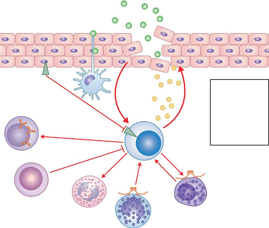
The role of IL-25, IL-33, and TSLP in promoting a Th2associated milieu is summarized in Fig. 1.7. In this model, injured epithelium has a central role in driving allergic inflammation through its ability to produce these cytokines. TSLP acts primarily on DCs to drive them to induce a Th2-like process. In addition, both IL-25 and IL-33 act directly on mast cells to drive their repertoire of Th2-associated cytokines. More important, IL-25, TSLP, and IL-33 act on ILC2 to increase their selective production of IL-5 and IL-13. These actions on ILC2 and mast cells can occur independent of ongoing allergen exposure, suggesting a mechanism for allergen-independent perpetuation of allergic inflammation.
Chemokines in Allergic Diseases
Asthma
Asthma is a chronic inflammatory lung disease characterized by airway inflammation, mucus hypersecretion, and bronchial hyperresponsiveness. The cellular inflammatory infiltrate in asthma is composed of eosinophils, lymphocytes, mast cells, and to a varying extent, basophils and neutrophils.
Airway exposure to proteases from common allergens, such as mites and molds, disrupts airway epithelial integrity and induces epithelial TSLP production (Fig. 1.8). TSLP expands the number of basophils, prolongs eosinophil survival, and increases eosinophil production of CCL2, CXCL1, and CXCL8. Two other epithelial cytokines, IL-25 and IL-33, also are produced on allergen exposure or epithelial damage. IL-25 and IL-33 upregulate the production of TSLP by epithelial cells and mast cells; induce mast cell release of IL-4, IL-5, IL-13, CCL1, and CXCL8; promote eosinophil survival; and enhance eosinophil production of CCL2 and CCL3. Activated basophils release IL-4, IL-13, granulocyte-macrophage colony-stimulating factor (GM-CSF), and CCL3 as well as histamine and leukotriene C4 (LTC4), which causes vasodilation and increases vascular permeability. Activated eosinophils generate IL-3, IL-4, IL-5, tumor necrosis factor-α (TNF-α), LTC4, platelet-activating factor (PAF), CCL3, CCL5, and CCL11. In addition to tryptase and chymase, activated mast cells are also a significant source of histamine, lipid mediators (LTB4, PGD2), cytokines (IL-3, IL-5, IL-13, IL-6, IL-10, TNF-α, GM-CSF), and chemokines (CCL1, CCL2, CCL3, CCL5, CCL17, CCL22, CXCL8).
Activation and differentiation of naive T cells into Th2 cells are marked by downregulation of L-selectin and CCR7 and appearance of CCR4, CCR8, CRTh2, and the BLT1 receptor
Fig. 1.7 Epithelium-derived cytokines in Th2 differentiation and allergic inflammation. The interleukins IL-25 and IL-33 and thymic stromal lymphopoietin (TSLP) are produced by injured epithelium and play critical roles in driving expression of Th2 cytokines. TSLP acts on dendritic cells to direct them to promote the differentiation of naive T cells into Th2 cells. By contrast, IL-25 and IL-33 act directly on the naive T cells to promote Th2 immune deviation. In addition, these three cytokines can generate a Th2 cytokine milieu independent of the adaptive immune system. TSLP and IL-33 directly induce the full repertoire of Th2 cytokine secretion from mast cells. Similarly, IL-25, TSLP, and IL-33 act on type 2 innate lymphoid cells (ILC2) to drive their more restricted secretion of IL-5 and IL-13. DC, Dendritic cell; IL, interleukin; Th, T helper.
Smooth muscle
CCL11
CCL13 CCL5
IL-4, IL-5, IL-13, CCL17, CCL22, CCL1, LTB4, PGD2 Mast
IL-4, IL-5, CCL2, CCL11 IL-4, IL-13
Amplification ↑IL-4, IL-5, IL-13
Chemoattractant receptors
Fig. 1.8 Chemokines and asthma. Asthma is characterized by the infiltration of lung tissue with T helper type 2 (Th2) cells producing IL-4, IL-5, and IL-13. Allergen proteases disrupt airway epithelial integrity and induce thymic stromal lymphopoietin (TSLP), IL-25, and IL-33, while epithelial toll-like receptor activation leads to IL-1β and tumor necrosis factor (TNF) production. These cytokines upregulate CC chemokine and Th2 cytokine production and release by smooth muscle cells, fibroblasts, mast cells, eosinophils, and basophils. Activated antigen-presenting cells travel to the draining lymph nodes and promote the generation of Th2 cells, which enter the lung and release more Th2 cytokines, thus amplifying the allergic response in the lung. CCL, C–C chemokine ligand; CCR, C–C chemokine receptor; DC, dendritic cell; HEV, high endothelial venules; IgE, immunoglobulin E; IL, interleukin; LPS, lipopolysaccharide.
for leukotriene B4 (LTB4). These receptors enable Th2 cells to move down the concentration gradient in response to CCL17, CCL22, CCL1, prostaglandin D2 (PGD2), and LTB4, mediators released by DCs and activated mast cells. IL-4 and IL-13 induce lung-residing macrophages, DCs, epithelial cells, and endothelial cells to produce CCL11, CCL24, CCL26, CCL1, CCL17, and CCL22, thus amplifying the allergic inflammatory response by attracting more eosinophils and Th2 cells.
Atopic Dermatitis
AD is a pruritic chronic inflammatory disease of the skin in which CD4+ memory T lymphocytes, DC subsets, eosinophils, and mast cells infiltrate the perivascular, subepidermal, and intraepidermal areas. A number of chemokines are aberrantly expressed in the skin of patients with AD and help recruit the
inflammatory infiltrate in this disorder. These include CCR2 and CCR3 ligands (CCL13, CCL11, and CCL26) for eosinophil and mast cell recruitment, CCR4 and CCR8 ligands (CCL22 and CCL1) for Th2 cell recruitment, CCR10 ligand (CCL27) for T cell entry into the epidermis, and CCL18.
The pathophysiology of AD begins with intense pruritus and the mechanical injury that results from chronic scratching (Fig. 1.9). Mechanical trauma can directly activate mast cells, which release histamine, neuropeptides, proteases, kinins, and cytokines, many of which further exacerbate pruritus. Furthermore, TSLP levels increase acutely in the skin after mechanical trauma. TSLP induces DC activation and DC production of CCL17 and CCL22.
The trafficking of memory T cells into the skin requires cutaneous lymphocyte antigen (CLA), which interacts with E-selectin on inflamed endothelium, and initiates rolling. The
CXCR3
Skin
Chemoattractant receptors
L-selectin IgE Antigen
Degranulation CCL2
CCL22, CCL17, CCL1, IL-4, IL-5, IL-13
Degranulation
Sur vival
IL-4, IL-5 CCL2 CCL11 Eosinophil
Fig. 1.9 Chemokines and atopic dermatitis. Atopic dermatitis begins with intense pruritus, chronic scratching, and mechanical injury to the skin. Mechanical trauma leads to mast cell release of Th2 cytokines and CC chemokines and upregulates local TSLP production, while loss of normal barrier function increases exposure to allergens and SEB. TSLP-activated dendritic cells travel to the draining lymph nodes and promote Th2 cell differentiation. Th2 cells enter the skin and release Th2 cytokines, thus amplifying the allergic response in the skin. CCL, C–C chemokine ligand; CCR, C–C chemokine receptor; CLA, cutaneous lymphocyte antigen; DC, dendritic cell; IgE, immunoglobulin E; IL, interleukin; SEB, staphylococcal enterotoxin B; Th2, T helper type 2; TSLP, thymic stromal lymphopoietin.
trafficking molecules most highly expressed by T cells isolated from healthy skin are CLA, CCR4, CCR6 (>80%–90%), and, to a lesser extent, CCR8 (50%). Whereas the ligands for CCR6 and CCR8 are upregulated in inflammation, skin endothelial cells and keratinocytes constitutively express CCL17 (one of the ligands for CCR4) and CCL27 (only known ligand for CCR10), respectively.
Eczema lesions as the hallmark of AD and allergic contact dermatitis lesions are induced by keratinocyte apoptosis, related to IFN-γ, Fas-Fas–ligand interaction, TNF-α, TNF-related weak inducer of apoptosis (TWEAK), and IL-32.33,34
BIOLOGY OF IMMUNE CELLS
T Lymphocytes
Two classes of α/β T lymphocytes that bear the co-receptors CD4 or CD8 are involved in adaptive immune responses. CD4+ T cells are traditionally called Th cells because they activate and direct other immune cells. There are also populations of CD4+ Treg cells that modulate immune responses. CD4+ T cells recognize antigen presented by class II MHC molecules on APCs, including DCs, B cells, and macrophages. Exogenous protein antigens are taken up by APCs and processed into peptides in endocytic
vesicles, which are presented on the cell surface bound to class II MHC molecules. The CD8+ cytotoxic T cells (CTLs) recognize antigen presented on MHC class I molecules. Class I MHC molecules are present on the surface of all nucleated cells. Their cytotoxic functions are carried out by release of preformed effector molecules and by interactions of cell surface molecules.
Antigen-activated CD4+ T cells have the potential to differentiate into effector cells, each with distinct functional properties conferred by the pattern of cytokines they secrete (Fig. 1.10).35 Th1 cells are a subset of CD4+ T cells that secrete IFN-γ, whereas Th2 cells produce IL-4, IL-5, IL-9, IL-10, and IL-13. Th17 cells produce IL-17A, IL-17F, and IL-22. Treg cells produce IL-10 and TGF-β1, are naturally occurring and induced, suppress T cell differentiation and APC activation, and are not considered effector cells. Th1 cells stimulate strong cell-mediated immune responses, particularly against intracellular pathogens. Th2 cells are elicited in immune responses that require a strong humoral component and in antiparasitic responses. Th17 serve critical host defense functions at mucosal surfaces.
Cytokines are the primary factors that influence the CD4+ Th cell generation and are considered the third signal in CD4+ T cell differentiation.20 IFN-γ and IL-12 stimulate the induction of Th1 cells. IL-4 drives Th2 cell generation by direct action on CD4+ T cells. IL-13 is involved in the induction of Th2 cells by an unknown mechanism, although not through direct effects on
CD4+ T cells. IL-6, IL-1β, TGF-β1, and in some situations, IL-23 promote Th17 development.
In the secondary lymphoid tissue, a naive T cell differentiates into an effector cell. Compared with naive T cells, effector cells do not require costimulation to be activated, allowing these cells to respond to antigen with hair-trigger rapidity to produce high levels of cytokines and chemokines, which then direct the immune response. Most activated effector CD4+ T cells die subsequent to an immune response through the process of activation-induced cell death, but a subset of CD4+ T cells will persist as memory cells for the life of the host. CD4+ memory T cells persist in lymphoid organs as central memory cells and in nonlymphoid tissues as effector memory cells. The effector memory T cells respond rapidly to repeat exposures to antigen, whereas central memory T cells are slower to be mobilized.
B Lymphocytes
MHC + peptide
GATA-3lo MAFlo
Fig. 1.10 Generation of helper T cell types 1, 2, and 17 (Th1, Th2, and Th17) from a naive CD4+ T cell. A naive CD4+ T cell does not secrete cytokines and has low expression levels of transcription factors GATA-3 and MAF. Differentiation along the Th1, Th2, or Th17 pathway is triggered by stimulation by antigen presented to the T cell receptor in the context of the major histocompatibility complex (MHC) by the appropriate antigen-presenting cell (APC) and a second signal imparted by ligation of costimulatory molecules CD80/CD86 and CD28. Dendritic cells (DCs) represent the key APCs for naive T cells. Those that produce interleukin-10 (IL10) favor Th2 differentiation, and those that produce interleukin-12 (IL-12) stimulate Th1 differentiation. Th17 cells can be generated in the presence of interleukin-6 (IL-6) and transforming growth factor-β1 (TGF-β1), presumably produced by DCs.
The humoral immune response is generated by B cells. Mature B cells express immunoglobulin on its cell surface, which constitutes the antigen-specific BCR. BCR is a molecular complex made up of antigen-binding or variable (V) regions. This region of the protein varies among immunoglobulins, allowing each antibody to bind to any foreign structure that the individual may encounter. To generate this diverse immunoglobulin repertoire, during development in the bone marrow, B cells undergo somatic deoxyribonucleic acid (DNA) recombination of the variability (V), diversity (D), and joining (J) regions of the immunoglobulin heavy and light chains. The invariant or constant region of the antibody is specialized for different effector functions in the immune system after antibody is secreted. There are five main constant-region forms: IgM, IgD, IgG, IgE, and IgA. The BCR in the membrane-bound form recognizes and binds antigen and transmits activation signals into the cell. Naive B cells recirculate through peripheral lymphoid tissues until it binds specific antigen through surface immunoglobulin and is activated (i.e., signal 1). Most antibody responses, including antibody responses to protein antigens, require antigen-specific T cell help. Antigen bound to surface immunoglobulin is internalized, processed, complexed with MHC class II molecules, and displayed on the cell surface. Previously primed CD4+ T cells that recognize the peptide-MHC class II complex on the B cell provide the second signal for activation. The cytokines secreted by CD4+ Th cells during B cell activation regulate which immunoglobulin heavy-chain constant regions will be selected during class-switch recombination to best serve the functions of the specific immune response. Th2 responses to allergens stimulate B cell activation and result in elevated levels of allergen-specific IgE.
Innate Lymphoid Cells
Populations of lymphoid cells that lack rearranged antigen receptors, which were called ILCs, have been recently identified. These ILC populations can be divided into three groups, based on shared phenotypic and functional properties like T cells. Type 1 ILC (ILC1) constitutively express T-bet and are able to produce IFN-γ upon activation. Type 2 ILC (ILC2) constitutively express GATA-3 and in response to IL-25, IL-33, and
TSLP stimulation produce IL-5 and IL-13. Type 3 ILC (ILC3) constitutively express ROR-γ and in response to IL-1β and IL-23 produce IL-17, IL-22, and IFN-γ. 36
ILC type 2 seems to be important in allergic responses. The ILC2/ILC1 ratio is high in patients with perennial AR sensitized to house dust mite; however, it turns to normal levels following a successful AIT. In the presence of retinoic acid, ILC2 cells transformed into regulatory ILCs (ILCregs) which produce IL-10. These cells can suppress Th2 cell and ILC2 activation. DCs that have the capability of retinoic acid production also induce peripheral Treg cell differentiation. Putting these together, one may suggest that ILCregs may participate in tolerance induction in the mechanisms of AIT.13,19 ILC2s take place in many functions during the inflammatory process in asthma and AD (Fig. 1.11)
Another type of ILC, ILC type 3, may have essential roles in immune tolerance induction. CD40L-expressing ILC3s locate in close contact with B cells in tonsils. Both cells work interdependently, as ILC3s induce IL-15 production in B cells and IL-15 which is a potent growth factor for ILC3s increases CD40L expression on ILC3s. CD40L+ ILC3s induce IL-10–secreting Breg cells through the CD40L and BAFF-receptor–dependent pathway. ILC3-induced Breg cells are characterized by CD27–IgD+IgM+CD24highCD38highCD1d+ immature transitional (itBreg) phenotype. This interaction is important for the maintenance of immune tolerance against innocuous antigens and is inadequate in allergic diseases. In tonsils, generation of functional allergen-specific Treg cells takes place. ILC3s, Breg cells, and Treg cells localize side by side in the interfollicular regions of palatine tonsils. CD40L+ ILC3s may be essential in the maintenance of immune tolerance in tonsils through induction of
functional itBreg cells. These cells can contribute to immune tolerance induction and suppression of T cell responses both by a cell-to-cell contact through programmed cell death-ligand 1 and by secretion of IL-10.13
Dendritic Cells
DCs are the most important APCs found throughout the body and are mainly recognized for their exceptional potential to generate a primary immune response and sensitization to allergens. DCs determine the T cell polarization process that produces Th1 cells (generating mainly IFN-γ), Th2 cells (generating mainly IL-4, IL-5, and IL-13), Th17 cells (generating mainly IL-17), and Treg cells (generating mainly IL-10 and TGF-β). These cells are also recognized for their ability to produce ongoing effector responses that are crucial in maintaining allergic inflammation. In humans, circulating DCs can be broadly divided into two groups: (1) mDCs and (2) pDCs. Both subsets express a different repertoire of TLRs and display a diverse cytokine signature after microbial stimulation. mDCs selectively express TLR2–6 and TLR8 and respond to bacterial and viral infections by producing large amounts of IL-12. In contrast, pDCs constitutively express the endosome-associated TLR7 and TLR9, and they are the main producers of type 1 IFNs in humans.6
Mast Cells
Mast cells are present throughout connective tissues and mucosal surfaces and are especially prominent at the interface with the external environment, such as the skin, respiratory tract, conjunctiva, and gastrointestinal tract. Mast cells contribute to the maintenance of tissue homeostasis, with important roles in
ILC Type 2
LineageIL-7R+(CD127) c-KITInt(CD117) CRTH2+ ST2+ IL-25R+ IL-1R+
Fig. 1.11
of
during